Embed Size (px)
Citation preview

ARTICLE IN PRESS
Contents lists available at ScienceDirect
Signal Processing
Signal Processing 90 (2010) 428–439
0165-16
doi:10.1
� Cor
E-m
ejk@gm
gkook@
journal homepage: www.elsevier.com/locate/sigpro
Despeckling of medical ultrasound images using Daubechiescomplex wavelet transform
Ashish Khare a, Manish Khare b, Yongyeon Jeong c, Hongkook Kim d, Moongu Jeon d,�
a Department of Electronics and Communication, University of Allahabad, Allahabad, Indiab Centre for Development of Advanced Computing, Noida, Indiac Department of Radiology, Chonnam National University Hwasun Hospital, Hwasun, Chonnam, Republic of Koread Department of Information and Communications, Gwangju Institute of Science and Technology, Gwangju, Republic of Korea
a r t i c l e i n f o
Article history:
Received 15 October 2008
Received in revised form
3 June 2009
Accepted 9 July 2009Available online 17 July 2009
Keywords:
Medical image denoising
Speckle noise removal
Daubechies complex wavelet transform
Wavelet shrinkage
84/$ - see front matter & 2009 Elsevier B.V. A
016/j.sigpro.2009.07.008
responding author.
ail addresses: [email protected] (
ail.com (M. Khare), [email protected]
gist.ac.kr (H. Kim), [email protected] (M. Jeo
a b s t r a c t
The paper presents a novel despeckling method, based on Daubechies complex wavelet
transform, for medical ultrasound images. Daubechies complex wavelet transform is
used due to its approximate shift invariance property and extra information in
imaginary plane of complex wavelet domain when compared to real wavelet domain. A
wavelet shrinkage factor has been derived to estimate the noise-free wavelet
coefficients. The proposed method firstly detects strong edges using imaginary
component of complex scaling coefficients and then applies shrinkage on magnitude
of complex wavelet coefficients in the wavelet domain at non-edge points. The proposed
shrinkage depends on the statistical parameters of complex wavelet coefficients of noisy
image which makes it adaptive in nature. Effectiveness of the proposed method is
compared on the basis of signal to mean square error (SMSE) and signal to noise ratio
(SNR). The experimental results demonstrate that the proposed method outperforms
other conventional despeckling methods as well as wavelet based log transformed and
non-log transformed methods on test images. Application of the proposed method on
real diagnostic ultrasound images has shown a clear improvement over other methods.
& 2009 Elsevier B.V. All rights reserved.
1. Introduction
Among all modalities of medical imaging system,ultrasonography has been considered as one of the mostnon-invasive and powerful technique for imaging organsand soft-tissue structures of the human body. Ultrasono-graphy is often preferred due to its relatively low-cost andalso there are no ionizing radiations. Beside theseadvantages, medical ultrasonographic images are of poorvisibility, resulting from speckle noise [1]. Speckle occursespecially in the images of liver and kidney whose
ll rights reserved.
A. Khare), mkhar-
(Y. Jeong), hon-
n).
underlying structures are too small to be resolved bylarge wavelength [2]. The presence of speckle resultsdegradation in image quality and makes it difficult forhuman interpretation and diagnosis. Thus speckle reduc-tion (despeckling) is an important issue for analysis ofultrasound images. Many algorithms have been developedfor despeckling. They can be broadly classified in thefollowing categories [28]: local statistics, homomorphic,anisotropic diffusion, and the most recent one is waveletfiltering.
The standard example of local statistics filters are theKuan Filter [3], the Frost filter [4], the Lee filter [5], theGamma MAP filter [6], etc. and their variations [6].Although these filters perform well for removal of specklenoise, they have major limitations in preserving sharpfeatures of the original image. Homomorphic filter isapplied in FFT domain. In the homomorphic filtering [29]

ARTICLE IN PRESS
A. Khare et al. / Signal Processing 90 (2010) 428–439 429
the image is denoised in fast Fourier transform (FFT)domain, then the inverse FFT is calculated. Anisotropicdiffusion [30] and its variant [31] are also used for speckleremoval. These are nonlinear filtering techniques forsimultaneously performing contrast enhancement andnoise reduction by using the coefficient of variation.
Uses of discrete wavelet transform (DWT) basedtechniques are the recent trends for speckle removal[7–9,11,14,22,26,29,32,40]. Since speckles are multiplica-tive in nature, so many DWT-based approaches use alogarithmic transform to convert signal-dependentspeckle noise to additive white noise. Then waveletthresholding is used to remove the noise. After thresh-olding, an exponential operation is employed to convertthe log-transformed image back to non-logarithmicformat. These approaches are based on the assumptionthat the mean of log-transformed speckle noise is equal tozero. However, exact value of the mean of log-transformedspeckle noise is not equal to zero and thus requirescorrection to avoid extra distortion in the restored image[9]. There are some wavelet based techniques that avoidlog-transform. Another drawback is the use of down-sampling operation in DWT, which results in shift-variant[10] and makes it difficult to preserve original imagediscontinuities in the wavelet domain [11]. Phaseinformation also plays an important role in ultrasoundimage and DWT is inefficient for providing the phaseinformation. All these shortcomings will be reduced bythe use of complex wavelet transform. Several complexwavelet transforms like the dual tree complex wavelettransform (DTCWT) [10], the projection-based complexwavelet transform [12], the steerable pyramid complexwavelet transform [13], etc. have been proposed. In theabove mentioned complex transforms, real-valued filtersinstead of complex-valued filters are used and due topresence of redundancy, they are computationally costlyas well. For avoiding these shortcomings, the Daubechiescomplex wavelet transform has been used [14–16], whichis approximately shift-invariant and also provides phaseinformation. The Daubechies complex wavelet transform[16] is a natural extension of the concepts of Daubechiesreal-valued wavelet transform. As Daubechies [17]has given real solutions for wavelet equation, Lina [16]has explored the complex solutions for the equation andproved that complex solutions do exist leading complexDaubechies wavelet transform. Complex Daubechies wa-velets can be made symmetric. The symmetry property ofthe filter makes it easy to handle the boundary problemsfor finite length signal [18]. A linear phase filter is requiredto preclude the nonlinear phase distortion and to keep theshape of the signal. However obtaining linear phase forthe complex Daubechies wavelet is difficult. Recently amethod to achieve both symmetry and linear phase oncomplex Daubechies wavelet was proposed [19]. In thepresent paper, we used symmetric and approximatelylinear phase complex Daubechies wavelet filter [20],which is nearly shift-invariant and non-redundant.
As compared a vast literature for denoising using real-valued wavelet transform, only a small literature isavailable for denoising using complex wavelet transform.Clonda et al. [20], Romberg [33] and Jalobeanu [34]
restored images by using hidden markov model ofcomplex wavelet transform and complex packets, respec-tively. Lina and MacGibbon [35] applied shrinkagefunctions using Bayesian method on real images. Achim[36] proposed a shrinkage function for removal of additivenoise in dual tree complex wavelet domain, for whichbivariate a-stable distribution of complex wavelet coeffi-cients has been assumed.
Most of the available literature assumes the log-transformed multiplicative noise to behave as additive noiseand follow a Gaussian distribution in wavelet domain. ThisGaussian assumption is over simplified and unnatural [37].Recently some researchers [9,11,22,37] have proposed otherdistributions for log-transformed speckle image. Michailo-vich [37] assumed that the speckle noise in wavelet domainis likely to obey the Fisher–Tippet distribution. Bhuiyan et al.[32] modeled the wavelet coefficients of log-transformedimage with a symmetric normal inverse Gaussian distribu-tion, while those of log-transformed noise are assumed to beGaussian distributed. Achim [36] have introduced theconcept of the bivariate a-stable distribution for denoisingin complex wavelet domain, but this proposed distributionand noise removal technique works well only for Gaussianadditive noise. This is major limitation of the proposedmethod by Achim [36]. In the present work we have shownthat the distribution of log-transformed image is a mixtureof two Gaussian distributions and based on this assumptiona wavelet-shrinkage parameter has been derived.
The parameters of shrinkage function are calculatedthrough the observed image statistics. This has made themethod highly adaptive. While most of the denoisingmethods smooth the image at all points, the proposedmethod preserves the strong edge points during thedenoising process. These edge points are detectedadaptively using imaginary component of complex scalingcoefficients and apply shrinkage at non-edge points. Thisis very helpful for preserving the contrast of objects in 2-Dimages. The proposed method has been compared withthe median filter, the wiener filter and standard speckleremoval methods like the Kuan, the Frost, the Lee and theGamma MAP filters. The proposed method is alsocompared with other wavelet based speckle removalmethods like the statistical log-transformed waveletspeckle noise removal [8], non-log transformed general-ized likelihood ratio based wavelet domain noise removal(GenLik) [9] method, and total least square denoisingalgorithm (TLS-denoise) [41]. By means of experimentalresults it has been shown that the present method yieldsfar better results than others [3,4,8,9]. There are severalvisual and numerical image quality measurements existsin the literature [38,39]. For the image quality perfor-mance measure we used signal to mean square error(SMSE) and signal to noise ratio (SNR), as they are bettermeasurements for speckle noise.
The rest of paper is organized as follows: Section 2describes the basic concepts of complex Daubechieswavelet transform and its properties. In Section 3, thedetails of the proposed algorithm, including the methodfor edge detection in complex wavelet domain and theshrinkage function are provided. In Section 4, the resultsof the proposed method for speckle removal are shown

ARTICLE IN PRESS
0 2 4 6 8 10 12 14 16 18 200
0.10.20.30.40.50.60.70.80.9
1
0 2 4 6 8 10 12 14 16 18 200
0.10.20.30.40.50.60.70.80.9
1
0 2 4 6 8 10 12 14-0.7
-0.6
-0.5
-0.4
-0.3
-0.2
-0.1
0
0.1
0.2
0 2 4 6 8 10 12 14-0.1
0
0.1
0.2
0.3
0.4
0.5
0.6
0.7
0.8
1 2 3 4 5 6 7 8 9 100
0.1
0.2
0.3
0.4
0.5
0.6
0.7
1 2 3 4 5 6 7 8 9 100
0.1
0.2
0.3
0.4
0.5
0.6
0.7
Fig. 1. (a) Original signal and the shifted signal by one sample, (b) high-pass wavelet coefficient of the original signal and the shifted signal using real db4
wavelet, and (c) magnitude of high-pass complex wavelet coefficients of the original signal and the shifted signal using SDW6 wavelet.
A. Khare et al. / Signal Processing 90 (2010) 428–439430
and compared to other methods, and finally conclusionsare given in section 5.
2. Daubechies complex wavelet transform
In the present work we used complex Daubechieswavelets that are endowed with symmetry property[16,42].
2.1. Construction of Daubechies complex wavelet
The basic equation of the multiresolution analysis isthe scaling equation:
fðtÞ ¼ 2X
n
anfð2t � nÞ (1)
where f(t) is scaling function and ans are coefficients. Theans can be real as well as complex valued and
Pan ¼ 1.
Daubechies’s wavelet bases {cj,k(t)} in one dimensionare defined through the above scaling function andmultiresolution analysis of L2ð<Þ [17].
Daubechies has given the general solution as follows:Let hðzÞ ¼ 1
2ð1þ zÞ. Consider the following polynomial:
pMðzÞ ¼XMk¼0
ð�1Þk2M þ 1
k
� �hðzÞ2M�2khð�zÞ2k (2)
that satisfies pMðzÞ � pMð�zÞ ¼ z.If we denote Zn, n ¼ 1,2,y,M, the set of roots of pM(z)
inside the unit circle (|Zn|o1), then any selection R among
ARTICLE IN PRESS
A. Khare et al. / Signal Processing 90 (2010) 428–439 431
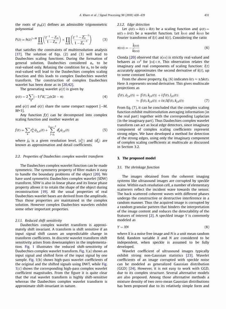
the roots of pM(z) defines an admissible trigonometricpolynomial
FðzÞ ¼ hðzÞ1þMYm2R
z�1 � Zm
1� Zm
� ��YneR
z�1 � Zn�1
1� Zn�1
!(3)
that satisfies the constraints of multiresolution analysis[17]. The solution of Eqs. (2) and (3) will lead toDaubechies scaling functions. During the formation ofgeneral solution, Daubechies considered an to bereal-valued only. Relaxing the condition for an to be onlyreal-valued will lead to the Daubechies complex scalingfunction and this leads to complex Daubechies wavelettransform. The construction of complex Daubechieswavelet has been done as in [20,42].
The generating wavelet c(t) is given by
cðtÞ ¼ 2X
n
ð�1Þna1�nfð2t � nÞ (4)
and c(t) and f(t) share the same compact support [�M,M+1].
Any function f(t) can be decomposed into complexscaling function and mother wavelet as
f ðtÞ ¼X
k
cj0
k fj0 ;kðtÞ þ
Xjmax�1
j¼j0
djkcj;kðtÞ (5)
where j0 is a given resolution level, fcj0
k g and fdjkg are
known as approximation and detail coefficients.
2.2. Properties of Daubechies complex wavelet transform
The Daubechies complex wavelet function can be madesymmetric. The symmetry property of filter makes it easyto handle the boundary problems of the object [20]. Wehave used symmetric Daubechies complex wavelet (SDW)transform. SDW is also in linear phase and its linear phaseproperty allows it to retain the shape of the object duringreconstruction [19]. All the usual properties of realDaubechies wavelet bases are derived from the amplitude.Thus those properties are maintained in the complexsolution. However complex Daubechies wavelets exhibitsome other important properties.
2.1.1. Reduced shift sensitivity
Daubechies complex wavelet transform is approxi-mately shift invariant. A transform is shift sensitive if aninput signal shift causes an unpredictable change intransform coefficients. In discrete wavelet transform shiftsensitivity arises from downsamplers in the implementa-tion. Fig. 1 illustrates the reduced shift-sensitivity ofDaubechies complex wavelet transform. Fig. 1(a) shows aninput signal and shifted form of the input signal by onesample. Fig. 1(b) shows high-pass wavelet coefficients ofthe original and the shifted signals using DWT, while Fig.1(c) shows the corresponding high-pass complex waveletcoefficient magnitudes. From the figure it is quite clearthat the real wavelet transform is highly shift-sensitivewhereas the Daubechies complex wavelet transform isapproximate shift-invariant in nature.
2.1.2. Edge detection
Let fðtÞ ¼ kðtÞ þ ilðtÞ be a scaling function and cðtÞ ¼uðtÞ þ ivðtÞ be a wavelet function. Let l oð Þ and k oð Þ beFourier transforms of l(t) and k(t). Considering the ratio
aðoÞ ¼ � lðoÞkðoÞ
(6)
Clonda [20] observed that a(o) is strictly real-valued andbehaves as o2 for |o|op. This observation relates theimaginary and real components of scaling function: l(t)accurately approximates the second derivative of k(t), upto some constant factor.
From the above property, Eq. (6) indicates lðtÞ � aDkðtÞ.Here D represents second derivative. This gives multiscaleprojections as
hf ðtÞ;fj;kðtÞi ¼ hf ðtÞ; kj;kðtÞi þ ihf ðtÞ; lj;kðtÞi
� hf ðtÞ; kj;kðtÞi þ iahDf ðtÞ; kj;kðtÞi (7)
From Eq. (7), it can be concluded that the complex scalingfunction exhibit multiresolution averaging information (inthe real part) together with the corresponding Laplacian(in the imaginary part). Thus Daubechies complex wavelettransform can act as local edge detectors, since imaginarycomponent of complex scaling coefficients representstrong edges. We have developed a method for detectionof the strong edges, using only the imaginary componentof complex scaling coefficients at multiscale as discussedin Section 3.2.
3. The proposed model
3.1. The shrinkage function
The images obtained from the coherent imagingsystems like ultrasound images are corrupted by specklenoise. Within each resolution cell, a number of elementaryscatterers reflect the incident wave towards the sensor.The back scattered coherent waves with different phasesundergo the constructive or destructive interference in arandom manner. Thus the acquired image is corrupted bya random granular pattern that hinders the interpretationof the image content and reduces the detectability of thefeatures of interest [2]. A speckled image Y is commonlymodeled as
Y ¼ XN (8)
where X is a noise free image and N is a unit mean randomfield. Random variable X and N are considered to beindependent, when speckle is assumed to be fullydeveloped.
Wavelet coefficient of ultrasound images typicallyexhibit strong non-Gaussian statistics [23]. Waveletcoefficients of an image corrupted with speckle noisecan be modeled as generalized Gaussian distribution(GGD) [24]. However, it is not easy to work with GGD,due to its complex structure. Several alternative modelsare also proposed. Among those alternative methods amixture density of two zero-mean Gaussian distributionshas been proposed due to its relatively simple form and

ARTICLE IN PRESS
A. Khare et al. / Signal Processing 90 (2010) 428–439432
high accuracy in modeling the distribution of waveletcoefficients [25].
We have observed that the distribution of Daubechiescomplex wavelet coefficients follow the mixture of twoGaussian distribution. Fig. 2 shows the performance of themixture Gaussian model in matching the distributions ofreal and imaginary components of complex waveletcoefficients of an ultrasound image. Fig. 2 shows thatthe mixture Gaussian model follows the actual histogram
-150 -100 -50 0 50 100 1500
0.05
0.1
0.15
0.2
0.25
0.3
0.35
Real Part of Wavelet Coefficient
Pro
babi
lity
blue - real PDFblack - mix PDFgreen - Gauss 1 PDFred - Gauss 2 PDF
-150 -100 -50 0 50 100 1500
0.05
0.1
0.15
0.2
0.25
0.3
0.35
Imaginary Part of Wavelet Coefficient
Pro
babi
lity
blue - real PDFblack - Mix PDFgreen - Gauss 1 PDFred - Gauss 2 PDF
Fig. 2. PDF of complex wavelet coefficient matching with Gaussian PDF
model for an ultrasound image.
Fig. 3. (a) An original ultrasound image of hepatic vein
very well. A small discrepancy near zero does not reducethe effectiveness of the model. Based on the mixture-Gaussian model, a denoising method has been establishedas follows.
In wavelet domain, the decomposition operation canbe written as [11,26]
WY ¼W½Y � ¼W½XN�
¼W½X� þW½XðN � 1Þ�
¼WX þWS (9)
Since N is a unit mean random field, therefore
E½N� ¼ 1 and E½N � 1� ¼ 0 (10)
Therefore WX ¼W½X� and WS ¼W½XðN � 1Þ� ¼W½XNC �
are centered and uncorrelated random process [11,45] and
E½WX � ¼ 0; E½WS� ¼ 0; and E½WXWS� ¼ 0 (11)
The a posteriori probability density function conditionedto the observation can be written as [11]
PWX jWYðwX jwY Þ ¼
PWY jWXðwY jwXÞPWX
ðwXÞ
PWYðwXÞ
¼PWSjWX
ðwSjwXÞPWXðwXÞ
PWYðwY Þ
The estimate wX maximizing the a posteriori pdf, is
wX ¼ arg maxwX
ðPWX jWYðwX jwY ÞÞ
� �
d
dwXðlnðPWSjWX
ðwSjwXÞÞ þ lnðPWXðwXÞÞÞwX¼wX
¼ 0 (12)
We assume Gaussian model for PWSjWXðwSjwXÞ and
PWXðwXÞ [11]. The MAP criteria is then equivalent to
applying MMSE criteria.We assumed Gaussian distributions for both WX and
WS and in the Gaussian case, the uncorrelationðE½WXWS� ¼ 0Þ is sufficient to ensure the independencebetween WX and WS. Therefore, the likelihood termPWS jWX
ðwSjwXÞ in the Bayesian relation is equal to pdfPWSðwSÞ of WS.For removal of signal dependent noise, a shrinkage in
wavelet domain is most popular. Several shrinkageestimates are used for removal of signal dependent noise[14,27,43,44]. In the present work, we used a different
and (b) detected edges by the proposed method.

ARTICLE IN PRESS
A. Khare et al. / Signal Processing 90 (2010) 428–439 433
adaptive shrinkage estimate for speckle removal similar tothat of Foucher [11].
Shrinkage estimate of the noise-free wavelet coeffi-cient WX is WX ¼ ZWY , where Z is shrinkage factor. Basedon minimum mean square error (MMSE) criteria, theoptimal shrinkage factor Z0 is obtained by minimizingmean square error between WX and WX
Z0 ¼ arg minZ
E½ðWX �WXÞ2�
0 5 10 15 20 250
5
10
15
20
25
30
SMSE (in dB) of noisy image
SM
SE
(in
dB) o
f den
oise
d im
age Black - The Proposed Method
Red - The Weiner filterBlue - The Frost filterGreen - The Gamma filterMagenta - The Median filter
0 5 10 15 20 250
5
10
15
20
25
30
SMSE (in dB) of noisy image
SM
SE
(in
dB) o
f den
oise
d im
age Black - The proposed method
Red - The Kuan filterBlue - The Lee filterGreen - Log-transformed wavelet denoising
Fig. 4. Comparison of the proposed method with other methods at
different noise levels of speckle noise.
Table 1Signal to noise ratio (SNR) (in dB) measures obtained by different speckle rem
Method Image 1 Image 2
Noisy image 40.4365 37.0248
Weiner filter 43.1113 42.0356
Frost filter 42.2444 41.2529
Gamma filter 39.9324 39.3890
Median filter 40.3738 39.0865
Kuan filter 42.1492 41.3597
Lee filter 45.0725 42.7658
Log wavelet speckle filter 43.7598 41.2973
GenLik method 45.2470 43.4482
TLS-denoise method 45.3266 42.8197
The proposed method 46.0243 43.5243
Substituting WX ¼ ZWY in above equation, we get
Z0 ¼ arg minZ
E½ðZWY �WXÞ2�
Thus, the MMSE solution is in the form of
Z0 ¼ E½W2Y � � E½WY WS�
E½W2Y �
(13)
To calculate Z0, it is necessary to estimate all unknownparameters. In the above equation E½W2
Y � can be knowneasily, but it is difficult to estimate E½WY WS� due todependence of noise WS upon WY.
After replacing WY with WX+WS in E½WY WS�, we obtain
E½WY WS� ¼ E½WXWS� þ E½W2S � (14)
Because of the zero-mean mixture Gaussian distributionmodel of WY [11], its expectation E[WY] is also zero.Therefore
E½W2Y � ¼ s2
WYand E½W2
S � ¼ s2WS
and from Eq. (11), E½WXWS� ¼ E½WX �E½WS� ¼ 0Therefore, E½WY WS� becomes
E½WY WS� ¼ 0þ E½W2S � ¼ s2
WS(15)
Thus the new shrinkage factor is
Z0 ¼ E½W2Y � � E½WY WS�
E½W2Y �
¼s2
WY� s2
WS
s2WY
(16)
Here s2WY
is known and s2WS
have to be estimated.Foucher [11] estimated s2
WS, by assuming zero-mean
mixture Gaussian distribution model for WY as
s2WS¼ cjm2
XC2Nð1þ C2
XÞ (17)
where mX ¼ E½X� and C2X ¼ C2
WY� cjC
2N=cjð1þ C2
NÞ
CN ¼ffiffiffiffiffiffiffiffi1=L
p, for L-look image with L ¼ 1 for ultrasound
images.The parameter,
cj ¼X
k
ðhkÞ2
!2 Xl
ðglÞ2
!2ðj�1Þ
where h and g are the high-pass and the low-pass filters atdecomposition level j, respectively.
CWY¼ sWY
=mY is called normalized standard deviationof noisy wavelet coefficients.
oval methods.
Image 3 Image 4 Image 5 Image 6
34.3720 31.2663 29.0466 27.1947
40.6713 37.2728 31.4130 27.2094
40.3678 38.4806 36.7043 34.8135
38.5900 38.6799 36.2117 33.3733
37.6680 35.2590 33.2556 30.2942
40.2710 38.6264 36.7439 34.7515
40.1807 36.5213 33.6769 31.3951
39.7478 36.6351 23.8631 23.5672
41.3571 37.7249 36.5907 35.7721
41.7626 38.2518 36.9642 34.3399
42.0984 38.7468 37.8801 35.9165
ARTICLE IN PRESS
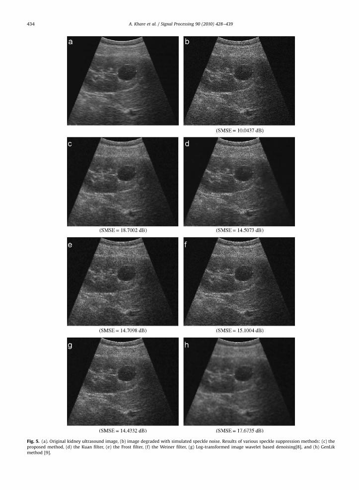
Fig. 5. (a). Original kidney ultrasound image, (b) image degraded with simulated speckle noise. Results of various speckle suppression methods: (c) the
proposed method, (d) the Kuan filter, (e) the Frost filter, (f) the Weiner filter, (g) Log-transformed image wavelet based denoising[8], and (h) GenLik
method [9].
A. Khare et al. / Signal Processing 90 (2010) 428–439434

ARTICLE IN PRESS
A. Khare et al. / Signal Processing 90 (2010) 428–439 435
Substituting the value of C2X in Eq. (17), we obtain
s2WS¼ cjm2
XC2N 1þ
C2WY� cjC
2N
cjð1þ C2NÞ
!
¼ cjm2XC2
N
cjð1þ C2NÞ þ C2
WY� cjC
2N
cjð1þ C2NÞ
¼ m2XC2
N
cj þ C2WY
1þ C2N
(18)
Since E[Y] ¼ E[X] (or mY ¼ mX) with normalized specklenoise N, and CWY
¼ sWY=mY ,
so we obtain
s2WS¼ m2
Y C2N
cj þs2
WY
m2Y
ð1þ C2NÞ
(19)
s2WS¼
C2Nðcjm2
Y þ s2WYÞ
ð1þ C2NÞ
(20)
Now the shrinkage factor Z0 can be computed easily bysubstituting the value of s2
WSin Eq. (16).
3.2. Edge detection in complex wavelet domain
Based on the observation and Eq. (7), that sharp edgeshave large values of imaginary component of scalingcoefficients over many scales, we have used an edgedetector that uses the interscale product of the imaginaryvalues of scaling coefficients, similar to the noise filtrationtechnique developed by Xu et al. [21]. Large values ofdirect multiplication locate important edges. This ap-proach is straightforward, easier to implement and robust.
In order to calculate edge coefficients, correlation(actually products) among imaginary component of scal-ing coefficients at adjacent scales has been computed. Inthe 1-D case, the correlation is defined as
CLð2j; kÞ ¼
YL
r¼0
ImðWf ð2jþr ; kÞÞ (21)
where ImðWf ð2jþr ; kÞÞ denotes imaginary component ofcomplex scaling coefficient at kth point and j+rth level.The optimal value of L for computing correlation can bedetermined experimentally and by means of severalexperiments, we have found that the choice for L ¼ 3gives good results for edge detection.
Given the observations made above, we can offer thefollowing process of edge detection:
Table 2Average processing time required for different speckle removal methods.
(1)Method Average processing time (in seconds)
Rescale the power of fCLð2j; kÞg1�k�n to that of
fWf ð2j; kÞg1�k�n.
(2) Identify an edge at position k if jCLð2j; kÞj4jWf ð2j; kÞj.
Frost filter 5.195Gamma filter 4.374
Median filter 1.186
Kuan filter 5.835
Lee filter 5.694
Log wavelet speckle filter 1.607
GenLik method 183.706
TLS-denoise method 1051.973
The proposed method 1.334
The above procedure can be easily extended fordetection of edges in 2-D images.
Fig. 3(a) and (b) shows an ultrasound image of hepaticvein and its edges computed by the above proposedalgorithm. Here L ¼ 3, where this value has been deter-mined experimentally considering the quality of theobtained results. From these figures, it is clear that
imaginary component of complex scaling coefficientshave good capability of edge detection at multiple levels.The edges detected by the proposed method are sharp.Multiple and false edge detection problem is not presentin the proposed method.
3.3. The algorithm
The proposed denoising algorithm is as follows:
1.
Transform the noisy image y into Daubechies complexwavelet coefficient w.2.
Detection of strong edges:(a) Separate real and imaginary components of wave-let coefficient.(b) Detect strong edges in noisy image using the
procedure described in Section 3.2.
3. Wavelet shrinkage:(a) Shrink the complex wavelet coefficients (except atedge points) with shrinkage factor described in Eq.(16).
(b) Apply the inverse wavelet transform to getestimated image.
4. Experiments and results
Here we present performance of the proposed methodand compare our results with other conventional despecklingmethods like the median, the Weiner, the Frost, the Lee, theGamma MAP and the Kuan filters. The proposed method isalso compared with recent wavelet based speckle removalmethods, viz. wavelet based statistical log-transformed imagedenoising [8], generalized likelihood ratio based waveletdomain noise removal technique (GenLik) [9], and total leastsquare denoising algorithms (TLS-denoise) [41].
Speckles occur when a coherent source and a non-coherent detector are used to interrogate the medium, whichis rough on the scale of the wavelength. Ultrasound B-scanmedical images represent the back-scattering of an ultra-sound beam from structures inside the body. Diffusescattering arises when there are a large number of scattererswith random phase within the resolution cell of theultrasound beam and this diffuse scattering causes specklein medical images. Speckle noise is in the form of granulartexture. Speckles degrade the ability to resolve details anddetect objects of size comparable to the speckle size.
ARTICLE IN PRESS
A. Khare et al. / Signal Processing 90 (2010) 428–439436
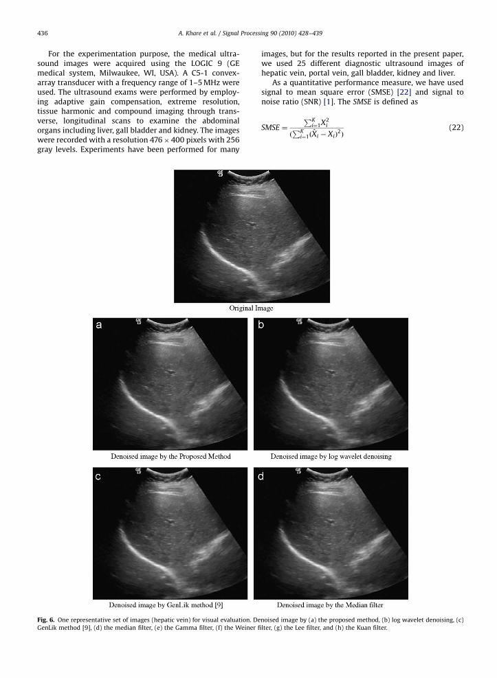
For the experimentation purpose, the medical ultra-sound images were acquired using the LOGIC 9 (GEmedical system, Milwaukee, WI, USA). A C5-1 convex-array transducer with a frequency range of 1–5 MHz wereused. The ultrasound exams were performed by employ-ing adaptive gain compensation, extreme resolution,tissue harmonic and compound imaging through trans-verse, longitudinal scans to examine the abdominalorgans including liver, gall bladder and kidney. The imageswere recorded with a resolution 476� 400 pixels with 256gray levels. Experiments have been performed for many
Fig. 6. One representative set of images (hepatic vein) for visual evaluation. De
GenLik method [9], (d) the median filter, (e) the Gamma filter, (f) the Weiner fi
images, but for the results reported in the present paper,we used 25 different diagnostic ultrasound images ofhepatic vein, portal vein, gall bladder, kidney and liver.
As a quantitative performance measure, we have usedsignal to mean square error (SMSE) [22] and signal tonoise ratio (SNR) [1]. The SMSE is defined as
SMSE ¼
PKi¼1X2
i
ðPK
i¼1ðXi � XiÞ2Þ
(22)
noised image by (a) the proposed method, (b) log wavelet denoising, (c)
lter, (g) the Lee filter, and (h) the Kuan filter.

ARTICLE IN PRESS
Fig. 6. (Continued)
A. Khare et al. / Signal Processing 90 (2010) 428–439 437
where X is original image, X is denoised image, and K isnumber of pixels in image
For denoising in real wavelet domain we used db6wavelet, as its performance is better, as reported in [7] andfor complex wavelet domain shrinkage, we apply thesymmetric Daubechies wavelet 14 (SDW14) as it isoptimal for denoising in complex domain [14]. For thesimulation experiments on 2D ultrasonic medical images,we selected a clean 256�256 liver grayscale ultrasoundimage as a reference image. For making noisy images, wehave artificially added speckle noise of different amountby using our MATLAB program. These noisy images havebeen denoised by different methods. Fig. 4 showsperformance of the proposed method over others in termsof SMSE values. Fig. 4(a) shows the performance of theproposed method in comparison with the weiner filter,the frost filter, the gamma filter and the median filter,while Fig. 4(b) shows the comparison of the proposedmethod with the Kuan filter, the Lee filter and the log-transformed wavelet based denoising method. From thesefigures it is clear that the performance of the proposedtechnique is far better in all noise regions, or in otherwords, the proposed shrinkage in complex waveletdomain, with edge preservation, results in removing morenoise compared to the conventional spatial domainmethod as well as real wavelet based speckle removalmethod in logarithmic transform domain.
Table 1 presents the performance of the proposedmethod in term of signal to noise ratio (SNR) for sixdifferent simulated noisy images. High value of SNRindicates better noise removal. It is evident from the tablethat the proposed method is more successful in specklenoise suppression than others. The visualization of theperformance of the proposed method on one representa-tive simulated image is shown in Fig. 5.
Processing time required by the proposed method iscomparatively less than other methods. It is presented inTable 2. We have taken one clean ultrasound image andrun the MATLAB programs of different methods, for 100random simulated speckle noisy images, on Pentium IVmachine with 2.8 GHz processing speed and 2 GB RAM.Finally the average run time of each method is noted.From Table 2, it can be observed that the computationtime required by the proposed method is less than thetime required by conventional methods—the Frost, theGamma, the Kuan and the Lee filters. Computation time ofthe median filter is slightly lower than the proposedmethod, but its performance is poorer than the proposedmethod. The proposed method is faster than the real-wavelet based despeckling method (log wavelet specklefilter) due to use of complex wavelet transform. Themethods like GenLik and TLS-denoise are model basedmethods. Although the denoising performance of TLSdenoise and GenLik are close to the proposed method, as
ARTICLE IN PRESS
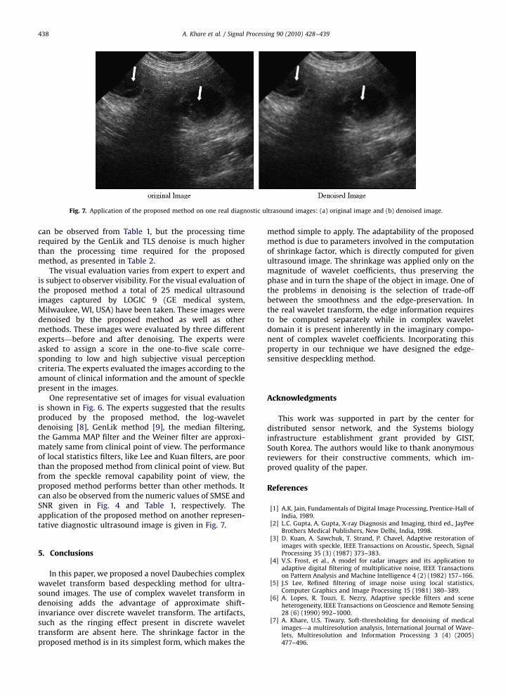
Fig. 7. Application of the proposed method on one real diagnostic ultrasound images: (a) original image and (b) denoised image.
A. Khare et al. / Signal Processing 90 (2010) 428–439438
can be observed from Table 1, but the processing timerequired by the GenLik and TLS denoise is much higherthan the processing time required for the proposedmethod, as presented in Table 2.
The visual evaluation varies from expert to expert andis subject to observer visibility. For the visual evaluation ofthe proposed method a total of 25 medical ultrasoundimages captured by LOGIC 9 (GE medical system,Milwaukee, WI, USA) have been taken. These images weredenoised by the proposed method as well as othermethods. These images were evaluated by three differentexperts—before and after denoising. The experts wereasked to assign a score in the one-to-five scale corre-sponding to low and high subjective visual perceptioncriteria. The experts evaluated the images according to theamount of clinical information and the amount of specklepresent in the images.
One representative set of images for visual evaluationis shown in Fig. 6. The experts suggested that the resultsproduced by the proposed method, the log-waveletdenoising [8], GenLik method [9], the median filtering,the Gamma MAP filter and the Weiner filter are approxi-mately same from clinical point of view. The performanceof local statistics filters, like Lee and Kuan filters, are poorthan the proposed method from clinical point of view. Butfrom the speckle removal capability point of view, theproposed method performs better than other methods. Itcan also be observed from the numeric values of SMSE andSNR given in Fig. 4 and Table 1, respectively. Theapplication of the proposed method on another represen-tative diagnostic ultrasound image is given in Fig. 7.
5. Conclusions
In this paper, we proposed a novel Daubechies complexwavelet transform based despeckling method for ultra-sound images. The use of complex wavelet transform indenoising adds the advantage of approximate shift-invariance over discrete wavelet transform. The artifacts,such as the ringing effect present in discrete wavelettransform are absent here. The shrinkage factor in theproposed method is in its simplest form, which makes the
method simple to apply. The adaptability of the proposedmethod is due to parameters involved in the computationof shrinkage factor, which is directly computed for givenultrasound image. The shrinkage was applied only on themagnitude of wavelet coefficients, thus preserving thephase and in turn the shape of the object in image. One ofthe problems in denoising is the selection of trade-offbetween the smoothness and the edge-preservation. Inthe real wavelet transform, the edge information requiresto be computed separately while in complex waveletdomain it is present inherently in the imaginary compo-nent of complex wavelet coefficients. Incorporating thisproperty in our technique we have designed the edge-sensitive despeckling method.
Acknowledgments
This work was supported in part by the center fordistributed sensor network, and the Systems biologyinfrastructure establishment grant provided by GIST,South Korea. The authors would like to thank anonymousreviewers for their constructive comments, which im-proved quality of the paper.
References
[1] A.K. Jain, Fundamentals of Digital Image Processing, Prentice-Hall ofIndia, 1989.
[2] L.C. Gupta, A. Gupta, X-ray Diagnosis and Imaging, third ed., JayPeeBrothers Medical Publishers, New Delhi, India, 1998.
[3] D. Kuan, A. Sawchuk, T. Strand, P. Chavel, Adaptive restoration ofimages with speckle, IEEE Transactions on Acoustic, Speech, SignalProcessing 35 (3) (1987) 373–383.
[4] V.S. Frost, et al., A model for radar images and its application toadaptive digital filtering of multiplicative noise, IEEE Transactionson Pattern Analysis and Machine Intelligence 4 (2) (1982) 157–166.
[5] J.S Lee, Refined filtering of image noise using local statistics,Computer Graphics and Image Processing 15 (1981) 380–389.
[6] A. Lopes, R. Touzi, E. Nezry, Adaptive speckle filters and sceneheterogeneity, IEEE Transactions on Geoscience and Remote Sensing28 (6) (1990) 992–1000.
[7] A. Khare, U.S. Tiwary, Soft-thresholding for denoising of medicalimages—a multiresolution analysis, International Journal of Wave-lets, Multiresolution and Information Processing 3 (4) (2005)477–496.

ARTICLE IN PRESS
A. Khare et al. / Signal Processing 90 (2010) 428–439 439
[8] S. Gupta, R.C. Chauhan, S.C. Saxena, Wavelet-based statisticalapproach for speckle reduction in medical ultrasound images, IEE:Medical & Biological Engineering & Computing 42 (2004) 189–192.
[9] A. Pizurica, W. Philips, I. Lemahieu, M. Acheroy, A versatile waveletdomain noise filtration technique for medical imaging, IEEETransactions on Medical Imaging 22 (3) (2003) 323–331.
[10] I.W. Selesnick, R.G. Baraniuk, N. Kingsbury, The dual-tree complexwavelet transform, IEEE Signal Processing Magazine 22 (6) (2005)123–151.
[11] S. Foucher, G.B. Benie, J.-M. Boucher, Multiscale MAP filtering of SARimages, IEEE Transactions on Image Processing 10 (2001) 49–60.
[12] F.C.A. Fernandes, R.L.C. Spaendonck, C.S. Burrus, A new frameworkfor complex wavelet transform, IEEE Transactions on SignalProcessing 51 (7) (2003) 1825–1837.
[13] A.A. Bharath, J. Ng, A steerable complex wavelet construction andits application to image denoising, IEEE Transactions on ImageProcessing 14 (7) (2005) 948–959.
[14] A. Khare, U.S. Tiwary, Daubechies complex wavelet transform basedtechnique for denoising of medical images, International Journal ofImage and Graphics 7 (4) (October 2007) 663–687.
[15] A. Khare, U.S. Tiwary, Daubechies complex wavelet transform basedmoving object tracking, in: Proceedings of IEEE Symposium onComputational Intelligence in Image and Signal Processing, April2007, pp. 36–40.
[16] J.-M. Lina, M. Mayrand, Complex Daubechies wavelets, Applied andComputational Harmonic Analysis 2 (1995) 219–229.
[17] I. Daubechies, Ten Lectures on Wavelets, SIAM, Philadelphia, PA, 1992.[18] G. Strang, T. Nguyen, Wavelets and Filter Banks, Wellesley-Cam-
bridge Press, Cambridge, 1996.[19] X.-P. Zhang, M.D. Desai, Y.-N. Peng, Orthogonal complex filter banks
and wavelets: some properties and design, IEEE Transactions onSignal Processing 47 (4) (1999) 1039–1048.
[20] D. Clonda, J.-M. Lina, B. Goulard, Complex Daubechies wavelets:properties and statistical image modeling, Signal Processing 84(2004) 1–23.
[21] Y. Xu, J.B. Weaver, D.M. Healy, J. Lu, Wavelet transform domainfilters: a spatially selective noise filtration technique, IEEE Transac-tions on Image Processing 3 (1998) 133–155.
[22] A. Achim, A. Bezerianos, P. Tsakalides, Novel Bayesian multiscalemethod for speckle removal in medical ultrasound images, IEEETransactions on Medical Imaging 20 (8) (August 2001) 772–783.
[23] H. Xie, L.E. Pierce, F.T. Ulaby, Statistical properties of logarithmicallytransformed speckle, IEEE Transactions on Geoscience RemoteSensing 40 (March 2002) 721–727.
[24] S.G. Chang, M. Vetterli, Spatial adaptive wavelet thresholding forimage denoising, in: Proceedings of IEEE International Symposiumon Image Processing, 1997.
[25] H. Chipman, E. Kolaczyk, R. McCulloch, Adaptive Bayesian waveletshrinkage, Journal of American Statistics Association 92 (1997).
[26] F. Argenti, L. Alparone, Speckle removal from SAR images in theundecimated wavelet domain, IEEE Transactions on GeoscienceRemote Sensing 40 (November 2002) 2363–2374.
[27] D.L. Donoho, Denoising by soft thresholding, IEEE Transactions onInformation Theory 41 (3) (May 1995) 613–627.
[28] C.P. Loizou, C.S. Pattichis, C.I. Christodoulou, R.S.H. Istepanian, M.Pantziaris, A Nicolaides, Comparative evaluation of despecklefiltering in ultrasound imaging of the carotid artery, IEEE Transac-tions on Ultrasonics, Ferroelectrics, and Frequency control 52 (10)(October 2005) 1653–1669.
[29] S. Solbo, T. Eltoft, Homomorphic wavelet based statistical despeck-ling of SAR images, IEEE Transactions on Geoscience and RemoteSensing 42 (4) (2004) 711–721.
[30] Y. Yongjian, S.T. Acton, Speckle reducing anisotropic diffusion, IEEETransactions on Image Processing 11 (11) (November 2002)1260–1270.
[31] K.Z. Abd-Elmoniem, A.-B.M. Youssef, Y.M. Kadah, Real-time specklereduction and coherence enhancement in ultrasound imaging vianonlinear anisotropic diffusion, IEEE Transactions on BiomedicalEngineering 49 (9) (September 2002) 997–1014.
[32] M.I.H. Bhuiyan, M.O. Ahmad, M.N.S. Swamy, Wavelet baseddespeckling of medical ultrasound images with the symmetricnormal inverse Gaussian prior, in: Proceedings of InternationalConference on Acoustics, Speech and Signal Processing, Honolulu,USA, 2007, pp. 721–724.
[33] J.K. Romberg, H. Choi, R.G. Baraniuk, N. Kingsbury, A hidden Markovtree model for the complex wavelet transform, Available at /http://www.acm.caltech.edu/�jrom/publications/complexS HMTv2.pdf.
[34] A. Jalobeanu, N. Kingsbury, J. Zerubia, Image deconvolution usingmarkov tree modeling of complex wavelet packets, in: Proceedingsof IEEE International Conference on Image Processing (ICIP’01),2001, pp. 201–204.
[35] J.-M. Lina, B. MacGibbon, Non-linear shrinkage estimation withcomplex Daubechies wavelets, Proceedings of SPIE 3169 (1997)67–79.
[36] A. Achim, E.E. Kuruoglu, Image denoising using bivariate a-stabledistributions in the complex wavelet domain, IEEE Signal Proces-sing Letters 12 (1) (January 2005) 17–20.
[37] O.V. Michailovich, A. Tannenbaum, Despeckling of medical ultra-sound images, IEEE Transactions on Ultrasonics, Ferroelectrics, andFrequency Control 53 (1) (January 2006) 64–78.
[38] Z. Wang, A.C. Bovik, H.R. Sheikh, E.P. Simoncelli, Image qualityassessment: from error visibility to structural similarity, IEEETransactions on Image Processing 13 (4) (April 2004) 600–612.
[39] C.P. Loizou, C.S. Pattichis, M. Pantzaris, T. Tyllis, A. Nicolaides,Quality evaluation of ultrasound imaging in the carotid arterybased on normalization and speckle reduction filtering, MedicalBiology Engineering Computing 44 (5) (2006) 414–426.
[40] H. Guo, J.E. Odegard, M. Lang, R.A. Gopinath, I.W. Selesnick, C.S.Burrus, Wavelet based speckle reduction with application to SARbased ATD/R, in: Proceedings of the International Conference onImage Processing, vol. 1, 1994, pp. 75–79.
[41] K. Hirakawa, T.W. Parks, Image denoising for signal-dependentnoise, in: Proceedings of the IEEE International Conference onAcoustics, Speech and Signal Processing, vol. 2, 2005, pp. 29–32.
[42] J.-M. Lina, P. Turcotte, B. Goulard, Complex dyadic multiresolutionanalyses, Advances in Imaging and Electron Physics 109 (1999)163–197.
[43] A. Khare, U.S. Tiwary, M. Jeon, Daubechies complex wavelettransform based multilevel shrinkage for deblurring of medicalimages in presence of noise, to appear in International Journal ofWavelets, Multiresolution and Information Processing 7 (5) (2009).
[44] I.M. Johnstone, G. Kerkyacharian, D. Picard, M. Raimondo, Waveletdeconvolution in a periodic setting, Journal of the Royal StatisticalSociety, Series B 66 (2004) 547–573.
[45] M. Dai, C. Peng, A.K. Chan, D. Loguinov, Bayesian wavelet shrinkagewith edge detection for SAR image despeckling, IEEE Transactionson Geoscience and Remote Sensing 42 (8) (August 2004)1642–1648.


![PERIODIC WAVELET TRANSFORMS AND PERIODICITY DETECTIONpfander/pubs/peridet.pdf · Fundamental works on wavelet theory are due to Meyer [16], Daubechies [9], and Mallat [15]. This paper](https://img.dokumen.tips/doc/110x75/5f2f24b9e0d19805ea25893b/periodic-wavelet-transforms-and-periodicity-detection-pfanderpubsperidetpdf.jpg)


![Basis Selection for Wavelet Regression - NeurIPS · 2.1 DISCRETE WAVELET TRANSFORM The Discrete Wavelet Transform (DWT) [Daubechies, 92] is implemented as a series of projections](https://img.dokumen.tips/doc/110x75/60d408b2fe3b0d42d144857b/basis-selection-for-wavelet-regression-neurips-21-discrete-wavelet-transform.jpg)