Embed Size (px)
Citation preview

659
Design, synthesis and photochemical properties of the firstexamples of iminosugar clusters based on fluorescent coresMathieu L. Lepage1, Antoine Mirloup2, Manon Ripoll1, Fabien Stauffert1,Anne Bodlenner1, Raymond Ziessel*2 and Philippe Compain*1,3
Full Research Paper Open Access
Address:1Laboratoire de Synthèse Organique et Molécules Bioactives(SYBIO), Université de Strasbourg/CNRS (UMR 7509), EcoleEuropéenne de Chimie, Polymères et Matériaux (ECPM), 25 rueBecquerel, 67087 Strasbourg, France, 2Institut de Chimie et Procédéspour l’Energie, l’Environnement et la Santé (ICPEES), Laboratoire deChimie Organique et Spectroscopie Avancées (LCOSA), Universitéde Strasbourg/CNRS (UMR 7515), Ecole Européenne de Chimie,Polymères et Matériaux (ECPM), 25 rue Becquerel, 67087Strasbourg, France and 3Institut Universitaire de France, 103 BdSaint-Michel, 75005 Paris, France
Email:Raymond Ziessel* - [email protected]; Philippe Compain* [email protected]
* Corresponding author
Keywords:BODIPY; fluorescent probes; iminosugars; 4-methylumbelliferone;multivalency; pyrene
Beilstein J. Org. Chem. 2015, 11, 659–667.doi:10.3762/bjoc.11.74
Received: 27 February 2015Accepted: 17 April 2015Published: 06 May 2015
This article is part of the Thematic Series "Multivalency as a chemicalorganization and action principle".
Guest Editor: R. Haag
© 2015 Lepage et al; licensee Beilstein-Institut.License and terms: see end of document.
AbstractThe synthesis and photophysical properties of the first examples of iminosugar clusters based on a BODIPY or a pyrene core are
reported. The tri- and tetravalent systems designed as molecular probes and synthesized by way of Cu(I)-catalysed azide–alkyne
cycloadditions are fluorescent analogues of potent pharmacological chaperones/correctors recently reported in the field of Gaucher
disease and cystic fibrosis, two rare genetic diseases caused by protein misfolding.
659
IntroductionSince the isolation in the 1970’s of 1-deoxynojirimycin (DNJ)
from natural sources and the finding of its biological activity as
an α-glucosidase inhibitor, thousands of sugar mimetics with a
nitrogen atom replacing the endocyclic oxygen have been
reported in the literature [1,2]. Iminosugars are mainly known
to be inhibitors of a number of carbohydrate-processing
enzymes with an emphasis on glycosidases [1,2]. In the early
2000’s, iminosugars were, remarkably, found to inhibit metallo-
proteinases [3], protein kinases [4] and cholinesterases [5],
which are enzymes that act on non-sugar substrates. The versa-
tility of iminosugars as inhibitors of enzymes of therapeutic
interest has been harnessed to cure a diversity of diseases in-
cluding diabetes, viral infection, lysosomal storage disorders,
tumour metastasis and cystic fibrosis [1]. First therapeutic
successes have been obtained as demonstrated by the number of
structures involved in clinical trials and two medicines on the
Beilstein J. Org. Chem. 2015, 11, 659–667.
660
market: Glyset (N-hydroxyethyl DNJ) for the treatment of
complications associated with type II diabetes and Zavesca
(N-Bu DNJ, 1), the first oral treatment for Gaucher and
Niemann–Pick diseases (Figure 1) [1,6-8]. Despite their high
therapeutic potential, the extensive studies in the field and the
myriad of compounds synthesized, very few examples of multi-
valent iminosugars were reported in the literature until recently
[9,10]. From 2010, the field has however experienced a major
take-off with the discovery of the first strong multivalent effects
in glycosidase inhibition observed with DNJ clusters based on
β-cyclodextrin or C60 cores showing strong affinity enhance-
ments over the corresponding monomers (up to 610-fold per
DNJ unit) [11,12]. In the following years, an impressive ever-
growing number of multivalent iminosugars based on various
scaffolds, ligands and linkers have been synthesized to further
investigate the impact of multivalency on glycosidase inhibi-
tion [9-26]. The interest of the inhibitory multivalent effect for
drug discovery was demonstrated by targeting glycosidases
involved in rare genetic diseases linked to misfolded proteins
[24-26]. The first examples of multivalent iminosugars such as
2 and 3 acting as pharmacological chaperones were thus
disclosed in the field of Gaucher disease, the most common
lysosomal storage disorder (Figure 1) [24,25]. DNJ clusters 2
and 3 are indeed able to increase mutant β-glucocerebrosidase
(GCase) residual activity levels as much as 3.3-fold in cells of
Gaucher patients at micromolar concentrations. In another rare
genetic disease, the rescue by multimeric correctors of the
mutant CFTR protein implied in cystic fibrosis led to the first
report of a multivalent effect for amending protein folding
defects in cells [26]. As judged by EC50 (half-maximal effec-
tive concentration) values, trivalent DNJ clusters 2 were indeed
up to 225-fold more efficient as CFTR correctors than the clin-
ical candidate N-Bu DNJ (1), a potent inhibitor of trimming ER
glucosidases [26]. Taken together, these recent studies provide
new therapeutic answers for a number of protein folding disor-
ders [27,28] but also raise many fundamental questions
concerning the mechanisms at play. In the present paper, we
report the first examples of fluorescently-labeled multivalent
iminosugars designed as molecular tools to investigate the mode
of action of pharmacological chaperones/correctors in cells and
in vivo, and get insights into the multivalent effect observed in
CFTR correcting activity. The originality of our approach relies
on the fact that, in the structures designed, this is the scaffold
itself [29,30], based on a pyrene or a boron-dipyrromethene
(F-BODIPY) dye, which has fluorescence activity.
Results and DiscussionSynthetic designThe fluorescent probes were designed as analogues of the best
multivalent pharmacological chaperones/correctors reported so
far that typically display three to four copies of a DNJ ligand
Figure 1: N-Bu DNJ (1) and examples of potent multivalent pharmaco-logical chaperones and CFTR correctors (2 and 3).
linked to a central core via an alkyl chain spacer (Figure 1) [24-
26]. The choice of the fluorophore core is naturally primordial
for the design of photostable, water-soluble and biocompatible
probes with the required photophysical properties. An addition-
al challenge is that, as the central core of a multivalent system,
the fluorophore structure defines also its valency, size and
shape. Difluoroboradiaza-s-indacenes, commonly named boron-
dipyrromethene dyes (F-BODIPY), were logically selected for
the construction of the probes. These compounds indeed
combine high fluorescence quantum yields and high molar
extinction coefficients, strong chemical and photochemical
stability in solution and in solid state. In addition, they can be
easily derivatized [31-37]. If the optical properties of BODIPY
are very sensitive to modification of the pyrrole core [38,39],
they are less sensitive to the substitution of the central pseudo
meso position [40,41]. Additionally, the fluorine substitution at
the boron has less influence on the spectroscopic properties of
the dyes [42]. So far, major endeavors have been dedicated to
the preparation of classical F-BODIPY structures and less
common E-BODIPY (E for ethynyl) and the examination of
their spectroscopic and salient physical properties [43-47]. We
have recently argued the case that the fluoro-substitution of

Beilstein J. Org. Chem. 2015, 11, 659–667.
661
boraindacene was a mean to considerably increase the solu-
bility, the stability and the steric hindrance avoiding the forma-
tion of aggregates [48]. In some cases, special sensing prop-
erties [49] may be induced by adequate tailoring, including
fluorescence amplification [50], and ratiometric pH reporter for
imaging protein–dye conjugates in living cells [51], or display
physiological binding of D-glucose [52]. The pyrene nucleus
was also selected as an alternative fluorophore since it may be
easily tetrafunctionalized at the 1, 3, 6 and 8 positions to give a
suitable core for the synthesis of tetravalent clusters [53]. In
addition, this fluorophore was chosen for its biological/chem-
ical stability and its photophysical properties including high
extinction coefficient with reliable fluorescence [54,55].
Another interest of the pyrene scaffold lies in its rigidity, a
property that may favourably impact inhibitory multivalent
effects [9,11,16,19]. A convergent approach comprising the
attachment of azide-armed iminosugars 4 [11,12] on polyalkyne
“clickable” scaffolds 5 and 6 via Cu(I)-catalyzed azide–alkyne
cycloaddition (CuAAC) was performed for achieving our syn-
thetic goals (Figure 2) [56,57]. With the objective of increasing
water solubility and chemical stability in biological medium,
triyne 6b, an analogue of F-BODIPY-based scaffold 6a was
prepared by replacing the fluoro groups on the boron center
with ethynyl tetra(ethylene glycol)methyl groups [58,59].
Figure 2: Azide-armed DNJ derivatives 4 and polyalkyne “clickable”scaffolds 5 and 6.
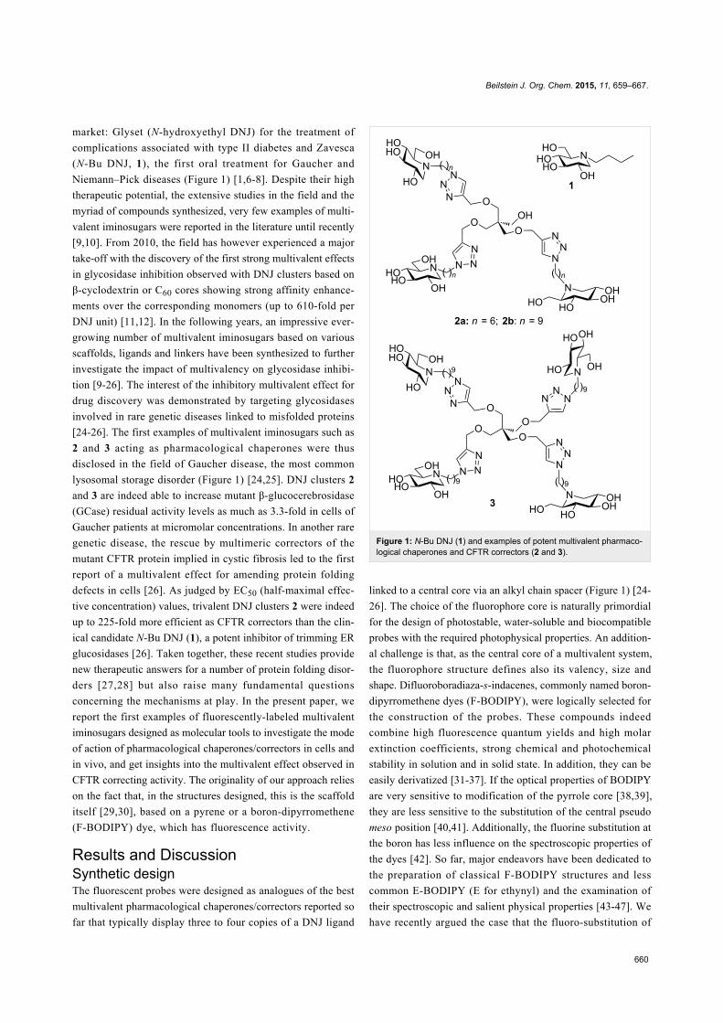
Synthesis of the BODIPY precursorsThe synthesis of the tris-iodo functionalized BODIPY dyes and
their acetylenic derivatives is sketched in Scheme 1. The syn-
thesis of derivatives 7 and 8 have previously been reported
using a regioselective iodination reaction positions 2 and 6 of
the BODIPY [60]. Substitution of both fluoro groups on the
boron was realized using the Grignard reagent of 1-[2”’-(2”-
{2’-(2-methoxyethoxy)ethoxy}ethoxy)ethoxy]prop-2-yne [61]
and the BODIPY derivative 8. With these precursors in hands it
was easy to transform the iodo function to yield the tri-
methylsilylacetylene derivatives 9 and 11 using standard Sono-
gashira–Hagihira cross-coupling reactions promoted by low
valent palladium precursors [62]. Excellent yields were
obtained for the trisubstituted derivatives (88 to 95%). Two
diagnostic NMR signals of the poly(ethylene glycol) chains at
4.16 ppm (protons a, integration 4H) and at 3.65 ppm for the
methoxy groups (protons b, integration 6H) in addition to the
presence of two TMS singlets at 0.20 and 0.28 ppm (respective
integration 18 and 9H) confirmed the substitution. Finally,
deprotection of the trimethylsilyl group using mild basic condi-
tions provided the target compounds 6a and 6b in good yields.
Terminal alkynes located in the 2,6 positions were found to
resonate at 3.32 ppm and the one in the pseudo meso position 8
resonates at 3.20 ppm.
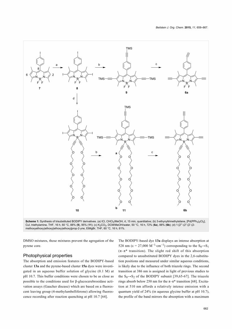
Fluorescent DNJ cluster synthesisFollowing a robust strategy developed in our group [11,12], the
last stages of the multivalent probe synthesis involved the
attachment of peracetylated azido iminosugars 4 on the scaf-
folds via CuAAC reaction and afterwards O-deacetylation using
an anion exchange resin. First attempts to perform CuAAC
reactions with triyne substrate 6b bearing a tetraethylene glycol
chain tethered to the boron center via an ethynyl bond proved
difficult. The use of copper(I) bromide dimethyl sulfide com-
plex [63] at room temperature led to a complex mixture of prod-
ucts. Better results were obtained with copper(II) sulfate and
sodium ascorbate under carefully degassed conditions and the
desired protected cluster 12b could be obtained in 56% yield
after purification on silica gel (Scheme 2). The major side-prod-
uct observed which could not be isolated in pure form may
correspond to CuAAC reaction of the azido iminosugar 4a with
the terminal alkyne resulting from the cleavage of the
carbon–boron bond in 6b. The same experimental protocol was
applied to functionalized BODIPY 6a, leading to the desired
trivalent cluster 12a in 83% yield. O-Deacetylation of com-
pounds 12 using anion exchange Amberlite IRA-400 (OH−)
resin provided the desired water-soluble clusters 13 in high
yields. As judged by 11B NMR, no fluoride displacement
occurred at the boron center during the deprotection step.
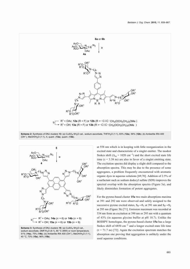
The synthesis of the 4-valent pyrene-based iminosugars 15 was
performed in a similar manner than for BODIPY-based clusters
13 (Scheme 3). The tetrayne 5 synthesized in 3 steps from
pyrene [53] was reacted with the azide precursors 4, and after-
wards deprotected to give the desired tetravalent iminosugars 15
in 37 to 72% yields for the two steps. Despite the good water
solubility of alkylated DNJ ligands, pyrene-based multivalent
iminosugars were only soluble in water/methanol or water/
Beilstein J. Org. Chem. 2015, 11, 659–667.
662
Scheme 1: Synthesis of trisubstituted BODIPY derivatives. (a) ICl, CHCl3/MeOH, rt, 15 min, quantitative; (b) 3-ethynyltrimethylsilane, [Pd(PPh3)2Cl2],CuI, triethylamine, THF, 16 h, 60 °C, 88% (9), 95% (11); (c) K2CO3, DCM/MeOH/water, 50 °C, 16 h, 72% (6a), 88% (6b); (d) 1-[2”’-(2”-{2’-(2-methoxyethoxy)ethoxy}ethoxy)ethoxy]prop-2-yne, EtMgBr, THF, 60 °C, 16 h, 61%.
DMSO mixtures, those mixtures prevent the agregation of the
pyrene core.
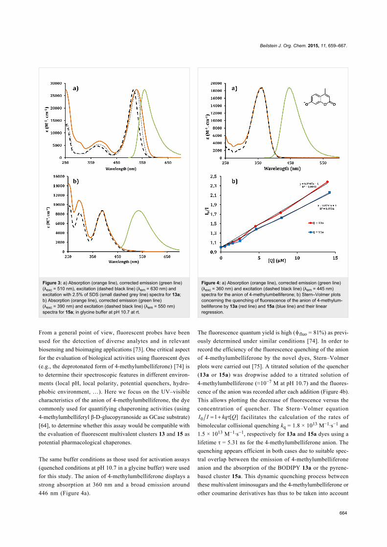
Photophysical propertiesThe absorption and emission features of the BODIPY-based
cluster 13a and the pyrene-based cluster 15a dyes were investi-
gated in an aqueous buffer solution of glycine (0.1 M) at
pH 10.7. This buffer conditions were chosen to be as close as
possible to the conditions used for β-glucocerebrosidase acti-
vation assays (Gaucher disease) which are based on a fluores-
cent leaving group (4-methylumbelliferone) allowing fluores-
cence recording after reaction quenching at pH 10.7 [64].
The BODIPY-based dye 13a displays an intense absorption at
528 nm (ε = 27,000 M−1·cm−1) corresponding to the S0→S1
(π–π* transition). The slight red shift of this absorption
compared to unsubstituted BODIPY dyes in the 2,6-substitu-
tion positions and measured under similar aqueous conditions,
is likely due to the influence of both triazole rings. The second
transition at 386 nm is assigned in light of previous studies to
the S0→S2 of the BODIPY subunit [39,65-67]. The triazole
rings absorb below 250 nm for the π–π* transition [68]. Excita-
tion at 510 nm affords a relatively intense emission with a
quantum yield of 24% (in aqueous glycine buffer at pH 10.7),
the profile of the band mirrors the absorption with a maximum
Beilstein J. Org. Chem. 2015, 11, 659–667.
663
Scheme 2: Synthesis of DNJ clusters 13: (a) CuSO4·5H2O cat., sodium ascorbate, THF/H2O (1:1), 83% (12a), 56% (12b); (b) Amberlite IRA 400(OH−), MeOH/H2O (1:1), rt, quant. (13a), quant. (13b).
Scheme 3: Synthesis of DNJ clusters 15: (a) CuSO4·5H2O cat.,sodium ascorbate, DMF/H2O (6:1), 80 °C (MW) or room temperature,51% (14a), 75% (14b); (b) Amberlite IRA 400 (OH−), MeOH/H2O (1:1),40 °C, 73% (15a), 96% (15b).
at 558 nm which is in keeping with little reorganization in the
excited state and characteristic of a singlet emitter. The modest
Stokes shift (Δss = 1020 cm−1) and the short excited state life
time (τ = 3.38 ns) are also in favor of a singlet emitting state.
The excitation spectra did display a slight shift compared to the
absorption spectra. This may be due to the presence of some
aggregates, a problem frequently encountered with aromatic
organic dyes in aqueous solutions [69,70]. Addition of 2.5% of
a surfactant such as sodium dodecyl sulfate (SDS) improves the
spectral overlap with the absorption spectra (Figure 3a), and
likely diminishes formation of potent aggregates.
For the pyrene-based cluster 15a two main absorptions maxima
at 391 and 292 nm were observed and safely assigned to the
successive pyrene excited states, S0→S1 at 391 nm and S0→S2
at 295 nm (Figure 3b) [71]. Emission maximum was recorded at
534 nm from an excitation at 390 nm or 295 nm with a quantum
of 43% (in aqueous glycine buffer at pH 10.7). Unlike the
BODIPY homologue, the pyrene-based cluster 15a has a large
Stokes shift of 6850 cm−1 and a longer excited state life time
(τ = 71.7 ns) [72]. Again the excitation spectrum matches the
absorption one proving that aggregation is unlikely under the
used aqueous conditions.
Beilstein J. Org. Chem. 2015, 11, 659–667.
664
Figure 3: a) Absorption (orange line), corrected emission (green line)(λexc = 510 nm), excitation (dashed black line) (λem = 630 nm) andexcitation with 2.5% of SDS (small dashed grey line) spectra for 13a;b) Absorption (orange line), corrected emission (green line)(λexc = 390 nm) and excitation (dashed black line) (λem = 550 nm)spectra for 15a; in glycine buffer at pH 10.7 at rt.
From a general point of view, fluorescent probes have been
used for the detection of diverse analytes and in relevant
biosensing and bioimaging applications [73]. One critical aspect
for the evaluation of biological activities using fluorescent dyes
(e.g., the deprotonated form of 4-methylumbelliferone) [74] is
to determine their spectroscopic features in different environ-
ments (local pH, local polarity, potential quenchers, hydro-
phobic environment, …). Here we focus on the UV–visible
characteristics of the anion of 4-methylumbelliferone, the dye
commonly used for quantifying chaperoning activities (using
4-methylumbelliferyl β-D-glucopyranoside as GCase substrate)
[64], to determine whether this assay would be compatible with
the evaluation of fluorescent multivalent clusters 13 and 15 as
potential pharmacological chaperones.
The same buffer conditions as those used for activation assays
(quenched conditions at pH 10.7 in a glycine buffer) were used
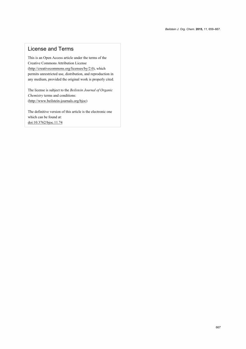
for this study. The anion of 4-methylumbelliferone displays a
strong absorption at 360 nm and a broad emission around
446 nm (Figure 4a).
Figure 4: a) Absorption (orange line), corrected emission (green line)(λexc = 360 nm) and excitation (dashed black line) (λem = 445 nm)spectra for the anion of 4-methylumbelliferone; b) Stern–Volmer plotsconcerning the quenching of fluorescence of the anion of 4-methylum-belliferone by 13a (red line) and 15a (blue line) and their linearregression.
The fluorescence quantum yield is high ( fluo = 81%) as previ-
ously determined under similar conditions [74]. In order to
record the efficiency of the fluorescence quenching of the anion
of 4-methylumbelliferone by the novel dyes, Stern–Volmer
plots were carried out [75]. A titrated solution of the quencher
(13a or 15a) was dropwise added to a titrated solution of
4-methylumbelliferone (≈10−7 M at pH 10.7) and the fluores-
cence of the anion was recorded after each addition (Figure 4b).
This allows plotting the decrease of fluorescence versus the
concentration of quencher. The Stern–Volmer equation
facilitates the calculation of the rates of
bimolecular collisional quenching kq = 1.8 × 10¹³ M−1·s−1 and
1.5 × 10¹³ M−1·s−1, respectively for 13a and 15a dyes using a
lifetime τ = 5.31 ns for the 4-methylumbelliferone anion. The
quenching appears efficient in both cases due to suitable spec-
tral overlap between the emission of 4-methylumbelliferone
anion and the absorption of the BODIPY 13a or the pyrene-
based cluster 15a. This dynamic quenching process between
these multivalent iminosugars and the 4-methylumbelliferone or
other coumarine derivatives has thus to be taken into account

Beilstein J. Org. Chem. 2015, 11, 659–667.
665
during the quantitative analyses of dedicated biological
processes.
ConclusionWe have reported the preparation of multivalent iminosugar
clusters based on two fluorescent cores by way of Cu(I)-catal-
ysed azide–alkyne cycloadditions. To our knowledge these are
the first examples of the use of BODIPY or pyrene as a scaf-
fold to display multivalent ligands. Although the trivalent
BODIPY-derived DNJ clusters are water soluble, a co-solvent
is necessary to dissolve the tetravalent pyrene-derived DNJ
clusters in water. Photophysical properties of those multivalent
dyes in aqueous media (glycine buffer at pH 10.7), are interest-
ing, providing high quantum yields, 24% for 13a and 43% for
15a, and well-defined spectroscopic features. Altogether, these
results augur well for a new class of molecular tools dedicated
to rationalize the mode of action of pharmacological chaper-
ones and CFTR correctors by probing uptake and mapping
biodistribution in cells and in vivo.
Supporting InformationSupporting Information File 1Characterization data and NMR spectra of all new
compounds.
[http://www.beilstein-journals.org/bjoc/content/
supplementary/1860-5397-11-74-S1.pdf]
AcknowledgementsThis work was supported by the Institut Universitaire de France
(IUF), the CNRS (UMR 7509), the University of Strasbourg,
the Agence Nationale de la Recherche (ANR, grant number
11-BS07-003-02), the International Centre for Frontier
Research in Chemistry (icFRC) and doctoral fellowships from
the French Department of Research to Mathieu L. Lepage and
Fabien Stauffert. Antoine Mirloup thanks the Rhin-Solar EC
Network supported by the European Fund for Regional Devel-
opment (FEDER) in the framework of the Programme
INTERREG IV Upper Rhine, Project no. C25 for financial
support of this work.
References1. Iminosugars: from Synthesis to Therapeutic Applications; Compain, P.;
Martin, O. R., Eds.; Wiley & Sons: Chichester, United Kingdom, 2007.doi:10.1002/9780470517437
2. Stütz, A. E., Ed. Iminosugars as Glycosidase Inhibitors: Nojirimycin andBeyond; Wiley-VCH: New York, NY, U.S.A., 1999.
3. Moriyama, H.; Tsukida, T.; Inoue, Y.; Yokota, K.; Yoshino, K.;Kondo, H.; Miura, N.; Nishimura, S.-I. J. Med. Chem. 2004, 47,1930–1938. doi:10.1021/jm0304313
4. Orsato, A.; Barbagallo, E.; Costa, B.; Olivieri, S.; De Gioia, L.;Nicotra, F.; La Ferla, B. Eur. J. Org. Chem. 2011, 5012–5019.doi:10.1002/ejoc.201100452
5. Decroocq, C.; Stauffert, F.; Pamlard, O.; Oulaïdi, F.; Gallienne, E.;Martin, O. R.; Guillou, C.; Compain, P. Bioorg. Med. Chem. Lett. 2015,25, 830–833. doi:10.1016/j.bmcl.2014.12.071
6. Winchester, B. G. Tetrahedron: Asymmetry 2009, 20, 645–651.doi:10.1016/j.tetasy.2009.02.048
7. Horne, G.; Wilson, F. X. Prog. Med. Chem. 2011, 50, 135–176.doi:10.1016/B978-0-12-381290-2.00004-5
8. Nash, R. J.; Kato, A.; Yu, C.-Y.; Fleet, G. W. J. Future Med. Chem.2011, 3, 1513–1521. doi:10.4155/fmc.11.117
9. Compain, P.; Bodlenner, A. ChemBioChem 2014, 15, 1239–1251.doi:10.1002/cbic.201402026
10. Gouin, S. G. Chem. – Eur. J. 2014, 20, 11616–11628.doi:10.1002/chem.201402537
11. Compain, P.; Decroocq, C.; Iehl, J.; Holler, M.; Hazelard, D.;Mena Barragán, T.; Ortiz Mellet, C.; Nierengarten, J.-F.Angew. Chem., Int. Ed. 2010, 49, 5753–5756.doi:10.1002/anie.201002802
12. Decroocq, C.; Rodríguez-Lucena, D.; Russo, V.; Mena Barragán, T.;Ortiz Mellet, C.; Compain, P. Chem. – Eur. J. 2011, 17, 13825–13831.doi:10.1002/chem.201102266
13. Decroocq, C.; Joosten, A.; Sergent, R.; Mena Barragán, T.;Ortiz Mellet, C.; Compain, P. ChemBioChem 2013, 14, 2038–2049.doi:10.1002/cbic.201300283
14. Joosten, A.; Schneider, J. P.; Lepage, M. L.; Tarnus, C.; Bodlenner, A.;Compain, P. Eur. J. Org. Chem. 2014, 1866–1872.doi:10.1002/ejoc.201301583
15. Bonduelle, C.; Huang, J.; Mena-Barragán, T.; Ortiz Mellet, C.;Decroocq, C.; Etamé, E.; Heise, A.; Compain, P.; Lecommandoux, S.Chem. Commun. 2014, 50, 3350–3352. doi:10.1039/c3cc48190e
16. Lepage, M. L.; Meli, A.; Bodlenner, A.; Tarnus, C.; De Riccardis, F.;Izzo, I.; Compain, P. Beilstein J. Org. Chem. 2014, 10, 1406–1412.doi:10.3762/bjoc.10.144
17. Marradi, M.; Cicchi, S.; Sansone, F.; Casnati, A.; Goti, A.Beilstein J. Org. Chem. 2012, 8, 951–957. doi:10.3762/bjoc.8.107
18. Cardona, F.; Isoldi, G.; Sansone, F.; Casnati, A.; Goti, A. J. Org. Chem.2012, 77, 6980–6988. doi:10.1021/jo301155p
19. Brissonet, Y.; Ortiz Mellet, C.; Morandat, S.; Garcia Moreno, M. I.;Deniaud, D.; Matthews, S. E.; Vidal, S.; Šesták, S.; El Kirat, K.;Gouin, S. G. J. Am. Chem. Soc. 2013, 135, 18427–18435.doi:10.1021/ja406931w
20. Rísquez-Cuadro, R.; García Fernández, J. M.; Nierengarten, J.-F.;Ortiz Mellet, C. Chem. – Eur. J. 2013, 19, 16791–16803.doi:10.1002/chem.201303158
21. Moreno-Clavijo, E.; Carmona, A. T.; Moreno-Vargas, A. J.; Molina, L.;Wright, D. W.; Davies, G. J.; Robina, I. Eur. J. Org. Chem. 2013,7328–7336. doi:10.1002/ejoc.201300878
22. D’Adamio, G.; Parmeggiani, C.; Goti, A.; Moreno-Vargas, A. J.;Moreno-Clavijo, E.; Robina, I.; Cardona, F. Org. Biomol. Chem. 2014,12, 6250–6266. doi:10.1039/C4OB01117A
23. Marra, A.; Zelli, R.; D’Orazio, G.; La Ferla, B.; Dondoni, A. Tetrahedron2014, 70, 9387–9393. doi:10.1016/j.tet.2014.10.035
24. Decroocq, C.; Rodríguez-Lucena, D.; Ikeda, K.; Asano, N.;Compain, P. ChemBioChem 2012, 13, 661–664.doi:10.1002/cbic.201200005
25. Joosten, A.; Decroocq, C.; de Sousa, J.; Schneider, J. P.; Etamé, E.;Bodlenner, A.; Butters, T. D.; Compain, P. ChemBioChem 2014, 15,309–319. doi:10.1002/cbic.201300442

Beilstein J. Org. Chem. 2015, 11, 659–667.
666
26. Compain, P.; Decroocq, C.; Joosten, A.; de Sousa, J.;Rodríguez-Lucena, D.; Butters, T. D.; Bertrand, J.; Clément, R.;Boinot, C.; Becq, F.; Norez, C. ChemBioChem 2013, 14, 2050–2058.doi:10.1002/cbic.201300312
27. Gavrin, L. K.; Denny, R. A.; Saiah, E. J. Med. Chem. 2012, 55,10823–10843. doi:10.1021/jm301182j
28. Gomes, C. M. Curr. Top. Med. Chem. 2012, 12, 2460–2469.doi:10.2174/1568026611212220002
29. Ribeiro-Viana, R.; García-Vallejo, J. J.; Collado, D.;Pérez-Inestrosa, E.; Bloem, K.; van Kooyk, Y.; Rojo, J.Biomacromolecules 2012, 13, 3209–3219. doi:10.1021/bm300998cSee for examples of glycoclusters with a BODIPY tag and see ref. [30].
30. Hoogendoorn, S.; van Puijvelde, G. H. M.; Kuiper, J.;van derMarel, G. A.; Overkleeft, H. S. Angew. Chem., Int. Ed. 2014, 53,10975–10978. doi:10.1002/anie.201406842
31. Ziessel, R.; Ulrich, G.; Harriman, A. New J. Chem. 2007, 31, 496–501.doi:10.1039/b617972j
32. Loudet, A.; Burgess, K. Chem. Rev. 2007, 107, 4891–4932.doi:10.1021/cr078381n
33. Ulrich, G.; Ziessel, R.; Harriman, A. Angew. Chem., Int. Ed. 2008, 47,1184–1201. doi:10.1002/anie.200702070
34. Boens, N.; Leen, V.; Dehaen, W. Chem. Soc. Rev. 2012, 41,1130–1172. doi:10.1039/C1CS15132K
35. Willems, L. I.; Beenakker, T. J. M.; Murray, B.; Scheij, S.;Kallemeijn, W. W.; Boot, R. G.; Verhoek, M.; Donker-Koopman, W. E.;Ferraz, M. J.; van Rijssel, E. R.; Florea, B. I.; Codée, J. D. C.;van der Marel, G. A.; Aerts, J. M. F. G.; Overkleeft, H. S.J. Am. Chem. Soc. 2014, 136, 11622–11625.
36. Yadav, A. K.; Shen, D. L.; Shan, X.; He, X.; Kermode, A. R.;Vocadlo, D. J. J. Am. Chem. Soc. 2015, 137, 1181–1189.doi:10.1021/ja5106738
37. Davison, H. R.; Taylor, S.; Drake, C.; Phuan, P.-W.; Derichs, N.;Yao, C.; Jones, E. F.; Sutcliffe, J.; Verkman, A. S.; Kurth, M. J.Bioconjugate Chem. 2011, 22, 2593–2599. doi:10.1021/bc2004457
38. Haugland, R. P.; Kang, H. C. U.S. Patent 4,774,339, 1998.39. Thoresen, L. H.; Kim, H.; Welch, M. B.; Burghart, A.; Burgess, K.
Synlett 1998, 1276–1278. doi:10.1055/s-1998-192440. Goze, C.; Ulrich, G.; Charbonnière, L.; Ziessel, R. Chem. – Eur. J.
2003, 9, 3748–3755. doi:10.1002/chem.20030508441. Qin, W.; Baruah, M.; Van der Auweraer, M.; De Schryver, F. C.;
Boens, N. J. Phys. Chem. A 2005, 109, 7371–7384.doi:10.1021/jp052626n
42. Ulrich, G.; Goze, C.; Guardigli, M.; Roda, A.; Ziessel, R.Angew. Chem., Int. Ed. 2005, 44, 3694–3698.doi:10.1002/anie.200500808
43. Kim, H.; Burghart, A.; Welch, M. B.; Reibenspies, J.; Burgess, K.Chem. Commun. 1999, 1889–1890. doi:10.1039/a905739k
44. Chen, J.; Reibenspies, J.; Derecskei-Kovacs, A.; Burgess, K.Chem. Commun. 1999, 2501–2502. doi:10.1039/a907559c
45. Rurack, K.; Kollmannsberger, M.; Daub, J. New J. Chem. 2001, 25,289–292. doi:10.1039/b007379m
46. Rurack, K.; Kollmannsberger, M.; Daub, J. Angew. Chem., Int. Ed.2001, 40, 385–387.doi:10.1002/1521-3773(20010119)40:2<385::AID-ANIE385>3.0.CO;2-F
47. Ziessel, R.; Harriman, A. Chem. Commun. 2011, 47, 611–631.doi:10.1039/C0CC02687E
48. Ziessel, R.; Ulrich, G.; Haefele, A.; Harriman, A. J. Am. Chem. Soc.2013, 135, 11330–11344. doi:10.1021/ja4049306
49. Turfan, B.; Akkaya, E. U. Org. Lett. 2002, 4, 2857–2859.doi:10.1021/ol026245t
50. Bricks, J. L.; Kovalchuk, A.; Trieflinger, C.; Nofz, M.; Büschel, M.;Tolmachev, A. I.; Daub, J.; Rurack, K. J. Am. Chem. Soc. 2005, 127,13522–13529. doi:10.1021/ja050652t
51. Han, J.; Loudet, A.; Barhoumi, R.; Burghardt, R. C.; Burgess, K.J. Am. Chem. Soc. 2009, 131, 1642–1643. doi:10.1021/ja8073374
52. Hansen, J. S.; Ficker, M.; Petersen, J. F.; Christensen, J. B.;Hoeg-Jensen, T. Tetrahedron Lett. 2013, 54, 1849–1852.doi:10.1016/j.tetlet.2013.01.101
53. Bernhardt, S.; Kastler, M.; Enkelmann, V.; Baumgarten, M.; Müllen, K.Chem. – Eur. J. 2006, 12, 6117–6128. doi:10.1002/chem.200500999
54. Somerharju, P. Chem. Phys. Lipids 2002, 116, 57–74.doi:10.1016/S0009-3084(02)00020-8
55. Pownall, H. J.; Smith, L. C. Chem. Phys. Lipids 1989, 50, 191–211.doi:10.1016/0009-3084(89)90050-9
56. Leen, V.; Leemans, T.; Boens, N.; Dehaen, W. Eur. J. Org. Chem.2011, 4386–4396. doi:10.1002/ejoc.201100324
57. Hornillos, V.; Amat-Guerri, F.; Acuña, A. U.J. Photochem. Photobiol., A: Chem. 2012, 243, 56–60.doi:10.1016/j.jphotochem.2012.06.005
58. Mula, S.; Ulrich, G.; Ziessel, R. Tetrahedron Lett. 2009, 50,6383–6388. doi:10.1016/j.tetlet.2009.08.091
59. Bura, T.; Ziessel, R. Org. Lett. 2011, 13, 3072–3075.doi:10.1021/ol200969r
60. Bonardi, L.; Ulrich, G.; Ziessel, R. Org. Lett. 2008, 10, 2183–2186.doi:10.1021/ol800560b
61. Jahnke, E.; Weiss, J.; Neuhaus, S.; Hoheisel, T. N.; Frauenrath, H.Chem. – Eur. J. 2009, 15, 388–404. doi:10.1002/chem.200801668
62. Sonogashira, K.; Tohda, Y.; Hagihara, N. Tetrahedron Lett. 1975, 16,4467–4470. doi:10.1016/S0040-4039(00)91094-3
63. Isobe, H.; Fujino, T.; Yamazaki, N.; Guillot-Nieckowski, M.;Nakamura, E. Org. Lett. 2008, 10, 3729–3732. doi:10.1021/ol801230k
64. Valenzano, K. J.; Khanna, R.; Powe, A. C., Jr.; Boyd, R.; Lee, G.;Flanagan, J. J.; Benjamin, E. R. Assay Drug Dev. Technol. 2011, 9,213–235. doi:10.1089/adt.2011.0370
65. Chen, T.; Boyer, J. H.; Trudell, M. L. Heteroat. Chem. 1997, 8, 51–54.doi:10.1002/(SICI)1098-1071(1997)8:1<51::AID-HC7>3.0.CO;2-5
66. Sathyamoorthi, G.; Wolford, L. T.; Haag, A. M.; Boyer, J. H.Heteroat. Chem. 1994, 5, 245–249. doi:10.1002/hc.520050309
67. Burghart, A.; Kim, H.; Wech, M. B.; Thoresen, L. H.; Reibenspies, J.;Burgess, K. J. Org. Chem. 1999, 64, 7813–7819.doi:10.1021/jo990796o
68. Potts, K. T. Chem. Rev. 1961, 61, 87–127. doi:10.1021/cr60210a00169. Niu, S.-L.; Ulrich, G.; Retailleau, P.; Harrowfield, J.; Ziessel, R.
Tetrahedron Lett. 2009, 50, 3840–3844.doi:10.1016/j.tetlet.2009.04.017
70. Niu, S. L.; Ulrich, G.; Ziessel, R.; Kiss, A.; Renard, P.-Y.; Romieu, A.Org. Lett. 2009, 11, 2049–2052. doi:10.1021/ol900302n
71. Berlman, I. B. Handbook of Fluorescence Spectra of AromaticMolecules; Academic Press: New York, NY, USA, 1971.
72. Geiger, M. W.; Turro, N. J. Photochem. Photobiol. 1975, 22, 273.doi:10.1111/j.1751-1097.1975.tb06749.x
73. Kaur, M.; Choi, D. H. Chem. Soc. Rev. 2015, 44, 58–77.doi:10.1039/C4CS00248B
74. Chen, R. F. Anal. Lett. 1968, 1, 423–428.doi:10.1080/00032716808051147
75. Stern, O.; Volmer, M. Phys. Z. 1919, 20, 183–188.
Beilstein J. Org. Chem. 2015, 11, 659–667.
667
License and TermsThis is an Open Access article under the terms of the
Creative Commons Attribution License
(http://creativecommons.org/licenses/by/2.0), which
permits unrestricted use, distribution, and reproduction in
any medium, provided the original work is properly cited.
The license is subject to the Beilstein Journal of Organic
Chemistry terms and conditions:
(http://www.beilstein-journals.org/bjoc)
The definitive version of this article is the electronic one
which can be found at:
doi:10.3762/bjoc.11.74