Embed Size (px)
Citation preview

Optimising Comprehension and Shaping Impressions
Bruce Hilliard© 2013 Page 1
DESCRIPTION OF THE NEURAL PROCESSING OF VISUAL INFORMATION
By Bruce Hilliard
TABLE OF CONTENTS
Section Title Page
APPENDIX 1 : DESCRIPTION OF THE KEY NEURAL PROCESSES USED FOR MANAGING
VISUAL INFORMATION ........................................................................................................................ 3
1.1. OVERVIEW ................................................................................................................................. 3
1.1.1. The Intent of this Appendix .................................................................................................. 3
1.1.2. Overview of the Visual System Elements ............................................................................ 3
1.2. AWARENESS AND ATTENTION ................................................................................................... 6
1.2.1. Perception ............................................................................................................................. 7
1.2.1.1. Receiving Visual Input ..............................................................................................................8
1.2.1.2. Sensory Memory ........................................................................................................................8
1.2.2. Attention ............................................................................................................................... 9
1.2.3. Visual Cognition ................................................................................................................ 10
1.2.3.1. The Continuum of Awareness ................................................................................................. 11
1.2.3.1.1. Vigilance ............................................................................................................................ 12
1.2.3.1.2. Unconscious and Conscious Processes ............................................................................... 13
1.2.3.1.3. Awareness .......................................................................................................................... 13
1.2.3.1.4. Unconscious Unattended Level .......................................................................................... 14
1.2.3.1.5. Subliminal Level ................................................................................................................ 14
1.2.3.1.6. Preconscious Awareness..................................................................................................... 15
1.2.3.1.7. Conscious Awareness ......................................................................................................... 16
1.2.3.1.8. Active Attention ................................................................................................................. 17
1.2.3.2. Changing the Levels of Awareness .......................................................................................... 17
1.3. PERCEPTION SYSTEM ELEMENTS ............................................................................................. 20
1.3.1. The Eye .............................................................................................................................. 20
1.3.1.1. General Description ................................................................................................................. 20
1.3.1.2. The Retina................................................................................................................................ 21
1.3.1.2.1. Photoreceptor Cells ............................................................................................................ 21
1.3.1.2.2. Horizontal Cells, Bipolar Cells and Amacrine Cells .......................................................... 26
1.3.1.2.3. Ganglion Cells .................................................................................................................... 27
1.3.2. The Optic Nerves................................................................................................................ 28
1.3.2.1. Parvocellular ............................................................................................................................ 29
1.3.2.2. Magnocellular .......................................................................................................................... 30
1.3.2.3. Koniocellular ........................................................................................................................... 30

Optimising Comprehension and Shaping Impressions
Bruce Hilliard© 2013 Page 2
Section Title Page
1.3.2.4. Synopsis ................................................................................................................................... 31
1.3.3. Interbrain Region ................................................................................................................ 32
1.3.3.1. Thalamus ................................................................................................................................. 32
1.3.3.1.1. Lateral Geniculate Nuclei ................................................................................................... 32
1.3.3.1.2. Thalamic Reticular Nucleus ............................................................................................... 34
1.3.3.1.3. The Pulvinar ....................................................................................................................... 34
1.3.3.2. Suprachiasmatic Nuclei ........................................................................................................... 35
1.3.4. Midbrain Region ................................................................................................................ 35
1.3.4.1. Superior Colliculus .................................................................................................................. 35
1.3.4.2. Pretectum ................................................................................................................................. 36
1.3.4.3. Reticular Formation ................................................................................................................. 37
1.3.5. Occipital Lobes .................................................................................................................. 37
1.4. FROM PERCEPTION TO COGNITION ........................................................................................... 40
1.4.1. Ventral Stream .................................................................................................................... 42
1.4.1.1. Overview of the Temporal Lobe Functions ............................................................................. 42
1.4.1.2. Wernicke’s Area ...................................................................................................................... 43
1.4.1.3. Subcortical Regions ................................................................................................................. 44
1.4.1.3.1. Parahippocampal Place Areas............................................................................................. 44
1.4.1.3.2. Hippocampi ........................................................................................................................ 44
1.4.1.3.3. Striatum and Basal Ganglia ................................................................................................ 46
1.4.1.3.4. Other Limbic System Areas................................................................................................ 47
1.4.1.3.5. Insula .................................................................................................................................. 48
1.4.1.3.6. Broca’s Area ....................................................................................................................... 48
1.4.2. Dorsal Stream ..................................................................................................................... 49
1.4.2.1. This Stream is Very Fast .......................................................................................................... 50
1.4.2.2. Focussed on Change and Motion ............................................................................................. 51
1.4.2.3. Facilitation of Bottom-up and Top-down processes ................................................................ 52
1.4.2.4. Spatial Awareness Focus ......................................................................................................... 52
1.4.2.5. Sublexical Language Analysis ................................................................................................. 53
1.4.2.6. Part of a Network ..................................................................................................................... 53
1.4.3. Frontal Lobe ....................................................................................................................... 54
1.4.3.1. Motor and Premotor Cortex ..................................................................................................... 54
1.4.3.2. Prefrontal Cortex ..................................................................................................................... 54
1.4.3.2.1. Frontal Eye Fields............................................................................................................... 55
1.4.3.2.2. Dorsolateral Prefrontal Cortex ............................................................................................ 55
1.4.3.2.3. Ventromedial Prefrontal Cortex ......................................................................................... 56
1.4.3.3. Orbitofrontal Cortex ................................................................................................................ 56
1.4.4. Interaction between the Two Streams ................................................................................ 57
1.4.4.1. General Interaction .................................................................................................................. 57
1.4.4.2. Working Memory .................................................................................................................... 58
1.4.4.3. Reverberation – The Underlying Process for Working Memory Management ........................ 60
1.5. METENCEPHALON .................................................................................................................... 61
1.6. EYE CONTROL PROCESS........................................................................................................... 61
1.6.1. Saccades ............................................................................................................................. 63
1.6.2. Smooth Pursuits .................................................................................................................. 64
1.7. PRACTICAL IMPLICATIONS ....................................................................................................... 64

Optimising Comprehension and Shaping Impressions
Bruce Hilliard© 2013 Page 3
Appendix 1: Description of the Key Neural
Processes used for Managing Visual
Information
1.1. Overview
1.1.1. The Intent of this Appendix
The objective of this appendix is to outline the key elements of the visual systems,
which are utilised for top-down and bottom-up processing. Additional implications
related to this physiology, and the psychophysical consequences, are described in
Chapters 3, and 6 within Volume 1 of this thesis.
1.1.2. Overview of the Visual System Elements
According to Grill-Spector & Malach (2004, p. 649) ‘visual perception is achieved via a
gradual stage wise process in which information is first represented in a localized and
simple form and, through a sequence of processes, is transformed into more abstract,
holistic, and even multimodal representations’. Additionally, within this processing
model there are specialised neural pathways ‘that process information about different
aspects of the visual scene’ (Grill-Spector & Malach, 2004, p. 650). The visual system
is therefore ‘much more complicated than the notion of taking a picture’ (Fulton, 2003b,
p. 33).
It is important to note that, as specified by Schutz, Braun, and Gegenfurtner (2011, p.
1), the scientific community are yet to reach a ‘satisfactory consensus’ on all of the
processes that are utilised to manage visual stimuli within the human brain. However,
there is enough research available on this topic to create a suitable working model for
utilisation within the experimental framework applied in this thesis.
Figure 1.1 provides a simplified overview of the key elements of this process(1)
and
illustrates how this links to the top-down and bottom-up processes discussed in Chapter
2 of the thesis.
1. This model was developed by the author, through amalgamation and rationalisation of
information provided by Fulton (2008); Underwood (2005); Sekuler (1977); Garrett
(2003); Feldman (2005); Graven & Browne (2008); Cao, Pokorny, Smith & Zele (2008);
Mohand-Said et al (2001); Calvo, Nummenmaa & Hyönä (2008); Bargmann (2012);
Doré-Mazars, Pouget & Beauvillain (2004); Crick & Koch (1998); Pins & ffytche (2003);
Wurtz, McAlonan, Cavanaugh & Berman (2011); Muggleton, Juan, Cowey & Walsh
(2003); Lisberger (2010); Sprague (1972); Schutz et al. (2011); Silvanto, Lavie & Walsh
(2006) and Tatler, Hayhoe, Land & Ballard (2011).

Optimising Comprehension and Shaping Impressions
Bruce Hilliard© 2013 Page 4
Figure 1.1: Overview of the Visual Processing System
This model is separated horizontally into specific regions of the human visual system,
as shown in the top line of the diagram. At the top left is the eye (and specifically the
retina), which is utilised for detection and initial manipulation of the visual stimuli.
Signals produced within the retina are then transferred to the processing areas within the
brain, through the optic nerves. Functional areas within the ‘Interbrain’
(Diencephalon) and ‘Midbrain’ (Mesencephalon) then process these signals, and
provide some fundamental interpretation and management of the visual information. In
particular, the regions within the thalamus, which are known as the Lateral Geniculate
Nuclei (LGN), Thalamic Reticular Nucleus (TRN), and Pulvinar; and other regions
within that vicinity, which are known as the Pretectum, and the Superior Colliculus
(SC) play important roles in feature extraction, primary perception, and low level
utilisation of the visual information received through the eyes.
From these interbrain and midbrain elements, signals are then passed to various parts of
the:
• Cerebrum(2)
. Visual signals are transmitted to a range of different regions within
the human brain. The most important of these appear to be within the Occipital
Lobes (OL), Parietal Lobes (PL), Temporal Lobes (TL), and Frontal Lobes (FL).
The heavy blue arrows within Figure 1.1 illustrate the primary pathways
connecting the OL, PL, TL and FL.
2. The cerebrum (or neo cortex) consists of two cerebral hemispheres (Fulton, 2011), which
are delineated into the lobes illustrated in Figure 1.2 and Figure 1.3.

Optimising Comprehension and Shaping Impressions
Bruce Hilliard© 2013 Page 5
• Subcortical Regions. Whilst processing the visual stimuli through the cerebrum,
a range of subcortical areas are also utilised.
• Efferent Systems. The preceding elements fit into a grouping known as afferent
systems, because they carry sensory information inward from the sensing organ
(e.g. the eyes) to the brain for processing (Fulton, 2011). However, these also
feed into sensory loops, which are known as efferent systems. Efferent systems
are directed away from the central processing systems to an effector, to cause
some relevant action (e.g. moving the eyes to a new location through
oculomotor(3)
systems) (Fulton, 2011). These efferent systems are shown within
Figure 1.1 below the brown dotted line.
Additionally the red connectors within Figure 1.1 illustrate some of the key feed-
forward and feed-back connections that are utilised for processing the visual
information.
The approximate location of these key physiological elements, and others that will be
discussed in this thesis, are shown in Figure 1.2 and Figure 1.3(4)
.
Figure 1.2: Top view of the key physiology of the visual processing system
3. Oculomotor systems support the conscious or autonomous movement of the eyes
(Sekuler, 1977). These movements are discussed in more detail later in Section 1.6 of
this appendix.
4. The physiology diagrams (which were developed by the author), apply information listed
in the sources at Footnote 1 and other references listed within this appendix. Some
aspects of the physiology in these diagrams have been simplified, to facilitate their
illustration.

Optimising Comprehension and Shaping Impressions
Bruce Hilliard© 2013 Page 6
Figure 1.3: Side view of the key physiology of the visual processing system
Each of these physiological elements, and the part they play in perception, is explained
in the following sections. For the purpose of brevity, this thesis only discusses the
elements of the physiology that are most pertinent to the research questions. However,
to put each of these elements into context, the next section addresses the important
general concepts of awareness and attention.
1.2. Awareness and Attention
As illustrated within the preceding overview, human vision utilises
multiple areas within the brain to implement processes that allow people
to develop an understanding of their environment, and then use this
information through the efferent systems. For this thesis the visual
processing channels have been delineated in terms of generating
awareness and attention.

Optimising Comprehension and Shaping Impressions
Bruce Hilliard© 2013 Page 7
The relationship between these aspects is illustrated in Figure 1.4(5)
.
Figure 1.4: The relationship between perception, awareness, cognition, and attention
As illustrated within this diagram, perception, attention and cognition are interlinked, so
they can process visual stimuli, and allow people to focus on the most important aspects
within their environment. Each element within this diagram is discussed in the
following subsections.
1.2.1. Perception
Perception is the process utilised to translate and begin the process of interpreting
sensory information (Feldman, 2005; Garrett, 2003). Within the thesis, this term is
applied to the primary (lower-level) aspects of perception, which process the visual
stimuli to develop percepts(6)
, without generating awareness(7)
. This differentiation is
5. This model was developed by the author, by synthesising information provided in Lamme
(2003); Irvine (2011); Jennings (2012); Carrasco (2011); Crick & Koch (1998); Crick &
Koch (2003); Taylor (2011); Itti & Koch (2001); Suchow & Alvarez (2011); Neisser
(2012); Badgaiyan (2012); Gallese (2007); Wallis & Bex (2011); Tatler & Land (2011);
Ries (2007); Lamme & Roelfsema (2000); Smith, Cotton, Bruno & Moutsiana (2010);
and Kanwisher & Wojciulik (2000). It is noteworthy that there are still numerous
competing views within the scientific community on how visual attention and awareness
work (Carrasco, Eckstein, Verghese, Boynton, & Treue, 2009; Jennings, 2012).
However, this model aligns to commonly accepted principles applicable to the hypotheses
being tested in this research.
6. A percept is a blended construct of visual stimuli, which allows the brain to handle
congruent or incomplete information (Crick & Koch, 2003; Navarra, Alsius, Soto-Faraco,
& Spence, 2010). This term aligns to Gibson’s (1960, 1973, 1979) direct perception
concept, which equates percepts to the ‘physical variables of the stimulus’ (Gibson, 1960,
p. 695). However, the model used in this thesis diverges from the direct perception
concept, by limiting percepts to lower level constructs, and treating higher level
perceptual structures as ‘representations’ (See Footnote 13). This approach reflects more
recent investigations by researchers like Ullman (1980), Costall & Still (1989), Stoffregen

Optimising Comprehension and Shaping Impressions
Bruce Hilliard© 2013 Page 8
designed to separate the lower level processes from visual cognition (which is described
in Section 1.2.3). The key physiological elements utilised to achieve perception are
covered in Section 1.3 of this appendix.
1.2.1.1. Receiving Visual Input
As illustrated in Figure 1.4, the process begins when the visual stimuli are
received through the eyes. This visual information is then managed and
assessed unconsciously and consciously(8)
, to generate attention and awareness, as
described in the following sections.
1.2.1.2. Sensory Memory
Sensory memory initially relates to the very short retention(9)
of stimuli within the
sensory systems (Feldman, 2005). However, as the visual stimuli
are processed through the perception system, they are then handled
within a network of working memory elements. Working
memory(10)
‘refers to the temporary representation of information
that was just experienced or just retrieved from long-term memory.
These active representations are short-lived’ (Curtis & D'esposito, 2003, p. 415), and
typically last only for about 15 to 25 seconds (Feldman, 2005).
This level of awareness:
• is considered unconscious, which means that the viewer is not consciously aware
of (and cannot report on) all of the visual stimuli collected through the eyes
(Irvine, 2011); and
& Bardy (2001), and Mira & Delgado (2009), whose concepts and theories have been
better able to reflect the neural processes that are applied within the human brain.
7. See Section 1.2.3.1.3 for a definition of the term awareness. The delineation in terms of
generating awareness was used to separate perception without awareness and perception
with awareness, as defined by Merikle, Smilek, & Eastwood (2001). Additionally, this
separation aligns to the physiological division at the visual cortex as detailed in Zeman
(2004) and Tong (2003), and then link this to the visual cognition model detailed in
Cavanagh (2011) and the awareness model developed by Dehaene, Changeux, Naccache,
Sackur, & Sergent (2006).
8. See Section 1.2.3.1.2 for a definition of the terms unconscious and conscious.
9. The general duration of visual sensory memory is typically only around 250-300
milliseconds (ms), but it can also vary with age, getting shorter as the individual gets
older (Walsh & Thompson, 1978).
10. For the purposes of this thesis the term Working Memory (WM) will be used, rather than
Short Term Memory (STM), or visual Short Term Memory (vSTM) This approach
conforms to the recognition that working memory is a more suitable term, noting that this
type of memory is not so much a ‘holding store’ (which better aligns to the STM or vSTM
concepts), but ‘the cognitive system’s processing engine’ (Sweller, 2002, p. 1502). See
Section 1.4.4.2 for more information on working memory.

Optimising Comprehension and Shaping Impressions
Bruce Hilliard© 2013 Page 9
• may hold a large amount of visual stimuli, which cannot all be processed
effectively (Jennings, 2012; Wallis & Bex, 2011), ‘because there are severe limits
on our capacity to process visual information’ (Carrasco, 2011, p. 1486).
Because of this limitation in mental capacity, the attention processes are very important.
1.2.2. Attention
‘Attention is a selective process’ (Carrasco, 2011, p. 1486),
which is used ‘to inform cognition or action’ (Wu, 2011, p.
96), in response to particular aspects within the field of
view(11)
. For example, Lamme (2003) refers to visual
attention as the method that is applied to implement faster
and more detailed processing of certain stimuli. Such
attention can be applied consciously or unconsciously (Kanai,
Tsuchiya, & Verstraten, 2006; Kramer, Irwin, Theeuwes, &
Hanh, 1999).
The application of conscious or unconscious attention is applied within a continuum,
which is defined within the following categories:
• Overt Attention. In overt attention the area of central vision, which is known as
the fovea (see Section 1.3.1.2), is moved consciously or unconsciously to the point
of interest within the field of view (Wu, 2011). These types of movements are
known as saccades, or smooth pursuits (see Section 1.6 of this appendix for an
explanation of these eye movements), and they are important because they move
the area of reference for the high definition visual areas within the retina, so the
point of interest is visualised through the area of the eye that typically has the
greatest visual acuity(12)
(Rossi & Roorda, 2010).
• Covert Attention. This type of attention is achieved without moving the eye, so
the fovea does not necessarily focus on the point of interest (Wu, 2011). Covert
attention is therefore used to monitor the environment (Carrasco, 2011), and it can
also trigger overt attention (Carrasco, et al., 2009; Kramer, et al., 1999; Peterson,
Kramer, & Irwin, 2004).
11. The field of view refers to the angle over which visual information can be projected onto
photoreceptor cells on the retina (see Section 1.3.1.2.1). This aspect is influenced by the
structure of the retina, and the positioning of the eyes (e.g. looking left or right, up or
down) (Fulton, 2005, 2009). As a general guide, the horizontal coverage when looking
straight ahead is illustrated in Figure 1.12 on Page 38. There is also another conceptual
variation to the term field of view. This is referred to as the Functional Field of View
(FFOV), which ‘is defined as the range of the visual field around the fixation point where
recognition is possible’ (Nobata, Hakoda, & Ninose, 2009, p. 887). The FFOV is also
referred to in other papers as the Useful Field Of View (UFOV) (e.g. Couperus (2009)
and Richards, Bennett, & Sekuler (2006)). In this thesis the FFOV/UFOV concepts will
be utilised and simply referred to as the field of view.
12. The term visual acuity is used to define the clearness of the vision, and it is dependent on
the sharpness of the retinal focus and the brain’s ability to interpret the visual stimuli
(Cline, Hofstetter, & Griffin, 1997).

Optimising Comprehension and Shaping Impressions
Bruce Hilliard© 2013 Page 10
The application of overt and covert attention is driven by exogenous factors
(environmental factors that induce visual stimuli), and endogenous factors (factors that
are internally driven within the brain of the observer) (Chua, 2009; Jennings, 2012).
Theeuwes (2004) identified that these exogenous and endogenous factors interact, to
shape human attention and awareness. As explained by Turatto & Galfano (2001), the
processes utilised to manage these factors are known as:
• Bottom-up processes. Exogenous factors can draw attention covertly or overtly,
to process the visual information through bottom-up processes (Chua, 2009).
Bottom-up processes are typically identified as:
unconscious (Breitmeyer, Ogmen, & Chen, 2004; Breitmeyer, Ro, Ogmen,
& Todd, 2007; Ro, Singhal, Breitmeyer, & Garcia, 2009; Taylor, 2011; Wu,
2011; Zhaoping, 2008), or producing fleeting consciousness (Crick & Koch,
2003); and
involuntary, in response to the visual stimuli (Fischer & Weber, 1998; Neo
& Chua, 2006), so these bottom-up processes are assessed as being more
mechanistic than top-down processes (Rusanen & Lappi, 2006).
• Top-down processes. The endogenous factors are handled through top-down,
goal-directed processes (Chua, 2009). Top-down processes can be unconscious
(Kanai, et al., 2006; Shipp, 2006), or conscious (Tapia & Breitmeyer, 2011), and
are typically driven by the intent of the individual viewing the content (Inukai,
Kumada, & Kawahara, 2010).
• These processes are described in more detail in Chapter 2 of this thesis, but it is
important to note that these perceptive and cognitive activities interact with each
other (Inukai, et al., 2010). For example, as explained by Turatto & Galfano
(2001) colour, which can be treated as an exogenous factor, can be handled
through both bottom-up (e.g. salience) and top-down (e.g. planned visual search)
processes. Top-down and bottom-up processes should therefore not be seen as
separate methods, but techniques which interact to shape attention (Inukai, et al.,
2010; Sarter, Givens, & Bruno, 2001; Sawaki & Luck, 2010). For instance, D.
Wang, Kristjánsson, & Nakayama (2005) identified that efficient
‘multiconjunction’ processing was successfully achieved in situations where
disassociated top-down and bottom-up processing models would have indicated
that the visual search should have been inefficient. It is therefore the interaction
between these processes that is of particular import in terms of this thesis, and for
this reason, top-down and bottom-up processes are illustrated in Figure 1.4 within
an integrated continuum.
1.2.3. Visual Cognition
Visual cognition (VC) is a ‘central aspect of human cognition’ (Sweller, 2002, p. 1501).
The term visual cognition:

Optimising Comprehension and Shaping Impressions
Bruce Hilliard© 2013 Page 11
• relates to the integration and analysis of the information that was collected
through the perception process, which is then utilised within higher level
representations(13)
(Logan, 1999);
• covers processes that occur on a subset of the visual stimuli (e.g. percepts or
representations), which were developed and selected using attention related
processes (Cavanagh, 2011; Müller & Krummenacher, 2006); and
• incorporates neural systems that apply local inferential processes, to develop an
understanding of what is seen in the environment (Cavanagh, 2011; Henderson &
Hollingworth, 2003).
As illustrated in Figure 1.4 visual cognition is conducted
consciously and unconsciously. These differentiators create a
continuum of awareness, which is discussed in more detail in the
following subsection. The physiological elements of the brain that
are used to manage visual cognition within the continuum of
awareness, are covered in Sections 1.3.5 (particularly the
extrastriate cortex) and 1.4 of this appendix.
1.2.3.1. The Continuum of Awareness
Figure 1.5(14)
illustrates the key levels, which define the continuum of awareness.
13. Representations are higher level (Crick & Koch, 2003; Pugh et al., 2000) mental
constructs (Wu, 2011), which are based on feature (e.g. percept) conjunctions and these
can then be linked to previous knowledge (Becker & Horstmann, 2009; Lewis, Borst, &
Kosslyn, 2011). Each representation is developed as required (Kristjánsson &
Nakayama, 2003), and they are typically volatile, which means that they are forgotten
quickly unless attention is focussed on them (Chua, 2009).
14. This model was developed by the author, by rationalising and coalescing information
provided in: Jennings (2012); Bradley (1902); Dehaene, et al (2006); Wu (2011); Hassin,
Bargh, Engell, & McCulloch (2009); Yates (1985); Bishop (2008); Merikle, et al., (2001);
Kida, Wasaka, Nakata, Akatsuka, & Kakigi (2006); Rahnev, Huang, Lau (2012);
Konstantinou, Bahrami, Rees, & Lavie (2010) and Lamme (2004).

Optimising Comprehension and Shaping Impressions
Bruce Hilliard© 2013 Page 12
Figure 1.5: The continuum of awareness
The demarcation of perception and cognition is illustrated at the far left of this model.
The next vertically aligned elements (vigilance, conscious, unconscious, and
awareness), which are shown towards the left of this diagram, are designed to broadly
scope key drivers and thresholds. The five levels of awareness utilised in this model
are not illustrated with defined boundaries, because there is considerable debate about
the differentiation between these levels. For this reason, although the following
subsections discuss them as defined elements, these should only be seen as tags, to
characterise broad levels within the continuum.
1.2.3.1.1. Vigilance
The term vigilance relates to the concept of providing sustained attention
(Maclean et al., 2009). Vigilance is therefore used within this framework to
bind the concepts of bottom-up and top-down attention processes, while
separating this from the identified levels of consciousness(15)
. This separation is
applied in light of the research conducted by Koch & Tsuchiya (2012), which
indicated that the neural processes that give rise to attention and consciousness
are related, but different. For example, bottom-up processes can lead directly
to the development of consciousness, or top-down processes may generate or
suppress consciousness (Van Boxtel, Tsuchiya, & Koch, 2010). This concept is
discussed in more detail in Section 1.2.3.2.
Therefore, a more effective framework is provided by assessing the level of engagement
with the stimuli. For instance, in terms of the model cited by Jennings (2012),
15. This approach is modelled on the framework utilised by Dehaene, et al (2006), which
separates vigilance from consciousness, and then links this to activations within the
brain’s neural networks, as discussed in Section 1.2.3.2.

Optimising Comprehension and Shaping Impressions
Bruce Hilliard© 2013 Page 13
vigilance (attention processes) can be applied within a continuum, which is defined
within the following range:
• at the top of the range the viewer is actively paying attention, and is likely to be
applying top-down processes (e.g. Task, Plans, Object Recognition, Value and
Reward), which may suppress some bottom-up processes, as discussed in Chapter
2 of Volume 1 in this thesis; and
• at the bottom of the continuum the person is only passively attending to the
stimuli, but may be applying both top-down and bottom-up processes.
The amount of active or passive vigilance applied to specific percepts or representations
will then affect the level of consciousness and vice versa (Campbell & Macdonald,
2011).
1.2.3.1.2. Unconscious and Conscious Processes
The term unconscious refers ‘to those mental processes of which the
individual is not aware while they occur’ (Borchert, 2006, p. 570). Although
the individual may not be aware of these processes, they can play an
important role in perception and cognition (Mayer & Merckelbach, 1999).
These unconscious processes are also identified as being fast and
automatic (Evans, 2008).
Consciousness can be classified as the level of neural activity, which
generates ‘awareness of the sensations, thoughts, and feelings being
experienced at a given moment. Consciousness is our subjective
understanding of both the environment around us and our private internal
world’ (Feldman, 2005, p. 148). These processes are typically identified as being
slower, and more deliberate than unconscious methods (Evans, 2008).
Although they are shown in Figure 1.5 as being separate, there is significant evidence to
support the ‘notion that perceptual processing does not always achieve consciousness,
despite the fact that at some level the mental representations of conscious and
unconscious percepts, and presumably their neural correlates, are quantitatively similar.’
(Milner & Goodale, 2008, p. 775). For this reason, this continuum of awareness
includes a level known as preconsciousness, which relates to percepts and
representations that are capable of entering the conscious state (Dehaene, et al., 2006)
(as discussed in Section 1.2.3.1.6).
1.2.3.1.3. Awareness
Awareness is a continuous (Bugental, 1964), and subjective (Badgaiyan, 2012)
mental state, which relates to the active processing of information, but it does not
necessarily imply consciousness (Norman, 2002). For instance, significant
elements of the cognitive processes are managed without conscious awareness
being generated (Merikle, et al., 2001; Ro, et al., 2009). Therefore awareness is
delineated as preconscious and conscious (as described in Sections 1.2.3.1.6 and
1.2.3.1.7 respectively).

Optimising Comprehension and Shaping Impressions
Bruce Hilliard© 2013 Page 14
Awareness is an important concept in terms of this thesis, because it is required as a
minimum state to achieve visual cognition (Fulton, 2005), and learning will typically
not take place without conscious or unconscious awareness (Norman, Price, Duff, &
Mentzoni, 2007)(16)
. As an example, conscious awareness is required to achieve
explicit learning (Rose, Haider, & Büchel, 2010), while preconscious awareness can
generate implicit learning(17)
(Schnotz & Kurschner, 2007). In turn, implicit and
explicit learning can then shape active attention (Couperus, 2009).
1.2.3.1.4. Unconscious Unattended Level
The term unconscious unattended is used to identify the situation in
which visual stimuli receive little or no attention. This equates to
the lowest level of what Mayer & Merckelbach (1999) refers to as
the ‘dumb’ unconscious. In practical terms, this level is delimited by the absence of
significant stimulation within the visual cortex (Dehaene, et al., 2006), as discussed in
Section 1.2.3.2. This means that the stimuli may be processed within the early visual
systems (see Sections 1.3.1 to 1.3.5 of this appendix), but they are not consciously
perceived or attended to, as a part of the visual cognition process.
1.2.3.1.5. Subliminal Level
The term subliminal ‘refers to perception so subtle it cannot reach
conscious awareness’ (Baumeister & Vohs, 2007, p. 954). For
instance, visual stimuli of less than 10-15 milliseconds duration cannot be accessed
consciously, but ‘they can have brief and subtle effects on our feelings and thinking’
(Baumeister & Vohs, 2007, p. 954). The type of attention applied to stimuli at this level
of awareness is initially associated with bottom-up processing, but it can also be driven
by top-down feedback (Dehaene, et al., 2006). Although this information is handled
unconsciously, subliminal activity can influence:
• impressions about the content, and the persuasiveness of the communication
(Lakhani, 2008);
• emotional arousal (Krosnick, Betz, Jussim, & Lynn, 1992);
• task related behaviours (Lau & Passingham, 2007); and
• subconscious motivation (Hart & Albarracín, 2009; Pessiglione et al., 2007),
which has direct implications for the application of reward and goal processes
(Custers & Aarts, 2010), within the framework utilised in this thesis.
16. Note: E. Norman, Price, Duff, & Mentzoni (2007) refer to preconsciousness as ‘fringe
consciousness’, which can be equated to preconscious awareness.
17. ‘Implicit learning is defined as the ability to learn without conscious awareness’
(Couperus, 2009, p. 342). As cited by Perruchet, Vinter, & Gallego (1997) processes
related to implicit learning shape percepts and representations. Shaping the percepts and
representations in this way may then support contextual cueing (e.g. allowing the visual
information to be linked to the relevant task), which appears to be required for implicit
learning to take place (Jiang & Chun, 2001).

Optimising Comprehension and Shaping Impressions
Bruce Hilliard© 2013 Page 15
For these reasons, subliminal advertising techniques have been used for many years
(Rogers, 1992). However, as pointed out by Beato (2010), the powerful subliminal
controls that were promised by marketers are not realistic. That being said, there is
significant research, which indicates that subliminal information can create subtle
influences on cognition (Custers & Aarts, 2010).
These influences appear to be created through cognitive priming(18)
(Strahan, Spencer,
& Zanna, 2002), in which the unconscious processing shapes percepts and
representations of the information being received (Perruchet, et al., 1997). For
example, Tsai, Wen-ko, & Liu (2007) identified that subliminal product placement in
movies can influence impressions. However, this effect worked best when the viewers
were already knowledgeable about the product’s trade mark (Tsai, et al., 2007). These
findings can be explained by the research published by Brooks et al. (2012), which
infers that priming is most successful when it can be linked with memories accessed
through the ventral stream processing(19)
.
1.2.3.1.6. Preconscious Awareness
Preconscious awareness can be defined as a perceptual state in which
the percepts, or representations, are capable of entering
consciousness, but are not consciously perceived at that time (Dehaene, et al., 2006). A
useful model for understanding this level of awareness is based on James’ (2010)
‘Ignore-ance’ concept. Ignore-ance reflects the lack of conscious acknowledgement of
specific information. In other words, the knowledge may be available within working
memory, but the active level of attention paid to the percept or representation is
insufficient to elevate the information into consciousness.
However, endogenous or exogenous factors can change this. For example, when a
person looks at a picture like Figure 1.6, numerous percepts and representations will be
handled at the preconscious level(20)
. For example, the lack of other people, the
colours, the harsh sterility, and the body language of the person in the picture may all be
assessed as percepts or representations. Many of these percepts and representations
would be handled at the preconscious level, but the aggregation of these various visual
aspects can then create conscious feelings in the viewer.
18. Priming is an unconscious process, which creates perceptual associations that facilitate
cognition (Schacter, Dobbins, & Schnyer, 2004). For example, showing an arrow
pointing to a particular location can prime the brain to look at a particular point in space
even before the salient object appears (Becker & Horstmann, 2009). This priming can
therefore enhance perception and shape attention (Kristjánsson & Nakayama, 2003).
19. See Section 1.2.3.2 for a diagram (Figure 1.7), which illustrates how subliminal
information is processed within the ventral stream. More detailed information on the
ventral stream is provided in Section 1.4.1.
20. This scenario is based on the findings identified in the experimentation published by M.L.
Smith (2012).

Optimising Comprehension and Shaping Impressions
Bruce Hilliard© 2013 Page 16
Figure 1.6: An example picture for assessing preconscious awareness
If that person is then asked to explain why they have those feelings, without looking at
the picture again, they would then discuss the different aspects of what they had seen.
This appraisal would be achieved by elevating individual preconscious percepts into
conscious awareness.
This move from preconsciousness to consciousness is important. As mentioned earlier,
implicit learning can be achieved through preconscious awareness (Jiang & Chun,
2001). However, the duration of retention for implicitly learnt material tends be shorter
than information gained through explicit learning (Scott, Minati, Dienes, Critchley, &
Seth, 2011). The effective presentation of material should therefore be aiming to
support the viewer to elevate key information into conscious awareness.
1.2.3.1.7. Conscious Awareness
Conscious awareness (CA) is defined as the level at which an
individual becomes conscious of a percept or representation. In other words, this is the
level of awareness at which a person can report on their perceptions (Dehaene, et al.,
2006).
CA can comprise a mixture of memory, and the momentary awareness of the
environment (Tulving, 2002). This combination allows higher order processing (visual
cognition) to be implemented within the framework of subjective experience (Lau &
Rosenthal, 2011).
Additionally, conscious awareness is typically the minimum level needed to implement
context relevant (Gawronski & Walther, 2012) volitional activities (Hassin, et al.,
2009).

Optimising Comprehension and Shaping Impressions
Bruce Hilliard© 2013 Page 17
1.2.3.1.8. Active Attention
Active attention(21)
refers to the volitional application of attentive
processes to specific items within the visual field (Bradley, 1902). In
other words, this level of awareness reflects the situation in which mostly endogenous
factors drive attention to focus on specific percepts or representations (Dayan, et al.,
2000). This type of active attention also typically triggers overt attentional shifts
(Berman & Colby, 2009). Such focus is important, because it helps to resolve
competition between the percepts and representations being managed within working
memory, so the brain can consciously focus on the most important visual aspects
(Serences & Yantis, 2006).
1.2.3.2. Changing the Levels of Awareness
The attention processes (as described in Section 1.2.2) are the drivers which move
elements of working memory through differing levels of awareness (Shin, Stolte, &
Chong, 2009), so the information can be managed and manipulated (Baddeley, 2000).
For instance the following scenarios explain how the level of awareness can change
within the continuum, through combinations of bottom-up and top-down attentional
drivers:
• Bottom-up can influence Top-down. Stimuli could initially be received and
managed using bottom-up processes at the subliminal level. If this stimulus is
sufficiently strong, the percept or representation could then influence top-down
cognition, even if the person is not initially aware of the visual information
(Rahnev, et al., 2012).
• Bottom-up can drive Top-down. A new visual stimulus (e.g. the appearance of a
highly salient object) can capture active attention immediately (Chua, 2009; Folk,
Remington, & Wu, 2009). In other words, if the bottom-up processing is
sufficiently strong, it can immediately trigger active attention which applies top-
down processing.
• Top-Down can Suppress Bottom-up. Alternatively, the viewer may be utilising
active attention with top-down processing, to focus attention on a specific task.
In these circumstances the bottom-up processing of other percepts is likely to be
suppressed (Desimone, 2007; Kida, et al., 2006; Theeuwes & Chen, 2005), which
means awareness of other items within the visual field is likely to remain at the
unconscious or preconscious levels.
21. Many publications refer to this as ‘selective attention’ (e.g. Ban, Lee, & Lee (2008);
Dayan, Kakade, & Montague (2000) and Schupp et al. (2007)). Others, such as Wu
(2011), refer to this as ‘action awareness’, which is contained within the awareness
continuum. Alternatively, Kida, et al. (2006) refer to active and passive attention. It is
this nomenclature, combined with the action awareness concept, which formed the core
within the model that was applied in this thesis. However, it also takes into account
selective attention, and the ‘focal awareness’ model utilised by Merker (2007).
Additionally, it allows this model to avoid the ambiguity generated by the implications of
covert selective attention, which are described in papers such as Serences & Yantis
(2006).

Optimising Comprehension and Shaping Impressions
Bruce Hilliard© 2013 Page 18
These changes in the level of awareness, and the neural activation through top-down
and bottom-up processes, can be mapped to the brain’s physiology as illustrated in
Figure 1.7(22)
.
Figure 1.7: Top-down and Bottom-up processes and the effect on awareness
As shown in this diagram, weak or interrupted bottom-up stimulus produces
Unconscious Unattended or Subliminal levels of awareness. As the strength of the
bottom-up stimulus increases, preconscious awareness can be generated without the
presence of top-down attention drivers. Conscious levels of awareness are created
when the bottom-up stimuli are sufficiently strong, and top-down processing is also
applied.
The neural activations associated with each of these levels of awareness can be
characterised as follows(23)
:
22. This diagram is based on Figure 1 in Dehaene, et al. (2006, p. 206). The orientation of
the diagram has been amended to align with the bottom-up and top-down diagrammatic
paradigm used in this appendix. Additionally, the text has been removed from the
diagram to minimise visual distractions, and these related issues are discussed below.
Finally, a nomenclature change has been applied within the diagram. Dehaene et al.
(2006) uses the term ‘Subliminal (unattended)’. In Figure 1.7, this has been modified to
better align with the awareness continuum model (Figure 1.5), because some level of
attention is required to achieve the subliminal effects referred to in other papers (e.g.
Hsieh, Colas, & Kanwisher (2011); Rahnev, et al. (2012)). Therefore, the term
‘Unconscious (unattended)’ has been used to replace Subliminal (unattended).
23. This information is drawn from Dehaene, et al. (2006) unless specified otherwise.

Optimising Comprehension and Shaping Impressions
Bruce Hilliard© 2013 Page 19
• Unconscious Unattended. At this level of
awareness there is very little activation within the
visual cortex, and the stimulation is already very
weak by the time it is being processed within the
extrastriate areas (see Section 1.3.5). In this
situation, the stimuli typically decay quickly (within
about 1-2 seconds), so the percept cannot be recalled
after that time.
• Subliminal (Attended). In subliminal activation, the
percepts are processed more extensively within the
visual cortex, and begin to stimulate the ventral
stream (see Section 1.4.1) for processing within the
temporal lobe. However, the depth of processing is
dependent on the strength of the stimuli, and the
amount of bottom-up and top-down attention that are
applied. This type of stimulation is short lived, and it is therefore impossible to
report on the stimuli through conscious awareness. However, as discussed in
Section 1.2.3.1.5, stimulation of this type can generate neural priming, which can
create subconscious impressions, or higher levels of awareness.
• Preconscious. When the bottom-up stimuli is
sufficiently strong, the viewer can become
preconsciously aware of the percept. In this state of
awareness significant activation is achieved within
the occipital lobe and the ventral stream.
Additionally, feed-forward(24)
and feed-back is
implemented with the interbrain and midbrain
regions (see Sections 1.3.3 and 1.3.4) to stimulate attentional control. However,
activation of the streams that transfer the percepts and representations to the
frontal lobes (see Section 1.4.3) for higher level cognition are suppressed, while
attention is focussed elsewhere. This means that preconscious percepts can only
be processed consciously once the brain changes the focus of attention, and this is
normally achieved by applying top-down drivers. To assist in this transition,
preconscious primers appear to feed forward very quickly through the dorsal
stream (see Sections 1.4.2 ) to trigger the elevation of awareness for that percept
(Bishop, 2008). If the triggered top-down drivers are sufficiently strong this will
generate conscious awareness.
• Conscious. Once the percept is being handled
consciously it is also processed through the dorsal
stream and within the frontal lobes. In these
situations, significant feed-back and feed-forward
systems are implemented to shape the focus of
attention. For example, an exogenous percept (e.g. a
highly salient object) will be processed within the
24. See Section 1.4.4 for a discussion of feed-forward and feed-back systems.

Optimising Comprehension and Shaping Impressions
Bruce Hilliard© 2013 Page 20
frontal lobe, which will provide feedback that shapes where the eye will look, so
active attention can be focussed on the salient object (Kanwisher & Wojciulik,
2000; Lamme, 2003).
Each of the key elements of the physiology utilised to support the attentional processes,
and these levels of awareness, are described in the following sections of this appendix.
1.3. Perception System Elements
1.3.1. The Eye
1.3.1.1. General Description
In simplistic terms the eye acts as the camera through which
visual information is captured. The key elements of the eye
covered in this paper, are those specified by Graven &
Browne (2008). They are the cornea, lens, iris, and retina.
These elements of the physiology, and others that will be
discussed later in this appendix, are illustrated in Figure 1.8.
Figure 1.8: Key aspects of the physiology within the eye
The first three of these physiological elements shape the way the light reaching the eye
is controlled. These elements are:
• The Cornea. The cornea is the opaque layer covering the lens, which allows
light to enter the eye, and actually has more effect on focus than the lens itself
(Fulton, 2003a, p. 14).
• The Iris. The iris is autonomously controlled to dilate or contract the size of
the pupil through which light enters the eye. It does this to balance two
competing factors. Firstly, a small pupil ‘increases the range of distances at
which objects are in focus. With a wide-open pupil, the range is relatively small
and details are harder to discern.’ (Feldman, 2005, p. 106). In other words, if the
pupil is dilated, more light enters the eye, but it can be harder to focus.
Alternatively, if too much light enters the eye, this can over-energise the
photoreceptor cells within the retina, and reduce visual acuity (Fulton, 2003a).

Optimising Comprehension and Shaping Impressions
Bruce Hilliard© 2013 Page 21
• The Lens. Muscles around the lens change its shape (through squeezing or
stretching) to modify the focus of the eye, and therefore the image reaching the
retina (Sekuler, 1977).
• The Retina. The retina covers most of the inside structure of the eye (Fulton,
2008), and it is ‘made up of light-sensitive receptor cells, and the neural cells that
are connected to them’ (Garrett, 2003, p. 253). Because of the importance of the
retina, it is discussed in more detail in the following section.
1.3.1.2. The Retina
The key element of the eye (in terms of this thesis) is the retina, because it ‘carries out
the first steps in the conversion of light into vision’ (Neves & Lagnado, 1999, p. 674).
There are five pertinent types of cell within the retina, and these are illustrated in Figure
1.9(25)
.
Figure 1.9: A simplified model of the cells within the Retina
These cells are described in the following subsections.
1.3.1.2.1. Photoreceptor Cells
Types of Cells
The human eye typically contains about six million cone cells and around 120
25. This basic diagram reflects information provided in Neves & Lagnado (1999) and Garrett
(2003). Graphics used to illustrate the physiology have been sourced as follows:
Rods and Cones: http://www.sciencephoto.com/media/308896/enlarge;
Horizontal Cell:http://wellcometrust.wordpress.com/2011/10/25/wellcome-to-the-cell-
matrix/; Bipolar Cell: http://bmcdb.wordpress.com/2012/05/10/;
Amacrine cell: http://viperlib.york.ac.uk/categories/75-retina;
Ganglion Cell: http://www.olympusfluoview.com/gallery/retinaganglionsmall.html.

Optimising Comprehension and Shaping Impressions
Bruce Hilliard© 2013 Page 22
million rod cells (Garrett, 2003). These types of cell can be classified as follows:
• Cone Cells. As explained by Feldman (2005) the cone cells support high quality
perception of colour, and work best in relatively bright light. There are three
main types of cone cell, which respond maximally to different wavelengths of
light, as illustrated in Figure 1.10(26)
. These photoreceptor cells are classified as
follows, in accordance to the wavelengths of light that stimulate them (Hunt,
2004):
Short (S) Wave. The S cones respond best to so called ‘cool’(27)
colours,
like purple (light wavelengths of about 420-440 nanometres (nm)) or blues.
This type is commonly described as being a ‘Blue’ cone.
Medium (M) Wave. The M cones are best stimulated by light of a
yellowish-green colour (534-545 nm), and they are commonly described as
‘Green’ cones.
Long (L) Wave. L cones respond best to yellow/red light (564-580 nm)(28)
,
and they are classified as ‘Red’ cones.
Figure 1.10: Relative absorption of light for S, M, L Cones and Rods
26. Developed from information provided in Bowmaker & Dartnall (1980) and Figure 9.11 in
Garrett (2003, p. 262).
27. See Section 3.4.1 in Volume 1 for a more detailed description of the concept of cool
colours.
28. Fulton (2003) also argues that human vision is tetrachromatic, which means that the
photoreceptors are sensitive not only to the S, M, and L wavelength, which are typically
considered as the visible spectrum, but also to ultraviolet and other shorter and longer
wavelength regions of the wider spectrum. However, this capability in humans is
suppressed by the eye’s lens and cornea. Therefore, for the sake of this paper, the UV
spectrum is included in Figure 1.1 for correctness, but the more widely used trichromatic
vision model will be utilised, as the exclusion of UV does not directly affect the model,
hypotheses, or findings.

Optimising Comprehension and Shaping Impressions
Bruce Hilliard© 2013 Page 23
• Rod Cells. The rod cells are much more sensitive to light, and therefore work
better in lower luminance than cone cells (Mustafi, Engel, & Palczewski, 2009).
However, they provide lower visual acuity, and are slower to respond to changes
in light (Fulton, 2003a). These cells are most sensitive to bluish-green colours
(498 nm) and are relatively insensitive to light wavelengths above 640 nm (red)
(Brown & Wald, 1964).
The Influence of Luminance on the Retina
The photoreceptor cells are also stimulated in different ways by the level of luminance
(the intensity of the light) reaching the retina (Fulton, 2008). Figure 1.11 gives an
overview of the different luminance levels, and their effect on the various types of
photoreceptor cells(29)
.
Figure 1.11: Luminance Levels and Rod and Cone Mediation
This diagram uses a logarithmic scale, measured in candelas per square metre (cd/m2).
Zones related to the level of luminosity have been illustrated, which range from
Scotopic (no light to very low light vision), to Hyperopic (high levels of luminosity
needed for far sighted vision). In terms of this thesis, the key range relates to Photopic
luminance (similar to normal daylight vision). However, some aspects will also refer
to the Mesopic range (lower luminance, where colour rendition is limited and acuity is
affected), due to the display mechanisms and techniques that are utilised to present
some visual information (e.g. using projectors or computer monitors to deliver
PowerPoint® presentations). Some practical examples of each of these levels of
luminance are provided in the top row of the diagram. Additionally, the range of
luminance levels provided by most computer monitors and projectors are contextualised
towards the bottom of the diagram.
29. This model was developed by the author from information provided in Fulton (2007, p.
168); Schubert (2006, p. 276); Fotios & Cheal (2011); Hayashi (2006); Shin, Yaguchi &
Shoioiri (2004); Stojmenovik (2008) and Read (2012).

Optimising Comprehension and Shaping Impressions
Bruce Hilliard© 2013 Page 24
Figure 1.11 also illustrates how the luminance levels affect the different photoreceptors
in the retina. As shown in this diagram, the cones are the critical mediator for vision
through the Photopic range. However, as the luminance falls into the Mesopic range
the cone cells become less effective, and colour constancy and general acuity generated
by the cones becomes poorer. Once the luminance levels fall below the chromatic
threshold the cones can no longer accurately generate visual input (Wilson, 1969).
Because the rod cells are more sensitive to light, they mediate vision from around the
middle of the Photopic range, and remain activated down into the Scotopic levels of
luminance. In people with normal vision, these cells can generate visual information
down to the achromatic(30)
threshold. Therefore, in the zone marked as comfortable
reading within Figure 1.11, both the cones and rods are being stimulated to provide
visual information. However, at this level of illumination, the cones tend to be much
more important. As the level of luminance falls, the rods become more important to
visualisation.
Cell Distribution
The rod and cone cells are not evenly distributed across the retina (Sekuler, 1977), and
their general distribution is illustrated in Figure 1.12(31)
.
Figure 1.12: Distribution of Rod and Cone cells within the Retina
30. The achromatic threshold reflects the smallest amount of luminance that can be detected
by a dark adapted eye (which equates to about 10-6
cd/m2). All colours therefore lose
their hue as this level of luminance is approached (Bouman & Walraven, 1972).
31. Adapted from Figure 2 in Mustafi, et al. (2009, p. 292) and includes zone information
provided in Kuchenbecker, Sahay, Tait, Neitz, & Neitz (2008), and Fulton (2011).

Optimising Comprehension and Shaping Impressions
Bruce Hilliard© 2013 Page 25
The grey zone reflects the optic disk, which is an ‘area on the surface of the retina
where the optic nerve(32)
leaves the eye.’ (Fulton, 2011, p. 44). Because there are no
photoreceptor cells in this region, a blind spot is created at this point on the retina
(Fulton, 2011). This diagram also demarcates the retina into three key zones, which are
known as the fovea, parafovea, and peripheral regions. As shown in Figure 1.12, the
fovea mostly comprises closely packed cone cells, which provide high quality colour
vision. Outside the foveal region there are relatively few cone cells, in relation to the
number of rod cells. Just as importantly, the density of the photoreceptor cells
decreases markedly, further from the fovea.
Other key aspects related to the general distribution of these photoreceptor cells within
the retina are illustrated in Figure 1.13(33)
.
Figure 1.13: Additional key aspects related to the distribution of Photoreceptor cells
Within this general distribution, the different types of cone (S, M and L) and rod
photoreceptor cells are distributed within a mosaic, which is not uniformly dispersed,
and this is particularly true outside the fovea. This type of distribution can cause
significant ‘fluctuations in the colour appearance’ (Roorda & Williams, 1999, pp. 521-
522) within different parts of the visual field. These uneven mosaics of photoreceptor
cells are also grouped into receptive fields, because of their linkage to individual, or
groups of, ganglion cells.
32. See Section 1.3.2 for information on the optic nerves.
33. This model was developed by the author from information provided in Fulton (2011);
Kuchenbecker, Sahay, Tait, Neitz, & Neitz (2008); Hofer, Carroll, Neitz, Neitz, &
Williams (2005); Larson & Loschky (2009); Sekuler (1977); Roorda & Williams (1999)
and Garrett (2003).

Optimising Comprehension and Shaping Impressions
Bruce Hilliard© 2013 Page 26
Effects on Visual Acuity
Because of this distribution of rod and cone cells, and the way in which the receptive
fields adaptively link to different ganglion cells (see Section 1.3.1.2.2 below), the acuity
within the visual field varies markedly. Figure 1.14 illustrates the reduction in visual
resolution (in terms of cycles per degree) at varying eccentricities from the centre of
gaze(34)
.
Figure 1.14: Resolution of Visual Acuity in Terms of the Zones in the Retina
As illustrated in this graph, the visual resolution falls rapidly outside of the fovea, and is
relatively poor within peripheral vision. The fovea and parafovea are therefore critical
in developing high quality visual information. However, as pointed out by Larson &
Loschky (2009), the peripheral vision also has an important role to play in developing
scene awareness.
1.3.1.2.2. Horizontal Cells, Bipolar Cells and Amacrine Cells
The receptive fields are connected to the ganglion cells
through the horizontal, bipolar and amacrine cells. However,
rather than just providing a conduit between the photoreceptors and the ganglion, these
intermediate cells provide a ‘gain control’ (Neves & Lagnado, 1999, p. 676) through
autonomous adaptation. For example, in lower light, the receptive field for ganglions
can increase due to chemical changes within horizontal and bipolar cells, which means
that vision can be improved in lower light by creating more visual linkages within the
receptive field for each ganglion, but this is also likely to provide less visual acuity
(Roorda & Williams, 1999). Additionally the amacrine cells play a key role in
signalling change, because they are optimised to adapt to moving stimuli (Neves &
Lagnado, 1999).
34. This graphic has been adapted from Figure 2 in Larson & Loschky (2009, p. 2).

Optimising Comprehension and Shaping Impressions
Bruce Hilliard© 2013 Page 27
1.3.1.2.3. Ganglion Cells
The ganglion cells receive the messages passed through from the photoreceptors
and then channel these through the optic nerves. Because of their
configuration, and their use of an adaptive receptive field, the ganglion cells are
typically stimulated by edges, and particularly high contrast edges and content (Neves
& Lagnado, 1999). Additionally, ganglion cells adapt over time, so their response to
steady light typically decays as time passes(35)
. This means that the ganglion will not
send a new signal to the brain for processing unless the information within the receptive
field changes significantly (Amthor, Tootle, & Gawne, 2005).
Colour perception is also achieved in some ganglion cells (Goldstein, 2002), by linking
inputs from groups of colour opponent photoreceptor cells within the associated
receptive field (Garrett, 2003; Murray, Parry, & Mckeefry, 2006). For example, Figure
1.15 illustrates this colour opponent principle for cone cells(36)
.
Figure 1.15: Colour Opponent Groupings in Photoreceptor Cells
As shown in this diagram the L (Red) and M (Green) cones are typically linked in
common receptor fields. The S (Blue) cones are linked with yellow as the opponent
colour. This perception of yellow is created by linking L (Red) and M (Green) inputs
within the ganglion, as illustrated in Figure 1.16(37)
.
35. Such changes appear to begin within as little as 125 milliseconds (ms), as the ganglion
adjust to the mean luminance, so contrast can be detected more readily (Freeman, Graña,
& Passaglia, 2010). However, longer periods in stable lighting conditions help to set the
mean rating widely across the retina, and this affect appears to become highly significant
within about 3 seconds, in Photopic conditions (Freeman, et al., 2010).
36. Adapted from Figure 9.12 in Garrett (2003, p. 263).
37. This model was developed by the author, by merging information from Figure 9.10 in
Garrett (2003, p. 261) and including information from Fulton (2009).

Optimising Comprehension and Shaping Impressions
Bruce Hilliard© 2013 Page 28
Figure 1.16: Interconnection of Ganglion for Colour Development
Therefore, as illustrated in Figure 1.16, colour perception from the cone cells is created
by merging inputs from different photoreceptors in the receptive field, to create three
base colour groups (Red, Green, Blue), and the yellow colour group, which is based on
merging input from Red (L) and Green (M) cone cells. Rod cell inputs can also be
combined with cone cell input (Neves & Lagnado, 1999), or can feed directly to specific
ganglion cells (Stabell & Stabell, 1996). Because of this approach within the receptive
fields, many millions of colour combinations can be processed. As an example, within
the standard Red, Green, Blue (RGB) model utilised within most computer monitors, a
total of 16,777,216 colour combinations can be created by mixing:
• the three basic colours that align to the S (Blue), M (Green), and L (Red) cones,
and the rods (Blue) sensitivities (Lee, Smith, Pokorny, & Kremers, 1997); and
• different levels of luminance.
However, it appears that the normal human eye can only detect differentiation for
around two million different colours (Marín-Franch & Foster, 2010).
1.3.2. The Optic Nerves
Visual stimulus information generated by the ganglion cells is transferred
to the interbrain region through the optic nerves. As illustrated in Figure
1.2, the information from the retina is bifurcated at the optic chiasm, so
the left side of the brain conjoins the information from the temporal side
(the side away from the nose) of the retina in the left eye, and the nasal
region (on the side of the nose) from the right eye. The right side of the
brain then handles the nasal region of the left eye, and the temporal region
of the right eye. This physiology assists in the development of binocular
vision(38)
(Feldman, 2005; Sekuler, 1977), which supports stereopsis(39)
.
38. Binocular vision (also sometimes called stereoscopic vision) refers to the linkage of
visual information obtained from both eyes (Fulton, 2011).

Optimising Comprehension and Shaping Impressions
Bruce Hilliard© 2013 Page 29
Several different models are utilised to explain the delineation of information passed
through the optic nerves. For simplicity within this thesis, only one model will be
utilised, and this relates to the provision of three pathways, which are described as
parvocellular, magnocellular and koniocellular. As pointed out by Cheong, Tailby,
Martin, Levitt, & Solomon (2011, p. 1) ‘these parallel visual pathways not only carry
different kinds of visual signals but also contribute differentially to brain circuits’.
These pathways are broadly outlined in the following subsections.
1.3.2.1. Parvocellular
The parvocellular (PC) channel predominantly receives input from M (Green) and L
(Red) cones in Photopic conditions (Shapley & Hawken, 2011). However, as the
luminance is reduced (and particularly within the Mesopic range), this channel also
receives signals from the rods (Cao, et al., 2008). Key characteristics of this channel
are as follows:
• it provides high levels of visual acuity (Cao, et al., 2008; Lee, et al., 1997) with
small receptor fields being managed by this channel (Chica & Christie, 2009);
• it is highly receptive to chromatic (colour) changes and contrasts, particularly
within the Red/Green (L/M) ranges (Martin, 2004);
• it is less sensitive than the magnocellular channel to changes in the levels of
luminance within the Photopic range, (Peterson, Belopolsky, & Kramer, 2003;
Skottun & Skoyles, 2006; Szmajda, Grunert, & Martin, 2008); and
• it is relatively slow to react to temporal changes within the visual field (Hong &
Blake, 2009; Peterson, et al., 2003), so it is less able to support motion tracking,
and even appears to have a moderating or inhibitory effect on motion tracking, in
the other channels (Chica & Christie, 2009; Martin, 2004).
Because of these attributes, the parvocellular channel is thought to provide the high
quality vision needed to support the viewer’s attentional focus (Skottun & Skoyles,
2006). Additionally, the parvocellular pathway can be delineated further, in terms of
the areas within the visual cortex (in the occipital lobe), which this channel of
information predominantly feeds (Allen, Smith, Lien, Kaut, & Canfield, 2009; Chica &
Christie, 2009). These are known as the Blob and Interblob feeds, because of the
physiology of the cells being stimulated within the visual cortex within the occipital
lobes. The Blob feed supports higher quality spectral composition (e.g. more sensitive
to changes in colour and luminance), and the Interblob sub-channel is optimised to
manage edges and orientations (Allen, et al., 2009).
39. Stereopsis relates to the process of discerning depth or distance by merging the
information generated through binocular vision (Lee & Bingham, 2010; Ponce & Born,
2008). For example, at shorter distances the determination of distance is achieved by
assessing the parallax differences between the two eyes, whereas at longer distances
apparent motion and perspective are utilised to assess range (Feldman, 2005; Fulton,
2011).

Optimising Comprehension and Shaping Impressions
Bruce Hilliard© 2013 Page 30
1.3.2.2. Magnocellular
In Photopic lighting conditions, the magnocellular channel also receives information
from the M (Green) and L (Red) cone cells (Cao, et al., 2008)(40)
. However, as the
level of luminance falls into the Mesopic range, this channel also receives stimuli from
the rods (Lee, et al., 1997). Although this channel is receiving the information from the
same types of photoreceptor cells as those used in the parvocellular pathway, it handles
this stimuli very differently. The magnocellular pathway can therefore be characterised
as channelling visual information as follows:
• it’s conductance of the visual information is much faster than the parvocellular
pathway (Allen, et al., 2009);
• it is sensitive to motion (Lambert, Wells, & Kean, 2003; Peterson, et al., 2003)
and this channel appears to contribute significantly to motion perception and
tracking (Seno, Sunaga, & Ito, 2010);
• it provides relatively low levels of visual acuity, with moderately low spatial
frequencies in relation to the parvocellular path (Schutz, et al., 2011), and
relatively large receptor fields associated with this channel (Martin, 2004;
Szmajda, et al., 2008), which means that it appears to be better adapted to
managing form rather than detail (Breitmeyer, Koc, Ogmen, & Ziegler, 2008);
• this channel responds significantly to changes in luminance levels (Hong & Blake,
2009); and
• it is relatively insensitive to colour, but may be desensitised by long (L) wave
(Red) colours (Seno, et al., 2010), which has implications that are discussed in
Section 3.2.3 in Volume 1 of this thesis.
As this channel provides rapid response to changing stimuli, but lower quality vision,
this channel has been predominantly linked to the provision of general awareness for
objects, their context, and movement within the environment (Allen, et al., 2009).
1.3.2.3. Koniocellular
The koniocellular pathway is the least understood of the three (Madary, 2011).
However, it appears that it receives information from the S (Blue) cones (Roy et al.,
2009), and also from rod cells in lower light conditions (e.g. Mesopic luminance range)
(Cao, et al., 2008). The koniocellular channel appears to support the communication of
the following information:
• it may be supporting the mediation and transmission of Blue/Yellow colour
information (Lee, et al., 1997; Martin, 2004) to round out the four colour
chrominance model illustrated in Figure 1.16, when conjoined with the Red/Green
(L/M) light stimuli;
• this channel is also linked to the transfer of coherent motion information (Skottun
& Skoyles, 2006);
40. It is also possible that this pathway receives inputs from the S (Blue) cones, but the
evidence in this respect is not conclusive (Martin, 2004).

Optimising Comprehension and Shaping Impressions
Bruce Hilliard© 2013 Page 31
• it appears to pass information through to key areas of the cortex faster than the
other channels (Morand et al., 2000); and
• the acuity of information transferred within this channel is considered relatively
low (in relation to the content within the parvocellular pathway), because it
mostly appears to transmit information from wide receptor fields within the retina
(Szmajda, et al., 2008).
Therefore, the koniocellular pathway supports the rapid transfer of relatively low acuity
information. It has therefore been postulated that the koniocellular information
supports the implementation of divided attention(41)
, but experimentation has not proven
this empirically (Maeda, Nagy, & Watamaniuk, 2009).
1.3.2.4. Synopsis
Table 1.1(42)
provides a synopsis of the characteristics of the three channels.
Table 1.1: Characteristics of the pathways and the information they appear to conduct
Characteristics Magnocellular
(MC)
Parvocellular (PC) Koniocellular
(KC) Interblob Blob
Spatial frequency
sensitivity (Level of
acuity)
Low (This also
displays
characteristics
of surround
suppression)
Highest High Low
Sensitive to luminance
variation
Yes (Responds
better to low
contrast stimuli
and have
relatively long
latencies)
Less sensitive
than Blob
Yes (within
certain
ranges)
Less sensitive
than MC channel
and has long
latencies.
Sensitivity to hue
variation
No (lacks
colour
selectivity)
Yes (but not
as sensitive
as Blob)
Yes
(particularly
colour
contrast)
Yes (Particularly
Blue/Yellow)
Sensitivity to movement High Low Low High
Conduction Rate (speed
of processing) Fast Slower Slower Very fast
41. Divided attention refers to the ability to attend to multiple stimuli within one or more
modality (Loose, Kaufmann, Auer, & Lange, 2003). For instance, this refers to
situations in which a person can attend to aural and visual information at the same time,
or pay attention to more than one object in the field of view (Mcmains & Somers, 2005).
42. This table was developed by the author, by consolidating information provided in Allen,
et al. (2009, pp. 282, 291); Morand, et al. (2000); Skottun & Skoyles (2006); Hong &
Blake (2009); Lambert, et al. (2003); Peterson, et al. (2003) ; Martin (2004); Lee, et al.
(1997); Wiesel & Hubel (1966); Pammer & Lovegrove (2001); Roy, et al. (2009); Shipp
(1995); Kandel, Schwartz & Jessell (2000); Briggs & Usrey (2011) and Saalmann &
Kastner (2011).

Optimising Comprehension and Shaping Impressions
Bruce Hilliard© 2013 Page 32
Characteristics Magnocellular
(MC) Parvocellular (PC) Koniocellular
(KC) Interblob Blob
Processing Motion, form,
depth and
luminance
Colour,
texture &
patterns in
fine detail
Brightness,
hue, texture
& shape
information
Colour (B/Y)
and motion
1.3.3. Interbrain Region
1.3.3.1. Thalamus
The thalamus acts as a pivotal driver and modulator for the neuronal
transfer of information within the afferent and efferent systems
(Sherman & Guillery, 2000). To achieve this objective it contains a group of
interconnected nuclei, which support multi-modal interaction (Glimcher & Lau, 2005).
For the purpose of this thesis only the key elements of the thalamus that relate directly
to visual processing are discussed in the following subsections.
1.3.3.1.1. Lateral Geniculate Nuclei
The Lateral Geniculate Nuclei (LGN) are the primary relay nucleus for
afferent visual signals (Wurtz, et al., 2011). However, as specified by Saalmann &
Kastner (2011) the LGN are more than just a relay, and receive modulatory information
from other areas of the brain, which they appear to use to moderate visual information
processing (which has ramifications for attention suppression).
The basic physiology of the LGN, the key areas to which they feed information, and the
areas from which they appear to receive modulatory information, are illustrated in
Figure 1.17(43)
.
43. This graphic was developed by the author from information provided in Saalmann &
Kastner (2011); Martin (2004); Graven & Browne (2008); Briggs & Usrey (2011) and
Wurtz et al. (2011).

Optimising Comprehension and Shaping Impressions
Bruce Hilliard© 2013 Page 33
Figure 1.17: Lateral Geniculate Nucleus
As shown in this diagram, there are six primary layers with six koniocellular layers
interspersed between them. The first two primary layers manage input from the
magnocellular pathway. One layer within the magnocellular region is allocated to the
input from each eye. The top four primary layers handle information from the
parvocellular pathway, so it contains much more processing capacity than the
magnocellular layers.
Visual stimuli information received from the optic nerves appears to be processed
within these layers within a separated retinotopic(44)
format (Rowland & Kentros, 2008;
Saalmann & Kastner, 2011). The LGN then appears to conduct spatial-frequency
filtering (rather than feature analysis) on this visual feed (Allen, et al., 2009) prior to:
• forwarding the stimuli information to the visual cortex (predominantly through
the optic radiations) (Fulton, 2003a; Saalmann & Kastner, 2011; Wurtz, et al.,
2011); and
• providing visual feeds to the TRN and Pulvinar (Saalmann & Kastner, 2011) as a
part of the visual moderation process, and to feed the efferent systems.
Just as importantly, there appears to be clear feed-back loops from the visual cortex
(e.g. V1 and V5), the TRN, and the brain stem (pedunculopontine tegmentum and
parabigeminal nuclei) (Briggs & Usrey, 2011; Saalmann & Kastner, 2011). This
feedback is likely to modulate attention and perception, so the visual system can focus
very early in the processing on important aspects within the environment (Saalmann &
Kastner, 2011). For example, Fulton (2003a) has postulated that this early processing
is also critical for generating ‘alarm’ mode signals, within an efferent loop which draws
the human eye to rapidly focus on important items within the visual field.
44. Retinotopic means that the information is organised topographically in a way that is
directly relatable to the geometry of the retina’s perception of object space (Fulton, 2011).

Optimising Comprehension and Shaping Impressions
Bruce Hilliard© 2013 Page 34
1.3.3.1.2. Thalamic Reticular Nucleus
The Thalamic Reticular Nucleus (TRN) ‘forms a thin shell of
neurons that cover the lateral and anterior surface of the dorsal
thalamus’ (Saalmann & Kastner, 2011, p. 209). The TRN receives signals from all of
the sensory modalities (Fulton, 2004; Mcalonan, Cavanaugh, & Wurtz, 2006), so this
structure serves as a nexus for interactions between the thalamic nuclei and a variety of
cortical areas (Fitzgibbon, 2006; Sherman & Guillery, 2000). As such, it appears to
play an important role in modulating single modality processing, and also providing
linkages across modalities (e.g. linking sight and sound) (Saalmann & Kastner, 2011).
In terms of the visual modality, the TRN appears to contain ‘topographically organised
representations of the visual field’ (Saalmann & Kastner, 2011, p. 217) and this
information is applied to influence other processing areas (Min, 2010), such as the LGN
and pulvinar (Fitzgibbon, 2007; Min, 2010; Shipp, 2003). For this reason, it is believed
that the TRN influences ‘selective attention by regulating thalamo-cortical information
transmission’ (Saalmann & Kastner, 2011, p. 217), and also appears to support
important aspects of shaping attention in multi-modal communication (e.g. matching
sight and sound) (Cavanaugh, Mcalonan, & Wurtz, 2008; Mcalonan, et al., 2006). In
addition to shaping and controlling attention, the TRN appears to fulfil the role of
‘filtering irrelevant signal processing in conscious perception’ (Min, 2010, p. 8).
To conduct the visual attention modulation, the TRN receives inputs from V1 (Layer 6),
the Superior Colliculus, the LGN (Wurtz, et al., 2011), and other related cortical areas
(Min, 2010).
1.3.3.1.3. The Pulvinar
Although there are a range of other thalamic areas that support visual
processing, this thesis will only focus on one of these, because of its
importance in visual perception. This is the pulvinar, which ‘can be regarded as much
farther up the visual pathway than are the LGN and TRN’ (Mcalonan, et al., 2006, p.
4449), ‘because it forms input-output loops almost exclusively with the cortex’
(Saalmann & Kastner, 2011, p. 209). This includes connections to both the occipital
lobes, and the posterior parietal cortex (that are both associated with vision) (Smith, et
al., 2010). These connections provide both feed-forward and feed-back
communication (Saalmann & Kastner, 2011). For example, the pulvinar appears to
send and receive information from areas V1 (Berman & Wurtz, 2011; Briggs & Usrey,
2011), and V5 (Breitmeyer, et al., 2008; Ffytche, Blom, & Catani, 2010; Zeki &
Ffytche, 1998), and also the Superior Colliculus (Wurtz, et al., 2011).
In addition to providing connectivity for higher order visual processing, the pulvinar
appears to play a key role in shaping visual attention, localising visual stimuli, filtering
distractors, orienting responses, searching visual space, binding/segregating(45)
visual
information (e.g. linking colour and shape) (Saalmann & Kastner, 2011), managing
45. Binding is the process related to combining the sensory information that belongs to one
object, whereas segregation is the process of disassociating information between objects
(Mcgovern, Hancock, & Peirce, 2011).

Optimising Comprehension and Shaping Impressions
Bruce Hilliard© 2013 Page 35
aspects of salience (Fecteau & Munoz, 2006), and moving attention (Desimone &
Duncan, 1995).
1.3.3.2. Suprachiasmatic Nuclei
Although this element is not specifically represented within Figure 1.1, the
Suprachiasmatic Nuclei (SCN) are included in this discussion, because they have
significant implications for the level of arousal experienced during visualised
presentations. The SCN are located on the anterior of the hypothalamus, and they are
responsible for regulating the circadian rhythm(46)
(Schwartz, 2002). This cycle is
moderated by the presence, or lack, of luminance reaching specialised ganglion cells in
the retina (Berson, Dunn, & Takao, 2002; Moore, 2007). For example, higher
luminance received by the ganglion cells creates stimulation that is received by the SCN
(Garrett, 2003). In turn the SCN generate signals that cause arousal within the brain
(Moore, 2007). On the other hand low light conditions can reinforce lower levels of
arousal and induce sleepiness (Ospeck, Coffey, & Freeman, 2009).
Additionally, research conducted by Berson, et al. (2002) to isolate the ganglion
responsible for generating the signals for the SCN, identified that they were optimally
stimulated by blue (500 nm) light frequencies. Later experiments carried out by
Figueiro, Bierman, Plitnick & Rea (2009, p. 105) indicated that the ‘higher level of blue
light resulted in a reduction in melatonin(47)
levels relative to the other lighting
conditions’. However, higher levels of red light also appeared to generate stimulation
(Figueiro, et al., 2009), so the level of luminance may be the most important factor in
determining the effects of lighting on the SCN.
1.3.4. Midbrain Region
According to Francisco, Javier & Vladimir (2007), and Merker (2007), the midbrain
region (which sits at the top of the brain stem) plays an important role in controlling
attention and consciousness. For the purposes of this thesis only three elements of the
midbrain will be discussed, because of their importance in visual processing.
1.3.4.1. Superior Colliculus
The superior colliculus (SC) appears to be intrinsically involved in
visual control, spatial orientation and localisation (Sprague, 1972).
For example the SC:
• is involved in generating pursuit (Schutz, et al., 2011) and (predominantly
reflexive) saccadic eye movements (Lisberger, 2010; Ludwig & Gilchrist, 2003),
which shift the eye to points of attention (e.g. target selection (Francisco, et al.,
2007; Krauslis, 2007)) by directly projecting efferent signals to pre-motor areas of
46. In this context the term circadian rhythm relates to the roughly 24 hour cycle of
wakefulness and sleepiness, as specified by Garrett (2003).
47. Melatonin is a sleep inducing neurochemical (Garrett, 2003).

Optimising Comprehension and Shaping Impressions
Bruce Hilliard© 2013 Page 36
the brain (Desimone & Duncan, 1995; Merker, 2007; Theeuwes, Devries, &
Godijn, 2003);
• appears to be the source of Corollary Discharges (CD) that compensate for the
disruption of perception during saccadic eye movements, and thus assist in
providing stable vision (Wurtz, et al., 2011);
• is associated with the implementation of multi-modal attention, monitoring, and
control (Arieh & Marks, 2008; Bizley & King, 2008);
• may be responsible for mediating motion detection (Merker, 2007) and saliency
processing (Fecteau & Munoz, 2006; Saalmann & Kastner, 2011);
• influences the level of alertness based on the level of luminance being received
(Miller, Obermeyer, Behan, & Benca, 1998);
• is important in pattern discrimination (Cardu, Ptito, & Dumont, 1980);
• appears to play an important part in learning (Sprague, 1972), which may be due
to attention issues (as cited above), or due to the mesodiencephalic bottleneck (of
which the SC is a key part) on the size of working memory (Merker, 2007); and
• shows responses to reward dependent effects (Schutz, et al., 2011), which are
important for shaping attention and visual processing, as a part of the top-down
and bottom-up processes.
To assist in achieving these objectives the SC receives retinal input, and may provide a
second route to the cortex that bypasses the LGN (Breitmeyer, et al., 2008) using either
direct connections, or connections through the pulvinar (Merker, 2007), to areas such as
V5 (Bizley & King, 2008; Skottun & Skoyles, 2006; Zeki & Ffytche, 1998), the
Forward Eye Fields (FEF) (Wurtz, et al., 2011), and the posterior parietal cortex
(Merker, 2007)(48)
.
1.3.4.2. Pretectum
The pretectum is closely associated with the SC (Merker, 2007; Sprague, 1972), and it
is therefore similarly linked to most of the functions of the superior colliculus, which
were cited earlier. In fact it may be more important than the SC for some aspects such
as perception of luminance and learning (Sprague, 1972). However, the main reason
that the pretectum is cited separately is its role of pupillary control (Tsujimura &
Tokuda, 2011). The pretectum is predominantly responsible for initiating changes in
pupil dilation and constriction with changes in luminance (Kourouyan & Horton, 1997).
These changes are achieved by sending efferent signals through the Edinger-Westphal
nucleus (Kourouyan & Horton, 1997). Such changes in the size of the pupil then have
implications on the eye’s ability to focus, as specified in Section 1.3.1.1.
48. There are a range of other connections, such as the midbrain cerebral aquaduct (in
particular the periaquaeductal grey), and hypothalamus (Merker, 2007). These have
been omitted for the purpose of simplicity.
Pretectum
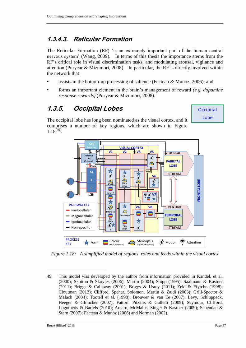
Optimising Comprehension and Shaping Impressions
Bruce Hilliard© 2013 Page 37
1.3.4.3. Reticular Formation
The Reticular Formation (RF) ‘is an extremely important part of the human central
nervous system’ (Wang, 2009). In terms of this thesis the importance stems from the
RF’s critical role in visual discrimination tasks, and modulating arousal, vigilance and
attention (Puryear & Mizumori, 2008). In particular, the RF is directly involved within
the network that:
• assists in the bottom-up processing of salience (Fecteau & Munoz, 2006); and
• forms an important element in the brain’s management of reward (e.g. dopamine
response rewards) (Puryear & Mizumori, 2008).
1.3.5. Occipital Lobes
The occipital lobe has long been nominated as the visual cortex, and it
comprises a number of key regions, which are shown in Figure
1.18(49)
.
Figure 1.18: A simplified model of regions, roles and feeds within the visual cortex
49. This model was developed by the author from information provided in Kandel, et al.
(2000); Skottun & Skoyles (2006); Martin (2004); Shipp (1995); Saalmann & Kastner
(2011); Briggs & Callaway (2001); Briggs & Usrey (2011); Zeki & Ffytche (1998);
Cloutman (2012); Clifford, Spehar, Solomon, Martin & Zaidi (2003); Grill-Spector &
Malach (2004); Tootell et al. (1998); Brouwer & van Ee (2007); Levy, Schluppeck,
Heeger & Glimcher (2007); Fattori, Pitzalis & Galletti (2009); Seymour, Clifford,
Logothetis & Bartels (2010); Arcaro, McMains, Singer & Kastner (2009); Schendan &
Stern (2007); Fecteau & Munoz (2006) and Norman (2002).

Optimising Comprehension and Shaping Impressions
Bruce Hilliard© 2013 Page 38
On the left of the visual cortex within this model(50)
is area V1, which is also commonly
referred to as the striate cortex, because of the striations within this area (Kandel, et al.,
2000). The remaining areas (V2 to V8) are collectively referred to as the extrastriate
areas (Grill-Spector & Malach, 2004; Kandel, et al., 2000). These areas work together
through a staged process (a partially hierarchical processing model), in which different
areas handle diverse functional aspects (the functional specialisation model) (Grill-
Spector & Malach, 2004), as shown in Figure 1.18. However, there is also significant
intra-cortical connectivity between these areas (Koulakov & Chklovskii, 2001; Wurtz,
et al., 2011), and these cortical regions therefore handle and share a wide range of
different stimuli types, to develop percepts and representations about the environment
(Grill-Spector & Malach, 2004).
In terms of this thesis the following key points related to the visual cortex are pertinent:
• The visual cortex utilises a retinotoptic framework(51)
(Grill-Spector & Malach,
2004; Ward, Macevoy, & Epstein, 2010) for handling the information, so it is
analysing a representation of the environment (Kandel, et al., 2000). However, it
is also utilising non-retinotopic aspects within the processing continuum (Grill-
Spector & Malach, 2004). In practical terms, this means that the stimuli is
predominantly handled in a more abstract way in the higher processing areas
(Kandel, et al., 2000).
• Each area handles both central (foveal and parafoveal) and peripheral vision
feeds, but central vision is magnified to optimise the quality and acuity of the
information received (Duncan & Boynton, 2003). This means that visual acuity
declines for stimuli further away from the fovea, which reinforces the issues
related to the distribution of photoreceptor cells in the retina (as discussed in
Section 1.3.1.2.1). There is therefore a strong bias toward processing central
vision. For example, Azzopardi & Cowey (1993) showed that the amount of
cortical tissue in V1 allocated to the processing foveal information was 3.3 to 5.9
times more than that allocated to peripheral vision. When this is also equated to
the cone to ganglion ratios within the retina, it means that there is between 12 and
24 times more cells within the primary visual cortex, which are allocated to
processing central vision, than there is to support peripheral vision (Duncan &
Boynton, 2003). Additionally, it is likely that this focus on foveal and parafoveal
50. Some areas within this model are actually located within, or on the boundary of, the
temporal and parietal lobes. For example, area V5 is located at the juncture of the
temporo-parietal occipital lobes (Grill-Spector & Malach, 2004). For the purposes of
simplification, all of the areas from V1 to V8 have been discussed within the region of the
occipital lobe. Additionally, this is a highly simplified model and the functional and
hierarchical connections within this model do not fully reflect the complex
interconnections applied within this area, such as those identified in McDonald, Mannion,
Goddard, & Clifford (2010).
51. This retinotopic map is represented in a log-polar form, so it is not an exact mapping, but
stretched by the visual processing. This takes into account the magnification aspects
created by the curved shape of the retina and the bias toward the foveal area (Grill-
Spector & Malach, 2004). The mapping therefore ‘preserves the qualitative spatial
relations but distorts quantitative ones’ (Grill-Spector & Malach, 2004, p. 251).

Optimising Comprehension and Shaping Impressions
Bruce Hilliard© 2013 Page 39
vision to improve acuity extends to the higher cortical (extrastriate) areas as well
(Duncan & Boynton, 2003).
• Because of the physiology of this area (particularly in the striate cortex), the cells
typically respond well to:
Edges. Neurons in the ‘visual cortex respond best to edges in their
receptive field’ (Koulakov & Chklovskii, 2001, p. 519). For example, ‘the
interblob pathway sketches boundaries’ (Shipp, 1995, p. 118). In
particular, edges are defined by boundary contrast, which can be caused by
differences such as luminance, colour, spatial frequency, or disparity
(Grossberg, 1994). It is these aspects that make an object stand out
(Grossberg, 1994)(52)
.
Orientation. Cells within the visual cortex respond to a ‘narrow range of
orientations of elongated bars or gratings presented within a certain spatial
position of the visual field.’ (Okamoto, Ikezoe, Tamura, Watanabe, &
Aihara, 2010, p. 151).
Other Aspects. In addition to edges and orientation, there are a range of
other aspects, which are pertinent in developing visual perception. For
example, colour, form and movement are of particular importance
(Grossberg, 1994; Sasaki & Watanabe, 2004, 2012; Seymour, et al., 2010;
Shipp, 1995)(52)
.
• Within the visual cortex ‘colour and form and motion are inextricably linked as
properties of objects’ (Shapley & Hawken, 2011, p. 701). In particular, colour
and form are linked reciprocally, because colour and shape (spatial relationships)
are so important in the perception of objects (Shapley & Hawken, 2011).
• There is a strong bias within the cells in V1 to manage colour opponent
information, with around two thirds (2/3) of the cells allocated to this task
(Friedman, Zhou, & Von Der Heydt, 2003; Johnson, Hawken, & Shapley, 2008).
• Conscious awareness of what is seen appears to occur higher in this processing
than V1 (Pylyshyn, 2002), so in the early stages of processing the viewer is
probably not consciously aware of the visual stimuli (Skottun & Skoyles, 2006).
• It appears that area V4 plays an important role in linking top-down and bottom-up
processes (Mazer & Gallant, 2003). This ‘convergence of bottom-up and top-
down processing streams in area V4 results in an adaptive dynamic map of
salience that guides oculomotor planning during natural vision.’ (Mazer &
Gallant, 2003, p. 1241)
• Area V5(53)
plays a very important role in perception processing within the visual
cortex. Key attributes of this area are:
52. These equate to key salience factors as discussed in Chapter 2 of the thesis.
53. Area V5 is often referred to by other terms in various publications. For example it is also
called the Middle Temporal (MT) area (e.g. Merchant, Battaglia-Mayer, & Georgopoulos
(2001), or by McGraw & Roach (2008)), or the human Middle Temporal + (hMT+) area
(Grill-Spector & Malach, 2004), or sometimes the Dorsolateral Occipital (DLO) area
(Wandell, Dumoulin, & Brewer, 2007).

Optimising Comprehension and Shaping Impressions
Bruce Hilliard© 2013 Page 40
This area contains direction selective cells (Huk, Ress, & Heeger, 2001),
which are attuned to perceive coherent, patterned (Grill-Spector & Malach,
2004), and biological movement (Grossman & Blake, 2002), as a part of
global motion detection (Castelo-Branco et al., 2002).
V5 appears to play an important role in shaping attention within the visual
cortex (Hupe et al., 1998; Zeki & Ffytche, 1998). For example, Foxe &
Simpson (2002) indicated that V5 can be activated prior to V1 (e.g. during
the presentation of fast or slow movement (Zeki & Ffytche, 1998)), and the
two areas then appear to interact to modulate attention.
V5 is also important within the process for driving optical flow (Morrone et
al., 2000), which affects pursuit tracking (Schutz, et al., 2011) and saccadic
eye movements (Kandel, et al., 2000). As explained in Section 1.6 this has
important implications for shaping attention.
• There are direct feed-back loops from the visual cortex (in particular V1 and V5)
to areas such as the LGN, pulvinar and superior colliculus, which appear to
provide visual maps which may be used for afferent attention shaping and efferent
control (Shipp, 2003).
• Although the visual cortex initially supports bottom-up processing of the visual
stream, perception is also modulated through top-down processing (Dodwell &
Humphrey, 1990; Ludwig & Gilchrist, 2003; Pourtois, Rauss, Vuilleumier, &
Schwartz, 2008; Rauss, Schwartz, & Pourtois, 2011). In particular, processing
through the visual cortex is specifically modulated by attention (Briggs & Usrey,
2011; Karns & Knight, 2008), and this appears to be shaped by feed-back systems
within the visual cortex, other areas within the interbrain and midbrain, and the
temporal, parietal and frontal lobes (Foxe & Simpson, 2002).
1.4. From Perception to
Cognition
From the visual cortex in the occipital lobe, the visual stimuli
take two paths to the frontal cortex (Cloutman, 2012), where
higher level cognition primarily takes place (Buchsbaum, 2004;
Gilbert & Fiez, 2004). These two paths are known as the
dorsal and ventral streams (Crick & Koch, 2003; Mishkin,
Ungerleider, & Macko, 1983) (54)
.
54. The dual stream approach discussed in this section is designed to merge a range of
models used to describe this phenomenon. For example Milner & Goodale (2008) refer
to these using the terms ‘vision for perception’ and ‘vision for action’. Fulton (2003a)
refers to these as the ‘Awareness’ and ‘Analytical’ channels. As another alternative,
Crick & Koch (1998) talk about ‘on-line’ and ‘seeing’ systems, and in an earlier paper
they refer to ‘awareness’ (Crick & Koch, 1995). In particular, the terminology used in
this paper is designed to merge these models with the affordances (Gibson, 1966, 1979)
approach cited by Young (2006), the linked ‘ecological-dorsal’ or ‘constructivist-ventral’
model specified by Norman (2002), the ‘Spatial Relationships’ and ‘Object Recognition’
paths explained by Essen, Anderson, & Felleman (1992), and the ‘Object Recognition’

Optimising Comprehension and Shaping Impressions
Bruce Hilliard© 2013 Page 41
A simplified model of the paths that these two streams take is illustrated in Figure
1.19(55)
.
Figure 1.19: A simplified model showing the generalised pathways for the two streams
These two streams appear to be functionally specialised (Ikkai, Jerde, & Curtis, 2010;
Kandel, et al., 2000; Norman, 2002), and can be generally characterised as follows:
• Ventral Stream. The ventral stream ‘has been functionally conceptualised as the
“what” stream, responsible for visual and auditory stimulus identification and
object recognition, the mapping of information into conceptual representations,
and the sound-to-meaning (and written word-to-meaning) mapping underlying the
comprehension of spoken (and written) language’ (Cloutman, 2012, p. 1). The
ventral stream is typically dominated by the processing of the parvocellular
information (Lamme & Roelfsema, 2000; Shipp, 1995), and also some aspects
from the koniocellular pathway (possibly colour and spatial frequency related
stimuli (Van Essen & Gallant, 1994)) (Brown & Goodale, 2008; Nassi &
Calloway, 2009; Simmons et al., 2009).
• Dorsal Stream. This second pathway ‘constitutes the “where/how” stream,
responsible for spatial processing (including location, relative position, and
motion), sensorimotor mapping and the guidance of action towards objects in
space’ (Cloutman, 2012, p. 1). The dorsal stream appears to predominantly
handle the magnocellular path stimuli (Sehatpour P et al., 2010) and some aspects
of the koniocellular information (e.g. motion related (Kaas & Lyon, 2007)).
and ‘Context’ model applied by Bar (2004). The pertinent aspects of these models
are described in the appropriate sections of this thesis.
55. This model was developed by the author from information provided in Garrett (2003);
Merchant, et al. (2001); Bisley (2011); Cloutman (2012); Hickok & Poeppel (2004) and
Schendan & Stern (2007). The model is designed to be indicative only, and does not
contain all of the linkages and connections that are related to the two streams. More of
these connections are illustrated in Figure 1.2, Figure 1.3, and Figure 1.18.

Optimising Comprehension and Shaping Impressions
Bruce Hilliard© 2013 Page 42
Although the two streams are explained within this framework as independently
handling the visual information, there is substantial cross pollination and sharing of
information across the two streams (Skottun & Skoyles, 2006), as explained in Section
1.4.4. Additionally, some of the subcortical elements (e.g. hippocampi, striatum,
limbic system, etc., which are discussed below) support both streams.
The following subsections explain some of the key neural mechanisms utilised within
these two pathways and their interaction.
1.4.1. Ventral Stream
As illustrated in Figure 1.18 the ventral stream is
mainly receiving information from areas within
the visual cortex that are dominated by the
processing of aspects such as colour and form
(Ninomiya, Sawamura, Inoue, & Takada, 2011).
The ventral stream therefore appears to utilise
these visual cues to create detailed mental
representations of the world (Cloutman, 2012). For this reason the ventral stream is
linked to the provision of awareness (Crick & Koch, 2003; Mazer & Gallant, 2003), and
object categorisation (Gallese, 2007).
Within the context of this thesis, key physiological elements utilised within this stream
can be categorised in terms of their processing activities, as discussed in the following
subsections.
1.4.1.1. Overview of the Temporal Lobe Functions
The temporal lobes (TL) appear to play an important role in object
recognition (Bartolomeo & Chokron, 2002; Mishkin, et al., 1983) and constancy
(Ffytche, et al., 2010). Some of the regions within these two lobes are also highly
focussed on responding to specialised features, such as faces(56)
or hands (Kandel, et al.,
2000). Additionally, the TL appear to be actively involved in linking new information
with other memories, such as the recognition of familiar places (Ffytche, et al., 2010),
colour recognition (Kandel, et al., 2000), recognition of lists of information (e.g.
declarative(57)
memory recall) (Aggleton, Sanderson, & Pearce, 2007), the processing of
semantic(58)
information (Mason & Just, 2007), and orientation (in conjunction with
56. The associated Fusiform Face Area (FFA) is not discussed in detail in this thesis, as it is
considered less important in terms of the hypotheses.
57. The term declarative memory relates to the storage and recall of factual information (e.g.
specific facts like dates, names, faces, etc.) (Feldman, 2005). Declarative information
appears to be stored as either semantic (see Footnote 58), or episodic (see Footnote 63)
memory (Feldman, 2005). Further information on declarative memory and the
differentiation with non-declarative memory is provided in Footnote 65.
58. ‘Semantic memory is the subcomponent of memory that is responsible for the acquisition,
representation, and processing of conceptual information. This critically important system

Optimising Comprehension and Shaping Impressions
Bruce Hilliard© 2013 Page 43
areas within the dorsal stream) (Brocki, Fan, & Fossella, 2008). The temporal lobes
therefore play an important role in cognitive discrimination (Magnussen & Greenlee,
1999).
In particular, this area is linked to the processing of foveal and parafoveal vision,
although some aspects of peripheral vision are also processed in these lobes (Grill-
Spector & Malach, 2004; Larson & Loschky, 2009). This utilisation of the peripheral
vision may be associated with scene(59)
‘gist’ analysis (Larson & Loschky, 2009;
Shimozaki, Chen, Abbey, & Eckstein, 2007). This aspect is important when
considering layout issues, such as those required to optimise the design of PowerPoint®
slides for teaching.
This area also demonstrates persistent stimulus-selective activity, and it also appears to
play an important role in working memory (Bellgrove & Mattingley, 2008; D'esposito,
2007).
Additionally, other regions in this area are linked to language (Hickok & Poeppel,
2004), and the recall of words (Kandel, et al., 2000). In particular, fluent reading that
draws on lexical memory(60)
, has been associated with this region of the brain
(Borowsky, et al., 2006). For instance, much of the passive (e.g. reading/listening)
language related functions appear to be linked to Wernicke’s area.
1.4.1.2. Wernicke’s Area
In about 90% of people, Wernicke’s area is located in the posterior portion of the left
temporal lobe (Hickok & Poeppel, 2004)(61)
, and it also extends up into the area of the
parietal lobe (Tanner, 2007) (see Figure 1.3). This region is linked with developing
understanding and comprehension of both written and verbal language (Johnson,
Williams, Pipe, Puppe, & Heiserman, 2001; Tanner, 2007).
of brain function is implicated in a wide range of cognitive functions, including the ability
to assign meaningful interpretations to words and sentences, recognize objects, recall
specific information from previously learned concepts, and acquire new information from
reasoning and perceptual experience’ (Saumier & Chertkow, 2002, p. 516).
59. A scene is a ‘semantically coherent (and often nameable) view… comprising background
elements and multiple discrete objects arranged in a spatially licensed manner’
(Henderson & Hollingworth, 1999, p. 244). Often this term is used to denote scenes in
the real world, but for the purpose of this thesis the term will be utilised for any form of
visual construct that will be viewed by an observer.
60. The term lexical memory refers to the storage of language related words and sounds,
which are recalled during the process of comprehension (Wise et al., 2001). Some
articles delineate this further into lexical and sublexical categories (Giroux & Rey, 2009).
Lexical memory relates to familiar words, which have already been encoded for recall,
and the sublexical systems are used to phonetically decode words (Borowsky et al.,
2006). The development and utilisation of lexical memory is associated with the ventral
stream, and the sublexical processes are associated with the dorsal stream (Borowsky, et
al., 2006; Saur et al., 2008)
61. In the remainder of the population it is located in a similar location in the right
hemisphere.

Optimising Comprehension and Shaping Impressions
Bruce Hilliard© 2013 Page 44
In terms of fluent reading, Wernicke’s area draws on lexical memory, by linking to the
hippocampus, thalamus, frontal cortex, and other cortical areas (Mason & Just, 2007;
Wise, et al., 2001).
1.4.1.3. Subcortical Regions
The following subcortical areas are pertinent to the processing of visual
information within the framework of this thesis.
1.4.1.3.1. Parahippocampal Place Areas
The Parahippocampal Place Areas (PPA) respond more strongly to scenes and pictures
of places, than other types of visual stimuli (Arcaro, et al., 2009; Dickinson & Intraub,
2009; Epstein, Harris, Stanley, & Kanwisher, 1999). In particular, there is evidence
that these areas assess the whole scene, rather than lower order bottom-up aspects
(Doran & Hoffman, 2010), or individual visual objects within the scene (Epstein, 2005).
For instance, the two PPA ‘represents the geometric structure of scenes as defined
primarily by their background elements’ (Epstein, 2005, p. 954).
Additionally, the PPA are involved with encoding new information about the
appearance and layout of what is being viewed (Epstein, et al., 1999). To achieve this
objective, the PPA appear to be important in linking the visual stimuli in representations
with memories provided by the associated hippocampus (see below) (Ffytche, et al.,
2010).
Although Shipp (2003) indicated that the PPA are predominantly processing
information received through the foveas (the fixation point) they also appear to receive
significant inputs from peripheral vision (Epstein, 2005). The PPA and the associated
Transverse Occipital Sulcus (TOS) also seem to play important roles in salience
mapping (Ward, et al., 2010).
Processing within the PPA has also been strongly linked to the application of attention,
with:
• the provision of novel visual material increasing processing attention; and
• repetition of visual material reducing the level of attention, and the amount of
processing that is applied (Yi & Chun, 2005).
1.4.1.3.2. Hippocampi
The two hippocampi receive convergent afferent information ‘from virtually all cortical
association areas’(62)
(Eichenbaum, 2004, p. 110). These structures appear to be very
important in visual processing (Meyer, 2002), and managing both short term storage
62. The hippocampi receive projections from, and provide inputs to, both the dorsal and
ventral streams (Wang, Gao, & Burkhalter, 2011), the frontal lobe (Brogaard, 2011;
Pennartz, Ito, Verschure, Battaglia, & Robbins, 2011) as well as other elements within the
midbrain (Sprague, 1972) and interbrain areas (Mason & Just, 2007; Parikh & Sarter,
2008).

Optimising Comprehension and Shaping Impressions
Bruce Hilliard© 2013 Page 45
and the retrieval of information (Eichenbaum, 2004). Most importantly (in terms of
this thesis), the hippocampi seem to be ‘critically involved in the rapid encoding of
events as associations among stimulus elements and context’ (Eichenbaum, 2004, p.
117). The following key points related to the hippocampi are important in terms of the
hypotheses being investigated:
• They support top-down Processing. The hippocampi may be utilised to ‘rapidly
encode high-fidelity representations’ of semantic elements (Ezzyat & Olson,
2006, p. 38), and link these with episodic memories(63)
. In doing this, the
hippocampi provide associations with episodic and other forms of memory to
generate understanding through top-down processes (Eichenbaum, 2004). For
example, the hippocampi are intrinsically involved in relational processing, and
providing linkages to generate meaning (Sullivan Giovanello, Schnyer, &
Verfaellie, 2004), so this affects aspects such as object recognition. Additionally,
the hippocampi are intrinsically involved in generating plans (Lisman & Redish,
2009) and other top-down signals (D'esposito, 2007).
• They aid understanding. The hippocampi link knowledge ‘in support of the
flexibility of their expressions through comparisons and generalisations across
memories’ (Eichenbaum, 2004, p. 114). Such linkages assist in the rapid
development of understanding, by relating new information to what is already
known.
• They play an important role in learning. The two hippocampi play a critical
role in learning (Schnotz & Kurschner, 2007; Tamminga, 2005), and in particular:
structural learning(64)
where the stimuli are bound by their relationships with
each other (e.g. temporal relationships) (Aggleton, et al., 2007);
the utilisation and memorisation of declarative information (Jurd, 2011;
Sokolov, 2002), and the interconnection of explicit (declarative) memory
(Rowland & Kentros, 2008) and non-declarative (implicit) memories(65)
(Rose, et al., 2010);
63. The term episodic memories ‘refer to our ability to recall specific past events about what
happened where and when. Episodic memory is distinct from other kinds of memory in
being explicitly located in the past and accompanied by the feeling of remembering,
whereas other knowledge that we acquire is purely factual, without any personalised
pastness [sic] attached to it’ (Clayton, Salwiczek, & Dickinson, 2007, p. 189). Episodic
memory does not just include information of a specific event or situation, but also
includes links to other related events (e.g. ones that precede or follow the episode)
(Ferbinteanu, Kennedy, & Shapiro, 2006; Tulving, 2002).
64. Structural learning is the process of ‘binding stimulus elements to make unique arrays (as
in all configural learning), the relationship of these elements to each other, be it spatial or
temporal, is specified’ (Aggleton, et al., 2007, p. 723).
65. In traditional models ‘explicit (declarative) memory is thought to be accessible to
awareness, whereas the contents of implicit (non-declarative) memory are unconscious’
(Berry, Shanks, & Henson, 2008, p. 367). In traditional models these memory types
were seen to be disassociated. However, the research conducted by Berry, et al., (2008)
has indicated that there are significant synergies between the two types. To avoid
ambiguity generated by using this term in relation to explicit and implicit learning (which
was discussed earlier), this thesis will use the terms declarative and non-declarative.

Optimising Comprehension and Shaping Impressions
Bruce Hilliard© 2013 Page 46
learning information that requires the application of spatial relationships
(Maviel, Durkin, Menzaghi, & Bontempi, 2004); and
understanding information that needs to be assessed in terms of its context
(Eichenbaum, 2004).
• Repetition plays an important part in learning. The repetition of experiences, or
visual inputs assists in developing declarative memory, because of the
biophysiological constructs within the hippocampi (Nakazawa, Mchugh, Wilson,
& Tonegawa, 2004; Salimpoor, Chang, & Menon, 2009).
• They assist in making inferential judgements. The hippocampi draw on a range
of memories collected from a network of cortical areas, to assist in making
inferential judgements (Eichenbaum, 2004).
• They play an important role in sequential management of memories. The
hippocampi appear to play a significant role in ordering both semantic and
episodic information (Eichenbaum, 2004; Levy, 1996). For this reason, the
hippocampi are important in supporting the ordering of semantic information in
learning. (Levy, 1996).
• They link emotions to new information. As a part of the relational and
association processes supported by the hippocampi, emotional memories are also
stimulated (Nalloor, Bunting, & Vazdarjanova, 2012). Such connections are
facilitated within the limbic system (Rajmohan & Mohandras, 2007) (see Section
1.4.1.3.4), and through connections with the frontal cortex (see Section 1.4.3).
• The input order affects the quality of memory. Typically the most recently
presented item in a sequence is best remembered (Eichenbaum, 2004).
1.4.1.3.3. Striatum and Basal Ganglia
Whereas the hippocampi are associated predominantly with declarative memory, the
striatum is more closely associated with non-declarative (procedural) memory (Jurd,
2011), and habit formation (Yin & Knowlton, 2006). In particular, the striatum and
basal ganglia(66)
appear to interact to support the utilisation of procedural memory for
behaviour related motor activity (Pennartz, et al., 2011). Additionally, areas within the
striatum mediate:
66. The basal ganglia were not illustrated in Figure 1.3, because their inclusion cluttered the
diagram. The basal ganglia consist of a range of different nuclei, which are located in
the vicinity of the mid-brain and ‘receive inputs from the neocortex, and by way of their
output nuclei, the basal ganglia project massively to thalamic nuclei, which in turn project
to the frontal cortex. This anatomy means the basal ganglia are in a prime position to
influence the executive functions of the forebrain, such as planning for movement and
even cognitive behaviours’ (Graybiel, 2000, p. 509). Additionally, ‘the basal ganglia
send outputs to brainstem nuclei involved in motor control, including the superior
colliculus, which controls axial orientation and saccadic eye movements’ (Graybiel, 2000,
p. 509). The striatum acts as the gateway for the basal ganglia (Kreitzer & Malenka,
2008) and the basal ganglia also communicate with the cerebellum (Carras, Feger, Hoshi,
Strick, & Tremblay, 2005).

Optimising Comprehension and Shaping Impressions
Bruce Hilliard© 2013 Page 47
• stimulus-response learning(67)
(Voorn, Vanderschuren, Groenewegen, Robbins, &
Pennartz, 2004);
• motivational and affective learning(68)
, which has direct implications for cognitive
assessments of value and reward within top-down processes (Balleine, et al.,
2009; Schmidt, Lebreton, Clery-Melin, Daunizeau, & Pessiglione, 2012); and
• goal seeking behaviours, appear to be generated through interaction between the
striatum and hippocampi (Pennartz, et al., 2011).
To facilitate these aspects, the striatum and basal ganglia also interact with limbic
system elements such as the amygdala (Everitt, Morris, O'brien, & Robbins, 1991), so
feelings, fears and aspirations are intrinsically involved in goal seeking behaviours (e.g.
top-down processes such as plans, or the assessment of value and reward).
1.4.1.3.4. Other Limbic System Areas
The limbic system includes a range of physiological structures, which include the
hippocampi(69)
. Other elements within the limbic system are associated with a range of
fundamental behaviours (e.g. hunger and reproduction) (Feldman, 2005) and emotions,
which include fear, rage, placidity, and motivation (Rajmohan & Mohandras, 2007).
Elements within the limbic system, such as the amygdala appear to have a moderating
effect on regions within the mid-brain area (e.g. thalamus) (Fitzgibbon, 2007; Rowland
& Kentros, 2008), interbrain (Merker, 2007) and ventral stream, which shape attention
(Behrendt & Young, 2004; Mohanty, Gitelman, Small, & Mesulam, 2008).
Additionally, the limbic system seems to modulate goal oriented behaviours and
motivation (Goto & Grace, 2008), which has implications for reward systems (Gilbert
& Fiez, 2004). For example, the limbic systems respond strongly to reward signals,
and link these to a wide range of salient and arousing events, which include reactions to
high intensity and novel stimuli (Boksem & Tops, 2008).
Additionally, the limbic system seems to directly affect cognitive processing within the
prefrontal cortex (Behrendt & Young, 2004; O'reilly, 2010; Parikh & Sarter, 2008).
Just as importantly, the limbic system elements have key implications related to feelings
67. This can be equated to simplistic learning, such as the generation of pavlovian responses
(e.g. ring the bell and the dog salivates), which are aligned to the classical conditioning
approach to learning. Classical conditioning indicates that people and animals will
respond in predictable ways if there is some stimulus (e.g. reward, pain, etc.) provided
from within their environment (Biggs & Telfer, 1987).
68. This approach links to the instrumental conditioning model. Instrumental conditioning
posits that humans and animals learn through encoding the consequences of their actions,
which then leads to goal driven behaviours (Balleine, Liljeholm, & Ostlund, 2009).
69. Although different papers utilise varying lists of the physiological structures involved in
the limbic system (Rowland & Kentros, 2008), the elements in the following list are fairly
widely accepted: (1) The limbic cortex, comprising the cingulate gyrus, the
parahippocampal gyrus; (2) The hippocampal formation, which contains the dentate
gyrus, hippocampus (including the fornix), and subicular gyrus; (3) Amygdala; (4)
Septal Area; and (5) Hypothalamus (Rajmohan & Mohandras, 2007).

Optimising Comprehension and Shaping Impressions
Bruce Hilliard© 2013 Page 48
and emotions (Changeux, 2012; Rowland & Kentros, 2008; Van Honk, Morgan, &
Schutter, 2007; Wood & Grafman, 2003) that the viewer might have in relation to the
visual information that is provided (Behrendt & Young, 2004).
1.4.1.3.5. Insula
The insula are located in each hemisphere within the inner part of a fissure between the
temporal and parietal cortices(70)
and they also extend into the frontal lobe (Kandel, et
al., 2000). These regions receive strong ‘inputs from the sensory thalamus and from
parietal, occipital and temporal association cortices’ (Jones, Ward, & Critchley, 2010, p.
611) and exchange information with the frontal cortex, striatum, limbic system
(Pennartz, et al., 2011), broca’s area (Kljajević, 2011), and the cingulate gyrus (Koch,
2011; Mason & Just, 2007; Mohanty, et al., 2008). The two insular regions are
implicated in ‘language(71)
, auditory processing, pain processing, taste and flavour
perception in addition to, or in conjunction with, autonomic(72)
processing’ (Jones, et al.,
2010, p. 611). Additionally, the two insula appear to support the assessment of value
and reward within goal-directed behaviour (O’doherty, 2011), and the application of
focussed attention (Nelson et al., 2010).
Most importantly (in terms of this thesis) the insula appear to be primary nodes for
integrating cognitive and emotional processing (Gu, Liu, Van Dam, Hof, & Fan, 2013).
These physiological regions may therefore provide one of the key areas in which the
first two dimensions of the first unifying model(73)
(comprehension and impressions) are
directly integrated within the human brain.
1.4.1.3.6. Broca’s Area
For most people Broca’s area is located in the left hemisphere of the brain, within the
inferior frontal gyrus in the frontal lobe (Garrett, 2003)(74)
. Although Figure 1.3 shows
this as a specific region, it is not an isolated anatomical area and appears to be a much
more diffuse neural zone than was originally thought (Kljajević, 2011).
It has long been known that Broca’s area ‘controls speech and provides grammatical
structure to language’ (Garrett, 2003, p. 64). However, more recent research has shown
70. Although this region is listed as a cortical area (Fulton, 2011), it has been included in this
Appendix as a sub-cortical area for simplicity, and because it is sometimes listed as a part
of the limbic system (e.g. Mier et al (2010)).
71. In particular the insular regions appear to be sensitive to phonological processing, with
particular emphasis on sublexical spelling translation in reading (Borowsky, et al., 2006)
and the processing of lexical ambiguity (Mason & Just, 2007). This infers linkage to the
dorsal sublexical processing stream.
72. The term autonomic refers to the automated regulation of general activities in the body
(e.g. heart rate, metabolic rate, etc.) (Garrett, 2003). These are controlled through the
autonomic nervous system, and emotional responses have a direct influence on arousal
generated through this system (Feldman, 2005).
73. See Figure 2.1 in Volume 1 of this thesis for a description of this model.
74. For others it is located in similar areas within the right hemisphere.

Optimising Comprehension and Shaping Impressions
Bruce Hilliard© 2013 Page 49
that this area contributes to many other aspects, which include ‘phonetic, phonological,
lexical, semantic and syntactic tasks; in rhythmic perception, harmonic incongruity
perception, tonal frequency discrimination, comprehension of artificial languages, time
perception, calculation tasks, memory tasks, action observation and mental imagery of
movement, musical syntax, processing of complex geometric patterns, in prediction of
sequential patterns’ (Kljajević, 2011, p. 20), and even in body language (Yu & Ballard,
2005).
1.4.2. Dorsal Stream
Whereas the ventral stream appears to be
dominated by the processing of focussed
information provided mainly by the parvocellular
pathway (Doniger, Foxe, Murray, Higgins, &
Javitt, 2002), the dorsal stream appears to
predominantly handle information supplied
through the magnocellular pathway (Mendes et
al., 2005) (75)
. Information managed within the dorsal stream is directly involved in
visuospatial perception (Chan & Newell, 2008), which assists in the development of
awareness (and particularly conscious awareness (Gallese, 2007)) about the
environment (Madary, 2011), and then uses this information to apply visuomotor
guidance (Mcintosh & Lashley, 2008) (e.g. generating attention focussed saccades
and/or other physical movements).
The dorsal stream passes through the parietal lobes to the frontal
cortex. Rather than define each region independently, it will be more
useful in terms of this thesis to aggregate the processes managed
within the parietal lobe(76)
. The following subsections therefore
describe the key features of the dorsal stream activity.
75. The key to this statement is the word ‘dominated’. There is significant evidence that the
ventral stream also handles some stimuli provided through the magnocellular pathway
and the dorsal system also receives inputs from the parvocellular path (Laycock,
Crewther, & Crewther, 2007; Madary, 2011; Skottun & Skoyles, 2006).
76. For the purposes of simplicity adjoining neural regions to the parietal lobes, such as the
intraparietal sulcus, have been handled conjointly within this thesis. Additionally,
aspects such as the split dorsal stream model developed by Rizzolatti & Matelli (2003)
have not been included for the purposes of brevity. A discussion of the somatosensory
cortex has also not been included. This neural region is responsible for processing skin
senses, such as touch, warmth, cold and pain, and is also integral to body positioning and
movement (Garrett, 2003). As these senses are not directly pertinent to the thesis, a
discourse on this region has not been included. This merging of the regions, and the
omission of the more complex aspects of the parietal structures within the dorsal stream,
does not make any material difference to the hypotheses, or the findings.

Optimising Comprehension and Shaping Impressions
Bruce Hilliard© 2013 Page 50
1.4.2.1. This Stream is Very Fast
The dorsal stream transfers stimuli information more quickly to the frontal cortex than
the ventral stream (Fattori, et al., 2009; Foxe & Simpson, 2002). This is partly due to
the fact that this dorsal neural feed of information is:
• predominantly receiving inputs from the faster magnocellular and koniocellular
pathways, as discussed earlier;
• the percepts are originally not interpreted through the types of detailed cognitive
processes applied to the ventral stream (Norman, 2002); and
• the initial fast processing is predominantly unconscious (Irvine, 2011)(77)
.
This rapid processing supports the provision of feed-back and feed-forward systems
(see Section 1.4.4) that modulate attention within the occipital (Pins & Ffytche, 2003)
and temporal lobes (Singh-Curry & Husain, 2009). For example, Figure 1.20(78)
illustrates the approximate timeframes for activation of the various regions within the
visual pathways.
77. However, this is not true for all aspects, and some properties of the dorsal stream are
consciously perceived, just as some stimuli within the ventral stream are processed
unconsciously (Norman, 2002). This statement is therefore a generalisation based on
dominant aspects within each stream.
78. The timeframes are based on the research conducted by Foxe & Simpson (2002), and
these results are based on the presentation of primed (pre-cued) visual stimuli. These
figures are therefore probably indicative of the fastest likely timings. Other studies, such
as the work published by Clementz, Brahmbhatt, McDowell, Brown & Sweeney (2007)
showed slower activations. For instance, they identified that elements of the striate and
extrastriate visual cortex appeared to be processing the stimuli within around 150ms after
presentation. However, in spite of this slower start, they still indicated that volitional
top-down processes were triggered at around 200ms. Their experiments therefore still
indicate a dorsal stream transfer speed of less than 50ms, even for volitional activity (e.g.
active attention).

Optimising Comprehension and Shaping Impressions
Bruce Hilliard© 2013 Page 51
Figure 1.20: Timeframes for activation of various areas for visual processing
Therefore, as pointed out by Foxe and Simpson (2002, p. 145):
‘The rapid flow of activation through the visual system to parietal and
prefrontal cortices (less than 30ms), provides a context for appreciating the
100-400ms commonly needed for information processing prior to response
output in humans. It demonstrates that there is ample time for multiple
cortical interactions at all levels’.
It is these priming feeds that typically also trigger the transition from preconscious to
conscious processing, as discussed in Section 1.2.3.2.
1.4.2.2. Focussed on Change and Motion
The dorsal stream is also designed to manage rapidly changing stimuli, and for this
reason many of the regions in this stream tend to be more plastic(79)
(Au & Lovegrove,
79. Neural plasticity ‘describes the brain’s ability to be formed and moulded’ (Muscolino,
2011, p. 68). In practical terms, this describes ‘the ability of the neuronal tissue to adjust
activities and physical characteristics in order to adapt to changes in the environment or
changes in their use’ (Jensen & Overgaard, 2011, p. 1).

Optimising Comprehension and Shaping Impressions
Bruce Hilliard© 2013 Page 52
2007). This plasticity allows human awareness to keep up with the rapidly changing
environment, so that new salient content can be detected more rapidly (Singh-Curry &
Husain, 2009).
The plastic nature of some of the parietal regions is utilised to achieve motion
perception (Ffytche, et al., 2010), and in particular the discernment of coherent
motion(80)
(Skottun & Skoyles, 2006).
1.4.2.3. Facilitation of Bottom-up and Top-down processes
Tapia & Breitmeyer (2011, p. 934) identified that the information passed through the
dorsal stream had a direct bearing on conscious attention ‘via top-down modulation of
reentrant activity in the ventral object-recognition stream’. This means that the dorsal
stream appears to trigger the ventral stream (Singh-Curry & Husain, 2009) to implement
active attention (e.g. focussing central vision on the object of interest, which then
utilises the high definition parvocellular path feeds) (Rawley & Constantinidis, 2010;
Zimmer, 2008)(81)
. Such activations can create either overt (Domagalik, Beldzik,
Fafrowicz, Oginska, & Marek, 2012), or covert (Fischer & Weber, 1998) attention
through bottom-up and top-down processes (Ignashchenkova, Dicke, Haarmeier, &
Thier, 2004).
Neural regions such as the Lateral Intraparietal (LIP) area also act ‘as a priority map, in
which objects are represented by activity proportional to their behavioural priority’
(Bisley & Goldberg, 2010, p. 1). The resultant ‘priority map combines bottom-up
inputs like rapid visual response with an array of top-down signals like a saccade plan’
(Bisley & Goldberg, 2010, p. 1). In conjunction with the Parietal Eye Fields (PEF)
these priority maps are utilised to target saccades and attention, and support goal-
directed movements (Muggleton, Kalla, Juan, & Walsh, 2011) .
1.4.2.4. Spatial Awareness Focus
Spatial coordinates are processed within the parietal lobe (Zimmer, 2008), and these are
utilised to:
• manage awareness of the surroundings (Pins & Ffytche, 2003); and
80. The perception of coherent motion relates to a higher level within the processing of visual
movement than is generated within the visual cortex (Adelson & Movshon, 1983). This
is achieved through ‘spatiotemporal interpolation by direction selective integration of
spatial pattern information’(Nishida, 2003, p. 273). Coherent motion perception
therefore includes the application of subjective judgements (Delicato & Derrington,
2005), so it goes beyond basic bottom-up perception processes.
81. However, as pointed out by Lambert, et al. (2003), salient aspects such as colour can also
capture attention directly through the ventral stream, so the dorsal stream provides just
one mechanism for quickly shifting attention (albeit a very important one).

Optimising Comprehension and Shaping Impressions
Bruce Hilliard© 2013 Page 53
• provide spatial coordinates which support the rapid targeting of reflexive saccades
(see Section 1.6.1) by providing direct input to the superior colliculus (Ludwig &
Gilchrist, 2003)(82)
.
The spatial depth structures managed within this stream also provide important three
dimensional (3D) cues (Ellison & Cowey, 2007; Mendes, et al., 2005). These 3D cues
seem to feed into the ventral stream to support object recognition, and object
positioning in a spatial context (Farivar, 2009). Spatial awareness of this type
therefore appears to have a direct effect on the cognition of aspects related to shape,
size, and orientation (Farivar, 2009).
Additionally, because the dorsal stream is predominantly handling magnocellular and
koniocellular feeds, the granularity of the percepts and representations handled in this
area are typically considered to be of lower definition. For instance, whereas the
ventral stream predominantly applies central vision (foveal and parafoveal) with some
feeds from peripheral vision, the dorsal stream is dominated by the processing of
‘peripheral retinal input with a high temporal resolution’ (Madary, 2011, p. 424).
1.4.2.5. Sublexical Language Analysis
As discussed in Footnote 60, the ventral stream predominantly manages fluent lexical
reading (Saur, et al., 2008). However, for words that cannot be read fluently,
sublexical mechanisms are utilised to decode the words phonetically, and these actions
are associated with the dorsal stream (Borowsky, et al., 2006). Additionally, as
identified by Cohen, Dehaene, Vinckier, Jobert, & Montavont (2008) the parietal lobe is
also important in managing the reading of degraded text, which is handled sublexically.
1.4.2.6. Part of a Network
This region receives significant inputs from the superior colliculus, hippocampi,
cerebellum (Clower, West, Lynch, & Strick, 2001), and the limbic system (Fulton,
2004). As discussed earlier in this appendix, these neural structures appear to provide
stimulus for shaping attention. Areas such as the limbic system also support the
integration of feelings, impressions, and motivations to this processing.
82. The regions most commonly identified as providing these activations are the Parietal Eye
Fields, which code salient stimuli within coordinate frames that are linked in relation to
the point of gaze (Muggleton, et al., 2011). These regions are therefore an important
driver in generating rapid saccadic eye movements, and this activity is facilitated by
direct connectivity with the superior colliculus (Mcdowell, Dyckman, Austin, &
Clementz, 2008).

Optimising Comprehension and Shaping Impressions
Bruce Hilliard© 2013 Page 54
1.4.3. Frontal Lobe
The frontal lobe(83)
consists of the motor cortex (MC), premotor
cortex (PMC), the prefrontal regions (Yeterian, Pandya,
Tomaiuolo, & Petrides, 2012), and the orbitofrontal (OF) cortex
(Rolls, Everitt, & Roberts, 1996).
1.4.3.1. Motor and Premotor Cortex
The motor cortex and premotor cortex are specifically
responsible for the learning and retrieval of motor related memories, which support
movements within the human body (Kantak, Mummidisetty, & Stinear, 2012). As
these motor activities are beyond the scope of this thesis, these regions will not be
discussed in detail, with the exception of a sub-element known as the Supplementary
Eye Fields (SEF).
The SEF directly contribute to initiating saccadic controls(84)
(Lachaux, et al., 2006),
and pursuit eye movements (Missal & Heinen, 2004). Additionally, the SEF facilitate
these activities by monitoring the context and consequences of these actions (Stuphorn,
Taylor, & Schall, 2000). Research has therefore indicated that the SEF are intrinsically
involved in the brain’s value and reward systems, by appraising value based oculomotor
decisions and then assessing the consequential rewards (So & Stuphorn, 2012).
1.4.3.2. Prefrontal Cortex
The prefrontal cortex (PFC) comprises a large proportion of the frontal lobe. This area
is important for cognitive processing, because:
• it plays a significant role in developing comprehension (Hashimoto & Sakai,
2002) and impressions (Baron, Gobbini, Engell, & Todorov, 2011; Cassidy &
Gutchess, 2012);
• it is important in implementing top-down (Miller, 2000; Sawaki & Luck, 2010),
attention focussing processes, which are related to the management of visual
information (Vanderhasselt et al., 2007; Zanto, Rubens, Thangavel, & Gazzaley,
2011);
83. A simplified model of the frontal lobes has been utilised in this thesis. Different papers
delineate the frontal lobes into a range of different regions, using a variety of models.
For example, other naming conventions include regions such as the orbital, ventrolateral,
lateral, anterior, medial and posterior. The more complex aspects of these physiological
models have not been applied, to avoid overcomplicating this non-material aspect of the
content.
84. In particular the SEF is associated with anti-saccades (Lachaux, Hoffmann, Minotti,
Berthoz, & Kahane, 2006). These are consciously controlled eye movements, which
suppress or inhibit typical attractions toward an object (which are known as pro-
saccades) (Domagalik, et al., 2012).

Optimising Comprehension and Shaping Impressions
Bruce Hilliard© 2013 Page 55
• it appears to play a major role in planning (Crick & Koch, 1998; Kaller, Rahm,
Spreer, Weiller, & Unterrainer, 2011; Oku & Aihara, 2008; Tanji, Shima, &
Mushiake, 2007); and
• it affects numerous aspects of perception and cognition (Ciaramelli, Leo, Del
Viva, Burr, & Ladavas, 2007; Foxe & Simpson, 2002; Libedinsky & Livingstone,
2011; Miller, Freedman, & Wallis, 2002).
For the purposes of this thesis(85)
, only key areas of the PFC are discussed. These areas
are called the Frontal Eye Fields (FEF), dorsolateral (DL), and ventromedial (VM)
regions. The functions of these regions can be broadly delineated as explained in the
following subsections.
1.4.3.2.1. Frontal Eye Fields
The FEF form a part of the efferent systems utilised to shift the eyes to the appropriate
location, to gather information from the environment (Muggleton, et al., 2003), and
provide visual stability (Shin & Sommer, 2012). For example, the FEF receive feeds
emanating in the superior colliculus, to create saccadic controls and stabilise vision
(Wurtz, et al., 2011).
The FEF are therefore integral to visual search (Lane, Smith, Schenk, & Ellison, 2012;
Muggleton, et al., 2003; Wurtz, et al., 2011). For example the FEF are intrinsically
involved in both reflexive (e.g. automatic eye movements responding to stimuli) and
goal-driven voluntary saccades (e.g. the eyes are consciously moved to view specific
objects in the environment) (Ludwig & Gilchrist, 2003). These activities appear to
utilise aspects of salience and priority (Ptak, 2012; Schutz, et al., 2011). For example,
salience aspects such as colour appear to be important for efferent activations (Fecteau
& Munoz, 2006; Muggleton, Juan, Cowey, Walsh, & O'breathnach, 2010).
1.4.3.2.2. Dorsolateral Prefrontal Cortex
The DL area receives information from the dorsal stream (Kravitz, Saleem, Baker, &
Mishkin, 2011). Additionally, the DL maintains ‘reciprocal connections with brain
regions associated with motor control (basal ganglia, premotor cortex, supplementary
motor area), performance monitoring (cingulate cortex), and higher-order sensory
processing (association areas, parietal cortex)’(86)
(Wood & Grafman, 2003, p. 140) .
The DL also appears to play an important role in:
• working memory, because it directs ‘attention to internal representations of
sensory stimuli and motor plans’ (Curtis & D'esposito, 2003, p. 415);
85. A simplified model of the PFC has been utilised to minimise confusion, because the
application of other more complex models would not be materially different within the
context of this thesis.
86. The cingulate cortex plays a role in visuospatial orientation (Vogt, Vogt, & Laureys,
2006). The association areas referred to in this quote are linked to the somatosensory
regions (Roland, O'sullivan, Kawashima, & Ledberg, 1996) described earlier in this
appendix. These regions have not been covered in detail as their physiology and
function have been grouped with other aspects for the purposes of simplicity.

Optimising Comprehension and Shaping Impressions
Bruce Hilliard© 2013 Page 56
• ‘the decisional processes, preparing saccades by inhibiting unwanted reflexive
saccades(87)
(inhibition), maintaining memorised information for ongoing
intentional saccades (short-term spatial memory), or facilitating anticipatory
saccades (prediction), depending upon current external environmental and internal
circumstances’ (Pierrot‐Deseilligny et al., 2003, p. 1460), which directly affect
top-down processing; and
• ‘the regulation of behaviour and control of responses to environmental stimuli’
(Wood & Grafman, 2003, p. 140), which are modulated by emotional content (Ito
et al., 2011).
1.4.3.2.3. Ventromedial Prefrontal Cortex
The VM receives sensory inputs from the ventral stream (Kravitz, et al., 2011), and also
utilises reciprocal connections with the limbic system (e.g. amygdala) for the processing
of emotions (Bechara, Damasio , Damasio , & Lee 1999). Additionally, the VM
interacts with the hippocampi and striatum to support memory related, and higher order,
processing (Arco & Mora, 2009; Nieuwenhuis & Takashima, 2011), such as the
direction of top-down control on goal directed behaviour (Arco & Mora, 2009;
O’doherty, 2011). The ventromedial area is also closely linked with:
• ‘the integration of information about emotion, memory, and environmental
stimuli’ (Wood & Grafman, 2003); and
• assessing aspects of value and reward (Kable & Glimcher, 2009).
1.4.3.3. Orbitofrontal Cortex
The orbitofrontal area appears to be involved in self-evaluation behaviours (Beer,
Lombardo, & Bhanji, 2009). For example, the OF cortex is engaged in:
• Reward Judgements. The OF manages reward and value related processes and
the perception of negative reinforcers (Rolls, 2000), which are involved in
‘motivational control and goal-directed behaviour’ through conscious choice
(Tremblay & Schultz, 1999, p. 704).
• Associative Learning. The OF is responsible for linking stimuli across
modalities to create associations that aid learning (e.g. linking sight and taste
stimuli with reward and punishment behaviour) (Rolls, 2000; Schoenbaum &
Roesch, 2005).
• Influencing Planning. Associative learning helps to create expectations of
predicted outcomes from actions, which link to limbic system areas such as the
amygdala, and therefore emotionally affect goal directed behaviours (e.g. plans)
(Saddoris, Gallagher, & Schoenbaum, 2005). Additionally, it appears that the OF
and VM may interact when planning in terms of consequential behaviours (Zald
& Andreotti, 2010).
87. This appears to work in conjunction with the FEF.

Optimising Comprehension and Shaping Impressions
Bruce Hilliard© 2013 Page 57
1.4.4. Interaction between the Two Streams
1.4.4.1. General Interaction
Although there is functional and anatomical separation of the dorsal and ventral streams
(Yeterian, et al., 2012), which pass information between the occipital and frontal lobes,
it is now widely accepted that there are numerous connections and synergies between
them (Chan & Newell, 2008; Cloutman, 2012; Elliott, Tremblay, & Welsh, 2002). For
example, Weiller, Bormann, Saur, Musso, & Rijntjes (2011) illustrated how the dorsal
and ventral streams need to interact to process language. Three types of interaction
between the streams have been proposed, and each has impacts on top-down and
bottom-up processing. These interactions can be categorised as follows:
• Independent Operation. In some situations the two streams appear to
independently pass percepts to the prefrontal cortex (Lee & Donkelaar, 2002;
Sanocki, Michelet, Sellers, & Reynolds, 2006) for integration (Yeterian, et al.,
2012).
• Feed-back/Feed-forward Operations. In parallel with the independent
processing, it appears that feed-back and feed-forward systems modulate the
activities in the alternate stream. For instance, dorsal stream information may
reach the PFC much more quickly than ventral stream representations(88)
(Laycock, et al., 2007). Feed-back is then applied from the PFC to the visual
cortex (occipital lobe), which then moderates the attention within the ventral
stream. For example, Laycock, Crewther, Fitzgerald, & Crewther (2009)
identified that the magnocellular (dorsal stream) information appear to play an
important role in salience during word identification, because there is a feed-back
system. This feed-back then shapes attention within the parvocellular (ventral
stream) (Laycock, et al., 2009). Additionally, Foxe & Simpson (2002) identified
that there are likely to be multiple iterations of feed-back and feed-forward
between the two streams, and the frontal and occipital lobes, in the process of
developing conscious awareness. This is important, because it supports the
implementation of both bottom-up and top-down processing within very short
timeframes (e.g. < 400 milliseconds). As just one example, structures within the
temporal lobe receive inputs from the prefrontal cortex to facilitate top-down
processing of imagery (Kosslyn, Thompson, & Ganis, 2002).
• Cross-Talk Operation. There is also evidence of direct cross-talk between
elements within the ventral and dorsal streams (Chen et al., 2007). Such cross-
talk appears to be strongly reciprocal (Felleman & Van Essen, 1991; Verhoef,
Vogels, & Janssen, 2011). This reciprocal communication directly moderates
activities in the other stream and supports more effective cognition, such as the
achievement of object recognition (Farivar, 2009). Research indicates that the
thalamus (Madary, 2011), pulvinar (Kaas & Lyon, 2007) and superior colliculus
(Lyon, Nassi, & Callaway, 2010) are involved in such cross-talk activities.
These types of interaction support a ‘flexible interactive system’ that facilitates dynamic
cognition (Cloutman, 2012, p. 3). In fact this type of interaction is essential in
88. As explained in Section 1.4.2.1.

Optimising Comprehension and Shaping Impressions
Bruce Hilliard© 2013 Page 58
achieving numerous cognitive processes (Almeida, Mahon, Nakayama, & Caramazza,
2008; Koshino, Carpenter, Keller, & Just, 2005; Mcintosh & Lashley, 2008).
1.4.4.2. Working Memory
Much of the interaction described in the preceding section appears to take place within
what is generically referred to as working memory. ‘Working memory refers to the
system or systems that are assumed to be necessary in order to keep things in mind
while performing complex tasks such as reasoning, comprehension and learning’
(Baddeley, 2010, p. 136). The role of working memory goes well ‘beyond simple
storage, allowing it to play an important role in cognition generally’ (Baddeley, 2010, p.
138).
The key elements of, and connectivity associated with, working memory are illustrated
in the model provided at Figure 1.21(89)
.
Figure 1.21: Working Memory Model
89. This model has been adapted by the author from Figure 2 on Page 138 in Baddeley
(2010). The significant changes made to this model are: (1) The separation between
working and long term memory has been made more explicit in this model, than was
achieved in Baddeley’s version. (2) The top-down and bottom-up process model
described in Section 2.2 in Volume 1 of this thesis has been explicitly placed into the
central executive frame within this model. This allows the implicit aspects of the central
executive mechanisms raised by Baddeley (2007, 2010) to be fleshed out more
effectively. It is also noteworthy that this model does not illustrate other forms of
working memory associated with the other senses such as; olfactory (smell) (Zelano,
Montag, Khan, & Sobel, 2009), somatosensory (touch) (Savini, Brunetti, Babiloni, &
Ferretti, 2012), and gustation (taste) (Savic-Berglund, 2004). For the purposes of this
thesis, this oversight in Baddeley’s (2010) model will not be significant, because this
research is only focussed on visual input.

Optimising Comprehension and Shaping Impressions
Bruce Hilliard© 2013 Page 59
The key elements of this model are defined by Baddeley (2010) as the:
• Central Executive. The central executive is the attentional control system within
this model (Baddeley, 2010), and it is responsible for managing the information
within working memory (Baddeley, 1992). Although the processes applied for
attentional control are not specified explicitly within Baddeley’s model, they
effectively align to the framework described in Section 2.2 (in Volume 1) of this
thesis.
• Visuo-spatial Sketch Pad. This form of working memory handles and
manipulates visual information and imagery (Baddeley, 1992).
• Phonological Loop. The phonological loop appears to include two components,
which include the following:
a store (storing speech-based information for about 1 to 2 seconds); and
this is coupled with an articulatory control process (which is ‘analogous
with inner speech’) (Baddeley, 1992, p. 255)(90)
.
• Episodic Buffer. The episodic buffer ‘is capable of holding multidimensional(91)
episodes or chunks(92)
, which may combine visual and auditory information
possibly also with smell and taste. It is a buffer that provides a temporary store in
which the various components of working memory, each based on a different
coding system, can interface with information from perception and long-term
memory. The episodic buffer is assumed to have limited capacity of about four
chunks or episodes, and to be accessible through conscious awareness’ (Baddeley,
2010, p. 138).
• Long Term Memory (LTM). In addition to the interactions between the
elements of working memory, these active stores also:
90. These functions are closely associated with Wernicke’s areas (see Section 1.4.1.2) and
Broca’s area (see Section 1.4.1.3.6).
91. Within the context cited by Baddeley (2007, 2010) the term ‘multidimensional’ is better
aligned to the concept of being multimodal.
92. The concept of chunks was raised by Miller (1956) to represent the mental aggregation of
bits of information. In Miller’s (1956, p. 83) model a ‘bit of information is the amount of
information that we need to make a decision between two equally likely alternatives. If
we must decide whether a man is less than six feet tall or more than six feet tall and if we
know that the chances are 50-50, then we need one bit of information.’ A bit is therefore
not necessarily a single aspect (e.g. a visual feature), but a conceptual linking of
information for binary analysis, which in itself is an aggregation of discriminable
variables (e.g. points, colours, tones, etc.) (Miller, 1956). Miller’s (1956) chunk concept
is therefore not a definitive measure, but a construct for understanding how information is
organised or grouped, to allow it to be managed more effectively within working
memory. This concept is important, because it explains how the mental aggregation of
information through chunking can assist the brain to handle more information than would
be possible for individual items or bits (Miller, 1956). Such aggregation may be
influenced by aspects like relational pattern matching (Kirsch, Sebald, & Hoffmann,
2010) within representations.

Optimising Comprehension and Shaping Impressions
Bruce Hilliard© 2013 Page 60
recall information from long term memory(93)
to develop representations,
which are utilised for cognition; and
feed information into long term memory, so this can be recalled later
(Baddeley, 2007).
As illustrated in Figure 1.21, each of the working memory and long term memory
elements interact with each other through feed-forward, feed-back and cross-talk
processes. The neural activations that support these interactions are known as synaptic
reverberations (Mongillo, Barak, & Tsodyks, 2008), which are explained in the
following section.
1.4.4.3. Reverberation – The Underlying Process for Working Memory Management
In practical terms, reverberation simply refers to the stimulation of neurons to keep
them activated and processing the information within working memory (known as
persistence) (Wang, 2001). This is a crucial activity, because it allows the information
to be held on-line for long enough to:
• be utilised effectively for cognition (Mongillo, et al., 2008); and
• provide sufficient reverberations to initiate the ‘the structural changes that
underlie consolidation and formation of permanent LTM’ (Nadel & Hardt, 2011,
p. 252)(94)
.
Working memory related reverberations take place within specific areas such as the
frontal lobes (e.g. prefrontal cortex), and also between areas such as the thalamus,
temporal lobe (e.g. inferotemporal area within the ventral stream), and posterior
parietal areas (e.g. Posterior Parietal cortex/Lateral Intraparietal areas within the
dorsal stream) (Wang, 2001).
One of the key drivers that creates these reverberations is attention (Khan & Muly,
2011), and in the absence of attention the information within working memory will
93. Long term memory refers to ‘memory that stores information on a relatively permanent
basis, although it may be difficult to retrieve’ (Feldman, 2005, p. 226). The generic
concept of long term memory can also be further delineated as ‘long term’ (LT) (which
lasts hours to months), or long lasting (LL) (which lasts from months to a lifetime)
(Garrett, 2003). In either case, LT and LL memories are typically held as consolidations
(e.g. involving the melding of different memories and aspects of memory into a
consolidated memory structure) (Garrett, 2003), which can be categorised as episodic
(see Footnote 63), semantic (see Footnote 58), or non-declarative (procedural) memories
(see Footnote 65) (Feldman, 2005). At the biological level, the development of long
term and long lasting memories is supported through the process of Long Term
Potentiation (LTP), which entails creating new connections between brain cells (Garrett,
2003). These LTP connections are a critical element of the biological processes
underlying learning (Garrett, 2003). Alternatively, the process of forgetting is associated
with Long Term Depression (LTD), in which the synaptic connections between brain
cells are weakened, or lost (Garrett, 2003).
94. See Footnote 93 for more information on consolidation and long term memory.

Optimising Comprehension and Shaping Impressions
Bruce Hilliard© 2013 Page 61
typically be lost very quickly (Chua, 2009; Ptak, 2012). The generation of the top-
down and bottom-up processes described in Chapter 2 (in Volume 1) of this thesis are
therefore very important in terms of ensuring that the pertinent information will be held
in working memory, and then transferred to LTM.
Having addressed the matter of general interactions and working memory, the following
section returns to a description of the remaining element of the visual processing model
provided in Figure 1.1. This element is the metencephalon.
1.5. Metencephalon
The metencephalon contains structures within the hindbrain, such as the pons and
cerebellum (Raz et al., 2003). The pons forms a bulbous part of the brainstem, which
influences sleep and arousal (Garrett, 2003). Such arousal is in turn affected by neural
regions like the Suprachiasmatic nucleus (Mistlberger, 2005) (See Section 1.3.3.2 for
more details on the SCN).
The cerebellum plays an important role in efferent control (Glickstein
& Doron, 2008), and is implicated in maintaining balance and refining
‘movements initiated in the motor cortex by controlling their speed,
intensity and direction’ (Garrett, 2003, p. 438). Additionally, the cerebellum is also
involved in shaping aspects related to attention, language, and working memory
(Glickstein, 2007; Strick, Dum, & Fiez, 2009). However, these activities within the
cerebellum do not appear to be aligned to cognition directly, but predominantly to the
efferent controls related to these activities (Glickstein & Doron, 2008), and the
functional automation of cognitive activities (Merker, 2007; Miller, 2000).
To achieve these activities, the cerebellum is closely linked with key structures within
the interbrain and midbrain regions (Kandel, et al., 2000), and through these (Wurtz, et
al., 2011), to areas within the frontal lobe such as the FEF (Li, Medina, Frank, &
Lisberger, 2011; Schutz, et al., 2011). Such connections support the cerebellum to
play an important role in reading (and in particular sub-lexical reading) (Borowsky, et
al., 2006; Schurz et al., 2010), and writing (Katanoda, Yoshikawa, & Sugishita, 2001).
Of greatest importance for this thesis is the efferent saccadic control (Salman et al.,
2006), and smooth eye tracking control (Lisberger, 2010) provided by the cerebellum.
1.6. Eye Control Process
As illustrated in the preceding sections of this appendix, numerous physiological
elements within the human brain contribute to top-down and bottom-up processes,
which then affect comprehension and impressions. There are two additional
physiological operations, which are important within the context of this thesis, because
they are integral to shaping attention. These processes relate to:
• Saccadic Eye Movements. Saccades are used to move the fovea within the visual
field (Leigh & Zee, 1999), so the eye can fixate on specific objects (Liversedge &
Findlay, 2000). These saccadic eye movements are therefore rapid movements
of the eye, which are happening most of the time, to generate awareness of the

Optimising Comprehension and Shaping Impressions
Bruce Hilliard© 2013 Page 62
environment and focus attention on specific objects (Casey, 2004; Vanrullen &
Koch, 2001) .
• Smooth Pursuit Eye Movements. Smooth pursuit eye movements support
continual clear vision of objects, which are moving within the visual environment.
(Leigh & Zee, 1999). In this form of eye movement the fovea is aligned to the
object on which the person is focussing their attention, and the eye then tracks the
movement (Lisberger, 2010).
Figure 1.22(95)
provides a simplified explanation of the differences between these two
types of eye movements.
Figure 1.22: Illustration of the difference between saccades and pursuits.
These eye movements are controlled by interrelated systems (Fulton, 2003a; Orban De
Xivry & Lefevre, 2007; Schutz, et al., 2011), and they are designed to shift the high
definition foveal vision to important locations within the field of view (Schutz, et al.,
2011), to facilitate awareness and attention (Reynolds & Chelazzi, 2004).
Drivers for these aspects appear to be remarkably similar for most people (Rothkopf,
Ballard, & Hayhoe, 2007). However, the conscious or unconscious implementation of
saccades and smooth pursuits is also task and context dependent (Mcdowell, et al.,
2008; Rothkopf, et al., 2007). For example, the controls utilised to hit a nail with a
hammer (active engagement in the task) will be different from those applied to view a
presentation (passive reading and viewing) (Flanagan & Johansson, 2003). This thesis
will therefore focus only on those aspects that are pertinent to the hypotheses which
relate to passive reading and viewing.
95. Concepts illustrated in these diagrams and the associated text are drawn from Schutz, et
al. (2011); Ludwig & Gilchrist (2003); Liversedge & Findlay (2000); Au & Lovegrove
(2007); Lisberger (2010) and McDowell, Dyckman, Austin, & Clementz (2008).

Optimising Comprehension and Shaping Impressions
Bruce Hilliard© 2013 Page 63
Both types of eye movement are pertinent to this thesis, because saccades are utilised to
view information on the screen (e.g. reading), and smooth pursuits are used to track
moving objects (e.g. viewing animations). The following subsections explain these eye
movements, and their implications in more detail.
1.6.1. Saccades
Saccades pertinent to this thesis can be classified as either reflexive or volitional (Leigh
& Zee, 1999), and these can be categorised as follows(96)
:
• Reflexive. Reflexive saccades are made in response to visual stimuli (Leigh &
Zee, 1999) and these reposition the fovea in response to external cues, which are
typically initiated by bottom-up processing, but can then be modulated by top-
down processes. (Mcdowell, et al., 2008).
• Volitional. This type of saccade is related to purposeful behaviours, which are
normally linked to top-down processes (Leigh & Zee, 1999; Mcdowell, et al.,
2008). Some examples of volitional saccades include; shifts of active attention
based on plans (remembering a scene and the location of objects in the view),
object recognition (recognising a shape and scanning it in a standard way), or on
command (consciously controlling the point of fixation) (Leigh & Zee, 1999;
Mcdowell, et al., 2008).
Within these two categories, the most important form of eye movement for this thesis is
the pro-saccade(97)
, which involves the repositioning of the fovea to specific locations or
96. Different articles also utilise other related terms. In particular, the terms stimulus and
goal driven are commonly used. Stimulus driven is best aligned to reflexive saccades,
whereas goal driven best aligns to volitional eye movements (See Ludwig & Gilchrist
(2003) and Mazer & Gallant (2003) for examples of the use of these terms). However,
there also appears to be some ambiguous aspects related to the use of these terms,
because some goal-driven behaviours may be better aligned to reflexive saccades (e.g. it
is not clear if low-level goal-driven reward mechanisms utilised within the SEF are
specifically just volitional or reflexive). For this reason, the model used in this thesis
utilises the reflexive and volitional paradigm, because the differentials and thresholds for
stimulus/goal driven saccades are not directly material to the hypotheses being tested.
97. Leigh and Zee (1999) also describe three other types of saccade, which they call: (1)
Express saccades: Very short latency saccades that can be elicited when the novel
stimulus is presented after the fixation stimulus has disappeared (gap stimulus). (2)
Spontaneous saccades: Seemingly random saccades that occur when the subject is not
required to perform any particular behavioural task. (3) Quick phases: Quick phases
are nystagmus (this is where the eyes reset during prolonged rotation and direct gaze
toward the oncoming visual scene) generated during vestibular (this is where the eye
holds the image steady when briefly rotating the head) or optikokinetic (this is where the
eyes rotate to hold the scene steady during sustained head rotation) stimulation, or as
automatic resetting movements in the presence of spontaneous drift of the eyes. (This
footnote contains intermingled quotes from Pages 4 and 91 within Leigh and Zee (1999)).
For the purposes of this thesis the express saccades are being grouped with the other
reflexive pro-saccades, because they are created by changes in the content within the
visual field, and are therefore covered within the standard bottom-up and top-down

Optimising Comprehension and Shaping Impressions
Bruce Hilliard© 2013 Page 64
objects within the visual scene (Amlôt & Walker, 2006; Desouza, Iversen, & Everling,
2003). Alternatively, there are anti-saccades, which are inhibiting eye control
processes which suppress pro-saccades (Domagalik, et al., 2012). Whereas, pro-
saccades can be either reflexive or volitional (reflecting bottom-up and top-down
processes), anti-saccades are volitional (Domagalik, et al., 2012; Mcdowell, et al.,
2008).
The last important saccadic influence within the context of this thesis relates to the gap
effect. The gap effect is generated when visual stimulus is removed for a short period
(Coubard, Daunys, & Kapoula, 2004). For example this can be achieved by blanking a
screen, or removing the objects from a screen for a short time, before showing other
objects on the screen. There are two important outcomes related to the application of
the gap effect in terms of this thesis, and these are:
• the following saccade toward the new object becomes much faster, so the point of
attention is moved to the new object more quickly after a gap stimulus is used
(Anderson & Carpenter, 2007); and
• when all visual stimuli are removed (such as a cut between scenes in a film, or the
use of blank separators during a presentation) then the gaze is typically shifted
back to the centre of the screen (Schutz, et al., 2011).
1.6.2. Smooth Pursuits
Smooth pursuits are typically initiated reflexively (Tavassoli & Ringach, 2009), and are
generated by the perception of object motion within the visual field (Berryhill, Chiu, &
Hughes, 2006). Once the fovea has reflexively fixated on the movement it is ‘a high
probability’ that the eye will start to track the moving object (Schutz, et al., 2011, p.
19). To facilitate this tracking, top-down processing can also be applied (Santos, Gizzi,
& Kowler, 2010; Thier & Ilg, 2005).
The first part of the smooth pursuit is therefore referred to as open loop (responding
directly to the stimulus), but for extended tracking, feedback systems within the brain
close the loop to optimise the fixation on the moving target (Schutz, et al., 2011).
1.7. Practical Implications
The preceding sections of this appendix have illustrated some key aspects of
psychophysics and biopsychology, which support top-down and bottom-up processing
of visual stimuli. The following points are of particular importance in terms of the
hypotheses being investigated within this thesis:
• External stimuli can shape attention. As explained in Section 1.2, exogenous
stimuli can shape the level of attentiveness applied by the viewer. This is
important, because it can drive the way in which the viewer processes the
information. The intent of effective visual design should be to present the
processes. The spontaneous saccades and quick phase saccades are not covered
explicitly as they are not germane to the hypotheses being investigated.

Optimising Comprehension and Shaping Impressions
Bruce Hilliard© 2013 Page 65
information in ways that motivate higher levels of awareness for the key
information being provided.
• Comprehension and impressions are linked. Limbic system connectivity
ensures that impressions(98)
are interwoven with the cognitive processes discussed
in this appendix. These interactions must therefore be taken into account when
designing visual materials.
• The neural mechanisms are critical to design. As illustrated in the preceding
information, there is now a significant level of understanding of the mechanisms
utilised to process visual information. Failure to take into account these
mechanisms can lead to visual design solutions that are counterproductive, and
inhibit effective communication.
This thesis is therefore designed to apply the psychophysical, biopsychological, and
physiological principles discussed in this appendix to identify optimal visual design
principles. This approach therefore supports a deliberate design process, which is
based on relevant theories, principles and guidelines, rather than an iterative subjective
process. However, it is not intended that such design would be ultimately
deterministic, or that prototyping would not be useful in the development of optimised
visual materials.
98. The term impressions refers to the subjective feeling about the presentation, which results
from the way the material is presented. This concept is explained in more detail in
Chapter 1 of this thesis.
General Note by the Author
Due to the substantial advances in neural scanning and monitoring techniques, which
have been achieved over the past few years, there have been significant
enhancements in the general understanding, and specific functionality, of various
elements within the human brain. These advances have assisted in developing
knowledge within the fields of psychophysics, biopsychology, and cognitive science.
In particular, the expansion of knowledge in these fields within the last five years has
been an important facilitator for the development of the concepts that are being
addressed within this thesis. The author therefore wishes to take this opportunity to
thank the other researchers who have made this current analysis feasible.

Optimising Comprehension and Shaping Impressions
Bruce Hilliard© 2013 Page 66
REFERENCES
Adelson, E. H., & Movshon, J. (1983). The perception of coherent motion in two-
dimensional patterns. Paper presented at the ACM Siggraph and Sigart
Interdisciplinary Workshop on Motion: Representation and Perception, Toronto.
Aggleton, J. P., Sanderson, D. J., & Pearce, J. M. (2007). Structural learning and the
hippocampus. Hippocampus, 17(9), 723-734. doi: 10.1002/hipo.20323
Allen, P. A., Smith, A. F., Lien, M.-C., Kaut, K. P., & Canfield, A. (2009). A multistream
model of visual word recognition. Attention, Perception, & Psychophysics, 71(2),
281-296. doi: 10.3758/app.71.2.281
Almeida, J., Mahon, B. Z., Nakayama, K., & Caramazza, A. (2008). Unconscious processing
dissociates along categorical lines Proceedings of the National Academy of Sciences
of the United States of America, 105(39), 15214-15218.
Amlôt, R., & Walker, R. (2006). Are somatosensory saccades voluntary or reflexive?
Experimental Brain Research, 168(4), 557-565.
Amthor, F. R., Tootle, J. S., & Gawne, T. J. (2005). Retinal ganglion cell coding in simulated
active vision. Visual Neuroscience, 22(6), 789-806.
Anderson, A. J., & Carpenter, R. H. S. (2007). The effect of stimuli that isolate S-cones on
early saccades and the gap effect. Proceedings of the Royal Society B: Biological
Sciences, 275(1632), 335-344.
Arcaro, M. J., McMains, S. A., Singer, B. D., & Kastner, S. (2009). Retinoptic organisation
of human ventral visual cortex. Journal of Neuroscience, 29(34), 10638-10652.
Arco, A. D., & Mora, F. (2009). Neurotransmitters and prefrontal cortex–limbic system
interactions: implications for plasticity and psychiatric disorders. Journal of Neural
Transmission, 116(8), 941-952. doi: 10.1007/s00702-009-0243-8
Arieh, Y., & Marks, L. (2008). Cross-modal interaction between vision and hearing: A
speed-accuracy analysis. Perception & Psychophysics, 70(3), 412-421. doi:
10.3758/pp.70.3.412
Au, A., & Lovegrove, B. (2007). The contribution of rapid visual and auditory processing to
the reading of irregular words and pseudowords presented singly and in contiguity.
Perception & Psychophysics, 69(8), 1344-1359.
Azzopardi, P., & Cowey, A. (1993). Preferential representation of the fovea in the primary
visual cortex. Nature, 361(6414), 719-721.
Baddeley, A. (1992). Working Memory. Science, 255(5044), 556-556.

Optimising Comprehension and Shaping Impressions
Bruce Hilliard© 2013 Page 67
Baddeley, A. (2000). The episodic buffer: a new component of working memory? Trends in
Cognitive Sciences, 4(11), 417-423. doi: 10.1016/s1364-6613(00)01538-2
Baddeley, A. (2007). Working memory, thought, and action. New York: Oxford University
Press.
Baddeley, A. (2010). Working memory. Current Biology, 20(4), R136-R140. doi:
http://dx.doi.org/10.1016/j.cub.2009.12.014
Badgaiyan, R. D. (2012). Nonconscious perception, conscious awareness and attention.
Consciousness and Cognition, 21(1), 584-586. doi: 10.1016/j.concog.2012.01.001
Balleine, B. W., Liljeholm, M., & Ostlund, S. B. (2009). The integrative function of the basal
ganglia in instrumental conditioning. Behavioural Brain Research, 199(1), 43-52.
doi: 10.1016/j.bbr.2008.10.034
Ban, S.-W., Lee, I., & Lee, M. (2008). Dynamic visual selective attention model.
Neurocomputing, 71(4–6), 853-856. doi: 10.1016/j.neucom.2007.03.003
Bar, M. (2004). Visual objects in context. Nature Reviews. Neuroscience, 5(8), 617-629. doi:
10.1038/nrn1476
Bargmann, C. I. (2012). Beyond the connectome: How neuromodulators shape neural
circuits. BioEssays, 34(6), 458-465. doi: 10.1002/bies.201100185
Baron, S. G., Gobbini, M. I., Engell, A. D., & Todorov, A. (2011). Amygdala and
dorsomedial prefrontal cortex responses to appearance-based and behavior-based
person impressions. Social Cognitive and Affective Neuroscience, 6(5), 572-581. doi:
10.1093/scan/nsq086
Bartolomeo, P., & Chokron, S. (2002). Can we change our vantage point to explore imaginal
neglect? Behavioral and Brain Sciences, 25(2), 184-185.
Baumeister, R. F., & Vohs, K. D. (Eds.). (2007). Encyclopedia of Social Psychology.
Thousand Oaks, CA: Sage Publications.
Beato, G. (2010, 2010/07//). Who's afraid of subliminal advertising? "Behavior placement"
in television programming is neither new nor alarming. Reason, 42, 18+.
Bechara, A., Damasio , H., Damasio , A. R., & Lee , G. P. (1999). Different Contributions of
the Human Amygdala and Ventromedial Prefrontal Cortex to Decision-Making. The
Journal of Neuroscience, 19(13), 5473-5481.
Becker, S. I., & Horstmann, G. (2009). A feature-weighting account of priming in
conjunction search. Attention, Perception, & Psychophysics, 71(2), 258-272. doi:
10.3758/app.71.2.258
Beer, J. S., Lombardo, M. V., & Bhanji, J. P. (2009). Roles of Medial Prefrontal Cortex and
Orbitofrontal Cortex in Self-evaluation. Journal of Cognitive Neuroscience, 22(9),
2108-2119. doi: 10.1162/jocn.2009.21359

Optimising Comprehension and Shaping Impressions
Bruce Hilliard© 2013 Page 68
Behrendt, R.-P., & Young, C. (2004). Hallucinations in schizophrenia, sensory impairment,
and brain disease: A unifying model. Behavioral and Brain Sciences, 27, 771-830.
Bellgrove, M. A., & Mattingley, J. B. (2008). Molecular Genetics of Attention. Annals of the
New York Academy of Sciences, 1129(1), 200-212. doi: 10.1196/annals.1417.013
Berman, R. A., & Colby, C. (2009). Attention and active vision. Vision Research, 49(10),
1233-1248. doi: 10.1016/j.visres.2008.06.017
Berman, R. A., & Wurtz, R. H. (2011). Signals Conveyed in the Pulvinar Pathway from
Superior Colliculus to Cortical Area MT. The Journal of Neuroscience, 31(2), 373-
384. doi: 10.1523/jneurosci.4738-10.2011
Berry, C. J., Shanks, D. R., & Henson, R. N. A. (2008). A unitary signal-detection model of
implicit and explicit memory. Trends in Cognitive Sciences, 12(10), 367-373. doi:
10.1016/j.tics.2008.06.005
Berryhill, M. E., Chiu, T., & Hughes, H. C. (2006). Smooth pursuit of nonvisual motion.
Journal of Neurophysiology, 96(1), 461-465. doi: 10.1152/jn.00152.2006
Berson, D. M., Dunn, F. A., & Takao, M. (2002). Phototransduction by Retinal Ganglion
Cells That Set the Circadian Clock. Science, 295(5557), 1070-1073.
Biggs, J. B., & Telfer, R. (1987). The process of learning. (2nd ed.). Sydney, Australia:
Prentice-Hall.
Bishop, S. J. (2008). Neural mechanisms underlying selective attention to threat. Annals of
the New York Academy of Sciences, 1129(1), 141-152. doi: 10.1196/annals.1417.016
Bisley, J. W. (2011). The neural basis of visual attention. The Journal of Physiology, 589(1),
49-57. doi: 10.1113/jphysiol.2010.192666
Bisley, J. W., & Goldberg, M. E. (2010). Attention, Intention and Priority in the Parietal
Lobe. Annual Review of Neuroscience, 33(1), 1-21.
Bizley, J. K., & King, A. J. (2008). Visual-auditory spatial processing in auditory cortical
neurons. Brain Research(1242), 24-36.
Boksem, M. A. S., & Tops, M. (2008). Mental fatigue: Costs and benefits. Brain Research
Reviews, 59(1), 125-139. doi: 10.1016/j.brainresrev.2008.07.001
Borchert, D. M. (Ed.). (2006). Encyclopedia of Philosophy (2nd ed. Vol. 9). Detroit, USA:
Macmillan Reference.
Borowsky, R., Cummine, J., Owen, W. J., Friesen, C. K., Shih, F., & Sarty, G. (2006). FMRI
of ventral and dorsal processing streams in basic reading processes: Insular
sensitivity to Phonology. Brain Topography, 18(4), 233-239.
Bouman, M. A., & Walraven, P. L. (1972). On threshold mechanisms for achromatic and
chromatic vision. Acta Psychologica, 36(3), 178-189. doi: 10.1016/0001-
6918(72)90002-9

Optimising Comprehension and Shaping Impressions
Bruce Hilliard© 2013 Page 69
Bowmaker, J. K., & Dartnall, H. J. (1980). Visual pigments of rods and cones in a human.
The Journal of Physiology, 298(January 1980), 501-511.
Bradley, F. H. (1902). On Active Attention. Mind, 11(41), 1-30.
Breitmeyer, B. G., Koc, A., Ogmen, H., & Ziegler, R. (2008). Functional hierarchies of
nonconscious visual processing. Vision Research(48), 1509-1513.
Breitmeyer, B. G., Ogmen, H., & Chen, J. (2004). Unconscious priming by color and form:
Different processes and levels. Consciousness and Cognition, 13(1), 138-157. doi:
10.1016/j.concog.2003.07.004
Breitmeyer, B. G., Ro, T., Ogmen, H., & Todd, S. (2007). Unconscious, stimulus-dependent
priming and conscious, percept-dependent priming with chromatic stimuli.
Perception & Psychophysics, 69(4), 550-557.
Briggs, F., & Callaway, E. M. (2001). Layer-Specific Input to Distinct Cell types in Layer 6
of Monkey Primary Visual Cortex. The Journal of Neuroscience, 21(10), 3600-3608.
Briggs, F., & Usrey, W. M. (2011). Corticogeniculate feedback and visual processing in the
primate. The Journal of Physiology, 589(1), 33-40. doi:
10.1113/jphysiol.2010.193599
Brocki, K., Fan, J., & Fossella, J. (2008). Placing Neuroanatomical Models of Executive
Function in a Developmental Context. Annals of the New York Academy of Sciences,
1129(1), 246-255. doi: 10.1196/annals.1417.025
Brogaard, B. (2011). Conscious vision for action versus unconscious vision for action?
Cognitive Science, 35(6), 1076-1104. doi: 10.1111/j.1551-6709.2011.01171.x
Brooks, S. J., Savov, V., Allzén, E., Benedict, C., Fredriksson, R., & Schiöth, H. B. (2012).
Exposure to subliminal arousing stimuli induces robust activation in the amygdala,
hippocampus, anterior cingulate, insular cortex and primary visual cortex: A
systematic meta-analysis of fMRI studies. NeuroImage, 59(3), 2962-2973. doi:
http://dx.doi.org/10.1016/j.neuroimage.2011.09.077
Brouwer, G. J., & van Ee, R. (2007). Visual Cortex Allows Prediction of Perceptual States
during Ambiguous Structure-From-Motion. The Journal of Neuroscience, 27(5),
1015-1023. doi: 10.1523/jneurosci.4593-06.2007
Brown, L. E., & Goodale, M. A. (2008). Koniocellular projections and hand-assisted
blindsight. Neuropsychologia, 46(14), 3241-3242. doi:
10.1016/j.neuropsychologia.2008.09.001
Brown, P. K., & Wald, G. (1964). Visual Pigments in Single Rods and Cones of the Human
Retina. Science, 144(3614), 45-52.
Buchsbaum, M. S. (2004). Frontal Cortex Function. The American Journal of Psychiatry,
161(12), 2178-2178.

Optimising Comprehension and Shaping Impressions
Bruce Hilliard© 2013 Page 70
Bugental, J. F. T. (1964). The Third Force in Psychology. Journal of Humanistic
Psychology, 4(1), 19-26. doi: 10.1177/002216786400400102
Calvo, M. G., Nummenmaa, L., & Hyönä, J. (2008). Emotional scenes in peripheral vision:
Selective orienting and gist processing, but not content identification. Emotion, 8(1),
68-80.
Campbell, K., & Macdonald, M. (2011). The effects of attention and conscious state on the
detection of gaps in long duration auditory stimuli. Clinical Neurophysiology, 122(4),
738-747. doi: 10.1016/j.clinph.2010.10.036
Cao, D., Pokorny, J., Smith, V. C., & Zele, A. J. (2008). Rod contributions to color
perception: Linear with rod contrast. Vision Research, 48(26), 2586-2592. doi:
10.1016/j.visres.2008.05.001
Cardu, B., Ptito, M., & Dumont, M. (1980). Pretectum and superior colliculus in object vs
pattern discrimination in the monkey. Neuropsychologia, 18(4–5), 559-568. doi:
10.1016/0028-3932(80)90157-8
Carras, P. L., Feger, J., Hoshi, E., Strick, P. L., & Tremblay, L. (2005). The cerebellum
communicates with the basal ganglia. [Article]. Nature Neuroscience, 8(11), 1491+.
Carrasco, M. (2011). Visual attention: The past 25 years. Vision Research, 51(13), 1484-
1525. doi: 10.1016/j.visres.2011.04.012
Carrasco, M., Eckstein, M. P., Verghese, P., Boynton, G. M., & Treue, S. (2009). Visual
attention: Neurophysiology, psychophysics and cognitive neuroscience. Vision
Research, 49(10), 1033-1036. doi: 10.1016/j.visres.2009.04.022
Casey, E. S. (2004). Attending and glancing. Continental Philosophy Review, 37(1), 83-126.
doi: 10.1023/B:MAWO.0000049308.09311.8b
Cassidy, B. S., & Gutchess, A. H. (2012). Structural variation within the amygdala and
ventromedial prefrontal cortex predicts memory for impressions in older adults.
Frontiers in Psychology, 3, 319.
Castelo-Branco, M., Formisano, E., Backes, W., Zanella, F., Neuenschwander, S., Singer,
W., & Goebel, R. (2002). Activity patterns in human motion-sensitive areas depend
on the interpretation of global motion. Proceedings of the National Academy of
Sciences of the United States of America, 99(21), 13914-13919.
Cavanagh, P. (2011). Visual cognition. Vision Research, 51(13), 1538-1551. doi:
10.1016/j.visres.2011.01.015
Cavanaugh, J., McAlonan, K., & Wurtz, R. H. (2008). Guarding the gateway to cortex with
attention in visual thalamus. [Report]. Nature, 456(7220), 391+.
Chan, J. S., & Newell, F. N. (2008). Behavioral evidence for task-dependent “what” versus
“where” processing within and across modalities. Perception & Psychophysics,
70(1), 36-49. doi: 10.3758/pp.70.1.36

Optimising Comprehension and Shaping Impressions
Bruce Hilliard© 2013 Page 71
Changeux, J.-P. (2012). Beauty in the brain: for a neuroscience of art. Academia Nazionale
dei Lincei, 23, 315-320.
Chen, C.-M., Lakatos, P., Shah, A. S., Mehta, A. D., Givre, S. J., Javitt, D. C., & Schroeder,
C. E. (2007). Functional Anatomy and Interaction of Fast and Slow Visual Pathways
in Macaque Monkeys. Cerebral Cortex, 17(7), 1561-1569. doi:
10.1093/cercor/bhl067
Cheong, S. K., Tailby, C., Martin, P. R., Levitt, J. B., & Solomon, S. G. (2011). Slow
intrinsic rhythm in the koniocellular visual pathway. Proceedings of the National
Academy of Sciences. doi: 10.1073/pnas.1108004108
Chica, A. B., & Christie, J. (2009). Spatial attention does improve temporal discrimination.
Attention, Perception, & Psychophysics, 71(2), 273-280. doi: 10.3758/app.71.2.273
Chua, F. K. (2009). A new object captures attention—but only when you know it's new.
Attention, Perception, & Psychophysics, 71(4), 699-711. doi: 10.3758/app.71.4.699
Ciaramelli, E., Leo, F., Del Viva, M. M., Burr, D. C., & Ladavas, E. (2007). The
contribution of prefrontal cortex to global perception. Experimental Brain Research,
181(3), 427-434. doi: 10.1007/s00221-007-0939-7
Clayton, N. S., Salwiczek, L. H., & Dickinson, A. (2007). Episodic memory. Current
Biology, 17(6), R189-R191. doi: 10.1016/j.cub.2007.01.011
Clementz, B. A., Brahmbhatt, S. B., McDowell, J. E., Brown, R. A. M., & Sweeney, J. A.
(2007). When does the brain inform the eyes whether and where to move? an EEG
study in humans. Cerebral Cortex, 17(11), 2634-2643. doi: 10.1093/cercor/bhl171
Clifford, C. W. G., Spehar, B., Solomon, S. G., Martin, P. R., & Zaidi, Q. (2003).
Interactions between colour and luminance in the perception of orientation. Journal
of Vision, 3, 106-115.
Cline, D., Hofstetter, H. W., & Griffin, J. R. (1997). Dictionary of visual science (4th ed.).
Boston: Butterworth-Heinemann.
Cloutman, L. L. (2012). Interaction between dorsal and ventral processing streams: Where,
when and how? Brain and Language.
Clower, D. M., West, R. A., Lynch, J. C., & Strick, P. L. (2001). The Inferior Parietal Lobule
Is the Target of Output from the Superior Colliculus, Hippocampus, and Cerebellum.
The Journal of Neuroscience, 21(16), 6283-6291.
Cohen, L., Dehaene, S., Vinckier, F., Jobert, A., & Montavont, A. (2008). Reading normal
and degraded words: Contribution of the dorsal and ventral visual pathways.
NeuroImage, 40(1), 353-366. doi: 10.1016/j.neuroimage.2007.11.036
Costall, A. P., & Still, A. (1989). Gibson's Theory of Direct Perception and the Problem of
Cultural Relativism. Journal for the Theory of Social Behaviour, 19(4), 433-441. doi:
10.1111/j.1468-5914.1989.tb00159.x

Optimising Comprehension and Shaping Impressions
Bruce Hilliard© 2013 Page 72
Coubard, O., Daunys, G., & Kapoula, Z. (2004). Gap effects on saccade and vergence
latency. Experimental Brain Research, 154(3), 368-381. doi: 10.1007/s00221-003-
1670-7
Couperus, J. W. (2009). Implicit learning modulates selective attention at sensory levels of
perceptual processing. Attention, Perception, & Psychophysics, 71(2), 342-351. doi:
10.3758/app.71.2.342
Crick, F., & Koch, C. (1995). Are we aware of neural activity in primary visual cortex?
Nature, 375(6527), 121-123.
Crick, F., & Koch, C. (1998). Consciousness and neuroscience. Cerebral Cortex, 8(2), 97-
107. doi: 10.1093/cercor/8.2.97
Crick, F., & Koch, C. (2003). A framework for consciousness. Nature Neuroscience, 6(2),
119-126. doi: 10.1038/nn0203-119
Curtis, C. E., & D'Esposito, M. (2003). Persistent activity in the prefrontal cortex during
working memory. Trends in Cognitive Sciences, 7(9), 415-423. doi: 10.1016/s1364-
6613(03)00197-9
Custers, R., & Aarts, H. (2010). The Unconscious Will: How the Pursuit of Goals Operates
Outside of Conscious Awareness. Science, 329(5987), 47-50. doi:
10.1126/science.1188595
D'Esposito, M. (2007). From cognitive to neural models of working memory. Philosophical
Transactions of the Royal Society B: Biological Sciences, 362, 761-772.
Dayan, P., Kakade, S., & Montague, P. R. (2000). Learning and selective attention. Nature
Neuroscience, 3 Suppl 1(11s), 1218-1223.
Dehaene, S., Changeux, J.-P., Naccache, L., Sackur, J., & Sergent, C. (2006). Conscious,
preconscious, and subliminal processing: a testable taxonomy. Trends in Cognitive
Sciences, 10(5), 204-211. doi: 10.1016/j.tics.2006.03.007
Delicato, L. S., & Derrington, A. M. (2005). Coherent motion perception fails at low
contrast. Vision Research, 45(17), 2310-2320. doi: 10.1016/j.visres.2005.02.020
Desimone, R. (2007). How the brain pays attention. Paper presented at the Forum for the
Future of Higher Education, Cambridge, Massachusetts.
Desimone, R., & Duncan, J. (1995). Neural mechanisms of selective visual attention. Annual
Review of Neuroscience, 18, 193-222.
DeSouza, J. F., Iversen, S. D., & Everling, S. (2003). Neural correlates for preparatory set
associated with pro-saccades and anti-saccades in primate prefrontal cortex. Journal
of Vision, 3(9), 686. doi: 10.1167/3.9.686
Dickinson, C. A., & Intraub, H. (2009). Spatial asymmetries in viewing and remembering
scenes: Consequences of an attentional bias? Attention, Perception, & Psychophysics,
71(6), 1251-1262. doi: 10.3758/app.71.6.1251

Optimising Comprehension and Shaping Impressions
Bruce Hilliard© 2013 Page 73
Dodwell, P. C., & Humphrey, G. K. (1990). A functional theory of the McCollough effect.
Psychological Review, 97(1), 78-89.
Domagalik, A., Beldzik, E., Fafrowicz, M., Oginska, H., & Marek, T. (2012). Neural
networks related to pro-saccades and anti-saccades revealed by independent
component analysis. NeuroImage, 62(3), 1325-1333. doi:
10.1016/j.neuroimage.2012.06.006
Doniger, G. M., Foxe, J. J., Murray, M. M., Higgins, B. A., & Javitt, D. C. (2002). Impaired
visual object recognition and dorsal/ventral stream interaction in schizophrenia.
Archives of General Psychiatry, 59(11), 1011-1020. doi:
10.1001/archpsyc.59.11.1011
Doran, M. M., & Hoffman, J. E. (2010). The role of visual attention in multiple object
tracking: Evidence from ERPs. Attention, Perception, & Psychophysics, 72(1), 33-52.
doi: 10.3758/app.72.1.33
Doré-Mazars, K., Pouget, P., & Beauvillain, C. (2004). Attentional selection during
preparation of eye movements. Psychological Research, 69(1-2), 67-76. doi:
10.1007/s00426-003-0166-1
Duncan, R. O., & Boynton, G. M. (2003). Cortical magnification within human primary
visual cortex correlates with acuity thresholds. Neuron, 38, 659-671.
Eichenbaum, H. (2004). Hippocampus: Cognitive Processes and Neural Representations that
Underlie Declarative Memory. Neuron, 44(1), 109-120.
Elliott, D., Tremblay, L., & Welsh, T. N. (2002). A fast ventral stream or early dorsal-ventral
interactions? Behavioral and Brain Sciences, 25(01), 105-105. doi:
doi:10.1017/S0140525X02310021
Ellison, A., & Cowey, A. (2007). Time course of the involvement of the ventral and dorsal
visual processing streames in a visuospatial task. Neuropsychologia, 45, 3335-3339.
Epstein, R. (2005). The cortical basis of visual scene processing. Visual Cognition, 12(6),
954-978. doi: 10.1080/13506280444000607
Epstein, R., Harris, A., Stanley, D., & Kanwisher, N. (1999). The Parahippocampal Place
Area: Recognition, Navigation, or Encoding? Neuron, 23(1), 115-125. doi:
10.1016/s0896-6273(00)80758-8
Essen, D. C. V., Anderson, C. H., & Felleman, D. J. (1992). Information Processing in the
Primate Visual System: An Integrated Systems Perspective. Science, 255(5043), 419-
423.
Evans, J. S. B. T. (2008). Dual-processing accounts of reasoning, judgement and social
cognition. Annual Review of Psychology, 59, 255-278.
Everitt, B. J., Morris, K. A., O'Brien, A., & Robbins, T. W. (1991). The basolateral
amygdala-ventral striatal system and conditioned place preference further evidence of

Optimising Comprehension and Shaping Impressions
Bruce Hilliard© 2013 Page 74
limbic-striatal interactions underlying reward-related processes. Neuroscience, 42(1),
1-18.
Ezzyat, Y., & Olson, I. (2006). The hippocampus and the fidelity of representations in visual
working memory. Journal of Vision, 6(6), 38. doi: 10.1167/6.6.38
Farivar, R. (2009). Dorsal–ventral integration in object recognition. Brain Research Reviews,
61(2), 144-153. doi: 10.1016/j.brainresrev.2009.05.006
Fattori, P., Pitzalis, S., & Galletti, C. (2009). The cortical visual area V6 in macaque and
human brains. Journal of Physiology-Paris, 103(1–2), 88-97. doi:
10.1016/j.jphysparis.2009.05.012
Fecteau, J. H., & Munoz, D. P. (2006). Salience, relevance, and firing: a priority map for
target selection. Trends in Cognitive Sciences, 10(8), 382-390. doi:
10.1016/j.tics.2006.06.011
Feldman, R. S. (2005). Understanding Psychology (7th ed.). New York: McGraw Hill.
Felleman, D. J., & Van Essen, D. C. (1991). Distributed Hierarchical Processing in the
Primate Cerebral Cortex. Cerebral Cortex, 1(1), 1-47. doi: 10.1093/cercor/1.1.1
Ferbinteanu, J., Kennedy, P. J., & Shapiro, M. L. (2006). Episodic memory—From brain to
mind. Hippocampus, 16(9), 691-703. doi: 10.1002/hipo.20204
Ffytche, D. H., Blom, J. D., & Catani, M. (2010). Disorders of visual perception. Journal of
Neurological Neurosurgical Psychiatry, 81, 1280-1287.
Figueiro, M. G., Bierman, A., Plitnick, B., & Rea, M. S. (2009). Preliminary evidence that
both blue and red light can induce alertness at night. BMC Neuroscience, 10, 105.
Fischer, B., & Weber, H. (1998). Effects of pre-cues on voluntary and reflexive saccade
generation I. Anti-cues for pro-saccades. Experimental Brain Research, 120(4), 403-
416. doi: 10.1007/s002210050414
FitzGibbon, T. (2006). Does the development of the perigeniculate nucleus support the
notion of a hierarchical progression within the visual pathway? Neuroscience, 140(2),
529-546. doi: 10.1016/j.neuroscience.2006.02.038
FitzGibbon, T. (2007). Do first order and higher order regions of the thalamic reticular
nucleus have different developmental timetables? Experimental Neurology, 204, 339-
354. doi: 10.1016/j.neuroscience.2006.02.038
Flanagan, J. R., & Johansson, R. S. (2003). Action plans used in action observation. Nature,
424(6950), 769-771. doi: 10.1038/nature01861.
Folk, C. L., Remington, R. W., & Wu, S.-C. (2009). Additivity of abrupt onset effects
supports nonspatial distraction, not the capture of spatial attention. Attention,
Perception, & Psychophysics, 71(2), 308-313. doi: 10.3758/app.71.2.308

Optimising Comprehension and Shaping Impressions
Bruce Hilliard© 2013 Page 75
Fotios, S., & Cheal, C. (2011). Predicting lamp spectrum effects at mesopic levels. Part 1:
Spatial brightness. Lighting Research and Technology, 43(2), 143-157. doi:
10.1177/1477153510393932
Foxe, J. J., & Simpson, G. V. (2002). Flow of activation from V1 to frontal cortex in
humans. Experimental Brain Research, 142, 139-150.
Francisco, A., Javier, L.-C., & Vladimir, L. (2007). The mesencephalon as a source of
preattentive consciousness. Behavioral and Brain Sciences, 30(1), 81.
Freeman, D. K., Graña, G., & Passaglia, C. L. (2010). Retinal Ganglion Cell Adaptation to
Small Luminance Fluctuations. Journal of Neurophysiology, 104(2), 704-712. doi:
10.1152/jn.00767.2009
Friedman, H. S., Zhou, H., & von der Heydt, R. (2003). The coding of uniform colour
figures in monkey visual cortex. The Journal of Physiology, 548(2), 593-613. doi:
10.1111/j.1469-7793.2003.00593.x
Fulton, J. T. (2003a). Biological Vision A 21st Century Tutorial
Fulton, J. T. (2003b). Introduction, Phlogeny & Generic Forms - Chapter 1 Processes in
Biological Vision: Including Electrochemistry of the Neuron: Vision Concepts.
Fulton, J. T. (2004). Higher Level Perception - Chapter 15 (Part II)Processes in Biological
Vision. Paolo Alto: Vision Concepts.
Fulton, J. T. (2005). Higher Level Perception - Chapter 15 (Part 1)Processes in Biological
Vision. Paolo Alto: Vision Concepts.
Fulton, J. T. (2007). Processes in Biological Vision - Chapter 17 (1a) Including
Electrochemistry of the Neuron
Fulton, J. T. (2008). Environment, Coordinate Reference System and First Order Operation -
Chapter 2Processes in Biological Vision: Including Electrochemistry of the Neuron
(Vol. Chapter 2): Vision Concepts.
Fulton, J. T. (2009). Description of the Retina - Chapter 3Processes in Biological Vision:
Including Electrochmistry of the Neuron (Vol. 3): Vison Concepts.
Fulton, J. T. (2011). Glossary supporting Processes in Biological VisionProcesses in
Biological Vision (pp. 1-69): Vision Concepts.
Gallese, V. (2007). The "Conscious" dorsal stream: Embodied simulation and its role in
space and action conscious awareness. Psyche, 13(1), 1-20.
Garrett, B. (2003). Brain and behaviour. San Luis, Obispo: Wadsworth.
Gawronski, B., & Walther, E. (2012). What do memory data tell us about the role of
contingency awareness in evaluative conditioning? Journal of Experimental Social
Psychology, 48(3), 617-623. doi: 10.1016/j.jesp.2012.01.002

Optimising Comprehension and Shaping Impressions
Bruce Hilliard© 2013 Page 76
Gibson, J. J. (1960). The concept of the stimulus in psychology. American Psychologist
[PsycARTICLES], 15(11), 694-703.
Gibson, J. J. (1966). The senses considered as perceptual systems. Boston: Houghton
Mifflin.
Gibson, J. J. (1973). Direct visual perception: A reply to Gyr. Psychological Bulletin
[PsycARTICLES], 79(6), 396-397.
Gibson, J. J. (1979). The ecological approach to visual perception. Boston: Houghton
Mifflin.
Gilbert, A., & Fiez, J. (2004). Integrating rewards and cognition in the frontal cortex.
Cognitive, Affective, & Behavioral Neuroscience, 4(4), 540-552. doi:
10.3758/cabn.4.4.540
Giroux, I., & Rey, A. (2009). Lexical and Sublexical Units in Speech Perception. Cognitive
Science, 33(2), 260-272. doi: 10.1111/j.1551-6709.2009.01012.x
Glickstein, M. (2007). What does the cerebellum really do? Current biology : CB, 17(19),
R824-R827.
Glickstein, M., & Doron, K. (2008). Cerebellum: Connections and Functions. The
Cerebellum, 7(4), 589-594.
Glimcher, P. W., & Lau, B. (2005). Rethinking the thalamus. Nature Neuroscience, 8(8),
983-984. doi: 10.1038/nn0805-983
Goldstein, E. B. (2002). Sensation and perception (6th ed.). Pacific Grove, California:
Wadsworth-Thomson Learning.
Goto, Y., & Grace, A. A. (2008). Limbic and cortical information processing in the nucleus
accumbens. Trends in Neurosciences, 31(11), 552-558. doi:
10.1016/j.tins.2008.08.002
Graven, S. N., & Browne, J. V. (2008). Visual Development in the Human Fetus, Infant, and
Young Child. Newborn and Infant Nursing Reviews, 8(4), 194-201. doi:
10.1053/j.nainr.2008.10.011
Graybiel, A. M. (2000). The basal ganglia. Current Biology, 10(14), R509-R511. doi:
10.1016/s0960-9822(00)00593-5
Grill-Spector, K., & Malach, R. (2004). The Human Visual Cortex. Annual Revision
Neuroscience, 27, 649-677.
Grossberg, S. (1994). 3-D vision and figure-ground separation by visual cortex. Perception
& Psychophysics, 55(1), 48-121.
Grossman, E. D., & Blake, R. (2002). Brain Areas Active during Visual Perception of
Biological Motion. Neuron, 35(6), 1167-1175.

Optimising Comprehension and Shaping Impressions
Bruce Hilliard© 2013 Page 77
Gu, X., Liu, X., Van Dam, N. T., Hof, P. R., & Fan, J. (2013). Cognition–Emotion
Integration in the Anterior Insular Cortex. Cerebral Cortex, 23(1), 20-27. doi:
10.1093/cercor/bhr367
Hart, W., & Albarracín, D. (2009). The Effects of Chronic Achievement Motivation and
Achievement Primes on the Activation of Achievement and Fun Goals. Journal of
Personality and Social Psychology, 97(6), 1129.
Hashimoto, R., & Sakai, K. L. (2002). Specialization in the left prefrontal cortex for sentence
comprehension. Neuron, 35(3), 589-597.
Hassin, R. R., Bargh, J. A., Engell, A. D., & McCulloch, K. C. (2009). Implicit working
memory. Consciousness and Cognition, 18(3), 665-678. doi:
10.1016/j.concog.2009.04.003
Hayashi, T. (2006). Are Higher Brightness and Contrast Ratio Better? Retrieved 28 August
2012, from http://www.eizo.com.au/support/wp/pdf/wp_06-001A.pdf
Henderson, J. M., & Hollingworth, A. (1999). High-level scene perception. Annual Review
of Psychology, 50, 243-271.
Henderson, J. M., & Hollingworth, A. (2003). Eye movements and visual memory:
Detecting changes to saccade targets in scenes. Perception & Psychophysics, 65(1),
58-71.
Hickok, G., & Poeppel, D. (2004). Dorsal and ventral streams: a framework for
understanding aspects of the functional anatomy of language. Cognition, 92(1–2), 67-
99. doi: 10.1016/j.cognition.2003.10.011
Hofer, H., Carroll, J., Neitz, J. A. Y., Neitz, M., & Williams, D. R. (2005). Organization of
the Human Trichromatic Cone Mosaic. The Journal of Neuroscience, 25(42), 9669-
9679. doi: 10.1523/jneurosci.2414-05.2005
Hong, S. W., & Blake, R. (2009). Interocular suppression differentially affects achromatic
and chromatic mechanisms. Attention, Perception, & Psychophysics, 71(2), 403-411.
doi: 10.3758/app.71.2.403
Hsieh, P.-J., Colas, J. T., & Kanwisher, N. (2011). Pop-Out Without Awareness: Unseen
Feature Singletons Capture Attention Only When Top-Down Attention Is Available.
Psychological Science, 22(9), 1220-1226. doi: 10.1177/0956797611419302
Huk, A. C., Ress, D., & Heeger, D. J. (2001). Neuronal Basis of the Motion Aftereffect
Reconsidered. Neuron, 32(1), 161-172.
Hunt, R. W. G. (2004). The reproduction of colour. Chichester, UK: Wiley.
Hupe, J. M., James, A. C., Payne, B. R., Lomber, S. G., Girard, P., & Bullier, J. (1998).
Cortical feedback improves discrimination between figure and background by V1,
V2 and V3 neurons. [10.1038/29537]. Nature, 394(6695), 784-787.

Optimising Comprehension and Shaping Impressions
Bruce Hilliard© 2013 Page 78
Ignashchenkova, A., Dicke, P. W., Haarmeier, T., & Thier, P. (2004). Neuron-specific
contribution of the superior colliculus to overt and covert shifts of attention. Nature
Neuroscience, 7(1), 56-64.
Ikkai, A., Jerde, T. A., & Curtis, C. E. (2010). Perception and Action Selection Dissociate
Human Ventral and Dorsal Cortex. Journal of Cognitive Neuroscience, 23(6), 1494-
1506. doi: 10.1162/jocn.2010.21499
Inukai, T., Kumada, T., & Kawahara, J.-i. (2010). Attentional capture decreases when
distractors remain visible during rapid serial visual presentations. Attention,
Perception, & Psychophysics, 72(4), 939-950. doi: 10.3758/app.72.4.939
Irvine, E. (2011). Rich experience and sensory memory. Philosophical Psychology, 24(2),
159-176.
Ito, A., Abe, N., Fujii, T., Ueno, A., Koseki, Y., Hashimoto, R., . . . Mori, E. (2011). The role
of the dorsolateral prefrontal cortex in deception when remembering neutral and
emotional events. Neuroscience Research, 69(2), 121-128. doi:
10.1016/j.neures.2010.11.001
Itti, L., & Koch, C. (2001). Computational modelling of visual attention. Nature Reviews.
Neuroscience, 2(3), 194-203.
James, J. (2010, 2010/05//). Awareness, Article, Middle Way, p. 41+. Retrieved from
http://go.galegroup.com/ps/i.do?id=GALE%7CA231408074&v=2.1&u=murdoch&it
=r&p=AONE&sw=w
Jennings, C. D. (2012). The subject of attention. Synthese, 189(3), 535-554. doi:
10.1007/s11229-012-0164-1
Jensen, M., & Overgaard, M. (2011). Neural plasticity and consciousness. Frontiers in
Psychology, 2(191), 1-2.
Jiang, Y., & Chun, M. M. (2001). Selective attention modulates implicit learning. A
Quarterly Journal of Experimental Psychology, 54(4), 1105-1124.
Johnson, E. N., Hawken, M. J., & Shapley, R. (2008). The Orientation Selectivity of Color-
Responsive Neurons in Macaque V1. The Journal of Neuroscience, 28(32), 8096-
8106. doi: 10.1523/jneurosci.1404-08.2008
Johnson, S. C., Williams, K., Pipe, J. G., Puppe, P., & Heiserman, J. E. (2001). Mapping
language comprehension: Convergent auditory and visual activation of Wernicke's
area with fMRI. NeuroImage, 13(6, Supplement), 547. doi: 10.1016/s1053-
8119(01)91890-7
Jones, C. L., Ward, J., & Critchley, H. D. (2010). The neuropsychological impact of insular
cortex lesions. Journal of Neurology, Neurosurgery & Psychiatry, 81(6), 611-618.
doi: 10.1136/jnnp.2009.193672
Jurd, R. (2011). TiNS Special Issue: Hippocampus and Memory. Trends in Neurosciences,
34(10), 499-500. doi: 10.1016/j.tins.2011.08.008

Optimising Comprehension and Shaping Impressions
Bruce Hilliard© 2013 Page 79
Kaas, J. H., & Lyon, D. C. (2007). Pulvinar contributions to the dorsal and ventral streams of
visual processing in primates. Brain Research Reviews, 55(2), 285-296. doi:
10.1016/j.brainresrev.2007.02.008
Kable, J. W., & Glimcher, P. W. (2009). The Neurobiology of Decision: Consensus and
Controversy. Neuron, 63(6), 733-745. doi: 10.1016/j.neuron.2009.09.003
Kaller, C. P., Rahm, B., Spreer, J., Weiller, C., & Unterrainer, J. M. (2011). Dissociable
contributions of left and right dorsolateral rrefrontal cortex in planning. Cerebral
Cortex, 21(2), 307-317. doi: 10.1093/cercor/bhq096
Kanai, R., Tsuchiya, N., & Verstraten, F. A. J. (2006). The Scope and Limits of Top-Down
Attention in Unconscious Visual Processing. Current biology : CB, 16(23), 2332-
2336.
Kandel, E. R., Schwartz, J. H., & Jessell, T. M. (2000). Principles of Neural Science. New
York: McGraw-Hill.
Kantak, S. S., Mummidisetty, C. K., & Stinear, J. W. (2012). Primary motor and premotor
cortex in implicit sequence learning – evidence for competition between implicit and
explicit human motor memory systems. European Journal of Neuroscience, 36(5),
2710-2715. doi: 10.1111/j.1460-9568.2012.08175.x
Kanwisher, N., & Wojciulik, E. (2000). Visual attention: Insights from brain imaging.
Nature Reviews. Neuroscience, 1(2), 91-100.
Karns, C. M., & Knight, R. T. (2008). Intermodal Auditory, Visual, and Tactitle Attention
Modulates Early Stages of Neural Processing. Journal of Cognitive Neuroscience,
21(4), 669-683.
Katanoda, K., Yoshikawa, K., & Sugishita, M. (2001). The functional MRI study on the
neural substrates for writing. Human Brain Mapping, 13, 34-42.
Khan, Z. U., & Muly, E. C. (2011). Molecular mechanisms of working memory. Behavioural
Brain Research, 219(2), 329-341. doi: http://dx.doi.org/10.1016/j.bbr.2010.12.039
Kida, T., Wasaka, T., Nakata, H., Akatsuka, K., & Kakigi, R. (2006). Active attention
modulates passive attention-related neural responses to sudden somatosensory input
against a silent background. Experimental Brain Research, 175(4), 609-617. doi:
10.1007/s00221-006-0578-4
Kirsch, W., Sebald, A., & Hoffmann, J. (2010). RT patterns and chunks in SRT tasks: a reply
to Jiménez (2008). Psychological Research, 74(3), 352-358.
Kljajević, V. (2011). Broca's Area: Four Misconceptions. Current Top Nuerological
Psychiatric Related Disciplines, 19(1), 20-30.
Koch, B. E. (2011). Human emotion response to typographic design. Ph.D. 3490660,
University of Minnesota, United States -- Minnesota. Retrieved from
http://search.proquest.com/docview/917700180?accountid=12629 ProQuest
Dissertations & Theses (PQDT) database.

Optimising Comprehension and Shaping Impressions
Bruce Hilliard© 2013 Page 80
Koch, C., & Tsuchiya, N. (2012). Attention and consciousness: related yet different. Trends
in Cognitive Sciences, 16(2), 103-105. doi: 10.1016/j.tics.2011.11.012
Konstantinou, N., Bahrami, B., Rees, G., & Lavie, N. (2010). Visual Short-Term Memory
Load Induced Blindness. Journal of Vision, 10(7), 720. doi: 10.1167/10.7.720
Koshino, H., Carpenter, P. A., Keller, T. A., & Just, M. A. (2005). Interactions between the
dorsal and the ventral pathways in mental rotation: An fMRI study. Cognitive,
Affective and Behavioral Neuroscience (pre-2011), 5(1), 54-66.
Kosslyn, S. M., Thompson, W. L., & Ganis, G. (2002). Mental imagery doesn't work like
that. Behavioral and Brain Sciences, 25(2), 198-200.
Koulakov, A. A., & Chklovskii, D. B. (2001). Orientation Preference Patterns in Mammalian
Visual Cortex: A Wire Length Minimization Approach. Neuron, 29(2), 519-527.
Kourouyan, H. D., & Horton, J. C. (1997). Transneuronal retinal input to the primate
Edinger-Westphal nucleus. The Journal of Comparative Neurology, 381(1), 68-80.
doi: 10.1002/(sici)1096-9861(19970428)381:1<68::aid-cne6>3.0.co;2-i
Kramer, A. F., Irwin, D. E., Theeuwes, J., & Hanh, S. (1999). Occulomotor capture by
abrupt onsets reveals concurrent programming of voluntary and involuntary saccades.
Behavioral and Brain Sciences, 22(4), 689-690.
Krauslis, R. J. (2007). Target selection, attention, and the superior colliculus. Behavioral and
Brain Sciences, 30, 98-99.
Kravitz, D. J., Saleem, K. S., Baker, C. I., & Mishkin, M. (2011). A new neural framework
for visuospatial processing. Nature Reviews. Neuroscience, 12(4), 217-230.
Kreitzer, A. C., & Malenka, R. C. (2008). Striatal Plasticity and Basal Ganglia Circuit
Function. Neuron, 60(4), 543-554.
Kristjánsson, Á., & Nakayama, K. (2003). A primitive memory system for the deployment of
transient attention. Perception & Psychophysics, 65(5), 711-724.
Krosnick, J. A., Betz, A. L., Jussim, L. J., & Lynn, A. R. (1992). Subliminal Conditioning of
Attitudes. Personality and Social Psychology Bulletin, 18(2), 152-162. doi:
10.1177/0146167292182006
Kuchenbecker, J. A., Sahay, M., Tait, D. M., Neitz, M., & Neitz, J. A. Y. (2008).
Topography of the long- to middle-wavelength sensitive cone ratio in the human
retina assessed with a wide-field color multifocal electroretinogram. Visual
Neuroscience, 25(3), 301-306. doi: 10.1017/s0952523808080474
Lachaux, J.-P., Hoffmann, D., Minotti, L., Berthoz, A., & Kahane, P. (2006). Intracerebral
dynamics of saccade generation in the human frontal eye field and supplementary eye
field. NeuroImage, 30(4), 1302-1312. doi: 10.1016/j.neuroimage.2005.11.023

Optimising Comprehension and Shaping Impressions
Bruce Hilliard© 2013 Page 81
Lakhani, D. (2008). Subliminal Persuasion : Influence and marketing secrets they don't want
you to know Retrieved from
http://murdoch.eblib.com.au/patron/FullRecord.aspx?p=353410
Lambert, A., Wells, I., & Kean, M. (2003). Do isoluminant color changes capture attention?
Perception & Psychophysics, 65(4), 495-507.
Lamme, V. A. F. (2003). Why visual attention and awareness are different. Trends in
Cognitive Sciences, 7(1), 12-18. doi: 10.1016/s1364-6613(02)00013-x
Lamme, V. A. F. (2004). Separate neural definitions of visual consciousness and visual
attention; a case for phenomenal awareness. Neural Networks, 17(5–6), 861-872. doi:
10.1016/j.neunet.2004.02.005
Lamme, V. A. F., & Roelfsema, P. R. (2000). The distinct modes of vision offered by
feedforward and recurrent processing. Trends in Neurosciences, 23(11), 571-579.
doi: 10.1016/s0166-2236(00)01657-x
Lane, A. R., Smith, D. T., Schenk, T., & Ellison, A. (2012). The involvement of posterior
parietal cortex and frontal eye fields in spatially primed visual search. Brain
Stimulation, 5(1), 11-17. doi: 10.1016/j.brs.2011.01.005
Larson, A. M., & Loschky, L. C. (2009). The contributions of central versus peripheral
vision to scene gist recognition. Journal of Vision, 9(10). doi: 10.1167/9.10.6
Lau, H., & Rosenthal, D. (2011). Empirical support for higher-order theories of conscious
awareness. Trends in Cognitive Sciences, 15(8), 365-373. doi:
10.1016/j.tics.2011.05.009
Lau, H. C., & Passingham, R. E. (2007). Unconscious Activation of the Cognitive Control
System in the Human Prefrontal Cortex. The Journal of Neuroscience, 27(21), 5805-
5811. doi: 10.1523/jneurosci.4335-06.2007
Laycock, R., Crewther, D. P., Fitzgerald, P. B., & Crewther, S. G. (2009). TMS disruption of
V5/MT+ indicates a role for the dorsal stream in word recognition. Experimental
Brain Research, 197(1), 69-79.
Laycock, R., Crewther, S. G., & Crewther, D. P. (2007). A role for the ‘magnocellular
advantage’ in visual impairments in neurodevelopmental and psychiatric disorders.
Neuroscience & Biobehavioral Reviews, 31(3), 363-376. doi:
10.1016/j.neubiorev.2006.10.003
Lee, B. B., Smith, V. C., Pokorny, J., & Kremers, J. (1997). Rod inputs to macaque ganglion
cells. Vision Research, 37(20), 2813-2828. doi: 10.1016/s0042-6989(97)00108-9
Lee, J., & Donkelaar, P. (2002). Dorsal and ventral visual stream contributions to perception-
action interactions during pointing. Experimental Brain Research, 143(4), 440-446.
doi: 10.1007/s00221-002-1011-2

Optimising Comprehension and Shaping Impressions
Bruce Hilliard© 2013 Page 82
Lee, Y.-L., & Bingham, G. P. (2010). Large perspective changes yield perception of metric
shape that allows accurate feedforward reaches-to-grasp and it persists after the optic
flow has stopped! Experimental Brain Research, 204(4), 559-573.
Leigh, R. J., & Zee, D. S. (1999). The neurology of eye movements (3rd ed.). New York:
Oxford University Press.
Levy, I., Schluppeck, D., Heeger, D. J., & Glimcher, P. W. (2007). Specificity of human
cortical areas for reaches and saccades. The Journal of Neuroscience, 27(17), 4687-
4696.
Levy, W. B. (1996). A sequence predicting CA3 is a flexible associator that learns and uses
context to solve hippocampal-like tasks. Hippocampus, 6(6), 579-590. doi:
10.1002/(sici)1098-1063(1996)6:6<579::aid-hipo3>3.0.co;2-c
Lewis, K. J., Borst, G., & Kosslyn, S. M. (2011). Integrating visual mental images and visual
percepts: new evidence for depictive representations. Psychological Research, 75(4),
259-271. doi: 10.1037/0096-1523.28.2.315.
10.1126/science.7754376
10.1126/science. 171.3972.701
Li, J., X., Medina, J. F., Frank, L. M., & Lisberger, S. G. (2011). Acquisition of Neural
Learning in Cerebellum and Cerebral Cortex for Smooth Pursuit Eye Movements.
The Journal of Neuroscience, 31(36), 12716-12726. doi: 10.1523/jneurosci.2515-
11.2011
Libedinsky, C., & Livingstone, M. (2011). Role of Prefrontal Cortex in Conscious Visual
Perception. The Journal of Neuroscience, 31(1), 64-69. doi: 10.1523/jneurosci.3620-
10.2011
Lisberger, S. G. (2010). Visual Guidance of Smooth-Pursuit Eye Movements: Sensation,
Action, and What Happens in Between. Neuron, 66(4), 477-491.
Lisman, J., & Redish, A. D. (2009). Prediction, sequences and the hippocampus.
Philosophical Transactions of the Royal Society B: Biological Sciences, 364(1251),
1193-1201.
Liversedge, S. P., & Findlay, J. M. (2000). Saccadic eye movements and cognition. Trends
in Cognitive Sciences, 4(1), 6-14. doi: 10.1016/s1364-6613(99)01418-7
Logan, G. D. (1999). Selection for cognition: Cognitive constraints on visual spatial
attention. Visual Cognition, 6(1), 55-81. doi: 10.1080/713756797
Loose, R., Kaufmann, C., Auer, D. P., & Lange, K. W. (2003). Human prefrontal and
sensory cortical activity during divided attention tasks. Human Brain Mapping,
18(4), 249-259. doi: 10.1002/hbm.10082
Ludwig, C. J. H., & Gilchrist, I. D. (2003). Goal-driven modulation of oculomotor capture.
Perception & Psychophysics, 65(8), 1243-1251.

Optimising Comprehension and Shaping Impressions
Bruce Hilliard© 2013 Page 83
Lyon, D. C., Nassi, J. J., & Callaway, E. M. (2010). A Disynaptic Relay from Superior
Colliculus to Dorsal Stream Visual Cortex in Macaque Monkey. Neuron, 65(2), 270-
279. doi: 10.1016/j.neuron.2010.01.003
MacLean, K. A., Aichele, S. R., Bridwell, D. A., Mangun, G. R., Wojciulik, E., & Saron, C.
D. (2009). Interactions between endogenous and exogenous attention during
vigilance. Attention, Perception, & Psychophysics, 71(5), 1042-1058. doi:
10.3758/app.71.5.1042
Madary, M. (2011). The dorsal stream and the visual horizon. Phenomenology and the
Cognitive Sciences, 10, 423-438.
Maeda, S., Nagy, A., & Watamaniuk, S. (2009). Dividing attention between two
simultaneous visual tasks I: The Parvocellular system & the Koniocellular system.
Journal of Vision, 9(8), 210. doi: 10.1167/9.8.210
Magnussen, S., & Greenlee, M. W. (1999). The psychophysics of perceptual memory.
Psychological Research, 62, 81-92.
Marín-Franch, I., & Foster, D. H. (2010). Number of perceptually distinct surface colors in
natural scenes. Journal of Vision, 10(9). doi: 10.1167/10.9.9
Martin, P. R. (2004). Colour through the thalamus. Clinical and Experimental Optometry,
87(4-5), 249-257.
Mason, R. A., & Just, M. A. (2007). Lexical ambiguity in sentence comprehension. Brain
Research, 1146(0), 115-127. doi: 10.1016/j.brainres.2007.02.076
Maviel, T., Durkin, T. P., Menzaghi, F., & Bontempi, B. (2004). Sites of Neocortical
Reorganization Critical for Remote Spatial Memory. Science, 305(5680), 96-99.
Mayer, B., & Merckelbach, H. (1999). Unconscious processes, subliminal stimulation, and
anxiety. Clinical Psychology Review, 19(5), 571-590. doi: 10.1016/s0272-
7358(98)00060-9
Mazer, J. A., & Gallant, J. L. (2003). Goal-Related Activity in V4 during Free Viewing
Visual Search: Evidence for a Ventral Stream Visual Salience Map. Neuron, 40(6),
1241-1250.
McAlonan, K., Cavanaugh, J., & Wurtz, R. H. (2006). Attentional Modulation of Thalamic
Reticular Neurons. The Journal of Neuroscience, 26(16), 4444-4450. doi:
10.1523/jneurosci.5602-05.2006
McDonald, J. S., Mannion, D. J., Goddard, E., & Clifford, C. W. G. (2010). Orientation-
selective chromatic mechanisms in human visual cortex. Journal of Vision,
10((12):34), 1-12.
McDowell, J. E., Dyckman, K. A., Austin, B. P., & Clementz, B. A. (2008).
Neurophysiology and neuroanatomy of reflexive and volitional saccades: Evidence
from studies of humans. Brain and Cognition, 68(3), 255-270. doi:
10.1016/j.bandc.2008.08.016

Optimising Comprehension and Shaping Impressions
Bruce Hilliard© 2013 Page 84
McGovern, D. P., Hancock, S., & Peirce, J. W. (2011). The timing of binding and
segregation of two compound aftereffects. Vision Research, 51(9), 1047-1057.
McGraw, P. V., & Roach, N. W. (2008). Centrifugal propagation of motion adaptation
effects across visual space. Journal of Vision, 8(11), 1-11.
McIntosh, R. D., & Lashley, G. (2008). Matching boxes: Familiar size influences action
programming. Neuropsychologia, 46(9), 2441-2444. doi:
10.1016/j.neuropsychologia.2008.03.003
McMains, S. A., & Somers, D. C. (2005). Processing efficiency of divided spatial attention
mechanisms in human visual cortex. The Journal of Neuroscience, 25(41), 9444-
9448. doi: 10.1523/jneurosci.2647-05.2005
Mendes, M., Silva, F., Simões, L., Jorge, M., Saraiva, J., & Castelo-Branco, M. (2005).
Visual magnocellular and structure from motion perceptual deficits in a
neurodevelopmental model of dorsal stream function. Cognitive Brain Research,
25(3), 788-798. doi: 10.1016/j.cogbrainres.2005.09.005
Merchant, H., Battaglia-Mayer, A., & Georgopoulos, A. P. (2001). Effects of optic flow in
motor cortex and Area 7a. Journal of Neurophysiology, 86, 1937-1954.
Merikle, P. M., Smilek, D., & Eastwood, J. D. (2001). Perception without awareness:
perspectives from cognitive psychology. Cognition, 79(1–2), 115-134. doi:
10.1016/s0010-0277(00)00126-8
Merker, B. (2007). Consciousness without a cerebral cortex: A challenge for neuroscience
and medicine. Behavioral and Brain Sciences, 30, 63-134.
Meyer, G. E. (2002). Single cells in the visual system and images past. Behavioral and Brain
Sciences, 25(2), 200-201.
Mier, D., Lis, S., Neuthe, K., Sauer, C., Esslinger, C., Gallhofer, B., & Kirsch, P. (2010).
The involvement of emotion recognition in affective theory of mind.
Psychophysiology, 47(6), 1028-1039. doi: 10.1111/j.1469-8986.2010.01031.x
Miller, A. M., Obermeyer, W. H., Behan, M., & Benca, R. M. (1998). The superior
colliculus-pretectum mediates the direct effect of light on sleep. Proceedings of the
National Academy of Sciences of the United States of America, 95(15), 8957-8962.
Miller, E. K. (2000). The prefrontal cortex and cognitive control. Nature Reviews.
Neuroscience, 1(1), 59-65.
Miller, E. K., Freedman, D. J., & Wallis, J. D. (2002). The prefrontal cortex: categories,
concepts and cognition. Philosophical Transactions of the Royal Society B:
Biological Sciences, 357(1424), 1123-1136.
Miller, G. A. (1956). The magical number seven, plus or minus two: some limits on our
capacity for processing information. Psychological Review [PsycARTICLES], 63(2),
81-97.

Optimising Comprehension and Shaping Impressions
Bruce Hilliard© 2013 Page 85
Milner, A. D., & Goodale, M. A. (2008). Two visual systems re-viewed. Neuropsychologia,
46(3), 774-785. doi: 10.1016/j.neuropsychologia.2007.10.005
Min, B.-K. (2010). A thalamic reticular networking model of consciousness. Theoretical
Biology and Medical Modelling, 7(10), 1-13.
Mira, J., & Delgado, A. E. (2009). Sensory representation spaces in neuroscience and
computation. Neurocomputing, 72(4–6), 793-805. doi:
http://dx.doi.org/10.1016/j.neucom.2008.04.054
Mishkin, M., Ungerleider, L. G., & Macko, K. A. (1983). Object vision and spatial vision:
two cortical pathways. Trends in Neurosciences, 6, 414-417.
Missal, M., & Heinen, S. J. (2004). Supplementary Eye Fields Stimulation Facilitates
Anticipatory Pursuit. Journal of Neurophysiology, 92(2), 1257-1262. doi:
10.1152/jn.01255.2003
Mistlberger, R. E. (2005). Circadian regulation of sleep in mammals: Role of the
suprachiasmatic nucleus. Brain Research Reviews, 49(3), 429-454. doi:
10.1016/j.brainresrev.2005.01.005
Mohand-Said, S., Hicks, D., Léveillard, T., Picaud, S., Porto, F., & Sahel, J. A. (2001). Rod–
Cone Interactions:: Developmental and Clinical Significance. Progress in Retinal and
Eye Research, 20(4), 451-467. doi: 10.1016/s1350-9462(01)00006-4
Mohanty, A., Gitelman, D. R., Small, D. M., & Mesulam, M. M. (2008). The spatial
attention network interacts with limbic and monoaminergic systems to modulate
motivation-induced attention shifts. Cerebral Cortex, 18, 2604-2613.
Mongillo, G., Barak, O., & Tsodyks, M. (2008). Synaptic Theory of Working Memory.
Science, 319(5869), 1543-1546. doi: 10.1126/science.1150769
Moore, R. Y. (2007). Suprachiasmatic nucleus in sleep–wake regulation. Sleep Medicine, 8,
Supplement 3(0), 27-33. doi: 10.1016/j.sleep.2007.10.003
Morand, S., Thut, G., de Peralta, R. G., Clarke, S., Khateb, A., Landis, T., & Michel, C. M.
(2000). Electrophysiological Evidence for Fast Visual Processing through the Human
Koniocellular Pathway when Stimuli Move. Cerebral Cortex, 10(8), 817-825. doi:
10.1093/cercor/10.8.817
Morrone, M. C., Tosetti, M., Montanaro, D., Fiorentini, A., Cioni, G., & Burr, D. C. (2000).
A cortical area that responds specifically to optic flow, revealed by fMRI. Nature
Neuroscience, 3(12), 1322-1328. doi: 10.1038/81860
Muggleton, N. G., Juan, C.-H., Cowey, A., & Walsh, V. (2003). Human Frontal Eye Fields
and Visual Search. Journal of Neurophysiology, 89(6), 3340-3343. doi:
10.1152/jn.01086.2002
Muggleton, N. G., Juan, C.-H., Cowey, A., Walsh, V., & O'Breathnach, U. (2010). Human
frontal eye fields and target switching. Cortex, 46(2), 178-184. doi:
10.1016/j.cortex.2009.01.011

Optimising Comprehension and Shaping Impressions
Bruce Hilliard© 2013 Page 86
Muggleton, N. G., Kalla, R., Juan, C.-H., & Walsh, V. (2011). Dissociating the contributions
of human frontal eye fields and posterior parietal cortex to visual search. Journal of
Neurophysiology, 105(6), 2891-2896. doi: 10.1152/jn.01149.2009
Müller, H. J., & Krummenacher, J. (2006). Visual search and selective attention. Visual
Cognition, 14(4-8), 389-410. doi: 10.1080/13506280500527676
Murray, I., Parry, N., & McKeefry, D. (2006). Cone opponency in the near peripheral retina.
Visual Neuroscience, 23(3-4), 503-507.
Muscolino, J. (2011, 2011 Fall). Neural plasticity. Massage Therapy Journal, 50, 89+.
Mustafi, D., Engel, A. H., & Palczewski, K. (2009). Structure of cone photoreceptors.
Progress in Retinal and Eye Research, 28(4), 289-302. doi:
10.1016/j.preteyeres.2009.05.003
Nadel, L., & Hardt, O. (2011). Update on Memory Systems and Processes.
Neuropsychopharmacology, 36(1), 251-273. doi: 10.1523/jneurosci.5222-05.2006
10.1080/09658210902882399
10.1523/jneurosci.1923-05.2006
10.1126/science.1896849
10.1037/0033-295x.99.2.195
Nakazawa, K., McHugh, T. J., Wilson, M. A., & Tonegawa, S. (2004). NMDA receptors,
place cells and hippocampal spatial memory. Nature Reviews. Neuroscience, 5(5),
361-372.
Nalloor, R., Bunting, K. M., & Vazdarjanova, A. (2012). Encoding of emotion-paired spatial
stimuli in the rodent hippocampus. Frontiers in behavioral neuroscience, 6, 27. doi:
10.3389/fnbeh.2012.00027
Nassi, J. J., & Calloway, E. M. (2009). Parallel processing strategies of the primate visual
system. [Report]. Nature Reviews Neuroscience, 10(5), 360+.
Navarra, J., Alsius, A., Soto-Faraco, S., & Spence, C. (2010). Assessing the role of attention
in the audiovisual integration of speech. Information Fusion, 11(1), 4-11. doi:
10.1016/j.inffus.2009.04.001
Neisser, J. (2012). Neural correlates of consciousness reconsidered. Consciousness and
Cognition, 21(2), 681-690. doi: 10.1016/j.concog.2011.03.012
Nelson, S. M., Dosenbach, N. U. F., Cohen, A. L., Wheeler, M. E., Schlaggar, B. L., &
Petersen, S. E. (2010). Role of the anterior insual in task-level control and focal
attention. Brain Structure and Function, 214(506), 669-680.
Neo, G., & Chua, F. K. (2006). Capturing focused attention. Perception & Psychophysics,
68(8), 1286-1296.

Optimising Comprehension and Shaping Impressions
Bruce Hilliard© 2013 Page 87
Neves, G., & Lagnado, L. (1999). The retina. Current Biology, 9(18), R674-R677. doi:
10.1016/s0960-9822(99)80436-9
Nieuwenhuis, I. L. C., & Takashima, A. (2011). The role of the ventromedial prefrontal
cortex in memory consolidation. Behavioural Brain Research, 218(2), 325-334. doi:
10.1016/j.bbr.2010.12.009
Ninomiya, T., Sawamura, H., Inoue, K.-i., & Takada, M. (2011). Differential Architecture of
Multisynaptic Geniculo-Cortical Pathways to V4 and MT. Cerebral Cortex, 21(12),
2797-2808. doi: 10.1093/cercor/bhr078
Nishida, S. (2003). Perception of coherent pattern in motion. Journal of Vision, 3(9), 273.
doi: 10.1167/3.9.273
Nobata, T., Hakoda, Y., & Ninose, Y. (2009). The functional field of view becomes
narrower while viewing negative emotional stimuli. Cognition & Emotion, 24(5),
886-891. doi: 10.1080/02699930902955954
Norman, E., Price, M. C., Duff, S. C., & Mentzoni, R. A. (2007). Gradations of awareness in
a modified sequence learning task. Consciousness and Cognition, 16(4), 809-837.
doi: 10.1016/j.concog.2007.02.004
Norman, J. (2002). Two visual systems and two theories of perception: An attempt to
reconcile the constructivist and ecological approaches. Behavioural and Brain
Sciences, 25, 73-96.
O'Reilly, R. C. (2010). The what and how of prefontal cortical organisation. Trends in
Neurosciences, 33, 355-361.
O’Doherty, J. P. (2011). Contributions of the ventromedial prefrontal cortex to goal-directed
action selection. Annals of the New York Academy of Sciences, 1239(1), 118-129. doi:
10.1111/j.1749-6632.2011.06290.x
Okamoto, T., Ikezoe, K., Tamura, H., Watanabe, M., & Aihara, K. (2010). Detailed analysis
of orientation preference map in visual cortex. Paper presented at the IEEE/ICME
International Conference on Complex Medical Engineering, Gold Coast, Australia.
Oku, M., & Aihara, K. (2008). A mathematical model of planning in the prefrontal cortex.
Artificial Life and Robotics, 12(1-2), 227-231. doi: 10.1007/s10015-007-0472-6
Orban de Xivry, J.-J., & Lefevre, P. (2007). Saccades and pursuit: two outcomes of a single
sensorimotor process. The Journal of Physiology, 584(1), 11-23.
Ospeck, M. C., Coffey, B., & Freeman, D. (2009). Light-dark cycle memory in the
mammalian suprachiasmatic nucleus. Biophysical Journal, 97, 1513-1524.
Pammer, K., & Lovegrove, W. (2001). The influence of color on transient system activity:
Implications for dyslexia research. Perception & Psychophysics, 63(3), 490-500.
Parikh, V., & Sarter, M. (2008). Cholinergic Mediation of Attention. Annals of the New York
Academy of Sciences, 1129(1), 225-235. doi: 10.1196/annals.1417.021

Optimising Comprehension and Shaping Impressions
Bruce Hilliard© 2013 Page 88
Pennartz, C. M. A., Ito, R., Verschure, P. F. M. J., Battaglia, F. P., & Robbins, T. W. (2011).
The hippocampal–striatal axis in learning, prediction and goal-directed behavior.
Trends in Neurosciences, 34(10), 548-559. doi: 10.1016/j.tins.2011.08.001
Perruchet, P., Vinter, A., & Gallego, J. (1997). Implicit learning shapes new conscious
percepts and representations. Psychonomic Bulletin & Review, 4(1), 43-48. doi:
10.3758/bf03210772
Pessiglione, M., Schmidt, L., Draganski, B., Kalisch, R., Lau, H., Dolan, R. J., & Frith, C. D.
(2007). How the brain translates money into force: A neuroimaging study of
subliminal motivation. Science, 316(5826), 904-906. doi: 10.1126/science.1140459
Peterson, M. S., Belopolsky, A. V., & Kramer, A. F. (2003). Contingent visual marking by
transients. Perception & Psychophysics, 65(5), 695-710.
Peterson, M. S., Kramer, A. F., & Irwin, D. E. (2004). Covert shifts of attention precede
involuntary eye movements. Perception & Psychophysics, 66(3), 398-405.
Pierrot‐Deseilligny, C., Müri, R. M., Ploner, C. J., Gaymard, B., Demeret, S., & Rivaud‐Pechoux, S. (2003). Decisional role of the dorsolateral prefrontal cortex in ocular
motor behaviour. Brain, 126(6), 1460-1473. doi: 10.1093/brain/awg148
Pins, D., & ffytche, D. (2003). The Neural Correlates of Conscious Vision. Cerebral Cortex,
13(5), 461-474. doi: 10.1093/cercor/13.5.461
Ponce, C. R., & Born, R. T. (2008). Stereopsis. Current Biology, 18(18), R845-R850. doi:
http://dx.doi.org/10.1016/j.cub.2008.07.006
Pourtois, G., Rauss, K. S., Vuilleumier, P., & Schwartz, S. (2008). Effects of perceptual
learning on primary visual cortex activity in humans. Vision Research, 28, 55-62.
Ptak, R. (2012). The frontoparietal attention network of the human brain: action, saliency,
and a priority map of the environment. The Neuroscientist, 18(5), 502-515. doi:
10.1177/1073858411409051
Pugh, K. R., Mencl, W. E., Jenner, A. R., Katz, L., Frost, S. J., Lee, J. R., . . . Shaywitz, B.
A. (2000). Functional neuroimaging studies of reading and reading disability
(developmental dyslexia). Mental Retardation and Developmental Disabilities
Research Reviews, 6(3), 207-213. doi: 10.1002/1098-2779(2000)6:3<207::aid-
mrdd8>3.0.co;2-p
Puryear, C. B., & Mizumori, S. J. Y. (2008). Reward prediction error signals by reticular
formation neurons. Learning and Memory, 15, 895-898.
Pylyshyn, Z. W. (2002). Mental Imagery: In search of a theory. Behavioral and Brain
Sciences, 25, 157-238.
Rahnev, D. A., Huang, E., & Lau, H. (2012). Subliminal stimuli in the near absence of
attention influence top-down cognitive control. Attention, Perception, &
Psychophysics, 74(3), 521-532. doi: 10.3758/s13414-011-0246-z

Optimising Comprehension and Shaping Impressions
Bruce Hilliard© 2013 Page 89
Rajmohan, V., & Mohandras, E. (2007). The limbic system. Indian Journal of Psychiatry,
49(2), 132-139.
Rauss, K., Schwartz, S., & Pourtois, G. (2011). Top-down effects on early visual processing
in humans: A predictive coding framework. Neuroscience & Biobehavioral
Reviews, 35(5), 1237-1253. doi: 10.1016/j.neubiorev.2010.12.011
Rawley, J. B., & Constantinidis, C. (2010). Effects of task and coordinate frame of attention
in areas 7a of the primate posterior parietal cortex. Journal of Vision, 10(1), 1-16.
Raz, N., Rodrigue, K. M., Kennedy, K. M., Dahle, C., Head, D., & Acker, J. D. (2003).
Differential age-related changes in the regional metencephalic volumes in humans: a
5-year follow-up. Neuroscience Letters, 349(3), 163-166. doi: 10.1016/s0304-
3940(03)00820-6
Read, J. (2012). Binocular Vision Research Retrieved 28 August 2012, 2012, from
http://www.jennyreadresearch.com/research/lab-set-up/experience-with-christie-
matrix-2500-lcd-projectors/hotspotting-or-brightness-inhomogeneity/
Reynolds, J. H., & Chelazzi, L. (2004). Attentional modulation of visual processing. Annual
Review of Neuroscience, 27, 611-647.
Richards, E., Bennett, P. J., & Sekuler, A. B. (2006). Age related differences in learning with
the useful field of view. Vision Research, 46(25), 4217-4231. doi:
http://dx.doi.org/10.1016/j.visres.2006.08.011
Ries, A. J. (2007). Magnocellular and parvocellular influences on reflexive attention. Ph.D.
3257586, The University of North Carolina at Chapel Hill, United States -- North
Carolina. Retrieved from
http://search.proquest.com/docview/304841526?accountid=12629 ProQuest
Dissertations & Theses (PQDT) database.
Rizzolatti, G., & Matelli, M. (2003). Two different streams form the dorsal visual system:
anatomy and functions. Experimental Brain Research, 153(2), 146-157. doi:
10.1007/s00221-003-1588-0
Ro, T., Singhal, N. S., Breitmeyer, B. G., & Garcia, J. O. (2009). Unconscious processing of
color and form in metacontrast masking. Attention, Perception, & Psychophysics,
71(1), 95-103. doi: 10.3758/app.71.1.95
Rogers, S. (1992). How a publicity blitz created the myth of subliminal advertising. Public
Relations Quarterly, 37(4), 12-12.
Roland, P. E., O'Sullivan, B. T., Kawashima, R., & Ledberg, A. (1996). Somatosensory
perception of microgeometry and macrogeometry activate different somatosensory
association areas. NeuroImage, 3(3, Supplement), S338. doi: 10.1016/s1053-
8119(96)80340-5
Rolls, E. T. (2000). The Orbitofrontal Cortex and Reward. Cerebral Cortex, 10(3), 284-294.
doi: 10.1093/cercor/10.3.284

Optimising Comprehension and Shaping Impressions
Bruce Hilliard© 2013 Page 90
Rolls, E. T., Everitt, B. J., & Roberts, A. (1996). The Orbitofrontal Cortex [and Discussion].
Philosophical Transactions: Biological Sciences, 351(1346), 1433-1444.
Roorda, A., & Williams, D. R. (1999). The arrangement of the three cone classes in the
living human eye. Nature, 397(6719), 520-522. doi: 10.1038/17383
Rose, M., Haider, H., & Büchel, C. (2010). The Emergence of Explicit Memory during
Learning. Cerebral Cortex, 20(12), 2787-2797. doi: 10.1093/cercor/bhq025
Rossi, E. A., & Roorda, A. (2010). The relationship between visual resolution and cone
spacing in the human fovea. Nature Neuroscience, 13(2), 156-157.
Rothkopf, C. A., Ballard, D. H., & Hayhoe, M. M. (2007). Task and context determine
where you look. Journal of Vision, 7(14). doi: 10.1167/7.14.16
Rowland, D. C., & Kentros, C. G. (2008). Potential Anatomical Basis for Attentional
Modulation of Hippocampal Neurons. Annals of the New York Academy of Sciences,
1129(1), 213-224. doi: 10.1196/annals.1417.014
Roy, S., Jayakumar, J., Martin, P. R., Dreher, B., Saalmann, Yuri B., Hu, D., & Vidyasagar.
(2009). Segregation of short-wavelength-sensitive (S) cone signals in the macaque
dorsal lateral geniculate nucleus. European Journal of Neuroscience, 30, 1517-1526.
Rusanen, A., & Lappi, O. (2006). The limits of mechanistic explanation in neurocognitive
sciences. doi: citeulike-article-id:6171346
Saalmann, Yuri B., & Kastner, S. (2011). Cognitive and Perceptual Functions of the Visual
Thalamus. Neuron, 71(2), 209-223.
Saddoris, M. P., Gallagher, M., & Schoenbaum, G. (2005). Rapid associative encoding in
basolateral amygdala depends on donnections with orbitofrontal cortex. Neuron,
46(2), 321-331.
Salimpoor, V. N., Chang, C., & Menon, V. (2009). Neural Basis of Repetition Priming
during Mathematical Cognition: Repetition Suppression or Repetition Enhancement?
Journal of Cognitive Neuroscience, 22(4), 790-805. doi: 10.1162/jocn.2009.21234
Salman, M. S., Sharpe, J. A., Eizenman, M., Lillakas, L., Westall, C., To, T., . . . Steinbach,
M. J. (2006). Saccades in children. Vision Research, 46(8–9), 1432-1439. doi:
10.1016/j.visres.2005.06.011
Sanocki, T., Michelet, K., Sellers, E., & Reynolds, J. (2006). Representations of scene layout
can consist of independent, functional pieces. Perception & Psychophysics, 68(3),
415-427.
Santos, E. M., Gizzi, M., & Kowler, E. (2010). Anticipatory smooth pursuit eye movements
in response to global motion. Journal of Vision, 10(7), 548. doi: 10.1167/10.7.548
Sarter, M., Givens, B., & Bruno, J. P. (2001). The cognitive neuroscience of sustained
attention: where top-down meets bottom-up. Brain Research Reviews, 35(2), 146-
160. doi: 10.1016/s0165-0173(01)00044-3

Optimising Comprehension and Shaping Impressions
Bruce Hilliard© 2013 Page 91
Sasaki, Y., & Watanabe, T. (2004). The primary visual cortex fills in colour. Proceedings of
the National Academy of Sciences of the United States of America, 101(52), 18251-
18256.
Sasaki, Y., & Watanabe, T. (2012). The primary visual cortex fills in colour. Journal of
Vision, 12(10), 716.
Saumier, D., & Chertkow, H. (2002). Semantic memory. Current Neurology and
Neuroscience Reports, 2(6), 516-522.
Saur, D., Kreher, B. W., Schnell, S., Kummerer, D., Kellmeyer, P., Vry, M.-S., . . . Weiller,
C. (2008). Ventral and dorsal pathways for language. Proceedings of the National
Academy of Sciences of the United States of America, 105(46), 18035-18040.
Savic-Berglund, I. (2004). Imaging of Olfaction and Gustation. Nutrition Reviews, 62(11),
S205-207; discussion S224-241.
Savini, N., Brunetti, M., Babiloni, C., & Ferretti, A. (2012). Working memory of
somatosensory stimuli: An fMRI study. International Journal of Psychophysiology,
86(3), 220-228. doi: http://dx.doi.org/10.1016/j.ijpsycho.2012.09.007
Sawaki, R., & Luck, S. J. (2010). Capture versus suppression of attention by salient
singletons: Electrophysiological evidence for an automatic attend-to-me signal.
Attention, Perception, & Psychophysics, 72(6), 1455-1470. doi:
10.3758/app.72.6.1455
Schacter, D. L., Dobbins, I. G., & Schnyer, D. M. (2004). Specificity of priming: a cognitive
neuroscience perspective. Nature Reviews. Neuroscience, 5(11), 853-862.
Schendan, H. E., & Stern, C. E. (2007). Mental rotation and object categorization share a
common network of prefrontal and dorsal and ventral regions of posterior cortex.
NeuroImage, 35(3), 1264-1277. doi: 10.1016/j.neuroimage.2007.01.012
Schmidt, L., Lebreton, M., Clery-Melin, M.-L., Daunizeau, J., & Pessiglione, M. (2012).
Neural mechanisms underlying motivation of mental versus physical effort. [Report].
PLoS Biology, 10(2).
Schnotz, W., & Kurschner, C. (2007). A reconsideration of cognitive load theory.
Educational Psychology Review, 19, 469-508.
Schoenbaum, G., & Roesch, M. (2005). Orbitofrontal Cortex, Associative Learning, and
Expectancies. Neuron, 47(5), 633-636.
Schubert, E. F. (2006). Human eye sensitivity and photometric qualies. Palo Alto, California:
Cambridge University Press.
Schupp, H. T., Stockburger, J., Codispoti, M., Junghöfer, M., Weike, A. I., & Hamm, A. O.
(2007). Selective Visual Attention to Emotion. The Journal of Neuroscience, 27(5),
1082-1089. doi: 10.1523/jneurosci.3223-06.2007

Optimising Comprehension and Shaping Impressions
Bruce Hilliard© 2013 Page 92
Schurz, M., Sturm, D., Richlan, F., Kronbichler, M., Ladurner, G., & Wimmer, H. (2010). A
dual-route perspective on brain activation in response to visual words: Evidence for a
length by lexicality interaction in the visual word form area (VWFA). NeuroImage,
49(3), 2649-2661. doi: 10.1016/j.neuroimage.2009.10.082
Schutz, A. C., Braun, D. I., & Gegenfurtner, K. R. (2011). Eye movements and perception:
A selective review. Journal of Vision, 11(5), 1-30.
Schwartz, W. J. (2002). Suprachiasmatic nucleus. Current Biology, 12(19), R644. doi:
10.1016/s0960-9822(02)01155-7
Scott, R. B., Minati, L., Dienes, Z., Critchley, H. D., & Seth, A. K. (2011). Detecting
conscious awareness from involuntary autonomic responses. Consciousness and
Cognition, 20(3), 936-942. doi: 10.1016/j.concog.2010.11.009
Sehatpour P, C., D. E., D., B. P., Revheim, N., Guilfoyle, D. N., Foxe, J. J., & Javitt, D. C.
(2010). Impaired visual object processing across an occipital-frontal-hippocampal
brain network in schizophrenia: An integrated neuroimaging study. Archives of
General Psychiatry, 67(8), 772-782. doi: 10.1001/archgenpsychiatry.2010.85
Sekuler. (1977). Spatial contrast report of a workshop held in Amsterdam January 4-9 1976.
In H. Spekreijse & L. H. Van Der Teel (Eds.), (pp. p. 24). Amsterdam: North Holland
Publishing.
Seno, T., Sunaga, S., & Ito, H. (2010). Inhibition of vection by red. Attention, Perception, &
Psychophysics, 72(6), 1642-1653.
Serences, J. T., & Yantis, S. (2006). Selective visual attention and perceptual coherence.
Trends in Cognitive Sciences, 10(1), 38-45. doi: 10.1016/j.tics.2005.11.008
Seymour, K., Clifford, C. W. G., Logothetis, N. K., & Bartels, A. (2010). Coding and
Binding of Color and Form in Visual Cortex. Cerebral Cortex, 20(8), 1946-1954.
doi: 10.1093/cercor/bhp265
Shapley, R., & Hawken, M. J. (2011). Color in the Cortex: single- and double-opponent
cells. Vision Research, 51(7), 701-717. doi: 10.1016/j.visres.2011.02.012
Sherman, S. M., & Guillery, R. W. (2000). Exploring the Thalamus. New York: Academic
Press.
Shimozaki, S. S., Chen, K. Y., Abbey, C. K., & Eckstein, M. P. (2007). The temporal
dynamics of selective attention of the visual periphery as measured by classification
images. Journal of Vision, 7(12). doi: 10.1167/7.12.10
Shin, J. C., Yaguchi, H., & Shoioiri, S. (2004). Change of Color Appearance in Photopic,
Mesopic and Scotopic Vision. Optical Review, 11(4), 265-271.
Shin, K., Stolte, M., & Chong, S. C. (2009). The effect of spatial attention on invisible
stimuli. Attention, Perception, & Psychophysics, 71(7), 1507-1513. doi:
10.3758/app.71.7.1507

Optimising Comprehension and Shaping Impressions
Bruce Hilliard© 2013 Page 93
Shin, S., & Sommer, M. A. (2012). Division of labor in frontal eye field neurons during
presaccadic remapping of visual receptive fields. Journal of Neurophysiology,
108(8), 2144-2159. doi: 10.1152/jn.00204.2012
Shipp, S. (1995). Visual Processing: The odd couple. Current biology : CB, 5(2), 116-119.
Shipp, S. (2003). The functional logic of cortico-pulvinar connections. Philosophical
Transactions of the Royal Society.
Shipp, S. (2006). Parallel visual pathways. Visual Neuroscience, 6(1), 21-23.
Silvanto, J., Lavie, N., & Walsh, V. (2006). Stimulation of the Human Frontal Eye Fields
Modulates Sensitivity of Extrastriate Visual Cortex. Journal of Neurophysiology,
96(2), 941-945. doi: 10.1152/jn.00015.2006
Simmons, D. R., Robertson, A. E., McKay, L. S., Toal, E., McAleer, P., & Pollick, F. E.
(2009). Vision in autism spectrum disorders. Vision Research, 49, 2705-2739.
Singh-Curry, V., & Husain, M. (2009). The functional role of the inferior parietal lobe in the
dorsal and ventral stream dichotomy. Neuropsychologia, 47(6), 1434-1448.
Skottun, B. C., & Skoyles, J. R. (2006). Is coherent motion an appropriate test for
magnocellular sensitivity? Brain and Cognition(61), 172-180.
Smith, A. T., Cotton, P. L., Bruno, A., & Moutsiana, C. (2010). Dissociating vision and
visual attention in the human pulvinar. Journal of Neurophysiology, 101(2), 917-925.
Smith, M. L. (2012). Rapid processing of emotional expressions without conscious
awareness. Cerebral Cortex, 22(8), 1748-1760. doi: 10.1093/cercor/bhr250
So, N. Y., & Stuphorn, V. (2012). Supplementary Eye Field Encodes Reward Prediction
Error. The Journal of Neuroscience, 32(9), 2950-2963. doi: 10.1523/jneurosci.4419-
11.2012
Sokolov, E. N. (2002). Neural basis of imagery. Behavioral and Brain Sciences, 25(2), 210.
Sprague, J. M. (1972). The superior colliculus and pretectum in visual behaviour.
Investigative Opthamology, 11(6), 473-482.
Stabell, B., & Stabell, U. L. F. (1996). Peripheral Colour Vision: Effects of Rod Intrusion at
Different Eccentricities. Vision Research, 36(21), 3407-3414. doi: 10.1016/0042-
6989(96)00079-x
Stoffregen, T. A., & Bardy, B. G. (2001). On specification and the senses. Behavioral and
Brain Sciences, 24, 195-261.
Stojmenovik, G. (2008). The whole story behind display specifications and human vision (1)
Retrieved 28 August 2012, 2012, from
http://www.infocomm.org/cps/rde/xbcr/infocomm/WholeStoryDisplaySpecifications
HumanVision_part1.pdf

Optimising Comprehension and Shaping Impressions
Bruce Hilliard© 2013 Page 94
Strahan, E. J., Spencer, S. J., & Zanna, M. P. (2002). Subliminal priming and persuasion:
Striking while the iron is hot. Journal of Experimental Social Psychology, 38(6), 556-
568. doi: 10.1016/s0022-1031(02)00502-4
Strick, P. L., Dum, R. P., & Fiez, J. A. (2009). Cerebellum and nonmotor function. The
Annual Review of Neuroscience, 32, 413-434.
Stuphorn, V., Taylor, T. L., & Schall, J. D. (2000). Performance monitoring by the
supplementary eye field. Nature, 408(6814), 857-860.
Suchow, J. W., & Alvarez, G. A. (2011). Motion Silences Awareness of Visual Change.
Current biology : CB, 21(2), 140-143.
Sullivan Giovanello, K., Schnyer, D. M., & Verfaellie, M. (2004). A critical role for the
anterior hippocampus in relational memory: Evidence from an fMRI study comparing
associative and item recognition. Hippocampus, 14(1), 5-8. doi: 10.1002/hipo.10182
Sweller, J. (2002). Visualisation and instructional design Retrieved 16 April 2012, 2012,
from http://www.iwm-kmrc.de/workshops/visualization/sweller.pdf
Szmajda, B. A., Grunert, U., & Martin, P. R. (2008). Retinal Ganglion Cell Inputs to the
Koniocellular Pathway. The Journal of Comparative Neurology, 510, 251-268.
Tamminga, C. A. (2005). The Hippocampus. The American Journal of Psychiatry, 162(1),
25-25.
Tanji, J., Shima, K., & Mushiake, H. (2007). Concept-based behavioral planning and the
lateral prefrontal cortex. Trends in Cognitive Sciences, 11(12), 528-534. doi:
10.1016/j.tics.2007.09.007
Tanner, D. C. (2007). Redefining Wernicke's Area: Receptive Language and Discourse
Semantics. Journal of Allied Health, 36(2), 63-66.
Tapia, E., & Breitmeyer, B. G. (2011). Visual Consciousness Revisited. Psychological
Science, 22(7), 934-942. doi: 10.1177/0956797611413471
Tatler, B. W., Hayhoe, M. M., Land, M. F., & Ballard, D. H. (2011). Eye guidance in natural
vision: Reinterpreting salience. Journal of Vision, 11(5). doi: 10.1167/11.5.5
Tatler, B. W., & Land, M. F. (2011). Vision and the representation of the surroundings in
spatial memory. Philosophical Transactions of the Royal Society B: Biological
Sciences, 366(1564), 596-610. doi: 10.1098/rstb.2010.0188
Tavassoli, A., & Ringach, D. L. (2009). Dynamics of smooth pursuit maintenance. Journal
of Neurophysiology, 102(1), 110-118.
Taylor, J. G. (2011). Consciousness Versus Attention. Cognitive Computation, 3(2), 333-
340. doi: 10.1007/s12559-010-9091-y

Optimising Comprehension and Shaping Impressions
Bruce Hilliard© 2013 Page 95
Theeuwes, J. (2004). Top-down search strategies cannot override attentional capture.
Psychonomic Bulletin & Review (pre-2011), 11(1), 65-70. doi: 10.1016/s0042-
6989(99)00163-7
Theeuwes, J., & Chen, C. Y. D. (2005). Attentional capture and inhibition (of return): The
effect on perceptual sensitivity. Perception & Psychophysics, 67(8), 1305-1312.
Theeuwes, J., DeVries, G.-J., & Godijn, R. (2003). Attentional and oculomotor capture with
static singletons. Perception & Psychophysics, 65(5), 735-746.
Thier, P., & Ilg, U. J. (2005). The neural basis of smooth-pursuit eye movements. Current
Opinion in Neurobiology, 15(6), 645-652. doi: 10.1016/j.conb.2005.10.013
Tong, F. (2003). Primary visual cortex and visual awareness. Nature Reviews Neuroscience,
4, 219-229.
Tootell, R. B. H., Hadjikhani, N., Hall, E. K., Marrett, S., Vanduffel, W., Vaughan, J. T., &
Dale, A. M. (1998). The retinopy of visual spatial attention. Neuron, 21, 1409-1422.
Tremblay, L., & Schultz, W. (1999). Relative reward preference in primate orbitofrontal
cortex. Nature, 398(6729), 704-708.
Tsai, M.-T., Wen-ko, L., & Liu, M.-L. (2007). The Effects of Subliminal Advertising on
Consumer Attitudes and Buying Intentions. International Journal of Management,
24(1), 3-14.
Tsujimura, S.-i., & Tokuda, Y. (2011). Delayed response of human melanopsin retinal
ganglion cells on the pupillary light reflex. Ophthalmic and Physiological Optics,
31(5), 469-479. doi: 10.1111/j.1475-1313.2011.00846.x
Tulving, E. (2002). Episodic Memory: From Mind to Brain. Annual Review of Psychology,
53(1), 1-25. doi: doi:10.1146/annurev.psych.53.100901.135114
Turatto, M., & Galfano, G. (2001). Attentional capture by color without any relevant
attentional set. Perception & Psychophysics, 63(2), 286-297.
Ullman, S. (1980). Against direct perception. Massachusetts Institute of Technology,
Massachusetts. Retrieved from http://dspace.mit.edu/handle/1721.1/6339
Underwood, G. (2005). Cognitive processes in eye guidance. Oxford New York: Oxford
University Press.
van Boxtel, J. J. A., Tsuchiya, N., & Koch, C. (2010). Consciousness and attention: On
sufficiency and necessity. Frontiers in Psychology, 1(217).
Van Essen, D. C., & Gallant, J. L. (1994). Neural mechanisms of form and motion
processing in primate visual system. Neuron, 13, 1-10.
van Honk, J., Morgan, B. E., & Schutter, D. J. L. G. (2007). Raw feeling: A model for
affective consciousness. Behavioral and Brain Sciences, 2007(30), 1.

Optimising Comprehension and Shaping Impressions
Bruce Hilliard© 2013 Page 96
Vanderhasselt, M.-A., De Raedt, R., Baeken, C., Leyman, L., Clerinx, P., & D'Haenen, H.
(2007). The influence of rTMS over the right dorsolateral prefrontal cortex on top-
down attentional processes. Brain Research, 1137(0), 111-116. doi:
10.1016/j.brainres.2006.12.050
VanRullen, R., & Koch, C. (2001). The capacity of visual awareness. Journal of Vision, 1(3),
209. doi: 10.1167/1.3.209
Verhoef, B.-E., Vogels, R., & Janssen, P. (2011). Synchronization between the end stages of
the dorsal and the ventral visual stream. Journal of Neurophysiology, 105(5), 2030-
2042. doi: 10.1152/jn.00924.2010
Vogt, B. A., Vogt, L., & Laureys, S. (2006). Cytology and functionally correlated circuits of
human posterior cingulate areas. NeuroImage, 29(2), 452-466. doi:
10.1016/j.neuroimage.2005.07.048
Voorn, P., Vanderschuren, L. J. M. J., Groenewegen, H. J., Robbins, T. W., & Pennartz, C.
M. A. (2004). Putting a spin on the dorsal–ventral divide of the striatum. Trends in
Neurosciences, 27(8), 468-474. doi: 10.1016/j.tins.2004.06.006
Wallis, T. S. A., & Bex, P. J. (2011). Visual Crowding Is Correlated with Awareness.
Current biology : CB, 21(3), 254-258.
Walsh, D. A., & Thompson, L. W. (1978). Age Differences in Visual Sensory Memory.
Journal of Gerontology, 33(3), 383-387. doi: 10.1093/geronj/33.3.383
Wandell, B. A., Dumoulin, S. O., & Brewer, A. A. (2007). Visual field maps in human
cortex. Neuron, 56, 366-383.
Wang, D. (2009). Reticular formation and spinal cord injury. Spinal Cord, 47(3), 204-212.
Wang, D., Kristjánsson, Á., & Nakayama, K. (2005). Efficient visual search without top-
down or bottom-up guidance. Perception & Psychophysics, 67(2), 239-253.
Wang, Q., Gao, E., & Burkhalter, A. (2011). Gateways of Ventral and Dorsal Streams in
Mouse Visual Cortex. The Journal of Neuroscience, 31(5), 1905-1918. doi:
10.1523/jneurosci.3488-10.2011
Wang, X.-J. (2001). Synaptic reverberation underlying mnemonic persistent activity. Trends
in Neurosciences, 24(8), 455-463. doi: http://dx.doi.org/10.1016/S0166-
2236(00)01868-3
Ward, E. J., MacEvoy, S. P., & Epstein, R. A. (2010). Eye-centered encoding of visual space
in scene-selective regions. Journal of Vision, 10(14). doi: 10.1167/10.14.6
Weiller, C., Bormann, T., Saur, D., Musso, M., & Rijntjes, M. (2011). How the ventral
pathway got lost – And what its recovery might mean. Brain and Language, 118(1–
2), 29-39. doi: 10.1016/j.bandl.2011.01.005

Optimising Comprehension and Shaping Impressions
Bruce Hilliard© 2013 Page 97
Wiesel, T. N., & Hubel, D. H. (1966). Spatial and chromatic interactions in the lateral
geniculate body of the rhesus monkey. Journal of Neurophysiology, 29(6), 1115-
1156.
Wilson, M. E. (1969). Wave-length discrimination at the foveal chromatic threshold. Journal
of Physiology, 201, 453-463.
Wise, R. J. S., Scott, S. K., Blank, S. C., Mummery, C. J., Murphy, K., & Warburton, E. A.
(2001). Separate neural subsystems within `Wernicke's area'. Brain, 124(1), 83-95.
doi: 10.1093/brain/124.1.83
Wood, J. N., & Grafman, J. (2003). Human prefrontal cortex: processing and
representational perspectives. Nature Reviews. Neuroscience, 4(2), 139-147.
Wu, W. (2011). What is Conscious Attention? Philosophy and Phenomenological Research,
82(1), 93-120. doi: 10.1111/j.1933-1592.2010.00457.x
Wurtz, R. H., McAlonan, K., Cavanaugh, J., & Berman, R. A. (2011). Thalamic pathways
for active vision. Trends in Cognitive Sciences, 15(4), 177-184. doi:
10.1016/j.tics.2011.02.004
Yates, J. (1985). The Content of Awareness Is a Model of the World. Psychological Review
[PsycARTICLES], 92(2), 249-249.
Yeterian, E. H., Pandya, D. N., Tomaiuolo, F., & Petrides, M. (2012). The cortical
connectivity of the prefrontal cortex in the monkey brain. Cortex, 48(1), 58-81. doi:
10.1016/j.cortex.2011.03.004
Yi, D.-J., & Chun, M. M. (2005). Attentional modulation of learning-related repetition
attenuation effects in human parahippocampal cortex. The Journal of Neuroscience,
25(14), 3593-3600.
Yin, H. H., & Knowlton, B. J. (2006). The role of the basal ganglia in habit formation.
Nature Reviews. Neuroscience, 7(6), 464-476.
Young, G. (2006). Are different affordances subserved by different neural pathways? Brain
and Cognition, 62(2), 134-142. doi: 10.1016/j.bandc.2006.04.002
Yu, C., & Ballard, D. H. (2005). Language evolution: Body of evidence? Behavioral and
Brain Sciences, 28(02), 148-149. doi: doi:10.1017/S0140525X05460033
Zald, D. H., & Andreotti, C. (2010). Neuropsychological assessment of the orbital and
ventromedial prefrontal cortex. Neuropsychologia, 48(12), 3377-3391. doi:
10.1016/j.neuropsychologia.2010.08.012
Zanto, T. P., Rubens, M. T., Thangavel, A., & Gazzaley, A. (2011). Causal role of the
prefrontal cortex in top-down modulation of visual processing and working memory.
Nature Neuroscience, 14(5), 656-661.
Zeki, S., & Ffytche, D. H. (1998). The Riddoch syndrome: insights into the neurobiology of
conscious vision. Brain, 121(1), 25-45. doi: 10.1093/brain/121.1.25

Optimising Comprehension and Shaping Impressions
Bruce Hilliard© 2013 Page 98
Zelano, C., Montag, J., Khan, R., & Sobel, N. (2009). A specialised odor memory buffer in
primary olfactory cortex. PLOS One, 4(3), 1-11.
Zeman, A. (2004). Theories of visual awareness. Progress in Brain Research, 144, 321-329.
Zhaoping, L. (2008). Attention capture by eye of origin singletons even without awareness—
A hallmark of a bottom-up saliency map in the primary visual cortex. Journal of
Vision, 8(5). doi: 10.1167/8.5.1
Zimmer, H. D. (2008). Visual and spatial working memory: From boxes to networks.
Neuroscience & Biobehavioral Reviews, 32(8), 1373-1395. doi:
10.1016/j.neubiorev.2008.05.016