Embed Size (px)
Citation preview

Improving Oral Cancer Survival: The Role of Dental Providers
DIANA V. MESSADI, DDS, MMSC, DMSC,Division of Oral Biology and Medicine at the University of California, Los Angeles, School ofDentistry.
PETRA WILDER-SMITH, DDS, PHD, andBeckman Laser Institute, University of California, Irvine.
LAWRENCE WOLINSKY, DDS, PHDDivision of Oral Biology and Medicine at the University of California, Los Angeles, School ofDentistry.
AbstractOral cancer accounts for 2 percent to 4 percent of all cancers diagnosed each year in the United States.In contrast to other cancers, the overall U.S. survival rate from oral cancer has not improved duringthe past 50 years, mostly due to late-stage diagnosis. Several noninvasive oral cancer detectiontechniques that emerged in the past decade will be discussed, with a brief overview of most commonoral cancer chemopreventive agents.
Oral cancer is an important component of the worldwide burden of cancer. Although itsincidence ranges around 3 percent of all cancers, relative survival rates are among the lowestof major cancers. An estimated 28,000 new cases of oral cancer were diagnosed in 2007 in theUnited States despite routine screening exams in current medical and dental practices. Oralcancer is the eighth most-common cancer among white males and the sixth most commoncancer among black men in the United States.
Approximately 9,000 deaths occur as a result of these malignancies. More deadly than breastcancer, cervical cancer, and prostate cancer, it has been estimated that oral cancer kills oneperson, every hour, every day.1,2
Several studies suggest that head and neck cancer, particularly tongue cancer, is increasing inyoung adults both nationally and internationally.3 Factors that contribute to this rise are stillunknown, suspected etiologic agents include smokeless tobacco, various forms of drug abuse,environmental factors, and the human papilloma virus.4
The oral cancer survival rate after five years of diagnosis approximates 50-55 percent. Incontrast to other cancers (e.g., breast, colorectal, and prostate cancers) the overall U.S. survivalrate from oral and pharyngeal cancer has not improved during the past 50 years.1,2,5 The five-year survival rate is 75 percent for those with localized disease at diagnosis, but only 16 percentfor patients with late stages because in the majority of cases, the cancer is diagnosed in stages3 and 4 with lymph node metastasis.6,7 Of all oral cancer cases documented by the NationalCancer Institute Surveillance, Epidemiology and End Results Program, advanced lesionsoutnumbered localized lesions more than 2:1.8
TO REQUEST A PRINTED COPY OF THIS ARTICLE, PLEASE CONTACT Diana V. Messadi, DDS, MMSc, DMSc, University ofCalifornia, Los Angeles, School of Dentistry, 10833 Le Conte Ave., CHS 63-019, Los Angeles, Calif., 90095..
NIH Public AccessAuthor ManuscriptJ Calif Dent Assoc. Author manuscript; available in PMC 2010 May 10.
Published in final edited form as:J Calif Dent Assoc. 2009 November ; 37(11): 789–798.
NIH
-PA Author Manuscript
NIH
-PA Author Manuscript
NIH
-PA Author Manuscript

Such dismal statistics seem perverse since the disease primarily arises in the surface oralepithelium that is readily accessible to direct visual and tactile examination. The conclusionthat at least some lesions are ignored or missed by patients, health care professionals, or both,is inescapable. In part, this may be due to an incomplete understanding or awareness that evensmall asymptomatic lesions can have significant malignant potential. Health educationprograms aimed at motivating patients to present earlier have also been largely unsuccessful.A delay in diagnosis and presentation with late-stage disease may be due to patient delay orprofessional delay, although both may contribute.
The Importance of Early DetectionThe prevention of oral cancer and its associated morbidity and mortality hinges upon the earlydetection of neoplastic lesions, allowing for histologic evaluation and treatment as necessary.Any tool that improves the detection of such lesions should improve the effectiveness ofscreening methodologies. Although basic oral cancer examination to achieve early detectionrequires only a 90-second visual and tactile examination, too few practitioners, and dentists inparticular, are conducting these exams.9 Moreover, the identification of high-risk individualswould permit the development and implementation of efficient chemoprevention andmolecular targeting strategies.
There is general consensus that clinical stage at the time of diagnosis is the most importantpredictor of recurrence and death in head and neck cancer patients. The time to diagnosis isinfluenced by multiple clinical and sociodemographic variables, including patient reluctanceto consult a health care professional, due to lack of access that is all too common, especiallyin patients with low socioeconomic status, SES, as well as professional delay in diagnosingand treating the disease.
Studies have shown that dentists and other health care providers are in desperate need ofsystemic educational updates in oral cancer prevention and early detection, as they are remissin the provision of oral examinations and in the detection of early oral cancers.10 Clinicianscan increase survival rates if a cancerous lesion is detected at an early stage, or if a precursorlesion (dysplasia) is discovered and treated prior to malignant progression.11 Recent modelsdetermining the value of a populationbased oral cancer screening program show it to be apromising health promotion strategy (especially in high-risk individuals) with significantincreases in quality adjusted life years saved, QALY, which await further economic appraisal.12
The Need to Educate Dental Health Professionals on Early Cancer DetectionThe lack of prevention and early detection of oral cancer by health care providers is a worldwideproblem. Most dentists claim to perform an oral cancer examination on their patients, butseveral studies indicate the dentists’ lack knowledge in the area of oral cancer etiology anddiagnosis.11
Despite the wide availability of several written guidelines, no noticeable progress has beenmade in achieving earlier diagnosis and prognosis of oral cancer in the past decade.2 In 2000,Horowitz et al., in the conclusion of a nationwide U.S. survey conducted among practicingdentists, stated that there is a need for systematic educational updates in oral cancer preventionand early detection. The need is to reinforce the importance of 1) obtaining complete healthhistories, including history of risk factors such as tobacco and alcohol consumption; and 2)performing an increased number of oral mucosal examinations. Based on indications by apreponderance of the dentists surveyed, that the emphasis on oral cancer was not comparableto other content areas in their dental schools, the authors also concluded that greater emphasison oral cancer prevention and early detection should be incorporated into the dental school
MESSADI et al. Page 2
J Calif Dent Assoc. Author manuscript; available in PMC 2010 May 10.
NIH
-PA Author Manuscript
NIH
-PA Author Manuscript
NIH
-PA Author Manuscript
curriculum. Dental boards should also include in the clinical portion of their licensure theperformance by the applicants of an oral cancer examination.
The American Cancer Society recommends screening for cancers of the head and neck,including oral cancers, every three years in asymptomatic persons between the ages of 20 and40, and yearly in asymptomatic patients after age 40. Smokers and alcohol users, who areconsidered high risk, should be examined every year regardless of their age.13 Dentists needto know that a comprehensive oral cancer examination only takes 90 seconds of their time —a minimal effort, given the resulting benefits to both the patient and the dentist if cancer isdetected early.
Visual examination continues to be the gold standard for the detection of early epithelialchanges. Criteria for suspicion of an oral leukoplakia or squamous cell carcinoma includechanges in surface texture, loss of surface integrity, color, size, contour deviations, or mobilityof intraoral or extraoral structures.14
Emerging New Clinical Modalities for Early DetectionRecent advancements in oral cancer research have led to the development of potentially usefuldiagnostic tools at the clinical and molecular level for the early detection of oral cancer. Thegold standard for oral cancer diagnosis remains tissue biopsy with a pathologic assessment,but this technique needs a trained health care provider, and is considered invasive, painful,expensive, and time consuming. Recently, scientific research in the field of oral cancer hasfocused on finding alternative approaches to traditional biopsy, with high expectations infinding a test for oral cancer detection that mimics the Papanicolaou smear, Pap smear, whichhas significantly improved the early detection and subsequently lowered the mortality rate ofcervical cancer.15
Recent clinical diagnostic tools developed for the early detection of oral cancer includetolonium chloride or toluidine blue dye, Oral CDx brush biopsy kits, ViziLite, salivarydiagnostics, and several imaging devices such as Velscope and multispectral optical imagingsystems. To date, none has shown equivalency or been confirmed to be superior to clinicalexamination.16,17
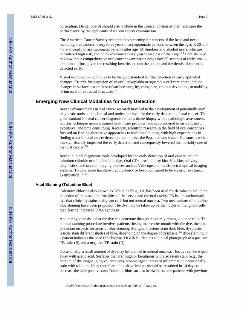
Vital Staining (Toluidine Blue)Tolonuim chloride also known as Toluidine blue, TB, has been used for decades to aid in thedetection of mucosal abnormalities of the cervix and the oral cavity. TB is a metachromaticdye that clinically stains malignant cells but not normal mucosa. Two mechanisms of toluidineblue staining have been proposed. The dye may be taken up by the nuclei of malignant cellsmanifesting increased DNA synthesis.
Another hypothesis is that the dye can penetrate through randomly arranged tumor cells. Theclinical staining procedure involves patients rinsing their entire mouth with the dye, then thephysician inspects for areas of blue staining. Malignant lesions stain dark blue; dysplasticlesions stain different shades of blue, depending on the degree of dysplasia.14 Blue staining ina patient indicates the need for a biopsy. FIGURE 1 depicts a clinical photograph of a positiveTB stain (B) and a negative TB stain (D).
Occasionally, a small amount of dye may be retained in normal mucosa. This dye can be wipedaway with acetic acid. Surfaces that are rough or keratinous will also retain stain (e.g., thedorsum of the tongue, gingival crevices). Nonmalignant areas of inflammation occasionallystain with toluidine blue; therefore, all positive lesions should be restained in 14 days todecrease the false positive rate. Toluidine blue can also be used to screen patients with previous
MESSADI et al. Page 3
J Calif Dent Assoc. Author manuscript; available in PMC 2010 May 10.
NIH
-PA Author Manuscript
NIH
-PA Author Manuscript
NIH
-PA Author Manuscript

carcinoma of the upper aerodigestive tract. These patients are known to be at high risk for arecurrence; therefore, clinicians may add toluidine rinses to their visual examination.14,18
From recent studies, a relationship between toluidine blue staining and genetic changesassociated with the progression of potentially malignant lesions to oral cancer — such as allelicloss or loss of heterozygosity (LOH) — was demonstrated.19 Furthermore, the authorsdemonstrated in a longitudinal study that toluidine blue identified LOH-positive lesions thatsubsequently progressed to oral cancer.
Chemiluminscence: ViziLiteChemiluminensce is a noninvasive screening tool targeted at dentists to assist in theidentification of suspicious superficial oral lesions. It consists of an acetic acid wash and asingle-use “chemi-light stick” that generates a moderately short wavelength light with peakoutputs near 430, 540, and 580 nm for illumination of the oral cavity (ViziLite). The use ofacetic acid followed by chemiluminescent illumination for visual diagnosis is similar tospeculoscopy (an adjunct to Pap smear of the uterine cervix), utilizing the samechemiluminescent light source for cervical diagnosis.14 Based on the rationale that the visualpresentation of cervical and oral/pharyngeal lesions, including SCC, is nearly identical underchemiluminescence, the cervical approach can be adapted to the diagnosis of any cancerouslesions of the oral cavity.
ViziLite is based on the typically greater nuclear content density and mitochondrial matrix ofabnormal cells than that of normal cells. The increased nuclear density and the resultingincrease in nuclear to cytoplasmic ratio reflect an increase in the proliferative rate and metabolicactivity of precancerous cells. After the patient rinses with a dilute acetic acid solution, thedense nuclei of abnormal squamous epithelial tissue will reflect light and appear white whenviewed under a diffuse low-energy wavelength light. Normal epithelium will absorb the lightand appear dark.20
The majority of studies investigating chemiluminescence evaluate subjective perceptions ofcharacteristics of intraoral lesions including brightness, sharpness and texture versus routineclinical examination. As these parameters are highly subjective, it is not surprising that resultshave been contradictory.16,18 Recently a combination of both TB and ViziLite systems(ViziLite Plus with TBlue system) has been introduced. FIGURE 2 demonstrates the use ofViziLite after a positive TB stain on the R-lateral border of tongue. The suspicious lesion isseen as a dense white lesion as compared to the adjacent dark normal mucosa. A newchemiluminescence device (MicroLux DL) has also recently been introduced on the market.14
Cytology (Oral CDx)The brush biopsy (CDx) was designed for use on clinical lesions that would otherwise not besubjected to biopsy because the level of suspicion for carcinoma, based upon clinical features,was low. When an abnormal CDx result is reported (atypical or positive), the clinician mustfollow-up with a scalpel biopsy of the lesion, as brush cytology does not provide a definitivediagnosis.21-23 The intent of the brush biopsy is to obtain cells from a suspicious oral sitewhile avoiding the pain and discomfort of a tissue biopsy, similar to a Pap smear for identifyingabnormal cervical cells in routine cervical cancer screening. The designed brush (Oral CDx)is used for epithelial cell collection and samples are eventually fixed onto a glass slide, stainedwith a modified Papanicolaou test and analyzed microscopically via a computer-based imagingsystem. Results are reported as “positive” or “atypical” when cellular morphology is highlysuspicious for epithelial dysplasia or carcinoma, or when abnormal epithelial changes are of
MESSADI et al. Page 4
J Calif Dent Assoc. Author manuscript; available in PMC 2010 May 10.
NIH
-PA Author Manuscript
NIH
-PA Author Manuscript
NIH
-PA Author Manuscript

uncertain diagnostic significance respectively.24 FIGURES 3A and 3B illustrate the use ofan oral CDx brush on a suspicious buccal mucosa lesion.
Controversy exists over the use of the Oral CDx product because some studies have indicateda high false-positive and a high false-negative rate.23 There are multiple examples in theliterature of studies with essentially opposite findings; therefore, most articles suggest furtherinvestigation of the product. A formal biopsy is still indicated if there is clinical suspicion ofa lesion regardless of the Oral CDx result.25-26
In conclusion, further research with clear objectives, well-defined population cohorts, andsound methodology are required before promoting the extensive use of the brush biopsy or anyother diagnostic tool for oral cancer detection.
Imaging Devices• Photosensitizers
• Spectroscopy and fluorescence
• In vivo microscopy
• Optical coherence tomography
1. PHOTOSENSITIZERS—Topical or systemic application of photosensitizers canselectively render pathologic tissues fluorescent when exposed to specific wavelengths of light,this technique has extensively been used for skin and esophageal cancer.27,28 This inducedfluorescence can be used to identify and delineate areas of pathology. Although thefluorescence may be strong enough to be detected with the naked eye (FIGURES 4A-4C),usually some sort of fluorescence detection device is used to enhance fluorescence detectionand assist with accurate lesion mapping. Whilst many agents are under investigation, or inclinical use outside of the United States, FDA approval for photosensitizing drugs remainslimited. Some promising agents for photodetection include aminolevulinic acid (ALA)(Levulan), hexyl aminolevulinate (Hexvix), methyl aminolevulinate (Metvix), tetra(metahydroxyphenyl) chlorin (mTHPC), as well as porfimer sodium (Photofrin).29-33
In a clinical study of 20 patients with oral neoplasms, three hours after the application of topicalPhotofrin solution, the photosensitized tissues showed a strong red fluorescence, withincreasing fluorescence intensity correlating with increasing levels of pathology. Guided bytheir visible fluorescence, lesions were biopsied at four suspicious sites for each patient. Thediagnostic sensitivity using unaided visual fluorescence diagnosis or fluorescence microscopyapproximated 93 percent. Diagnostic specificity was 95 percent for visual diagnosis, improvingto 97 percent using fluorescence microscopy. The differences between healthy tissue versusdysplasia versus malignancy were all significant (p<0.05).33
Advantages of a photosensitizer-based diagnostics approach include the capability for 3-Dsurface and subsurface mapping of lesion margins using available imaging technologies, theability to inspect large surface areas, noninvasiveness, and the capability for subsequentphotodestruction of the photosensitized lesion. Depending on the photosensitizer used and itsmode of application (systemic versus topical), imitations include systemic photosensitizationover prolonged periods of time, limited penetration depth, the need for specialized fluorescencedetection and mapping equipment, and lack of specificity when inflammation or scar tissue arepresent.
2. SPECTROSCOPY—This term refers to the process of measuring the emission andabsorption of different wavelengths (spectra) of visible and nonvisible light. Various types of
MESSADI et al. Page 5
J Calif Dent Assoc. Author manuscript; available in PMC 2010 May 10.
NIH
-PA Author Manuscript
NIH
-PA Author Manuscript
NIH
-PA Author Manuscript

optical spectroscopy have been investigated for oral diagnosis. All of these methods have onebasic principle in common: the optical spectrum of a tissue contains information about thebiochemical composition and/or the structure of the tissue, which conveys diagnosticinformation. Malignancy-related biochemical and morphologic changes perturb tissueabsorption, fluorescence, and scattering properties. The biochemical information can beobtained by measuring absorption/reflectance, fluorescence, or Raman scattering signals.34
Data is often in the form of graphs, some imaging devices that color code spectralcharacteristics of tissues also exist, such as the Velscope.
VELSCOPE SYSTEM: For decades the use of tissue autofluorescence has been described toscreen and diagnose precancers and early cancer lesions in organs such as the lung, uterinecervix, skin, and, more recently, the oral cavity.35-37 The concept behind tissueautoflorescence is that changes in the structure (e.g., hyperkeratosis, hyperchromatin, andincreased cellular/nuclear pleomorphism) and metabolism (e.g., concentration of flavinadenine dinucleotide [FAD] and nicotinamide adenine dinucleotide [NADH]) of theepithelium, as well as changes of the subepithelial stroma (e.g., composition of collagen matrixand elastin), alter their interaction with light. Specifically, these epithelial and stromal changescan alter the distribution of tissue fluorophores and as a consequence the way they fluoresceafter stimulation with intense light (typically, blue light excitation at 400 to 460 nm), a processcalled autoflorescence. The autoflorescence signal can be directly visualized by the clinician.35-37
One of the tissue fluorescence imaging system that have been marketed to dental offices is theVelscope system. In the oral cavity, normal oral mucosa emits a pale green autofluorescencewhen viewed through the instrument handpiece whilst abnormal tissue displays a decreasedautofluorescence and appears darker with respect to the surrounding healthy tissue.37-38
Studies have shown that Velscope can improve lesions contrast and therefore improve theclinician’s ability to distinguish between mucosal lesions and healthy mucosa. Whilstpreliminary data on Velscope’s specificity and sensitivity are predominantly based on caseseries and case reports, full-scale clinical trials using different patient populations are neededto establish the diagnostic efficacy of this tool.18
Structural and morphological information may be obtained by spectroscopic techniques thatassess the elastic-scattering properties of tissue.39 Pursuant to encouraging preliminary dataclinical trials of elastic scattering spectroscopy, sometimes in combination with fluorescencespectroscopy, or imaging, are under way.34 Other studies combine spectroscopy with polarizedlight and/or fluorescence imaging, and/or in vivo microscopy. Devices under development andtesting include the FastEEM4 System, the Indentafi and the PS2-oral. These clinical studiesare still at a relatively early stage, and preliminary results are encouraging.40-49
Significant challenges to the use of diagnostic spectroscopy include the often low signal-to-noise ratio, difficulty in identifying the precise source of signals, data quantification issues,and establishing definitive diagnostic milestones and endpoints, especially given the widerange of tissue types contained within the oral cavity. Limited tissue penetration and concernsabout mutagenicity when using UV light present further clinical challenges. The abundance ofdata/information generated in association with our incomplete understanding of thecarcinogenesis process tend to render data analysis and interpretation very complex, however,the development of diagnostic algorithms may be able to mitigate this challenge.39
3. IN VIVO MICROSCOPR—In vivo confocal or multiphoton imaging resembleshistological tissue evaluation, except that 3-D subcellular resolution is achieved noninvasivelyand without stains. In epithelial structure, resolution of 1 μm has been achieved with a 200–400-μm field of view. Confocal imaging of oral mucosa has resolved subcellular detail in the
MESSADI et al. Page 6
J Calif Dent Assoc. Author manuscript; available in PMC 2010 May 10.
NIH
-PA Author Manuscript
NIH
-PA Author Manuscript
NIH
-PA Author Manuscript

lip and tongue and oral squamous cell carcinoma from multiple sites.50,51 While thistechnology can provide detailed images of tissue architecture and cellular morphology, a verysmall field of view and limited penetration depth of 250-500 μm considerably reduce theclinical usefulness of this approach. Multiphoton microscopy resembles confocal, but affordsa greater tissue penetration depth, the use of many different wavelengths of light, and less tissueheating52 (FIGURE 4). Because of high cost and the specialized expertise required to operatesuch systems, neither approach is clinically feasible in the foreseeable future.
4. OPTICAL COHERENCE TOMOGRAPHY—Optical coherence tomography, OCT, isan optical imaging method first used to visualize human tissue in 1991. It has been since refinedand accepted as an imaging modality in ophthalmology. Several systems have been cleared byFDA for such use; one OCT system (Imalux) currently has FDA 510(k) clearance fornonophthalmalogic medical use.
OCT is a high-resolution optical technique that permits noninvasive imaging of surface andsubsurface tissues. It has been compared to ultrasound scanning conceptually. Both ultrasoundand OCT provide real-time structural imaging, but unlike ultrasound, OCT is based on low-coherence interferometry, using broadband light to provide crosssectional, high-resolutionsubsurface tissue images (FIGURE 5). With a tissue penetration depth of 1 mm to 2 mm, theimaging range of OCT technology is suitable for the oral mucosa.53-56 Previous studies usingOCT have demonstrated the ability to evaluate macroscopic characteristics of epithelial,subepithelial, and basement membrane structures and show the potential for nearhistopathological-level resolution and close correlation with histologic appearance.57
In one of the authors’ recent studies of 50 patients with oral dysplastic and malignant lesions,intraand interobserver agreement between diagnoses based on histopathology and imaging datawas excellent, with kappa values of 0.844-0.896.58 For detecting carcinoma in situ or squamouscell carcinoma, SCC, versus noncancer, sensitivity and specificity were 0.931; for detectingSCC versus all other pathologies, sensitivity was 0.931 and specificity was 0.973. These datademonstrate the excellent capability of in vivo OCT for screening high-risk patients,monitoring existing lesions, and detecting, diagnosing oral premalignancy and malignancy inhuman subjects. This study showed that the in vivo OCT image of a dysplastic lesion (FIGURE6B) parallels histolopathological status (FIGURE 6C), showing epithelial thickening, loss ofstratification in lower epithelial strata, epithelial downgrowth, and loss of epithelialstratification as compared to healthy oral mucosa (FIGURE 6D).
For oral cancer, FIGURES 7A AND 7C show clinical appearance and histopathology,respectively of an area of SCC on the buccal mucosa. In the OCT image (FIGURE 7B), theepithelium is highly variable in thickness, with areas of erosion and extensive downgrowth andinvasion into the subepithelial layers. The basement membrane is not visible as a coherentlandmark.58
As the technology and techniques evolve, this modality should progressively reduce the needfor biopsy, define surgical margins, and provide a direct evaluation of the effectiveness ofcomplete lesion removal.
Saliva as a Diagnostic ToolSalivary diagnostics has come to the forefront of biomedical research. The ability to usesalivary biomarkers (i.e., DNA, RNA, and proteins) as a predictive measure for systemicdisease has generated much interest among dental researchers in the United States and Europe.Laboratory-based methodologies, which allow the rapid identification of proteins, RNA andDNA have afforded scientists the ability to examine and quantify complex salivary profiles.At the University of California, Los Angeles, School of Dentistry, Dr. David Wong and
MESSADI et al. Page 7
J Calif Dent Assoc. Author manuscript; available in PMC 2010 May 10.
NIH
-PA Author Manuscript
NIH
-PA Author Manuscript
NIH
-PA Author Manuscript

collaborators are developing research platforms toward the global identification of diseasesignatures in saliva.
The premise of their approach is that serum contents, such as disease biomarkers, are largelypresent in saliva, thus rendering oral fluid a logical source to harness disease biomarkers.59
They employ both a proteome-wide as well as a genome-wide approach toward theidentification of disease biomarkers and signatures. Dr. Wong’s goal is to develop and utilizenovel patient-oriented genomewide molecular tools that may identify oral cancer specificmolecular markers.
Early work by Wong and his coworkers identified interleukin 6 and 8 as predictive biomarkersfor oral cancer.60-63 They are now validating these findings and have expanded this work tobreast and pancreatic cancer. Oral fluid–based tests presently exist or are being developed todetect a variety of infectious diseases (including HIV, parvovirus, acute hepatitis, dengue fever,and malaria), as well as to detect alcohol and drug use and steroid hormone levels.64
Other body fluids such as serum could possibly also be helpful in identifying common geneticmutations, promoter hypermethylation, and LOH.65,66
The UCLA group is at the final stage of developing an oral fluid nanosensor test device thatcould be used in the dental office. This portable, point-of-care, chairside device is to be usedfor saliva diagnostics, not only for oral cancer but other diseases such as diabetes, Sjögren’ssyndrome, and breast and prostate cancer (FIGURE 8).
Collectively, technology platform advancement and the identification and validation of robustand discriminatory suites of salivary biomarkers for disease diagnostics represent the necessarymarriage to propel saliva diagnostics into a clinical and commercial reality. At the same time,they are building the scientific foundation toward the use of saliva as a diagnostic fluid. Thisis a perfect example of translational research in reverse, based on a highly relevant clinicalobservation that saliva contains proteomic and genomic biomarkers for oral cancer detection,and building a scientific foundation toward the mechanistic background, thereby enablingbetter exploitation of the full clinical potential of saliva diagnostics.67-69
Oral Cancer ChemopreventionMany epidemiologic studies have consistently linked the abundant consumption of foods ofplant origin, such as fruit, vegetables, whole grains, legumes, nuts, seeds, and tea, with adecreased risk of developing various types of cancers.70,71 Chemoprevention is the use ofpharmacologic or natural agents that inhibit the development of invasive cancer. These workeither by blocking the DNA damage that initiates carcinogenesis, or by arresting or reversingthe progression of premalignant cells in which such damage has already occurred.
Recent advances in understanding the causes of cancer and the consequent ability to providea genetic diagnosis of susceptibility necessitate the identification of agents that can effectivelyreverse, halt, or at least delay the carcinogenic process. It is important that any agents selectedon the basis of trials in premalignant lesions have minimal or no toxicity since a large numberof subjects whose lesions are unlikely to progress to cancer will necessarily be exposed to theproduct. Therefore, the development of new agents or the use of old agents at nontoxic dosesis important.72
As in all cancers, the most effective way of approaching oral cancer prevention is to identifyindividuals who are at a high risk to develop such cancers (e.g., individuals with oralpremalignant lesions) and to treat them with agents that can suppress the development ofadditional premalignant lesions and inhibit the development of oral cancer in existing lesions.
MESSADI et al. Page 8
J Calif Dent Assoc. Author manuscript; available in PMC 2010 May 10.
NIH
-PA Author Manuscript
NIH
-PA Author Manuscript
NIH
-PA Author Manuscript

Oral carcinogenesis is a multistep process, which is characterized by genetic, epigenetic, andphenotypic changes. Many of these changes involve the activation of signaling or metabolicpathways that give the cells favorable growth and survival characteristics. Therefore,chemopreventive agents that can inhibit or reverse these changes by targeting specificmolecular pathways have received increased attention as novel candidates for cancerprevention and therapy.73
A wide range of compounds has been investigated as possible chemopreventive therapies forpotential oral cancer, such as vitamins and minerals, including vitamin A and other retinoids,beta carotene, vitamin E, vitamin C, folates, and selenium, and has been studied. Herbaltreatments have generated interest recently. Molecularly targeted approaches have includedcyclooxygenase-2 (COX-2) inhibitors, EGFR inhibitors, and adenovirus vectors.74 Severalpromising new compounds are currently in clinical chemoprevention trials in head and neckcancer. These include curcumin analogs, green tea extracts (GTEs), selenium, polyphenols ofpomegranate juice, Bowman-Birk inhibitor (BBI) from soybeans and others.75
The authors and other collaborators from multiple institutes are involved in a multicentric phaseII NCI-sponsored study to examine the effect of the protease inhibitor Bowman-Birk Inhibitorconcentrate (BBIC), on oral leukoplakia patients (FIGURES 9A and 9B). This compound hadpreviously been tested in patients with benign prostatic hypertrophy, ulcerative colitis, andthose studies suggested clinical activity without toxicity being noted.76 BBIC effect in the oralcavity is still not well established, but preliminary results are promising.77
Because of their easy accessibility to topical treatment and visual clinical examination, oralpremalignant lesions provide an excellent model to study the effects of variouschemopreventive agents on epithelial solid tumors. In case of oral premalignancy, the primary(clinical) endpoints are determined by clinical measurement of the lesion and confirmed byhistologic examination of oral biopsy. Both are feasible in the oral cavity. Secondary(biomarkers) endpoints are usually biochemical and molecular markers found on tissues orbuccal cells which are easily obtained from the oral cavity.
Current clinical trials that use clinical and biomarker endpoints will identify useful agents fororal cancer prevention and provide a better understanding of the carcinogenesis process byinvestigating the specific targeted cellular proteins and dependent cell signaling pathwaysmodulated by these agents. Such insights will not only shed a light on the mechanism of cancerprogression but will also guide future drug development and ultimately improve therapy.78
THE PREVENTION of oral cancer and its associated morbidity and mortality hinges uponthe early detection of neoplastic lesions.
AcknowledgmentsThe data presented in this manuscript was supported by the following grants: CA TRDRP 14IT-0097, NIH (LAMMP)RR01192, DOE DE903-91ER 61227, NIH EB-00293 CA91717, NSF BES-86924. NIH (EB-00293, NCI-91717,RR-01192, EB0002SS, EB002494, AR47551, and UO1 CA-72294.
REFERENCES1. Jemal A, Siegel R, et al. Cancer statistics, 2007. CA Cancer J Clin 2007;57(1):43–66. [PubMed:
17237035]2. Jemal A, Siegel R, et al. Cancer statistics, 2008. CA Cancer J Clin 2008;58(2):71–96. [PubMed:
18287387]
MESSADI et al. Page 9
J Calif Dent Assoc. Author manuscript; available in PMC 2010 May 10.
NIH
-PA Author Manuscript
NIH
-PA Author Manuscript
NIH
-PA Author Manuscript

3. Schantz SP, Yu GP. Head and neck cancer incidence trends in young Americans, 1973-1997, with aspecial analysis for tongue cancer. Arch Otolaryngol Head Neck Surg 2002;128:268. [PubMed:11886342]
4. Mork J, Lie AK, et al. Human papillomavirus infection as a risk factor for squamous cell carcinomaof the head and neck. N Engl J Med 2001;344:1125. [PubMed: 11297703]
5. American Cancer Society. cancer facts and figures, 2005. [Accessed Sept. 8, 2009].cancer.org/docroot/STT/stt_0.asp
6. Mashberg A, Samit AM. Early detection, diagnosis, and management of oral and oropharyngeal cancer.CA Cancer J Clin 1989;39:67–88. [PubMed: 2495159]
7. Silverman S, Gorsky M. Epidemiologic and demographic update in oral cancer: California and nationaldata, 1973-1985. J Am Dent Assoc 1990;120:495–9. [PubMed: 2335670]
8. NIH/NCI SEER cancer mortality data, 1973-2005. [Accessed Sept. 8, 2009].seer.cancer.gov/data/access.html
9. Horowitz AM. Perform a death-defying act: the 90-second oral cancer examination. J Am Dent Assoc2001;32(supplement):36s–40s.
10. Horowitz AM, Drury TF, et al. Oral pharyngeal cancer prevention and early detection. Dentists’opinions and practices. J Am Dent Assoc April;2000 131(4):453–62. [PubMed: 10770007]
11. Alfano MC, Horowitz AM. Professional and community efforts to prevent morbidity and mortalityfrom oral cancer. J Am Dent Assoc November;2001 132(Suppl):24S–29S. [PubMed: 11803649]
12. Downer MC, Jullien JA, Speight PM. An interim determination of health gain from oral cancer andprecancer screening: preselecting high risk individuals. Community Dent Health June;1998 15(2):72–6. [PubMed: 9793221]
13. American Cancer Society, California Division and Public Health Institute, California Cancer Registry.California Cancer Facts and Figures 2007. American Cancer Society, California Division; Oakland:Sep. 2006
14. Epstein JB, Silverman S Jr, et al. Analysis of oral lesion biopsies identified and evaluated by visualexamination, chemiluminescence and toluidine blue. Oral Oncology 2008;44:538–44. [PubMed:17996486]
15. Wong DT. Toward a simple, saliva-based test for the detection of oral cancer. Expert Rev Mol DiagnMay;2006 6(3):267–72. [PubMed: 16706730]
16. Lingen MW, Kalmar JR, et al. Critical evaluation of diagnostic aids for the detection of oral cancer.Oral Oncol 2008;44:10–22. [PubMed: 17825602]
17. Trullenque-Eriksson A, Muñoz-Corcuera M, et al. Analysis of new diagnostic methods in suspiciouslesions of the oral mucosa. Med Oral Patol Oral Cir Bucal May;2009 14(5):E210–6. (e-pub February2009). [PubMed: 19218907]
18. Patton LP, Epstein JB, Kerr RA. Adjunctive techniques for oral cancer examination and lesiondiagnosis: a systematic review of the literature. J Am Dent Assoc 2008;139:896–905. [PubMed:18594075]
19. Zhang L, Williams M, et al. Toluidine blue staining identifies high-risk primary oral premalignantlesions with poor outcome. Cancer Res 2005;65:8017–21. [PubMed: 16140975]
20. Farah CS, McCullough MJ. A pilot case control study on the efficacy of acetic acid wash andchemiluminescent illumination (ViziLite) in the visualisation of oral mucosal white lesions. OralOncol 2007;43(8):820–4. [PubMed: 17169603]
21. Potter TJ, Summerlin DJ, Campbell JH. Oral malignancies associated with negative transepithelialbrush biopsy. J Oral Maxillofac Surg 2003;61:674–7. [PubMed: 12796875]
22. Sciubba JJ. Improving detection of precancerous and cancerous oral lesions. Computer-assistedanalysis of the oral brush biopsy. U.S. collaborative OralCDx study group. J Am Dent Assoc1999;130:1445–57. [PubMed: 10570588]
23. Rick GM. Oral brush biopsy: the problem of false positives. Oral Surg Oral Med Oral Pathol OralRadiol Endod 2003;96:252. [PubMed: 14515857]
24. Fedele S. Diagnostic aids in the screening of oral cancer. Head Neck Oncol January;2009 1(1):5.[PubMed: 19284694]
MESSADI et al. Page 10
J Calif Dent Assoc. Author manuscript; available in PMC 2010 May 10.
NIH
-PA Author Manuscript
NIH
-PA Author Manuscript
NIH
-PA Author Manuscript

25. Frist S. The oral brush biopsy: separating fact from fiction. Oral Surg Oral Med Oral Pathol OralRadiol Endod 2003;96:654–5. [PubMed: 14723218]
26. Eisen D, Frist S. The relevance of the high-positive predictive value of the oral brush biopsy. OralOncol 2005;41:753–5. [PubMed: 15936979]
27. Kennedy JC, Pottier RH. Endogenous protoporphyrin IX, a clinically useful photosensitizer forphotodynamic therapy. J Photochem Photobiol B 1992;14(4):275–92. [PubMed: 1403373]
28. Cassas, A.; Fukuda, H.; Battle, A. Hexyl ALA ALA-based photodynamic therapy in epithelial tumors:in vivo and in vitro models. In: Dougherty, TJ., editor. optical methods for tumor treatment anddetection: mechanisms and techniques in photodynamic therapy IX; Proc. SPIE; 2002. p. 114-23.
29. Ebihara A, Liaw LH, et al. Detection and diagnosis of oral cancer by light-induced fluorescence.Lasers Surg Med 2003;32(1):17–24. [PubMed: 12516066]
30. Leunig A, Rick K, et al. Fluorescence imaging and spectroscopy of 5-aminolevulinic acid inducedprotoporphyrin IX for the detection of neoplastic lesions in the oral cavity. Am J Surg 1996;172(6):674–7. [PubMed: 8988675]
31. Leunig A, Mehlmann M, et al. Detection of squamous cell carcinoma of the oral cavity by imaging5-aminolevulinic acid-induced protoporphyrin IX fluorescence. Laryngoscope 2000;110(1):78–83.[PubMed: 10646720]
32. Leunig A, Mehlmann M, et al. Fluorescence staining of oral cancer using a topical application of 5-aminolevulinic acid: fluorescence microscopic studies. J Photochem Photobiol B 2001;60(1):44–9.[PubMed: 11386680]
33. Chang CJ, Wilder-Smith P. Topical application of photofrin for photodynamic diagnosis of oralneoplasms. Plast Reconstr Surg 2005;115(7):1877–86. [PubMed: 15923832]
34. Sharwani A, Jerjes W, et al. Assessment of oral premalignancy using elastic scattering spectroscopyAssessment of oral premalignancy using elastic scattering spectroscopy. Oral Oncology 2006;42(4):343–9. [PubMed: 16321565]
35. Rosin MP, Poh CF, et al. Visualization and other emerging technologies as change makers for oralcancer prevention. Ann N Y Acad Sci 2007;1098:167–83. [PubMed: 17332080]
36. Lane PM, Gilhuly T, et al. Simple device for the direct visualization of oral-cavity tissue fluorescence.J Biomed Opt March-April;2006 11(2):024006. [PubMed: 16674196]
37. Poh CF, Ng SP, et al. Direct fluorescence visualization of clinically occult high-risk oral premalignantdisease using a simple hand-held device. Head Neck 2007;29:71–6. [PubMed: 16983693]
38. Poh CF, Zhang L, et al. Fluorescence visualization detection of field alterations in tumor margins oforal cancer patients. Clin Cancer Res 2006;12(22):6716–22. [PubMed: 17121891]
39. Bigio IJ, Bown SG. Spectroscopic-sensing of cancer and cancer therapy: current status of ranslationalresearch. Cancer Biol Ther March;2004 3(3):259–67. [PubMed: 15107613]
40. Schwarz RA, Gao W, et al. Noninvasive evaluation of oral lesions using depth-sensitive opticalspectroscopy simple device for the direct visualization of oral-cavity tissue fluorescence. CancerApril 15;2009 115(8):1669–79. (e-pub Jan. 23, 2009). [PubMed: 19170229]
41. McGee S, Mirkovic J, et al. Model-based spectroscopic analysis of the oral cavity: impact of anatomy.J Biomed Opt November-December;2008 13(6):064034. [PubMed: 19123680]
42. Lane PM, Gilhuly T, et al. Simple device for the direct visualization of oral-cavity tissue fluorescence.J Biomed Opt 2006;11(2):024006. [PubMed: 16674196]
43. De Veld DC, Witjes MJ, et al. The status of in vivo autofluorescence spectroscopy and imaging fororal oncology. Oral Oncol 2005;41:117–31. [PubMed: 15695112]
44. Wagnieres GA, Star WM, Wilson BC. In vivo fluorescence spectroscopy and imaging for oncologicalapplications. Photochem Photobiol 1998;68(5):603–32. [PubMed: 9825692]
45. Ramanujam N. Fluorescence spectroscopy of neoplastic and non-neoplastic tissues. Neoplasia 2000;2(1-2):89–117. [PubMed: 10933071]
46. Culha M, Stokes D, Vo-Dinh T. Surface-enhanced Raman scattering for cancer diagnostics: detectionof the BCL2 gene. Expert Rev Mol Diagn September;2003 3(5):669–75. [PubMed: 14510186]
47. Choo-Smith LP, Edwards HG, et al. Medical applications of Raman spectroscopy: from proof ofprinciple to clinical implementation. Biopolymers 2002;67(1):1–9. [PubMed: 11842408]
MESSADI et al. Page 11
J Calif Dent Assoc. Author manuscript; available in PMC 2010 May 10.
NIH
-PA Author Manuscript
NIH
-PA Author Manuscript
NIH
-PA Author Manuscript

48. Bigio IJ, Mourant JR. Ultraviolet and visible spectroscopies for tissue diagnostics: fluorescencespectroscopy and elastic-scattering spectroscopy. Phys Med Biol 1997;42(5):803–14. [PubMed:9172260]
49. Farrell TJ, Patterson MS, Wilson B. A diffusion theory model of spatially resolved, steady-statediffuse reflectance for the noninvasive determination of tissue optical properties in vivo. Med Phys1992;19(4):879–88. [PubMed: 1518476]
50. White WM, Rajadhyaksha M, et al. Noninvasive imaging of human oral mucosa in vivo by confocalreflectance microscopy. Laryngoscope 1999;109(10):1709–17. [PubMed: 10522947]
51. Clark AL, Gillenwater AM, et al. Confocal microscopy for real-time detection of oral cavity neoplasia.Clin Cancer Res 2003;9(13):4714–21. [PubMed: 14581341]
52. Wilder-Smith P, Osann K, et al. In vivo multiphoton fluorescence imaging: a novel approach to oralmalignancy. Lasers Surg Med 2004;35(2):96–103. [PubMed: 15334611]
53. Wilder-Smith P, Woong-Gyu Jung, et al. In vivo optical coherence tomography for the diagnosis oforal malignancy. Lasers Surg Med 2004;35(4):269–75. [PubMed: 15493024]
55. Wilder-Smith P, Krasieva T, et al. Noninvasive imaging of oral premalignancy and malignancy. JBiomed Opt 2005;10(5):51601.
56. Kawakami-Wong H, Hammer-Wilson MJ, et al. In vivo optical coherence tomography-based scoringof oral mucositis in human subjects: a pilot study. J Biomed Opt 2007;12(5):051702. [PubMed:17994875]
57. Muanza TM, Cotrim AP, et al. Evaluation of radiation-induced oral mucositis by optical coherencetomography. Clin Cancer Res 2005;11(14):5121–7. [PubMed: 16033826]
58. Wilder-Smith P, Lee K, et al. In vivo diagnosis of oral dysplasia and malignancy using opticalcoherence tomography: preliminary studies in 50 patients. Laser Surg Med. 2009 Accepted forpublication.
59. Phillips, C. NCI Cancer Bulletin. 2006. Rinse and spit: saliva as a cancer biomarker source; p. 3-5.60. Li Y, St. John MA, et al. Salivary transcriptome diagnostics for oral cancer detection. Clin Cancer
Res Dec. 15;2004 10(24):8442–50. [PubMed: 15623624]61. Forde MD, Koka S, et al. Systemic assessments utilizing saliva: part 1 general considerations and
current assessments. Int J Prosthodont 2006;19(1):43–52. [PubMed: 16479760]62. Wong DT. Towards a simple, saliva-based test for the detection of oral cancer, oral fluid (saliva),
which is the mirror of the body, is a perfect medium to be explored for health and disease surveillance.Expert Rev Mol Diagn May;2006 6(3):267–72. [PubMed: 16706730]
63. Wong DT. Salivary diagnostics powered by nanotechnologies, proteomics and genomics. J Am DentAssoc 2006;137(3):313–21. [PubMed: 16570464]
64. Pesce MA, Spitalnik SL. Saliva and the clinical pathology laboratory. Ann N Y Acad Sci March;20071098:192–9. [PubMed: 17435128]
65. Glazer CA, Chang SS, et al. Applying the molecular biology and epigenetics of head and neck cancerin everyday clinical practice. Oral Oncol April-May;2009 45(4-5):440–6. (e-pub July 31, 2008).[PubMed: 18674958]
66. Molinolo AA, Amornphimoltham P, et al. Dysregulated molecular networks in head and neckcarcinogenesis. Oral Oncol April-May;2009 45(4-5):324–34. (e-pub Sept. 19, 2008). [PubMed:18805044]
67. Wong DT. Salivary transcriptome. Clin Cancer Res 2007;13(4):1350. [PubMed: 17317847]68. Wong DT. Salivary diagnostics for oral cancer. J Calif Dent Assoc April;2006 34(4):303–8. [PubMed:
16900988]69. Wong DT. Salivary diagnostics. J Calif Dent Assoc April;2006 34(4):283–5. [PubMed: 16900985]70. Doll R, Peto R. The causes of cancer: quantitative estimates of avoidable risks of cancer in the United
States today. J Natl Cancer Inst 1981;66:1191–308. [PubMed: 7017215]71. Béliveau R. Gingras Denis, Role of nutrition in preventing cancer. Can Fam Physician November;
2007 53(11):1905–11. [PubMed: 18000267]72. Hong WK, Spitz MR, Lippman SM. Cancer chemoprevention in the 21st century: genetics, risk
modeling, and molecular targets. J Clinical Oncol Nov. 1;2000 18(21 suppl):9s–18s. [PubMed:11060320]
MESSADI et al. Page 12
J Calif Dent Assoc. Author manuscript; available in PMC 2010 May 10.
NIH
-PA Author Manuscript
NIH
-PA Author Manuscript
NIH
-PA Author Manuscript

73. Jon, Sudbø; Magne, Bryne, et al. Molecular-based treatment of oral cancer. Oral Oncol 2003;39:749–58. [PubMed: 13679198]
74. Brown KS, Kane MA. Chemoprevention of squamous cell carcinoma of the oral cavity. OtolaryngolClin North Am 2006;39:349–63. [PubMed: 16580916]
75. Khuri FR, Shin DM. Head and neck cancer chemoprevention gets a shot in the arm. J Clin Oncol Jan.20;2008 26(3):345–7. [PubMed: 18202405]
76. Lichtenstein GR, Deren J, et al. The Bowman-Birk protease inhibitor: a novel therapy for the inductionof remission in patients with active ulcerative colitis. Gastroenterology 2002;67:236–40.
77. Armstrong WB, Kennedy AR, et al. Clinical modulation of oral leukoplakia and protease activity byBowman-Birk inhibitor concentrate in a phase IIa chemoprevention trial. Clin Cancer Res2000;6:4684–91. [PubMed: 11156220]
78. Klass CM, Shin DM. Current status and future perspectives of chemoprevention in head and neckcancer. Current Cancer Drug Targets 2007;7:623–32. [PubMed: 18045067]
MESSADI et al. Page 13
J Calif Dent Assoc. Author manuscript; available in PMC 2010 May 10.
NIH
-PA Author Manuscript
NIH
-PA Author Manuscript
NIH
-PA Author Manuscript

FIGURE 1A.Leukoplakia lesion on right buccal mucosa.
MESSADI et al. Page 14
J Calif Dent Assoc. Author manuscript; available in PMC 2010 May 10.
NIH
-PA Author Manuscript
NIH
-PA Author Manuscript
NIH
-PA Author Manuscript

FIGURE 1B.Positive blue staining after TB rinse.
MESSADI et al. Page 15
J Calif Dent Assoc. Author manuscript; available in PMC 2010 May 10.
NIH
-PA Author Manuscript
NIH
-PA Author Manuscript
NIH
-PA Author Manuscript

FIGURE 1C.Leukoplakia of R lateral border of tongue.
MESSADI et al. Page 16
J Calif Dent Assoc. Author manuscript; available in PMC 2010 May 10.
NIH
-PA Author Manuscript
NIH
-PA Author Manuscript
NIH
-PA Author Manuscript

FIGURE 1D.Negative stain after TB rinse.
MESSADI et al. Page 17
J Calif Dent Assoc. Author manuscript; available in PMC 2010 May 10.
NIH
-PA Author Manuscript
NIH
-PA Author Manuscript
NIH
-PA Author Manuscript

FIGURE 2.Chemiluminescence with ViziLite shows suspicious lesion on the right side of the tongue aswhite; normal epithelium is dark.
MESSADI et al. Page 18
J Calif Dent Assoc. Author manuscript; available in PMC 2010 May 10.
NIH
-PA Author Manuscript
NIH
-PA Author Manuscript
NIH
-PA Author Manuscript

FIGURE 3A.Oral CDx brush application on a suspicious buccal mucosal lesion, with suspicious erythroplkialesion on R buccal mucosa.
MESSADI et al. Page 19
J Calif Dent Assoc. Author manuscript; available in PMC 2010 May 10.
NIH
-PA Author Manuscript
NIH
-PA Author Manuscript
NIH
-PA Author Manuscript

FIGURE 3B.Application of the Oral CDx brush for cytology testing.
MESSADI et al. Page 20
J Calif Dent Assoc. Author manuscript; available in PMC 2010 May 10.
NIH
-PA Author Manuscript
NIH
-PA Author Manuscript
NIH
-PA Author Manuscript

FIGURE 4A.In vivo multiphoton microscopy images of the hamster cheek pouch. Healthy tissue, showingordered and dense collagen fiber network in blue.
MESSADI et al. Page 21
J Calif Dent Assoc. Author manuscript; available in PMC 2010 May 10.
NIH
-PA Author Manuscript
NIH
-PA Author Manuscript
NIH
-PA Author Manuscript

FIGURE 4B.Dysplastic lesion, showing engorged blood vessel, shortening of collagen fibers with reduceddensity and organization.
MESSADI et al. Page 22
J Calif Dent Assoc. Author manuscript; available in PMC 2010 May 10.
NIH
-PA Author Manuscript
NIH
-PA Author Manuscript
NIH
-PA Author Manuscript

FIGURE 4C.Malignant lesion, showing further collagen loss and inflammatory exudates (red).
MESSADI et al. Page 23
J Calif Dent Assoc. Author manuscript; available in PMC 2010 May 10.
NIH
-PA Author Manuscript
NIH
-PA Author Manuscript
NIH
-PA Author Manuscript

FIGURE 5.OCT performs 2-D imaging in biological tissues by projecting harmless near-infrared lightonto the tissue and measuring the backscattered intensity of light as a function of depth (left).It is able to capture tissue substructure images in real-time using lateral scanning (middle). In-depth spatial resolution is 5-20 μm in air, and is typically able to visualize tissue to a depth of2 mm (right).
MESSADI et al. Page 24
J Calif Dent Assoc. Author manuscript; available in PMC 2010 May 10.
NIH
-PA Author Manuscript
NIH
-PA Author Manuscript
NIH
-PA Author Manuscript

FIGURE 6.Dysplastic and normal buccal mucosa. (A) Photograph, (B) in vivo OCT image, and (C) H&E(10x) of dysplastic buccal mucosa. (D) In vivo OCT image of normal buccal mucosa. Key: 1-stratified squamous epithelium, 2-keratinized epithelial surface layer, 3-basement membrane4-submucosa.
MESSADI et al. Page 25
J Calif Dent Assoc. Author manuscript; available in PMC 2010 May 10.
NIH
-PA Author Manuscript
NIH
-PA Author Manuscript
NIH
-PA Author Manuscript

FIGURE 7.Squamous cell carcinoma of the buccal mucosa. (A) Photograph, (B) in vivo OCT image and(C) H&E (10x) of buccal mucosa with squamous cell carcinoma. (D) In vivo OCT image ofnormal buccal mucosa. Key: 1-stratified squamous epithelium, 2-keratinized epithelial surfacelayer, 3-basement membrane, 4-submucosa.
MESSADI et al. Page 26
J Calif Dent Assoc. Author manuscript; available in PMC 2010 May 10.
NIH
-PA Author Manuscript
NIH
-PA Author Manuscript
NIH
-PA Author Manuscript
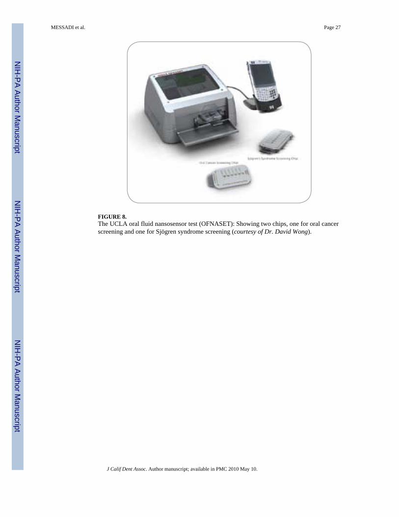
FIGURE 8.The UCLA oral fluid nansosensor test (OFNASET): Showing two chips, one for oral cancerscreening and one for Sjögren syndrome screening (courtesy of Dr. David Wong).
MESSADI et al. Page 27
J Calif Dent Assoc. Author manuscript; available in PMC 2010 May 10.
NIH
-PA Author Manuscript
NIH
-PA Author Manuscript
NIH
-PA Author Manuscript

FIGURE 9A.A patient with proliferating verrucous leukolplkia (PVL) of ventral surface of tongue.
MESSADI et al. Page 28
J Calif Dent Assoc. Author manuscript; available in PMC 2010 May 10.
NIH
-PA Author Manuscript
NIH
-PA Author Manuscript
NIH
-PA Author Manuscript

FIGURE 9B.Regression of the PVL lesion after six months of BBIC treatment.
MESSADI et al. Page 29
J Calif Dent Assoc. Author manuscript; available in PMC 2010 May 10.
NIH
-PA Author Manuscript
NIH
-PA Author Manuscript
NIH
-PA Author Manuscript