Embed Size (px)
Citation preview
Available online at www.sciencedirect.com
Journal of Human Evolution 54 (2008) 173e186
Dental trait expression at the enamel-dentine junctionof lower molars in extant and fossil hominoids
Matthew M. Skinner a,b,*, Bernard A. Wood c, Christophe Boesch d,Anthony J. Olejniczak b, Antonio Rosas e, Tanya M. Smith b, Jean-Jacques Hublin b
a Hominid Paleobiology Doctoral Program, Department of Anthropology, 2110 G Street NW,
The George Washington University, Washington, DC 20052, USAb Department of Human Evolution, Max Planck Institute for Evolutionary Anthropology,
Deutscher Platz 6, D-04103 Leipzig, Germanyc Center for the Advanced Study of Human Paleobiology, Department of Anthropology,
2110 G Street NW, The George Washington University, Washington, DC 20052, USAd Department of Primatology, Max Planck Institute for Evolutionary Anthropology,
Deutscher Platz 6, D-04103 Leipzig, Germanye Department of Paleobiologıa, Museo Nacional de Ciencias Naturales, CSIC,
C/Jose Gutierrez Abascal 2, 28006 Madrid, Spain
Received 9 November 2006; accepted 30 September 2007
Abstract
Discrete dental traits are used as proxies for biological relatedness among modern human populations and for alpha taxonomy and phylogenyreconstruction within the hominin clade. We present a comparison of the expression of lower molar dental traits (cusp 6, cusp 7, trigonid crestpattern, and protostylid) at the enamel-dentine junction (EDJ) in a variety of extant and fossil hominoid taxa, in order to assess the contributionof the EDJ to the morphology of these traits at the outer enamel surface (OES). Molars (n¼ 44) were imaged nondestructively using high-resolution microCT, and three-dimensional surface models of the EDJ and OES were created to compare trait expression at each surface.Our results indicate that these dental traits originate at the EDJ, and that the EDJ is primarily responsible for their degree of expression atthe OES. Importantly, variable trait morphology at the EDJ (often not easily recognizable at the OES) indicates that different developmentalprocesses can produce traits that appear similar at the enamel surface, suggesting caution in intra- and intertaxonomic comparisons. The resultsalso highlight the importance of the EDJ for understanding the morphological development of discrete traits, and for establishing graded scalesof variation to compare trait frequency among groups for the purpose of taxonomic and/or phylogenetic analysis. Finally, this study demonstratesthat imaging the EDJ of both worn and unworn fossil hominin teeth provides a novel source of information about tooth development and var-iation in crown morphology.� 2007 Elsevier Ltd. All rights reserved.
Keywords: Tooth morphology; Cusp 6; Cusp 7; Protostylid; Trigonid crest; Micro-computed tomography
* Corresponding author. Department of Human Evolution, Max Planck Insti-
tute for Evolutionary Anthropology, Deutscher Platz 6, D-04103 Leipzig, Ger-
many. Tel.: þ49 341 355 0767.
E-mail address: [email protected] (M.M. Skinner).
0047-2484/$ - see front matter � 2007 Elsevier Ltd. All rights reserved.
doi:10.1016/j.jhevol.2007.09.012
Introduction
Tooth crown morphology plays a critical role in homininsystematics in that it is relevant to hypotheses of alpha taxon-omy, the assignment of fossil specimens to hominin taxa, andthe reconstruction of the evolutionary history of the homininclade. The presence and degree of expression of discrete traitsat the outer enamel surface (OES) of teeth is an important

174 M.M. Skinner et al. / Journal of Human Evolution 54 (2008) 173e186
component of these morphological analyses. Over the lastthree decades, a concerted effort has been made to standardizethe classification of dental traits and to investigate how theirexpression varies within and among modern human popula-tions (e.g., Turner et al., 1991). Discrete dental traits havecome to play a central role in inferring biological relationshipsamong modern humans (Scott and Turner, 1997, and refer-ences therein), living nonhuman primates (Johanson, 1974;Uchida, 1996; Pilbrow, 2003), fossil hominoids (Pilbrow,2006), and fossil hominins (e.g., Weidenreich, 1937; Robin-son, 1956; Wood and Abbott, 1983; Suwa et al., 1996; Bailey,2002; Hlusko, 2004; Bailey and Lynch, 2005; Guatelli-Steinberg and Irish, 2005; Bailey and Wood, 2007; Marti-non-Torres et al., 2007).
Traditional analyses of discrete traits make two assump-tions. First, trait morphology that appears similar at the OESin different teeth is the result of developmental processesthat are similar enough to allow valid comparisons withinand between groups. Second, the OES is adequate for the for-mulation of standardized classifications of trait variation (oftentaking the form of grades from minor to marked trait expres-sion). However, given how teeth grow, it is possible that differ-ent developmental processes can result in similar morphologyat the OES, confounding the definition, classification, andcomparison of discrete-trait morphology. During molar toothdevelopment, major aspects of crown morphology such ascusps form on a basement membrane (membrana praeforma-tiva) that serves as the template upon which a layer of enamelis deposited (Butler, 1956, 1999). In mature teeth, the shape ofthis membrane is preserved as the enamel-dentine junction(EDJ), and trait expression at the OES is the culmination ofEDJ shape and differential enamel distribution. This studytests these assumptions about trait morphology at the OESby examining the shape of the enamel-dentine junction(EDJ) as a proxy of trait development that is preserved in fullyformed teeth.
To understand how different developmental processes canresult in similar trait morphology at the OES, consider the fol-lowing analogy. An artist is producing two sculptures (equiva-lent to the fully formed tooth crown) using a wireframetemplate (equivalent to the EDJ) and clay (equivalent toenamel). The artist can produce two sculptures that are identicalat their outer surfaces in two ways. First he can begin with iden-tically shaped wireframes and place an identical distribution ofclay over each. Conversely, he can begin with wireframes thatdiffer in shape but apply different distributions of clay overeach to produce an identical shape at the surface. Furthermore,two sculptures that differ in the shape of their surfaces can eachcontain identical wireframes. Few studies of discrete dentaltraits have included information about the shape of the wire-frame, or EDJ, template (but see Schwartz et al., 1998).
The utility of the EDJ for understanding the developmentalbasis of crown morphology has been demonstrated by a num-ber of previous studies (Kraus, 1952; Korenhof, 1960, 1961,1982; Nager, 1960; Kraus and Jordan, 1965; Sakai et al.,1965, 1967a,b, 1969; Sakai and Hanamura, 1971, 1973a,b;Corruccini, 1987a,b, 1998; Schwartz et al., 1998; Sasaki and
Kanazawa, 1999; Olejniczak et al., 2004; Macchiarelli et al.,2006). Most of these studies showed that the gross morphol-ogy of the OES is primarily determined by the shape of theEDJ, with the shape of the enamel cap having only a minor in-fluence on the morphology of the OES. Butler (1956: 32e33)noted: ‘‘Allowing for such modifications due to the depositionof enamel it remains true that the main features of the crownpattern, and many of its minor details, are already present inthe membrane praeformativa before the hard tissues havedeveloped.’’ However, the relative contribution of the EDJand the enamel cap to the expression of the smaller morpho-logical features of the tooth crown, which constitute discretetraits of fossil and living hominins and hominoids, remainsa topic of debate. Depending on the feature in question, bothconcordance and a lack of concordance between the EDJ sur-face and OES have been reported (e.g., Korenhof, 1960;Kraus, 1952; Corruccini and Holt, 1989; Schwartz et al.,1998; Olejniczak et al., 2004).
Nager (1960) decalcified 96 human teeth to compare theshapes of the OES and the EDJ of the same tooth. Based onhis observations he defined three types of structures. (Nager[1960] used the term crown relief to refer to morphologicalstructures, but for the purpose of this discussion, we will usethe term trait.) A ‘‘primary-definitive’’ trait consists of struc-tures that are present on both the EDJ and on the unwornOES. This category includes structures whose morphology isaltered slightly when enamel is deposited over the surface ofthe growing tooth (e.g., the discrepancy between a pointed den-tine horn and its overlying, more blunt enamel cusp does notpreclude the ‘‘cusp’’ from being a primary-definitive trait). A‘‘primary-temporary’’ trait consists of structures that are pres-ent on the EDJ, but cannot be observed on the unworn OES.An example of the latter is the hypocone-protocone ridge pres-ent at the EDJ of human upper molars, which is not visible at theenamel surface (Korenhof, 1960: Plate XIII, specimen MMSD381). A ‘‘secondary’’ trait consists of structures not seen on theEDJ, but which are evident on the OES (e.g., primary occlusalfissures present at the OES that have no corresponding fissure-like morphology at the dentine surface).
This study examines four dental traits of the lowermolarsdcusp 6, cusp 7, trigonid crest pattern, and theprotostyliddwith the aim of determining the relative contribu-tions of the EDJ and the enamel cap to their expression at theOES in a variety of extant and extinct hominoids (Fig. 1). Thesetraits are found in all hominin and extant great ape species, andthey are thought to provide information about taxonomy and/orphylogeny (e.g., Johanson, 1974; Wood and Abbott, 1983;Suwa et al., 1996; Uchida, 1996; Pilbrow, 2003; Bailey, 2002,2006; Bailey and Hublin, 2006; Bailey and Wood, 2007;Martinon-Torres et al., 2007). A cusp 6 (also called a tubercu-lum sextum or entoconulid, and referred to hereafter as C6) isa cusp or cuspule on a lower molar within the distal fovea, lin-gual to the hypoconulid, or cusp 5 (Turner et al., 1991). A cusp 7(also called a tuberculum intermedium, interconulid, or meta-conulid, and referred to hereafter as C7) is a cusp or cuspuleoccurring in the lingual groove between the metaconid andentoconid (Turner et al., 1991; Hlusko, 2002). Trigonid crest

Table 1
Composition of the lower molar sample
Taxon n M1 M2 M3 Source
P. t. verus 19 6 13 MPI-EVA
G. g. beringei 2 1 1 NMNH
P. pygmaeus ssp. 3 3 ZMB
G. blacki 1 1 SFN
H. sapiens 7 3 4 NMNH, MPI-EVA
H. neanderthalensis 4 4 MNCN
P. robustus 4 3 1 TM, UW
A. africanus 4 1 2 1 TM, UW
Source codes: MPI-EVA, Max Planck Institute for Evolutionary Anthropology,
Leipzig, Germany; NMNH, National Museum of Natural History, Washington,
DC, USA; ZMB, Museum fur Naturkunde, Humboldt Universitat, Berlin,
Germany; SFN, Senckenberg Forschungsinstitut und Naturmuseum, Frankfurt,
Germany; MNCN, Museo Nacional de Ciencias Naturales, Madrid, Spain,
TM, Transvaal Museum, Pretoria, South Africa; UW, University of Witwaters-
rand, Johannesburg, South Africa.
Fig. 1. Virtual reconstruction of a Gorilla gorilla beringei lower left second mo-
lar highlighting the four discrete traits examined in this study: cusp 6, cusp 7,
protostylid, and trigonid crest pattern (identified by black circles). The cusp 6 in
this molar could be considered a double cusp 6 (discussed in text).
175M.M. Skinner et al. / Journal of Human Evolution 54 (2008) 173e186
pattern refers to the midtrigonid crest (defined as a transverseridge or loph that connects the middle part of the two mesialcusps) and the distal trigonid crest (defined as a transverse ridgeor loph that connects the distal aspect of the two mesial cusps)(Korenhof, 1982; Wu and Turner, 1993). A protostylid was de-scribed by Dahlberg (1950: 16) as ‘‘an elevation or ridge ofenamel on the anterior part of the buccal surface of the lowermolars, which ascends from the gingival end of the buccalgroove and extends mesio-occlusally.’’
We ask three questions about each trait. Does it originate atthe EDJ? What is the relative contribution of the EDJ to traitexpression at the OES? Is the process of trait development, asinferred from the shape of the EDJ and overlying enamel cap,consistent among the study taxa? If a trait can be consideredprimary-definitive under Nager’s classification, then the EDJexpression of a trait can be incorporated into its formal defini-tion and may inform the establishment of taxon-specific trait-scoring standards; the importance of the latter has been notedby a number of authors (e.g., Reid and Van Reenen, 1995; VanReenen and Reid, 1995; Irish and Guatelli-Steinberg, 2003;Hlusko, 2004; Bailey and Wood, 2007). Furthermore, if it isshown that the EDJ is either a proxy for OES morphologyor is more informative than the OES, then worn fossil teethmay be used in the analysis of discrete traits.
Materials and methods
Study sample
Table 1 lists the molars included in this study. A range ofliving and extinct hominid and hominin taxa (following the
taxonomy of Wood and Richmond, 2000) were included tocapture variation for each trait within taxa, as well as variationin trait morphology among taxa. While sex is known for somespecimens, it is not incorporated as a variable in our analysisdue to the limited sample sizes for all taxa. First, second, andthird molars were included, and in a few cases, metamericteeth from the same individual were examined. Modern humanspecimens include North American aboriginals and EarlyBronze Age specimens from Great Britain. Extant hominoidsinclude Pan troglodytes verus, Gorilla gorilla beringei, andPongo pygmaeus ssp. The fossil taxa include Gigantopithecusblacki, Australopithecus africanus, Paranthropus robustus,and Homo neanderthalensis.
Micro-computed tomography and surface reconstruction
In order to produce three-dimensional reconstructions ofthe EDJ and the OES, each tooth was scanned using eitherthe SKYSCAN 1172 Desktop MicroCT (scan parameters:100 Kv, 94 mA, 2.0 mm aluminum and copper filter,2048� 2048 matrix, 0.12 rotation step, 360� of rotation, 2frame averaging) or SCANCO mCT40 (scan parameters:70 Kv, 114 mA, 1024� 1024 matrix, 0.36 rotation step, 180�
of rotation) computed tomographic scanner. Pixel dimensionsand slice thickness between reconstructed serial images wereisometric with resolutions ranging between 13 and 50 microns(mm) (e.g., isometric voxels of 13 mm� 13 mm� 13 mm to50 mm� 50 mm� 50 mm).
To facilitate tissue segmentation, the complete image stackfor each tooth was filtered using a three-dimensional medianfilter (kernel size of 3), followed by a mean of least variancefilter (kernel size of 3), implemented as a computer-programmed macro. This filtering process results in morehomogenous tissue classes (e.g., enamel vs. dentine) and allo-cates pixels with intermediate gray-scale values at tissue inter-faces (i.e., air-enamel, enamel-dentine, air-dentine) to theappropriate tissue (Schulze and Pearce, 1994). The effect ofthe filtering process on the morphology of the reconstructedsurfaces was assessed by overlaying surfaces derived from

176 M.M. Skinner et al. / Journal of Human Evolution 54 (2008) 173e186
unfiltered images over surfaces derived from filtered imagesand examining differences in shape. The effect of filteringon surface morphology is minimal compared to the size ofthe structures (e.g., cusps and crests) that constitute the dis-crete traits being analyzed. An exception is that the size andshape of very small tubercles (e.g., <0.5 mm in length) cannotbe discerned using this methodology (discussed below).
Filtered image stacks were imported into the Amirasoftware package (v4.1, www.amiravis.com), and enameland dentine tissues were segmented by evaluating the 3D-voxel-value histogram and its distribution of gray-scale values,which typically presents a trimodal distribution with one peakrepresenting dentine, another peak representing enamel, anda third peak representing air and background noise in the im-ages. For unfossilized teeth of extant taxa, in which enameland dentine tissues differ substantially in their degree of min-eralization (and therefore their densities and the ability of X-rays to pass through them), the filtering process results ingray-scale pixel-value distributions for each tissue that donot overlap. In fossil teeth, diagenetic alteration (e.g., dentineremineralization) sometimes results in similar tissue densitiesfor enamel and dentine, and thus overlapping gray-scale pixel-value ranges for each tissue (Olejniczak and Grine, 2006).Even after filtering, there is often an incomplete separation be-tween the two, and a decision must be made about the range ofgray-scale values allocated to each tissue. All of the teeth inthis study evinced a clear separation of enamel and dentine, re-sulting in well-distinguished gray-scale values and accuraterepresentations of the EDJ. Specimens that could not be
Fig. 2. Schematic representation of C6 morphology present at the EDJ in the study
the hypoconulid (Hypd) and entoconid (Ent). (a) No C6 manifestation at the EDJ;
ridge of the hypoconulid; (c) double hypoconulid-type C6 manifested as two DH-lik
manifested as a DH-like feature between the hypoconulid DH and entoconid DH w
ifested as two DH-like features between the hypoconulid DH and entoconid DH; (
between the hypoconulid DH and the entoconid DH.
segmented to produce accurate surface reconstructions wereexcluded from the study.
After segmentation, the OES and the EDJ were recon-structed as triangle-based surface models in Amira 4.0 (sur-face generation module using unconstrained smoothingparameter), which can be rotated and enlarged interactivelyto view and compare trait expression. In specimens that pre-served only the enamel cap, a surface model of the EDJ wascreated by digitally removing the occlusal surface of themodel of the reconstructed enamel cap surface.
Results
The manifestation of each trait at the EDJ and the influenceof enamel deposition on trait manifestation at the OES are de-scribed below. Trait distribution within and among taxa is ad-dressed, although sample sizes are too small to warrant stronginferences to be drawn regarding intertaxonomic differences intrait presence/expression.
Cusp 6
Variation in C6 manifestation at the EDJ of molars in thestudy sample is summarized schematically in Fig. 2 and canbe separated into two types. The first type, referred to hereas the ‘‘hypoconulid-type’’ C6, is characterized by a dentinehorn (DH) on the lingual slope of the hypoconulid DH(Figs. 2b and 3a). The shape of this feature resembles a smallerversion of the adjacent hypoconulid DH. In a number of
sample. View is towards the distal face of the EDJ surface between the DHs of
(b) single hypoconulid-type C6 manifested as a DH-like feature on the lingual
e features on the lingual ridge of the hypoconulid DH; (d) single fovea-type C6
ith no tendency for spatial association to either; (e) double fovea-type C6 man-
f) C6 complex exhibiting a single hypoconulid-type and single fovea-type DH

177M.M. Skinner et al. / Journal of Human Evolution 54 (2008) 173e186
specimens, this DH was duplicated, with both horns occurringin proximity to the hypoconulid DH and resembling seriallydeveloped structures. These specimens are characterized ashaving a double hypoconulid-type C6 (Figs. 2c and 3b). Thesetypes of C6 are most common in the P. t. verus sample butwere also present in the H. sapiens sample (Table 2). The closeassociation of this type of C6 with the hypoconulid is not al-ways apparent from the OES, particularly in partially wornteeth. Furthermore, in some chimpanzee teeth, a double hypo-conulid-type C6 can only be identified at the EDJ (Fig. 3b) dueto tooth wear on the distal margin of the tooth crown.
The second type of C6, referred to here as the ‘‘fovea-type’’C6, takes the form of a DH on the marginal ridge of the distalfovea between the hypoconulid and entoconid (Figs. 2d and3c). This type can be differentiated from the hypoconulid-type because its DH is separate from the hypoconulid DHand entoconid DH. In high-cusped teeth, such as those of G.g. beringei, this type of C6 can be a tubercle on a cingulumlikeshelf on the distal marginal ridge of the EDJ (seen on the OESof Fig. 1). Taxa exhibiting the fovea-type C6 include G. blacki,A. africanus, P. robustus, H. neanderthalensis, P. pygmaeusssp., and G. g. beringei. Similar to the hypoconulid-type dis-cussed above, the fovea-type C6 can also appear in a duplexform, with two DH-like features on the margin of the distal fo-vea (schematically represented in Fig. 2e). One specimen of H.sapiens had both types of C6 at the EDJ (schematically repre-sented in Fig. 2f).
In both A. africanus and P. robustus, which have the fovea-type C6 when present, enamel distribution has a marked influ-ence on the shape of the C6 at the OES. The small DH(s) at theEDJ corresponds with a relatively large cusp at the OES. Thiscontrasts with all of the other taxa, in which the relative size ofthe C6 DH(s) is similar to the relative size of the C6 cusps atthe OES. Thus, in these thickly enameled taxa (Grine andMartin, 1988), enamel distribution has a greater influence onC6 shape at the OES than in thinly enameled taxa.
Another variant (possibly similar developmentally to a C6)was observed in one P. t. verus molar and in one G. g. beringeimolar (Fig. 3d). In these specimens, a small cusp is present onthe OES on a crest that joins the entoconid to the hypoconulid(the presence of this crest is variable within and among mostof the study taxa). In both teeth, this cusp corresponds to
Table 2
C6 frequency (as a ratio) by taxon and type1
C6 Absent Fovea Hypoconulid Double-F2 Double-H2
P. t. verus 4/19 12/19 3/19
G. g. beringei 1/2 1/2
P. pygmaeus ssp. 2/3 1/3
G. blacki 1/1
H. sapiens3 4/7 1/7 1/7
H. neanderthalensis 3/4 1/4
P. robustus 1/4 1/4 2/4
A. africanus 3/4 1/4
1 Pooled analysis of all molar types (M1e3).2 Double-F: double fovea-type; Double-H: double hypoconulid-type.3 One H. sapiens molar presented both a fovea-type and a hypoconulid-type C6.
a DH at the EDJ located on a crest between the entoconidDH and hypoconulid DH.
Cusp 7
Variation in C7 manifestation at the EDJ of molars in thestudy sample is summarized schematically in Fig. 4. Likethe C6 trait, there are two main types of C7. The first typeof C7, referred to as the ‘‘metaconulid-type,’’ is manifestedat the EDJ as a protuberance or DH-like feature on the endof a shoulder on the distal ridge of the metaconid DH(Figs. 4c and 5a). This shoulder can be faint or pronounced,and it can occur at varying distances from the metaconidDH (Figs. 4d and 5b). This type seems consistent with grade1A of the ASUDAS classification of cusp 7 (Turner et al.,1991).
The second type of C7, referred to as the ‘‘interconulid-type,’’ is manifest at the EDJ by a DH of variable size onthe low point of the marginal ridge between the DHs of themetaconid and entoconid (Figs. 4f and 5c,d). In some cases,this type of C7 appears more spatially associated with themetaconid, as evidenced by a less-developed trough betweenthe C7 DH and the metaconid compared to the trough be-tween it and the entoconid DH (Fig. 4e). This type corre-sponds with grades 1e4 of the ASUDAS classification ofcusp 7. The metaconulid-type C7 is present in P. t. verus,G. g. beringei, and A. africanus, while the interconulid-type is present in H. sapiens, H. neanderthalensis, P. robus-tus, A. africanus, and G. blacki; no C7 was observed in P.pygmaeus.
Differential enamel distribution does not greatly influencethe manifestation of C7 at the OES, but distinguishing betweena cuspule at the OES over a pronounced shoulder of the meta-conid, and a cuspule at the OES over a small DH on the shoul-der of the metaconid, is difficult based on OES morphologyalone. Dental attrition does not obscure C7 morphology at theOES in this sample. In one A. africanus (STW 560A) specimen,the OES exhibits both of the types of C7 discussed here (theEDJ presenting a DH under the interconulid-type but onlya slight ridge elevation below the metaconulid-type C7).
Trigonid crest pattern (TCP)
Variation in trigonid crest patterning at the EDJ is summa-rized schematically in Fig. 6. The first type consists of eitherweakly pronounced or well-pronounced crests on the slopesof the protoconid and metaconid DHs, which extend fromthe tips of the DHs towards the occlusal basin (Figs. 6a,band 7a). The second type consists of pronounced but separatecrests that extend across the occlusal basin (Figs. 6c and 7c).The third type is a single crest between the tips of the metaco-nid and protoconid DHs, with or without accompanying minorcrests (Figs. 6e and 7b). In the fourth type, mesial and middlecrests (either both complete or one interrupted) link the proto-conid and metaconid DHs (Figs. 6f,g and 7d). Other minorvariations in the study sample are not discussed here (e.g.,Fig. 6d,h).

Fig. 3. Selected examples of C6 expression at the EDJ in the study sample (OES of each specimen is inset in top left corner). Abbreviations are: Ent¼ entoconid,
Hyp¼ hypoconid, Hypd¼ hypoconulid. (a) Single hypoconulid-type C6 on a lower second molar of P. t. verus (TAI 15012); (b) double hypoconulid-type C6 on
a lower second molar of P. t. verus (TAI 11800); (c) single fovea-type C6 on a lower first molar of A. africanus (STW 421B); (d) DH-like feature located on a crest
joining the entoconid and hypoconulid DHs of a lower second molar of G. g. beringei (NMNH 543034). The status of this feature as a manifestation of C6 is
unclear. Images are not to scale.
178 M.M. Skinner et al. / Journal of Human Evolution 54 (2008) 173e186
Homo neanderthalensis exhibits the most prominent trigo-nid crest expression at the EDJ, characterized in some speci-mens by a single sharp crest between the metaconid andprotoconid DHs with no other associated crest features. Pan.t. verus is most variable in TCP, while P. robustus, A. africa-nus, and G. blacki present little in the way of trigonid crestmorphology. The relationship between the informal types ofTCP above and two other discrete dental traits, mesial and dis-tal trigonid crests (Korenhof, 1982; Sasaki and Kanazawa,1999), is addressed below.
In unworn teeth, the TCP can, in most cases, be inferredaccurately from the OES; however, moderate wear can makethe interpretation of TCP difficult. In worn molars, the EDJpreserves trigonid crest features that form during the develop-ment of the tooth. Generally, our analysis supports the conclu-sion that trigonid crest pattern is primary-definitive in nature,with crest features present at the OES associated with match-ing crest features at the EDJ. Even small accessory crestsobservable at the OES have dentine analogues.
Protostylid
Variation in protostylid expression at the EDJ is summa-rized schematically in Fig. 8. In this schematic representation,we have included morphological features on the mesial bor-der of the protoconid DH and on the distal border of the hy-poconulid DH. In minor forms of protostylid expression(Figs. 8b,c and 9a), small wrinkles/depressions are presentat the EDJ. Such features may be located mesially on theslope of the protoconid DH, centrally between the protoconidand hypoconid DHs, and/or distally between the hypoconidand hypoconulid DHs. When the protostylid is strongly ex-pressed, the cingular shelf is large, and it is the shape ofthe buccal slopes of the protoconid and hypoconid DHsthat dictate the morphology of the buccal crown surface(Figs. 8def and 9bed). We see no reason to exclude mesialand distal cingular features from the ‘‘protostylid’’ complex,and thus our analysis differs from some previous studies ofthis trait (e.g., Hlusko, 2004). Examination of the EDJ of

Fig. 4. Schematic representation of C7 morphology present at the EDJ in the study sample. View is towards the lingual face of the EDJ surface between the DHs of
the metaconid (Me) and entoconid (Ent). (a) No C7 manifestation at the EDJ; (b) moderately pronounced shoulder on the distal ridge of the metaconid DH; (c)
metaconulid-type C7 on the distal shoulder of the metaconid DH, which in some cases can resemble a small DH-like feature; (d) a second example of a metaco-
nulid-type C7 in which a DH-like feature is not closely associated with the metaconid DH; (e) interconulid-type C7 with a DH-like feature on the distal ridge (but
separated from the shoulder by a trough) of the metaconid DH; (f) interconulid-type C7 with a DH-like feature at the low point on the ridge between the metaconid
DH and entoconid DH.
179M.M. Skinner et al. / Journal of Human Evolution 54 (2008) 173e186
molars in this sample suggests that these morphological fea-tures the result of the same developmental processes. Taxawith marked protostylid expression include A. africanus,P. robustus, and G. g. beringei. All of the other taxa presentminor expression of the protostylid (with the exception of thesingle G. blacki molar, which presents no protostylidmorphology).
All of the OES structures included as a protostylid originateat the EDJ, with only a minor influence due to the differential de-position of enamel. In almost all cases, there is a consistent re-lationship between protostylid manifestations at the EDJ andat the OES, and therefore the protostylid is a primary-definitive trait. Even when the OES expression of the protostylidis complex, it is matched by an equivalent morphology at theEDJ (Fig. 9d). In a small number of cases, minor surface featureson the buccal side of the buccal DHs could not be detected at theOES, but the influence of dental attrition on the manifestation ofthis trait at the OES could not be ruled out in these cases.
Discussion
The aim of this study was to address three questions relatedto the development of discrete dental traits: Do the four dentaltraits originate at the EDJ? What is the contribution of theEDJ to trait expression at the OES? Is the process of trait de-velopment, as inferred from the shape of the EDJ and overly-ing enamel cap, consistent among the study taxa? Withrespect to the first two questions, our results are consistent:
the presence and degree of morphological expression of C6,C7, trigonid crest pattern, and protostylid are dictated primar-ily by the EDJ. Enamel deposition rarely masks trait presenceat the EDJ, nor are there any OES traits in the absence of EDJexpression. These results mean that the EDJ of moderatelyworn teeth may be used to assess the presence or absenceof traits (e.g., the second C6 in Fig. 3b), and in unworn teeth,information about the EDJ may clarify the developmental ba-sis of traits present at the OES. Our results demonstratea strong correlation between the EDJ and OES morphologyfor the traits studied here, and they suggest a consistent pre-dictive relationship between EDJ and OES morphology forthe majority of dental traits incorporated into anthropologi-cal analyses (contra Kraus, 1952; Schwartz et al., 1998;Olejniczak et al., 2004).
The traits C6 and C7 are discussed together because theyare both accessory cusps, albeit present in different parts ofthe tooth crown. In all molars, a feature scored as a C6 atthe OES was located directly above a DH-like feature at theEDJ. In some cases, this DH was similar in shape to theDHs of the adjacent primary cusps (i.e., hypoconulid andentoconid) in its degree of pointedness and slope shape, whilein other cases, the DH was more like a tubercle with a low,blunt tip. The resolution of the microCT scan may influencethe morphology of small EDJ features, and this must be con-sidered in assessments of the original shape of diminutive DHsat the EDJ. Nonetheless, all C6s in the study sample can beclassified as primary-definitive traits (sensu Nager, 1960),

Fig. 5. Selected examples of C7 expression at the EDJ in the study sample (OES of each specimen is inset in top left corner). Abbreviations are: Me¼metaconid
and Ent¼ entoconid. (a) Metaconulid-type C7 on the distal shoulder of the metaconid DH of a lower third molar of P. t. verus (TAI 11790); (b) metaconulid-type
C7 farther removed from the metaconid DH of a lower second molar of G. g. beringei (NMNH 543034); (c) interconulid-type C7 on a lower second molar of
H. neanderthalensis (SD 756); (d) interconulid-type C7 on a lower second molar of G. blacki (CA 736). Images are not to scale.
180 M.M. Skinner et al. / Journal of Human Evolution 54 (2008) 173e186
with clear evidence that the trait originates at the EDJ. As withmost cusps, enamel deposition alters the shape of the DHthrough the creation of more convex cusp slopes and a blunter,rounded cusp tip.
In all teeth in which the C7 is large in relation to adjacentcusps, there is an equivalent DH-like feature at the EDJ. Evenin teeth in which a small cuspule is located in the valley be-tween the metaconid and entoconid, there is a correspondingelevation at the EDJ. Some manifestations of C7 at the OES(those corresponding to a type 1A under the ASUDAS) aremore ambiguous at the EDJ. For example, while a cuspulelikemorphology could be argued for the OES, the correspondingEDJ is marked by a protuberance rather than a distinct, iso-lated elevation of tissue that would be expected to underliean enamel cuspule. Nonetheless, although concordance be-tween the OES and EDJ in the metaconulid-type C7 is notas pronounced as the interconulid-type, it is still a primary-definitive trait under Nager’s classification.
The nature of the developmental processes underlyingaccessory-cusp formation relates directly to our third question
regarding developmental similarity of traits both within andbetween different taxa. Should a hypoconulid-type C6 ina P. t. verus specimen and a fovea-type C6 in a G. g. beringeispecimen be coded as the same discrete trait for the purpose ofphylogenetic analysis? From our observations, these two typesappear to be the result of subtly different developmental pro-cesses (as an aside, we suggest that the term ‘‘entoconulid’’as a synonym for C6 is inappropriate unless one is specificallyhighlighting the spatial association of this accessory cusp withthe entoconid). Similarly, the two types of C7, observed be-tween the metaconid and entoconid, appear to represent differ-ent underlying developmental processes. This conclusion issupported by one A. africanus specimen (STW 560A) thatexhibits both a metaconulid-type and interconulid-type C7(complicating attempts to classify these two features underone trait).
Can developmental genetic research throw any light onthese results? Recent research into cusp patterning on murineteeth (Jernvall and Jung, 2000; Jernvall and Thesleff, 2000)suggests that an accessory cusp at the OES, associated with

Fig. 6. Schematic representation of TCP present at the EDJ in the study sample. View is towards the occlusal surface of the EDJ including the mesial marginal ridge
(mmr) and the tips of the DHs (represented as solid black circles) of the protoconid (Pr) and metaconid (Me). (a) Small, minor crests associated with the Pr and Me
DHs; (b) multiple large crests running towards the occlusal basin; (c) pronounced but separate crests that extend across the occlusal basin; (d) single pronounced
crests running medially towards each other but not joined in the occlusal basin; (e) a single pronounced crest between the Pr and Me DHs with additional less
pronounced crests also present; (f) a mesial crest and a middle crest, one of which is not complete; (g) a mesial (originating at or mesial to the DHs) and middle
crest that are complete; (h) a middle crest with a small secondary crest in the occlusal basin. This schematic does not exhaust the manifestations of trigonid crest
pattern seen in the study sample.
181M.M. Skinner et al. / Journal of Human Evolution 54 (2008) 173e186
a DH at the EDJ, corresponds to a secondary enamel knot, thepresence and location of which is determined by the geneticpathways that control the expression domains of the geneproducts (Kassai et al., 2005). It has been suggested that thepattern of primary and secondary cusps on a mammalian toothcrown is the outcome of an iterative cusp-patterning program(Polly, 1998; Jernvall and Jung, 2000; Jernvall and Thesleff,2000; Salazar-Ciudad et al., 2003), which is influenced bothby the genes that control the spacing and size of DHs and/orthe overall size of the crown. Cai et al. (2007) showed that,in mice and rats, epithelial tissue dictates cusp size and theunderlying mesenchymal tissue dictates crown size. Underthis paradigm, it is unlikely that a tooth would exhibit mor-phology resulting from a gene for a C6 or double-C6; rather,the pattern of spacing of secondary enamel knots probably re-sults in the formation of (1) an accessory cusp adjacent to thehypoconulid and (2) another accessory cusp adjacent to that(Fig. 3b). Similarly, the difference between a metaconulid-type and an interconulid-type C7 may reflect patterns ofgrowth (or differentiation) rates within the developing toothgerm rather than a one-to-one relationship between a geneand a feature. Our observations indicate that DH-like featurescan appear in many locations on the EDJ (e.g., on a trigonidcrest, between the hypoconid and hypoconulid DHs, or ona crest between the hypoconulid and entoconid DHs). Withina developmental paradigm that incorporates a patterning-cascade mode of cusp development (sensu Jernvall and Jung,
2000), it may be difficult to devise a simple coding schemefor accessory cusps that is consistent with their developmentalorigin. Examination of EDJ expression of accessory cusps re-veals morphological variation that is less obvious at the OES,and that will need to be addressed as discrete-trait analysisextends beyond modern humans to other primate taxa.
While the majority of accessory cusps have correspondingDHs underlying them at the EDJ, in some specimens, there isonly a faint corresponding elevation at the EDJ. In a number ofspecimens, C6s at the OES (in the range of 0.5e1.0 mm indiameter) presented morphology that, in terms of cuspuleshape, must be attributed to the growth of the enamel ratherthan the EDJ template. Histological analyses of hominoidenamel formation have determined that the ameloblasts de-posit more enamel per day over cusp tips (on average) com-pared to lateral or cervical regions of the tooth crown(Beynon et al., 1991). Assuming that DHs of C6s form ina similar manner to the DHs of the primary cusps (i.e., withthe initiation of a secondary enamel knot), then ameloblastsin these locations may be depositing proportionately thickerenamel than adjacent regions. This could result in cusplikemorphology at the OES and explain the apparent discrepanciesbetween EDJ shape and the OES expression of accessorycusps seen in some teeth. Histological analysis of accessorycusps may throw light on the ontogeny of these features.
Examination of the EDJ proved especially valuable for theassessment and interpretation of trigonid crest expression in
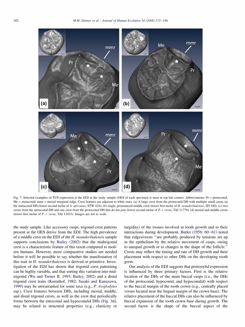
Fig. 7. Selected examples of TCP expression at the EDJ in the study sample (OES of each specimen is inset in top left corner). Abbreviations: Pr¼ protoconid,
Me¼metaconid, mmr¼mesial marginal ridge. Crest features are adjacent to white stars. (a) A large crest from the protoconid DH with multiple small crests on
the metaconid DH (lower second molar of A. africanus, STW 424); (b) single, pronounced middle crest (lower first molar of H. neanderthalensis, SD 540); (c) two
crests from the metaconid DH and one crest from the protoconid DH that do not join (lower second molar of P. t. verus, TAI 11779); (d) mesial and middle crests
(lower first molar of P. t. verus, TAI 13433). Images are not to scale.
182 M.M. Skinner et al. / Journal of Human Evolution 54 (2008) 173e186
the study sample. Like accessory cusps, trigonid crest patternspresent at the OES derive from the EDJ. The high prevalenceof a middle crest on the EDJ of the H. neanderthalensis samplesupports conclusions by Bailey (2002) that the midtrigonidcrest is a characteristic feature of this taxon compared to mod-ern humans. However, more comparative studies are neededbefore it will be possible to say whether the manifestation ofthis trait in H. neanderthalensis is derived or primitive. Inves-tigation of the EDJ has shown that trigonid crest patterningcan be highly variable, and that sorting this variation into mid-trigonid (Wu and Turner II, 1993; Bailey, 2002) and a distaltrigonid crest traits (Korenhof, 1982; Sasaki and Kanazawa,1999) may be unwarranted for some taxa (e.g., P. troglodytesssp.). Crest features between DHs, including mesial, middle,and distal trigonid crests, as well as the crest that periodicallyforms between the entoconid and hypoconulid DHs (Fig. 3d),may be related to structural properties (e.g., elasticity or
turgidity) of the tissues involved in tooth growth and to theirinteractions during development. Butler (1956: 60e61) notedthat ridges/crests ‘‘are probably produced by tensions set upin the epithelium by the relative movement of cusps, owingto unequal growth or to changes in the shape of the follicle.’’Crests may reflect the timing and rate of DH growth and theirplacement with respect to other DHs on the developing toothgerm.
Our analysis of the EDJ suggests that protostylid expressionis influenced by three primary factors. First is the relativelocation of the DHs of the main buccal cusps (i.e., the DHsof the protoconid, hypoconid, and hypoconulid) with respectto the buccal margin of the tooth crown (e.g., centrally placedversus located near the lingual margin of the crown base). Therelative placement of the buccal DHs can also be influenced bybuccal expansion of the tooth crown base during growth. Thesecond factor is the shape of the buccal aspect of the

Fig. 8. Schematic representation of protostylid variation present at the EDJ in the study sample. View is towards the buccal face of the EDJ including the buccal
margin, the tips of the DHs (represented as solid black circles) of the protoconid (Pr), hypoconid (Hyp), and hypoconulid (Hypd), and the marginal ridge that runs
between them. (a) No protostylid morphology along the buccal margin of the EDJ crown surface; (b) minor furrow on the mesial surface of the protoconid DH; (c)
similar to (b) and a second furrow on the distal surface of the hypoconulid DH; (d) similar to (c) with a cingulumlike furrow between the protoconid DH and
hypoconid DH; (e) pronounced furrow running from the mesial border of the protoconid DH to the mesial surface of the hypoconid DH with a furrow on the
distal surface of the hypoconulid DH (small crests are variably manifest on the surface of the protoconid DH); (f) a continuous, cingulumlike furrow across
the whole buccal margin of the EDJ in association with crests on the surfaces of the buccal DHs. This schematic is not exhaustive of the manifestations of pro-
tostylid morphology present in the study sample.
183M.M. Skinner et al. / Journal of Human Evolution 54 (2008) 173e186
protoconid and hypoconid DHs (e.g., the presence of crestsrunning towards the cervical margin of the tooth; Figs. 8e,fand 9d), and the third factor (though of a lesser influence) isthe thickness and distribution of enamel on the buccal sideof the dentine crown. Thus, protostylid morphology, includingcrests, depressions, cusps, cuspules, wrinkles, and fissures, de-rives from the EDJ with only minor modification caused by thedifferential deposition of enamel. A possible exception to theminor influence of enamel deposition on OES expression ofthe protostylid is P. robustus; an ongoing analysis of a largersample of molars from this taxon (Skinner et al., unpub. data)indicates that the relatively thick enamel of this taxon canmask EDJ expression in some cases. In answer to our thirdquestion, protostylid development appears similar among thestudy taxa.
If cingular remnants and furrowlike features on the mesialand distal portions of the buccal side of the crown are devel-opmentally related, then they should not be considered as sep-arate traits for the purpose of dental trait analysis (contraHlusko, 2004). Robinson (1956: 199e120) considered the pro-tostylid a complex character in South African fossil homininsdue to ‘‘remnants of a cingulum farther back on the buccal sur-face as well as on the whole of the mesial face of the crown.’’The complexity of this trait within the hominoid clade has alsobeen noted by Bailey and Wood (2007). If further evidencesupports developmental nonindependence among mesial, cen-tral, and distal components of protostylid morphology, this
trait could prove to be difficult to objectively and meaningfullysubdivide into an ordinal scale. Examination of crown size andDH size/spacing, which are thought to be under separategenetic control (Harris and Dinh, 2006; Cai et al., 2007),may lead to a better understanding of protostylid variationwithin and among taxa.
Further research will explore the interaction between DHheight and placement, the overall size of the molar crown,and the collective influence of all of these factors on theexpression of C6s, C7s, trigonid crest pattern, and protostylids.We agree with Bailey and Wood (2007) that new scoring cri-teria need to be developed for several of the traits relevant tothe study of early hominins, and we suggest that simultaneousexamination of the EDJ in addition to the OES may help in theidentification of independent discrete traits with a demonstra-bly similar pattern of development.
Conclusion
The results of this analysis indicate that the presence anddegree of morphological expression of four dental traits (C6,C7, trigonid crest pattern, and protostylid) is largely deter-mined by the factors controlling the shape of the EDJ, andthat differential enamel deposition does not significantly alterthe morphology of these traits or create traits at the outerenamel surface that are not present at the EDJ. For some traits,such as the protostylid and trigonid crest pattern, EDJ

Fig. 9. Selected examples of protostylid expression at the EDJ in the study sample (OES of each specimen is inset in top left corner). Abbreviations are:
Pr¼ protoconid, Hyp¼ hypoconid, Hypd¼ hypoconulid. Features associated with the protostylid are highlighted by white solid lines. (a) Minor furrows extending
across the buccal face of the protoconid DH and below the hypoconulid DH (lower second molar of P. t. verus (TAI 13437); (b) cingulumlike furrows extending
across most of the buccal face (lower second molar of G. g. beringei, NMNH 543037); (c) prominent furrows below the hypoconulid DH and between the hypo-
conid and protoconid DHs (lower second molar of P. robustus, SK 3974); (d) prominent furrows along the mesiobuccal face of the EDJ with small tubercle features
below the hypoconid DH that are also expressed on the enamel surface (lower second molar of A. africanus, STW 560E). Images are not to scale.
184 M.M. Skinner et al. / Journal of Human Evolution 54 (2008) 173e186
expression reveals considerable variation within nonhumanprimate taxa that will require new trait descriptions and stan-dardized classifications for comparison within and betweentaxa. In some cases, EDJ morphology suggests that traitsthat appear similar at the enamel surface in different taxamay not be the result of similar patterns of development,thus limiting their utility for phylogenetic analysis. Further-more, because of the significant morphological contributionof the EDJ to trait expression at the OES, the EDJ of wornteeth may be used as a proxy for the OES expression of thesetraits, and this may well result in increases in the size of studysamples of early hominin taxa.
Acknowledgments
This manuscript benefited from the comments of Bill Kim-bel, Shara Bailey, and from two anonymous reviewers. Accessto specimens was kindly provided by: Mike Raath of the
University of Witwatersrand; Francis Thackeray of the Trans-vaal Museum; Ottmar Kullmer of the Senckenberg Museum;Mandy Jay and Mike Richards of the MPI-EVA; and RickPotts and David Hunt of the National Museum of Natural His-tory. Fred Grine helped provide access to the hominin fossilmaterial and contributed many useful discussions. Mark F.Skinner provided critical comments on an earlier version ofthe manuscript. Heiko Temming of the MPI-EVA assisted inthe microCT scanning and contributed his expertise in the pro-cessing of the CT image stacks. Part of this manuscript waspresented at the 2007 AAPA meeting and was awarded theA.A. Dahlberg prize for best student paper by the Dental An-thropology Association. CB thanks the Ministry of Environ-ment and Eaux et Forets, the ministry of scientific research,the direction of the Ta€ı National Park as well as the SwissCentre of Scientific Research for constant support to theTa€ı chimpanzee project. This research is supported by NSFIGERT, EVAN Marie Curie Research Training Network

185M.M. Skinner et al. / Journal of Human Evolution 54 (2008) 173e186
MRTN-CT-019564, the Spanish MEC grant CGL-2006-02131, and the Max Planck Society. MMS is supported bya George Washington University Selective Excellence Fellow-ship. The participation of BW was supported by the HenryLuce Foundation.
References
Bailey, S.E., 2002. A closer look at Neanderthal postcanine dental morphol-
ogy: the mandibular dentition. Anat. Rec. (New Anat.) 269, 148e156.
Bailey, S.E., 2006. Beyond shovel-shaped incisors: Neandertal dental mor-
phology in a comparative context. Period. Biol. 108, 253e267.
Bailey, S.E., Lynch, J.M., 2005. Diagnostic differences in mandibular P4 shape
between Neandertals and anatomically modern humans. Am. J. Phys.
Anthropol. 126, 268e277.
Bailey, S.E., Hublin, J.-J., 2006. Dental remains from the Grotte du Renne at
Arcy-sur-Cure (Yonne). J. Hum. Evol. 50, 485e508.
Bailey, S.E., Wood, B.A., 2007. Trends in postcanine occlusal morphology
within the hominin clade: the case of Paranthropus. In: Bailey, S.E.,
Hublin, J.-J. (Eds.), Dental Perspectives on Human Evolution: State of
the Art Research in Dental Anthropology. Springer, Dordrecht, pp. 33e52.
Beynon, A.D., Dean, M.C., Reid, D.J., 1991. On thick and thin enamel in hom-
inoids. Am. J. Phys. Anthropol. 86, 295e309.
Butler, P.M., 1956. The ontogeny of molar pattern. Biol. Rev. 31, 30e70.
Butler, P.M., 1999. The relation of cusp development and calcification to
growth. In: Mayhall, J.T., Heikkinen, T. (Eds.), Proceedings of the 11th
International Symposium on Dental Morphology, Oulu, Finland, 1998.
Oulu University Press, Oulu, Finland, pp. 26e32.
Cai, J., Cho, S.-W., Kim, J.-Y., Lee, M.-J., Cha, Y.-G., Jung, H.-S., 2007. Pattern-
ing the size and number of tooth and its cusps. Dev. Biol. 304, 499e507.
Corruccini, R.S., 1987a. The dentinoenamel junction in primates. Int. J. Prima-
tol. 8, 99e114.
Corruccini, R.S., 1987b. Relative growth from the dentino-enamel junction in
primate maxillary molars. J. Hum. Evol. 2, 263e269.
Corruccini, R.S., 1998. The dentino-enamel junction in primate mandibular
molars. In: Lukacs, J.R. (Ed.), Human Dental Development, Morphology,
and Pathology: A Tribute to Albert A. Dahlberg. University of Oregon An-
thropological Papers, Portland, pp. 1e16.
Corruccini, R.S., Holt, B.M., 1989. The dentinoenamel junction and the hypo-
cone in primates. J. Hum. Evol. 4, 253e262.
Dahlberg, A.A., 1950. The evolutionary significance of the protostylid. Am. J.
Phys. Anthropol. 8, 15e25.
Grine, F.E., Martin, L.B., 1988. Enamel thickness and development in Austral-
opithecus and Paranthropus. In: Grine, F.E. (Ed.), Evolutionary History of
the ‘‘Robust’’ Australopithecines. Aldine de Gruyter, New York, pp. 3e42.
Guatelli-Steinberg, D., Irish, J.D., 2005. Early hominin variability in first
molar dental trait frequencies. Am. J. Phys. Anthropol. 128, 477e484.
Harris, E.F., Dinh, D.P., 2006. Intercusp relationships of the permanent max-
illary first and second molars in American whites. Am. J. Phys. Anthropol.
130, 514e528.
Hlusko, L.J., 2002. Expression types for two cercopithecoid dental traits
(interconulus and interconulid) and their variation in a modern baboon
population. Int. J. Primatol. 23, 1309e1318.
Hlusko, L.J., 2004. Protostylid variation in Australopithecus. J. Hum. Evol. 46,
579e594.
Irish, J., Guatelli-Steinberg, D., 2003. Ancient teeth and modern human
origins: an expanded comparison of African Plio-Pleistocene and recent
world dental samples. J. Hum. Evol. 45, 113e144.
Jernvall, J., Jung, H.-S., 2000. Genotype, phenotype, and developmental biol-
ogy of molar tooth characters. Yearb. Phys. Anthropol. 43, 171e190.
Jernvall, J., Thesleff, I., 2000. Reiterative signaling and patterning during
mammalian tooth morphogenesis. Mech. Dev. 92, 19e29.
Johanson, D.C., 1974. An odontological study of the chimpanzee with some
implications for hominoid evolution. Ph.D. Dissertation, University of
Chicago.
Kassai, Y., Munne, P., Hotta, Y., Penttila, E., Kavanagh, K.,
Ohbayashi, N., Takada, S., Thesleff, I., Jernvall, J., Itoh, N., 2005.
Regulation of mammalian tooth cusp patterning by ectodin. Science
309, 2067e2070.
Korenhof, C.A.W., 1960. Morphogenetical Aspects of the Human Upper
Molar. Uitgeversmaatschappij Neerlandia, Utrecht.
Korenhof, C.A.W., 1961. The enamel-dentine border: a new morphological
factor in the study of the (human) molar pattern. Proc. Koninkl., Nederl.
Acad. Wetensch. 64B, 639e664.
Korenhof, C.A.W., 1982. Evolutionary trends of the inner enamel anatomy of
deciduous molars from Sangiran (Java, Indonesia). In: Kurten, B. (Ed.),
Teeth: Form, Function and Evolution. Columbia University Press, New
York, pp. 350e365.
Kraus, B.S., 1952. Morphologic relationships between enamel and dentin
surfaces of lower first molar teeth. J. Dent. Res. 31, 248e256.
Kraus, B.S., Jordan, R., 1965. The Human Dentition before Birth. Lea and
Febiger, Philadelphia.
Macchiarelli, R., Bondioli, L., Debenath, A., Mazurier, A., Tournepiche, J.-F.,
Birch, W., Dean, C., 2006. How Neanderthal molar teeth grew. Nature 444,
748e751.
Martinon-Torres, M., Bermudez de Castro, J.M., Gomez-Robles, A., Arsuaga, J.L.,
Carbonell, E., Lordkipanidze, D., Manzi, G., Margvelashvili, A., 2007. Dental
evidence on the hominin dispersals during the Pleistocene. Proc. Natl. Acad.
Sci. U.S.A. 104, 13279e13282.
Nager, G., 1960. Der vergleich zwischen dem raumlichen verhalten des
dentin-kronenreliefs und dem schmelzrelief der zahnkrone. Acta Anat.
42, 226e250.
Olejniczak, A.J., Grine, F.E., 2006. Assessment of the accuracy of dental
enamel thickness measurements using micro-focal X-ray computed tomog-
raphy. Anat. Rec. 288A, 263e275.
Olejniczak, A.J., Martin, L.B., Ulhaas, L., 2004. Quantification of dentine
shape in anthropoid primates. Ann. Anat. 186, 479e485.
Pilbrow, V., 2003. Dental variation in African apes with implications for un-
derstanding patterns of variation in species of fossil apes. Ph.D. Disserta-
tion, New York University.
Pilbrow, V., 2006. Lingual incisor traits in modern hominoids and an assess-
ment of their utility for fossil hominoid taxonomy. Am. J. Phys. Anthropol.
129, 323e338.
Polly, P.D., 1998. Variability, selection, and constraints: development and evo-
lution in viverravid (Carnivora, Mammalia) molar morphology. Paleobiol-
ogy 24, 409e429.
Reid, C., Van Reenen, J., 1995. The Carabelli trait in early South African hom-
inids: a morphometric study. In: Moggi-Cecchi, J. (Ed.), Aspects of Dental
Biology: Paleontology, Anthropology and Evolution. International Institute
for the Study of Man, Florence, pp. 299e304.
Robinson, J.T., 1956. The Dentition of the Australopithecinae. Transvaal Mu-
seum, Pretoria.
Sakai, T., Hanamura, H., 1971. A morphology study of enamel-dentin border
on the Japanese dentition. Part V. Maxillary molar. J. Anthropol. Soc. Nip-
pon 79, 297e322.
Sakai, T., Hanamura, H., 1973a. A morphology study of enamel-dentin border
on the Japanese dentition. Part VI. Mandibular molar. J. Anthropol. Soc.
Nippon 81, 25e45.
Sakai, T., Hanamura, H., 1973b. A morphology study of enamel-dentin border
on the Japanese dentition. Part VII. General conclusion. J. Anthropol. Soc.
Nippon 81, 87e102.
Sakai, T., Sasaki, I., Hanamura, H., 1965. A morphology study of enamel-
dentin border on the Japanese dentition. Part I. Maxillary median incisor.
J. Anthropol. Soc. Nippon 73, 91e109.
Sakai, T., Sasaki, I., Hanamura, H., 1967a. A morphology study of enamel-
dentin border on the Japanese dentition. Part II. Maxillary canine.
J. Anthropol. Soc. Nippon 75, 155e172.
Sakai, T., Sasaki, I., Hanamura, H., 1967b. A morphology study of enamel-
dentin border on the Japanese dentition. Part III. Maxillary premolar.
J. Anthropol. Soc. Nippon 75, 207e223.
Sakai, T., Sasaki, I., Hanamura, H., 1969. A morphology study of enamel-den-
tin border on the Japanese dentition. Part IV. Mandibular premolar.
J. Anthropol. Soc. Nippon 77, 71e98.

186 M.M. Skinner et al. / Journal of Human Evolution 54 (2008) 173e186
Salazar-Ciudad, I., Jernvall, J., Newman, S.A., 2003. Mechanisms of pattern
formation in development and evolution. Development 130, 2027e2037.
Sasaki, K., Kanazawa, E., 1999. Morphological traits on the dentino-
enamel junction of lower deciduous molar series. In: Mayhall, J.T.,
Heikkinen, T. (Eds.), Proceedings of the 11th International Symposium
on Dental Morphology, Oulu, Finland, 1998. Oulu University Press,
Oulu, Finland, pp. 167e178.
Schulze, M.A., Pearce, J.A., 1994. A morphology-based filter structure for
edge-enhancing smoothing. In: Proceedings of the 1994 IEEE Interna-
tional Conference on Image Processing, pp. 530e534.
Schwartz, G.T., Thackeray, J.F., Reid, C., van Reenan, J.F., 1998. Enamel
thickness and the topography of the enamel-dentine junction in South
African Plio-Pleistocene hominids with special reference to the Carabelli
trait. J. Hum. Evol. 35, 523e542.
Scott, G.R., Turner II, C.G., 1997. The Anthropology of Modern Human
Teeth. Cambridge University Press, Cambridge.
Suwa, G., White, T.D., Howell, F.C., 1996. Mandibular postcanine dentition
from the Shungura Formation, Ethiopia: crown morphology, taxonomic
allocations, and Plio-Pleistocene hominid evolution. Am. J. Phys. Anthro-
pol. 101, 247e282.
Turner II, C.G., Nichol, C.R., Scott, G.R., 1991. Scoring procedures for
key morphological traits of the permanent dentition: the Arizona State
University Dental Anthropology System. In: Kelley, M.A., Larsen, C.S.
(Eds.), Advances in Dental Anthropology. Wiley-Liss, New York
pp. 13e31.
Uchida, A., 1996. Craniodental Variation among the Great Apes. Peabody
Museum of Archaeology and Ethnology, Harvard University, Cambridge.
Van Reenen, J., Reid, C., 1995. The Carabelli trait in early South African hom-
inids: a morphological study. In: Moggi-Cecchi, J. (Ed.), Aspects of Dental
Biology: Paleontology, Anthropology and Evolution. International Institute
for the Study of Man, Florence, pp. 291e298.
Weidenreich, F., 1937. The dentition of Sinanthropus pekinensis: a comparative
odontography of the hominids. Palaeontol. Sin. Series D I, 1e180.
Wood, B.A., Abbott, S.A., 1983. Analysis of the dental morphology of Plio-
Pleistocene hominids. I. Mandibular molars: crown area measurements
and morphological traits. J. Anat. 136, 197e219.
Wood, B.A., Richmond, B.R., 2000. Human evolution: taxonomy and paleobi-
ology. J. Anat. 196, 19e60.
Wu, L., Turner II, C.G., 1993. Variation in the frequency and form of the lower
permanent molar middle trigonid crest. Am. J. Phys. Anthropol. 91, 245e248.


![Cerámica y Vidrio - SECVboletines.secv.es/upload/20080123101152.46[5]225-231.pdf(1). The carbonate content present in human bones, dentine and enamel, fluctuates from 3 to 8wt% (1,2)](https://img.dokumen.tips/doc/110x75/60e91f0916c2df5a0402aec8/cermica-y-vidrio-5225-231pdf-1-the-carbonate-content-present-in-human-bones.jpg)


![Uneven distribution of enamel, dentine and cementum in ... / year) [4] caused by attrition (tooth to tooth contact) and abrasion (tooth to food contact) [5], CT erupt continuously](https://img.dokumen.tips/doc/110x75/5b0dd8407f8b9a2c3b8dd3cd/uneven-distribution-of-enamel-dentine-and-cementum-in-year-4-caused-by.jpg)


