Embed Size (px)
Citation preview
CentralBringing Excellence in Open Access
Journal of Fever
Cite this article: Solanki HA, Shankar SS, Pandya HA (2017) Dengue as a Druggable Disease: A Review. J Fever 1(1): 1001.
*Corresponding authorHitesh A. Solanki, Department of Botany, Bioinformatics and Climate Change Impact Management, Gujarat University, Ahmedabad, Gujarat 380009, India, Email:
Submitted: 26 November 2016
Accepted: 01 March 2017
Published: 06 March 2017
Copyright© 2017 Solanki et al.
OPEN ACCESS
Keywords•Dengue virus•Genome•Proteome•Drug compounds•Experimental analysis•Computational methods
Review Article
Dengue as a Druggable Disease: A ReviewHitesh A. Solanki*, Shetty Shilpa Shankar, and Himanshu A. PandyaDepartment of Botany, Bioinformatics and Climate Change Impact Management, Gujarat University, India
Abstract
Flavivirus covers pathologically significant pathogenic virus which causes several infections in humans globally. Dengue is one of the viral infection with potential deadly complications. It is an infectious disease transmitted by mosquitoes. It contains four different serotypes, which is based on the type of antibodies produced in the human body after infection such as DENV-1, DENV-2, DENV- 3 and DENV-4. The genome organisation is of single positive strand RNA genome virus which is like a large cellular mRNA molecule. Proteolysis yields ten proteins, including the three structural proteins C, prM and E and the seven nonstructural (NS) proteins involved in genome replication and capping NS1, NS2A, NS2B, NS3, NS4A, NS4B, and NS5. Availability of Dengue virus sequences and human genome sequences enabled to search for drug targets using various techniques. With number of experimental analysis, computational target-based identification method for distinguishing drugs which are approved for clinical use in humans, it may be able to interfere with Dengue virus proteins. A systematic in silico approach were applied to identify chemicals or natural drugs that been clinically permitted for human, but never evaluated against dengue virus proteins based on the principle of homologous drug target screening. The protein-protein interactions between the virus and mosquito or human as its host through which dengue bind and exploit its host cellular pathways and processes were also analyzed. It was also important to study the dengue virus genome and proteome arrangement through which one can identify novel target. This target can be used for further analysis of drug compounds using systematic method. This review study covers various methods which were useful for the experimental study to identify drug compounds.
DENGUEDengue is a severe viral infection with deadly complications.
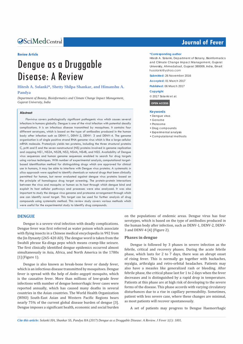
Dengue fever was first referred as water poison which associate with flying insects in a Chinese medical encyclopedia in 992 from the Jin Dynasty (265-420 AD). The dengue word is taken from the Swahili phrase Ka-dinga pepo which means cramp-like seizure. The first clinically identified dengue epidemics occurred almost simultaneously in Asia, Africa, and North America in the 1780s [1] (Figure 1).
Dengue is also known as break-bone fever or dandy fever, which is an infectious disease transmitted by mosquitoes. Dengue fever is spread with the help of Aedes aegypti mosquito, which is the causative fever. More than millions of low-grade fever infections with number of dengue hemorrhagic fever cases were reported annually, which has caused many deaths in several countries in the Asian countries. The World Health Organization (WHO) South-East Asian and Western Pacific Regions bears nearly 75% of the current global disease burden of dengue [3]. Dengue imposes a significant health, economic and social burden
on the populations of endemic areas. Dengue virus has four serotypes, which is based on the type of antibodies produced in the human body after infection, such as DENV-1, DENV-2, DENV- 3 and DENV-4 [4] (Figure 2).
Phases in dengue
Dengue is followed by 3 phases in severe infection as the febrile, critical and recovery phases. During the acute febrile phase, which lasts for 2 to 7 days, there was an abrupt onset of rising fever. This is normally go together with backaches, myalgia, arthralgia and retro-orbital headaches. Patients may also have a measles like generalized rash or bleeding. After febrile phase, the critical phase last for 1 to 2 days when the fever decreases and is distinguished by a rapid drop in temperature. Patients at this phase are at high risk of developing to the severe forms of the disease. This phase accords with varying circulatory disturbances due to a rise in capillary permeability. Sometimes, patient with less severe case, where these changes are minimal, as most patients will recover spontaneously.
A set of patients may progress to Dengue Haemorrhagic
CentralBringing Excellence in Open Access
Solanki et al. (2017)Email:
J Fever 1(1): 1001 (2017) 2/19
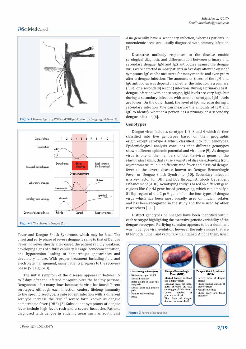
Fever and Dengue Shock Syndrome, which may be fatal. The onset and early phase of severe dengue is same to that of Dengue Fever, however shortly after onset, the patient rapidly weakens, developing signs of diffuse capillary leakage, hemoconcentration, and hypotension leading to hemorrhagic appearances and circulatory failure. With proper treatment including fluid and electrolyte management, many patients progress to the recovery phase [5] (Figure 3).
The initial symptom of the diseases appears in between 5 to 7 days after the infected mosquito bites the healthy persons. Dengue can infect many times because the virus has four different serotypes. Although each infection confers lifelong immunity to the specific serotype, a subsequent infection with a different serotype increase the risk of severe form known as dengue hemorrhagic fever (DHF) [3] Subsequent symptoms of dengue fever include high fever, rash and a severe headache. Patients diagnosed with dengue in endemic areas such as South East
Asia generally have a secondary infection, whereas patients in nonendemic areas are usually diagnosed with primary infection [7].
Distinctive antibody responses to the disease enable serological diagnosis and differentiation between primary and secondary dengue. IgM and IgG antibodies against the dengue virus were detected in most patients in five days after the onset of symptoms. IgG can be measured for many months and even years after a dengue infection. The amounts or titres, of the IgM and IgG antibodies was depend on whether the infection is a primary (first) or a secondary(second) infection. During a primary (first) dengue infection with one serotype, IgM levels are very high, but during a secondary infection with another serotype, IgM levels are lower. On the other hand, the level of IgG increase during a secondary infection. One can measure the amounts of IgM and IgG to identify whether a person has a primary or a secondary dengue infection [6].
Genotypes
Dengue virus includes serotype 1, 2, 3 and 4 which further classified into five genotypes based on their geographic origin except serotype 4 which classified into four genotypes. Epidemiological analysis concludes that different genotypes shown different epidemic potential and virulence [9]. As dengue virus is one of the members of the Flavivirus genus of the Flaviviridae family, that cause a variety of disease extending from asymptomatic, mild, undifferentiated fever and classical dengue fever to the severe disease known as Dengue Hemorrhagic Fever or Dengue Shock Syndrome [10]. Secondary infection is a key factor for DHF and DSS through Antibody Dependent Enhancement (ADE). Genotyping study is based on different gene regions like C-prM gene-based genotyping, which can amplify a 511bp region of the C-prM gene of all the four types of dengue virus which has been most broadly used on Indian isolates and has been recognized in the study and those used by other researchers [1,11].
Distinct genotypes or lineages have been identified within each serotype highlighting the extensive genetic variability of the dengue serotypes. Purifying selection appears to be a dominant way in dengue viral evolution, however the only viruses that are fit for both human and vector are maintained. Among them, Asian
Figure 1 Dengue figure by WHO and TDR publication on Dengue guidelines [2].
Figure 2 The phases in Dengue [5].
Figure 3 Forms of Dengue [6].
CentralBringing Excellence in Open Access
Solanki et al. (2017)Email:
J Fever 1(1): 1001 (2017) 3/19
genotypes of DENV-2 and DENV-3 are frequently associated with severe disease including secondary dengue infections. Intra-host viral diversity has also been described in human host. Three main contributing factors to the genetic diversity of the Dengue virus are:
a. Natural evolution of the Virus
b. Mutation of the virus
• towards resistance against treatment
• towards the state of the immune system of the patient
c. Recombination
Virus’s natural RNA replication process allows production of many different subtypes of its kind [6]. Although, these four serotypes are different strains of the same Dengue virus, thus they have approximately 60-80% homology between them. Phylogenetic studies have identified genetic subtypes within each of the four Dengue virus serotypes [3].
Dengue appeared in the second half of the 20th century as a major public health with major concern in many tropical and sub-tropical regions around the world. India became prevalent for both normal and severe dengue fever by means of transmission became sustained during the inter-epidemic periods in large parts of the country. Furthermore, dengue epidemics give rise to of hyperendemic areas, typically large, densely populated cities where one or all four DENV serotypes circulated in a sustained fashion [9].
Flavivirus
The first human infected virus was discovered by Walter Reed, who demonstrated that yellow fever could be experimentally transferred through the filtered serum of an infected individual and that same infectious agent was transmitted to humans by mosquitoes. Positive stranded RNA viruses have been classified into three superfamilies based on the evolutionary relatedness of their RNA-dependent RNA-polymerases (RdRP). The Flaviviridae are members of superfamily 2, which bear distant resemblance to coliphages and the plant infecting carmo-, tombus-, diantho- and subgroup I luteoviruses. Before the era of molecular biology, members of the family Flaviviridae had been previously classified as Togaviridae [3].
The genus Flavivirus includes medically significant pathogenic virus causing several infections in humans worldwide. It consists of more than 70 viruses, many of which are arthropod-borne human pathogens [1]. Flaviviruses cause a variety of diseases, including fevers, encephalitis and hemorrhagic fevers. Major global concern includes dengue virus (DENV) with its related infections, including dengue haemorrhagic fever (DHF) and dengue shock syndrome (DSS), Japanese encephalitis virus (JEV), West Nile virus (WNV) and Yellow Fever virus (YFV), which have been reviewed elsewhere. Other Flaviviruses of regional concern include Murray Valley encephalitis virus (MVEV), St. Louis encephalitis virus (SLEV) and tick-borne encephalitis virus (TBEV). Decreases in mosquito control efforts during the 20th century, united with societal factors like increased transportation and dense urbanization have contributed to the recurrence of Flaviviruses such as DENV in South and Central America [12].
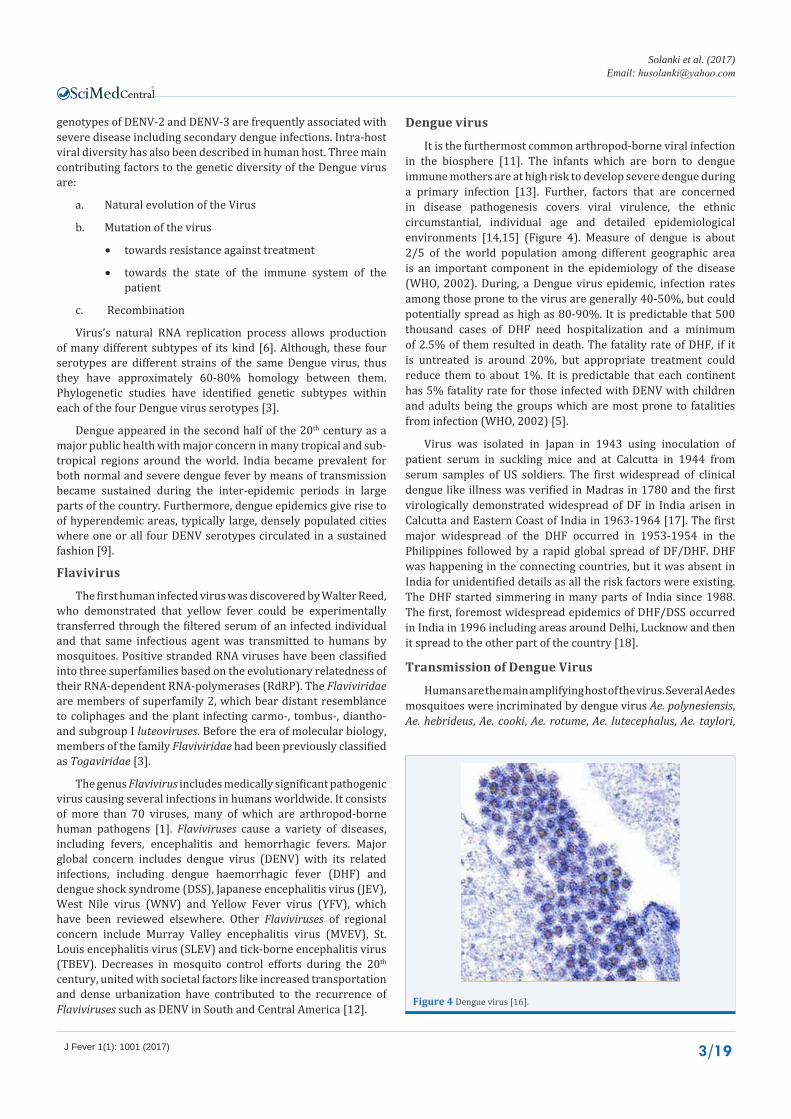
Dengue virus
It is the furthermost common arthropod-borne viral infection in the biosphere [11]. The infants which are born to dengue immune mothers are at high risk to develop severe dengue during a primary infection [13]. Further, factors that are concerned in disease pathogenesis covers viral virulence, the ethnic circumstantial, individual age and detailed epidemiological environments [14,15] (Figure 4). Measure of dengue is about 2/5 of the world population among different geographic area is an important component in the epidemiology of the disease (WHO, 2002). During, a Dengue virus epidemic, infection rates among those prone to the virus are generally 40-50%, but could potentially spread as high as 80-90%. It is predictable that 500 thousand cases of DHF need hospitalization and a minimum of 2.5% of them resulted in death. The fatality rate of DHF, if it is untreated is around 20%, but appropriate treatment could reduce them to about 1%. It is predictable that each continent has 5% fatality rate for those infected with DENV with children and adults being the groups which are most prone to fatalities from infection (WHO, 2002) [5].
Virus was isolated in Japan in 1943 using inoculation of patient serum in suckling mice and at Calcutta in 1944 from serum samples of US soldiers. The first widespread of clinical dengue like illness was verified in Madras in 1780 and the first virologically demonstrated widespread of DF in India arisen in Calcutta and Eastern Coast of India in 1963-1964 [17]. The first major widespread of the DHF occurred in 1953-1954 in the Philippines followed by a rapid global spread of DF/DHF. DHF was happening in the connecting countries, but it was absent in India for unidentified details as all the risk factors were existing. The DHF started simmering in many parts of India since 1988. The first, foremost widespread epidemics of DHF/DSS occurred in India in 1996 including areas around Delhi, Lucknow and then it spread to the other part of the country [18].
Transmission of Dengue Virus
Humans are the main amplifying host of the virus. Several Aedes mosquitoes were incriminated by dengue virus Ae. polynesiensis, Ae. hebrideus, Ae. cooki, Ae. rotume, Ae. lutecephalus, Ae. taylori,
Figure 4 Dengue virus [16].

CentralBringing Excellence in Open Access
Solanki et al. (2017)Email:
J Fever 1(1): 1001 (2017) 4/19
Ae. africanus, Ae. apok. Aedes aegypti is one of the most efficient vectors for arboviruses because it is highly anthropophilic which frequently bites before completing oogenesis and thrives near humans, which is supported by secondary vector Ae. Albopictus [18] (Figure 5). The transmission cycle of dengue virus by the mosquito Aedes aegypti begins with a dengue infected person. Apart from biological transmission, vertical transmission is also present mainly to maintain virus. The natural transmission of dengue virus from an infected female mosquito to its offspring known as the vertical transmission. Factors which can influence the dynamics of virus transmission, including environmental or climate factors, host-pathogen interactions and population immunological factors. Climate factors directly influence the biology of the vectors and so their abundance and distribution. It is consequently an important determinant of vector-borne disease epidemics [5].
If a person has virus circulating in the blood which is called a viremia that lasts about five days. During the viremic period, an uninfected female Aedes aegypti mosquito bites the person and ingests blood that contains dengue virus. There are some evidence of transovarial transmission of dengue virus in Aedes aegypti, through which mosquitoes are only infected by biting a viremic person. The virus then infects the mosquito mid-gut and later spreads systemically over a period of eight to twelve days. Then after within the mosquito, the virus replicates during an extrinsic incubation period of eight to twelve days. And if mosquito then bites a prone person can transmits the virus to individual as well as to every other prone person for the rest of its life. Again, the virus then replicates in the second individual and shows symptoms. The symptoms of infection begin to appear on an average of four to seven days after the mosquito bite, which is the intrinsic incubation period in humans. While the intrinsic incubation period exists for four to seven days, which can range from three to 14 days. The viremia activates somewhat before the start of symptoms. Symptoms may last for three to 10 days with an average of five days, after the onset of symptoms. The illness persists for several days after the viremia has ended [20].
Replication cycle
The full functional interaction that occur during viral replication are largely unknown. Thus, a general understanding was needed to understand as how dengue proteins regulate the virus replication cycle. Virus particles enter the cell by receptor-mediated endocytosis. Upon the acidification of the endocytic vesicle, the nucleocapsid enters the cytoplasm inside which the virus genome was released. Further, the genome is translated into a single polyprotein and further undergoes co-translational and post-translational conducted by host and viral proteases to yield the separate proteins which are essential for viral replication and its packaging. Replication takes place on the intracellular membranes and their assembly occurs on the endoplasmic reticulum (ER) membrane. A new assembled virus particles are then transported through the trans-golgi network and are released by exocytosis. Since, the viral proteins are important for the various stages of entry, replication, assembly. Thus, it serves as potential targets for the development of novel antiviral drugs [17] (Figure 6).
Safety measures to avoid dengue
There is no specific treatment for normal dengue fever and most of the people recover within 2 weeks. To help with recovery, it is recommended that patients should: (a) Get plenty of bed rest, (b) Drink lots of fluids and (c) Take medicine Paracetamol to reduce fever. For severe dengue symptoms, including shock and coma early and aggressive emergency treatment with fluid and electrolyte replacement may be life-saving ways [9].
The finest way to prevent infection is to take prior precautions to avoid being bitten by mosquitoes. Several dengue vaccines are being developed, but none is expected to be licensed in the next few years. Studies were going on for the development [9].
Figure 5 Transmission of Dengue Virus [19].
Figure 6 Replication of dengue virus [16].

CentralBringing Excellence in Open Access
Solanki et al. (2017)Email:
J Fever 1(1): 1001 (2017) 5/19
An early detection of the disease can also be helpful to decrease the fatality rates. Meet the doctor as soon as possible and take the prescribed medication which was mainly pain relievers, rest and drink maximum fluids. However, if the condition degrades in next 24 hours, visit the doctor for further medication and tests.
Dengue virus: genome
Dengue virus is a short virus that carries a single strand of RNA as its genome. The genome codes ten proteins. Three of the proteins are structural proteins that form the coat of the virus and send the RNA to target cells and seven of them are non-structural proteins that manage the production of new viruses once the virus gets inside the cell. The virus is enveloped by a lipid membrane. And 180 identical copies of the envelope protein are attached to the superficial area of the membrane by a short transmembrane segment. The duty of the envelope protein is to attach to a cell surface and begin the infection [21].
Cryoelectron microscopy has been used to study many characteristics of the life cycle of the dengue virus. In the structures, a low-resolution image of virus is obtained by the electron microscope, further the atomic structures of the distinct fragments are fit into the image to produce the final model [21] (Figure 7)
Many of the studies have been carried out using mouse to understand the immune response and the mechanisms of immunosuppression and pathogenesis of severe dengue disease. DENV mainly induces a humoral immune response, while the delayed type hypersensitivity response is poor. DENV infected sick mice develop immunosuppression to both homologous and heterologous antigens. Macrophages process DENV antigen by serine proteases and present it to B cells in vitro and in vivo, leading to their clonal development [18]. A tetravalent antigen was designed by splicing the EDIIIs of DENV-1, DENV-2, DENV-3 and DENV-4 using flexible Pentaglycyl linkers. A synthetic gene, which encode this tetravalent antigen was expressed in Pichia pastoris and purified to almost to homogeneity. This tetravalent antigen when injected into inbred BALB/c mice, provoked neutralizing antibodies specific to each of the four DENV in plaque reduction neutralization tests. Efforts are ongoing to produce the tetravalent antigen on a chimeric VLP platform. Some promising
dengue antigens have been developed using different systems [18].
Viral genome analysis
Using an electron microscope, protein biochemistry and nucleic acid sequencing takes systematic molecular characterization become possible. As their size is small, it was no wonder that the first completely sequenced genome was a phage MS2 [23]. More than 5000 viruses have been sequenced [24], although many have not been completely sequenced not even classified into any major virus families. The National Center for Biotechnology Information (NCBI) genome database lists more than 4000 complete sequences for various viral genomes [5,25]. Many RNA viruses have the capacity of changing genetic material with one another. The genetic exchange between different Dengue virus strain analysis comes under recombination study [26]. A single sequence can be produced by genetic exchange between viruses belonging to different subtypes. Thus, a new type of biologically important Dengue viruses may be created [10].
Genomics and Bioinformatics provide new opportunities to find novel targets. With the remarkable growth of microbial sequence databases, it has now become possible to use in silico method to compare different genomes to identify potential targets at the beginning of the drug discovery process [4]. Nucleic acid based therapy was introduced to control the replication of virus in the cell, but it could control DENV-2 strain only. Likewise, viral entry inhibitor compounds have been discovered, which are specific to stop only DENV-3 and to a lesser degree DENV-1 and DENV-4 [27] (Figure 8).
Dengue virus contains four serotypes which are transmitted by Aedes mosquitoes. Patients with DENV infection display various symptoms that range from no substantial illness or normal fever to severe Dengue Haemorrhagic Fever (DHF) and Dengue Shock Syndrome (DSS) [4,5]. To recognize vastly conserved regions in the genome of the all four DENV serotypes, a multiple sequence alignment was conducted by getting NS3 and E protein conserved (Figure 9).
For whole genome study, multiple sequence alignment was performed which show similarity among the different sequences. These similarities gave the region as a target gene for further analysis to identify variation among them. These variations can be useful to identify the changes among the sequences [3,23]. Further, recombination can be studied to identify the major parent for recombination between dengue serotypes.
Antigens: Recombinant dengue virus
Several studies have contributed in terms of developing new components or technology for diagnostic purposes. A recombinant DENV-3 envelope domain III protein has been produced in Escherichia coli for possible use in diagnosis [28]. A biotinylated chimeric dengue antigen to deed the high affinity of biotin-streptavidin interaction to detect anti-dengue antibodies have been developed which includes the envelope domain III of all four DENV serotypes [14]. Immunosensor has been recognized for label free and real time assay for the serological diagnosis of DENV infection. The main aim for development of biosensors Figure 7 Cryoelectron microscopy of dengue virus [21].

CentralBringing Excellence in Open Access
Solanki et al. (2017)Email:
J Fever 1(1): 1001 (2017) 6/19
Dengue virus genome organisation and polyprotein
The genome organisation contains single positive strand RNA genome, which is like a large cellular mRNA molecule. The genome is approximately 11 kb in size, bears a type I cap structure at its 5’-end and lacks a 3’-polyadenylate tail. The long open reading frame encodes a large polyprotein which is flanked in 5’ and 3’ by untranslated regions (UTRs). It carries many of cis-acting signals as stem loops, conserved sequences required for viral replication and possibly RNA capping. There are complementary sequences in these UTRs that are thought to be responsible for cyclization of the genome, which is essential for replication [3,10].
Flavivirus are enveloped viruses having two outer membrane proteins such as the envelope E protein and the membrane M processed from the precursor prM protein. The genome is thought to be wrapped or associated with the capsid protein C. A single polyprotein is translated from the genome and the former is cleaved by a combination of cellular proteases and a serine protease made of NS2B and NS3 protease (N-terminus domain) [3].
Figure 8 Schematic representation of DENV genome [22].
Figure 9 DENV positive RNA genome and its co-linear polyprotein [21].
for diagnosis was demonstrated [29]. The recombinant dengue multiepitope rDME-M protein specific to IgM in Escherichia coli was produced in a 5-L fermenter for use in diagnostic purpose [18,30]. Using a multiple sequence alignment study of dengue virus, tetravalent vaccine design has been done. The multiple sequence alignments were done with 102 dengue virus intra serotype of all four DENV serotype which shown high similarity of E protein among each of DENV intra serotype. Though mutation occurred on E protein, the antibody can recognize all DENV intra serotype as one dengue virus serotype [8].
Recombination could be the pre-dominant factor in determining the genome evolution [19]. Only definite homologous regions were retained for phylogenetic analysis [22]. Genome recombination events can be found in the genome alignments. Sequence recombination can be detected in whole and partial genome alignments using Bootscan and SimPlot like software [31], other comparable software also available [8,33]. Analysis of the sub-genomic sequences of dengue virus type 1 structural gene has recently acceptable to identify possible recombination breakpoints [3].

CentralBringing Excellence in Open Access
Solanki et al. (2017)Email:
J Fever 1(1): 1001 (2017) 7/19
Proteolysis yields ten proteins having three structural proteins C, prM, and E and seven nonstructural (NS) proteins involved in genome replication and capping are NS1, NS2A, NS2B, NS3, NS4A, NS4B, and NS5. Some of these NS proteins also participate in the pathogenesis and counteract the innate immunity of the host cell. Replication of the viral genome does not occur individually in the cytoplasm. Instead, there is an extensive intracellular membrane re-arrangement in the infected cell with various observable cell substructures containing most NS proteins organized along the virus replication cycle [10,22].
THE STRUCTURAL PROTEINSC Protein
Capsid C protein is a basic protein of 11 kd. Charged residues are clustered at the N and C-termini separated by an internal hydrophobic region that mediates membrane association. The C protein folds into a compact dimmer with each monomer containing four alpha helices. Nascent C (anchC) also contains a C-terminal hydrophobic anchor that serves as a signal peptide for ER translocation of prM. This hydrophobic domain is cleaved from mature C through viral serine protease. Based on the asymmetric distribution of positively charged and hydrophobic residues, RNA binding and membrane interaction surfaces have been tentatively allotted. Thus, yet it is not clear how C protein dimmers are organized within nucleocapsids, but the interaction with RNA or DNA can bring isolated C protein dimmers to accumulate into nucleocapsid like particles [3,10].
Membrane glycoprotein prM
The glycoprotein prM precursor of M protein of 26 kd and translocated into the ER by the C-terminal hydrophobic domain of C. Cleavage of the pr peptide from prM generates the M protein. This cleavage is mediated by the cellular protease furin which is required for virus maturation. Signal peptidase cleavage is delayed until the viral serine protease cleaves upstream of this signal sequence to generate the mature C protein [22]. This strategy seems to result from the combination of a short 14 to 22aa signal sequence, suboptimal residues at the signalase cleavage site and downstream regions of prM. Optimize the prM signalase cleavage site, so that it is no longer regulated by the viral serine protease leads to excess production of empty virions. Thus, coordinated anchC/prM cleavage helps to delay structural protein processing and virus production until viral serine protease levels are appropriately high and late in infection [10].
Envelope glycoprotein
The E protein is excellent with a dual function: (a) to recognize cellular receptor and (b) to fuse the viral membrane to cellular endosomic membranes. E is an attractive target because an antiviral molecule binding and impeding attachment would act before infection or spreading the virus for uninfected cells [22].
THE NON-STRUCTURAL PROTEINSThe non-structural proteins include NS1, NS2A, NS2B, NS3,
NS4A, NS4B and NS5.The role and structure of NS1 is unknown. It is a soluble protein detected much early during infection, but has received little attention as an antiviral target. NS2A, NS2B, NS4A and NS4B are membrane associated proteins supposed to anchor and regulate the replication complex during the virus life-cycle.
NS1
The NS1 glycoprotein of 46 kd which translocated into the ER during synthesis and cleaved from E protein by host signal peptidase, whereas an unknown ER-resident host enzyme cleaves the NS1/NS2A junction. The eight C-terminal residues of NS1 and more than 140 amino acids of NS2A are required for this process. NS1 is largely engaged within infected cells, but can localize to the cell surface and is also able to secrete from mammalian cells. NS1 has an important, yet unclear role in RNA replication. NS1 localizes to sites of RNA replication. The mutation in N linked glycosylation sites within NS1 can lead to defects in RNA replication and virus production [7,10].
NS2A and NS2B proteins
NS2A is relatively small of 22 kd and hydrophobic protein. Its N-terminus is generated through NS1/NS2A cleavage by an unknown ER-resident host enzyme. In addition, the serine protease can cleave at an internal site in NS2A to generate a C-terminally truncated form NS2A. As NS2A is an attractive candidate for coordinating the shift between RNA packaging and RNA replication processes. NS2B is also a small of 14 kd and membrane-associated protein. NS2B forms a stable complex with NS3 and acts as a cofactor for the NS2B-NS3 serine protease. The activity of co-factor lies in a central peptide which intercalates within the fold of the serine protease domain. Mutation of conserved residues in NS2B can show effects on autoproteolytic cleavage at the NS2B/NS3 junction and transcleavage activities [10,22].
NS3 protein
NS3 is 69 kd which carry two functional domains, a N-terminal serine protease which is 170 aa and a C-terminus helicase or RNA triphosphatase of 440 aa. The alone protease domain is inactive, which needs the presence of 40 aa of NS2B bound to form a protease active site. The NS2B/NS3 protease has been the first dengue protein target actively used in drug design. Unfortunately, the crystal structure reported in 1999 for this domain was false, thus the original article retracted in 2009. Two complementary approaches have been followed to discover antivirals to inhibit the enzyme. The first approach has been the screening of a large chemical library and the second approach has been to design of peptidomimetics, such approach which has also been followed in the case of HCV [21,22]. So far, the comparatively flat topology and the charge repartition of the NS2B/NS3 protease active site are believed to account for the difficulty of finding potent compounds.
Still, such difficulties have also been mentioned many of the themes for the HCV protease, but patience and abstention have finally paid off with the discovery of potent HCV NS3 protease inhibitors. The other potential problem is that the protease domain might be regulated by its C-terminus fellow helicase domain, thus, different conformations of the full length NS3 have been stated. A great knowledge has been gathered and published on this protease and progress are still accumulating in this field which can be seen in the future with other dengue drug target pairs. Similarly, The NS3 helicase domain is a fascinating target which contains features unique to flaviviruses, such as Domain III. Though, drug discovery and design against this enzyme has
CentralBringing Excellence in Open Access
Solanki et al. (2017)Email:
J Fever 1(1): 1001 (2017) 8/19
proven challenging for numerous reasons such as poor helicase activity in vitro, a ATPase active site and the absence of obvious pockets which able to accomodate small molecule inhibitors [5,22,23].
NS4A and NS4B proteins
The NS4A and NS4B are small as 16 kd and 27 kd, respectively and hydrophobic proteins. As mentioned, an important genetic interaction exists between NS1 and NS4A. A role for NS4A in RNA replication is supported by the co-localization of this protein with replication complexes. The studies show that NS4A/2K/4B cleavage is necessary for induction of membrane rearrangements by NS4A [22]. NS4B co-localizes with NS3 and viral double stranded RNA (dsRNA) in ER-derived membrane structures presumed to be sites of RNA replication.
NS4B is also posted translationally modified to migrate faster on sodium dodecyl sulphate-polyacrylamide gel electrophoresis (SDS-PAGE), although the identity and function of this modification remain to be determined. NS4A and NS4B can also block type I IFN signalling. NS4B has the strongest antagonistic effect, which requires either proper processing of the NS4–NS4B polyprotein by the NS2B–NS3 serine protease or expression of NS4B with an N-terminal signal peptide [10].
NS5 protein
NS5 is the largest and most conserved dengue protein. NS5 is 900 amino acids, protein which is 100 kd carrying the enzymatic activities required for RNA capping and synthesis of the dengue RNA genome [10]. The NS5 N-terminal domain has been shown in 2002 as 2’O Methyltransferase (MTase) through crystal structure analysis and later the N7-guanine MTase activity was also proved to be embedded in the 260-amino acid fragment. The NS5 C-terminus domain has been revealed to carry RdRp activity using in vitro assays and its crystal structure has been resolved in 2007 with West Nile virus structure [22]. The N terminal region of NS5 was found to exhibit homology with S-adenosyl-methionine (SAM) dependent MTase, signifying that this protein is involved in modification of the 5- cap. It was subsequently shown that the purified N-terminal domain of DENV-2 NS5 could transfer methyl groups from SAM to capped RNA substrates. The C-terminus of NS5 contains significant homology to RdRP of other positive-strand RNA viruses. The Polymerase activity of this protein has been confirmed with recombinant NS5 [10]. The complete structural picture will be completed when the full-length NS5 crystal structure will be available. In a parallel to HIV and HCV drug design programs, the knowledge of a structure of a tertiary complex made of NS5/RNA/NTP would certainly add excitement to this growing active field. Certainly, the naked polymerase structure used in drug discovery frequently points to inhibitor compounds that are sub-optimal when assayed against replication complexes [22].
VIRAL PROTEOME ANALYSISThe Dengue virus pass in a host cell once the E protein binds
to the virus receptor [31] on the host cell surface and activates its conformational rearrangement triggering the E protein in its dimeric pre-fusion form to transform into a trimeric post-fusion structure [6].
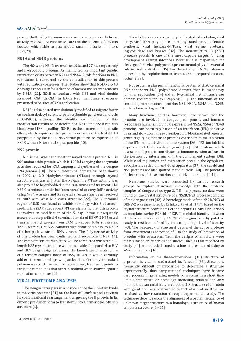
Targets for virus are currently being studied including viral entry, viral RNA polymerase or methyltransferase, nucleotide synthesis, viral helicase/NTPase, viral serine protease, R-glycosidase and kinases [32]. The non-structural 3 (NS3) protease protein is one of the most capable targets for drug development against infections because it is responsible for cleavage of the viral polyprotein precursor and plays an essential role in viral replication [36]. For the activity of NS3 protease a 40-residue hydrophilic domain from NS2B is required as a co-factor [8,33].
NS5 protein is a large multifunctional protein with a C-terminal RNA-dependent-RNA polymerase domain that is mandatory for viral replication [34] and an N-terminal methyltransferase domain required for RNA capping [35]. The functions of the remaining non-structural proteins NS1, NS2A, NS4A and NS4B, are less known (Figure 10).
Many functional studies, however, have shown that the proteins are involved in dengue pathogenesis and immune response in humans. Individual expression of NS2A, NS4A or NS4B proteins, can boost replication of an interferon (IFN) sensitive virus and slow down the expression of IFN-b-stimulated reporter genes, signifying that these proteins contribute to the inhibition of the IFN-mediated viral defence system [36]. NS5 too inhibits expression of IFN-stimulated genes [37]. NS1 protein, which is a secreted protein contributes to immune evasion at least in the portion by interfering with the complement system [38]. While viral replication and maturation occur in the cytoplasm, endoplasmic reticulum and Golgi apparatus [39], the capsid and NS5 proteins are also spotted in the nucleus [40]. The potential nuclear roles of these proteins are poorly understood [4,41].
Numerous studies were conducted by various research groups to explore structural knowledge into the protease complex of dengue virus type 2. Till many years, no data were found on the crystal structure of a NS2B/NS3 protease complex of the dengue virus [42]. A homology model of the NS2B/NS3 of DENV-2 was assembled by Brinkworth et al., 1999, based on the crystal structure coordinates of the hepatitis C virus NS3/NS4A as template having PDB id - 1JXP. The global identity between the two sequences is only 14.8%. Yet, regions nearby putative catalytic residues defined by indicating a high level of identity [43]. The deficiency of structural details of the active protease from experiments are not helpful to the study of interaction of proteins with substrates. Thus, the designs of inhibitors were mainly based on either kinetic studies, such as that reported by study [44] or theoretical considerations and explained using in silico simulations [16].
Information on the three-dimensional (3D) structure of a protein is vital to understand its function [33]. Since it is frequently difficult or impossible to determine a structure experimentally, thus computational techniques have become very popular in generating models of proteins in a short time limit. Comparative or homology modelling remains the only method that can unfailingly predict the 3D structure of a protein with great accuracy comparable to that of a protein structure resolved at low-resolution through experimental study. The technique depends upon the alignment of a protein sequence of unknown target structure to a homologous structure of known template structure [34,35].
CentralBringing Excellence in Open Access
Solanki et al. (2017)Email:
J Fever 1(1): 1001 (2017) 9/19
Figure 10 Schematic representation of Dengue virus genome [15].
However, possible problems can occur in structural determination when the target protein and template shows less than 25% sequence identity based on global length of amino acids. Nevertheless, satisfactorily long alignments can still conclude structural similarity, even if the sequence similarity is below 25% [16] (Table 1)
IMMUNNOPATHOGENESIS OF DENGUEThe main features of DHF/DSS include capillary leakage,
thrombocytopenia, and coagulopathy. Taking into consideration of several points such as: Dengue virus infection induces transient immune aberrant activation of CD4/CD8 ratio inversion and cytokine overproduction. Dengue virus infection encourages autoimmunity with the help of molecular mimicry. AntiNS1 or anti-prM antibodies can cross-react with platelets and endothelial cells. The binding to platelets causes platelet lysis in the presence of complement, where the binding to endothelial cells makes their NO-mediated apoptosis. Further, dengue virus infection activates both coagulation and fibrinolysis systems. The unbalance between coagulation and fibrinolysis induce hemorrhage in dengue. Dengue virus infection causes strong immune activation. Aberrant immune responses such as cytokine overproduction and generation of autoantibody act against platelets and endothelial cells, which occur after dengue virus infection. A molecular mimicry between platelets or endothelial cells with the NS-1 or prM of dengue virus would explain the cross-reactive of antiNS1 or anti-prM antibody to host cells, and take part in the attack of platelet and endothelial cells during the disease growth. High serum ferritin level, a macrophage activation marker in vivo was found to be highly elevated in the dengue patients, signifying that it was a common sign in the dengue patients. This suggests that the monocytes or macrophages were activated by cytokines such as IFN-γ during the dengue disease process after dengue virus infection. The triggered macrophages would then achieve the phagocytosis of the autoantibody-coated platelet and further contribute to the development of thrombocytopenia in Dengue fever. The anti-NS1 and anti-prM cross-reactive antibody to platelets and endothelial cells deliver a description for the target specificity and sole feature of thrombocytopenia and plasma leakage during the development of Dengue hemorrhagic fever. The macrophage activation might also be responsible for the sustained disease process with high fatality rate, as observed in the early elderly dengue virus-infected patients [46-48] (Figure 11).
TREATMENT AND THERAPEUTIC INTERVENTIONAs many cytokines and chemical mediators were released
in patients with DHF, thus theoretically there should be a major factor that inductees the amplification cycle of immunopathogenesis. Immunopathogenesis might be mediated by a few molecules produced during the early phase of immune responses. If the molecular basis of DHF is known, it is possible to prevent the disease and treat patients. Viral load was identified as the key element inducing the intense immune deviation and triggering the subsequent cascade leading to hemorrhage and plasma leakage. Ribavirin, an antiviral drug which is a guanosine analogue for RNA viruses can inhibit dengue virus replication and IL-6 and IL-8 production in endothelial cells. Another clinically used antiviral drug, amantadine can also inhibit dengue virus. Further, carboxyfullerene (C60), a novel compound with free radical scavenger activity can also inactivate dengue virus in a light-independent manner. It can block viral replication within the attachment and penetration stages, signifying a direct interaction between C60 and the envelope of the virion. C60 not only suppress the replication of enveloped viruses, but it also regulates immune responses by inhibiting cytokine production or modulating inflammatory cells. The inhibition of dengue virus-induced disease by C60 can be further evaluated in animal infection models [48-50].
PROTEIN-HOST INTERACTIONDENV is carried and spread to human host by the primary
mosquito vector Aedes aegypti and rarely by Aedes albopictus. Thus, DENV shows the notable capability to survive and replicate in two very different host organisms, proficient by a genome encoding 10 Proteins [45]. To perform the molecular functions which are required for invasion, replication, and spreading of the virus, proteins encoded by DENV must interact with and modify the actions of protein networks in both hosts. The computational method based on protein structures used to predict interactions between DENV and its human and insect hosts. The several interactions with many which are involved in known cell death, stress and immune system pathways were predicted [27]. Host–virus interactions have been studied since the discovery of viruses in 1898 [19], when the tobacco mosaic virus defined [4].
Some of the study performed a genome-wide siRNA screen for Drosophila melanogaster proteins whose depletion affected

CentralBringing Excellence in Open Access
Solanki et al. (2017)Email:
J Fever 1(1): 1001 (2017) 10/19
Table 1: Dengue protein structures in RCSB [37].
Target PDB Method Resolution Comments
E and M 1p58 cryo-EM 9.5 Å Mature virion
E 1thd cryo-EM, PA* 9.5Å Mature virus particle
E 1tge cryo-EM 12.5Å Immature virus particle
E 1tg8 X-ray 2.61Å
E 1ok8 X-ray 2.0Å Post-fusion conformation
E 1uzg X-ray 3.5Å Dengue 3 complex with ligands BMA, FUL and NAG
E 2b6b Cryo-EM Complexed with crd of DC-SIGN
E 1oke X-ray 2.4Å Complex with N-octyl-β-D-glucoside
E 1oan X-ray 2.75Å Ligand-binding pocket/apo-protein
E 1k4r cryo-EM 24Å First cryo-EM structure
E 2r29 X-ray 3Å Binding with neutralizing antibody
E 2r6p Cryo-EM, PA* 24Å E protein and Fab 1A1D-2 into the cryo-EM structure of Fab/Dengue complex
E 3c6r Cryo-EM 25Å Low pH immature virion
E 2r69 X-ray 3.8Å Fab 1A1D-2 / Domain III
prM/E 3c6d Cryo-EM 12.5Å Immature virion
E 2jsf NMR Domain III
E 2h0p NMR 16Å Dengue 4Domain III
prM 1n6g Cryo-EM 2.2Å Immature dengue-2 PrM
prM/E 3c5x X-ray 2.6Å PrM/E heterodimer at low pH
prM/E 3c6e X-ray 2.1Å PrM/E heterodimer at neutral pH
Capsid 1r6r NMR 21 structure
NS3 protease 1bef X-ray 2.1Å Apo structure
NS3 protease 1df9 X-ray 2.1Å Complex with Mung –Bean Bowman-Brik inhibitor
NS2B-NS3 protease/ cofactor 2fom X-ray 1.5Å Active protease
NS2B-NS3 protease/ cofactor 3U1J X-ray Dengue 3 complex with aprotinin
NS3 protease 2qid X-ray 2.1Å
NS3 protease/helicase 2vbc X-ray 3.15Å
NS3 protease/NTPase 2bhr X-ray 2.8Å
NS3 protease/NTPase 2bmf X-ray 2.41Å Complex with Mung –Bean Bowman-Brik inhibitor
Methyltransferase 2p3o X-ray 2.76Å Complex with 7MeGppA and S- adenosyl-L-homocysteine
Methyltransferase 2p3l X-ray 2.20Å Complex with GppA and S- adenosyl-L-homocysteine
Methyltransferase 2p3q X-ray 2.75Å Complex with GppG and S- adenosyl-L-homocysteine
Methyltransferase 2p40 X-ray 2.70Å Complex with 7MeGppG
Methyltransferase 2p41 X-ray 1.80Å Complex with 7MeGppG2’OMe and S- adenosyl-L-homocysteine
Methyltransferase 1r6a X-ray 2.60Å Complex with s-adenosylhomocysteine and ribavirin 5’ triphosphate
Methyltransferase 2p1d X-ray 2.90Å Complex with GTP and S-adenosyl-L-homocysteine
Methyltransferase 1l9k X-ray 2.40Å
Polymerase 2j7u X-ray 1.85Å Apo-structure
Polymerase 2j7w X-ray 2.60Å Complex with 3’DGTP

CentralBringing Excellence in Open Access
Solanki et al. (2017)Email:
J Fever 1(1): 1001 (2017) 11/19
Figure 11 Immunopathogenesis of severe dengue - an integrated model [14].
the capability of DENV-2 to infect the host cells. For insect host factors with human homologues, they verified 55 as human host factors also using siRNA [51]. The other study had recognized 123 human host factors for DENV in a study chiefly searching for West Nile Virus host factors, but also tested these proteins in DENV infection [34]. Overall, these two studies implicated 173 human proteins and 116 Drosophila melanogaster proteins as playing an important role in DENV infection study. It was noted that the similar studies in HIV together have discovered almost 1000 such human host factors and thus it was unlikely that these studies have identified all such factors in DENV hosts [27].
Structures for individual dengue proteins determined to atomic resolution, which is suitable for structure-based drug discovery and design. Vertical arrows in the figure (8) indicate major polyprotein cleavage sites recognized by the dengue NS2B-NS3 protease [46]. Many studies have investigated approaches to disrupt virus infection by interfering with viral entry to the host. Key proteins in these interactions are the E (envelope), prM (pre-membrane), and C (capsid) structural proteins. Potential entry-associated inhibitors that have been evaluated include small molecules [51,52] and peptide inhibitors from the study [42]. The highly basic regions of the capsid protein have been implicated in interactions with viral RNA [53,54]. An NMR structure of the capsid protein [55] provided supporting structural evidence that the protein’s highly basic region could interact with viral RNA and its hydrophobic region could interact with lipid molecules. The NS3 protease is a key target for the development of dengue antiviral drugs subsequently the NS2-NS3B protease is required for virus replication [56] and protease inhibitors have a successful history as being developed as antiviral drugs [57].
X-ray crystal structures of the dengue protein NS3 protease PDB id - 1BEF [58] and of the NS3 protease complexes with the
mung-bean Bowman-Birk inhibitor PDB id - 1DF9 [59] provided an initial structural source to understand NS3 protease function [46]. Scientists succeeded in developing tetravalent vaccines, but because of interfering of different attenuated Dengue virus, it required to be improved [27].
Molecular Modelling is a method of designing a 3D model for a protein of unknown structure based on one or more related protein of known structure [16]. Many of the unknown protein structures have been successfully modelled using in silico methods and their stability has been established by the molecular dynamics simulation studies [60,61]. Comparative modelling is quite useful in predicting the unknown 3D protein structures by both starting from a known primary structure and depend on known 3D structures of homologous proteins. Sequentially, related proteins are expected to show similar confirmations. The atomic positions in homologous regions are taken from known protein structures, while non-homologous portions are predicted using potential energy minimization, molecular dynamics and simulated annealing. The criteria for similarity analysis are (1) an identity of at least 25% for a sequence size >100 amino acids and (2) an expectation value (E) < 10−4, which gives the likelihood that the similarities are due to chance [5,62].
Within molecular modelling, molecular docking one can predict the ideal positioning of one molecule to a second when bound to each other to form a stable complex. Knowledge of the preferred positioning in turn may be used to predict the strength of binding affinity between two molecules using scoring functions. The protein structure and a database of potential ligands aid as inputs to a docking program. The success of a docking program depends on two components the search algorithm and the scoring function [16] (Figure 12).

CentralBringing Excellence in Open Access
Solanki et al. (2017)Email:
J Fever 1(1): 1001 (2017) 12/19
The computational methodology used to generate the chart assumes that proteins with comparable structures will share interaction partners. It was noted that the structural-based methodology provides a greater picture of the interaction network, while more understated changes at the sequence level are likely to explain experimentally observed differences in strain effects [63]. Dengue virus manipulates cellular processes to its own advantage through specific interactions with the host’s protein interaction network. The interaction networks deliver a set of hypothesis for further study into the DENV life cycle as well as potential therapeutic targets [36]. Computational approaches have also been used to predict dengue-host protein interactions [64], for example, it is predicted that 4,376 human-dengue and 176 mosquito-dengue protein interactions based on structural similarity between dengue proteins and host proteins [65].
MOLECULAR DOCKING Primary in vitro drug screening may provide insights into
their ability to inhibit infectious growth and if any promising activities are revealed, they could create important pave towards the discovery of novel target [3]. To understand the interface through which the pathogen connects and manipulates its host data, these require knowledge of the molecular points of interaction arrangement. Specifically, knowledge of the protein interactions between pathogen and host is important to understand the interaction. Understanding the interplay between DENV and its human host will help in understanding the viral life cycle and the ways through which it can manipulate its host [21].
PDB and Worldwide Protein Data Bank (PDB) have over 88000 or more protein structures, many of which are useful in critical metabolic pathways that may be considered as potential therapeutic targets and specific databases having the structures of binary complexes which become available along with information about their binding affinities in such as PDBBIND, PLD, AffinDB and BindDB database, thus molecular docking procedures improve by getting specific information [20].
The most important step in the computer-aided drug design is the design of suitable lead-like ligands, analysis of protein-ligand binding interactions and the identification of active binding sites in the target protein. The universal idea of a docking program is to position the ligand in possible binding modes in the protein active site and to calculate a score for the protein-ligand complex [66]. Another method pharmacophore corresponds to those features common to a set of compounds acting at the same receptor site, which are responsible for recognition and activation. Common pharmacophoric features are hydrogen bonding sites and hydrophobic regions. Atoms that can behave as hydrogen bond acceptors are like carbonyl oxygen atoms and donors could be hydroxyl or an amide NH group. One important hypothesis in drug design is that similar molecules can be expected to exhibit similar biological activity greatly. Substituents of the groups that produce broadly similar biological properties and facilitate the safe interactions are called bioisosteric groups. Such groups are therefore frequently interchangeable in drug design and they are used to maintain receptor interactions just to change other properties which enable a most straightforward synthesis or by avoiding toxic metabolites [30].
ENVELOPE PROTEIN AND NS3 PROTEIN AS TARGET FOR DOCKING
It is somewhat unclear that what a receptor dengue virus distinguishes on the cell surface. Also, the virus may recognize different receptors in the human host and mosquito vector cells respectively. It has been claimed that patches of positively charged residues on the surface of domain III bind to negatively charged heparin sulfate on the cell surface [16,67]. Many cell types express heparan sulfate and a more specific protein receptor is thought to be required to target the dengue virus to permissive cell types, immature dendritic cells particularly. Glycans on the virus surface have recently been drawn in receptor binding by the conclusion that DC-SIGN, a mannose-specific oligomeric C-type lectin on the cell surface is vital for
Figure 12 Dengue Protein structure [46].
CentralBringing Excellence in Open Access
Solanki et al. (2017)Email:
J Fever 1(1): 1001 (2017) 13/19
productive infection of dendritic cells. These glycans must belong to E, as it is the only glycoprotein on the surface of the mature virion. DC-SIGN is definite to immature dendritic cells, which considered to be the primary target cells upon introduction of dengue virus into the bloodstream [20,68,69]. Dengue virus infection of human dendritic cells is inhibited by anti- DC-SIGN antibodies and by the soluble ectodomain of DC-SIGN. DC-SIGN is a type II integral membrane protein. Its extracellular domain has a stalk and a C-terminal C-type carbohydrate recognition domain (CRD) [70]. The CRD recognizes fucose and high mannose N-glycans containing blood group antigens. Using DC-SIGN-specific monoclonal antibodies, its expression has been found in immature myeloid DCs in skin, intestine, lung, liver, placenta and lymph nodes. The participation of DC-SIGN in dengue virus infections was shown by competition assays employing either monoclonal antibodies against DC-SIGN or soluble DC-SIGN to inhibit DENV infection [72]. A computational screening on dengue envelope protein using receptor-guided de novo designed technique is presented to probe probable inhibitory properties of molecules with pharmacophore and molecular simulation study was also done by taking envelop as a target protein [73].
During fusion process, the dimeric form of the envelope proteins re-associates to the trimeric form on exposure to an acidic pH. The fusion peptide gets exposed to the host cell membrane due to a 37º conformational reorganization in the hinge region between domains I and II. The conformational change that occurs during this process is irreversible and fusion occurs, followed by viral entry into the host cell. The residue site corresponding to Asn-153 is highly conserved amongst most of the flaviviruses, while the Asn-67 residue is unique to the Dengue virus [20,67,74]. Inhibitors targeting dengue virus envelope protein to avoid DC-SIGN detention seem satisfactory to restrict with dengue dissemination. More data about DC-SIGN–Den-E interaction is required for the formation of these inhibitors [70,75].
NS3 viral protease required for virus replication, which is also a potential target for antiviral drugs. Targets currently being examined for viral entry, viral RNA polymerase/ methyltransferase, nucleotide synthesis, viral helicase/ NTPase, viral serine protease, R-glucosidases and kinases [76]. The non-structural 3 (NS3) protease is promising targets for drug development against flaviviridae because it is accountable for cleavage of the viral polyprotein precursor and plays a pivotal role in viral replication. For NS3 protease activity a 40-residue hydrophilic domain from NS2B is required as co-factor [30]. The overall structure of the DENV-3 protease is like that of the DENV-1 and DENV-2 structures with RMSD of 0.80 and 1.11 to DENV-1 and DENV-2 for 158 of 186 and 147 of 174 residues, respectively. Though, there is substantial movement in two loop regions of NS3 residues 119 to 120 and 155 to 159, which are disordered in DENV-1 and closer together in DENV-2, so there is not enough space to accommodate the NS2B hairpin. Aligning the beta-hairpin region of DENV-3 NS2B from the Bz-nKRR-H-bound structure into the DENV-3 aprotinin-bound structure shows that there would be similarly few direct interactions between aprotinin and the hairpin of NS2B. It is unlikely that the NS2B hairpin needs to bind to NS3 to make plan for the catalytically competent active site before aprotinin can bind to the NS2B-NS3
complex. A protinin is an effective inhibitor of both WNV and DENV proteases with a greater activity against DENV [77].
The NS3 protease is important as polyprotein processing which needs the presence of NS2B cofactor for its optimal catalytic activity. It has been indicated that the catalytic triad of this protease contains His51, Asp75 and Ser135 which is typical serine protease family. The conjugate NS3-NS2B cleave the precursor polyprotein at the NS2A/NS2B, NS2B/NS3, NS3/NS4A, NS4A/NS4B and NS4B/NS5 junctions also at internal sites inside C, NS2A, NS3 and NS4A. These sites have two basic amino acid residues Arg-Arg, Arg-Lys, Lys-Arg, or rarely Gln-Arg at the P2 and P1 positions, respectively. Other studies have specified that the P1 position has a strong preference for basic amino acid residues Arg/Lys, whereas the preference for P2 is Arg > Thr > Gln/Asn/Lys, while for P3 is Lys > Arg > Asn. Until now, there is no vaccine or anti-dengue viral drug currently available in the market against dengue infection. Therefore, there is an urgent necessity for the design of antiviral agents to fight against this disease. One of the antiviral agent strategies involves blocking and inhibiting viral enzyme activity, which prevent virus replication [39,78].
DENGUE AS A DRUGGABLE DISEASEDengue fever is not completely recognized as a major viral
disease in terms of public health and economic burden, although the situation is presently changing. However, the more aggressive expansion of the disease in the world as well as the emergence of a market is gaining the attention of novel vector in the field from both academic and corporate worlds. There is no vaccine or treatment available and the main aims are that both will be available nearly at the same time, that is within the coming 5 years. Since it is expected that cutting viremia as early as possible might show less severe dengue disease, dengue markers that parallel viremia is the greatest thing that one can expect. This expectation is becoming reality with the presence and detection of dengue NS1 in serum of patients, though ELISA technology prior the appearance of dengue reactive IgM in the first days of illness [1].
The study clearly discovered that genotype III (lineage III) of DENV-3 viruses is circulating chiefly and are the major cause of dengue infection in India since at least 1984. Though the exact time of introduction of the genotype into India could not be determined [23].
The development of vaccine need to be induce equally seroconversion against all four types of dengue [20]. Antisense oligos such as morpholinos have shown the ability to inhibit dengue viral translation in BHK cell and viral replication in Vero cells. Most of the non-specific antiviral drugs, such as the use of mycophenolic acid as an immunosuppressant agent have shown certain efficacy for inhibiting dengue virus replication in vitro and in cell culture system [6].
METHODOLOGY TO DISCOVER ANTIVIRALSNovel method for the discovery of antivirals was partially a
knowledge-based method, positioned around nucleobases and nucleotides known to be used by viruses for their replication. The initiation of AIDS and the discovery of non-nucleotide
CentralBringing Excellence in Open Access
Solanki et al. (2017)Email:
J Fever 1(1): 1001 (2017) 14/19
reverse transcriptase inhibitors unlocked the age of large-scale screening, which is entirely a trial and error procedure not based on earlier knowledge. Loads of compounds are tested as soon as possible using High Throughput Screening (HTS) techniques and only those showing activities are selected [24].
Before the antiviral properties of viruses are discovered, antiviral chemicals or molecules either occur somewhere in the world and are selected or discovered, or they do not exist and are invented and afterward synthesized. The existing molecules are either owned by someone or generally organized in a chemical library or repository or they are in the wild, plants, marine organisms, insects, etc. [1,19]. Tetracycline is discovered to inhibit the activity of envelope protein during virus entry [24].
VACCINE DEVELOPMENTAt present, there is no licensed dengue vaccine available.
Numerous studies on tetravalent attenuated dengue virus vaccines are under clinical trial. Other experimental vaccines such as a chimeric vaccine and a DNA vaccine are also under investigation. Most of these vaccines inhibit or delay viremia in a monkey infection model following inoculation with dengue virus [4]. The design of safe vaccines for virus must take into consideration the phenomenon of antibody-dependent enhancement and more importantly the problem of immune potentiation of the disease. To avoid the problem of antibody-dependent enhancement, NS1 protein are used in the place of the envelope E protein, and it was already reported that NS1 immunization can prevent mortality caused by intracerebral injection of dengue virus into mice. The immune-mediated pathogenesis is further degraded by immune memory and immune enhancement. The molecular mimicry between platelet or endothelial cells and NS1 of dengue virus further raises the importance of autoimmunity in dengue virus infection. Thus, an animal model with DHF display is urgently needed. Until the immunopathogenesis of DHF is clearly understood, cautious should be take about the long-term safety of dengue vaccines. Until the epitope that cross-reacts with the self-antigen is identified, it is likely to design a dengue virus vaccine with no side effects [24,27,39,79,80].
Status of vaccine development
The first dengue vaccine, Dengvaxia (CYD-TDV) by Sanofi Pasteur was first registered in Mexico in December, 2015. CYD-TDV is a live recombinant tetravalent dengue vaccine has been evaluated as a 3-dose series on a 0 or 6 or 12 month schedule in Phase III clinical studies. It has been registered for use in individuals 9-45 years of age living in endemic areas. There are other five vaccine candidates under evaluation in clinical trials, including other live-attenuated vaccines as well as subunit, also DNA and purified inactivated vaccine candidates. Additional technological approaches, such as virus-vectored and VLP-based vaccines are also under evaluation in preclinical studies. The growing global epidemic of dengue is a main concern and a safe and effective vaccine is urgently needed. WHO expects vaccines to be part of the Global dengue prevention and control strategy in between 2012-2020.
More research is needed on correlation of protection to DENV-2 in the Sanofi Pasteur phase IIb trial despite balanced
neutralizing antibody response to vaccination. It remains for the vaccine community to develop and implement plans for the strategic use of dengue vaccines by developing evidence-based policies to target high-risk groups and decrease virus transmission [4].
There is need to ensure that satisfactory observation is in place and is maintained for the safety and effectiveness of dengue vaccine during the post-licensure period. Early preparation and understanding of the true burden of disease is essential for successful vaccine introduction (WHO, 2011) and the ASEAN Member States Dengue Vaccination Advocacy Steering Committee (ADVASC) convened a regional workshop to review the current status of dengue surveillance and diagnostics in the ASEAN region [27,39].
Plant as anti-dengue
Medicinal plants have been traditionally used for diverse kinds of diseases including infectious diseases. There is an increasing need for constituents with antiviral activity since the treatment of viral infections with the available antiviral drugs frequently leads to the issue of viral resistance. The development of a dengue vaccine is complicated by the antibody-dependent enhancement effect [17].
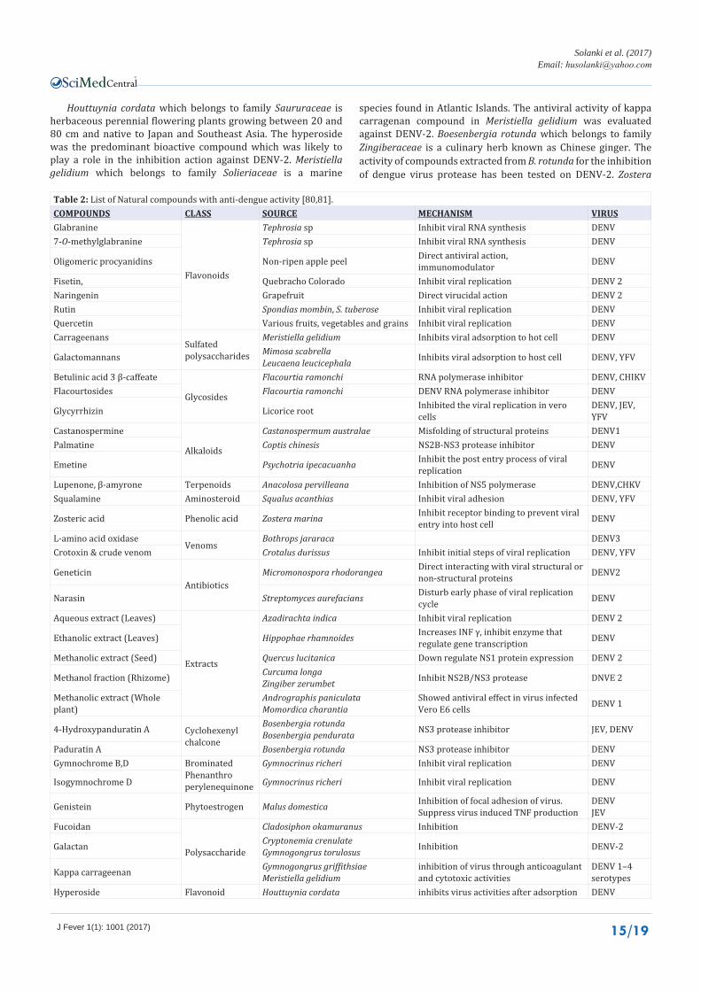
Castanospermum belongs to the Fabaceae family with only one species -Castanospermum australe, commonly known as the Black Bean. The investigation as anti-viral activity of castanospermine, as a natural alkaloid resulting from the tree C. australae by in vitro assay. Castanospermine has displayed good inhibitory anti-dengue activity over a broad range of doses from 10 to 250 mg/kg/day. The investigation reveals that castanospermine acts as an ER α-glucosidase I inhibitor and reduces infection of a subset of enveloped RNA and DNA viruses in vitro. Study of its mechanism of action proposes that castanospermine may interrupt the folding of some viral proteins by preventing the removal of the terminal glucose residue on N-linked glycans in dengue virus. Cladosiphon okamuranus which belongs to family Chordariaceae is brown seaweed found naturally in Okinawa. A sulfated polysaccharide named fucoidan from Cladosiphon okamuranus was found to greatly inhibit DENV-2 infection. The active compound is Fucoidan against dengue. Leucaena leucocephala which belongs to family Fabaceae. Compound Galactomannans extracted from seeds of Leucaena leucocephala have verified activity against the yellow fever virus (YFV) and DENV 1 in vitro and in vivo and L. leucocephala show protection against death in 96.5 % of YFV-infected mice. Mimosa scabrella which belongs to family Fabaceae and compound Galactomannans extracted from seeds of Mimosa scabrella too show activity against YFV and DENV-1 in vitro and in vivo. Tephrosia madrens also belongs to family Fabaceae and ompound Glabranine is the main active compound for dengue fever treatment. The flavonoids isolated from T. madrensis, glabranine and 7-O-methyl-glabranine apply strong inhibitory effects on dengue virus replication. Cryptonemia crenulata which belongs to family Halymeniaceae is a marine species found throughout the Indian Ocean Islands, Southeast Asia and Pacific Islands. Compound galactan the sulfated polysaccharides from Cryptonemia crenulate were selective inhibitors for DENV-2 multiplication [18,67] (Table 2).
CentralBringing Excellence in Open Access
Solanki et al. (2017)Email:
J Fever 1(1): 1001 (2017) 15/19
Houttuynia cordata which belongs to family Saururaceae is herbaceous perennial flowering plants growing between 20 and 80 cm and native to Japan and Southeast Asia. The hyperoside was the predominant bioactive compound which was likely to play a role in the inhibition action against DENV-2. Meristiella gelidium which belongs to family Solieriaceae is a marine
species found in Atlantic Islands. The antiviral activity of kappa carragenan compound in Meristiella gelidium was evaluated against DENV-2. Boesenbergia rotunda which belongs to family Zingiberaceae is a culinary herb known as Chinese ginger. The activity of compounds extracted from B. rotunda for the inhibition of dengue virus protease has been tested on DENV-2. Zostera
Table 2: List of Natural compounds with anti-dengue activity [80,81].COMPOUNDS CLASS SOURCE MECHANISM VIRUSGlabranine
Flavonoids
Tephrosia sp Inhibit viral RNA synthesis DENV7-O-methylglabranine Tephrosia sp Inhibit viral RNA synthesis DENV
Oligomeric procyanidins Non-ripen apple peel Direct antiviral action, immunomodulator DENV
Fisetin, Quebracho Colorado Inhibit viral replication DENV 2Naringenin Grapefruit Direct virucidal action DENV 2Rutin Spondias mombin, S. tuberose Inhibit viral replication DENVQuercetin Various fruits, vegetables and grains Inhibit viral replication DENVCarrageenans
Sulfated polysaccharides
Meristiella gelidium Inhibits viral adsorption to hot cell DENV
Galactomannans Mimosa scabrella Leucaena leucicephala Inhibits viral adsorption to host cell DENV, YFV
Betulinic acid 3 β-caffeate
Glycosides
Flacourtia ramonchi RNA polymerase inhibitor DENV, CHIKVFlacourtosides Flacourtia ramonchi DENV RNA polymerase inhibitor DENV
Glycyrrhizin Licorice root Inhibited the viral replication in vero cells
DENV, JEV, YFV
Castanospermine
Alkaloids
Castanospermum australae Misfolding of structural proteins DENV1Palmatine Coptis chinesis NS2B-NS3 protease inhibitor DENV
Emetine Psychotria ipecacuanha Inhibit the post entry process of viral replication DENV
Lupenone, β-amyrone Terpenoids Anacolosa pervilleana Inhibition of NS5 polymerase DENV,CHKVSqualamine Aminosteroid Squalus acanthias Inhibit viral adhesion DENV, YFV
Zosteric acid Phenolic acid Zostera marina Inhibit receptor binding to prevent viral entry into host cell DENV
L-amino acid oxidaseVenoms
Bothrops jararaca DENV3Crotoxin & crude venom Crotalus durissus Inhibit initial steps of viral replication DENV, YFV
GeneticinAntibiotics
Micromonospora rhodorangea Direct interacting with viral structural or non-structural proteins DENV2
Narasin Streptomyces aurefacians Disturb early phase of viral replication cycle DENV
Aqueous extract (Leaves)
Extracts
Azadirachta indica Inhibit viral replication DENV 2
Ethanolic extract (Leaves) Hippophae rhamnoides Increases INF γ, inhibit enzyme that regulate gene transcription DENV
Methanolic extract (Seed) Quercus lucitanica Down regulate NS1 protein expression DENV 2
Methanol fraction (Rhizome) Curcuma longa Zingiber zerumbet Inhibit NS2B/NS3 protease DNVE 2
Methanolic extract (Whole plant)
Andrographis paniculata Momordica charantia
Showed antiviral effect in virus infected Vero E6 cells DENV 1
4-Hydroxypanduratin A Cyclohexenyl chalcone
Bosenbergia rotunda Bosenbergia pendurata NS3 protease inhibitor JEV, DENV
Paduratin A Bosenbergia rotunda NS3 protease inhibitor DENVGymnochrome B,D Brominated
Phenanthro perylenequinone
Gymnocrinus richeri Inhibit viral replication DENV
Isogymnochrome D Gymnocrinus richeri Inhibit viral replication DENV
Genistein Phytoestrogen Malus domestica Inhibition of focal adhesion of virus. Suppress virus induced TNF production
DENV JEV
Fucoidan
Polysaccharide
Cladosiphon okamuranus Inhibition DENV-2
Galactan Cryptonemia crenulateGymnogongrus torulosus Inhibition DENV-2
Kappa carrageenan Gymnogongrus griffithsiaeMeristiella gelidium
inhibition of virus through anticoagulant and cytotoxic activities
DENV 1–4 serotypes
Hyperoside Flavonoid Houttuynia cordata inhibits virus activities after adsorption DENV
CentralBringing Excellence in Open Access
Solanki et al. (2017)Email:
J Fever 1(1): 1001 (2017) 16/19
marina which belongs to family Zosteraceae is an aquatic plant known as eelgrass and native to North America and Eurasia. A compound from the temperate marine eelgrasss Zostera marina possess antidengue virus activity. Myrtopsis corymbosa which belongs to family Rutaceae. Compound ramosin, myrsellinol and myrsellin are the main active compound of M. corymbosa from its bark. The bark extract is the strongest which inhibits 87% of DENV polymerase. Alkaloids content of leaves were also investigated, compounds which acknowledged as skimmianine, γ-fagarin and haplopin compounds, but isolated alkaloids were only somewhat active against the DENV-NS5 [67,68].
Alternanthera philoxeroides which belongs to family Amaranthaceae. It is also called as Alligator Weed which is an immersed aquatic plant. It originates from South America but currently conquering Australia. The effect of A. philoxeroides extracts against dengue virus was investigated in vitro. Coumarin extract of A. philoxeroides showed lowest toxicity on cells TD50 = 535.91, where a petroleum ether extract of A. philoxeroides had the strongest inhibitory effect on dengue virus ED50 = 47.43. Quercus lusitanica is also known as Quercus infectora which belongs to the family of Fagaceae. The gall oak common name for Quercus lusitanica is mainly found in Greece, Asia, Iran and India. In India gall extract is used to treat minor soar throat and chronic diarrhoea. The gall extracts of Q. infectoria, are widely used in traditional medication as karkatasringi for the preparation of Balachaturbhadra, Shringyadi churna, Karkatadi churna, Kantkaryavaleha. Sylvia et al., (2006) [10], was demonstrated in vitro inhibitory activity of Quercus lusitanica seed extract. The result showed the down regulation of NS1 protein expression of infected cells after treating with seed extract. In 2008 again the same plant extract was assessed for antidengue activity by Sylvia et al. The study showed seed extract of Quercus lusitanica inhibited Dengue type 2 virus in the concentration 0.032 to 0.25 mg/ml [67].
Carica papaya which belongs to family Caricaceae is an erect, fast-growing and unbranched tree or shrub indigenous to Central America and cultivated in Mexico and most tropical countries for its edible fruits. The aqueous extract of the leaves of this plant unveiled the potential activity against DF by increasing the platelet count, white blood cells and neutrophils in blood samples of a 45-year-old patient bitten by carrier mosquitoes. Lippia alba and Lippia citriodora which belong to family Verbenaceae are flowering plants native to Southern Texas, Mexico, the Caribbean, Central and South America. L. alba essential oil was more effective against DENV 2 than other serotypes, while for L. citriodora essential oil, the virucidal action against DENV 1, 2 and 3 were similar but lower than against DENV 4. Piper retrofractum which belongs to family Piperaceae is a flowering vine native to Southeast Asia and cultivated in Indonesia and Thailand mostly for its fruit. The in vitro anti-dengue activity of Piper retrofractum in Vero cells was investigated which shown inhibitory activity against DENV-2 infected cells were determined on dichloromethane ethanol extract by the MTT method [17,67,68]
The demand for plant-based medicines is growing as they are usually considered to be safer, cheaper, non-toxic and less harmful than synthetic drugs. Many of natural compounds stated in traditional medicinal plants to have anti-dengue properties
were considered and some of them were also screened for anti-dengue compounds structure [17].
The compounds had to check whether it is drug-likeness or not. The ADMET property will go to check compounds properties. Molecular docking of target protein as receptor and drug as ligand gives its binding affinity and score [34,83]. The active compounds from plants showed a wide range of activity against DENV serotypes in many of the study. The studied compounds belong to several chemical classes such as sulfated polysaccharides, flavonoids, quercetin and natural chalcone compounds. Number of study and protein-protein interaction analysis, envelope E protein and non-structural NS3 protein had shown their wide interaction with host protein during infection. Thus, the plants from which extract had taken were first tested to detect their inhibition activity against structural protein Envelope and non-structural protein NS3 of DENV-3 were considered for further analysis to determine the binding energy of the identified compounds. The computational analysis has shown many of the druggable compound as antiviral for further experimental study against dengue viral targets [83].
CONCLUSIONSDengue viral diseases is considered as a major public health
challenge, particularly in regions with tropical and subtropical climates. The genome and proteome of DENV give full knowledge about its outbreak in the world. One of the widely-spread serotype of dengue virus is serotype 2. After which, serotype 3 also showed some of the major breakouts similarly serotype 1 and 4 too. Genomic sequence alignment, recombination analysis and phylogenetic evolutionary analysis gave idea about major or minor parent for variation and conserved regions. Proteomic study gave knowledge about proteins which can be used as target to analyze some plant extracts or chemical compounds as lead or ligand. Molecular docking explores the interaction space and binding potential of each docked conformation.
Regardless of profound clinical and economic impact of flavivirus infections within large sections of the population, most treatments constitute medical management, while effective therapy is still indefinable. Many plants are available, which may act as a source of lead antiflaviviral compounds. These natural compounds, after appropriate pharmacological and toxicological screening, may ultimately leads to new therapeutics for dengue viral diseases. Further, the exploration for antiviral drugs and therapeutic agents which mediate in the development of DHF/DSS will become more dominant when the pathogenesis of dengue virus infection is more clearly understood. Vaccine development focuses on the development of a tetravalent dengue vaccine due to concerns of antibody-dependent enhancement, which is described as a form of enhanced virus uptake into macrophage cells through the binding of non-neutralizing antibody against another serotype of DENV. Thus, it is concluded that dengue is a druggable disease as there are several ways to inhibit it based on its activity. Early detection and precautions are very essential for proper treatment. Antidengue drug and vaccine were in their clinical stage thus, it can be said that in the coming years there can be an availability of drugs for dengue.
CentralBringing Excellence in Open Access
Solanki et al. (2017)Email:
J Fever 1(1): 1001 (2017) 17/19
REFERENCES1. Gupta N, Srivastava S, Jain A, Chaturvedi UC. Dengue in India. Indian J
Med Res. 2012; 136: 373-90.
2. Mannapperuma M M A T N, Wijesinghe M W A C R & Weerasinghe A R, Learning the diversity and evolutionary pattern of the dengue virus (2012 Int Conf on Bioscience, Biochemistry and Bioinformatics), IPCBBE. 2012; 3: 6-11.
3. Wilder-Smith A, Ooi EE, Vasudevan SG, Gubler DJ. Update on dengue: epidemiology, virus evolution, antiviral drugs, and vaccine development. CABI. 2010; 12: 157-164.
4. Kannabirran V, Madhu S, Priya PV, Uthaya R, Mozhi SS. An endemic challenge of Dengue-A crucial review. IJDD. 2012; 1: 51-58.
5. World Health Organization, Special Programme for Research, Training in Tropical Diseases, World Health Organization. Department of Control of Neglected Tropical Diseases, World Health Organization. Epidemic, Pandemic Alert. Dengue: guidelines for diagnosis, treatment, prevention and control. World Health Organization; 2009.
6. Garg P, Nagpal J, Khairnar P, Seneviratne SL. Economic burden of dengue infections in India. Trans R Soc Trop Med Hyg. 2008; 102: 570-577.
7. Lindenbach BD, Rice CM. Flaviviridae: the viruses and their replication. Fields virology. 2001; 1: 991-1041.
8. Somvanshi P, Singh V, Seth PK. Identification and analysis of putative promoter motifs in Flavivirus genome. Bioinformation. 2008; 3: 162-167.
9. Ilyaa M, Rahman Z, Shamas S, Alam M, Israr M, Masood K. Bioinformatics analysis of envelope glycoprotein e epitopes of Dengue virus type 3. African Journal of Biotechnology. 2011; 10: 3528-3533.
10. Mairiang D, Zhang H, Sodja A, Murali T, Suriyaphol P, Malasit P, Limjindaporn T, Finley Jr RL. Identification of new protein interactions between dengue fever virus and its hosts, human and mosquito. PLoS One. 2013; 8: e53535.
11. Mendez-Rios J, Uetz P. Global approaches to study protein protein interactions among viruses and hosts. Future microbiol. 2010; 5: 289-301.
12. Yang JM, Chen YF, Tu YY, Yen KR, Yang YL. Combinatorial computational approaches to identify tetracycline derivatives as flavivirus inhibitors. PLoS One. 2007; 2: e428.
13. Ronquist F, Huelsenbeck JP. MrBayes 3: Bayesian phylogenetic inference under mixed models. Bioinformatics. 2003; 19: 1572-1574.
14. Balajee R. using tetracycline analogues.
15. Abd Kadir SL, Yaakob H, Mohamed Zulkifli R. Potential anti-dengue medicinal plants: a review. J Nat Med. 2013; 67: 677-689.
16. Rodenhuis-Zybert IA, Wilschut J, Smit JM. Dengue virus life cycle: viral and host factors modulating infectivity. Cell Mol Life Sci. 2010; 67: 2773-2786.
17. Canard B. Antiviral research and development against dengue virus. Available: Accessed. 2012.
18. Hernandez-Santoyo A, Tenorio-Barajas AY, Altuzar V, Vivanco-Cid H, Mendoza-Barrera C. Protein-protein and protein-ligand docking. Protein Engineering-Technology and Application, Dr. Tomohisa Ogawa. 2013; 978-953.
19. Chakravarti A1, Arora R, Luxemburger C. Fifty years of dengue in India. Trans R Soc Trop Med Hyg. 2012; 106: 273-282.
20. Geis I. Dengue Virus.
21. Sharma S, Dash PK, Agarwal S, Shukla J, Parida MM, Rao PV. Comparative complete genome analysis of dengue virus type 3 circulating in India between 2003 and 2008. J Gen Virol. 2011; 92: 1595-1600.
22. Wiwanitkit V. Applied informatics manipulation for fight against dengue.
23. Seto J, Walsh MP, Mahadevan P, Zhang Q, Seto D. Applying genomic and bioinformatic resources to human adenovirus genomes for use in vaccine development and for applications in vector development for gene delivery. Viruses. 2010; 2: 1-26.
24. Malakar AK. Comparative Computational Studies and In silico Identification of Polyprotein in Japanese Encephalitis Virus. 2011; 1: Pb326.
25. Borovska P, Gancheva V, Asenov E, Georgiev I. Computational Aspects of In silico Experiments for Investigating the Impact of the Host Genome on the Influenza Virus A Variability. In Proceedings of the International Scientific Conference Supercomputing Applications in Science and Industry, Bulgaria 2011; 1-8.
26. Vlachakis D, Koumandou VL, Kossida S. A holistic evolutionary and structural study of flaviviridae provides insights into the function and inhibition of HCV helicase. PeerJ. 2013; 1: e74.
27. Somvanshi P, Seth P. Comparative Proteome Analysis of Distinct Variants of Dengue Virus using In silico Approach. IJGPG. 2008; 4: 1.
28. Murphy BR, Whitehead SS. Immune response to dengue virus and prospects for a vaccine. Annu Rev Immunol. 2011; 29: 587-619.
29. Doolittle JM, Gomez SM. Mapping protein interactions between Dengue virus and its human and insect hosts. PLoS Negl Trop Dis. 2011; 5: e954.
30. Patil JA, Cherian S, Walimbe AM, Bhagat A, Vallentyne J, Kakade M, et al. Influence of evolutionary events on the Indian subcontinent on the phylogeography of dengue type 3 and 4 viruses. Infect Genet Evol. 2012; 12: 1759-1769.
31. Rout PR, Satpathy R, Satpathy GR. Journal of Chemical and Pharmaceutical Research. JCPR. 2012; 4: 1200-1206.
32. Budhayash Gautam, Shashi Rani, Satendra Singh and Rohit. Farmer in silico comparative genome analysis of Hepatitis B and Hepatitis C virus. IJPSRR. 2010; 4: 023.
33. Kulkarni S, Patil P, Virupaksha B, Alpana G, Prashant K, Baikerikar S. Molecular dynamics, docking and QSAR analysis of napthoquinone derivatives as topoisomerase I inhibitors. Int J Comput Bioinfo In Silico. 2013; 2: 223-233.
34. Yadav PK, Pandey HK. In silico drug target identification in Legionella pneumophila strain Paris: modelling and molecular dynamics simulation of a candidate enzyme phosphoglyceromutase (PGM). 2013.
35. Guo X, Zhao Q, Wu C, Zuo S, Zhang X, Jia N, et al. First isolation of dengue virus from Lao PDR in a Chinese traveler. Virol J. 2013; 10: 70.
36. Tomlinson SM, Malmstrom RD, Watowich SJ. New approaches to structure-based discovery of dengue protease inhibitors. Infectious Disorders-Drug Targets (Formerly Current Drug Targets-Infectious Disorders). 2009; 9: 327-343.
37. Tolou HJ, Couissinier-Paris P, Durand JP, Mercier V, de Pina JJ, De Micco P, et al. Evidence for recombination in natural populations of dengue virus type 1 based on the analysis of complete genome sequences. J Gene Virol. 2001; 82: 1283-1290.
38. Vetrivel U, Subramanian G, Dorairaj S. A novel in silico approach to identify potential therapeutic targets in human bacterial pathogens. HUGO J. 2011; 5: 25.
CentralBringing Excellence in Open Access
Solanki et al. (2017)Email:
J Fever 1(1): 1001 (2017) 18/19
39. Singh KhD, Muthusamy K. In silico genome analysis and drug efficacy test of influenza A virus (H1N1) 2009. Indian J Microbiol. 2009; 49: 358-364.
40. Somvanshi P, Seth P. In silico primer designing of structural region of Dengue virus for molecular diagnostic. IJGP. 2008; 3: 2.
41. Riaz N, Aftab A, Malik F, Sheikh A, Akram W, Kalsoom S. Structural modeling of natural citrus products as potential cross-strain inhibitors of Dengue virus. Afri J Biotechnol. 2011; 10: 6633-6639.
42. Tambunan US, Parikesit AA. In silico analysis of envelope Dengue Virus-2 and envelope Dengue Virus-3 protein as the backbone of Dengue Virus tetravalent vaccine by using homology modeling method. Online J Biol Sci. 2009; 9: 6-16.
43. Othman R, Wahab HA, Yusof R, Rahman NA. Analysis of secondary structure predictions of dengue virus type 2 NS2B/NS3 against crystal structure to evaluate the predictive power of the in silico methods. In silico biology. 2007; 7: 215-224.
44. Luo H, Luo J. Initial SARS coronavirus genome sequence analysis using a bioinformatics platform. InProceedings of the second conference on Asia-Pacific bioinformatics. 2004; 29: 271-276
45. Holmes EC, Worobey M, Rambaut A. Phylogenetic evidence for recombination in dengue virus. Mol Biol Evol. 1999; 16: 405-409.
46. Yang Z, Lasker K, Schneidman-Duhovny D, Webb B, Huang CC, Pettersen EF, et al. an integrated modeling system. J struc biol. 2012; 179: 269-278.
47. Baugh EH, Lyskov S, Weitzner BD, Gray JJ. Real-time PyMOL visualization for Rosetta and PyRosetta. PLoS One. 2011; 6: e21931.
48. Villegas-Rosales PM, M ndez-Tenorio A, Ortega-Soto E, Barr n BL. Bioinformatics prediction of siRNAs as potential antiviral agents against dengue viruses. Bioinformation. 2012; 8: 519-522.
49. Martina BE, Koraka P, Osterhaus AD. Dengue virus pathogenesis: an integrated view. Clin Microbiol Rev. 2009; 22: 564-581.
50. Pandarinath P, Shashi P. Bioinformatic Approach for the Identification of Hepatitis B Viral Insert in The Exon Region of Human Genome. Journal of Computer Science & Systems Biology. 2012.
51. Slota M, ziemski M. The application of HIV-1 genomic sequence alignment in the virus subtype determination for further prognosis of possible disease progression pattern.
52. Cui G, Fang C, Han K. Prediction of protein-protein interactions between viruses and human by an SVM model. BMC Bioinformatics. 2012; Suppl 7:S5.
53. Doolittle JM, Gomez SM. Structural similarity-based predictions of protein interactions between HIV-1 and Homo sapiens. Virology journal. 2010; 7: 1.
54. Evans P, Dampier W, Ungar L, Tozeren A. Prediction of HIV-1 virus-host protein interactions using virus and host sequence motifs. BMC Med Genomics. 2009; 2: 27.
55. Dyer MD, Murali TM, Sobral BW. Computational prediction of host-pathogen protein protein interactions. Bioinformatics. 2007; 23: 159-166.
56. Wuchty S. Computational prediction of host-parasite protein interactions between P. falciparum and H. sapiens. PLoS One. 2011; 6: e26960.
57. Thompson JD, Gibson TJ, Plewniak F, Jeanmougin F, Higgins DG. The CLUSTAL_X windows interface: flexible strategies for multiple sequence alignment aided by quality analysis tools. Nucleic acids research. 1997; 25: 4876-4882.
58. Costa-Mattioli M, Ferré V, Casane D, Perez-Bercoff R, Coste-Burel
M, Imbert-Marcille BM, et al. Evidence of recombination in natural populations of hepatitis A virus. Virology. 2003; 311: 51-59.
59. Gubser C, Hué S, Kellam P, Smith GL. Poxvirus genomes: a phylogenetic analysis. J Gen Virol. 2004; 85: 105-117.
60. Dash PK, Parida MM, Saxena P, Abhyankar A, Singh CP, Tewari KN, et al. Reemergence of dengue virus type-3 (subtype-III) in India: implications for increased incidence of DHF & DSS. Virol J. 2006; 3: 55.
61. Soni A, Pandey KM, Ray P, Jayaram B. Genomes to hits in silico - a country path today, a highway tomorrow: a case study of chikungunya. Curr Pharm Des. 2013; 19: 4687-4700.
62. Larkin MA, Blackshields G, Brown NP, Chenna R, McGettigan PA, McWilliam H, et al. Clustal W and Clustal X version 2.0. Bioinformatics. 2007; 23: 2947-2948.
63. Fu X, Wang X, Ni B, Shen H, Wang H, Zhang X, et al. Recombination analysis based on the complete genome of bocavirus. Virology journal. 2011; 8: 1.
64. Tripathi SK, Gupta P, Khare V, Chatterjee A, Kumar R, Khan MY, et al. Emergence of new lineage of Dengue virus 3 (genotype III) in Lucknow, India. Iran J Microbiol. 2013; 5: 68-75.
65. kumar Sarangi M, Padhi S. Dengue and Its Phytotherapy: A Review. IJPPR. 2014; 1029:1024.
66. Swain SS. and Dubey, D. Anti-Dengue Medicinal Plants: A Mini-Review. Research and Reviews: Journal of Pharmacognosy and Phytochemistry. 2013; 1: 5-9.
67. Pigili RK, Runja C. Medicinal plants used in dengue treatment: an overview. Int J Chem Nat Sci. 2014; 2: 70-76.
68. Alen M, Schols D. Broad antiviral activity of carbohydrate-binding agents against dengue virus infection. INTECH Open Access Publisher. 2012; 1.
69. Modis Y, Ogata S, Clements D, Harrison SC. Variable surface epitopes in the crystal structure of dengue virus type 3 envelope glycoprotein. J Virol. 2005; 79: 1223-1231.
70. Baier A. Flaviviral Infections and Potential Targets for Antiviral Therapy. Flavivirus encephalitis. 2011: 89.
71. Shah M, Wadood A, Rahman Z, Husnain T. Interaction and inhibition of dengue envelope glycoprotein with mammalian receptor DC-sign, an in-silico approach. PLoS One. 2013; 8: e59211.
72. Prashantha CN, Patil A, Priya S. Designing of Anti Dengue Drug Molecule against In silco Modeled Target DC-Sign (CD-209). IJACR. 2013; 3: 215.
73. Parikesit AA, Tambunan US. Screening of commercial cyclic peptides as inhibitor envelope protein dengue virus (DENV) through molecular docking and molecular dynamics. Pak J Biol Sci. 2013; 16: 1836.
74. Senthilvel P, Lavanya P, Kumar KM, Swetha R, Anitha P, Bag S, et al. Flavonoid from Carica papaya inhibits NS2B-NS3 protease and prevents Dengue 2 viral assembly. Bioinformation. 2013; 9: 889-895.
75. Noble CG, Seh CC, Chao AT, Shi PY. Ligand-bound structures of the dengue virus protease reveal the active conformation. J Virol. 2012; 86: 438-446.
76. Selisko B, Guillemot JC, Alvarez K, Canard B. Opportunities in the development of ANTI-dengue drugs.
77. AI-Khafaji ZM, AI-Rikabi ZH, Moner AI. Druggability of Some Unexploited Proteins of Mycoplasma Pneumoniae M129. WJPPS. 2014; 3: 24-35.
78. Desai VH, Kumar SP, Pandya HA, Solanki HA. Receptor-Guided De Novo Design of Dengue Envelope Protein Inhibitors. Applied biochemistry

CentralBringing Excellence in Open Access
Solanki et al. (2017)Email:
J Fever 1(1): 1001 (2017) 19/19
Solanki HA, Shankar SS, Pandya HA (2017) Dengue as a Druggable Disease: A Review. J Fever 1(1): 1001.
Cite this article
and biotechnology. Springer. 2015; 177: 861-878.
79. Olagnier D, Amatore D, Castiello L, Ferrari M, Palermo E, Diamond MS, et al. Dengue Virus Immunopathogenesis: Lessons Applicable to the Emergence of Zika Virus. J Mol Biol. 2016; 428: 3429-3248.
80. Abubakr M, Mandal SC, Banerjee S. Natural compounds against flaviviral infections. Natural product communications. 2013; 8: 1487-1492.
81. Teixeira RR, Pereira WL, Oliveira AF, da Silva AM, de Oliveira AS, da Silva ML, et al. Natural products as source of potential dengue antivirals. Molecules. 2014; 19: 8151-8176.
82. Lei HY, Huang KJ, Lin YS, Yeh TM, Liu HS, Liu CC. Immunopathogenesis of dengue hemorrhagic fever. Am J Infect Dis. 2008; 4: 1-9.
83. Lei HY, Yeh TM, Liu HS, Lin YS, Chen SH, Liu CC. Immunopathogenesis of dengue virus infection. J Biomed Sci. 2001; 8: 377-388.