Embed Size (px)
Citation preview

fmicb-07-00289 March 7, 2016 Time: 19:34 # 1
ORIGINAL RESEARCHpublished: 09 March 2016
doi: 10.3389/fmicb.2016.00289
Edited by:Tzi Bun Ng,
The Chinese University of Hong Kong,China
Reviewed by:Joana Costa,
University of Coimbra, PortugalAlejandro De Las Penas,
Instituto Potosino de InvestigaciónCientífica y Tecnológica, Mexico
Vishvanath Tiwari,Central University of Rajasthan, India
*Correspondence:Elisa Andreozzi
Specialty section:This article was submitted to
Antimicrobials, Resistanceand Chemotherapy,
a section of the journalFrontiers in Microbiology
Received: 26 November 2015Accepted: 23 February 2016
Published: 09 March 2016
Citation:Andreozzi E, Barbieri F, Ottaviani MF,
Giorgi L, Bruscolini F, Manti A,Battistelli M, Sabatini L and Pianetti A
(2016) Dendrimersand Polyamino-Phenolic Ligands:Activity of New Molecules AgainstLegionella pneumophila Biofilms.
Front. Microbiol. 7:289.doi: 10.3389/fmicb.2016.00289
Dendrimers and Polyamino-PhenolicLigands: Activity of New MoleculesAgainst Legionella pneumophilaBiofilmsElisa Andreozzi1*, Federica Barbieri1, Maria F. Ottaviani2, Luca Giorgi2,Francesca Bruscolini1, Anita Manti1, Michela Battistelli1, Luigia Sabatini1 andAnna Pianetti1
1 Department of Biomolecular Sciences, University of Urbino Carlo Bo, Urbino, Italy, 2 Department of Pure and AppliedSciences, University of Urbino Carlo Bo, Urbino, Italy
Legionnaires’ disease is a potentially fatal pneumonia caused by Legionellapneumophila, an aquatic bacterium often found within the biofilm niche. In man-madewater systems microbial biofilms increase the resistance of legionella to disinfection,posing a significant threat to public health. Disinfection methods currently used inwater systems have been shown to be ineffective against legionella over the long-term,allowing recolonization by the biofilm-protected microorganisms. In this study, the anti-biofilm activity of previously fabricated polyamino-phenolic ligands and polyamidoaminedendrimers was investigated against legionella mono-species and multi-species biofilmsformed by L. pneumophila in association with other bacteria that can be found in tapwater (Aeromonas hydrophila, Pseudomonas aeruginosa, Escherichia coli, Klebsiellapneumoniae). Bacterial ability to form biofilms was verified using a crystal violetcolorimetric assay and testing cell viability by real-time quantitative PCR and Plate Countassay. The concentration of the chemicals tested as anti-biofilm agents was chosenbased on cytotoxicity assays: the highest non-cytotoxic chemical concentration wasused for biofilm inhibition assays, with dendrimer concentration 10-fold higher thanpolyamino-phenolic ligands. While Macrophen and Double Macrophen were the mostactive substances among polyamino-phenolic ligands, dendrimers were overall twofoldmore effective than all other compounds with a reduction up to 85 and 73% of legionellaand multi-species biofilms, respectively. Chemical interaction with matrix molecules ishypothesized, based on SEM images and considering the low or absent anti-microbialactivity on planktonic bacteria showed by flow cytometry. These data suggest that thestudied compounds, especially dendrimers, could be considered as novel moleculesin the design of research projects aimed at the development of efficacious anti-biofilmdisinfection treatments of water systems in order to minimize legionellosis outbreaks.
Keywords: Legionella pneumophila, biofilm formation, biofilm removal, polyamino-phenolic ligands, dendrimers
Frontiers in Microbiology | www.frontiersin.org 1 March 2016 | Volume 7 | Article 289
fmicb-07-00289 March 7, 2016 Time: 19:34 # 2
Andreozzi et al. Chemicals Efficacy on Legionella Biofilms
INTRODUCTION
Legionella genus includes aerobic, motile, gram-negative bacteriathat are the etiological agents of legionellosis. The clinicalspectrum and severity of the infection ranges from a self-limited,acute flu-like illness, to an atypical severe form of pneumoniacalled Legionnaires’ disease, characterized by a high mortalityrate of 10–15% (Bartram et al., 2007). The most pathogenicspecies is Legionella pneumophila, responsible for more than90% of described clinical cases (Steinert et al., 2002; Bartramet al., 2007), and strains belonging to serogroup 1 (Sg1) theones predominantly isolated (Doleans et al., 2004; Ng et al.,2009). The reason for the higher pathogenicity of L. pneumophilaSg1 is not completely understood; its clinical prevalence appearsnot to be correlated with predominance in the environment,where non-Sg1 strains show higher recovery percentages. Thelow non-Sg1 sensitivity of the commonly used diagnostic methodcould be a reason for Sg1 clinical prevalence, however, previousstudies link it to an increased resistance to the alternative serumcomplement pathway, due to variations in the outer-membraneO-antigen segment of the lipopolysaccharide. Moreover, Sg1strain Philadelphia was proven to induce bacteraemia anddisseminate to other organs in mice, unlike non-Sg1 strains(Khan et al., 2013).
Legionella sp. show ubiquitous distribution, occurring indifferent natural aquatic environments and artificial watersystems (Doleans et al., 2004). Legionella transmission mostlyoccurs via aerosols generated by several potential sources suchas air conditioning and hot water systems, cooling towers,humidifiers and shower heads (Doleans et al., 2004; Osawa et al.,2014). Legionella virulence is due to its ability to infect andgrow in macrophages and its environmental persistence largelydepends on the ability to proliferate in protozoa, especially inamoebae of genera Acanthamoeba and Hartmannella (Doleanset al., 2004; Messi et al., 2013).
Legionella sp. can survive in natural and man-made waterenvironments as planktonic cells or as surface-associated cells,embedded in an extracellular polysaccharide matrix, formingmixed microbial biofilms (Bartram et al., 2007; Declercket al., 2009; Stewart et al., 2012). Biofilm formation allowsnutrient distribution and sharing of genetic material, facilitatesintercellular communications by signal molecules and suppliescell protection from environmental stresses (Watnick andKolter, 2000) and from biocides or disinfectant treatments thatwould inactivate free-floating microorganisms (Sanli-Yuruduet al., 2007; Bridier et al., 2011). Biofilms positively influencethe persistence of Legionella in water systems and theymay enhance the virulence of the bacteria inside the host.Khweek et al. (2013) found that biofilm-derived legionellaewere able to avoid the phagosome-lysosome fusion in murinemacrophages and to proliferate more rapidly in host cellsin contrast to planktonic bacteria. Moreover, the presenceof protozoa, such as amoebae, in water systems increasesbacterial resistance to antimicrobial agents, acting as hosts forlegionella replication and protectors within the intracellularniche (Declerck et al., 2009; Lau and Ashbolt, 2009). Intra-amoeba grown Legionella sp. appear to be even more pathogenic
than the free-living form as they were found to be moreinvasive in several human cell lines (Lau and Ashbolt,2009).
In order to prevent legionellosis outbreaks, differentdisinfection methods (water heating, ultraviolet radiation,chlorination, monochloramine, ozonation, copper and silverionization, peroxides) have been used to eliminate legionellaeor at least control their growth in water supplies (Miyamotoet al., 2000; Levin, 2009; Jakubek et al., 2013). However, most ofthe physical and chemical treatments have been shown not tobe effective over a long time, mainly because of their inabilityto penetrate the biofilm, which strengthens the microbialtolerance to biocides, thereby favoring bacterial regrowth afterdecontamination (Ditommaso et al., 2005; Iatta et al., 2013).Even though monochloramine was found to cause a significantdecrease of L. pneumophila in a nuclear power plant coolingcircuit, the presence of biofilm allowed the re-colonizationof the system when the treatment was interrupted (Jakubeket al., 2013). Furthermore, the most clinically relevant strains,L. pneumophila Sg 1, were shown to be the most resistantserogroup to chlorination in several studies, as they mightdevelop or better adapt to protective biofilms (Cooper andHanlon, 2010; Iatta et al., 2013).
Removal of biofilms means elimination of one of themain sources of recontamination after water system treatmentand could be helpful to obtain a better performance of thecommonly used disinfection methods that are effective onplanktonic microorganisms but not on the sessile counterpart.Discovery and implementation of new potential and effectivedisinfection strategies is a major but essential challengeto minimize Legionella presence and consequently reducelegionellosis risk to public health. This goal could be achievedtesting new molecules that are active against bacteria resistantto traditional biocides, as referred by Tiwari et al. (2015), andalso active in biofilm control. Some anti-biofilm techniqueshave been developed and proposed, combining mechanicalwith physical or chemical processes. It is also suggested thataddition of enzymatic molecules contributes to biocide activityimprovement through destruction of the polysaccharide matrix(de Carvalho, 2007). However, new studies are necessary toimprove the efficiency of biofilm removal. In order to testcompounds that could be thought as novel molecules in thedevelopment of new and not only temporary disinfectionstrategies, we investigated the ability of polyamidoamine(PAMAM) dendrimers and polyamino-phenolic ligands tointerfere with biofilms.
PAMAM dendrimers are repetitively branched moleculestypically symmetric around the central core, where the numberof ramifications determines the number of generations. Thesurface of PAMAM dendrimers exhibit carboxy (half-generation,e.g., G0.5 dendrimer) or amino groups (full-generation, e.g.,G2 dendrimer), whose number increases with the number ofgenerations (Tomalia et al., 2012). Dendrimers have been studiedso far to act as drugs, drug carriers, vehicle of biologicalmaterials and antimicrobial agents (Chen and Cooper, 2000;Ottaviani et al., 2000, 2004; Klajnert et al., 2006; Tülü andErtürk, 2012). Dendrimers could also be very good candidates
Frontiers in Microbiology | www.frontiersin.org 2 March 2016 | Volume 7 | Article 289
fmicb-07-00289 March 7, 2016 Time: 19:34 # 3
Andreozzi et al. Chemicals Efficacy on Legionella Biofilms
to interact with and then remove biofilms since they offer alarge number of external surface groups controlling most ofthe physical properties and able to contemporaneously interactwith active sites (Zarena and Gopal, 2013). Furthermore, theyshow complete solubility in water and can be charged onthe surface to promote the interaction with the bacterial wall(Chen and Cooper, 2000; Tülü and Ertürk, 2012) or withbiofilm molecules, interfering with chemical bonds present inthe matrix. Dendrimers have also been functionalized withbiocide functional groups able to interact with specific bacterialtargets (Chen and Cooper, 2000; Lazniewska et al., 2012). Forexample, dendrimeric peptides inhibited E. coli viability in bothplanktonic and biofilm states in a dose-dependent manner atconcentrations ranging from 5 to 40 µM (Hou et al., 2009).Johansson et al. (2008) also referred that multivalent fucosyl-peptide dendrimers caused a complete inhibition of biofilmformation (IC50 ∼10 µM) and dispersion of already establishedbiofilms in Pseudomonas aeruginosa, without affecting bacterialgrowth.
Polyamino-phenolic ligands are polyfunctional moleculescontaining one or more phenol, biphenol, naphthol, cathecol, andother hydroxy-aryl groups connected to a polyamine skeletonwith linear or macrocyclic topology (Ambrosi et al., 2008).Due to the acidity of the phenol moieties and to the basicityof the amine groups, these compounds are amphiprotic andin aqueous solution can be present in different protonationforms depending on the pH, most of them with amphi-ioniccharacter. Polyamino-phenolic ligands are able to interact withseveral substrates as cations (including ammonium salts andmetal ions), anions, and neutral species with strong hydrogenbond donors, like carbohydrates, urea derivatives, peptidesand other highly polar biomolecules (Steed and Atwood,2009).
Therefore, considering that dendrimers have already beenshown acting as anti-microbial agents both in planktonic andbiofilm states (Chen and Cooper, 2000; Tülü and Ertürk, 2012;Zarena and Gopal, 2013), they were selected in this study toexperiment their action in the prevention and control of biofilmsformed by L. pneumophila. Furthermore, previously fabricatedpolyamino-phenolic ligands (Ambrosi et al., 2008) were chosenconsidering that the potential role of amphiprotic molecules inthe inhibition of biofilm is already well known (Banin et al., 2006;Chang et al., 2012).
MATERIALS AND METHODS
Sampling and IsolationPutative L. pneumophila strains were isolated from hot water ofseveral accommodation facilities (hotels, residences and campingsites) in Pesaro–Urbino area (Italy) and verified as belonging tothe genus Legionella according to Italian legislation (Guidelinesfor prevention and control of legionellosis, 2000).
Aeromonas hydrophila, Pseudomonas aeruginosa, Klebsiellapneumonia, Escherichia coli strains were isolated from theenvironment and identified as previously reported (Ottavianiet al., 2011; Sabatini et al., 2013; Boi et al., 2015).
Molecular and Serological IdentificationIsolated Legionella strains were cultured on Charcoal YeastExtract (CYE) agar added with Legionella Buffered CharcoalYeast Extract (BCYE) growth supplement (BCYE agar)(Liofilchem, Roseto degli Abruzzi, Italy). Genomic DNA wasextracted using DNeasy Blood and Tissue Kit (Qiagen, Hilden,Germany), according to the manufacturer’s instructions. Thestrains were confirmed as L. pneumophila by a multiplexspecies-specific PCR assay (Qasem et al., 2008), using a slightmodified protocol. The sequence of Legionella genus-specificprimers (L5SL9 – L5SR93) and L. pneumophila-specific primers(LmipL920 – LmipR1548), the target gene and the expectedsize of amplified DNA were previously reported. The reactionmixture (45 µL + 5 µL of sample DNA) consisted of 0.15 µMof L5SL9 and L5SR93 primers, 0.5 µM of LmipL920 andLmipR1548 primers, 200 µM dNTPs, 1.5 mM MgCl2, 1XPCR Buffer, and 1.25 U Hot-Rescue DNA polymerase (allfrom Diatheva, Fano, Italy). Amplification of target DNA wasperformed in a MultiGene Gradient Termal Cycler (LabnetInternational, Edison, NJ, USA): initial denaturation at 95◦C for10 min followed by 35 cycles (1 min at 95◦C, 1 min at 51◦C,1 min at 72◦C) and final extension at 72◦C for 10 min.
In order to distinguish L. pneumophila Sg 1 from serogroups2–15, a serological test was performed using Legionella Latex Kit(Liofilchem, Roseto degli Abruzzi, Italy), a rapid agglutinationtest.
Stock cultures were stored at −80◦C in Brain Heart InfusionBroth (Oxoid, Basingstoke, UK) containing glycerol at 20% (v/v)until they were used.
Biofilm AssayLegionella pneumophila strains were tested for their abilityto form biofilm in different media: tap chlorinated water,filtered tap chlorinated water, spring water, filtered spring waterand filtered Legionella Buffered Yeast Extract (BYE) broth,composed by 10 g/L Yeast Extract (Liofilchem, Roseto degliAbruzzi, Italy), 10 g/L ACES Buffer, 1 g/L alpha-ketoglutaricacid, 0.4 g/L L-cystein hydrochloride monohydrate, 0.25 g/LIron (III) pyrophosphate hydrate (all from Sigma–Aldrich, StLouis, MO, USA) (before filtering, pH was adjusted to 6.9 withKOH). Water was used to set up a condition very similar tothat in water systems. Two types of water were employed tohighlight the different bacterial ability to produce biofilm inchlorinated and non-chlorinated water. Filtered water (0.2 µmpore size filter) was also used to avoid the interference ofautochthonous microorganisms that could make the resultshard to interpret and reproduce. BYE broth was used toverify if water could be a limitation for Legionella biofilmformation.
The biofilm formation assay was performed using a slightmodification of a method reported by Baldassarri et al. (2001).Briefly, Legionella strains grown on BCYE agar were harvestedusing the waters and the broth described above. Single straincultures were then centrifuged at 3000 rpm for 30 min andthe pellet was resuspended in the same substrates to obtain anabsorbance (600 nm) of 0.7 (9 × 10⁸ CFU/mL). The suspensionswere inoculated in 96-well polystyrene plates (200 µL/well). After
Frontiers in Microbiology | www.frontiersin.org 3 March 2016 | Volume 7 | Article 289

fmicb-07-00289 March 7, 2016 Time: 19:34 # 4
Andreozzi et al. Chemicals Efficacy on Legionella Biofilms
3, 6, and 9 days of incubation at 37◦C in wet atmosphere,planktonic cells were removed and wells were gently rinsedwith sterile distilled water to eliminate non-adhering bacteria.Adhered bacteria were fixed at 80◦C for 10 min. After stainingat room temperature for 10 min with Hucker’s crystal violet,the dye was removed and the wells were washed two times.Hence, 200 µL 95% (v/v) ethanol were added in each well todissolve Hucker’s crystal violet. Biofilm formation was quantifiedby measuring the absorbance of the solubilized dye at 570 nm,using a plate reader (Thermo Electron Corporation, Vantaa,Finland). Strains were identified as strong (OD > 0.240), weak(0.120 < OD < 0.240) and non- (OD < 0.120) biofilm producers.ATCC 35984, ATCC 35983, and ATCC 12228 Staphylococcusepidermidis strains were used as controls for strong, weakand non-biofilm producers (Zhang et al., 2003; Taj et al.,2012). The major biofilm producer strain was used for furtheranalysis.
Pseudomonas aeruginosa, A. hydrophila, E. coli, andK. pneumoniae strains were simultaneously tested for theirability to form biofilm over 3 days in filtered tap chlorinatedwater at 37◦C in wet atmosphere, each strain alone and inassociation with L. pneumophila major biofilm producer. Mono-species biofilms were formed as described for Legionella strains;two-species and five-species biofilms were prepared using thesame amount of each species to reach the final concentration of9× 108 CFU/mL (200 µL/well).
16S rRNA Gene SequencingThe higher biofilm producer strain of L. pneumophila wascharacterized by sequencing of the 16S rRNA gene. PCRamplification of the 16S rRNA gene was performed by adding5 µL of the extracted DNA to 45 µL of the reaction mixture:0.8 µM of 27F (DeLong, 1992) and 1492R (Lane, 1991) primers,100 µM dNTPs, 1.5 mM MgCl2, 1X PCR Buffer, 0.075 U HotStartTaq DNA polymerase (all from Qiagen, Hilden, Germany).PCR was carried out on iCycler BioRad Thermal Cycler asdecribed by Kahlisch et al. (2010). PCR products were purifiedby MinElute PCR Purification Kit (Qiagen, Hilden, Germany)and used as a template for sequencing with Big Dye TerminatorSequencing Kit (Applied Biosystems, Foster City, CA, USA),using the same primers as those used in the PCR. SequencingPCR products were purified by DyeEx 2.0 Spin Kit (Qiagen,Hilden, Germany), following the manufacturer’s instructions,and sequenced on ABI Prism 3100 Genetic Analyser. Forwardand reverse sequences were analyzed and assembled usingSequencher 5.2.4 (Gene Codes Corporation, Ann Arbor, MI,USA) and blasted against Ribosomal Database Project (Cole et al.,2013).
Assessment of Bacterial Cell Viability inMulti-Species BiofilmsIn order to assess cell viability of L. pneumophila in multi-species biofilms, a reverse-transcription quantitative real-timePCR (RTqPCR) assay was used to quantify transcriptionallyactive (viable) Legionella cells, as described by Andreozzi et al.(2014). Briefly, a standard calibration curve was built using
appropriate dilutions (1 × 10⁻⁵ to 1 × 10⁻⁸ ng/µL) of apurified 5S rDNA amplicon obtained from a PCR reactionperformed using extracted DNA of L. pneumophila grownon BCYE agar. PCR, RTqPCR and melt curve analysis wereconducted as previously reported. Two-species and five-speciesbiofilms were grown in filtered tap water for 3 days in 12-well plates containing 2 mL/well bacterial suspensions, with thesame amount of each species to reach the final concentrationof 9 × 10⁸ CFU/mL. After incubation at 37◦C, planktonic cellswere removed and wells were washed. Adherent biofilm cellswere harvested in sterile distilled water, obtaining a final OD600of 1.0. RNA was extracted using Total RNA Purification Kit(Norgen Biotek, Thorold, ON, Canada), and retrotranscriptionof RNA in cDNA was carried out by QuantiTect ReverseTranscription Kit (Qiagen, Hilden, Germany), according tothe manufacturer’s instructions. RTqPCR reactions containingundiluted and diluted (1:10, 1:100, 1:1000) cDNA from everymixed biofilm were run together to account for potentialqPCR inhibition. Experiments were executed in triplicate foreach reaction and repeated three times in order to assessrepeatability and reproducibility of the standard curve. Thecoefficient of variation (CV) and the limit of detection(LOD) were determined. Transcriptionally active Legionella cellnumber/mL was calculated for every mixed biofilm as previouslydescribed.
The presence of P. aeruginosa, A. hydrophila, E. coli, andK. pneumoniae viable cells in mixed biofilms was detected byperforming a Plate Count assay. Serial 10-fold dilutions ofthe same two-species and five-species biofilm samples usedfor RNA extraction were prepared in Phosphate Buffer Saline(PBS) and five 10 µL drops of each dilution were placedonto selective media: Cetrimide Agar, Mac Conkey Agar (bothfrom Liofilchem, Roseto degli Abruzzi, Italy) and m-AeromonasSelective Agar Base (Havelaar) (Biolife, Milano, Italy). CFU countwas performed after incubation at 37◦C for 24 h.
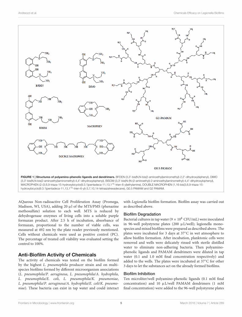
Chemical CompoundsFive polyamino-phenolic ligands (Macrophen, DoubleMacrophen, Bisom, Dimo, Bfden), synthesized by the Laboratoryof Supramolecular Chemistry (Department of Pure and AppliedSciences, University of Urbino) (Ambrosi et al., 2008), andtwo PAMAM dendrimers half-generation (G0.5) and second-generation (G2), a gift from Prof. Donald A. Tomalia, were usedto inhibit biofilm formation and promote biofilm degradation(Figure 1).
Cytotoxicity TestINT 407 (ATCC CCL-6) and HeLa (ATCC CCL-2) cell lineswere used for cytotoxicity assays. 100 µL cell suspensions wereseeded at numbers of 1 × 10⁵ cell/mL on 96-well plates thatwere incubated for 24 h at 37◦C in 5% CO2. Then the chemicalswere added to each well to obtain different final concentrations(0.01, 0.1, 0.5, 1.0, 5.0 mM). Chemical effect was evaluatedafter 24 and 48 h of incubation. Cell viability was assessedby 3-(4,5-dimethylthiazol-2-yl)-5-(3-carboxymethoxyphenyl)-2-(4-sulfophenyl)-2H-tetrazolium (MTS) assay using CellTiter 96
Frontiers in Microbiology | www.frontiersin.org 4 March 2016 | Volume 7 | Article 289
fmicb-07-00289 March 7, 2016 Time: 19:34 # 5
Andreozzi et al. Chemicals Efficacy on Legionella Biofilms
FIGURE 1 | Structures of polyamino-phenolic ligands and dendrimers. BFDEN (3,3′-bis(N,N-bis(2-aminoethyl)aminomethyl)-2,2′-dihydroxybiphenyl), DIMO(3,3′-bis(N,N-bis(2-aminoethyl)aminomethyl)-4,4′-dihydroxybiphenyl), BISOM (3,3′-bis(N-(N-(2-aminoethyl)-2-aminoethyl)aminomethyl)-4,4′-dihydroxybiphenyl),MACROPHEN (2-(3,6,9-triaza-15-hydroxybicyclo(9.3.1)pentadeca-11,13,115-trien-6-yl)ethylamine), DOUBLE MACROPHEN (1,16-bis(3,6,9-triaza-15-hydroxybicyclo(9.3.1)pentadeca-11,13,115-trien-6-yl)-3,7,10,14-tetraazahexadecane), G0.5 PAMAM and G2 PAMAM.
AQueous Non-radioactive Cell Proliferation Assay (Promega,Madison, WI, USA), adding 20 µl of the MTS/PMS (phenazinemethosulfate) solution to each well. MTS is reduced bydehydrogenase enzymes of living cells into a soluble purpleformazan product. After 2.5 h of incubation, absorbance offormazan, proportional to the number of viable cells, wasmeasured at 492 nm by the plate reader previously mentioned.Cells without chemicals were used as positive control (PC).The percentage of treated cell viability was evaluated setting thecontrol to 100%.
Anti-Biofilm Activity of ChemicalsThe activity of chemicals was tested on the biofilm formedby the highest L. pneumophila producer strain and on multi-species biofilms formed by different microorganism associations(L. pneumophila/P. aeruginosa, L. pneumophila/A. hydrophila,L. pneumophila/E. coli, L. pneumophila/K. pneumoniae,L. pneumophila/P. aeruginosa/A. hydrophila/E. coli/K. pneumo-niae). These bacteria can exist in tap water and could interact
with Legionella biofilm formation. Biofilm assay was carried outas described above.
Biofilm DegradationBacterial cultures in tap water (9× 10⁸ CFU/mL) were inoculatedin 96-well polystyrene plates (200 µL/well); legionella mono-species and mixed biofilms were prepared as described above. Theplates were incubated for 3 days at 37◦C in wet atmosphere toallow biofilm formation. After incubation, planktonic cells wereremoved and wells were delicately rinsed with sterile distilledwater to eliminate non-adhering bacteria. Then polyamino-phenolic ligands and PAMAM dendrimers were diluted in tapwater (0.1 and 1.0 mM final concentration respectively) andadded to the wells. The plates were incubated at 37◦C for other3 days to let the substances act on the already formed biofilms.
Biofilm InhibitionTen microliter/well polyamino-phenolic ligands (0.1 mM finalconcentration) and 10 µL/well PAMAM dendrimers (1 mMfinal concentration) were added to the 96-well polystyrene plates
Frontiers in Microbiology | www.frontiersin.org 5 March 2016 | Volume 7 | Article 289

fmicb-07-00289 March 7, 2016 Time: 19:34 # 6
Andreozzi et al. Chemicals Efficacy on Legionella Biofilms
inoculated with bacterial cultures in tap water (9× 10⁸ CFU/mL)(190 µL/well) at the inoculum time to test biofilm inhibitionactivity of the chemicals over 3-day contact time.
Afterward, biofilm degradation and inhibition were evaluatedby measuring the absorbance with the plate reader, as describedabove. Wells containing bacteria without chemical compoundswere used as PC. Chemical activity was expressed as thepercentage of biofilm reduction relative to the untreated controlbiofilm.
Planktonic Microbial Cell Viability afterChemical TreatmentIn order to determine if anti-biofilm activity of the chemicalswas due to their microbicidal action or to direct interactionwith the biofilm matrix, antimicrobial activity was determined onplanktonic bacteria.
Microbial viability after compound treatments was testedon planktonic bacteria by flow cytometric analyses with aFACScalibur (Becton Dickinson, Milan, Italy) equipped withan Argon Ion Laser set at 15 mV and tuned to an excitationwavelength of 488 nm. Chemical microbicidal activity on free-floating microorganisms was evaluated: bacterial suspensionsin PBS (9 × 10⁸ CFU/mL) were treated with chemicals atthe concentration used for biofilm experiments and incubatedat 37◦C for 24 h with agitation at 150 rpm, to avoidcell adhesion to flask bottom. Bacterial suspensions in PBSwithout chemicals were used as negative controls, while bacteriatreated with 50 mg/L chlorine were used as PCs. To detectcell viability, bacterial cells were stained for 15 min withSYBR Green I (1:10000 final concentration) and PropidiumIodide (PI, 1 µg/mL final concentration) (both from LifeTechnologies, Waltham, MA, USA), fluorochromes with ahigh affinity for nucleic acids. Then cells were fixed inparaformaldehyde (4% final concentration) for 1 h, washedby centrifugation (3500 rpm for 15 min) and suspendedin PBS. Cell count was performed by adding CytocountTM
counting beads (DakoCytomation, Denmark) to the stainedsamples before processing by flow cytometry (Manti et al.,2011). Multi-parametric analyses were performed on bothscattering signals (FSC, SSC), and FL1 to detect SYBR GreenI fluorescence and FL3 to detect PI fluorescence. Threepopulations were evidenced in dot plots: viable cells, damagedcells and dead cells. All parameters were collected in logarithmicscale. All data were statistically analyzed with Cell Questsoftware.
Scanning Electron Microscopy (SEM)The successful biofilm formation by the major biofilm producerLegionella strain and the anti-biofilm activity of dendrimerswere confirmed by electron microscopy. The strain grown onBCYE agar was harvested in filtered tap water as describedabove. One mililiter suspension adjusted at concentration of9 × 108 CFU/mL with or without dendrimers (1 mM) wasinoculated in 24-well plate with coverslips. After 3 days ofincubation at 37◦C in wet atmosphere, non-adhering bacteriawere removed by gently washing. Adhering bacteria growing
on coverslips, were fixed with 2.5% glutaraldehyde in 0.1 Mphosphate buffer (pH 7.3) for 1 h. The monolayer was washedand postfixed with 1% OsO4 in the same buffer for 1 h.A progressive alcohol dehydration was performed, followed byspecimen critical point drying. After mounting on conventionalSEM stubs by means of silver glue, slides were gold sputtered(Battistelli et al., 2005). Observations were carried out with aPhilips 515 scanning electron microscope.
Statistical AnalysisAll experiments were executed in triplicate for each sampleand repeated three times. Data were presented as means andstandard deviations of the measurements. The reduction ofbiofilm was analyzed by two-way ANOVA model with respectto microorganisms and different compounds. Experiment wisesignificance level was fixed at P < 0.05. All the analyses wereperformed using Stata 12.1 SE (Stata corporation, College Station,TX, USA).
RESULTS
Isolation and Identification ofLegionella sp.Ten putative Legionella strains were isolated fromaccommodation facilities in Pesaro–Urbino area (Italy) andconfirmed as Legionella by biochemical tests. The genus/species-specific PCR assay reconfirmed the isolates belonged toLegionella genus and allowed to identify six L. pneumophilastrains. Legionella Latex Kit revealed that all L. pneumophilastrains were Sg 1.
Microorganism Biofilm Producing AbilityOverall, L. pneumophila biofilm production assays showed thattwo strains were not able to form any biofilm (OD < 0.120),three of them were identified as weak biofilm producersand one strain (L. pneumophila 155) resulted a high biofilmproducer (OD > 0.240). Based on the 16S rRNA sequenceidentity level, L. pneumophila strain 155 was classified asL. pneumophila subspecies pneumophila (GenBank accessionnumber: KM657957) and as serogroup 1, after Legionella-specificserotyping assay.
Biofilm producer strains were able to form biofilms inall different growth substrates (data not shown). OD valuesshowed that biofilm formation ability was not decreased whensupplying water as growth medium instead of BYE broth.Biofilm formation in spring and tap chlorinated water wassimilar and not related to the substrate type: weak biofilmproducer strains showed mean OD values of 0.144, 0.123, 0.186in spring water and 0.120, 0.145, 0.172 in tap water for eachstrain, respectively; L. pneumophila 155 exhibited mean ODvalues of 0.265 in spring water and 0.281 in tap water. Thesedata mean that chlorine concentration in tap water was not alimiting factor for biofilm formation. Bacteria were also ableto form biofilm in filtered water as much as in unfilteredwater (P > 0.05). Hence, filtered tap water was chosen as
Frontiers in Microbiology | www.frontiersin.org 6 March 2016 | Volume 7 | Article 289

fmicb-07-00289 March 7, 2016 Time: 19:34 # 7
Andreozzi et al. Chemicals Efficacy on Legionella Biofilms
substratum for further experiments. Biofilm formation over 6and 9 days did not show any increase in OD values comparedwith biofilms over 3 days, therefore 3-day biofilm was chosen forfurther experiments. L. pneumophila 155 was selected to studybiofilm formation in association with the other environmentalmicroorganisms.
Aeromonas hydrophila and K. pneumoniae were classified asweak biofilm producers over 3 days in tap water (mean OD valuesof 0.123 and 0.131 respectively), whilst they strongly increasedbiofilm formation when in association with L. pneumophila 155.Instead E. coli and P. aeruginosa were not able to make biofilm ontheir own (OD < 0.120), but they enhanced the biofilm formationability in presence of L. pneumophila 155 (Figure 2).
Assessment of Bacterial Cell Viability inMulti-Species BiofilmsReverse-transcription quantitative real-time PCR resultsdemonstrated the presence of viable L. pneumophila 155cells in both two-species and five-species biofilms. Inparticular, the highest viable cell number was found inL. pneumophila 155/K. pneumoniae biofilm (8.92 × 10⁷
cell/mL), followed by L. pneumophila 155/A. hydrophilabiofilm (8.71 × 10⁷ cell/mL) (Table 1). CV intra- and inter-assay values (not higher than 8.5 and 8.7% respectively)proved the repeatability and the reproducibility of the assay.The LOD was 3.89 × 101 copies of 5S rDNA/reaction,corresponding to 130 L. pneumophila 155 cells/mL. TheRTqPCR assay displayed an overall efficiency of 91% and aPearson’s R coefficient of 0.99. Melt curve analysis showeda clear and reproducible melting peak between 81.8 and82.3◦C.
Plate Count assay showed the presence of viable non-legionella cells in all two-species biofilms (Table 1). However,it was not possible to quantify non-legionella viable strains infive-species biofilms because of the incomplete selectivity of themedia, which allowed a widespread growth of P. aeruginosa.
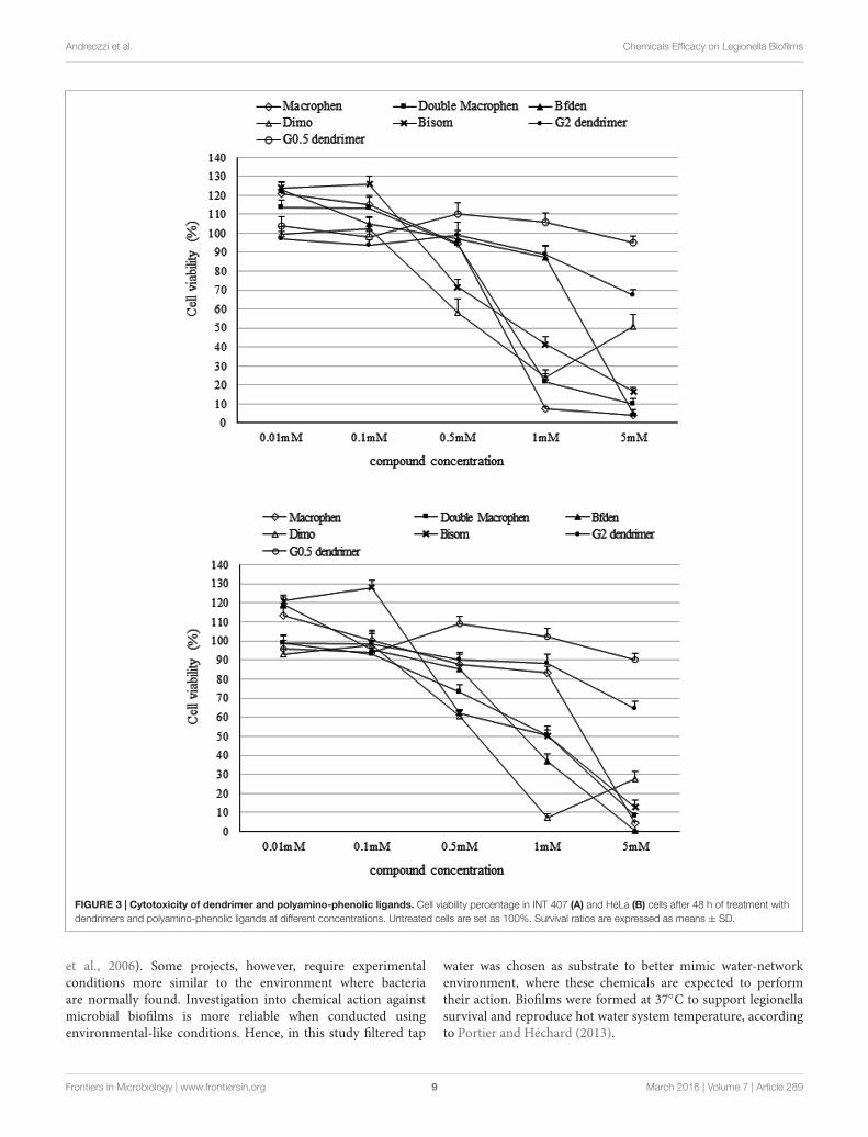
Chemical CytotoxicityCytotoxicity assay was executed to determine the highest non-cytotoxic concentration that could be used against the microbialbiofilm. Compound concentrations maintaining cell viabilityhigher than 85% were chosen to test their anti-biofilm activity.A higher threshold than the standard value usually used toestablish cytotoxic concentrations (IC50: half maximal inhibitoryconcentration), was used in this study.
Figure 3 shows the toxicity of polyamino-phenolic ligandsand dendrimers in INT 407 (Figure 3A) and HeLa (Figure 3B)cells respectively. Forty eight hour cell treatment exhibiteda slightly higher cytotoxicity behavior compared with 24 htreatment, hence only 48 h data are illustrated below. Everycompound showed nearly the same trend in both cell lines.When treated with polyamino-phenolic ligands, INT 407 andHeLa cells exhibited viability higher than 85% in the presenceof all compounds at concentrations up to 0.1 mM. On theother hand, INT 407 cell viability was higher than 85% onlyafter treatment with Macrophen, Double Macrophen or Bfdenat 0.5 mM (94, 95, and 97% respectively). Treatment with0.5 mM Bisom or Dimo led to a INT 407 cell viability of 72and 58% respectively. Regarding HeLa cells, only Macrophenshowed low cytotoxicity at the 0.5 mM concentration witha cell viability value of 88%, whereas cell viability wasbelow the 85% threshold with the other compounds. G0.5and G2 dendrimer treated cells maintained viability values
FIGURE 2 | Biofilm formation ability of bacterial strains. OD values of biofilms formed by single bacterial strains and by L. pneumophila 155 in association withenvironmental strains. ATCC 35984, ATCC 35983, and ATCC 12228 S. epidermidis strains were used as controls for strong, weak and non-biofilm producers.
Frontiers in Microbiology | www.frontiersin.org 7 March 2016 | Volume 7 | Article 289
fmicb-07-00289 March 7, 2016 Time: 19:34 # 8
Andreozzi et al. Chemicals Efficacy on Legionella Biofilms
TABLE 1 | Count of viable Legionella pneumophila 155 cells and viable non-legionella cells in multi-species biofilms based on quantification of 5SrRNAcopies by RTqPCR and on Plate Count assay, respectively.
Biofilms Trascriptionally active L. pneumophila cells/mL Non-legionella CFU/mL
L. pneumophila/A. hydrophila 8.71 × 10⁷± 6.92 × 10⁶ 7.00 × 10⁸
± 1.00 × 10⁸
L. pneumophila/E. coli 2.02 × 10⁷± 4.21 × 10⁶ 5.67 × 10⁸
± 1.53 × 10⁸
L. pneumophila/K. pneumoniae 8.92 × 10⁷± 6.30 × 10⁶ 1.10 × 10⁹
± 1.59 × 10⁸
L. pneumophila/P. aeruginosa 6.96 × 10⁷± 6.59 × 10⁶ 2.11 × 10⁸
± 6.49 × 10⁷
Five-species 4.98 × 10⁷± 3.20 × 10⁶
Cell number is expressed as mean ± SD.
higher than 85% up to 5 and 1 mM in both cell lines,respectively.
Therefore polyamino-phenolic ligands were used at 0.1 mMand dendrimers were used at 1 mM for further experiments onbiofilms.
In addition, polyamino-phenolic ligand concentrations≤0.1 mM and G0.5 dendrimer concentrations ≤0.5 mMincreased MTS assay OD values of the cells up to 124%. Theincreased OD values at low chemical concentrations may reflectan increase of metabolic activity, due to cell-chemical interaction.
Chemical ActivityThe percentages of biofilm reduction and inhibition by thechemical compounds are reported in Figures 4 and 5.
Biofilm DegradationDendrimers showed the highest biofilm reduction activity withboth legionella mono-species and multi-species systems whencompared to all other compounds (Figure 4). In general, G2dendrimer was the most active in biofilm degradation, withstatistically significant differences with relation to all compounds(P < 0.05), causing a reduction of 78% on L. pneumophilabiofilm, while the highest decrease in multi-species biofilmswas observed in L. pneumophila/P. aeruginosa (69%) andL. pneumophila/E. coli (66%). G0.5 dendrimer caused a similarreduction (63%) only on L. pneumophila/P. aeruginosa biofilm.
Among the polyamino-phenolic compounds, Macrophen andDouble Macrophen were overall the most effective chemicals,but no statistically significant differences were found (P > 0.05).In particular, Double Macrophen showed the highest reductionon L. pneumophila/E. coli biofilm (39%), while Macrophenwas the most effective on L. pneumophila/A. hydrophila andL. pneumophila/K. pneumonia (both 33%). Macrophen, DoubleMacrophen and Dimo were the most efficacious compoundsagainst L. pneumophila biofilm (27, 32, and 27% respectively).
Biofilm InhibitionWhen biofilm formation inhibition was evaluated (Figure 5),dendrimers showed the highest inhibition activity, withstatistically significant differences in relation to all othercompounds (P < 0.05). G0.5 dendrimer was, overall, thecompound that showed the best efficacy, abundantly inhibitinglegionella mono-species (85%) and L. pneumophila/A. hydrophilaand L. pneumophila/E. coli (both 73%) two-species biofilms.G2 dendrimer also caused a rather high biofilm inhibition,although less effective, on the same biofilms as G0.5 (61, 59,
61% for L. pneumophila, L. pneumophila/A. hydrophila andL. pneumophila/E. coli biofilms respectively). Moreover, five-species biofilm was highly inhibited by G0.5 dendrimer, with avalue of 68%, more than twice as much as the other compounds.
Among the polyamino-phenolic ligands, Double Macrophenwas the most effective on L. pneumophila biofilm (48%).With respect to multi-species biofilms, Macrophen caused thehighest inhibition on L. pneumophila/P. aeruginosa (26%),L. pneumophila/K. pneumoniae (24%) and five-species (27%)biofilm formation, whereas Double Macrophen was themain inhibitor of L. pneumophila/A. hydrophila (34%) andL. pneumophila/E. coli (42%) biofilms.
Planktonic Microbial Cell Viability afterChemical TreatmentThe most active chemicals (G2 and G0.5 dendrimers, Macrophenand Double Macrophen) against biofilms were selected todetermine their antimicrobial activity on planktonic bacteriaby flow cytometry. Viability test showed that Macrophen andDouble Macrophen treatments did not reduce the number ofviable cells in all microbial species, while G2 and G0.5 dendrimertreatments reduced the viability only in A. hydrophila (60 and37% respectively) and E. coli (53 and 47% respectively) (Figure 6shows a representative dot plot), with a parallel increase indamaged cell number (Figure 7).
Scanning Electron MicroscopyThe anti-biofilm activity of dendrimers was tested by SEM imageanalysis. After 3 days incubation, SEM revealed the formationof an abundant L. pneumophila 155 biofilm, extensively spreadon the coverslip (1 cm diameter). Figures 8A,B (control sample)shows bacteria adhering to the substrate and embedded inan extracellular matrix, clearly visible on the surface of theaggregated cells. In the treated samples no extracellular matrixis visible: naked bacteria, mainly forming a monolayer, areattached to the surface, but there is no connection between cells(Figure 8C). The presence of dendrimers caused the lack ofextracellular matrix, resulting in the detachment of a relevant cellamount that may justify the high reduction percentages obtainedin the biofilm inhibition assay.
DISCUSSION
Legionella pneumophila biofilm studies are usually carried outin culture media and at different incubation temperatures (Piao
Frontiers in Microbiology | www.frontiersin.org 8 March 2016 | Volume 7 | Article 289
fmicb-07-00289 March 7, 2016 Time: 19:34 # 9
Andreozzi et al. Chemicals Efficacy on Legionella Biofilms
FIGURE 3 | Cytotoxicity of dendrimer and polyamino-phenolic ligands. Cell viability percentage in INT 407 (A) and HeLa (B) cells after 48 h of treatment withdendrimers and polyamino-phenolic ligands at different concentrations. Untreated cells are set as 100%. Survival ratios are expressed as means ± SD.
et al., 2006). Some projects, however, require experimentalconditions more similar to the environment where bacteriaare normally found. Investigation into chemical action againstmicrobial biofilms is more reliable when conducted usingenvironmental-like conditions. Hence, in this study filtered tap
water was chosen as substrate to better mimic water-networkenvironment, where these chemicals are expected to performtheir action. Biofilms were formed at 37◦C to support legionellasurvival and reproduce hot water system temperature, accordingto Portier and Héchard (2013).
Frontiers in Microbiology | www.frontiersin.org 9 March 2016 | Volume 7 | Article 289

fmicb-07-00289 March 7, 2016 Time: 19:34 # 10
Andreozzi et al. Chemicals Efficacy on Legionella Biofilms
FIGURE 4 | Biofilm degradation activity of chemicals. Degradation of legionella mono-species and multi-species 3-day-old biofilms by chemicals. Biofilms weretreated with 0.1 mM polyamino-phenolic ligands and 1 mM dendrimers after 3-day biofilm formation. Untreated biofilms were acted as controls (100%). Degradationpercentages are expressed as means ± SD. Asterisks indicate significant (∗P < 0.05) and non significant (∗∗P > 0.05) differences.
FIGURE 5 | Biofilm inhibition activity of chemicals. Inhibition of legionella mono-species and multi-species biofilm formation by chemicals. Biofilms were treatedwith 0.1 mM polyamino-phenolic ligands and 1 mM dendrimers during 3-day biofilm formation period. Untreated biofilms were acted as controls (100%). Inhibitionpercentages are expressed as means ± SD. Asterisks indicate significant (∗P < 0.05) differences.
Legionella pneumophila 155 was shown to be a biofilmproducer both in BYE medium and in water based on theOD value obtained by Hucker’s crystal violet assay. Moreover,demonstration of biofilm formation in water was confirmed bySEM, showing bacterial adhesion to surface and extracellularmatrix connecting cells.
Pécastaings et al. (2010) demonstrated that L. pneumophilawas able to form mono-species biofilm in low-nutrient mediumnot supporting planktonic growth and allowing bacterialreplication only in the sessile phase, unlike classical richmedia. Here, Legionella biofilm formation seemed not to becompromised in filtered water compared with culture media
Frontiers in Microbiology | www.frontiersin.org 10 March 2016 | Volume 7 | Article 289

fmicb-07-00289 March 7, 2016 Time: 19:34 # 11
Andreozzi et al. Chemicals Efficacy on Legionella Biofilms
FIGURE 6 | Representative dot plot (FL1 vs. FL3). Planktonic E. coli cells labeled with SYBR Green I and PI treated with G2 dendrimer (B) respect to theuntreated control (A).
after 3-day incubation. Matrix presence allows us to refer to abiofilm in the initial formation phase, even though its productionwas probably due to planktonic cell deposition rather then tocell replication. In fact, matrix production could result fromthe high initial bacterial inoculum that might determine theactivation of the Quorum Sensing and, consequently, biofilmformation. However, matrix exopolysaccharides could act assource of nutrients, supporting bacteria survival and possiblegrowth.
In experimental conditions mono-species biofilms show ahigher reproducibility than multi-species biofilms. On the otherhand, multi-species biofilm assays are necessary to investigatethe actual chemical power in a model closer to the environmentwhere the compounds should be used.
The formation of mixed biofilms was shown using Hucker’scrystal violet assay, while the ability of viable bacteria to coexistwere assessed by RTqPCR and CFU count assays. These methodswere used only with the aim to verify the presence of viablebacteria; comparison between cell numbers of different bacterialspecies cannot be proposed because of the different quantificationmethods used (RTqPCR assay and CFU count) and the use ofdifferent selective media in CFU counts, that may vary in theirability to recover viable cells.
Interactions of different bacterial strains in mixed biofilmsmay turn into both synergy and reduction in biofilm production(Carpentier and Chassaing, 2004; Marouani-Gadri et al., 2009).Microbial interactions can determine appreciable biomassvariation in two species biofilms in comparison with mono-species biofilms, and strains considered poor biofilm formersby themselves may be present in multi-species biofilms (Simõeset al., 2007; Røder et al., 2015). Ren et al. (2015) alsoobserved synergistic interactions with greater biofilm biomassproduction in multi-species communities compared with single-species cultures. Based on CV assay OD values, K. pneumoniaeshowed the highest synergistic effect (probably due to the
positive interaction of the molecules involved in legionella andklebsiella Quorum Sensing mechanisms), proving to be thebest species for L. pneumophila 155 to form biofilm with,followed by A. hydrophila. According to this result, Stewartet al. (2012) recently demonstrated that L. pneumophila isable to adhere and persist in K. pneumoniae dynamic biofilm.Considering bacteria that are not able to form biofilm, theycould be able to adhere to an already formed biofilm or toother bacteria that have colonized a surface. In this study,E. coli and P. aeruginosa seem to be unable to form biofilmon their own, but biofilm formation happens in presence ofL. pneumophila. They probably adhere to L. pneumophila cells,attached to the well surface, taking part in the biofilm biomass.L. pneumophila/E. coli biofilm OD is lower than L. pneumophilamono-species biofilm OD: in this case the presence of E. colicould slightly decrease L. pneumophila ability to produce biofilm.On the other hand L. pneumophila/P. aeruginosa biofilm OD ishigher than L. pneumophila monospecies biofilm OD: it can beassumed that P. aeruginosa could enhance legionella ability toform biofilm.
In this work non-functionalized PAMAM dendrimers [strongantibacterial agents, less expensive and less tricky to besynthesized than the functionalized ones (Calabretta et al.,2007)] were used for the first time against Legionella biofilms.The efficacy of the chemicals not only to prevent bacteriafrom producing biofilms but also to reduce previously formedbiofilms appeared to be dependent on the microorganism speciesand on the chemical properties. Overall, dendrimers were themolecules that generally reached the highest percentage ofbiofilm inhibition and reduction. It has to be pointed out thatdendrimers were ten times more concentrated than polyamino-phenolic ligands, due to the cytotoxicity results, and this couldbe the reason for the dendrimer best performance. Althoughpolyamino-phenolic ligands could exhibit an increased efficacyat higher concentrations, chemical cytotoxicity has to be taken
Frontiers in Microbiology | www.frontiersin.org 11 March 2016 | Volume 7 | Article 289

fmicb-07-00289 March 7, 2016 Time: 19:34 # 12
Andreozzi et al. Chemicals Efficacy on Legionella Biofilms
FIGURE 7 | Microbial viability. Planktonic cell viability percentage obtained by flow cytometry analysis after 24 h chemical treatment with dendrimers (1 mM) andpolyamino-phenolic ligands (0.1 mM). Untreated bacteria were used as negative controls (NC) (100%), while bacteria treated with chlorine (50 mg/L) were used aspositive controls (PC).
into account because it is a relevant feature when dealing withsubstances that could be used in water systems.
PAMAM dendrimers have been previously shown to be highlytoxic to some bacteria: Calabretta et al. (2007) showed thatG5 amino-terminated PAMAM dendrimer killed 50% (EC50)of P. aeruginosa at concentrations of 1.5 µg/mL; Lopez et al.(2009) also reported that the Minimum Inhibitory Concentration(MIC) values against P. aeruginosa were 6.3 µg/mL for G3and 12.5 µg/mL for G5 PAMAM dendrimer, while the MICvalues against Staphylococcus aureus were 12.5 µg/mL for
both dendrimers. In our study, flow cytometric data revealedthat both G2 and G0.5 dendrimers were able to decreaseplanktonic A. hydrophila and E. coli microbial viability, probablyinteracting with and damaging the bacterial outer membrane,while they were not microbicidal on planktonic L. pneumophila,P. aeruginosa, and K. pneumoniae, at the used concentration.Results regarding E. coli and K. pneumoniae agree with thoseby other authors (Xue et al., 2013), reporting that E. coliMinimum Bactericidal Concentration (MBC) ranges from 12.5and 25 µg/mL for G2 dendrimer. On the contrary, results
Frontiers in Microbiology | www.frontiersin.org 12 March 2016 | Volume 7 | Article 289

fmicb-07-00289 March 7, 2016 Time: 19:34 # 13
Andreozzi et al. Chemicals Efficacy on Legionella Biofilms
FIGURE 8 | Legionella pneumophila 155 biofilm scanning electronmicrographs. Images showing biofilms formed by untreated L. pneumophila155 cells (A,B) and L. pneumophila 155 + G2 dendrimer (1 mM) treatment(C). An abundant formation of diffuse masses of biofilm with extracellularmatrix is visible in (A,B), while naked bacteria on the surface withoutextracellular matrix are shown in (C). (B) is a higher magnification of (A).(A–C) Bar = 5 µm.
regarding P. aeruginosa contrast with literature data: Calabrettaet al. (2007) referred that the EC50 for G5 PAMAM was about170-fold lower than the concentration used in our work, whereasXue et al. (2013) showed that the MIC for G2 PAMAM was about35-fold lower. The different effect of the dendrimers on the same
bacterial species may be due to the different behavior of the singlestrains, but also to the different bacterial concentrations used invarious studies. To the best of our knowledge, data concerningL. pneumophila and A. hydrophila has not been reported yet.
Non-functionalized PAMAM dendrimers act through non-specific mechanisms, unlike functionalized dendrimers, actingthrough specific molecule interactions (Calabretta et al., 2007;Wang et al., 2010; Xue et al., 2013). Cationic amine groupson full-generation dendrimer (such as PAMAM G2) surfacemay favor the adsorption onto negatively charged bacterial wallby electrostatic attraction, and change membrane permeabilityeventually resulting in cell death (Calabretta et al., 2007; Tülüand Ertürk, 2012; Xue et al., 2013); particularly, in gram-negative bacteria the polycation dendrimer may bind thepolyanion lipopolysaccharide (replacing the stabilizing divalentions, calcium and magnesium), may diffuse through the cellwall and destroy the inner cytoplasmic membrane. Also half-generation carboxyl terminated dendrimers (such as PAMAMG0.5) may alter the structure and permeability of the bacterialouter membrane acting as a polyanion chelating calcium andmagnesium ions (which bind and stabilize the phosphate groupin the lipid layer) and leading to cell wall disruption (Wang et al.,2010).
In this study, despite the lack of microbicidal activity inthe majority of plancktonic bacteria, dendrimers were overallhighly active against biofilms, suggesting that the anti-biofilmefficacy was not mainly due to the chemical microbicidal activity.Bacterial adhesion, the initial phase of biofilm formation, couldbe limited by dendrimer interaction with cell surface or with ions(that promote microbial adhesion), changing wall properties andavoiding microbial attachment to the substrate. However, the lackof microbicidal activity might suggest that biofilm inhibition bydendrimers was more likely to be due to chemical interactionwith bonds and differently charged molecules in the matrix thanwith bacterial cells. This hypothesis was also confirmed by SEMimages showing that L. pneumophila 155 biofilm treated withdendrimers was lacking in extracellular matrix; dendrimers couldprevent the molecules produced by legionellae from binding toform the extracellular polymeric substances (EPS) of the matrix,weakening the adhesion of the cells to the surface and cell to cellinteraction. Nevertheless, these hypotheses have to be confirmedby further analysis on dendrimer mechanisms of action.
Polyamino-phenolic ligands showed a lower anti-biofilmactivity compared to dendrimer action at the concentrationsused, even if they also caused biofilm inhibition and degradation,especially Macrophen and Double Macrophen on mono-speciesbiofilms. Polyamino-phenolic ligands can act as chelatingagents, forming complexes with cations, such as calciumand magnesium. Deficiency in these divalent ions mightdestabilize the biofilm matrix, usually constituted by proteins,extracellular DNA and especially polysaccharides (Brandaet al., 2005), which normally bind a large amount ofcations, whose lack may damage the biofilm structure andcause its detachment (Turakhia et al., 1983). Furthermore,the interaction with the extracellular matrix is more likelythan the interaction with bacterial wall, because of the lackof Macrophen and Double Macrophen microbicidal activity.
Frontiers in Microbiology | www.frontiersin.org 13 March 2016 | Volume 7 | Article 289

fmicb-07-00289 March 7, 2016 Time: 19:34 # 14
Andreozzi et al. Chemicals Efficacy on Legionella Biofilms
Our work shows for the first time the reduction efficiencyof polyamino-phenolic ligands and G2 and G0.5 PAMAMdendrimers on L. pneumophila biofilms in the initial stage of theirformation in filtered tap water. In particular, dendrimers werethe most effective compounds, thanks to their lower cytotoxicitythat allowed the use of a higher concentration. The anti-biofilmactivity was not achieved by chemical antimicrobial properties,but probably by chemical interaction with the electrostatic bondspresent in the biofilm that allowed the partial detachment.
These data suggest that the studied compounds, especiallydendrimers, could be seen as potentially interesting moleculesthat might be studied for a possible rule in support ofnew efficacious anti-biofilm disinfection treatments, releasingbacteria, as Legionella sp., from protective biofilms, makingmicroorganisms susceptible to disinfectant concentrationsusually used in water systems. Nevertheless, further experimentsare needed to study biofilm inhibition efficacy in presence ofprotozoa and at different chemical concentrations and incubationtimes to identify the time required to obtain the maximumbiofilm reduction not only in static but also in dynamic flowconditions. Furthermore, the study of the interaction of thesecompounds with the biofilm is crucial to better understandthe mechanism of action that allows them to inhibit bacterialattachment or promote matrix detachment.
AUTHOR CONTRIBUTIONS
EA and AP conceived and designed the experiments. EA, FB,and LS performed the experiments. EA analyzed the data. MOmade the dendrimers available for usage, was involved in thewriting of the chemical description and dispensed helpful advicefor dendrimer usage. LG sinthetized polyamino-phenolic ligands,
was involved in the writing of the chemical description anddispenced helpful advice for polyamino-phenolic ligand usage.AM supplied cytometry analysis tools and methods, was involvedin sample cytometric analysis and in the writing of the cytometricsection. MB supplied SEM analysis tools and methods and wasinvolved in SEM image capture. EA wrote the paper. AP and FBwere involved in the writing and review of the article. AP andFB managed founds. All authors have approved the final versionof the article. All authors agree to be accountable for all aspectsof the work in ensuring that questions related to the accuracy orintegrity of any part of the work are appropriately investigatedand resolved.
FUNDING
This study was partly supported by “Fondazione Cassa DiRisparmio di Pesaro.”
ACKNOWLEDGMENTS
We thank Francesco Marinelli, Doctor in Statistical Science(University of Bologna, Italy), for his collaboration in thestatistical analysis. We would like to acknowledge Prof. DonaldA. Tomalia (NanoSynthons LLC, The National Dendrimer andNanotechnology Center, Mt. Pleasant), for kindly providing thePAMAM dendrimers. We are grateful to Marina Pecellin, whowas involved in the 16S rRNA gene sequencing, and Prof.Dr. Manfred Höfle, (Helmholtz Centre for Infection Research,Braunschweig, Germany), for kindly authorizing the publicationof the sequencing data. We wish to thank Rui P. A. Pereira for thereviewing of the manuscript and helpful comments.
REFERENCESAmbrosi, G., Formica, M., Fusi, V., Giorgi, L., and Micheloni, M. (2008).
Polynuclear metal complexes of ligands containing phenolic units. Coordin.Chem. Rev. 252, 1121–1152. doi: 10.1016/j.ccr.2007.09.027
Andreozzi, E., Di Cesare, A., Sabatini, L., Chessa, E., Sisti, D., Rocchi, M.,et al. (2014). Role of biofilm in protection of the replicative form ofLegionella pneumophila. Curr. Microbiol. 69, 769–774. doi: 10.1007/s00284-014-0648-y
Baldassarri, L., Cecchini, R., Bertuccini, L., Ammendolia, M. G., Iosi, F., Arciola,C. R., et al. (2001). Enterococcus spp. produces slime and survives in ratperitoneal macrophages. Med. Microbiol. Immunol. 190, 113–120.
Banin, E., Brady, K. M., and Greenberg, E. P. (2006). Chelator-induced dispersaland killing of Pseudomonas aeruginosa cells in a biofilm. Appl. Environ.Microbiol. 72, 2064–2069. doi: 10.1128/AEM.72.3.2064-2069.2006
Bartram, J., Chartier, Y., Lee, J. V., Pond, K., and Surman-Lee, S. (2007). Legionellaand the Prevention of Legionellosis. Geneva: World Health Organization.
Battistelli, M., De Sanctis, R., De Bellis, R., Cucchiarini, L., Dachà, M., and Gobbi, P.(2005). Rhodiola rosea as antioxidant in red blood cells: ultrastructural andhemolytic behavoiur. Eur. J. Histochem. 49, 243–254.
Boi, P., Manti, A., Pianetti, A., Sabatini, L., Sisti, D., Rocchi, M. B., et al.(2015). Evaluation of Escherichia coli viability by flow cytometry: a methodfor determining bacterial responses to antibiotic exposure. Cytometry B Clin.Cytom. 88, 149–153. doi: 10.1002/cyto.b.21214
Branda, S. S., Vik, A., Friedman, L., and Kolter, R. (2005). Biofilms: the matrixrevisited. Trends Microbiol. 13, 20–26. doi: 10.1016/j.tim.2004.11.006
Bridier, A., Briandet, R., Thomas, V., and Dubois-Brissonnet, F. (2011). Resistanceof bacterial biofilms to disinfectants: a review. Biofouling 27, 1017–1032. doi:10.1080/08927014.2011.626899
Calabretta, M. K., Kumar, A., McDermott, A. M., and Cai, C. (2007).Antibacterial activities of poly(amidoamine) dendrimers terminated withamino and poly(ethylene glycol) groups. Biomacromolecules 8, 1807–1811. doi:10.1021/bm0701088
Carpentier, B., and Chassaing, D. (2004). Interactions in biofilms betweenListeria monocytogenes and resident microorganisms from food industrypremises. Int. J. Food Microbiol. 97, 111–122. doi: 10.1016/j.ijfoodmicro.2004.03.031
Chang, Y., Gu, W., and McLandsborough, L. (2012). Low concentration ofethylenediaminetetraacetic acid (EDTA) affects biofilm formation of Listeriamonocytogenes by inhibiting its initial adherence. Food Microbiol. 29, 10–17.doi: 10.1016/j.fm.2011.07.009
Chen, Z. C., and Cooper, S. L. (2000). Recent advances in antimicrobialdendrimers. Adv. Mater. 12, 843–846. doi: 10.1002/(SICI)1521-4095(200006)12:11<843::AID-ADMA843>3.3.CO;2-K
Cole, J. R., Wang, Q., Fish, J. A., Chai, B., McGarrell, D. M., Sun, Y., et al. (2013).Ribosomal database project: data and tools for high throughput rRNA analysis.Nuclic Acids Res. 42, D633–D642. doi: 10.1093/nar/gkt1244
Cooper, I. R., and Hanlon, G. W. (2010). Resistance of Legionella pneumophilaserotype 1 biofilms to chlorine-based disinfection. J. Hosp. Infect. 74, 152–159.doi: 10.1016/j.jhin.2009.07.005
de Carvalho, C. C. C. R. (2007). Biofilms: recent developments on an old battle.Recent Pat. Biotechnol. 1, 49–57. doi: 10.2174/187220807779813965
Frontiers in Microbiology | www.frontiersin.org 14 March 2016 | Volume 7 | Article 289

fmicb-07-00289 March 7, 2016 Time: 19:34 # 15
Andreozzi et al. Chemicals Efficacy on Legionella Biofilms
Declerck, P., Behets, J., Margineanu, A., van Hoef, V., De Keersmaecker, B.,and Ollevier, F. (2009). Replication of Legionella pneumophila inbiofilms of water distribution pipes. Microbiol. Res. 164, 593–603. doi:10.1016/j.micres.2007.06.001
DeLong, E. F. (1992). Archaea in coastal marine environments. Proc. Natl. Acad.Sci. U.S.A. 89, 5685–5689. doi: 10.1073/pnas.89.12.5685
Ditommaso, S., Biasin, C., Giacomuzzi, M., Zotti, C. M., Cavanna, A., andRuggenini Moiraghi, A. (2005). Peracetic acid in the disinfection of a hospitalwater system contaminated with Legionella species. Infect. Control. Hosp.Epidemiol. 26, 490–493. doi: 10.1086/502573
Doleans, A., Aurell, H., Reyrolle, M., Lina, G., Freney, J., Vandenesch, F. J.,et al. (2004). Clinical and environmental distributions of Legionella strains inFrance are different. J. Clin. Microbiol. 42, 458–460. doi: 10.1128/JCM.42.1.458-460.2004
Guidelines for prevention and control of legionellosis (2000). “Linee-guida per laprevenzione e il controllo della legionellosi,” in Proceedings of the PermanentConference for the Relationship Between the Government, the Regions,Trento and Bolzano, 103.
Hou, S., Zhou, C., Liu, Z., Young, A. W., Shi, Z., Ren, D., et al. (2009). Antimicrobialdendrimer active against Escherichia coli biofilms. Bioorg. Med. Chem. Lett. 19,5478–5481. doi: 10.1016/j.bmcl.2009.07.077
Iatta, R., Cuna, T., Napoli, C., De Giglio, O., and Montagna, M. T. (2013).Environmental surveillance and molecular investigation of Legionella spp. inApulia, in the years 2008-2011. Ann. Ig. 25, 435–441.
Jakubek, D., Le Brun, M., Leblon, G., DuBow, M., and Binet, M. (2013). The impactof monochloramine on the diversity and dynamics of Legionella pneumophilasubpopulations in a nuclear power plant cooling circuit. FEMS Microbiol. Ecol.85, 302–312. doi: 10.1111/1574-6941.12121
Johansson, E. M. V., Crusz, S. A., Kolomiets, E., Buts, L., Kadam, R. U.,Cacciarini, M., et al. (2008). Inhibition and dispersion of Pseudomonasaeruginosa biofilms by glycopeptide dendrimers targeting the fucose-specificlectin, LecB. Chem. Biol. 15, 1249–1257. doi: 10.1016/j.chembiol.2008.10.009
Kahlisch, L., Henne, K., Draheim, J., Brettar, I., and Höfle, M. G. (2010).High-resolution in situ genotyping of Legionella pneumophila populationsin drinking water by multiple-locus variable-number tandem-repeat analysisusing environmental DNA. Appl. Environ. Microbiol. 76, 6186–6195. doi:10.1128/AEM.00416-10
Khan, M. A., Knox, N., Prashar, A., Alexander, D., Abdel-Nour, M., Duncan, C.,et al. (2013). Comparative genomics reveal that host-innate immune responsesinfluence the clinical prevalence of Legionella pneumophila serogroups. PLoSONE 8:e67298. doi: 10.1371/journal.pone.0067298
Khweek, A. A., Fernández Dávila, N. S., Caution, K., Akhter, A., Abdulrahman,B. A., Tazi, M., et al. (2013). Biofilm-derived Legionella pneumophila evades theinnate immune response in macrophages. Front. Cell. Infect. Microbiol. 3:18.doi: 10.3389/fcimb.2013.00018
Klajnert, B., Cladera, J., and Bryszewska, M. (2006). Molecular interactions ofdendrimers with amyloid peptides: pH dependence. Biomacromolecules 7,2186–2191. doi: 10.1021/bm060229s
Lane, D. J. (1991). “16S/23S rRNA sequencing,” in Nucleic Acid Techniques inBacterial Systematics, eds E. Stackebrandt and M. Goodfellow (New York, NY:Wiley), 115–175.
Lau, H. Y., and Ashbolt, N. J. (2009). The role of biofilms and protozoa in Legionellapathogenesis: implications for drinking water. J. Appl. Microbiol. 107, 368–378.doi: 10.1111/j.1365-2672.2009.04208.x
Lazniewska, J., Milowska, K., and Gabryelak, T. (2012). Dendrimers - revolutionarydrugs for infectious diseases. WIREs Nanomed Nanobiotechnol. 4, 469–491. doi:10.1002/wnan.1181
Levin, A. S. (2009). Nosocomial legionellosis: prevention and management. Exp.Rev. Anti-Infect. Ther. 7, 57–68. doi: 10.1586/14787210.7.1.57
Lopez, A. I., Reins, R. Y., McDermott, A. M., Trautner, B. W., andCai, C. (2009). Antibacterial activity and cytotoxicity of pegylatedpoly(amidoamine) Dendrimers. Mol. BioSyst. 5, 1148–1156. doi: 10.1039/b904746h
Manti, A., Boi, P., Amalfitano, S., Puddu, A., and Papa, S. (2011). Experimentalimprovements in combining CARD-FISH and flow cytometry forbacterial cell quantification. J. Microbiol. Methods 87, 309–315. doi:10.1016/j.mimet.2011.09.003
Marouani-Gadri, N., Augier, G., and Carpentier, B. (2009). Characterization ofbacterial strains isolated from a beef-processing plant following cleaning anddisinfection - influence of isolated strains on biofilm formation by Sakaïand EDL 933 E. coli O157:H7. Int. J. Food Microbiol. 133, 62–67. doi:10.1016/j.ijfoodmicro.2009.04.028
Messi, P., Bargellini, A., Anacarso, I., Marchesi, I., de Niederhäusern, S.,and Bondi, M. (2013). Protozoa and human macrophages infection byLegionella pneumophila environmental strains belonging to differentserogroups. Arch. Microbiol. 195, 89–96. doi: 10.1007/s00203-012-0851-9
Miyamoto, M., Yamaguchi, Y., and Sasatsu, M. (2000). Disinfectant effects of hotwater, ultraviolet light, silver ions and chlorine on strains of Legionella andnontuberculous Mycobacteria. Microbios 101, 7–13.
Ng, V., Tang, P., Jamieson, F., Guyard, C., Low, D. E., and Fisman, D. N.(2009). Laboratory-based evaluation of legionellosis epidemiology in Ontario,Canada, 1978 to 2006. BMC Infect. Dis. 9:68–77. doi: 10.1186/1471-2334-9-68
Osawa, K., Shigemura, K., Abe, Y., Jikimoto, T., Yoshida, H., Fujisawa, M.,et al. (2014). A case of nosocomial Legionella pneumonia associated witha contaminated hospital cooling tower. J. Infect. Chemother.9:68. doi:10.1016/j.jiac.2013.07.007
Ottaviani, D., Parlani, C., Citterio, B., Masini, L., Leoni, F., Canonico, C.,et al. (2011). Putative virulence properties of Aeromonas strains isolatedfrom food, environmental and clinical sources in Italy: a comparativestudy. Int. J. Food Microbiol. 144, 538–545. doi: 10.1016/j.ijfoodmicro.2010.11.020
Ottaviani, M. F., Furini, F., Casini, A., Turro, N. J., Jockusch, S., Tomalia,D. A., et al. (2000). Formation of supramolecular structures between DNAand starburst dendrimers studied by EPR, CD, UV and melting profiles.Macromolecules 33, 7842–7851. doi: 10.1021/ma000877i
Ottaviani, M. F., Jockusch, S., Turro, N. J., Tomalia, D. A., and Barbon, A.(2004). Interactions of dendrimers with selected amino acids and proteinsstudied by Continuous Wave EPR and Fourier Transform EPR. Langmuir 20,10238–10245. doi: 10.1021/la0485881
Pécastaings, S., Bergé, M., Dubourg, K. M., and Roques, C. (2010). Sessile Legionellapneumophila is able to grow on surfaces and generate structured monospeciesbiofilms. Biofouling 26, 809–819. doi: 10.1080/08927014.2010.520159
Piao, Z., Sze, C. C., Barysheva, O., Iida, K., and Yoshida, S. (2006). Temperature-regulated formation of mycelial mat-like biofilms by Legionella pneumophila.Appl. Environ. Microbiol. 72, 1613–1622. doi: 10.1128/AEM.72.2.1613-1622.2006
Portier, E., and Héchard, Y. (2013). “Natural biofilm formation with Legionellapneumophila,” in Legionella: Methods and Protocols. Methods in MolecularBiology, eds C. Buchrieser and H. Hilbi (New York, NY: Springer Science &Buisness Media), 213–217.
Qasem, J. A., Mustafa, A. S., and Khan, Z. U. (2008). Legionella in clinicalspecimens and hospital water supply facilities: molecule, detection andgenotyping of the isolates. Med. Princ. Pract. 17, 49–55. doi: 10.1159/000109590
Ren, D., Madsen, J. S., Sørensen, S. J., and Burmølle, M. (2015). High prevalenceof biofilm synergy among bacterial soil isolates in cocultures indicates bacterialinterspecific cooperation. ISME J. 9, 81–89. doi: 10.1038/ismej.2014.96
Røder, H. L., Raghupathi, P. K., Herschend, J., Brejnrod, A., Knøchel, S., Sørensen,S. J., et al. (2015). Interspecies interactions result in enhanced biofilm formationby co-cultures of bacteria isolated from a food processing environment. FoodMicrobiol. 51, 18–24. doi: 10.1016/j.fm.2015.04.008
Sabatini, L., Pianetti, A., Cecchetti, G., Bruner, A., Citterio, B., Barbieri, F.,et al. (2013). Chemical and microbiological monitoring of air in two wasteincineration plants. Ig Sanita Pubbl. 69, 13–37.
Sanli-Yurudu, N. O., Kimiran-Erdem, A., and Cotuk, A. (2007). Studies on theefficacy of chloramine T trihydrate (N-chloro-p-toluene sulfonamide) againstplanktonic and sessile populations of different Legionella pneumophila strains.Int. J. Hyg. Environ. Health 210, 147–153. doi: 10.1016/j.ijheh.2006.08.004
Simões, L. C., Simões, M., and Maria João Vieira, M. J. (2007). Biofilm interactionsbetween distinct bacterial genera isolated from drinking water. Appl. Environ.Microbiol. 73, 6192–6200. doi: 10.1128/AEM.00837-07
Steed, J. W., and Atwood, J. L. (2009). Supramolecular Chemistry, 2nd Edn.Hoboken, NJ: John Wiley & Sons, Ltd.
Frontiers in Microbiology | www.frontiersin.org 15 March 2016 | Volume 7 | Article 289

fmicb-07-00289 March 7, 2016 Time: 19:34 # 16
Andreozzi et al. Chemicals Efficacy on Legionella Biofilms
Steinert, M., Hentschel, U., and Hacker, J. (2002). Legionella pneumophila:an aquatic microbe goes astray. FEMS Microbiol. Rev. 26, 149–162. doi:10.1111/j.1574-6976.2002.tb00607.x
Stewart, C. R., Muthye, V., and Cianciotto, N. P. (2012). Legionella pneumophilapersists within biofilms formed by Klebsiella pneumoniae, Flavobacteriumsp., and Pseudomonas fluorescens under dynamic flow conditions. PLoS ONE7:e50560. doi: 10.1371/journal.pone.0050560
Taj, Y., Essa, F., Aziz, F., and Kazmi, S. U. (2012). Study on biofilm-formingproperties of clinical isolates of Staphylococcus aureus. J. Infect. Dev. Ctries 6,403–409. doi: 10.3855/jidc.1743
Tiwari, W., Roy, R., and Tiwari, M. (2015). Antimicrobial active herbal compoundsagainst Acinetobacter baumannii and other pathogens. Front. Microbiol. 6:18.doi: 10.3389/fmicb.2015.00618
Tomalia, D. A., Christensen, J. B., and Boas, U. (2012). Dendrimers, Dendrons,and Dendritic Polymers: Discovery, Applications, and the Future. Cambridge:Cambridge University Press.
Tülü, M., and Ertürk, A. S. (2012). “Dendrimers as antibacterial agents,” in A Searchfor Antibacterial Agents, ed. V. Bobbarala (Rijeka: InTech), 89–106.
Turakhia, M. H., Cooksey, K. E., and Characklis, W. G. (1983). Influence ofa calcium-specific chelant on biofilm removal. Appl. Environ. Microbiol. 46,1236–1238.
Wang, B., Navath, R. S., Menjoge, A. R., Balakrishnan, B., Bellair, R., Dai, H., et al.(2010). Inhibition of bacterial growth and intramniotic infection in a guineapig model of chorioamnionitis using PAMAM dendrimers. Int. J. Pharm. 395,298–308. doi: 10.1016/j.ijpharm.2010.05.030
Watnick, P., and Kolter, R. (2000). Biofilm, city of microbes. J. Bacteriol. 182,2675–2679. doi: 10.1128/JB.182.10.2675-2679.2000
Xue, X., Chen, X., Mao, X., Hou, Z., Zhou, Y., Bai, H., et al. (2013). Amino-terminated generation 2 poly(amidoamine) dendrimer as a potential broad-spectrum, nonresistance-inducing antibacterial agent. AAPS J. 15, 132–142. doi:10.1208/s12248-012-9416-8
Zarena, A. S., and Gopal, S. (2013). Dendrimer a new dimensionin targeting biofilms. Mini-Rev. Med. Chem. 13, 1448–1461. doi:10.2174/13895575113139990064
Zhang, Y. Q., Ren, S. X., Li, H. L., Wang, Y. X., Fu, G., Yang, J., et al.(2003). Genome-based analysis of virulence genes in a non-biofilm-formingStaphylococcus epidermidis strain (ATCC 12228). Mol. Microbiol. 49, 1577–1593. doi: 10.1046/j.1365-2958.2003.03671.x
Conflict of Interest Statement: The authors declare that the research wasconducted in the absence of any commercial or financial relationships that couldbe construed as a potential conflict of interest.
Copyright © 2016 Andreozzi, Barbieri, Ottaviani, Giorgi, Bruscolini, Manti,Battistelli, Sabatini and Pianetti. This is an open-access article distributed under theterms of the Creative Commons Attribution License (CC BY). The use, distribution orreproduction in other forums is permitted, provided the original author(s) or licensorare credited and that the original publication in this journal is cited, in accordancewith accepted academic practice. No use, distribution or reproduction is permittedwhich does not comply with these terms.
Frontiers in Microbiology | www.frontiersin.org 16 March 2016 | Volume 7 | Article 289