Embed Size (px)
Citation preview

Dementia and Vision
W I L L I A M L H I L L S , M D , O D
A S S O C I AT E P R O F E S S O R
O P H T H A L M O C L O G Y A N D N E U R O L O G Y
O C T O B E R 1 3 , 2 0 1 6

Nothing to disclose

Dementia is a general term for a decline in mental ability severe enough to interfere with daily life.
Dementia is not a specific disease. It's an overall term that describes a wide range of symptoms
Alzheimer’s disease is the most common form of dementia, accounting for 60-80%
Vascular dementia, after a stroke, is the second most common form of dementia
Dementia

While symptoms of dementia can vary greatly, at least two of the following core mental functions must be significantly impaired to be considered dementia:
Memory
Communication and language
Ability to focus and pay attention
Reasoning and judgment
Visual perception
Dementia

Many people have memory loss issues — this does not mean they have Alzheimer's or another dementia
People with dementia may have problems with:
-- short-term memory
-- keeping track of a purse or wallet
-- paying bills
-- planning and preparing meals
-- remembering appointments
-- traveling out of the neighborhood.
Many dementias are progressive, meaning symptoms start out slowly and gradually get worse.
Dementia

Dementia with Lewy bodies
Frontotemporal dementia
Posterior Cortical Atrophy
Parkinson’s disease dementia
Vascular Dementia
Dementia: Types
Anatomical relationship to functionBased on observation of patients with deficits and post mortem examination
Brodman’s areas

Organization of Visual FunctionsWernicke (1874) and Lissauer (1890)
First model of sequential organization of visual functions
Theory based on anatomical separation of:◦ Primary center of sensation◦ Secondary center of imagination

Holmes and Lister (1916)
Explored possibility that specialized areas could be distributed over two visual pathways
Distinction between visual agnosias

The visual system: SeeingConceptually two componentsCentral ◦ Anatomically = macula/fovea◦ Cones are highest density in fovea (20 degrees) ◦ Mediates color vision, high spatial frequency resolution◦ Functions optimally in conditions of light adaptation
Peripheral◦ Rods are more abundant in periphery ◦ 20 degrees from the fovea Rods are in highest conc◦ Mediates motion detection, low spatial frequency◦ Functions best in dim illumination

The dualistic approach to visionThe ‘where’ system◦ Magnocellular pathway◦ Perceptual processing◦ Memorization of spatial relationships
◦ Dorsal stream
The ‘what’ system◦ Parvocellular pathway◦ Object identification
◦ Ventral stream

The dualistic approach to vision
https://visionhelp.files.wordpress.com/2012/08/ventral-dorsal-stream.png

Dorsal StreamLinks primary visual cortex (V1), through the middle temporal area, to the posterior parietal lobe
http://cercor.oxfordjournals.org/content/early/2011/02/21/cercor.bhq285/F2.large.jpg

Ventral StreamLinks primary visual cortex (V1), through area V4, to the inferotemporal region
http://cercor.oxfordjournals.org/content/early/2011/02/21/cercor.bhq285/F2.large.jpg
http://i305.photobucket.com/albums/nn224/engleon/homer-simpson-wallpaper-brain-1024.jpg

Visual impairments of the Primary visual cortex‘Blindsight’
‘Cortically blind’◦ Loss of conscious vision in part of their visual
field due to primary visual cortex lesion◦ Able to produce accurate visuomotor responses
to visual stimuli under forced choice situations/automatic responses

Visual impairments of the Primary visual cortex:Deafferentation of the ventral pathway from retinal inputsDorsal pathway should still receive retinal information via subcortical routesRetinal projections to the optic tectum and pulvinar reach the parietal cortexConfirms the role of the parietal cortex in the automatic visuomotor transformation

Afferent visual impairmentsANTON SYNDROME
Cortical blindness
Patients denied any visual problems
Claim that they can see
No demonstrable visual behavior
Most common with bilateral occipital lobe infarctions
http://www.japi.org/november_2012/images/13_cr_reversible_posterior_leukoencephalopathy_syndrome_01.jpg
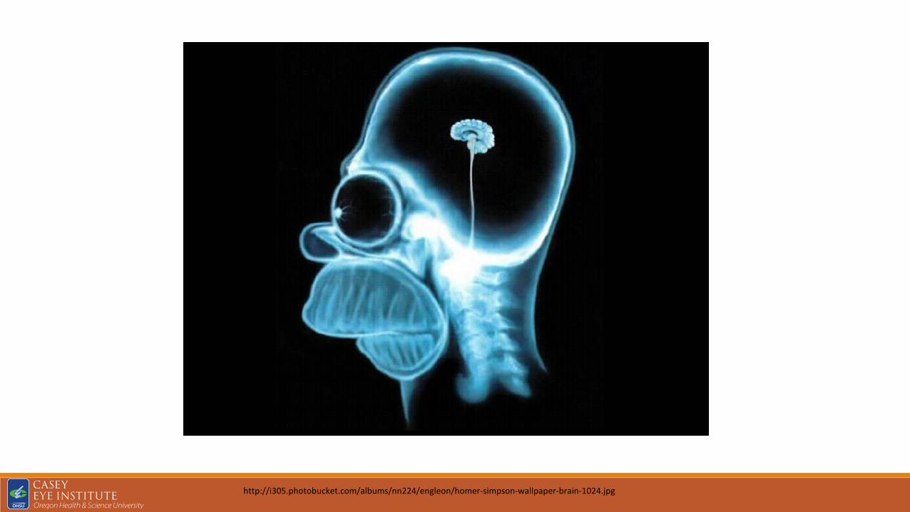
Afferent visual impairmentsHEMISPATIAL NEGLECT
Do not see objects in areas of known visual function
Double simultaneous stimulation
Confrontation visual fields
Damage to the right hemisphere◦ Posterior parietal cortex◦ Frontal eye fields◦ Cingulate gyrus
http://plato.stanford.edu/entries/mental-imagery/unineglect.gif
Dissociation of the visual processing mechanismsVisual agnosias◦ Apperceptive◦ Associative◦ Alexia◦ Prosopagnosia◦ Simultagnosia◦ Unilateral spatial neglect◦ Optic ataxia◦ Spatial disorientation

Visual Agnosia‘mind blindness’ Lissauer 1890
‘Agnosia’ Freud 1891
Unable to perceive and/or recognize visual objects
not blind since they clearly respond to visual stimuli

Case:47 yo man found comatose and low BP
RHH c macular sparing
Normal memory, language and intelligence
Could not name objects nor group them
Could not demonstrate or describe the use of
PET – specific storage of functional knowledge of objects
Middle portion of temporal lobe
Category specific deficits

Associative agnosia‘normal precept stripped of meaning’
‘psychic blindness’’
Normal perception of the object, although object recognition is impaired

Neural mechanisms for object recognitionSequential processing of informationV1- prestriate cortex – inferotemporal cortex – superior temporal sulcusProcessed serially from simpler to complexEg V1 & V2 extract oriented edges and contoursV4 responds shape or colorInferotemporal cells respond to 2D or 3D
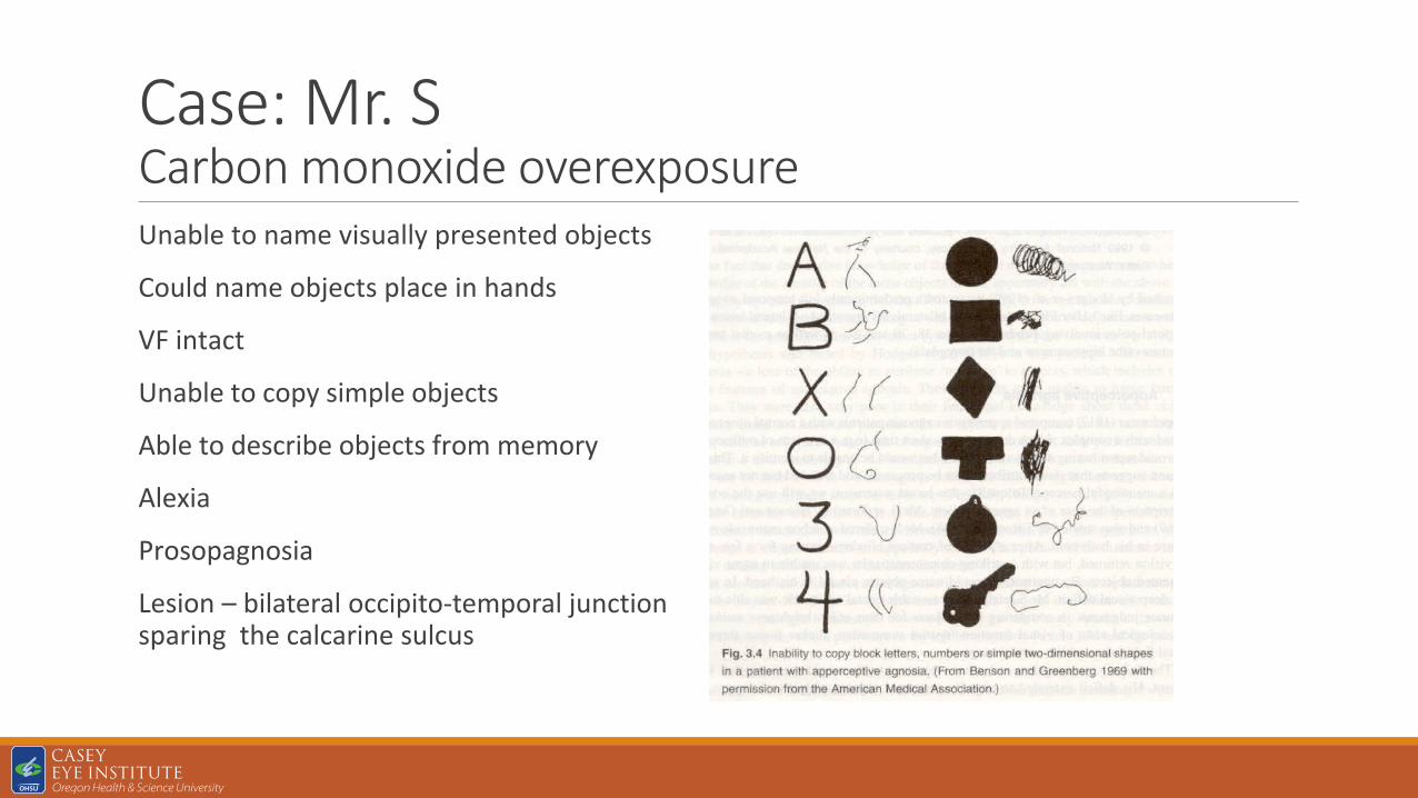
Case: Mr. SCarbon monoxide overexposureUnable to name visually presented objects
Could name objects place in hands
VF intact
Unable to copy simple objects
Able to describe objects from memory
Alexia
Prosopagnosia
Lesion – bilateral occipito-temporal junction sparing the calcarine sulcus

Apperceptive agnosiaVisual form agnosia◦ Object agnosia◦ Alexia◦ prosopagnosia
Benson and Greenberg (1969)‘Identification of shape or form is the only precondition for identifying an object, a letter or a face. Other visual attributes, like color or brightness, are not essential for object identification’

Neural mechanisms for object recognitionOne exception to single cell discharge
15% of elaborate anterior inferotemporal (STS) respond to the sight of a face, parts of a face, hand, and certain hand actions
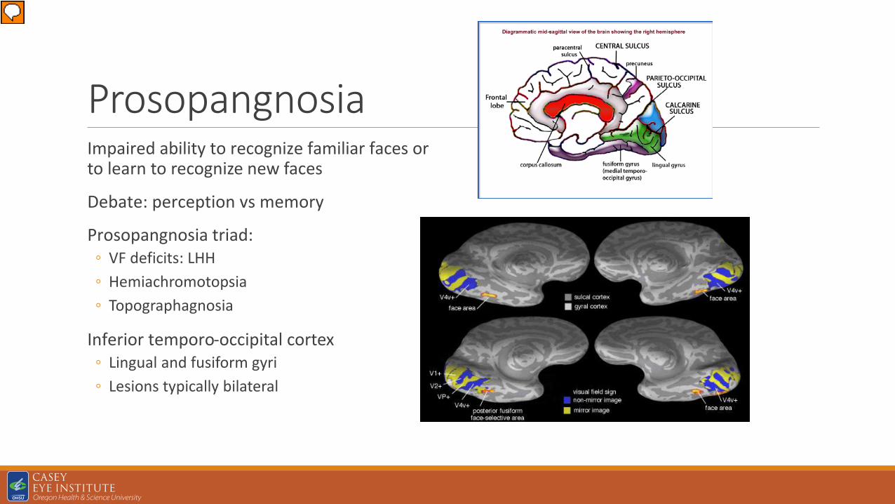
ProsopangnosiaImpaired ability to recognize familiar faces or to learn to recognize new faces
Debate: perception vs memory
Prosopangnosia triad: ◦ VF deficits: LHH◦ Hemiachromotopsia◦ Topographagnosia
Inferior temporo-occipital cortex ◦ Lingual and fusiform gyri◦ Lesions typically bilateral

AlexiaLoss of efficient reading for comprehension
Slow laborious letter by letter reading to complete global alexia
Left medial and inferior temporo-occipital region
Left angular gyrus◦ Stores visual representation of words
Associated findings: ◦ RHH◦ Dyschromotopsia◦ Anomia
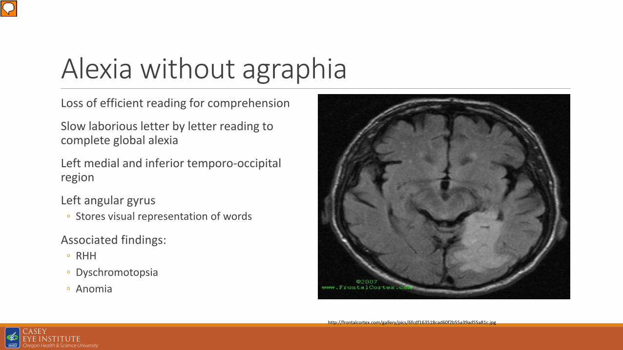
Alexia without agraphiaLoss of efficient reading for comprehension
Slow laborious letter by letter reading to complete global alexia
Left medial and inferior temporo-occipital region
Left angular gyrus◦ Stores visual representation of words
Associated findings: ◦ RHH◦ Dyschromotopsia◦ Anomia
http://frontalcortex.com/gallery/pics/6fcdf163518cad60f2b55a39ad55a81c.jpg

Cerebral achromotopsiaColors look dull to complete colorless◦ Vertebrobasilar ischemia◦ HSV encephilitis◦ Cerebral mets◦ Focal recurrent seizures◦ Dementia
Ventromedial sector of Occipital lobe
Middle third of the Lingual gyrus and fusiform gyri
White matter immediately behind posterior tip of the lateral ventricle
Bilateral hemispheric capacity to perceive color
Hemiachromatopsia

Cerebral akinetopsiaComplete loss of movement perceptionRequires bilateral lesionsLateral temporo-occipital cortex◦ Conjunction of the inferior temporal sulcus and the lateral
occipital sulcus
Subtle symptoms – unilateralObject motion guides ◦ Limb reaching movements◦ Smooth pursuits◦ Influencing saccadic accurracy
Self motion ◦ Distinguish self vs object motion◦ Complements VOR
Smooth pursuits and OKN

Dorsal simultagnosiaPatient accurately perceives the individual elements or details of a complex picture, but cannot appreciate the overall meaning of the pictureBilateral or unilateral parieto-occipital damagePosterior cortical atrophy, CBG degeneration, ADBalint (1909) – as part of a syndromeDisorder of visual attention i.e. inability to disengage attention from current location

Simultagnosia: case

Cookie theft picture

Simultanagnosia in DementiaPatients from the Aging and Alzheimer’s Clinic◦ Probable Alzheimer’s disease◦ mild cognitive impairment◦ frontotemporal dementia◦ Lewy body disease◦ corticobasal ganglionic degeneration
Mini-Mental Status Examination (MMSE)
Clinical Dementia Rating
VA at least 20/60 in the better eye. Simultanagnosia was determined if the patient could not see the Ishihara control (IC) plate or the American Optical Hardy Rand Rittler control (AC) plates with both eyes open
They were also shown two diagrams of a large number or letter constructed from smaller numbers and letters termed the 3/4 diagram and the A/M diagram

Simultanagnosia in DementiaThe mean MMSE score was 23.4
Average age patients was 72.5
Simultanagnosia by AC testing had either probable AD or Lewy body disease◦ Mean MMSE was 20.3
Simultanagnosia by IC ◦ Mean MMSE was 14.5
Study suggests simultanagnosia is not an early finding in dementing illness
AC plates may assist in the diagnosis of some forms of dementia or lend information regarding severity of disease


The Clinical Spectrum of Posterior Cortical Atrophy(IRB00010075)
WILL IAM L HILLS, MD, OD
ASSOCIATE PROFESSOR
SEPTEMBER 15, 2016

Nothing to disclose Jennifer Olds, MD (Lowe)
Victoria Palek, MD
Judith EA Warner, MD
Samuel Passi, MD
Wayne Cornblath, MD (Univ of Michigan)
Roger Turbin, MD (Rutgers Newark Campus)
David Katz, MD (Georgetown Univ and Howard Univ)
Andrew Lee, MD (Houston Methodist Hospital)
Marilyn Kay, MD (Univ of Wisc)
Ben Frishberg, MD (The Neurology Center)

Posterior cortical atrophy (PCA)Progressive neurodegenerative condition
Selective decline in higher order visual processing
Dysfunction of the parietal, occipital, and occipito-temporal regions
First described by Benson in 1988 in patients with marked parieto-occipital atrophy
Histopathologic studies subsequently identified AD as most common pathologyVisual variant of Alzheimer’s diseaseBiparietal AD
Benson F, Davis J, Snyder BD. Posterior Cortical atrophy. Archives of Neurology. 1988;45:789–793.

Posterior cortical atrophyPrevalence and Incidence relatively unknown Estimated to be 5% of Alzheimer’s population (Snowden et al) Increasingly prevalent disease with today's aging population, yet rare conditionDifficult to define due to a lack of clear diagnostic criteria, a general lack of awareness…
Underdiagnosed, misdiagnosed or significantly delayed diagnosis

Posterior cortical atrophy Clinical presentation is heralded by subjective vision loss, yet a normal ophthalmic examination
Earlier age of onset – 50’s – mid 60’s (range 40 – 80)
Gender though to be equal, however, some studies suggest women are affected more
Neuro-psychological deficits
Visual spatial impairments
Alexia
Balint’s syndrome
Gertmann’s syndrome
Working memory deficits

Posterior cortical atrophyBALINT’S SYNDROME
Oculomotor apraxia
Optic ataxia
Simultanagnosia
GERTMANN’S SYNDROME
Acalculia
Agraphia
Finger agnosia
Left/right disorientation

Posterior cortical atrophyBASIC VISUAL PROCESSING
Form
Motion
Color
Point localization
IQ TESTING
Performance IQ is often up to 30–40 points lower than verbal IQ scores
Performance on cognitive tasks with any significant visual component (e.g. visual memory recall, Trail Making, Stroop test) are vulnerable to impairment and isinterpretation

Incomplete Left Homonymous HemianopsiaMarch 2012
Progressive Left Homonymous HemianopsiaJuly 2012

Posterior cortical atrophy Poor visuospatial skills on neuropsychological testing
Occipito-parietal hypometabolism on PET imaging
CSF analysis detected reduced AB(1-42) Tau Index and elevated phosphorylated-tau
0.0
0.4
0.8
1.2
1.6
2.0
2.4
2.8
0 30 60 90 120 150
AT
Inde
x
Phospho-Tau
ATI versus P-tau
Patient DataSeries2

Posterior Cortical Atrophy
Introduction: Posterior cortical atrophy (PCA) is a rare progressive neurodegenerative disease with prominent cortical visual dysfunction, first described by Benson in 1988. Studies have shown visual field defects, on formal testing, increased phosphorylated-tau protein in the CSF, and hypometabolism in the parieto-occipital regions on PET imaging. We describe one patient with PCA whose symptoms are classic but whose progression has been atypically slow. To further investigate this disease entity, we retrospectively reviewed 9 additional charts and report the commonalities in all 10 cases.
33sec27sec
Ms. LR PCA Key Points: Visuoperceptual disturbances and Cortical visual dysfunction Progressive neurodegenerative disease Visual neglect is common Less memory impairment than Alzheimer’s Simultanagnosia Parieto-occipital atrophy Diminished metabolic activity in the posterior aspect of the brain on PET imaging
* History of Ambylopia
Conclusion: Posterior cortical atrophy is an uncommon atypical variant of Alzheimer’s disease, sometimes termed “Benson’s syndrome”. Patients commonly report difficulty reading despite normal visual acuities. Balint’s syndrome is common in PCA, yet difficult to detect at the bedside. Our observations and previous studies describe visual signs and symptoms attributable to the parieto-occipital cortex. More recently, CSF phosphorylated-tau has been utilized as a bio-marker. A decreased AT index and elevated phosphorylated Tau concentration can assist in differentiation from other forms of dementia. The most common symptoms were difficulty with reading and depth perception. Most common findings were simultanagnosia, decreased color vision, decreased stereopsis, abnormal Amsler’s grid and visual field defects. Posterior cortical atrophy should be considered in patients whose chief complaint is difficulty reading despite normal visual acuities. A systematic approach using clinical, laboratory, and radiographic data can aid in diagnosis of PCA. Complementary diagnostic studies can include MRI, PET, and CSF studies. Early diagnosis is important to prevent unnecessary costly investigations, allow for appropriate therapeutic and psychosocial interventions and reduce the stress of an “unknown diagnosis”.
Symptoms/Signs at Presentation Ms. LR Mr. DH Mr. AB Ms. MK Mr. MM Mr. PR Ms. CT Ms. JF Ms. RC Ms. ND Total
Difficulty reading Yes Yes n/a Yes Yes n/a Yes Yes Yes Yes 8/8
Difficulty with depth perception Yes Yes n/a Yes Yes Yes * Yes n/a n/a n/a 6/6
Simultanagnosia Yes No Yes Yes Yes Yes Yes Yes n/a Yes 8/9
Descreased color vision or Achromatopsia
Yes Yes Yes Yes Yes Yes Yes Yes Yes Yes 10/10
n/a 7 OD, 8 OS 3 OD, 2 OS 0/10 OU 2/10 OU 0/10 OU 7/10 OU 0/10 0/10 OU 8/10 OU n/aTitmus, Circles (x/9) 7 5 3 3 0 * 7 n/a n/a n/a 6/6
Abnormal Amsler's grid Yes Yes Yes No Yes Yes n/a n/a n/a Yes 6/7
Abnormal PET Yes Yes n/a n/a n/a n/a n/a n/a n/a n/a 2/2
Abnormal VF Yes Yes Yes Yes Yes n/a Yes n/a Yes Yes 8/10
Figure 1: MRI and PET
Figure 2: Incomplete Left Homonymous HemianopsiaMarch 2012
Figure 3: Progressive Left Homonymous HemianopsiaJuly 2012
0.00.40.81.21.62.02.42.80 30 60 90 120 150
T In
dex
Phospho-Tau
ATI versus P-tau
P
Picture by Bev Doolittle
Methods: We reviewed the index subject’s symptoms and findings on presentation and follow up. We reviewed 8 additional cases of PCA evaluated by Neuro-Ophthalmology. We analyzed the signs and symptoms in, as well as the demographics of, these cases to look for common themes at presentation.
Results: A 58 year old woman presents with a progressive 10 year history of difficulty processing spatial information, reading handwriting and recognizing faces. She described visual hallucinations, photophobia, poor depth perception, and visual recall. Poor visuospatial skills were found on neuropsychological testing and occipito-parietal hypometabolism on PET imaging. (Figure 1). Visual field testing found progressive incomplete homonymous hemianopsia (Figures 2 and 3). CSF analysis detected reduced AB(1-42) Tau Index and elevated phosphorylated-tau. (Figure 4)
Jennifer Lowe, MS41; Julie Falardeau, MD2; Robert Egan, MD3; and William L. Hills, MD2
1Oregon Health and Science University, School of Medicine; 2Oregon Health and Science University, Casey Eye Institute; 3St. Helena Hospital, Neuro-ophthalmology.
Figure 4

PCA
Visual dysfunction in posterior cortical atrophy Individuals with posterior cortical atrophy have difficulty identifying objects and faces, particularly when they consist of many parts or are viewed from an unfamiliar (non-canonical) perspective. Eye-tracking studies contrasting scene perception in healthy individuals (A) and people with posterior cortical atrophy (B) suggest that patients have poor top-down guidance and control of oculomotor function. Circles represent fixation locations and circle size represents fixation duration. Patients with posterior cortical atrophy fixate prominent features initially (eg, dome on pier), but subsequently fixate relatively uninformative aspects of the scene (eg, sea or sky) and miss important contextual details (eg, beachfront or near the end of the pier). Images from Tim Shakespeare and Sebastian Crutch (unpublished).
Crutch, Sebastian J et al. Posterior cortical atrophy. The Lancet Neurology , Volume 11 , Issue 2 , 170 - 178
William L. Hills, MD
Neuro-ophthalmology
Associate Professor of Ophthalmology and Neurology
Casey Eye Institute/OHSU
Diplomate of Neurology, ABPN
3303 SW Bond Ave, 11th Floor
Portland, OR 97239
503-494-7250
503-494-3017 (fax)
Pager 12322