Embed Size (px)
Citation preview
931
Brazilian Journal of Microbiology (2010) 41: 931-945 ISSN 1517-8382
DEGRADATION OF H-ACID BY FREE AND IMMOBILIZED CELLS OF ALCALIGENES LATUS
Usha M.S.*, Sanjay M.K., Gaddad S.M., Shivannavar C.T.
Department of P. G. Studies and Research in Microbiology, Gulbarga University, Gulbarga-585106, Karnataka, India.
Submitted: January 07, 2010; Returned to authors for corrections: February 11, 2010; Approved: April 26, 2010.
ABSTRACT
Alcaligenes latus, isolated from industrial effluent, was able to grow in mineral salts medium with 50 ppm
(0.15 mM) of H-acid as a sole source of carbon. Immobilization of Alcaligenes latus in Ca-alginate and
polyurethane foam resulted in cells embedded in the matrices. When free cells and immobilized cells were
used for biodegradation studies at concentration ranging from 100 ppm (0.3 mM) to 500 ppm (1.15 mM)
degradation rate was enhanced with immobilized cells. Cells immobilized in polyurethane foam showed
100% degradation up to 350 ppm (1.05 mM) and 57% degradation at 500 ppm (1.5 mM). Degradation rate
of Ca-alginate immobilized cells was less as compared to that of polyurethane foam immobilized cells.
With Ca-alginate immobilized cells 100% degradation was recorded up to 200 ppm (0.6 mM) of H-acid
and only 33% degradation was recorded at 500 ppm (1.5 mM) of H-acid. Spectral analysis of the products
after H-acid utilization showed that the spent medium did not contain any aromatic compounds indicating
H-acid degradation by A. latus.
Key words: H-acid, biodegradation, immobilized cells, Alcaligenes latus
INTRODUCTION
Synthetic dyes have been extensively used in textile,
power loom and dying industries. Azo dyes constitute more
than 50% of the dyes produced globally (22). Almost 10 –
15% of the dyes produced are discharged in the effluents (49).
The amino and hydroxy naphthalene sulphonic acids are
building blocks of azo dyes and the sulphonic acid group
confers a xenobiotic character to this class of chemicals (41).
The amino groups as additional substituents add polarity to
these xenobiotics, which may further resist biodegradation.
Even these xenobiotic compounds are partially degraded by
certain bacteria and algae; they utilize naphthalene sulphonic
acids as a source of sulfur.
Certain bacteria and algae have been reported to utilize
naphthalene sulphonic acids as a source of sulfur (21, 43, 48,
50). Kneifel et al. (21) showed that under sulfate limitation,
axenic batch cultures of the green alga Scenedesmus obliquus
could metabolize 1-naphthalene sulfonic acid and partially use
the sulfonate as a source of sulfur. A small amount of 1-
naphthalene sulfonic acid was desulfonated. The resulting 1-
naphthol was mostly transformed into 1-naphthyl �-D-
glucopyranoside. According to Soeder et al. (43) 1-naphthalene
sulfonate was utilized by axenic cultures of Scenedesmus
obliquus and by five other green microalgae as the sole source
of sulfur. 1-naphthol appeared as the major metabolite of 1-
naphthalene sulfonate. Hence, they concluded that 1-
naphthalene sulfonate underwent a desulfonation. Zurrer et al.
(50) showed that a Pseudomonas sp., an Arthrobacter sp. and
an unidentified bacterium isolated from sewage could
desulfonate at least 16 aromatic compounds none of which
served as carbon source. Pseudomonas sp. strain S-313
*Corresponding Author. Mailing address: Lecturer, Department of Microbiology, Centre for P.G. Studies, Jain University, Bangalore-11.; Tel.: +91 9845342346, +91 80 41210691 Fax. +91 80 41210692.; E-mail: [email protected]

932
Usha, M.S. et al. Degradation of H-acid by A. latus
converted 1-naphthalene sulfonic acid, 2-naphthalene sulfonic
acid, 5-amino 1-naphthalene sulfonic acid, benzene sulfonic
acid and 3-aminobenzene sulfonic acid to 1-naphthol, 2-
naphthol, 5-amino 1-naphthol, phenol and 3-aminophenol
respectively. Feigel and Knackmuss (15) have reported degradation of
4-aminobenzene sulphonic acid in a co-metabolism by two
species of bacteria. Hydrogenophaga deaminated 4-
aminobenzene sulphonic acid by regioselective 3,4-
dioxygenation. The major part of the metabolite was catechol
4-sulfonate which was further metabolized by Agrobacterium
radiobacter. Nortemann et al. (33) showed that Pseudomonas
sp. BN6 could oxidize 1- and 2-naphthalene sulfonate, 1-
hydroxynaphthalene 2-sulfonate, 2,6-naphthalene disullfonate
and all monosulfonated naphthalene 2-sulfonates which carry
one or two substitutents in the positions 4-, 5-, 6-, 7- or 8- of
the naphthalene ring system with the exception of 4- or 5-
amino and 4-hydroxynaphthalene 2-sulfonates. These
compounds were converted to the corresponding salicylates.
However the strain BN6 did not oxidize substituted
naphthalene 1-sulfonates, naphthalene 3-sulfonates and
naphthalene disulfonates. 5-Hydroxyquinoline 2-carboxylic
acid was obtained as an end product from the degradation of 5-
Amino naphthalene 2-sulfonic acid by Pseudomonas sp. (32).
The formation of 5-hydroxyquinoline 2-carboxylate prevented
NADH regeneration and further oxidation of 5-amino
naphthalene 2-sulfonic acid was limited by the internal NADH
pool. Moraxella sp. isolated from industrial sewage plant could
degrade Naphthalene 2, 6 and Naphthalene 1, 6 disulfonic
acids (48). Regioselective 1,2-dioxygenation caused
desulfonation of the compound resulting in accumulation of 5-
sulfosalicylic acid which also could be used as the sole carbon
source. 5-Sulfosalicylic acid grown cells exhibited high
gentisate 1,2-dioxygenase activity. Bacteria degrading amino
hydroxy naphthalene sulphonic acids have been isolated from
river Elbe by Nortemann et al. (31). The complete degradation
of 6-aminonaphthalene 2-sulfonic acid was carried out by a
mutualistic interaction of two Pseudomonas strains. Strain BN6
effected the initial conversion of the compound into 5-
aminosalicylate through regioselective attack of the
naphthalene skeleton in 1,2- position . 5-aminosalicylate was
totally degraded by strain BN9. After prolonged adaptation of
strain BN6 to growth on 6-aminonaphthalene 2-sulfonic acid,
this organism readily converted all naphthalene 2-sulfonates
with OH- or NH2- substituents in 5-, 6-, 7- or 8- position. The
corresponding hydroxy or aminosalicylates were excreted in
stoichiometric amounts.
H-acid (1-amino 8-hydroxy naphthalene 3, 6-disulfonic
acid) is a xenobiotic compound (PAH) used as a precursor in
the preparation of several azo dyes and is resistant to
degradation by all most all microorganisms. Degradation of H-
acid by physico chemical methods has been worked out by few
investigators (30, 38, 45). However, there are limited reports
on biodegradation of H-acid (40, 27).
Immobilized cells have been defined as cells that are
entrapped within or associated with an insoluble matrix.
Mattiasson (26) discussed various methods of immobilization:
covalent coupling, adsorption, entrapment in a three-
dimensional polymer network, confinement in a liquid-liquid
emulsion and entrapment within a semi permeable membrane.
Entrapment in three-dimensional polymer network is widely
used for immobilization studies. Various matrices, such as K-
carrageenan, alginate, agar, polyacrylamide-hydrazide and
polyurethane foam, have been successfully used for
immobilization of microorganisms (7, 8, 16, 26).
Under many conditions, immobilized cells have
advantages over either free cells or immobilized enzymes.
Immobilization imparts more operational flexibility due to the
fact that it prevents biomass washout in continuous flow
reactors, allows the use of higher cell densities than those
obtainable with free cell systems, facilitates the separation of
biomass from the treated effluent and offers the potential for
improving wastewater treatment and solves the problems
associated with solid-liquid separation in settling tanks (5, 11).
Use of immobilized cells permits the operation of bioreactors
at flow rates that are independent of the growth rate of the
microorganisms employed (34). Catalytic stability can be
greater for immobilized cells than for free cells and some
933
Usha, M.S. et al. Degradation of H-acid by A. latus
immobilized microorganisms tolerate higher concentrations of
toxic compounds than do their non-immobilized counter parts
(20, 47).
Bioremediation using immobilized bacterial cells for the
degradation of benzene was first shown by Somerville et al.
(44) using polyacrylamide as immobilizing matrix.
Bioremediation with immobilized cells in various matrices has
been widely investigated for numerous toxic chemicals such as
phenol (2, 19, 29), pentachlorophenol (6, 35), 4-chlorophenol
(1, 47), pyridine (23) and naphthalene (24, 25).
Manohar et al. (25) reported the higher rate of naphthalene
degradation by Pseudomonas sp. strain NGK1 immobilized in
polyurethane foam than other matrices (alginate, agar and
polyacrylamide) tested and free cells. Polyurethane foam was
used as an excellent support for the immobilization of
microbial cells for their use in the production of fuels
chemicals (16). Joshi and D’souza (18) reported the
immobilization of activated sludge for the degradation of
phenol. Mordocco et al. (28) studied the effect of parameters
such as pH, temperature and dilution rate and bead diameter on
the alginate-immobilized cells of Pseudomonas putida for the
continuous degradation of phenol at low concentration. There
was an enhanced mineralization of pentachlorophenol when
Pseudomonas sp. UG30 cells were immobilized in K-
carrageenan compared to free cells (6).
Pseudomonas paucimobilis cells immobilized in Ca-
alginate-phytagel was able to degrade sulfanilic acid (4-amino
benzene sulphonic acid). The compound was used as sole
carbon and nitrogen source. Immobilization in Ca-alginate-
phytagel did not alter biodegradation activity of the organism
(36). Production rate of CO2 was checked during degradation
of 6-amino-2-naphthalene sulphonic acid by bacteria
immobilized on sand particles. A linear correlation between
CO2 formation and growth rate was found for submerged
growing cultures as well as for bacteria immobilized on sand
particles (13). The present work reports the enhancement of the
degradation of H-acid by Alcaligenes latus.
MATERIALS AND METHODS
Chemicals
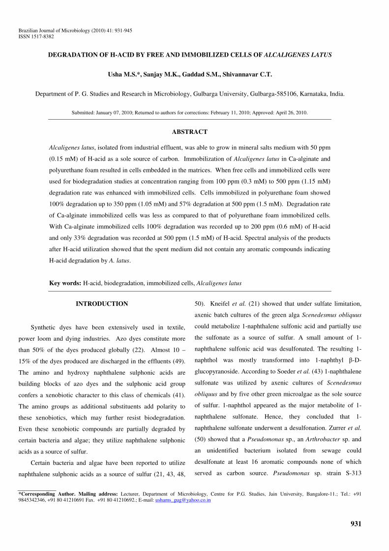
H-acid and other chemicals used in the present
investigation were obtained from Hi-media (Bombay) (Figure
1).
OH NH2
SO3NaHO3S
Figure 1. H-acid (1-Amino 8-Hydroxy Naphthalene 3,6-
disulfonic acid)
Media for H-acid degradation studies
Mineral salts medium of Brilon et al. (3) (12 g
Na2HPO4.2H2O, 2 g KH2PO4, 0.5 g NH4NO3, 0.1 g
MgCl2.6H2O, 50 mg Ca(NO3)2.4H2O, 7.5 mg FeCl2.4H2O and
0.1 ml trace elements solution per liter of medium (37))
containing 50 ppm (0.15 mM) H-acid, 100 mg/l of yeast extract
and 100 mg/l of dextrose was used for the degradation of H-
acid by A. latus.
For alginate-entrapped cells, the degradation medium
contained (g/l) K2HPO4 0.15, MgSO4.7H2O 0.2, NH4Cl 1.0,
FeCl3 0.05 and CaCl2 0.2. The pH of the medium was adjusted
to 7 (24). Different concentrations of H-acid (100 ppm (0.3
mM) to 500 ppm (1.5 mM)) were added as the sole carbon
source.
Immobilization of Bacterial cells
Immobilization in Ca-alginate: The alginate entrapment
of cells was performed according to the method of Bettman and
Rehm (2). Alginate (4% w/v) was dissolved in boiling water
and autoclaved at 121°C for 15 min. Bacterial suspension (5%
w/v) was added to 100 ml sterilized alginate solution and

934
Usha, M.S. et al. Degradation of H-acid by A. latus
mixed by stirring on a magnetic stirrer. This alginate/cell
mixture, with stirring, was extruded drop by drop into a cold,
sterile 0.2 M CaCl2 solution through a burette connected to a
tapered pipette tip, by blowing air from the other end of the
burette. Gel beads of approximately 2 mm diameter were
obtained. The beads were hardened by resuspending into a
fresh CaCl2 solution for 2 h with gentle agitation. Finally these
beads were washed with distilled water and used for
experimentation.
Immobilization in Polyurethane foam
A cell suspension was prepared by mixing 10 g of the cell
paste (centrifuged pellet with little bit of moisture) with 20 ml
of buffer (carbon-free growth medium). One part of
polyurethane pre-polymer was cooled on ice. One part
(weight/weight) of buffer was added and the mixture was
stirred well for one min. One part of the cell suspension was
added, and mixing was continued for an additional one min.
An additional part of the cell suspension was then added and
mixing was continued for another one min. Cell-free foam was
made for use as a control by substituting buffer for the cell
suspensions. The reaction vessel was kept on ice for 2 h while
the polyurethane foam hardened. The foam was removed from
the reaction vessel, rinsed with buffer to remove free cells and
stored at 4°C. At the beginning of each experiment the foam
was rinsed three times with buffer to remove any free cells
released during cutting of the foam (35).
Biodegradation studies
Batch degradation studies:
1. With free cells system
To carry out biodegradation studies with free cells 10 ml
(8 × 10¹⁰ CFU/ml) of Alcaligenes latus culture was added to
each flask with 100 ml of mineral salts medium, 10 mg of yeast
extract and different concentrations of H-acid (100 ppm (0.3
mM) to 500 ppm (1.5 mM)). The flasks were incubated at
37°C and at 180 rpm for 24 h.
2. With immobilized systems
For biodegradation experiment with Ca-alginate, 5 g (wet
weight) of beads, containing the immobilized Alcaligenes latus
cells, were placed in 250 ml flasks containing 100 ml mineral
salts medium and H-acid was added at concentrations ranging
from 100 ppm (0.3 mM) to 500 ppm 1.5 mM). The flasks were
placed in a rotary shaker at 180 rpm at 37°C for 24 h.
Similarly 2 g of polyurethane foam with immobilized
Alcaligenes latus cells were added to 100 ml of mineral salts
medium containing H-acid at varying concentrations from 100
ppm (0.3 mM) to 500 ppm (1.5 mM). Flasks were placed in a
rotary shaker at 180 rpm at 37°C for 24 h.
To determine whether the H-acid was absorbed by the
immobilization matrices or not control flasks were kept for
incubation with cell free Ca-alginate beads and polyurethane
foams separately with different concentrations of H-acid in 100
ml of mineral medium.
Repeated Batch degradation studies
To observe the long-term stability of H-acid degradation by
immobilized cells in different matrices, the system was used for
repeated batch degradation. After each cycle of incubation
period (24 h), the spent medium was decanted and beads/ foam
were washed with sterile water and transferred into a fresh sterile
mineral salts medium (100 ml) containing 200 ppm (0.6 mM) of
H-acid. The degradation process was carried out as described
above and the spent medium was used for the analysis of H-acid
degraded.
Continuous degradation studies
The continuous treatment of H-acid was carried out in a
continuous flow reactor. The reactor was filled with immobilized A.
latus cells in different matrices. The degradation process was carried
out by continuous supply of sterile mineral salts medium/minimal
mineral salts medium containing H-acid at different concentrations
with constant flow rate (ml/h) with the help of peristaltic pump
(Miclins PP10-4C, India).
Design of bioreactor for continuous treatment: A
cylindrical glass column (4×50 cm, volume 650 ml) with inlet
and outlet facilities was used. The bottom of the column was

935
Usha, M.S. et al. Degradation of H-acid by A. latus
packed with a glass wool (4 cm diameter) followed by a porous
glass-frit. Then the reactor was packed with the respective
immobilized cell matrix to a height of 30 cm. The reactor was
attached to a reservoir containing mineral salts
medium/minimal mineral salts medium with H-acid. The
medium was fed into the column continuously with the help of
a peristaltic pump (Miclins PP10-4C, India) through a side arm
present near the bottom of the column. The medium after H-
acid degradation was continuously removed from the side arm
situated just above the packed bed. The detention time was
calculated by the following formula.
Detention time: void volume / flow rate (ml/h).
Degradation Rate (R)= (Ci-Ce) × D
Where Ci = Concentration of H-acid in the influent
Ce= Concentration of H-acid in the effluent
D = Dilution rate= Flow rate (ml/h) / Void volume of the
reactor (ml)
Estimation of per cent degradation of H-acid
At regular intervals a portion of the cultures were
withdrawn and collected from the effluent in case of
continuous system, centrifuged to remove cells and debris. The
absorbance of clear supernatants was read at 390 nm. The
concentrations of the H-acid were calculated by referring to the
calibrated curve and the percent of the H-acid degraded were
then estimated.
Cell viability enumeration
To enumerate viable cells in the Ca-alginate beads, beads
were washed in saline and one bead was soaked in 1 ml of
saline. Soaked bead was shaken with glass beads for 15 min.
Sample was plated onto nutrient agar and viable cells were
expressed as CFU/ml after overnight incubation (15 h) at 37°C.
For enumeration of viable cells in the PUF (polyurethane
foam), PUF were rinsed with saline. One PUF was then torn
into fine pieces using sterile forceps, suspended in saline and
vortexed to dislodge the immobilized cells. Sample was then
plated onto nutrient agar and viable cells were counted after
overnight incubation (15 h) at 37°C.
Confirmation of H-acid degradation
In order to confirm degradation of H-acid by A. latus, the
products were extracted from the large amount of spent medium
(after centrifugation at 8000 × g for 10 min) using two volumes
of diethyl ether. Thus extracted products were separated and
characterized using TLC, 1H NMR and IR analysis.
The separated diethyl ether fractions were dried over
anhydrous sodium sulphate and the traces of sulphate from the
filtrate were removed by anhydrous barium chloride. The
metabolites were isolated and purified by TLC.
Thin layer chromatography was carried out by using a
glass plate (20 × 10 cm) coated with silica gel G slurry (1:2
w/v in water), dried and kept in oven at 100°C for 2 h. Solvent
systems used were chloroform: acetone (80:20) and
cyclohexane: ethylacetate: acetone (4:1:1). Spots were
observed after exposing the plates to iodine vapors. H-acid and
other aromatic compounds were used as a control to compare
with degraded products of H-acid.
The bands obtained on the chromatogram were separated
and compounds were eluted with diethyl ether. The compounds
were then kept for evaporation and sent to I.I.Sc., Bangalore
and CDRI, Lucknow for IR and NMR analysis.
RESULTS
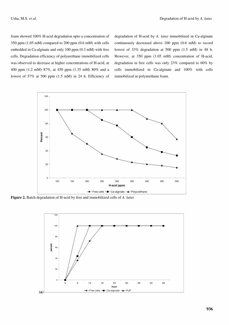
The results of H-acid degradation in batch system by free
cells are presented in Figures 2 and 3. It can be observed that
100% degradation upto 100 ppm (0.3 mM) of H-acid within 18
h. Further increase in H-acid concentration resulted in decrease
in the efficiency of H-acid degradation to 65% at 150 ppm
(0.45 mM), 50% at 200 ppm (0.6 mM), and 35% at 300 ppm
(0.9 mM) and less than 25% at 350 ppm (1.05 mM) and above.
At 500 ppm (1.5 mM), the percent degradation was observed to
be 15% only even after 48 h of incubation.
The results of degradation of H-acid in batch systems by
A. latus immobilized in Ca-alginate and polyurethane foam are
shown in Figures 2 and 3. A. latus immobilized in polyurethane
936
Usha, M.S. et al. Degradation of H-acid by A. latus
foam showed 100% H-acid degradation upto a concentration of
350 ppm (1.05 mM) compared to 200 ppm (0.6 mM) with cells
embedded in Ca-alginate and only 100 ppm (0.3 mM) with free
cells. Degradation efficiency of polyurethane immobilized cells
was observed to decrease at higher concentrations of H-acid, at
400 ppm (1.2 mM) 87%, at 450 ppm (1.35 mM) 80% and a
lowest of 57% at 500 ppm (1.5 mM) in 24 h. Efficiency of
degradation of H-acid by A. latus immobilized in Ca-alginate
continuously decreased above 200 ppm (0.6 mM) to record
lowest of 33% degradation at 500 ppm (1.5 mM) in 48 h.
However, at 350 ppm (1.05 mM) concentration of H-acid,
degradation in free cells was only 23% compared to 60% by
cells immobilized in Ca-alginate and 100% with cells
immobilized in polyurethane foam.
0
20
40
60
80
100
120
100 150 200 250 300 350 400 450 500
H-acid (ppm)
Per
cent
Free cells Ca-alginate Polyurethane Figure 2. Batch degradation of H-acid by free and immobilized cells of A. latus
(a)
0
20
40
60
80
100
120
0 6 12 18 24 30 36 42 48
hour
per
cent
Free cells Ca-alginate PUF
937
Usha, M.S. et al. Degradation of H-acid by A. latus
(b)
0
20
40
60
80
100
120
0 6 12 18 24 30 36 42 48
hour
perc
ent
Free cells Ca-alginate PUF
(c)
0
20
40
60
80
100
120
0 6 12 18 24 30 36 42 48
hour
perc
ent
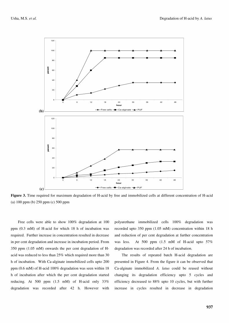
Free cells Ca-alginate PUF Figure 3. Time required for maximum degradation of H-acid by free and immobilized cells at different concentration of H-acid
(a) 100 ppm (b) 250 ppm (c) 500 ppm
Free cells were able to show 100% degradation at 100
ppm (0.3 mM) of H-acid for which 18 h of incubation was
required. Further increase in concentration resulted in decrease
in per cent degradation and increase in incubation period. From
350 ppm (1.05 mM) onwards the per cent degradation of H-
acid was reduced to less than 25% which required more than 30
h of incubation. With Ca-alginate immobilized cells upto 200
ppm (0.6 mM) of H-acid 100% degradation was seen within 18
h of incubation after which the per cent degradation started
reducing. At 500 ppm (1.5 mM) of H-acid only 33%
degradation was recorded after 42 h. However with
polyurethane immobilized cells 100% degradation was
recorded upto 350 ppm (1.05 mM) concentration within 18 h
and reduction of per cent degradation at further concentration
was less. At 500 ppm (1.5 mM of H-acid upto 57%
degradation was recorded after 24 h of incubation.
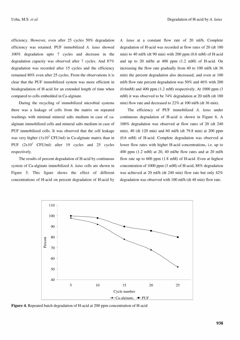
The results of repeated batch H-acid degradation are
presented in Figure 4. From the figure it can be observed that
Ca-alginate immobilized A. latus could be reused without
changing its degradation efficiency upto 5 cycles and
efficiency decreased to 88% upto 10 cycles, but with further
increase in cycles resulted in decrease in degradation
938
Usha, M.S. et al. Degradation of H-acid by A. latus
efficiency. However, even after 25 cycles 50% degradation
efficiency was retained. PUF immobilized A. latus showed
100% degradation upto 7 cycles and decrease in the
degradation capacity was observed after 7 cycles. And 87%
degradation was recorded after 15 cycles and the efficiency
remained 80% even after 25 cycles. From the observations it is
clear that the PUF immobilized system was more efficient in
biodegradation of H-acid for an extended length of time when
compared to cells embedded in Ca-alginate.
During the recycling of immobilized microbial systems
there was a leakage of cells from the matrix on repeated
washings with minimal mineral salts medium in case of ca-
alginate immobilized cells and mineral salts medium in case of
PUF immobilized cells. It was observed that the cell leakage
was very higher (1x10³ CFU/ml) in Ca-alginate matrix than in
PUF (2x101 CFU/ml) after 19 cycles and 25 cycles
respectively.
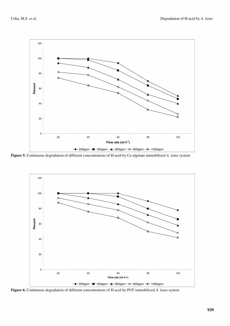
The results of percent degradation of H-acid by continuous
system of Ca-alginate immobilized A. latus cells are shown in
Figure 5. This figure shows the effect of different
concentrations of H-acid on percent degradation of H-acid by
A. latus at a constant flow rate of 20 ml/h. Complete
degradation of H-acid was recorded at flow rates of 20 (dt 180
min) to 40 ml/h (dt 90 min) with 200 ppm (0.6 mM) of H-acid
and up to 20 ml/hr at 400 ppm (1.2 mM) of H-acid. On
increasing the flow rate gradually from 40 to 100 ml/h (dt 36
min) the percent degradation also decreased, and even at 100
ml/h flow rate percent degradation was 50% and 46% with 200
(0.6mM) and 400 ppm (1.2 mM) respectively. At 1000 ppm (3
mM) it was observed to be 74% degradation at 20 ml/h (dt 180
min) flow rate and decreased to 22% at 100 ml/h (dt 36 min).
The efficiency of PUF immobilized A. latus under
continuous degradation of H-acid is shown in Figure 6. A
100% degradation was observed at flow rates of 20 (dt 240
min), 40 (dt 120 min) and 60 ml/h (dt 79.8 min) at 200 ppm
(0.6 mM) of H-acid. Complete degradation was observed at
lower flow rates with higher H-acid concentrations, i.e. up to
400 ppm (1.2 mM) at 20, 40 ml/hr flow rates and at 20 ml/h
flow rate up to 600 ppm (1.8 mM) of H-acid. Even at highest
concentration of 1000 ppm (3 mM) of H-acid, 88% degradation
was achieved at 20 ml/h (dt 240 min) flow rate but only 42%
degradation was observed with 100 ml/h (dt 48 min) flow rate.
40
50
60
70
80
90
100
110
5 10 15 20 25
Cycle number
Perc
ent
Ca-alginate, PUF Figure 4. Repeated batch degradation of H-acid at 200 ppm concentration of H-acid
939
Usha, M.S. et al. Degradation of H-acid by A. latus
0
20
40
60
80
100
120
20 40 60 80 100
Flow rate (ml h-1)
Per
cen
t
200ppm 400ppm 600ppm 800ppm 1000ppm
Figure 5. Continuous degradation of different concentrations of H-acid by Ca-alginate immobilized A. latus system
0
20
40
60
80
100
120
20 40 60 80 100
Flow rate (ml h-1)
Per
cent
200ppm 400ppm 600ppm 800ppm 1000ppm
Figure 6. Continuous degradation of different concentrations of H-acid by PUF immobilized A. latus system
940
Usha, M.S. et al. Degradation of H-acid by A. latus
Cell viability was greater in PUF than Ca-alginate beads.
After 2 h of immobilization cell viability was found to be 5 ×
106 CFU/ml in PUF whereas it was 4 × 10⁴ CFU/ml in Ca-
alginate bead.
The products of H-acid degradation by A. latus were
separated and identified by TLC and spectral analyses.
The products of H-acid degradation by A. latus were
extracted with diethyl ether and subjected to TLC by various
solvent systems. With Cyclohexane:Ethyl acetate:Acetone
(4:1:1) and Chloroform:Acetone (80:20) as solvent system, a
single broad band with tailing was observed not corresponding
to the H-acid band. Rf values of H-acid in two different solvent
systems were found to be 0.23 and 0.19 and those of the
product were found to be 0.33 and 0.28. TLC bands were
scraped out, extracted with diethyl ether and subjected to
spectral analysis.
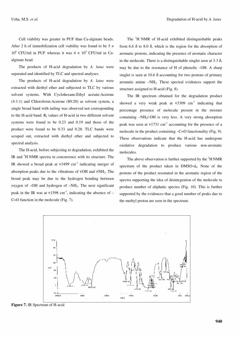
The H-acid, before subjecting to degradation, exhibited the
IR and 1H NMR spectra in concurrence with its structure. The
IR showed a broad peak at ν3499 cm-1 indicating merger of
absorption peaks due to the vibrations of νOH and νNH2. The
broad peak may be due to the hydrogen bonding between
oxygen of –OH and hydrogen of –NH2. The next significant
peak in the IR was at ν1598 cm-1, indicating the absence of –
C=O function in the molecule (Fig. 7).
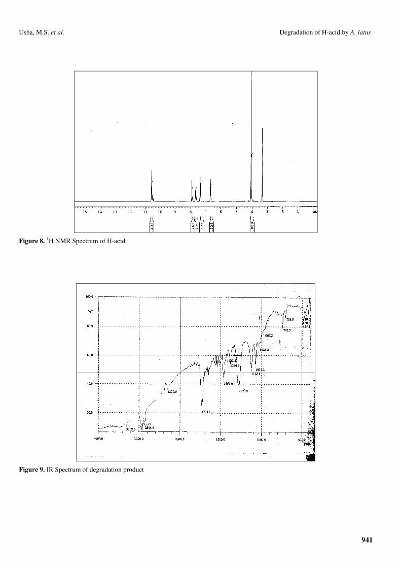
The 1H NMR of H-acid exhibited distinguishable peaks
from 6.6 δ to 8.0 δ, which is the region for the absorption of
aromatic protons, indicating the presence of aromatic character
in the molecule. There is a distinguishable singlet seen at 3.3 δ,
may be due to the resonance of H of phenolic –OH. A sharp
singlet is seen at 10.6 δ accounting for two protons of primary
aromatic amine –NH2. These spectral evidences support the
structure assigned to H-acid (Fig. 8).
The IR spectrum obtained for the degradation product
showed a very weak peak at ν3309 cm-1 indicating that
percentage presence of molecule present in the mixture
containing –NH2/-OH is very less. A very strong absorption
peak was seen at ν1731 cm-1 accounting for the presence of a
molecule in the product containing –C=O functionality (Fig. 9).
These observations indicate that the H-acid has undergone
oxidative degradation to produce various non-aromatic
molecules.
The above observation is further supported by the 1H NMR
spectrum of the product taken in DMSO-d6. None of the
protons of the product resonated in the aromatic region of the
spectra supporting the idea of disintegration of the molecule to
produce number of aliphatic species (Fig. 10). This is further
supported by the evidences that a good number of peaks due to
the methyl proton are seen in the spectrum.
Figure 7. IR Spectrum of H-acid
941
Usha, M.S. et al. Degradation of H-acid by A. latus
Figure 8. 1H NMR Spectrum of H-acid
Figure 9. IR Spectrum of degradation product

942
Usha, M.S. et al. Degradation of H-acid by A. latus
Figure 10. 1H NMR Spectrum of degradation product
DISCUSSION
Results of batch degradation studies revealed that
polyurethane foam immobilized cells were able to degrade H-
acid completely upto 350 ppm (1.05 mM) compared to 200
ppm (0.6 mM) with Ca-alginate immobilized cells and 100
ppm (0.3 mM) with free cells in 48 h. H-acid degradation
efficiency of A. latus was observed to be reduced as the
concentration of H-acid increases.
The A. latus cells immobilized in the polyurethane foam
showed higher degradation rate than the cells immobilized in
Ca-alginate. This high rate of degradation may be attributed to
the better availability of substrate (H-acid) to the cells as they
are immobilized by adsorption process, whereas, with Ca-
alginate matrix the cells are embedded in the matrix, where the
slow diffusion of substrate (H-acid) and air into the polymer
beads may decrease the rate of degradation. Manohar and
Karegoudar (30) and Sharanagouda and Karegouder (42)
observed similar results with naphthalene degradation by
immobilized Pseudomonas sp. It was also suggested that the
storage stability and microbial activity of immobilized cells in
polyurethane foam are known to be better than those cells
immobilized in Ca-alginate (4, 9).
A. latus immobilized in Ca-alginate and polyurethane
foam showed complete degradation of H-acid upto 400 ppm
and 600 ppm respectively at a flow rate of 20 ml/h. Increased
degradation by immobilized cells was due to the accelerated
reaction rates caused by the high local cell density in or on the
immobilized matrix and also the immobilization of cells could
improve the tolerance capacity against toxicity of high
concentration of H-acid. Immobilization also provides a kind of
membrane stabilization, which was assumed to be responsible
for the cell protection and better degradation rates in
immobilized cells. Similar observations have been made by
Hall and Rao (16) in the production of fuels and chemicals and
Lee et al. (23) in the degradation of pyridine by immobilized
Pimetobacter sp. It has been reported that immobilization of
cells in a polymer matrix can confer protection from high
levels of toxic compounds normally lethal to free cells. The
protective effect may be due to adsorption of the toxic
compound by the matrix, lowering the available concentration
of the toxic compound in solution as observed with
polyurethane foam (17), activated carbon (29) and beet chips
(10). Alternatively it may be due to alteration in the membrane
composition of cell in the matrix, rendering the membrane less
permeable to toxic substances (12, 19). Immobilized viable

943
Usha, M.S. et al. Degradation of H-acid by A. latus
cells have been shown to have altered metabolism, enhanced
enzyme induction, altered macromolecules composition, and
reduced specific cell growth and cell yield (14).
Repeated batch degradation studies were conducted to
observe the effect of physicochemical factors on the
degradation efficiency of A. latus immobilized in Ca-alginate
and polyurethane foam. The results revealed that the
polyurethane foam immobilized cells retained complete H-acid
degradation capacity for longer period (7 cycles) compared to
cells immobilized in Ca-alginate (5 cycles). Cell leakage was
almost three times more in Ca-alginate compared to
polyurethane foam immobilized cells after 25 cycles.
Polyurethane foam showed higher mechanical stability,
resistance to chemicals and biological degradation and lower
cell leakage, that made it suitable for a long period reuse.
Trevors et al. (46) observed mechanical instability and gradual
cell leakage from Ca-alginate beads, decreasing the
degradation rate. Enhancement in the activity of immobilized
cells on reuse has been reported for the other systems (18, 39)
and this may be due to post immobilization
modifications/acclimatization. In contrast, our results on the
reuse of immobilized beads/foam cubes did not show
enhancement in the degradation of H-acid but degradation rate
decreased after several cycles of reuse. Similar observations
were made by Sharanagouda and Karegoudar (42) in the
degradation of 2-Methyl naphthalene using Pseudomonas sp.
The metabolic products produced from H-acid degradation
by A. latus were analyzed. Spectral scanning of the product
showed a single peak at 260 nm and a single band on TLC
which were not corresponding to H-acid peak. Further analysis
of TLC band by spectral analysis revealed the presence of a
large number of small molecules suggesting the formation of
aliphatic molecules during microbial utilization and aromatic
byproducts were not observed. The IR spectrum of the products
indicated the absence of sulphonic acid group suggesting that
complete desulphonation occurs during the degradation of H-
acid. Similar reports have been given by Wittich et al. (48).
ACKNOWLEDGEMENT
I owe a deep sense of gratitude and appreciation to my
Research Supervisor, Dr. C. T. Shivannavar, Reader,
Department of Microbiology, Gulbarga University, Gulbarga
for his valuable guidance. Thanks are due to SAIF, C.D.R.I.,
Lucknow and SAIF, I.I.Sc., Bangalore for providing NMR and
IR and IMTECH, Chandigarh for identification of bacterium.
Thanks are also due to Dr. Purohit, Retired professor,
Department of Chemistry, Gulbarga University for interpreting
spectral analysis data. I thank Gulbarga University, Gulbarga
for providing Research Student Fellowship during my research
work.
REFERENCES
1. Balfanz, J.; Rehm, H.J. (1991). Biodegradation of 4-chlorophenol by
adsorptive immobilized Alcaligens sp. A 7-2 in soil. Appl. Microbiol.
Biotechnol. 35, 662-668.
2. Bettman, H.; Rehm, H.J. (1984). Degradation of phenol by polymer
entrapped microorganisms. Appl. Microbiol. Biotechnol. 20, 285-290.
3. Brilon, C.; Beckmann, W.; Hellwig, M.; Knackmuss, H.J. (1981).
Enrichment and isolation of naphthalene sulphonic acid utilizing
Pseudomonads. Appl. Environ. Microbiol. 42, 39.
4. Brouers, M.; Collard, F.; Jeanfils, J. et al. (1983). Soil Energy. Res. Dev.
Eur. Community ser, D2, 171.
5. Cassidy, M.B.; Lee, H.; Trevors, J.T. (1996). Environmental applications
of immobilized microbial cells: A review. J. Ind. Microbiol. 16, 79-101.
6. Cassidy, M.B.; Shaw, K.W.; Lee, H.; Trevors, J.T. (1997). Enhanced
mineralization of pentachlorophenol by K-carrageenan – encapsulated
Pseudomonas sp. UG30. Appl. Microbiol. Biotechnol. 47, 108-113.
7. Cheetham, P.S.J. (1980). Developments in immobilized cells and their
applications. In: Wiseman A. (ed) Topics in enzyme and fermentation
technology, Vol. 4, Ellis Harwood, Chichester, 189-238.
8. Chibata, I.; Tosa, T.; Sato, T. (1978). Preparation of immobilized
enzymes and microbial cells. In: Chibata I(ed). Immobilized enzymes.
Wiley, N.Y., 9-108.
9. Cocquempot, M.F.; Thomasset, B.; Barbotin, J.N. et al. (1981).
Comparative stabilization of biological photosystems by several
immobilization procedures. Eur. J. Appl. Microbiol. Biotechnol. 11, 193-
198.
10. Curtain, C.C. (1986). Understanding and avoiding ethanol inhibition.
Trends Biotechnol. 4, 110.
944
Usha, M.S. et al. Degradation of H-acid by A. latus
11. D’souza, S.F. (1989). Immobilized cells: Techniques and applications.
Indian J. Microbiol. 29, 83-117.
12. Diefenbach, R.; Keweloh, H.; Rehm, H.J. (1992). Fatty acid impurities in
alginate influence the phenol tolerance of immobilized Escherichia coli.
Appl. Microbiol. Biotechnol. 36, 530-534.
13. Diekmann, R.; Hempel, D.C. (1989). Production rates of CO2 by
suspended and immobilized bacteria degrading 6-amino 2-naphthalene
sulfonic acid. Appl. Microbiol. Biotechnol. 32, 113-117.
14. Doran, P.M.; Bailey, J.E. (1986). Effects of immobilization on growth,
fermentation properties, and macromolecular composition of
Saccharomyces cerevisiae attached to gelatin. Biotechnol. Bioeng. 28,
73-87.
15. Feigel, B.J.; Knackmuss, H.J. (1993). Syntrophic interactions during
degradation of 4-amino benzene sulphonic acid by a two species bacterial
culture, Arch. Microbiol. 159, 124-130.
16. Hall, C.O.; Rao K.K. (1989). Immobilized photosynthetic membranes
and cells for the production of fuels and chemicals. Chimicaoggi. 1, 41-
47.
17. Hu, Z.C.; Korus, R.A.; Levinson, W.E.; Crawford, R.L. (1994).
Adsorption and biodegradation of pentachlorophenol by polyurethane-
immobilized Flavobacterium. Environ. Sci. Technol. 28, 491-496.
18. Joshi, N.T.; D’Souza, S.F. (1999). Immobilization of activated sludge for
the degradation of phenol. J. Environ. Sci. Health-A. 34, 1689-1700.
19. Keweloh, H.; Heipieper, H.J.; Rehm, H.J. (1989). Protection of bacteria
against toxicity of phenol by immobilization in calcium alginate. Appl.
Microbiol. Biotechnol. 31, 383-389.
20. Klein, J.; Hackel, U.; Wagner, F. (1979). Phenol degradation by Candida
tropicalis whole cells entrapped in polymeric ionic networks, p. 101-118.
In. K. Venkatasubramanian (ed.). Immobilized microbial cells. ACS
Symposium Series no.106. American Chemical society, Washington D.
C.
21. Kneifel, H.; Elmendorff, K.; Hegewald, E.; Soeder, C.J. (1997).
Biotransformation of 1-Naphthalene sulphonic acid by the green algae
Scenedesmus obliquus. Arch. Microbiol. 167, 32-37.
22. Kulkarni, S.V.; Blackwell, C.D.; Blackard, A.L.; Stackocese, C.W.;
Alexander, M.W. (1985). Textile dyes and dying equipment,
classification, properties and environmental aspects, U.S. Environmental
Protection Agency, EPA – 600/2 – 85/010. Research Trainage Par, NC.
23. Lee, S.T.; Rhee, S.K.; Lee, G.M. (1994). Biodegradation of pyridine by
freely suspended and immobilized Pimelobacter sp. Appl. Microbiol.
Biotechnol. 41, 652-657.
24. Manohar, S. (1999). Microbial degradation of aromatic hydrocarbons.
Ph. D. Thesis, Gulbarga University, Gulbarga.
25. Manohar, S.; Kim, C.K.; Karegoudar, T.B. (2001). Enhanced degradation
of naphthalene by immobilization of Pseudomonas sp. strain NGK1 in
polyurethane foam. Appl. Microbiol. Biotechnol. 55, 311-316.
26. Mattiasson, B. (1983). Immobilization methods. P. 3-26. In. B.
Mattiasson (ed.), Immobilized cells and organelles, Vol. I. CRC press,
Inc., Boca Raton, Fla.
27. Mohanty, S.; Rao, N.N.; Khare, P.; Kaul, S.N. (2005). A coupled
photocatalytic–biological process for degradation of 1-amino-8-naphthol-
3, 6-disulfonic acid (H-acid). Wat. Res. 39, 5064-5070
28. Mordocco, A.; Kuek, C.; Jenkins, R. (1999). Continuous degradation of
phenol at low concentration using immobilized Pseudomonas putida.
Enzyme Microb. Technol. 25, 530-536.
29. Morsen, A.; Rehm, H.J. (1987). Degradation of phenol by a mixed
culture of Pseudomonas putida and Cryptococcus elinovii adsorbed on
activated carbon. Appl. Microbiol. Biotechnol. 26, 283-288.
30. Noorjahan, M.; Reddy, P.M.; Kumari, D.V. et al., (2003). Photocatalytic
degradation of H-acid over a novel TiO2 thin film fixed bed reactor and
in aqueous suspensions. J. Photochem. Photobiol. A. 156, 179-187.
31. Nortemann, B.; Baumgarten, J.; Rast, H.G.; Knackmuss, H.J. (1986).
Bacterial communities degrading Amino and Hydroxy Naphthalene 2-
sulfonates. Appl. Environ. Microbiol. 52, 1195-1202.
32. Nortemann, B.; Glasser, A.; Machinek, R.; Remberg, G.; Knackmuss,
H.J. (1993). 5-Hydroxy quinoline 2-Carboxylic Acid, a dead end
metabolite from the bacterial oxidation of 5-Aminonaphthalene 2-
sulfonic acid. Appl. Environ. Microbiol. 59, 1898-1903.
33. Nortemann, B.; Kuhm, A.E.; Knackmuss, H.J.; Stolz, A. (1994).
Conversion of substituted Naphthalene sulfonates by Pseudomonas sp.
BN6. Arch. Microbiol. 161, 320-327.
34. Nunez, M.J.; Lema, J.M. (1987). Cell immobilization: Application to
alcohol production. Enzyme Microb. Technol. 9, 642-661.
35. O’Reilly, K.; Crawford, R.L. (1989). Degradation of pentachlorophenol
by polyurethane-immobilized Flavobacterium cells. Appl. Microbiol.
Biotechnol. 55, 2113-2118.
36. Perei, K.; Rakhely, G.; Kiss, I. et al. (2001). Biodegradation of sulfanilic
acid by Pseudomonas paucimobilis. Appl. Microbiol. Biotechnol. 55,
101-107.
37. Pfennig, N.; Lippert, K.D. (1966). Uber das Vitamin B12-Bedurfnis
phototropher Schwefelbakterien. Arch. Mikrobiol. 55, 245-256.
38. Rao, B.Y.K.; Godbole, S.S.; D’Souza, S.F. (1988). Preparation of lactose
free milk by fermentation using immobilized Saccharomyces fragilis
Biotechnol. Lett., 6, 427-430.
39. Rao, N.N.; Bose, G.; Khare, P. et al. (2006). Fenton and Electro-Fenton
methods for oxidation of H-acid and Reactive Black 5. J. Environ. Eng.
132, 367-376.
40. Rehorek, A.; Urbig, K.; Meurer, R.; Schäfer, C.; Plum, A.; Braun, G.
(2002). Monitoring of azo dye degradation processes in a bioreactor by
on-line high-performance liquid chromatography J Chromatogr A. 949,
263-8.
41. Reineke, W.; Knackmuss, H.J. (1978). Chemical structure and
biodegradability of halogenated aromatic compounds: substituents effects
on 1,2 dioxygenation of benzoic acid. Biochim. Biophys. Acta, 542, 412.
42. Sharanagouda, U.; Karegoudar, T.B. (2002). Degradation of 2-
methylnaphthalene by free and immobilized cells of Pseudomonas sp.
945
Usha, M.S. et al. Degradation of H-acid by A. latus
strain NGK1.�World J. Microbiol. Biotechnol. 18, 225-230.
43. Soeder, C.J.; Hegewald, E.; Kneifel, H. (1987). Green microalgae can use
Naphthalene sulphonic acids as sources of sulfur. Arch. Microbiol. 148,
260-263.
44. Somerville, H.J.; Mason, J.R.; Ruffell, R.N. (1977). Benzene degradation
by bacterial cells immobilized in polyacrylamide gel.Eur. J. Appl.
Microbiol., 4, 75-85.
45. Swaminathan, K.; Sandhya, S.; Sophia, C.A. et al. (2003). Decolorization
and degradation of H-acid and other dyes using ferrous-hydrogen
peroxide system. Chemosphere. 50, 619-625.
46. Trevors, J.T.; Elsas, V.J.D.; Lee, H. et al. (1992). Use of alginate and
other carriers for encapsulation of microbial cells for use in soil
Microbiol. Rel. 1, 61-69.
47. Westmeier, F.; Rehm, H.J. (1987). Degradation of 4-chlorophenol in
municipal wastewater by adsorptive immobilized Alcaligenes sp. A7-2.
Appl. Microbiol. Biotechnol. 26, 78-83.
48. Wittich, R.M.; Rast, H.G.; Knackmuss, H.J. (1988). Degradation of
Naphthalene 2, 6 and Naphthalene 1, 6 – disulfonic acid by a Moraxella
sp. Appl. Environ. Microbiol. 54, 1842-1847.
49. Zollinger, H. (1987). Colour chemistry – synthesis, properties and
application of organic dyes and pigments 92-102, VCH publishers, N.Y.
50. Zurrer, D.; Cook A.M.; Leisenger, T. (1987). Microbial desulfonation of
substituted Naphthalene sulphonic acids and Benzene sulphonic acids.
Appl. Environ. Microbiol. 53, 1459-1463.


![Study on the Microbial Degradation Tannic Acid by Alcaligenes … · 2017-08-14 · for the treatment of the effluents. Some of them are primary treatment in fill and draw tanks [5],](https://img.dokumen.tips/doc/110x75/5f30565d191f3a661c0a3ad3/study-on-the-microbial-degradation-tannic-acid-by-alcaligenes-2017-08-14-for-the.jpg)