Embed Size (px)
Citation preview
Degenerative joint disease
(DJD)
รศ.นพ.อาร ีตนาวล ีAree Tanavalee, MD
Associate Professor
Department of
Orthopaedics
Faculty of Medicine
Chulalongkorn University

What is DJD?
• Commonly known
– Osteoarthritis, Osteoarthrosis
• Disease character:
– Different etiologies
– Similar findings
• Biologic
• Morphologic
• Clinical outcomes
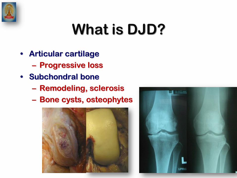
What is DJD?
• Articular cartilage
– Progressive loss
• Subchondral bone
– Remodeling, sclerosis
– Bone cysts, osteophytes
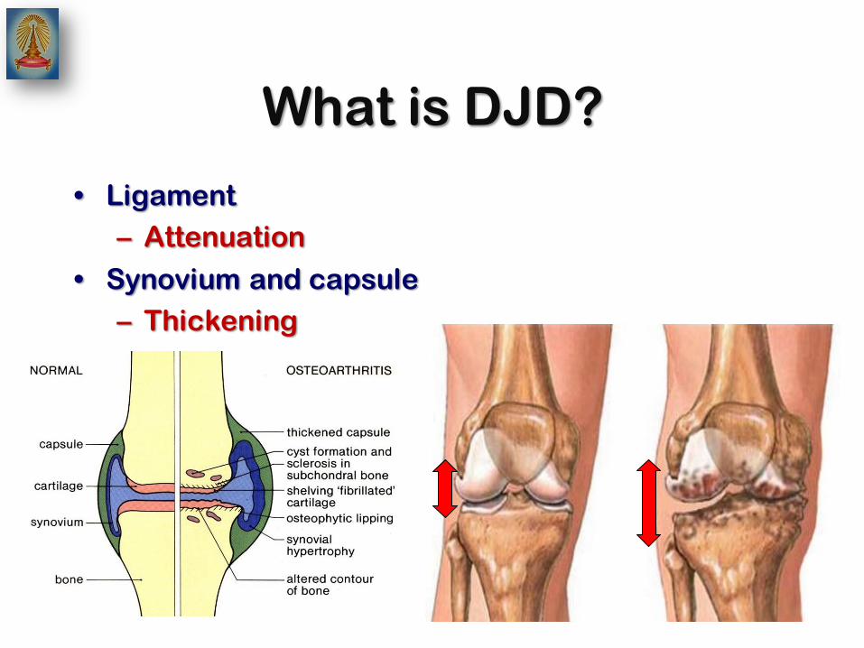
What is DJD?
• Ligament
– Attenuation
• Synovium and capsule
– Thickening
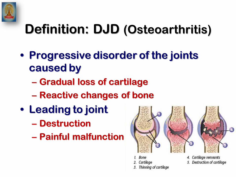
Definition: DJD (Osteoarthritis)
• Progressive disorder of the joints
caused by
– Gradual loss of cartilage
– Reactive changes of bone
• Leading to joint
– Destruction
– Painful malfunction

Prevalence
• Clinically defined OA.
– on the basis of symptoms and physical
examination findings
– Ages 25–74years
• 12.1% of the US population
• Radiographically defined OA
– According to the Kellgren/Lawrence
scale (presence of osteophytes)
Lawrence RC, Felson DT, Helmick CG, et al. Estimates of the prevalence of arthritis and other
rheumatic conditions in the United States. Part II. Arthritis Rheum 2008;58(1):26–35.
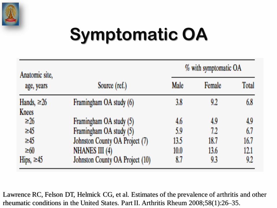
Symptomatic OA
Lawrence RC, Felson DT, Helmick CG, et al. Estimates of the prevalence of arthritis and other
rheumatic conditions in the United States. Part II. Arthritis Rheum 2008;58(1):26–35.
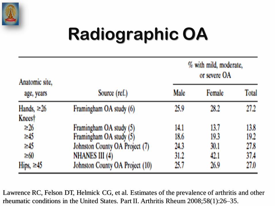
Radiographic OA
Lawrence RC, Felson DT, Helmick CG, et al. Estimates of the prevalence of arthritis and other
rheumatic conditions in the United States. Part II. Arthritis Rheum 2008;58(1):26–35.

Comparing OA diagnosis
S R
7
3
3
Higher prevalence of OA
• Genetic mutation
– Mutation in type II collagen
• Heritability
– Radiographic OA of hands & knees
– 39-65%
• Knee
– History of meniscectomy
– Repetitive kneeling and squatting
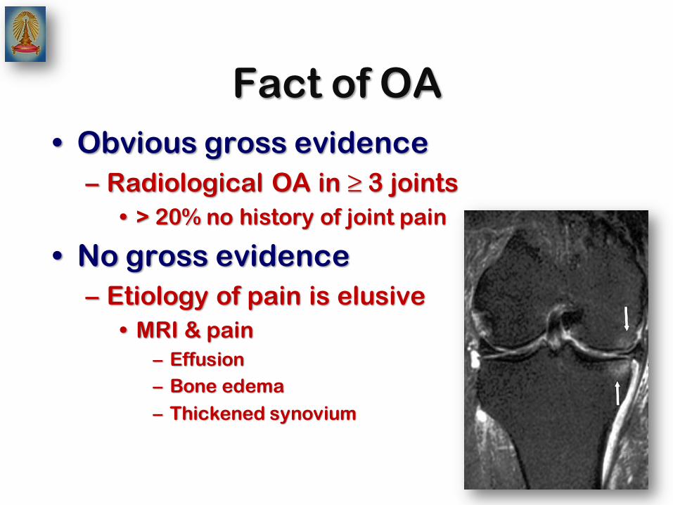
Fact of OA
• Obvious gross evidence
– Radiological OA in ≥ 3 joints
• > 20% no history of joint pain
• No gross evidence
– Etiology of pain is elusive
• MRI & pain
– Effusion
– Bone edema
– Thickened synovium
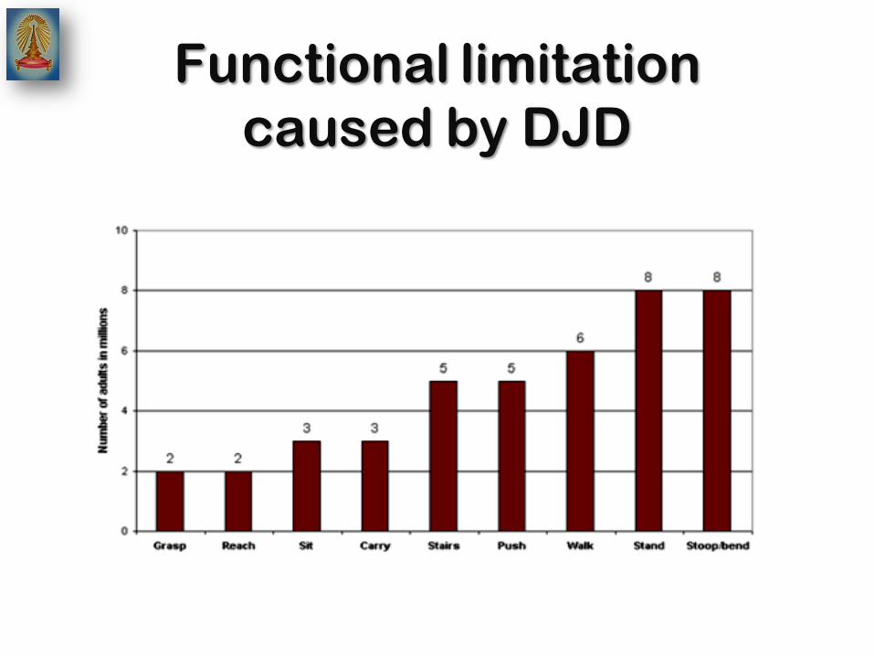
Functional limitation
caused by DJD
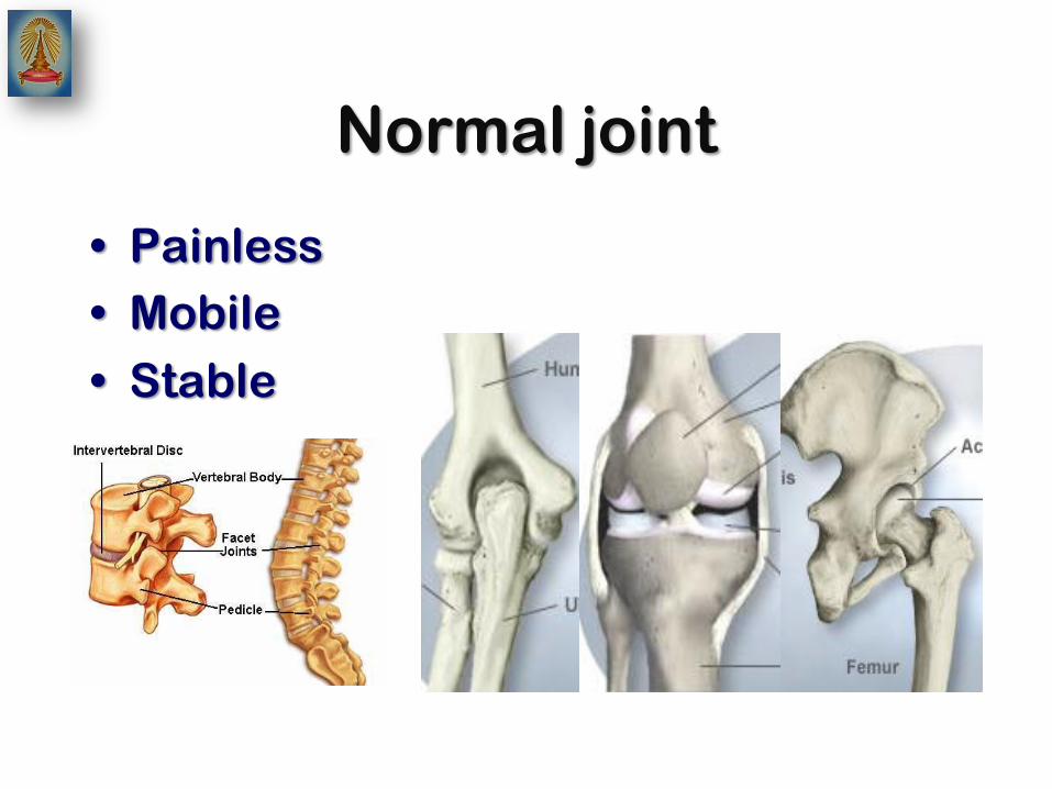
Normal joint
• Painless
• Mobile
• Stable

Mature articular cartilage
• 5 layers
– Avascular
– Aneural
– Alymphatic
• Matrix
• Chondrocyte
• Nutrients – Diffusion
– Synovial fluid
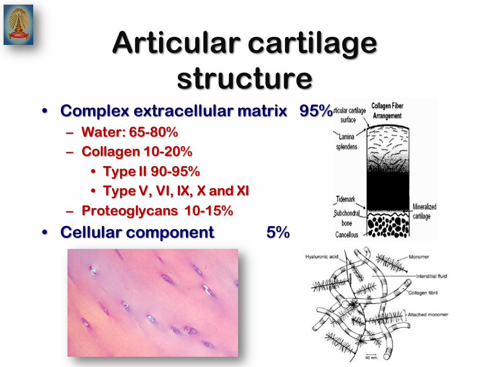
Articular cartilage
structure • Complex extracellular matrix 95%
– Water: 65-80%
– Collagen 10-20%
• Type II 90-95%
• Type V, VI, IX, X and XI
– Proteoglycans 10-15%
• Cellular component 5%
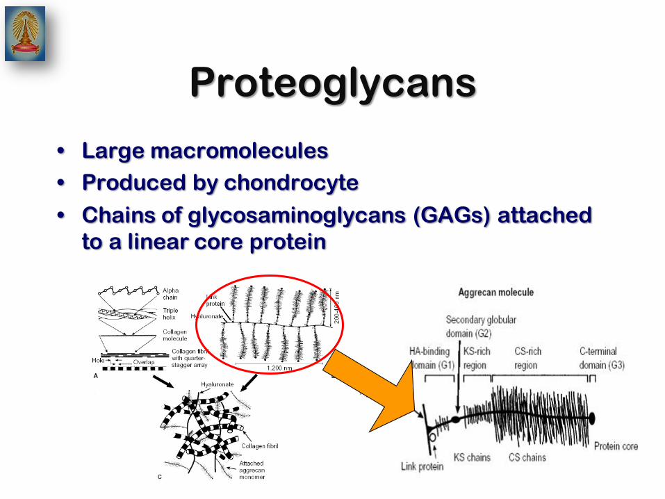
Proteoglycans
• Large macromolecules
• Produced by chondrocyte
• Chains of glycosaminoglycans (GAGs) attached
to a linear core protein
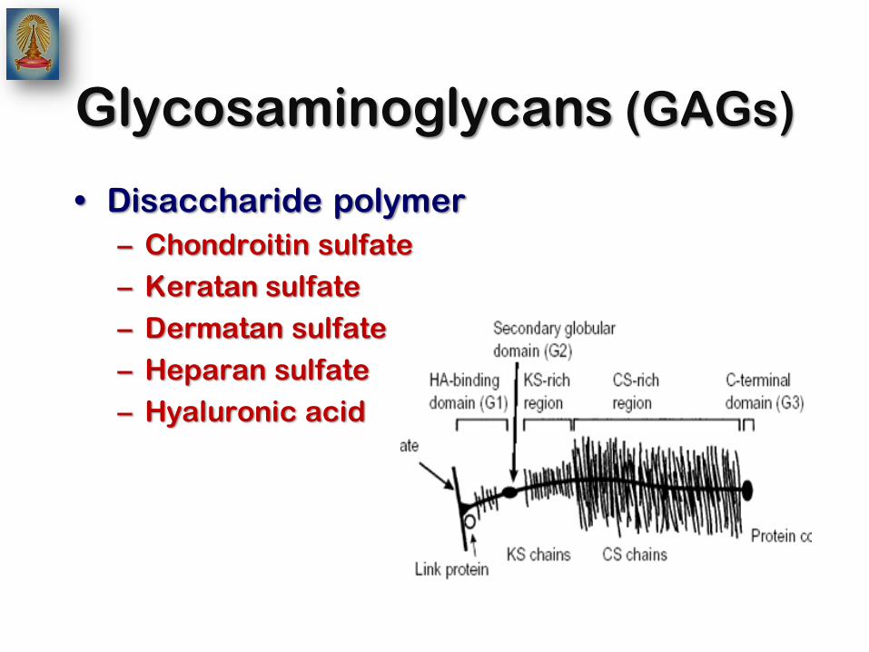
Glycosaminoglycans (GAGs)
• Disaccharide polymer
– Chondroitin sulfate
– Keratan sulfate
– Dermatan sulfate
– Heparan sulfate
– Hyaluronic acid
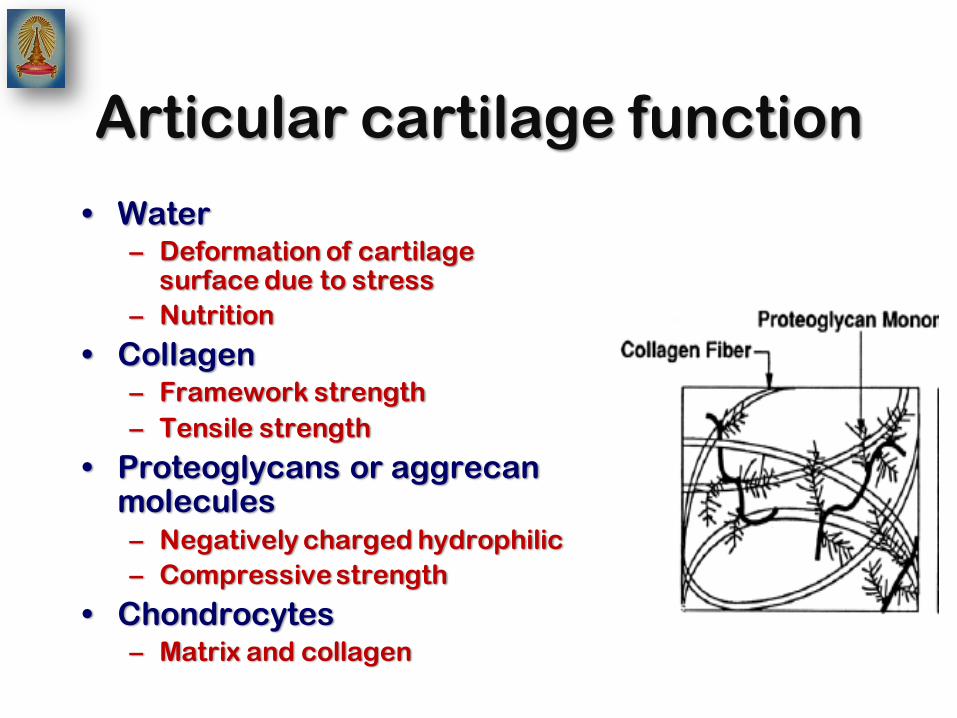
Articular cartilage function
• Water – Deformation of cartilage
surface due to stress
– Nutrition
• Collagen – Framework strength
– Tensile strength
• Proteoglycans or aggrecan molecules – Negatively charged hydrophilic
– Compressive strength
• Chondrocytes – Matrix and collagen

Normal articular cartilage
• Load distribution
• Decrease friction
• Resistance to compressive, tensile, and shear
forces
• Minimizing loads on subchondral bone
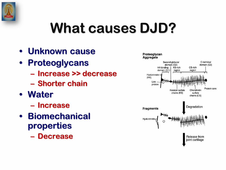
What causes DJD?
• Unknown cause
• Proteoglycans
– Increase >> decrease
– Shorter chain
• Water
– Increase
• Biomechanical properties – Decrease

Pathology of DJD
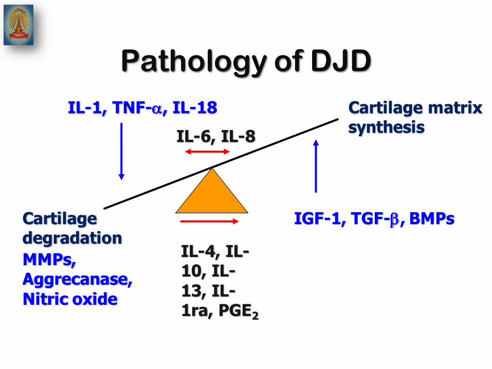
IL-1, TNF-, IL-18
IL-6, IL-8
Cartilage degradation
Cartilage matrix synthesis
IL-4, IL-10, IL-
13, IL-1ra, PGE2
IGF-1, TGF-, BMPs
MMPs, Aggrecanase,
Nitric oxide
Pathology of DJD

Pathology of DJD
• Fissuring and focal erosive cartilage lesions
• Cartilage loss and destruction
• Subchondral bone sclerosis and cyst
• Large osteophyte formation

Subchondral bone change
• Bone marrow
edema
• Subchondral
sclerosis
• Osteophyte
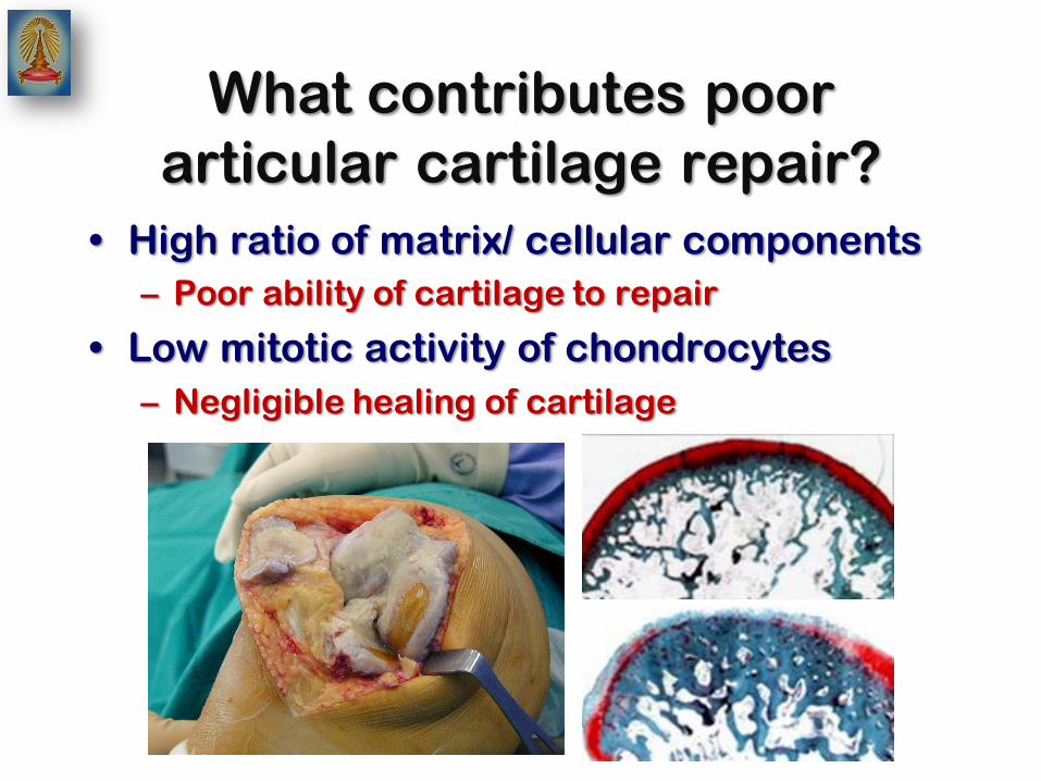
What contributes poor
articular cartilage repair?
• High ratio of matrix/ cellular components
– Poor ability of cartilage to repair
• Low mitotic activity of chondrocytes
– Negligible healing of cartilage
What are risk factors for OA
• Older age
• Female sex
• Repetitive stress and joint overload
• Genetic factors
• Major trauma
• Obesity
• Prior inflammatory joint disease
• Metabolic disorder
• Proprioceptive defects

Etiologic factors in OA
• Normal load – Abnormal biomaterial
(joint vulnerability)
• Normal biomaterial
– Excessive mechanical stress (excessive joint loading)
What are the symptoms of
DJD? • With or without symptoms for a long time
• Symptoms
– Subtle development of morning stiffness
– Pain with movement and activity
– Improve with rest
– Decreased range of motion
– Abnormal sound
– Unstable joints

How is DJD diagnosed?
• Symptoms and signs
– Joint pain
– Restriction of motion
– Crepitus with motion
– Joint effusions
– Deformity
– Instability
• Radiography – Abnormal findings
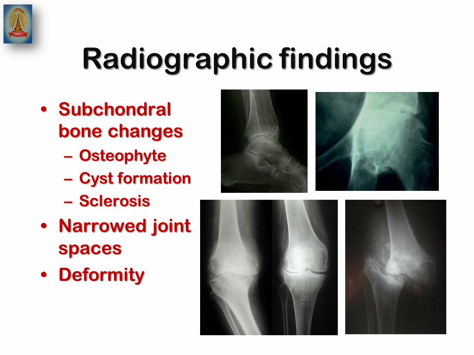
Radiographic findings
• Subchondral
bone changes
– Osteophyte
– Cyst formation
– Sclerosis
• Narrowed joint
spaces
• Deformity
Classification
• Primary or idiopathic
– Most common type
– No identifiable etiology or predisposing
cause
• Secondary
– Identifiable underlying cause
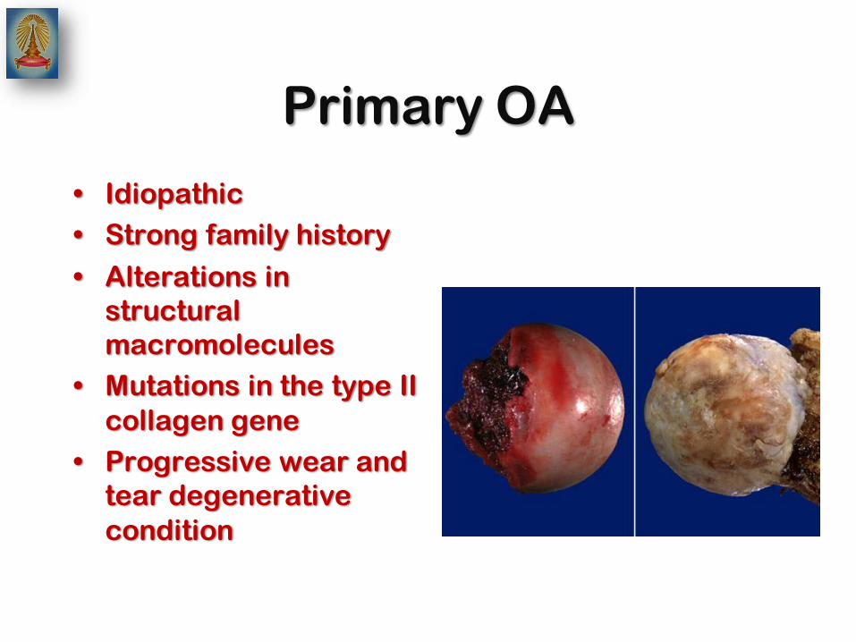
Primary OA
• Idiopathic
• Strong family history
• Alterations in
structural
macromolecules
• Mutations in the type II
collagen gene
• Progressive wear and
tear degenerative
condition
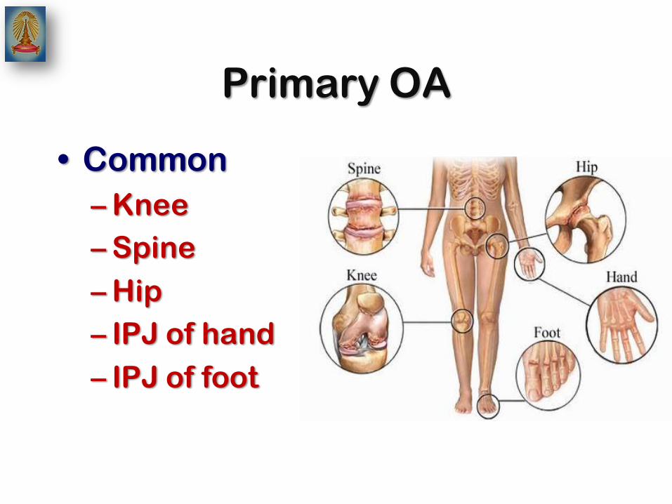
Primary OA
• Common
– Knee
– Spine
– Hip
– IPJ of hand
– IPJ of foot
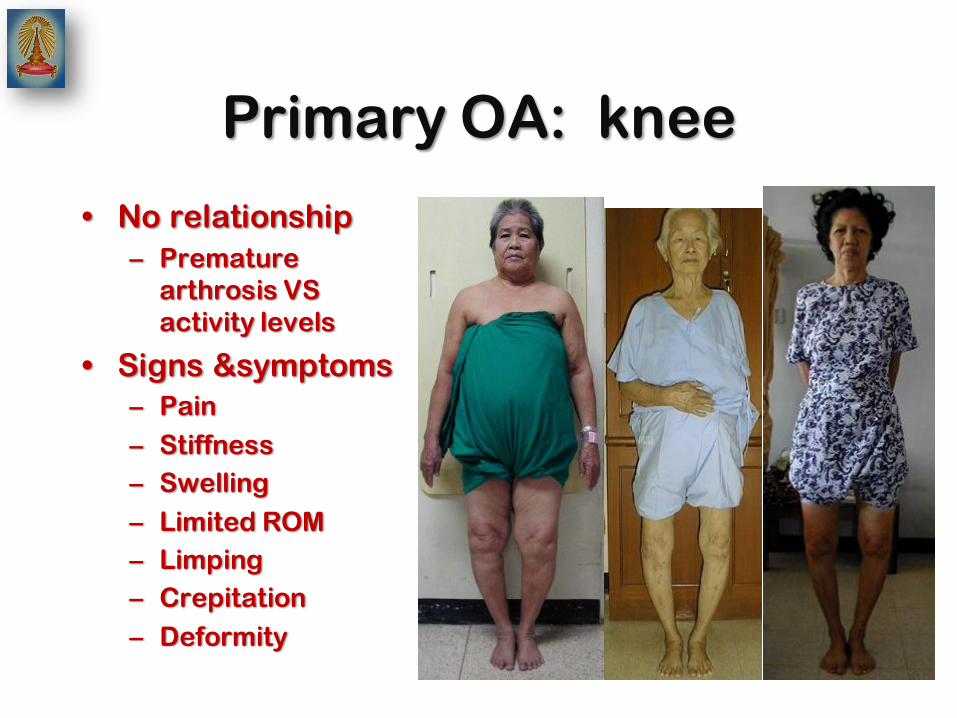
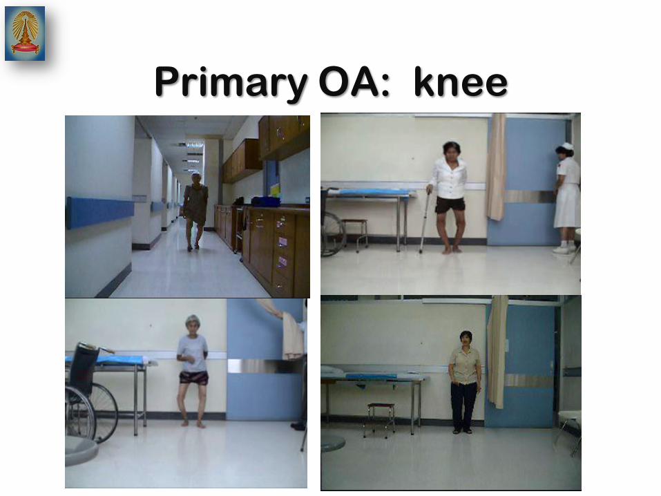
Primary OA: knee
• No relationship
– Premature
arthrosis VS
activity levels
• Signs &symptoms
– Pain
– Stiffness
– Swelling
– Limited ROM
– Limping
– Crepitation
– Deformity
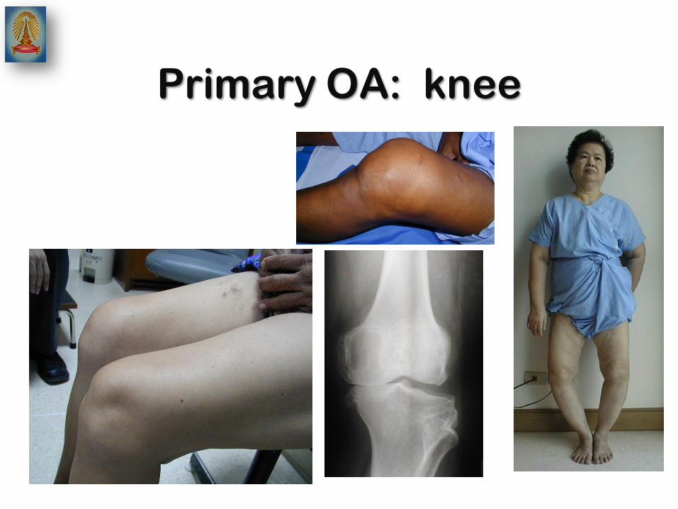
Primary OA: knee
Primary OA: knee
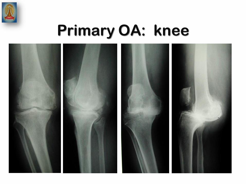
Primary OA: knee
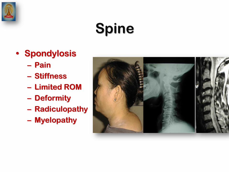
Spine
• Spondylosis
– Pain
– Stiffness
– Limited ROM
– Deformity
– Radiculopathy
– Myelopathy

Spine

Primary OA: hip
• Pain
• Stiffness
• Limited ROM
• Limping
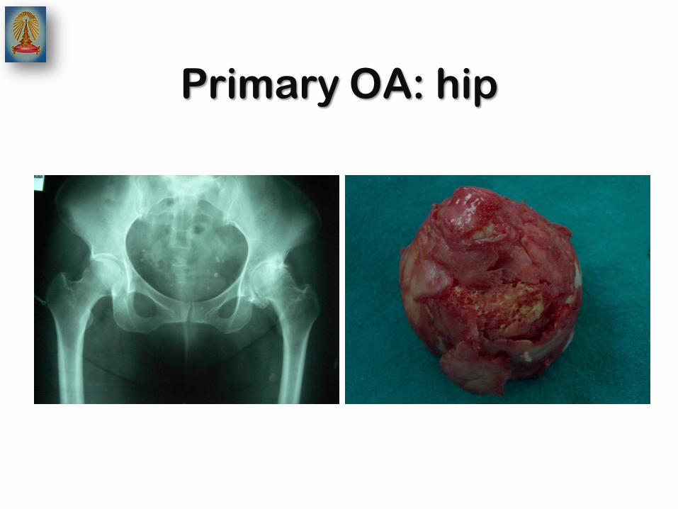
Primary OA: hip

Primary OA: hand & finger
• Pain
• Swelling
• Limited ROM
• Heberden's nodes
• Bouchard's nodes

Primary OA: other joints

Secondary OA
• Direct response
– Abnormal mechanical loading with
articular cartilage
• Various causes
– Metabolic condition
– Anatomic factors
– Traumatic events
– Inflammatory disorders
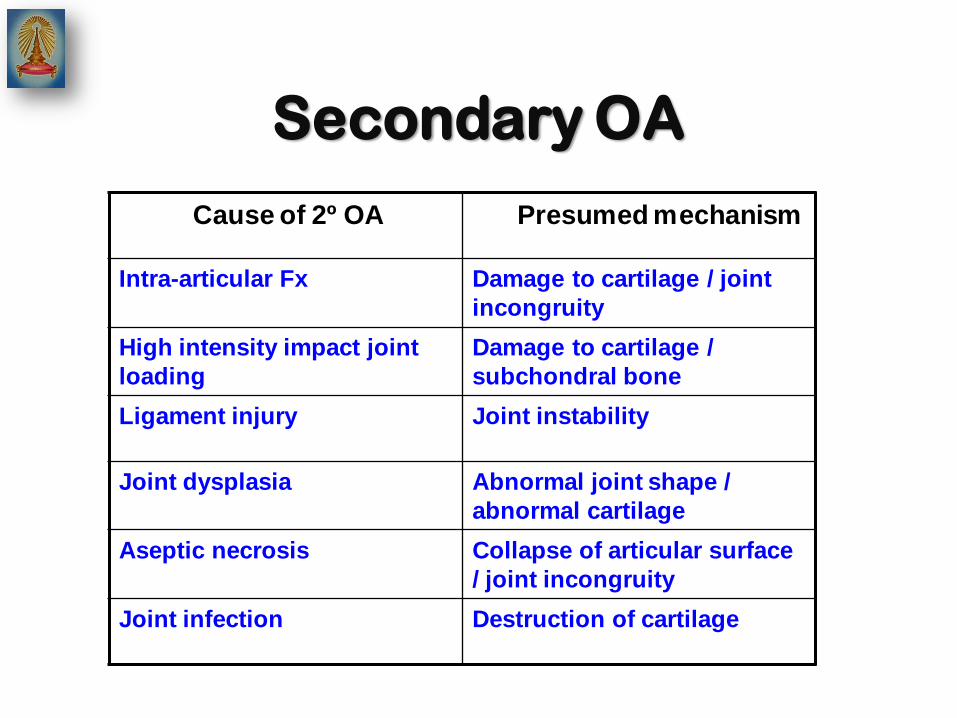
Cause of 2º OA Presumed mechanism
Intra-articular Fx Damage to cartilage / joint
incongruity
High intensity impact joint
loading
Damage to cartilage /
subchondral bone
Ligament injury Joint instability
Joint dysplasia Abnormal joint shape /
abnormal cartilage
Aseptic necrosis Collapse of articular surface
/ joint incongruity
Joint infection Destruction of cartilage
Secondary OA
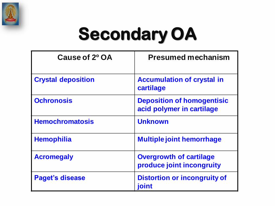
Cause of 2º OA Presumed mechanism
Crystal deposition Accumulation of crystal in
cartilage
Ochronosis Deposition of homogentisic
acid polymer in cartilage
Hemochromatosis Unknown
Hemophilia Multiple joint hemorrhage
Acromegaly Overgrowth of cartilage
produce joint incongruity
Paget’s disease Distortion or incongruity of
joint
Secondary OA
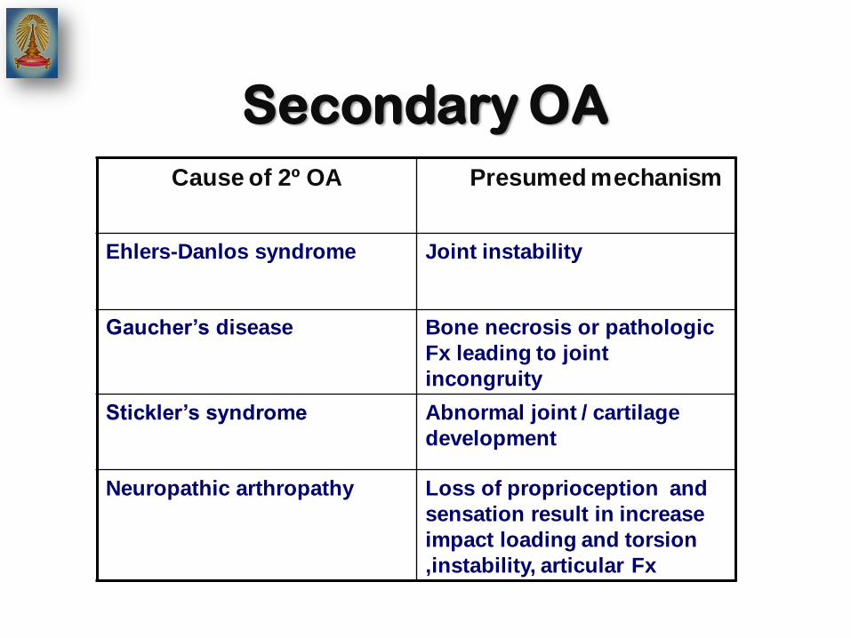
Cause of 2º OA Presumed mechanism
Ehlers-Danlos syndrome Joint instability
Gaucher’s disease Bone necrosis or pathologic
Fx leading to joint
incongruity
Stickler’s syndrome Abnormal joint / cartilage
development
Neuropathic arthropathy Loss of proprioception and
sensation result in increase
impact loading and torsion
,instability, articular Fx
Secondary OA
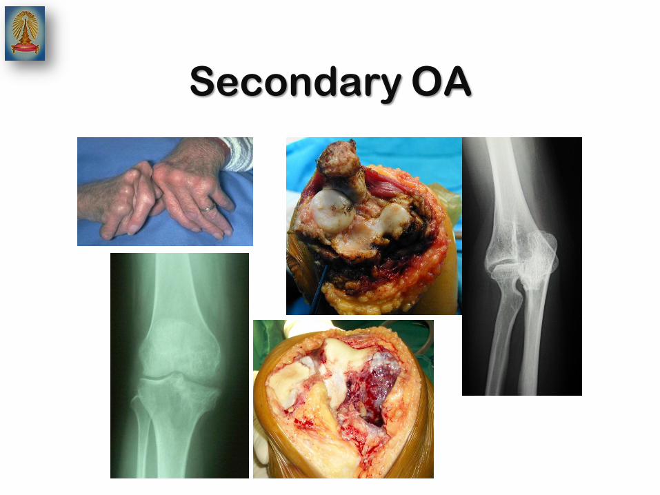
Secondary OA
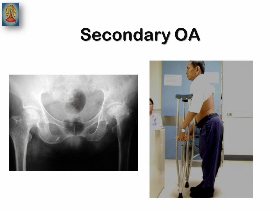
Secondary OA
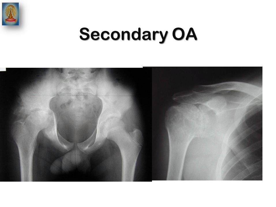
Secondary OA
How is DJD treated?
• Goal of treatment
– Decrease pain
– Muscle strengthening
– Improve or maintain joint function
• Activity of daily living
• Sports activity
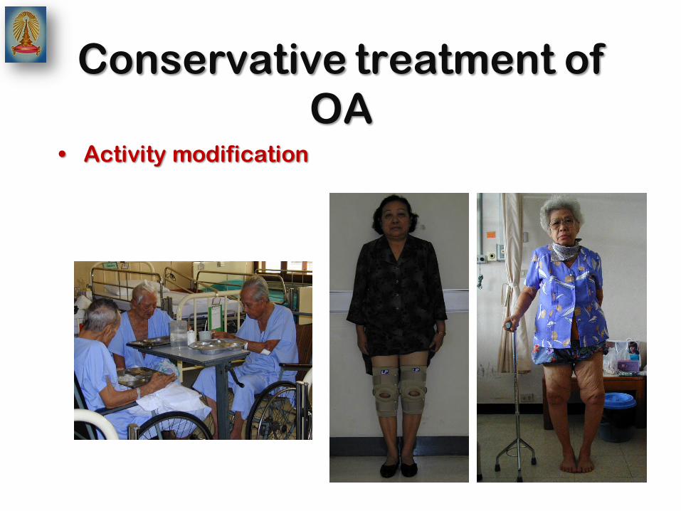
Conservative treatment of
OA • Activity modification
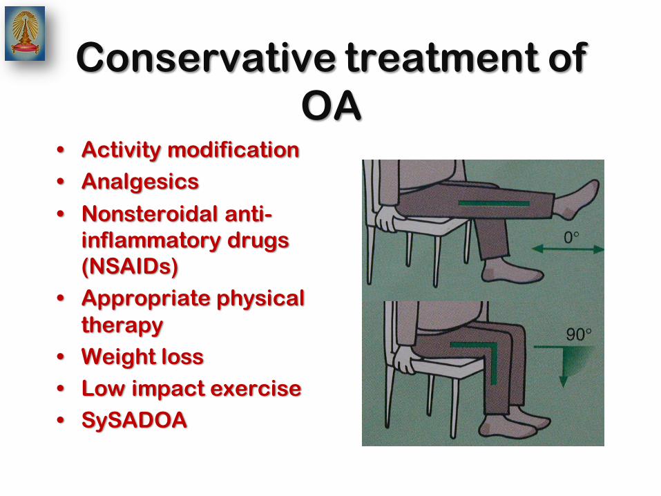
Conservative treatment of
OA • Activity modification
• Analgesics
• Nonsteroidal anti-
inflammatory drugs
(NSAIDs)
• Appropriate physical
therapy
• Weight loss
• Low impact exercise
• SySADOA

Conservative treatment of
OA • SySADOA
• Symptomatic Slow Acting
Drug for Osteoarthritis
– Glucosamine
– Chondroitin
– Diacerein
– Hyalulonic acid injection

Conservative treatment of
OA • Intra-articular administration of
hyaluronic acid

Surgical treatment of OA
• Arthroscopy
– Early symptom
– Mild pathology –less deformity
– Mechanical causes
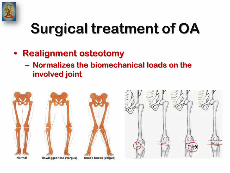
Surgical treatment of OA
• Realignment osteotomy
– Normalizes the biomechanical loads on the
involved joint
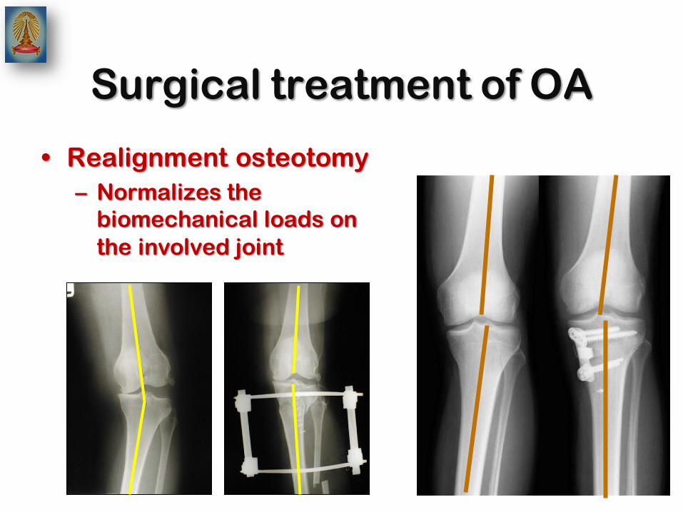
Surgical treatment of OA
• Realignment osteotomy
– Normalizes the
biomechanical loads on
the involved joint

Surgical treatment of OA

Surgical treatment of OA
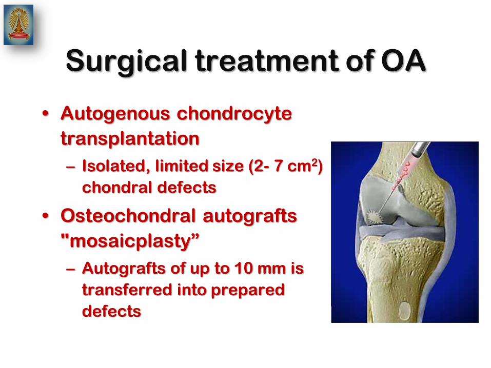
Surgical treatment of OA
• Autogenous chondrocyte
transplantation
– Isolated, limited size (2- 7 cm2)
chondral defects
• Osteochondral autografts
"mosaicplasty”
– Autografts of up to 10 mm is
transferred into prepared
defects

Surgical treatment of OA
• Knee arthroplasty
– Unicompartmental knee arthroplasty
– Total knee arthroplasty

Surgical treatment of OA
• Conventional total knee replacement
(arthroplasty)
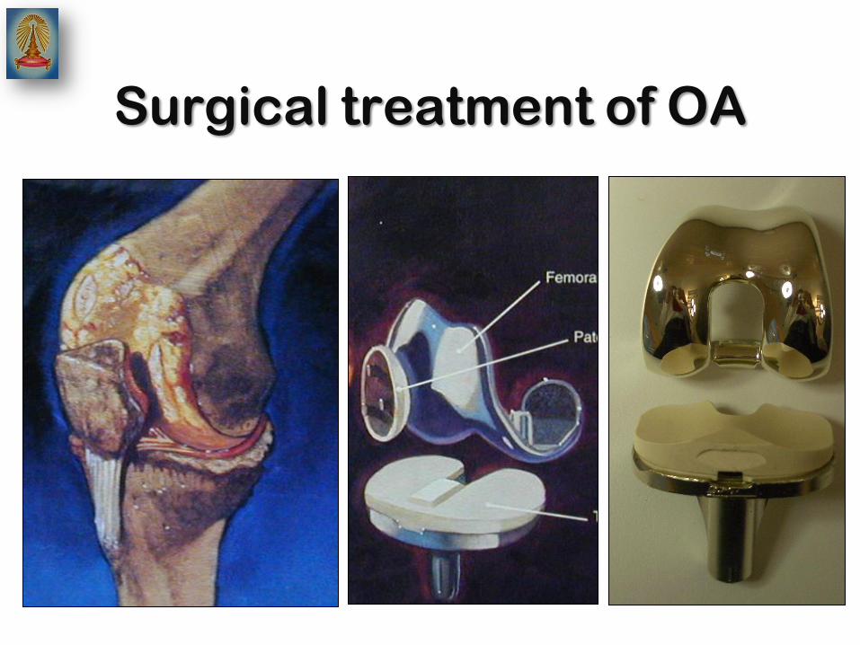
Surgical treatment of OA

Surgical treatment of OA
• Unicompartmental knee replacement
(arthroplasty)
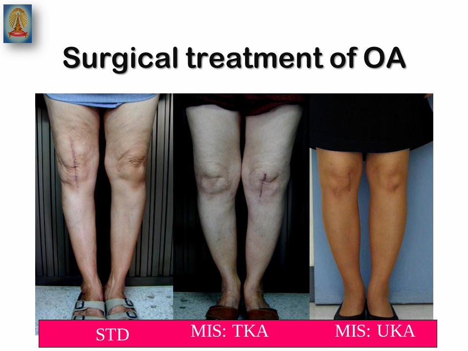
Surgical treatment of OA
STD MIS: TKA MIS: UKA
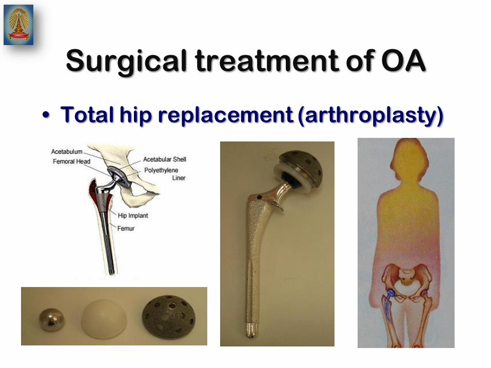
Surgical treatment of OA
• Total hip replacement (arthroplasty)
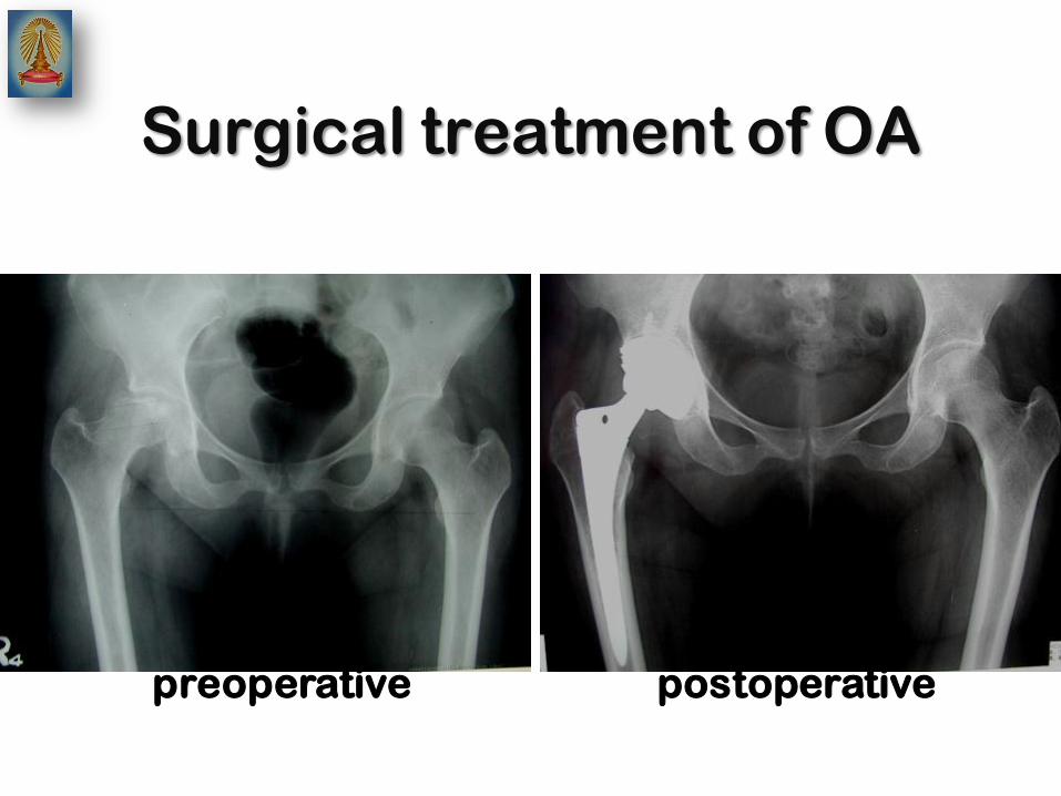
Surgical treatment of OA
preoperative postoperative

Surgical treatment of OA
• Conventinal surgical technique

Surgical treatment of OA
• Minimally invasive surgical technique

Thank you