Embed Size (px)
Citation preview
INVITED MEDICAL REVIEW
Defining the genomic landscape of head and neck cancersthrough next-generation sequencing
G Rizzo1,2,3,4, M Black1,2,3,4, JS Mymryk1,2,3,5,6, JW Barrett1,2,3, AC Nichols1,2,3,4,6
1Department of Otolaryngology Head & Neck Surgery, Western University, London, ON; 2London Regional Cancer Program, London,ON; 3Lawson Health Research Institute, London, ON; 4Department of Anatomy and Cell Biology, Western University, London, ON;5Department of Microbiology and Immunology, The University of Western Ontario, London, ON; 6Department of Oncology, WesternUniversity, London, ON, Canada
Next-generation sequencing (NGS) has revolutionized
the field of genomics and improved our understanding
of cancer biology. Advances have been achieved by
sequencing tumor DNA and using matched normal
DNA to filter out germ line variants to identify cancer-
specific changes. The identification of high incidences of
activating mutations in head and neck squamous cell
carcinoma (HNSCC) amenable to drug targeting has
been made, with clear distinctions between the muta-
tional profile of HPV-positive and HPV-negative tumors.
This wealth of new understanding undoubtedly amelio-
rates our understanding of HNSCC cancer biology and
elucidates clear targets for drug targeting which will
guide future personalized medicine.
Oral Diseases (2014) doi: 10.1111/odi.12246
Keywords: head and neck cancer; genomics; sequencing;
therapeutic targets; human papillomavirus; HPV; head and neck
squamous cell carcinoma; next-generation sequencing; PI3K/Akt
pathway; NOTCH pathway; cell cycle control; E6; E7
Introduction
The genomic era for cancer biology has arrived. Theinvaluable information obtained through the human gen-ome project paired with technological advances hasallowed the development of rapid, accurate platforms tocarry out comprehensive genetic characterization of germline and tumor samples. This technology, which is oftenreferred to as next-generation sequencing (NGS), hasallowed characterization of many of the major cancertypes at either the exome, or less frequently whole-gen-
ome level (Ferlay et al, 2010). These studies have pro-vided a wealth of information that has already begun totransform cancer diagnostics and therapeutics (Danceyet al, 2012).
Head and neck squamous cell cancer (HNSCC) includesmalignant squamous lesions arising in the oral cavity,nasal cavity, larynx, and pharynx. It is the sixth mostcommon cancer by incidence worldwide, with approxi-mately 600 000 new diagnoses per year (Ferlay et al,2010). Heavy tobacco use and alcohol consumption havelong been recognized as etiologic factors for HNSCC;however, infection with high-risk human papillomavirus(HPV) has more recently been identified as an indepen-dent cause of HNSCC. In fact, HPV infection nowaccounts for approximately 25% of all HNSCC cases (Fer-lay et al, 2010; Chaturvedi et al, 2011). HPV-related headand neck cancers tend to occur in younger, non-smokingpatients who experience superior survival outcomes whencompared with HPV-negative patients (Ang et al, 2010).Indeed, HPV-positive HNSCC is a distinct epidemiologic,molecular, and clinical entity and is increasing at an epi-demic rate (Chaturvedi et al, 2011; Nichols et al, 2013).Despite a favorable prognosis for most HPV-positivepatients, approximately 20% fail therapy, with more thanhalf due to distant metastases (Nichols et al, 2013). Incontrast, HPV-negative patients are at a higher risk oflocal, regional and distant relapse, with approximately50% succumbing to the disease (Ferlay et al, 2010).
In addition to mortality, both the disease and its treat-ment can often have marked patient morbidity. Indeed,HNSCC is unique as treatments often impact the mostpersonal characteristics of an individual, including facialappearance and the ability to eat and speak. Other frequentside effects include pain, fibrosis, hearing loss, renalimpairment, and neurotoxicity (Machtay et al, 2008).Thus, there are tremendous clinical challenges to delivereffective therapy that maximizes cure, while minimizingthese sequelae. Understanding the mutational landscape ofHNSCC through NGS has the potential to develop preci-sion, targeted treatment strategies to achieve this goal.
Correspondence: Anthony Nichols, Victoria Hospital, London Health Sci-ence Centre, Department of Otolaryngology – Head and Neck Surgery,Room B3-431A, 800 Commissioners Road East, London, ON, CanadaN6A 5W9. Tel.: 519 685 8804, Fax: 519-685-8567, E-mail: [email protected] 24 March 2014; accepted 30 March 2014
Oral Diseases (2014) doi:10.1111/odi.12246© 2014 John Wiley & Sons A/S. Published by John Wiley & Sons LtdAll rights reserved
www.wiley.com

Tumors of the head and neck possess a large degree ofheterogeneity with few consistent mutations across patientcohorts. This underscores the difficulty in identifyingeffective molecular targeting agents that will benefit a sub-stantial population of HNSCC patients (Stransky et al,2011; Lui et al, 2013). The majority of HNSCC tumorshave loss of function mutations in tumor suppressor genes,thereby making effective drug targeting challenging(Stransky et al, 2011; Liu et al, 2013). Confounding theproblem is the rise of a virally induced second distincttype of HNSCC. As stated previously, these two head-and-neck-associated cancers are very distinct diseases withdivergent clinical manifestations and treatment outcomes(Gillison, 2004; Chaturvedi et al, 2011; Stransky et al,2011). Historically, HNSCCs were caused by environmen-tal carcinogens introduced by heavy tobacco and alcoholuse, which lead to a wide variety of mutations and epige-netic modifications (Gillison, 2004; Stransky et al, 2011).Tobacco and alcohol act as mutagenic agents by damagingcellular DNA and introducing mutations in vital DNArepair genes. In contrast, HPV-related tumors express viraloncoproteins (e.g., HPV-E6 and HPV-E7), which functionto inhibit and degrade critical tumor suppressor gene pro-ducts [e.g., p53 and the retinoblastoma protein (pRb),respectively], thereby overriding these critical cellular con-trol mechanisms without the introduction of cellular genemutations. Consistent with this mechanism, HPV-negativeHNSCCs have been found to harbor more than twice asmany mutations as HPV-positive HNSCC tumors (4.83mutations/MBp and 2.28 mutations/MBp, respectively)(Stransky et al, 2011). These higher mutation rates areconsistent with other smoking-related cancers (Stranskyet al, 2011).Next-generation sequencing has provided valuable
insight into the genetic integrity of genes encoding pro-teins involved in signaling pathways that may contributeto the development, maintenance, and progression of thedisease. Although head and neck cancers are dominatedby mutations in tumor suppressors, NGS has helpeduncover activating mutations that can be targeted withsmall molecule inhibitors (SMIs). This is pivotal to under-standing tumor biology and thus developing individualizedstrategies for patient care. However, much work remainsto be done, as cure rates have remained relatively constantover the past 30 years. This review offers insight into per-sonalized therapy by summarizing the current findings ofNGS by highlighting the most commonly mutated path-ways in head and neck malignancies.
Next-generation sequencing
NGS has revolutionized the field of genomics. It is nowpossible to routinely extract an organism’s DNA and per-form large-scale, and cost-effective, highly accurate, gen-ome-scale sequence data acquisition. These more recentsequencing technologies have succeeded the traditionalSanger method (Cabelguenne et al, 2000), while replacingmicroarray analysis as the platform for discovery andgenotyping (Alkan et al, 2011). As the technology contin-ues to improve, the once-high cost of sequencing will con-tinue to decline, allowing this technology to be much
more readily available for studies of cancer biology(Kozarewa et al,2012). In the near future, this will affordresearchers the power to study, understand, and proactive-ly treat a wide panel of mutations found in malignanttumors.
Although NGS can be highly useful to detect germ linevariants that predict cancer risk or treatment toxicity (e.g.,BRCA1 and 2), the clearest advances have been obtainedby sequencing tumors along with matched normal DNA,which is often obtained from blood. This strategy allowsexclusion of germ line variants to report solely somaticchanges. It is increasingly recognized that many changesrepresent less significant ‘passenger’ mutations with asmall number of true ‘driver’ mutations (Lawrence et al,2013). As bioinformatics expertise increases, identifiedmutations can be further analyzed by expression rates,gene size, and gene replication timing to identify thosedriver mutations truly responsible for carcinogenesis (Law-rence et al, 2013).
A variety of NGS techniques exist, ranging fromfocused DNA sequencing for the detection of only well-established clinically relevant point mutations, to the char-acterization of the coding genome (whole-exome sequenc-ing – WES), the whole genome (whole-genomesequencing – WGS), copy number alterations (derivedfrom copy number arrays, WES or WGS), assessment ofmRNA abundance and transcript sequence (RNA sequenc-ing), and translocation detection (WGS and RNA sequenc-ing). These techniques are complementary and allow acomplete molecular profile of each cancer. Indeed, recentpublications from The Cancer Genome Atlas (TCGA)have highlighted the need for multiplatform integratedanalysis of these techniques with the aim of comprehen-sively identifying molecular genomic targets in each can-cer type (Koboldt et al, 2012; Muzny et al, 2012;Creighton et al, 2013).
NGS of HNSCC
The study of HNSCC biology through NGS efforts hasled to a clearer understanding of the molecular and etio-logical aspects of HNSCC. The presence or absence ofHPV has resulted in two different forms of head and neckcancer. These are etiologically and molecularly distinct,sharing only similarities in their anatomical locations andtheir targets for hijacking cell function. The first NGSscreens of HNSCC patient tumors described mutationalevents in key cell cycle components, as well as down-stream effectors of mitogenic signaling (Agrawal et al,2011; Stransky et al, 2011). Additional discoveriesincluded mutations in cell differentiation pathways, partic-ularly NOTCH1 and FXBW7 (Agrawal et al, 2011).
HPV-negative tumors, arising from repeated exposureto carcinogens, tend to harbor approximately twice asmany mutations per mega-base pair as compared to theirHPV-positive counterparts (Stransky et al, 2011; Nicholset al, 2012; Lui et al, 2013). HPV-negative tumors have ahigher degree of transversions at CpG sites, which is char-acteristic of smoking malignancies (Stransky et al, 2011).In contrast, HPV-positive tumors have more transitions atTpC sites based on TCGA data (accessed through www.
Oral Diseases
NGS and HNSCC
G Rizzo et al
2

cbioportal.org). In addition, mutations in tumor suppressorgenes tend to occur more often in HPV-negative tumors(Agrawal et al, 2011; Stransky et al, 2011). For example,TP53 loss is almost ubiquitous across HPV-negativetumors and is typically associated with high incidences ofCDK2NA loss and/or CCN1 gain (Stransky et al, 2011;Lechner et al, 2013; Pickering et al, 2013).Conversely, HPV-positive tumors, despite their lower
incidence of mutations, appear to have more mutations inthe oncogene PIK3CA (Liu et al, 2013; Nichols et al,2013). HPV-positive tumors have activating mutations in28% of cases, versus only 10% of HPV-negative tumors(Stransky et al, 2011; Liu et al, 2013; Nichols et al,2013). This is significant, as PIK3CA is the only fre-quently mutated, directly targetable oncogene in HNSCC.In contrast, it is much more problematic to re-establishwild-type expression of a mutated tumor suppressor geneor to target the HRAS mutations that occur in approxi-mately 5% of cases (Stransky et al, 2011). Together withobserved outcome differences, these findings confirm thatHPV-positive and HPV-negative HNSCC are distinct clin-ical and molecular diseases.The following sections highlight the most common
mutations or pathway alterations in HNSCC, which arethought to play an important role in oncogenicity. To date,there is no known genetic predisposition to HNSCC, noris there a common targetable mutational event (excludingTP53) seen across nearly all patients. This review high-lights a large number of potentially targetable mutations,all made known through NGS studies.
The role of HPVHPV infection and progression to HPV-induced cancerrelies on the ability of the virus to avoid host immunedetection and establish a persistent infection (Andersenet al, 2013). This is considered a key risk factor in HPV-induced cancer, as the likelihood of integration of theHPV genome into the DNA of the host cell becomes evergreater with increasing time. In addition to immune eva-
sion mechanisms encoded by the virus, mutations that aidin immune evasion are present in HPV-related HNSCC,including focal deletions and point mutations in theTRAF3 gene, which contributes to both innate andacquired anti-viral response (Oganesyan et al, 2006,TCGA [http://www.cbioportal.org]).
It has long been accepted that the integrated HPV ge-nomes found in HPV-induced cancers have preferentiallylost a functional viral E2 open reading frame, due in partto the fact that E2 antagonizes the role of the oncogenicE6 and E7 proteins (Thierry and Yaniv, 1987; Akagi et al,2013). The viral E6 and E7 oncogenes are critical as theyare consistently retained and expressed in HPV-positivetumors and cell lines despite the frequent fragmentation ofHPV-integrants (Akagi et al, 2013; Khoury et al, 2013).
Genomic instability is also considered a necessity forviral-mediated carcinogenesis, and this is in part achievedby abrogating cell cycle checkpoints. Indeed, the HPVviral proteins E6 and E7 simultaneously attack TP53 andpRB, inducing their degradation by the proteasome(Moody and Laimins, 2010) (Figure 1). However, a moredirect role has been proposed that credits HPV integrationwith a much greater role in the generation of genomicinstability (Akagi et al, 2013). In a panel of 5 HPV-posi-tive HNSCC cell lines, 2 HPV-positive HNSCC tumors,as well as 2 HPV-positive cervical cancer cell lines, arecurrent pattern of extensive, focal copy number variation(CNVs), and/or other forms of structural variation immedi-ately adjacent to the HPV integration events wereobserved (Akagi et al, 2013). Mechanistically, this mayresult from the dependence of HPV on the host cell DNAreplication apparatus for its own replication. Cellular DNApolymerase and accessory factors are recruited to the viralorigin of replication (OR) by the E2 and E1 HPV-replica-tion proteins (Rowley, 1973; Sakakibara et al, 2011).Once integrated into the host cell genome, the viral originmay continue to function in this capacity, resulting inlocal expansion of integrated viral sequences and flankingcellular DNA. This has been confirmed experimentally, as
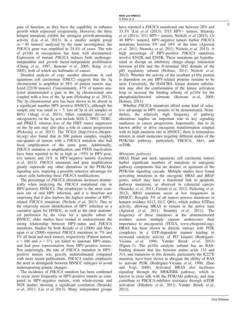
Figure 1 The oncogenic and tumor suppressivecomponents most commonly altered in headand neck cancers as discussed in this review,and their resultant effects on terminaldifferentiation, cell proliferation, and apoptosis.Red squares denote oncogenes, blue circlesdenote tumor suppressors, yellow circlesindicate components with an unknown function,and green circles denote HPV viral oncogenes
Oral Diseases
NGS and HNSCC
G Rizzo et al
3

reintroduction of the E1 and E2 HPV-replication proteinsinto HPV-positive cancers induces overamplification ofthe genomic locus where the HPV OR has been integrated(Kadaja et al, 2007).As a result, concatemers containing both viral and host
sequences adjoined by identical HPV-breakpoint sequencesare produced (Akagi et al, 2013; Khoury et al, 2013). Thisamplification step helps to explain how HPV16-infected can-cer cells have up to 1000 copies of HPV-integrants in theirgenome (Flores and Lambert, 1997). Replication of the chi-meric host-viral DNA sequence has been proposed to occurby rolling circle mode of DNA replication from the HPV OR(Flores and Lambert, 1997; Akagi et al, 2013). This rollingamplification is achieved by the formation of a transient‘looping’ structure comprised of HPV-integrants and adja-cent host chromosomal regions. The accumulation of thesehost-viral concatemeric sequences and their subsequent post-replication repair results in the high incidences of amplifica-tions, and rearrangements in HNSCC (Akagi et al, 2013).HPV-directed amplification provides a novel means by
which genetic alterations are introduced into cells, greatlyincreasing the prospect of progression to a cancerous phe-notype. What remains to be understood is whether thisHPV integration is site-directed, which would help explainthe high incidences of genomic amplifications in HPV-positive HNSCC. Additionally, understanding the func-tional relationship between specific mutations and HPVneeds to be elucidated. The roles of HPV E6 and E7 oncell cycle progression have been established, yet HPV’seffect on the host genome integrity as a whole remains tobe understood. It can be supposed that viral effects arefunctionally distinct from the effects of tobacco and alco-hol insult. This is evident by the significant difference inthe mutational profile of the two HNSCC subtypes. How-ever, much remains to be learned about the specific rolesof these changes in cancer progression.
PI3K/Akt PathwayThe phosphatidylinositol-4,5-bisphosphate 3-kinase(PI3K)/Akt signaling cascade is the most commonlymutated pathway, with alterations observed in many can-cers, including a subset of head and neck cancers (Samu-els et al, 2004; Ikenoue et al, 2005; Kang et al, 2005).PI3Ks phosphorylate phosphatidylinositol and its phos-phorylated derivatives, generating second messengers thatcontrol proliferation, survival, motility, and morphology(Osaki et al, 2004) and are composed of a regulatory sub-unit (p85) and a catalytic subunit (p110) (Osaki et al,2004). The binding of the p85 src-homology 2 (SH2)domain to particular phosphotyrosine residues, often inresponse to receptor tyrosine kinase (RTK) activation,localizes the PI3K complex to the plasma membranewhere it phosphorylates phosphatidylinositol-4,5-bisphos-phate (PIP2) to its active phosphatidylinositol-3,4,5-tri-phosphate (PIP3) form. This localizes and activates Akt,which will eventually alleviate mammalian target of rapa-mycin (mTOR) inhibition by phosphorylating the TSC1/2proteins. mTOR is then able to activate downstream cellproliferation, motility, adhesion, and survival pathways(Samuels et al, 2004; Kozaki et al, 2006). In normalphysiology, PI3K is regulated by the tumor suppressor
phosphatase and tensin homolog (PTEN), which catalyzesthe reverse reaction from active PIP3 to inactive PIP2(Vander Broek et al, 2013).
Aberrant PI3K/Akt signaling is activated throughsomatic mutations, RTK stimulation, particularly the epi-dermal growth factor receptor (EGFR) and the insulin-likegrowth factor receptor 1 (IGF-1R), or G-protein-coupledreceptors (Vander Broek et al, 2013). This pathway haseven been suggested to be so important to oncogenicitythat cancer cells will actively maintain a certain homeo-static level of PI3K-AKT signaling in face of therapeuticintervention (Holmes, 2011).
Mutations within the PI3K pathway have also beenlinked to genomic instability. Specifically, PI3K pathway-mutated HNSCC tumors harbor 2.3 times more non-synonymous mutations, many of which occur withincancer-associated genes, than tumors with a wild-type PI3Kpathway (Stransky et al, 2011; Liu et al, 2013). Thissuggests that PI3K pathway mutations in HNSCC mayfacilitate the expansion or selection of tumor cells withgreater genomic instability, particularly with mutations inknown cancer-associated genes (Liu et al, 2013).
Several groups have confirmed high incidences ofPI3K/Akt pathway mutations in HNSCCs. Anatomically,laryngeal tumors tend to harbor the greatest number ofPI3K pathway mutations compared with other sites. Liuet al (2013) found a 30.5% (46/151 tumors) mutation ratein the PI3K/Akt pathway, more than double the amount ofmutations found in any of the other major signaling axes(JAK/STAT or MAPK). Similarly, Lechner et al (2013)reported PIK3CA mutation or amplification, as well asPTEN inactivation by gene copy loss or mutation, in bothHPV-positive and HPV-negative tumors (n = 40 of eachtype, 60% and 31% of any change, respectively). Muta-tions at multiple levels of the PI3K pathway appear to becommon in HPV-negative HNSCC (21.7%, 10/46 tumors)(Stransky et al, 2011; Liu et al, 2013). Similar compound-ing mutations are rare in the JAK/STAT and MAPK sig-naling pathways in HNSCC (Liu et al, 2013).Interestingly, tumors with multiple mutations in the PI3Ksignaling axis were found only in advanced-stage (StageIV) HNSCC (Liu et al, 2013). The role of multiple PI3Kmutations on disease progression remains to be elucidated,but this could be a direct byproduct of continued genomicinsult.
PIK3CA. The most common mutation in the PI3Kpathway occurs within the coding region of PIK3CA,which is mutated in 10–15% of all HNSCCs (Liu et al,2013) (Figure 1). PIK3CA is located at chromosomalposition 3q26.3 and encodes the p110a catalytic subunitof the class 1A of the PI3K family (Ikenoue et al, 2005;Kozaki et al, 2006). Ninety percent of PIK3CA mutationsare clustered at three hotspots: two missense mutations inexon 9, which alter amino acids 542 [glutamic acid (E) tolysine (K)] and 545 (E to K) in the helical domain, and amissense mutation in exon 20, which alters amino acid1047 [histidine (H) to arginine (R)/leucine (L)] (Holmes,2011). An additional four novel PIK3CA mutations(R115L, G363A, C971R, and R975S) have been reported(Liu et al, 2013). All of these mutants appear to exhibit
Oral Diseases
NGS and HNSCC
G Rizzo et al
4
gain of function, as they have the capability to enhancegrowth when expressed exogenously. However, the threehotspot mutations exhibit the strongest growth-promotingactivity (Liu et al, 2013). In a smaller sample group(n = 45 tumors) analyzed by the same investigators, thePIK3CA gene was amplified in 24.4% of cases. The roleof p110a in oncogenesis has been well documented.Expression of mutant PIK3CA induces both anchorage-independent and growth factor-independent proliferation(Chang et al, 1997; Ikenoue et al, 2005; Kang et al,2005), both of which are hallmarks of cancer.Detailed analysis of copy number alterations in oral
squamous cell carcinomas (OSCC) suggests that the 3qchromosome is amplified in 58% of patient tumors ana-lyzed (22/38 tumors). Concomitantly, 47% of tumors ana-lyzed demonstrated a gain in the 3q chromosomal armcoupled with a loss of the 3p arm (Pickering et al, 2013).The 3p chromosomal arm has been shown to be absent ina significant number HPV-positive HNSCCs, although thesample size was small (n = 7, loss of 3p in six samples or86%) (Akagi et al, 2013). Other candidate drivers ofoncogenicity on the 3q arm include SOX-2, TP63, TERC,and PRKCI, whereas loss of the FHIT tumor suppressoron the 3p arm may also play a role in cancer progression(Pickering et al, 2013). The TCGA (http://www.cbiopor-tal.org) also found that in 500 patient samples, roughlyone-quarter of tumors with a PIK3CA mutation also hadfocal amplification of the same gene. Additionally,PIK3CA mutation or amplification, and PTEN inactivationhave been reported to be as high as >55% in HPV-posi-tive tumors and 31% in HPV-negative tumors (Lechneret al, 2013). PIK3CA mutations and gene amplificationgreatly supersede any other alterations in the PI3K/Aktsignaling axis, implying a possible selective advantage forcancer cells harboring these PIK3CA modifications.The percentage of PIK3CA alterations increases dramat-
ically when analyzing the PIK3CA mutational rate inHPV-positive HNSCCs. The oropharynx is the most com-mon site of oral HPV infection, and it is therefore notsurprising that it also harbors the highest number of HPV-related PIK3CA mutations (Nichols et al, 2013). Due tothe relatively recent identification of HPV infection as acausative agent for HNSCC, as well as the clear anatomi-cal preference by the virus for a specific subset ofHNSCC, older studies have tended to underestimate thestrong relationship between HPV-status and PIK3CAmutations. Studies by both Kozaki et al (2006) and Mur-ugan et al (2008) reported PIK3CA mutations in 7% and5% all head and neck tumors, respectively (Patient tumors,n = 108 and n = 37), yet failed to annotate HPV-status,and had poor representation from HPV-positive tumors.Not surprisingly, the rate of PIK3CA mutation in HPV-positive tumors was gravely underestimated comparedwith more recent publications. PIK3CA studies emphasizethe need to distinguish between HNSCC subtypes to avoidunderreporting genetic changes.The incidence of PIK3CA mutation has been confirmed
to occur more frequently in HPV-positive tumors as com-pared to HPV-negative tumors, with whole-exome andNGS studies showing a significant correlation (Stranskyet al, 2011; Liu et al 2013). Many independent groups
have reported a PIK3CA mutational rate between 28% and33.3% [Liu et al (2013): 5/15 HPV+ tumors; Stranskyet al (2011): 3/11 HPV+ tumors; Nichols et al (2013) 13/46 HPV+ tumors]. HPV-negative tumors harbor PIK3CAmutations between 5% and 10% of the time (Agrawalet al, 2011; Stransky et al, 2011; Nichols et al, 2013). Ahigh percentage of HPV-positive PIK3CA mutationsinvolve E542K and E545K. These mutations are hypothe-sized to disrupt an inhibitory charge–charge interactionbetween p110a and the N-terminal SH2 domain of thep85 regulatory subunit (Holmes, 2011; Nichols et al,2013). Whether the activity of the resultant p110a proteinis dependent on any HPV-related proteins remains to beseen. Conversely, the H1047R/L kinase domain substitu-tion may alter the conformation of the kinase activationloop or increase the binding affinity of p110a for thephosphatidylinositol substrate (Ikenoue et al, 2005;Holmes, 2011).
Whether PIK3CA mutations afford some kind of selec-tive advantage to HPV remains to be demonstrated. None-theless, the relatively high frequency of pathwayalterations implies an important role in key signalingmediators in cancer progression. Importantly, due to itsknown ability to drive oncogenic transformation coupledwith its high mutation rate in HNSCC, there is tremendousinterest in small molecules targeting different nodes of thePI3K/Akt pathway, particularly PIK3CA, Akt1, andmTOR.
Mitogenic pathwaysHRAS. Head and neck squamous cell carcinoma tumorsharbor significant numbers of mutations in mitogenicpathway components that are distinct, or upstream of thePI3K/Akt signaling cascade. Multiple studies have foundactivating mutations in the oncogenic HRAS and BRAFgenes, which may have a functional link to apoptoticpathway mutations, as observed in colorectal cancers(Stransky et al, 2011; Cerami et al, 2012; Pickering et al,2013). HRAS mutations occur at low frequencies inHNSCC. Roughly 5% of all tumors possess mutations inhotspot residues (G12, G13, Q61), which reduce GTPaseactivity, allowing HRAS to remain in the active state(Agrawal et al, 2011; Stransky et al, 2011). Thefrequency of these mutations at the aforementionedresidues across multiple cancers underscores theirimportance to oncogenesis (TCGA, www.cbioportal.org).HRAS has been shown to directly interact with PI3Kcomplexes in a GTP-dependent manner leading toincreased catalytic activity of PI3 kinases (Rodriguez-Viciana et al, 1996; Vander Broek et al, 2013)(Figure 1). The p110a catalytic subunit has an RAS-binding domain that lies between amino acids 133 and314, and mutations in this domain, particularly the K227Emutation, have been shown to abrogate the ability of RASto activate PI3K (Rodriguez-Viciana et al, 1996; Zhaoand Vogt, 2008). Activated HRAS also facilitatessignaling through the MEK/ERK pathway, which isknown to cross talk with the PI3K/Akt pathway, and maycontribute to PIK3CA-inhibitor resistance through mTORactivation (Elkabets et al, 2013; Vander Broek et al,2013).
Oral Diseases
NGS and HNSCC
G Rizzo et al
5
Importantly, of the three RAS family members,HNSCCs almost exclusively seems to exhibit HRAS muta-tions, which is contrary to other cancers where KRAS isthe predominant mutated RAS (Paterson et al, 1996). Thisis significant, because activating mutations are rare inHNSCC, and the oncogenic nature of HRAS mutationspermits potential targeting with SMIs. Unfortunately, thereare no successful strategies to directly target RAS despitesignificant drug development efforts (Downward, 2003).Thus, current areas of investigation target upstream anddownstream pathway members that are directly druggableincluding mitogen-activated protein kinase kinase (MEK)(Roberts and Der, 2007).
Epidermal growth factor receptor. Epidermal growthfactor receptor, also known as ErbB-1 or HER1 (VanderBroek et al, 2013), is an RTK upstream of multiple cellsignaling cascades. It particularly influences the PI3K/Aktand HRAS pathways (Yarden, 2001) (Figure 1). EGFRgenetic alterations are frequent in epithelial cancers, withHNSCC being no exception (Bonner et al, 2006). Incontrast to lung cancer, activating point mutations inEGFR are extremely rare in HNSCC. However, >10% oftumors have amplifications in the EGFR gene (Lechneret al, 2013). In addition, EGFR CNVs or increased EGFRprotein expression has been shown to correlate with poorprognosis in HNSCC (Grandis and Tweardy, 1993). Morerecently, our group has showed a positive correlationbetween expression of the HPV E5 oncogene and EGFRexpression in HNSCC patient samples (S.H. Um, N.Mundi, J. Yoo, D.A. Palma, K. Fung, D. MacNeil, B.Wehrli, J.S. Mymryk, J.W. Barrett, A.C. Nichols,unpublished data). Additionally, Lechner et al (2013) haveidentified a low frequency of mutations in other RTKs,notably the fibroblast growth factor receptors (FGFR)genes FGFR1 and FGFR3 at low frequencies in HPV-negative HNSCC. Taken together, these data suggest thattreatment with anti-EGFR therapies may have some utilityin a subset of HNSCC.Cetuximab, a monocloncal antibody directed against
EGFR, is the only FDA approved targeted agent for thetreatment of HNSCC in light of level 1 evidence demon-strating increased survival when added to radiation alone(Bonner et al, 2006). However, resistance to EGFR inhibi-tors is common (Chong and J€anne, 2013), and there is asuggestion that these agents only benefit HPV-positivepatients (Eriksen et al, 2010). Resistance to EGFR inhibi-tors in HNSCC remains challenging, possibly becausemany cancers contain a significant number of downstreammutations in this pathway (Stransky et al, 2011; Pickeringet al, 2013). As one specific example, treatment ofHNSCC cell lines expressing mutant-EGFR with erlotinib,an EGFR-targeting SMI, was ineffective when the cellharbored an HRAS mutation (Hah et al, 2013). Thisunderscores the need for predictive biomarkers of sensitiv-ity and resistance to EGFR-targeted therapy to identify thepatient population most likely to benefit from these oftenexpensive targeted agents (Hansen and Siu, 2013). A strat-egy to overcome resistance in genetically complex tumorsharboring multiple mutations is the use of a combinationof inhibitors. NGS therein becomes paramount in the
analysis of patient tumor genetics to properly direct theapplication of this precise treatment.
FGFR3–TACC3 fusions. Recurrent gene fusion mutationsare an important class of driver mutations that possessoncogenic potential. Perhaps the best-characterizedtranslocation in cancer is the BCR-ABL gene fusion inchronic myeloid leukemia (CML), in which nearly allCML patients harbor this mutation (Rowley, 1973).Importantly, this fusion predicts response to BCR-ABLinhibitors, resulting in markedly improved patient survival.Outside hematological cancers, multiple other cancersharbor fusions that predict drug sensitivity, such as ALKfusions in non-small cell lung cancer and the androgen-regulated gene TMPRSS2 with E26-transformation-sequences (ETS) in prostate cancer (Tomlins et al, 2005).Gene fusions can be resolved through whole-genomesequencing and RNA sequencing. As increasing numbersof tumor types are characterized on these platforms, thenumber of identified clinically important translocations israpidly increasing.
Recently, FGFR fusions have been identified in multipletumor types. Wu et al (2013) identified new FGFRfusions with intact kinase domains in lung, bladder, brain,and thyroid cancers, as well as three HNSCC samples.Importantly, these FGFR protein fusion partners showedthe propensity for oligomerization, and overexpression ofFGFR fusions promoted cell proliferation (Wu et al,2013). In HNSCC, FGFR3 was fused to the transforming,acidic coiled-coil containing protein 3, TACC3 (Wu et al,2013), whose function has yet to be elucidated, althoughit is thought to play a role in cell growth and differentia-tion (Schneider et al, 2007) (Figure 1). It is highly likelythat the dimeric activation of FGFR3 is achieved throughthe dimerization of TACC3’s coiled-coil domains. This isstrengthened by the observation that FGFR3 cannot dimer-ize on the cell membrane without the addition of fibroblastgrowth factor (FGF), further emphasizing the oncogenicsynergy produced by this fusion with TACC3 (Wu et al,2013). All FGFR fusion partners tested exhibited oligo-merization capability, perhaps utilizing a shared mode ofkinase activation. More importantly, FGFR inhibition sup-pressed tumor growth in vivo, suggesting that this isindeed a clinically relevant genetic change (Wu et al,2013). Review of the HNSCC TCGA data (www.cbiopor-tal.org) revealed two additional TACC3-FGFR3 fusions,both from HPV-positive patients. This suggests the poten-tial to perform prospective screening for this fusion inHNSCC, similar to ALK fusions in lung cancer, allowingfusion-positive patients to selectively receive FGFR inhibi-tor therapy.
FAT1. Changing the profile of a cell’s surface to modifyadhesion is an important step in cancer cell progression.Cancer cells must undergo a dedifferentiation process thatpermits them to metastasize and gain access to newmicroenvironments. Although mutations have beenidentified in many genes encoding cell surface adhesionproteins, HNSCCs tend to have a high incidence ofmutations in the protocadherin FAT1 gene found at the4q35 chromosomal region (TCGA: www.cbioportal.org;
Oral Diseases
NGS and HNSCC
G Rizzo et al
6
Morris et al, 2013) (Figure 1). It has been reported thatFAT1 mutations and homozygous deletion in HNSCCtumors have an incidence rate of 8–12% and 6%,respectively, (Stransky et al, 2011; Morris et al, 2013;TCGA: www.cbioportal.org). HPV-negative tumors have asignificantly higher incidence of FAT1 inactivation with32% of tumors (n = 78/243) having either loss of functionmutations or outright 4q35 chromosomal deletion (TCGA:www.cbioportal.org). FAT1 is a member of the cadherinsuperfamily of proteins and has five EGF-like repeats.Hypothetically, this allows it to function not only as anadhesion molecule, but also as a signaling capacity(Dunne et al, 1996). FAT1 functions as a tumorsuppressor, binding to b-cadherin and blocking nuclearlocalization (Morris et al, 2013). Loss of FAT1, by eithersomatic mutation, homozygous deletions, or inactivatingstructural rearrangements, promotes tumorigenesis throughactivation of Wnt signaling (Morris et al, 2013).Specifically, loss of FAT1 permits b-catenin nuclearlocalization thereby activating expression of targetoncogenes, such as CCND1 and C-MYC (Morris et al,2013).
SOX2. SOX2 has long been recognized as a keytranscription factor in development and cancer, with animportant function in the maintenance of pluripotency inundifferentiated stem cells (Cerami et al, 2012; Wanget al, 2012). As such, SOX2 mutations could have a majorimpact on the development and progression of HNSCC.While a direct role for SOX2 in HNSCC has yet to bedemonstrated, studies have shown that SOX2 is amplifiedin 12% of HNSCC tumors (Lechner et al, 2013)(Figure 1). This is coupled with similar findings insquamous lung cancer (Cerami et al, 2012) andesophageal squamous cell carcinoma (Wang et al, 2012).Moreover, SOX2 is located on the often-amplified 3qchromosomal arm, particularly in the 3q26.3 region wherePIK3CA is also found (Lechner et al, 2013; Pickeringet al, 2013). PIK3CA amplifications have long beenthought of as the oncogenic driver on the 3q chromosome;however, due to the close proximity of SOX2, PIK3CAmay not be solely responsible. Coupled with the fact it isknown to be amplified in HNSCC, Schulein et al (2011)have reported an upregulation of SOX2 expression inputative HNSCC stem cells, which seem to show anepithelial to mesenchymal transition phenotype suggestiveof a role in metastasis.
Cell cycle and DNA repair pathwaysTP53. p53 is regarded as the ‘guardian of the genome’and ‘gatekeeper of the cell’ (Levine, 1997; Surget et al,2013). The primary functions of this transcription factorare to initiate cell cycle arrest thereby allowing theappropriate DNA repair enzymes to rectify genomic insult(Levine, 1997). If cellular repair cannot be accomplished,p53 can initiate cellular apoptosis and/or senescence.Without functional p53, cell cycle control is compromised,allowing incessant cell growth, the accumulation ofgenomic insult, and subsequent progression to cancerousphenotypes (Levine, 1997) (Figure 1). TP53 is the most
commonly mutated gene in HNSCC (Gillison, 2004;Stransky et al, 2011; Liu et al 2013). Disruption of thenormal functions of p53, either by mutation (HPV-negative HNSCC) or by degradation via the HPV16 E6protein (HPV-positive HNSCC), appears to be paramountto HNSCC development (Gillison, 2004). HPV-negativetumors contain TP53 mutations in 73–100% of cases(Agrawal et al, 2011; Stransky et al, 2011; Lechner et al,2013). In contrast, HPV-positive tumors never appear tohave TP53 mutations (Agrawal et al, 2011; Stransky et al,2011), most likely due to the fact that the E6 viral proteinfacilitates the degradation of p53, and mutationalinactivation of TP53 is therefore not required (Akagi et al,2013). The majority of HNSCC TP53 mutations occur asmissense mutations, with a significantly lower incidenceof nonsense, indel, or splice site mutations resulting in theloss of active protein (Agrawal et al, 2011; Stransky et al,2011; Pickering et al, 2013). TP53 mutations primarilyaffect p53’s ability to bind to DNA (Loyo et al, 2013;Surget et al, 2013). Furthermore, TP53, as well as otherfrequently mutated genes, often undergo copy numberchanges. Indeed, Agrawal et al (2011) noted that alongwith being the most frequently mutated gene, TP53 wasalso affected by CNVs.
It has been suggested that p53 and p110a mutations aremutually exclusive, Akt phosphorylates murine double-minute two homolog (MDM2) causing it to localize fromthe cytoplasm to the nucleus. MDM2 is an ubiquitin-pro-tein ligase that induces the degradation of p53 when pres-ent in the nucleus (Vander Broek et al, 2013). Thus,p110a-dependent activation of Akt confers resistance top53-mediated apoptosis, which can be overcome with aPI3K/mTOR inhibitor (Herzog et al, 2013). Conversely,p53-mediated apoptosis requires transcriptional inhibitionof wild-type p110a (Singh et al, 2002). Despite this seem-ingly mutually exclusive relationship, Stransky et al(2011) reported two patient tumors that harbored bothmutations. Given the heterogeneous nature of manytumors, it remains to be determined whether these muta-tions actually exist in the same tumor cells. However,these data suggest that further investigation of the relation-ship between the TP53 and PIK3CA pathways may berequired.
The presence of TP53 mutations can be an early sign ofHNSCC. Indeed, TP53 mutations have been observed inmild dysplastic lesions that progress to invasive carcino-mas (Boyle et al, 1993). Moreover, a large, multicenterstudy of TP53 status in 420 HNSCC patients found thatpatients with TP53 mutations exhibited a 1.5-fold decreaseis survival as compared to those with wild-type TP53. Notsurprisingly, TP53-mutant tumors tend to respond poorlyto cisplatin and fluorouracil-based chemoradiation (Cab-elguenne et al, 2000) and are associated with local recur-rence after radiotherapy (Ganly et al, 2000). This mightexplain why HPV-positive HNSCC patients respond betterthan their HPV-negative counterparts to similar treatment,as HPV-driven oncogenesis is facilitated by the degrada-tion of wild-type p53 rather than its outright loss.
CCND1 and CDKN2A. Head and neck squamous cellcarcinoma tumors have amplification of CCND1, which
Oral Diseases
NGS and HNSCC
G Rizzo et al
7
encodes cyclinD1, and deletion of CDKN2A/B, whichencodes p16INK4A (p16) and p14ARF (Figure 1). Similaralterations are found in certain non-small cell lung cancersand esophageal squamous cell carcinomas, suggesting thatthese mutations could be hallmarks of smoking-inducedmalignancies (Cerami et al, 2012; Wang et al, 2012;Lechner et al, 2013). It is not surprising then to see asignificantly higher number of these chromosomal changesin HPV-negative tumors (Lechner et al, 2013). Again, theabsence of these alterations is consistent with themechanistic action of HPV, where the viral E7 proteininactivates pRb, a key mediator of cell cycle initiation. Asboth cyclin D1 and p16 regulate pRb function, their rolesare largely irrelevant in the presence of E7. Therefore,much like with p53, despite a significantly lowerincidence of mutation in HPV-positive tumors,deregulation of a key cell cycle process is achieved by aviral oncoprotein without the need for mutation. Asmentioned, the CCND1 gene encodes for cyclin D1,which is responsible for inducing G1/S phase transitionand is located at 11q13 (TCGA, www.cbioportal.org). TheCDKN2A gene encodes for the p16INK4A tumorsuppressor responsible for inhibiting the formation of theCDK4/6 and cyclin D1 complex and plays a vital role inthe cell’s oncogene-induced senescence (McLaughlin-Drubin et al, 2013). Gain of CCND1 and loss ofCDK2NA events occurred in 94% (33/35 tumors) ofOSCC samples and was considerably more common inHPV-negative tumors (Lechner et al, 2013; Pickeringet al, 2013). Interestingly, CNA analysis by the samegroup showed that the CDKN2A locus on the 9pchromosomal arm had roughly equal numbers of gainsand losses, suggesting the presence of a possibleoncogenic driver in the 9p region in addition to the well-established CDKN2A loss (Pickering et al, 2013).Additionally, cyclin D1 levels were only elevated in>10% of tumors (8/38). Relative copy number (RCN)(normalized to ploidy number) and gene expressionanalysis for CCND1 were strongly correlated suggestingelevated cyclin D1 activity (Pickering et al, 2013). Thiscannot be definitively identified as the prime oncogenicdriver, as other potential oncogenes in the same ampliconas CCND1 also showed a strong correlation between RCNand gene expression (Pickering et al, 2013).Loss of heterozygosity at the CDKN2A locus is recog-
nized as an early event for the progression from mild dys-plasia to invasive carcinoma (Schwarz et al, 2008;McLaughlin-Drubin et al, 2013). However, unlike TP53 itis unlikely that CDKN2A mutations alone would be ableto drive oncogenesis, as CDKN2A mutations are present inbenign epithelial lesions with low transforming potential,which do not progress to cancer (Loyo et al, 2013).
Retinoblastoma protein (pRb). Although mutations in pRbare uncommon in HNSCC, mutations in the transcriptionfactor E2F1 have been observed (Lechner et al, 2013;TCGA, www.cbioportal.org). E2F1 is a transcriptionfactor that promotes cell cycle entry and is negativelyregulated by pRb (Chen et al, 2009) (Figure 1). TheseE2F1 mutations prevent the inhibitory association of pRbwith E2F1, thereby pushing the cell into S phase (Chen
et al, 2009). E2F1 activating mutations have beenobserved in 20% of the TCGA samples, with particularenrichment in HPV-positive tumors (www.cbioportal.org).The HPV viral-E7 protein performs a similar function tothat of activating E2F1 mutations in that it facilitates pRbdegradation and cell cycle progression into S phase.Whether these mutations work in concert remains to bedetermined. As HPV E7 and E2F1 have similar cell cyclecontrol functions on pRb, E2F1 mutations in HPV-positive tumors may confer a distinct and cell cycle-independent oncogenic advantage on the cell. Indeed, pRbis activated after DNA damage causing it to bind E2F1,blocking the transcription of cell cycle promoters(Carnevale et al, 2012). However, DNA-damaging agentsalso lead to the stabilization of E2F1 via either ataxiatelangiectasia mutated (ATM) kinase or Rad3-related(ATR) kinase phosphorylation of the serine 31 residue,leading to transcription at proapoptotic promoters (Linet al, 2001; Biswas and Johnson, 2012; Carnevale et al,2012). Interestingly, both the pRB-bound and pRb-freemodifications of E2F1 are essential for the activation ofkey proapoptotic promoters, particularly TA-p73 andINK4A (Bates et al, 1998; Carnevale et al, 2012). TA-p73is able to induce further expression of additional pro-apoptotic genes, whereas ARF, the gene product ofINK4A, blocks MDM2 from degrading p53 (Bates et al,1998). Mutations in E2F1 that promote loss of pRb-regulation would appear to be redundant in HPV-positiveHNSCCs, especially considering the high affinity of HPV-16 E7 for pRB (Munger et al, 2001). Therefore, thenature of activating E2F1 mutations probably impactsE2F1’s transcriptional ability of pro-apoptotic genes.Based on speculation, mutations that abrogate E2F1’spRb-independent, proapoptotic function, either throughchange in promoter-binding or reduced protein stability,would antagonize proapoptotic function, leading to theevasion of apoptosis and the promotion of cancerousphenotypes (Hanahan and Weinberg, 2011). Additionalresearch is required to clearly elucidate the interplaybetween E2F1 and E7.
Activation of p16INK4A, the gene product ofCDK2NA, is heavily associated with HPV-positiveHNSCC, specifically E7 viral oncoprotein expression(Ang et al, 2010; Chaturvedi et al, 2011; McLaughlin-Drubin et al, 2013). In fact, p16 expression is often a sur-rogate marker for HPV-positivity in many cancers (Loet al, 1996; El-Nagger et al, 1997) .Conversely, HPV-neg-ative tumors show frequent loss of p16INK4A or epige-netic silencing by hypermethylation at its 5’ CpG island(Lo et al, 1996; Nichols et al, 2012; Loyo et al, 2013). Incervical cancer studies, elevated p16INK4A is mediatedby epigenetic derepression via the H3K27-specific demeth-ylase (KDM6B), which is itself induced by E7 (El-Naggeret al, 1997; McLaughlin-Drubin et al, 2013). Furthermore,p16INK4A, classically thought of as a tumor suppressor,is essential for HPV-positive cell survival, suggesting apotential oncogenic function that is addictive (McLaugh-lin-Drubin et al, 2013). Moreover, the potential oncogenicrole for p16INK4A may explain the high amounts 9pchromosomal gains reported previously (Pickering et al,2013).
Oral Diseases
NGS and HNSCC
G Rizzo et al
8

Taken together, abrogation of p53 function (mutation ordegradation) and deregulation of key processes mediatingthe cell cycle initiation seems essential to oncogenic pro-gression in both HPV-positive and HPV-negativeHNSCCs. In HPV-negative cancers, TP53 is almostalways mutated resulting in no functional protein synthe-sis, whereas in HPV-positive cancers, p53 function issuppressed by the viral oncoprotein E6. As well,HPV-negative cancers have a high frequency of CCND1amplification and CDKN2A deletion, as well as few acti-vating mutations in E2F1 leading to uncontrolled cellcycling. Conversely in HPV-positive cancers, the tumorsuppressive role of pRb is abrogated by E7 resulting inuncontrolled cell cycling. Despite the two subtypes ofHNSCC being considerably different, both cancer typestarget the same key pathway elements to initiate and driveoncogenesis.
NOTCH pathwayNOTCH1. The oncogenic activity of the NOTCH pathwaywas first recognized in hematopoietic cancers, whereactivating mutations in the proline (P), glutamic acid (E),serine (S), and threonine-rich (T) (PEST) andheterodimerization domains (HD) drove oncogenesis(Wang et al, 2012). In contrast, mutations in the NOTCHpathway in HNSCC appear to be tumor-suppressive(Stransky et al, 2011; Wang et al, 2012; Pickering et al,2013). NOTCH1 is the third most commonly mutatedgene in HNSCC (Stransky et al, 2011). NOTCH1mutations are good candidates as primary drivers ofoncogenesis as it plays an important role in regulatingnormal cell differentiation, lineage commitment, andembryonic development, all of which are dysregulated incancer (Loyo et al, 2013) (Figure 1).Terminal differentiation in squamous epithelia is con-
trolled through the single-pass transmembrane NOTCH1receptor. It is also activated by genotoxic stress in a mech-anism that is dependent on p53 (Stransky et al, 2011). Asthe majority of NOTCH ligands are also transmembraneproteins, receptor activation is primarily achieved by cell–cell interactions. This paracrine activation allows effectiveorganization of its signaling activity across a large popula-tion of cells (Oswald et al, 2001). The smaller intracellu-lar domain of NOTCH1 (NCID) is cleaved uponactivation and localizes to the nucleus to activate down-stream components and drive differentiation (Oswald et al,2001).Aberrant NOTCH1 activity in HNSCC is often caused
by nonsense mutations resulting in a truncated proteinlacking the C-terminal ankyrin repeat domain. This dis-rupts its ability to activate target gene transcription (Agra-wal et al, 2011; Stransky et al, 2011). Moreover,mutations in highly conserved residues in both the intra-cellular and extracellular domains are also common, andthey lead to poor ligand binding (Stransky et al, 2011).Two other mutations found in HNSCC result in impropersplicing and presumably produce defective products(Stransky et al, 2011). Conversely, hematopoietic tumorswith activating NOTCH1 mutations tend to occur in thehighly conserved PEST domain, or in the HD (Agrawalet al, 2011).
NOTCH1 mutations are found in approximately 15% ofHNSCC (Agrawal et al, 2011; Stransky et al, 2011; Pic-kering et al, 2013). In addition, copy number analysisreveals loss of heterozygosity at the NOTCH1 locus in aseries of tumors (Agrawal et al, 2011). In vitro studiesusing retroviral constructs expressing full-length or acti-vated NOTCH1 in HNSCC lines with abrogated or absentNOTCH1 lead to loss of proliferation and senescence, fur-ther strengthening the notion that it serves a tumor sup-pressive function in HNSCC (Pickering et al, 2013).Additionally, double-knockout NOTCH1�/� mice developepithelial cancers suggesting a fundamental role forNOTCH1 in squamous cell differentiation that is protec-tive against cancer formation (Nicolas et al, 2003; Agra-wal et al, 2011).
Along with NOTCH1mutations, alterations in other Notchpathway components have also been reported in OSCC (Pic-kering et al, 2013). Enrichment of epidermal developmentgene expression has shown significant IRF6 and TP63 muta-tions occurring in parallel with NOTCH1 mutations (Stran-sky et al, 2011) (Figure 1). The majority of these mutationsare expected to contribute to oncogenesis based onNOTCH1’s tumor suppressive role in head and neck malig-nancies. TP63, transformation-related protein 63, encodesDNp63 and TA-p63 isoforms, is a phylogenic relative ofp53, and is also found on the often-amplified 3q chromo-somal arm (Skipper, 2007; Agrawal et al, 2011). DNp63 isimportant in the renewal of basal keratinocytes, whichrequire the downregulation of NOTCH1 and CDKN2Aexpression, and is expressed at much higher frequencies intumors (Agrawal et al, 2011). TA-p63 has a tumor suppres-sor role, yet the more common DNp63 isoform blocks itsfunction in a dominant negative fashion (Agrawal et al,2011). IRF6 conversely has been implicated in the degrada-tion of DNp63 and appears to have a tumor suppressive role(Stransky et al, 2011).
Mutations in other NOTCH-related genes important insquamous cell differentiation that may prime immaturebasal-like cells to prematurely divide have been identifiedin HNSCC (Agrawal et al, 2011; Stransky et al, 2011).These include JAG1 and JAG2, which encode theNOTCH1 ligand Jagged proteins, and have the ability toboth inhibit and activate Notch signaling either by cis-inhibition, or activation of the NOTCH1 receptor (Agra-wal et al, 2011). The NOTCH pathway is so critical that66% of HNSCC tumors carry some sort of genetic alter-ation to at least one member of the pathway (Agrawalet al, 2011). Considering that the Notch pathway inHNSCC tends to be tumor suppressive, it can be reasonedthat complementary mutations must inhibit or prevent nor-mal activity of NOTCH1. Upstream pathway componentsinvolved in nuclear polarity, SYNE-1 and SYNE-2, andcalcium sensing mediators, RIMS2 and PCLO, all ofwhich are important in some capacity for terminal squa-mous cell differentiation, are also mutated in a modestnumber of HNSCC cancers, and may also contribute tothe disease (Stransky et al, 2011) (Figure 1).
FBXW7. Mutations have also been reported in theubiquitin ligase complex F-box/WD repeat-containingprotein 7 (FBXW7) at the R505G/L hotspot (TCGA,
Oral Diseases
NGS and HNSCC
G Rizzo et al
9

www.cbioportal.org). Lechner et al (2013) reported anincidence rate of FBXW7 alterations in 15% of all tumors,with a significant enrichment of FBXW7 in HPV+HNSCC disease. FBXW7 targets the c-myc and cyclin Eoncoproteins, as well as mTOR for degradation (Lechneret al, 2013; Loyo et al, 2013). More relevantly, one of itsmain targets is NOTCH1, and in HNSCC patient tumorsmutations are clustered in hotspots known to block thedegradation of active NOTCH1 (Baldus et al, 2009;Agrawal et al, 2011) (Figure 1). In T-ALL leukemia,FBXW7 loss occurs in concert with NOTCH1 gain offunction mutations implicating a regulatory role ofFBXW7 with NOTCH1 activity (Kox et al, 2010). InHNSCC, NOTCH1 mutations result in loss of functionwhich may implicate FBXW7 in other pathways. Furtherstudies are required to elucidate the function andpenetrance of these mutations in HNSCC.Targeting NOTCH1 therapeutically is complex in
HNSCC due to its apparent tumor suppressive role.Gamma secretase inhibitors that prevent NCID cleavageand nuclear translocation would not be effective inHNSCC because NOTCH1 is already inactive. Indeed,such inhibitors actually cause epithelia malignancies (Ex-tance, 2010). Alternative strategies for targeting this path-way must involve restoring Notch function or targetingdownstream pathway members.
Apoptotic pathwaysCASP8. Evasion of apoptosis has long been known as ahallmark of cancer (Hanahan andWeinberg, 2011). Caspase-8 (CASP8) functions upstream of caspase-3 and is activatedby cleavage via interactions with the FADD-death-likereceptor (Porter, 2006). Simultaneous mutations of FADDand CASP8 are extremely rare, and CASP8 has been reportedto be mutated in 10% of OSCC tumors (n = 4/40) (Pickeringet al, 2013), and 8% of tumors by the TCGA (www.cbioportal.org) (n = 500) (Figure 1). These appear to beinactivating mutations clustered in the first death effectordomain, intron, and caspase peptidase domains. CASP8plays an important role in chemoresistance to genotoxicdrugs in 6 HNSCC cell lines carrying p53 mutations (Kanedaet al, 2006). A strong correlation between inactivatingCASP8 and activating HRAS mutations has been observedwith 75% of CASP8mutants having HRASmutations as well(Pickering et al, 2013) (Figure 1). Abrogating oncogenicHRAS activity with an HRAS-ribozyme in laryngeal cancercells activates apoptosis through the effector caspase-8 andcaspase-9, as well as the effector caspase-3, showing aninherent link between HRAS and CASP8 in HNSCC (Wanget al, 2002). These data are consistent with what are reportedin the TCGA (www.cbioportal.org) for HNSCC where sevenof eight tumors with HRAS mutations also harbored CASP8mutations.
Amplifications. Lechner et al (2013) have reportedamplifications in several genes involved in theprevention of apoptosis, including BCL2L1 (6%amplification) and MCL1 (3% amplification). BCL2L1and MCL1 are a part of BCL family of proteins thatcluster on the outer mitochondrial membrane and are
known to regulate the voltage-dependent anion channelopening, which controls the production of reactiveoxygen species (ROS) and the release of cytochrome C,both being potent activators of apoptosis (Boise et al,1993) (Figure 1). While suppression of apoptosis is alogical mechanism of HNSCC pathogenesis, the preciserole of all apoptotic mutations in HNSCC remains to befully elucidated.
The often-amplified 11q13 chromosomal arm showed afrequent co-amplification event with a narrow region atthe 11q22 loci, harboring BIRC2 and BIRC3, which aremembers of the cell death/NF-jB pathways (TCGA,www.cbioportal.org). The 11q22 locus was rarely ampli-fied without 11q13 amplification, which contains not onlyCCND1, but also FADD. Considering this, it is not sur-prising that in 500 patient samples, FADD and CASP8mutations were determined to be inversely associated,probably due to their redundant yet mutually exclusivemechanism of controlling cell survival, and activating celldeath/NF-jB pathways (TCGA, www.cbioportal.org) (Fig-ure 1).
The key goal in cancer treatment is inducing cancer celldeath. The discovery of apoptotic pathway mutations andmoreover combinatory mutations that protect the cell fromtherapy adds to the complexity of treating HNSCCpatients and undoubtedly contributes to poor clinicalresponse. Whether targeting apoptosis pathways to restoreor enhance therapeutic response is possible remains to beseen. Further understanding of the precise impact of thesealterations still needs to be elucidated, and how thesemutations impact resistance and sensitivity to particulartherapies.
Clinical trials
Epidermal growth factor receptorBonner et al (2006) have shown that adjuvant cetuximaband radiation treatment were superior to radiation alonefor patients with advanced-stage HNSCC as evidenced byimproved locoregional control. Additionally, Vermorkenet al (2008) found that patients with recurrent or meta-static HNSCCs, who experienced disease progression onplatinum-based therapies, had an objective response rateof 13% to cetuximab after 4 months. However, the effectsdo not appear as dramatic as in other cancers (Hansen andSiu, 2013).
The results observed with EGFR-targeting SMIs areeven less effective when compared to cetuximab treat-ment. This is not particularly surprising, as most EGFR-alterations in HNSCC are amplifications, and SMIs arebetter at targeting specific mutated domains (Hansen andSiu, 2013). Erlotinib did not manage to impart a greatercomplete response rate or progression-free survival com-pared to cisplatin-based chemoradiation in clinical trials(Martins et al, 2013). Moreover, a second EGFR SMI gef-itinib also proved ineffective compared to placebo in arandomized clinical study in 270 patients with poor prog-nosis (Argiris et al, 2013). This reinforces the notion thatdownstream mutations in this pathway contribute to inef-fective EGFR targeting as previously discussed. More
Oral Diseases
NGS and HNSCC
G Rizzo et al
10

specifically, activated HRAS is known to mitigate theeffects of erlotinib (Argiris et al, 2013).More recent efforts to overcome EGFR resistance
involve the use of irreversible, broad pan-ErbB receptorSMIs, particularly afatinib and dacomitinib (Hansen andSiu, 2013). ErbB3 has become a particularly interestingtherapeutic target because upon heterodimerizing with itspartner, either EGFR or ErbB2, it can allosterically acti-vate its partner’s kinase domain. Additionally, it is knownto be a potent activator of the PI3K/Akt signaling cascade(Hansen and Siu, 2013).The proposed mechanism, however, for EGFR resis-
tance still relates back to downstream activation of otherpathways, particularly the PI3K/Akt/mTOR pathway. Dueto the high incidence of PIK3CA mutations, particularly inHPV-positive tumors, as well as a whole host of otherpathway-related proteins, it seems that inhibition of multi-ple pathways will be necessary for improved therapeuticoutcomes.
PI3K/AktThe large degree of cross talk and often direct influenceof the PI3K/Akt pathway in cancer would imply that itshould be viewed as a very logical set of targets for thetreatment of not just HNSCC, but also all cancers. More-over, the presence of activating mutations in PIK3CA inHNSCC provides a viable target for targeted therapy, as itis far more practical to design inhibitors than to restorefunction to mutated tumor suppressors. Currently, the pan-isoform PI3K inhibitor BKM120 (Novartis) is the furthestalong in development for HNSCC. Current phase I andphase II clinical studies have looked at BKM120 alone, aswell as in combination with cetuximab, with reported suc-cess in patients with recurrent, metastatic or incurable,progressive HNSCC (Vander Broek et al, 2013). BEZ-235(Novartis) is a dual mTOR/PI3K inhibitor, with a highsensitivity to PI3 kinases p110a (IC50 = 4 nM), p110c(IC50 = 5 nM), and p110d (IC50 = 7 nM) (Maira et al,2008). Liu et al (2013) have shown that HNSCC cell linesharboring PIK3CA H1047R mutations are highly sensitiveto BEZ235 as compared to HNSCC cell lines with wild-type PIK3CA. These data were confirmed in xenograftmodels using cell lines and patient-derived HPV-positivePIK3CA-mutated (E545K) tissue (Liu et al, 2013). More-over, the combinatory treatment of BEZ235 and cetux-imab was more effective than cetuximab alone in thesemodels.Our group, as well as others (Gonzalez-Angulo, 2013),
has looked at the preliminary effects of a-specific PI3Kinhibitor BYL719 (Novartis), both in vitro (G Rizzo andNichols, unpublished data) and in phase I trials forpatients with advanced solid tumors with PIK3CA altera-tions. BYL719 has roughly equal association coefficients(Ka) for WT as well as E542K, E545K, and H1047R-mutant PI3KCA (Ka = 4–5 nM), but has a considerablyhigher Ka for all other PI3K catalytic subunits, as well asmTOR. In vitro data suggest a preferential effect on celllines with PIK3CA mutations, with minimal effects in celllines with PIK3CA CNVs (G Rizzo and AC Nichols,unpublished data). Early clinical results examiningBYL719 safety indicate that BYL719 is well tolerated
with only mild toxicity including reversible hyperglyce-mia, nausea, and diarrhea (Gonzalez-Angulo, 2013).
In terms of other components of this pathway, the Akt-inhibitor MK-2206 (Merck) has shown promising effectswhen used concurrently with paclitaxel in preclinical andphase I trials, and is currently being evaluated in phase IItrials as a single agent for recurrent metastatic HNSCC(Ahmed et al, 2013). Rapamycin, as well as several ana-logs, has also shown promise in early clinical, as well aspreclinical, models with regard to targeting mTOR (Van-der Broek et al, 2013). A major problem in targetingmTOR, however, is the negative feedback loop that ensueswhereby decreased mTOR signaling also blocks the insu-lin receptor substrate 1 (IRS1) causing the activation ofPIK3CA and reinstitution of cell growth (Huang and Man-ning, 2009). This observation stresses the need for theinvestigation and development of dual PIK3CA-mTORinhibitors. Currently two such drugs, the orally adminis-tered PF-502 and intravenously administered PF-384 (Pze-ifer), have shown promising preliminary results, yet moreresearch is needed.
The PI3K/Akt signaling axis remains a very logical areato focus our attention for the furthering of targeted thera-pies in HNSCC. This is based on the fact that PIK3CA,particularly in HPV-positive cancers, has a significantlyhigher mutation rate and can be targeted due to its onco-genic nature. Despite HRAS also being oncogenic andoften mutated in HNSCC, understanding the complexitiesof this G-coupled protein and how to overcome itsremarkable affinity for GTP remains an unresolved chal-lenge. PIK3CA, on the other hand, has such a fundamentalrole in all cancer progression that being able to controlthis pathway as well as its upstream and downstream com-ponents may be the key to unlocking the proper treatmentsfor HNSCC.
Significance
Next-generation sequencing has provided valuable insightand allowed the construction of a genetic profile of typicalmutations in HNSCC. Despite being etiologically differ-ent, HPV-positive and HPV-negative HNSCC both appearto rely on compromising the activity of key cell cycle reg-ulatory proteins. In HPV-negative tumors, the most com-mon mutation is p53, arguably the most important proteinin regulating cell cycle, apoptosis, and DNA damagerepair. Moreover, HPV-negative tumors also show highincidences of cyclin D1 and p16INK4A alterations, bothkey effectors of the pRb pathway, which controls the G1–S transition. While mutations are far less common inHPV-positive tumors, the same key genes are targeted toinitiate oncogenesis: The HPV viral proteins E6 and E7stimulate proteasomal degradation of the tumor suppressorgenes p53 and pRb, resulting in aberrant cell growth.These cells eventually acquire other mutations to elicit fulltransformation.
Designing SMIs in HNSCC remains challenging due tothe overwhelming number of mutations in ‘difficult to tar-get’ tumor suppressors, rather than more easily targetedoncogenes. Unlike melanoma, in which highly mutatedBRAF can be targeted, there has not been a single
Oral Diseases
NGS and HNSCC
G Rizzo et al
11
targetable gene identified in HNSCC that is mutated at ahigh frequency. Therefore, elucidating the genetic basis oftumors becomes paramount for personalized medicine.Analysis of the existing TCGA data and future datasetswill inevitably reveal a multitude of additional potentialtherapeutic targets. The biological importance of each ofthese candidate genes will need to be carefully evaluatedwith robust preclinical investigation and ultimately clinicaltrials. However, this invaluable wealth of genetic informa-tion provides tremendous hope for improved outcomes forpatients suffering from HNSCC.
Author contribution
Rizzo drafted and wrote the paper, Barrett drafted andwrote the paper, and Nichols drafted and wrote the paper.
References
Agrawal N, Frederick MJ, Pickering CR et al (2011). Exomesequencing of head and neck squamous cell carcinoma revealsinactivating mutations in NOTCH1. Science 333: 1154–1157.
Ahmed O, Kuo WL, Nagilla M et al (2013). Synergy with com-bination of AKT inhibitor (MK-2206) and paclitaxel in headand neck squamous cell carcinoma. J Clin Oncol 31: Abstracte13532.
Akagi K, Li J, Broutian TR et al (2013). Genome-wide analysisof HPV integration in human cancers reveals recurrent, focalgenomic instability. Genome Res 24: 185–199.
Alkan C, Coe BP, Eichler EE (2011). Genome structural varia-tion discovery and genotyping. Nat Rev 12: 363–375.
Andersen AS, Koldjaer SA, Ovesen T, Rusan M (2013). Theinterplay between HPV and host immunity in head and necksquamous cell carcinoma. Int J Cancer 134: 2755–2763.
Ang KK, Harris J, Wheeler R (2010). Human Papillomavirusand survival of patients with oropharyngeal cancer. N Engl JMed 363: 24–37.
Argiris A, Ghebremichael M, Gilbert J et al (2013). Phase IIIrandomized, placebo-controlled trial of docetaxel with or with-out gefitinib in recurrent or metastatic head and neck cancer:an Eastern Cooperative Oncology Group trial. J Clin Oncol31: 1405–1414.
Baldus CD, Thibaut J, Goekbuget N et al (2009). Prognosticimplications of NOTCH1 and FBXW7 mutations in adultacute T-lymphoblastic leukemia. Haematologica 94: 1383–1390.
Bates S, Philips AC, Clark PA et al (1998). p14ARF links thetumour suppressors RB and p53. Nature 395: 124–125.
Biswas AK, Johnson DG (2012). Transcriptional and nontran-scriptional functions of E2F1 in response to DNA damage.Cancer Res 72: 13–17.
Boise LH, Gonzalez-Garcia M, Postema CE et al (1993). Bcl-x,a bcl-2-related gene that functions as a dominant regulator ofapoptotic cell death. Cell 74: 597–608.
Bonner JA, Harari PM, Giralt J et al (2006). Radiotherapy pluscetuximab for squamous-cell carcinoma of the head and neck.N Engl J Med 354: 449–459.
Boyle JO, Hakim J, Koch W et al (1993). The incidence of p53mutations increases with progression of head and neck cancer.Cancer Res 53: 4477–4480.
Cabelguenne A, Blons H, de Waziers I et al (2000). P53 altera-tions predict tumor response to neoadjuvant chemotherapy inhead and neck squamous cell carcinoma: a prospective series.J Clin Oncol 18: 1465–1473.
Carnevale J, Palander O, Seifried LA, Dick FA (2012). DNAdamage signals through differentially modified E2F1 mole-cules to induce apoptosis. Mol Cell Biol 32: 900–912.
Cerami E, Gao J, Dogrusoz U et al (2012). The cBio cancer ge-nomics portal: an open platform for exploring multidimen-sional cancer genomics data. Cancer Discov 2: 401–404.
Chang HW, Aoki M, Fruman D et al (1997). Transformation ofchicken cells by the gene encoding the catalytic subunit ofPI3-kinase. Science 276: 1848.
Chaturvedi AK, Engels EA, Pfeiffer RM et al (2011). Humanpapillomavirus and rising oropharyngeal cancer incidence inthe United States. J Clin Oncol 29: 4294–4301.
Chen HZ, Tsai SY, Leone G (2009). Emerging roles of E2Fs incancer: an exit from cell cycle control. Nat Rev Cancer 9:785–797.
Chong CR, J€anne PA (2013). The quest to overcome resistanceto EGFR-targeted therapies in cancer. Nat Med 19: 1389–1400.
Creighton CJ, Morgan M, Gunaratne PH et al (2013). Compre-hensive molecular characterization of clear cell renal cell carci-noma. Nature 499: 43–49.
Dancey JE, Bedard PL, Onetto N, Hudson TJ (2012). Thegenetic basis for cancer treatment decisions. Cell 148: 409–420.
Downward J (2003). Targeting RAS signaling pathway in cancertherapy. Nat Rev Cancer 3: 11–22.
Dunne J, Hanby AM, Poulsom R et al (1996). Molecular cloningand tissue expression of FAT, the human homologue of theDrosophila fat gene that is located on chromosome 4q34-q35and encodes a putative adhesion molecule. Genomics 30: 207–223.
Elkabets M, Vora S, Juric D et al (2013). mTORC1 inhibition isrequired for sensitivity to PI3K p110a inhibitors in PIK3CA-mutant breast cancer. Cancer Res 5: 196ra99.
El-Nagger AK, Lai S, Clayman G et al (1997). Methylation, amajor mechanism of p16/CDKN2 gene inactivation in headand neck squamous carcinoma. Am J Pathol 151: 1767–1773.
Eriksen JG, Lassen P, Overgaard J (2010). Do all patients withhead and neck cancer benefit from radiotherapy and concurrentcetuximab? Lancet Oncol 11: 312–313.
Extance A (2010). Alzheimer’s failure raises questions about dis-ease-modifying strategies. Nat Rev Drug Discov 9: 749–751.
Ferlay J, Shin HR, Bray F, Forman D, Mathers C, Parkin DM(2010). Estimates of worldwide burden of cancer in 2008GLOBOCAN 2008. Int J Cancer 127: 2893–2917.
Flores ER, Lambert PF (1997). Evidence for a switch in themode of human papillomavirus type 16 DNA replication dur-ing the viral life cycle. J Virol 71: 7167–7179.
Ganly I, Soutar DS, Brown R, Kaye SB (2000). P53 alterationsin recurrent squamous cell cancer of the head and neck refrac-tory to radiotherapy. Br J Cancer 82: 392–398.
Gillison ML (2004). Human papillomavirus-associated head andneck cancer is a distinct epidemiologic, clinical, and molecularentity. Semin Oncol 31: 744–754.
Gonzalez-Angulo A (2013). Safety, pharmokinetics, and preli-minary activity of the alpha-specific PI3K inhibitor BYL719:results from the first-in-human study. J Clin Oncol 31:Abstract 2531.
Grandis JR, Tweardy DJ (1993). Elevated levels of transforminggrowth factor alpha and epidermal growth factor receptor mes-senger RNA are early markers of carcinogenesis in head andneck cancer. Cancer Res 53: 3579–3584.
Hah JH, Zhao M, Pickering CR et al (2013). HRAS mutationsand resistance to the epidermal growth factor receptor tyrosinekinase inhibitor erlotinib in head and neck squamous cell car-cinoma cells. Head Neck. doi:10.1002/hed.23499.
Oral Diseases
NGS and HNSCC
G Rizzo et al
12
Hanahan D, Weinberg RA (2011). Hallmarks of cancer: the nextgeneration. Cell 144: 646–674.
Hansen AR, Siu LL (2013). Epidermal growth factor receptortargeting in head and neck cancer: have we just been skim-ming the surface. J Clin Oncol 31: 1381–1383.
Herzog A, Bian Y, Vander Broek R et al (2013). PI3K/mTORinhibitor PF-04691502 Antitumor activity is enhanced withinduction of wild-type TP52 in human xenograft and murineknockout models of head and neck cancer. Clin Cancer Res19: 3808–3819.
Holmes D (2011). PI3K pathway inhibitors approach junction.Nat Rev Drug Discov 10: 563–564.
Huang J, Manning BD (2009). A complex interplay betweenAkt, TSC2, and the two mTOR complexes. Biochem SocTrans 37: 217–222.
Ikenoue T, Kanai F, Hikibi Y et al (2005). Functional analysisof PIK3CA gene mutations in human colorectal cancer. CancerRes 65: 4562–4567.
Kadaja M, Sumerina A, Verst T, Ojarand M, Ustav E, Ustav M(2007). Genomic instability of the host cell induced by thehuman papillomavirus replication machinery. EMBO J 26:2180–2191.
Kaneda Y, Shimamoto H, Matsumura K et al (2006). Role ofcaspase-8 as a determinant in chemosensitivity of p53-mutatedhead and neck squamous cell carcinoma cell lines. J Med DentSci 53: 57–66.
Kang S, Bader AG, Vogt PK (2005). Phosphatidylinositol 3-kinase mutations identified in human cancer are oncogenic.PNAS 102: 802–807.
Khoury JD, Tannir NM, Williams MD et al (2013). The Land-scape of DNA Virus Associations Across Human MalignantCancers Using RNA-seq. J Virol 87: 8916–8926.
Koboldt DC, Fulton RS, McLellan MD et al (2012). Compre-hensive molecular portraits of human breast tumors. Nature490: 61–70.
Kox C, Zimmerman M, Stanulla M et al (2010). The favorableeffect of activating NOTCH1 receptor mutations on long-termoutcome in T-ALL patients treated on the ALL-BFM 2000protocol can be separated from FBXW7 loss of function. Leu-kemia 24: 2005–2013.
Kozaki K, Imoto I, Pimkhaokham A et al. (2006). PIK3CAmutation is an oncogenic aberration at advanced stages of oralsquamous cell carcinoma. Cancer Sci 97: 1351–1358.
Kozarewa I, Rosa-Rosa JM, Wardell CP et al (2012). A modifiedmethod for whole exome resequencing from minimal amountsof starting DNA. PLoS One 7: e32617–e32625.
Lawrence MS, Stojanov P, Polak P et al (2013). Mutational het-erogeneity in cancer and the search for new cancer-associatedgenes. Nature 499: 214–218.
Lechner M, Frampton G, Fenton T et al (2013). Targeted next-generation sequencing of head and neck squamous cell carci-noma identifies novel genetic alterations in HPV+ and HPV-tumors. Genome Med 5: 2–12.
Levine AJ (1997). p53, the cellular gatekeeper for growth anddivision. Cell 88: 323–331.
Liu VW, Hedberg ML, Li H et al (2013). Frequent mutation ofthe PI3K pathway in head and neck cancer defines predictivebiomarkers. Cancer Discov 3: 761–769.
Lin WC, Lin FT, Nevins JR (2001). Selective induction of E2F1in response to DNA damage, mediated by ATM-dependentphosphorylation. Genes Dev 15: 1833–1844.
Lo KW, Cheung ST, Leung SF, et al (1996). Hypermethylationof the p16 gene in nasopharyngeal carcinoma. Cancer Res 56:2721–2725.
Loyo M, Li RJ, Agrawal N (2013). Lessons learned from next-generation sequencing in head and neck cancer. Head Neck35: 454–463.
Machtay M, Moughan J, Trotti A et al (2008). Factors associatedwith severe late toxicity after concurrent chemoradiation forlocally advanced head and neck cancer: an RTOG analysis. JClin Oncol 26: 3582–3589.
Maira SM, Stauffer F, Brueggen J et al (2008). Identificationand characterization of NVP-BEZ235, a new orally availabledual phosphatidylinositol 3-kinase/ mammalian target of rapa-mycin inhibitor with potent in vivo antitumor activity. MolCancer Ther 7: 1851.
Martins RG, Parvathaneni U, Bauman JE et al (2013). Cisplatinand radiotherapy with or without erlotinib in locally advancedsquamous cell carcinoma of the head and neck: a randomizedphase II trial. J Clin Oncol 31: 1415–1421.
McLaughlin-Drubin ME, Park D, Munger K (2013). Tumorsuppressor p16INK4A is necessary for survival of cervical carci-noma cell lines. Proc Natl Acad Sci USA 110: 16175–16180.
Moody CA, Laimins LA (2010). Human papillomavirus onco-proteins: pathways to transformation. Nat Rev Cancer 10:550–560.
Morris LG, Kaufman AM, Gong Y et al (2013). Recurrentsomatic mutation of FAT1 in multiple human cancers leads toaberrant Wnt activation. Nat Genet 45: 253–261.
Munger K, Basile JR, Duensing S et al (2001). Biological activi-ties and molecular targets of the human papillomavirus E7oncoprotein. Oncogene 20: 7888–7898.
Murugan AK, Hong NT, Fukui Y, Munirajan AK, Tsuchida N(2008). Oncogenic mutations of the PIK3CA gene in head andneck squamous cell carcinomas. Int J Oncol 32: 101–111.
Muzny DM, Bainbridge MN, Chang K et al (2012). Comprehen-sive molecular characterization of human colon and rectal can-cer. Nature 487: 330–337.
Nichols AC, Chan-Seng-Yue M, Yoo J et al (2012). A pilotstudy comparing HPV-positive and HPV-negative head andneck squamous cell carcinomas by whole exome sequencing.ISRN Oncol 2012: 1–9.
Nichols AC, Palma DA, Chow W et al (2013). High frequencyof activating PIK3CA mutations in HPV-positive oropharyn-geal cancer. Arch Otolaryngol Head Neck Surg 139: 617–622.
Nicolas M, Wolfer A, Raj K et al (2003). Notch1 functions as atumor suppressor in mouse skin. Nat Genet 33: 416–421.
Oganesyan G, Saha SK, Guo B et al (2006). Critical role ofTRAF3 in the Toll-like receptor-dependent and independentantiviral response. Nature 439: 208–211.
Osaki M, Oshimura M, Ito H (2004). PI3K-Akt pathway: itsfunctions and alterations in human cancer. Apoptosis 9: 667–676.
Oswald F, T€auber B, Dobner T et al (2001). p300 acts as a tran-scriptional coactivator for mammalian notch-1. Mol Cell Biol21: 7761–7774.
Paterson IC, Eveson JW, Prime SS (1996). Molecular changes inoral cancer may reflect aetiology and ethnic origin. Eur J Can-cer B Oral Oncol 32B: 150–153.
Pickering CR, Zhang J, Yoo SY et al (2013). Integrative geno-mic characterization of oral squamous cell carcinoma identifiesfrequent somatic drivers. Cancer Discov 3: 770–781.
Porter AG (2006). Flipping the safety catch of procaspase-3. Nat-ure Chem Biol 2: 509–510.
Roberts PJ, Der CJ (2007). Targeting the Raf-MEK-ERK mito-gen-activated protein kinase cascade for the treatment ofcancer. Oncogene 26: 3291–3310.
Rodriguez-Viciana P, Warne PH, Vanhaesebroeck B, WaterfieldMD, Downward J (1996). Activation of phosphoinositide 3-kinase by interaction with Ras by point mutation. EMBO J 15:2442–2451.
Rowley JD (1973). A new consistent chromosomal abnormalityin chronic myelogenous leukemia identified by quinacrine flu-orescence and Giemsa staining. Nature 243: 290–293.
Oral Diseases
NGS and HNSCC
G Rizzo et al
13

Sakakibara N, Mitra R, McBride AA (2011). The papillomavirusE1 helicase activates a cellular DNA damage response in viralreplication foci. J Virol 85: 8981–8995.
Samuels Y, Wang Z, Bardelli A et al (2004). High frequency of muta-tions of the PIK3CA gene in human cancers. Science 304: 554.
Schneider L, Essmann F, Kletke A et al (2007). The transform-ing acidic coiled coil 3 protein is essential for spindle-depen-dent chromosome alignment and mitotic survival. J Biol Chem282: 29273–29283.
Schulein C, Eilers M, Popov N (2011). PI3K-dependent phos-phorylation of Fbw7 modulates substrate degradation andactivity. FEBS Lett 585: 2151–2157.
Schwarz S, Bier J, Driemel O et al (2008). Losses of 3p14 and9p21 as shown by fluorescence in situ hybridization are earlyevents in tumorigenesis of oral squamous cell carcinoma andalready occur in simple keratosis. Cytometry A 73: 305–311.
Singh B, Reddy PG, Goberdhan A et al (2002). P53 regulatescell survival by inhibiting PIK3CA in squamous cell carcino-mas. Genes Dev 16: 984–993.
Skipper M (2007). Dedicated protection for the female germline.Nat Rev Mol Cell Biol 8: 4–5.
Stransky N, Egloff AM, Tward AD et al (2011). The mutationallandscape of head and neck squamous cell carcinoma. Science333: 1157–1160.
Surget S, Khoury MP, Bourdon JC (2013). Uncovering the roleof p53 splice variants in human malignancy: a clinical per-spective. Onco Targets Ther 19: 57–68.
Thierry F, Yaniv M (1987). The BPV1-E2 trans-acting proteincan be either an activator or a repressor of the HPV18 regula-tory region. EMBO J 6: 3391–3397.
Tomlins SA, Rhodes DR, Perner S et al (2005). Recurrentfusions of TMPRSS2 and ETS transcription factor genes inprostate cancer. Science 310: 644–648.
Vander Broek R, Mohan S, Eytan DF, Chen Z, Van Waes C(2013). The PI3K/Akt/mTOR axis in head and neck cancer:functions, aberrations, cross talk, and therapies. Oral Dis.doi:10.1111/odi.12206.
Vermorken JB, Mesia R, Rivera F et al (2008). Platinum-basedchemotherapy plus cetuximab in head and neck cancer. N EnglJ Med 359: 1116–1127.
Wang CH, Tsai LJ, Tsao YP et al (2002). Recombinant ade-novirus encoding H-ras ribozyme induces apoptosis in laryn-geal cancer cells through caspase- and mitochondria-dependent pathways. Biochem Biophys Res Commun 298:805–814.
Wang MT, Chen G, An SJ et al (2012). Prognostic significanceof cyclinD1 amplification and the co-alteration of cyclingD1/pRb/ppRb in patients with esophageal squamous cell carci-noma. Dis Esophagus 25: 2558–2568.
Wu YM, Su FY, Kalyana-Sundaram S et al (2013). Identificationof targetable FGFR Gene fusions in diverse cancers. CancerRes 3: 636–647.
Yarden Y (2001). The EGFR family and its ligands in humancancer: signaling mechanisms and therapeutic opportunities.Eur J Cancer 37: 3–8.
Zhao L, Vogt PK (2008). Helical domain and kinase domainmutations in p110a of phosphatidylinositol 3-kinase inducegain of function by different mechanisms. PNAS 105: 2652–2657.
Oral Diseases
NGS and HNSCC
G Rizzo et al
14