Embed Size (px)
Citation preview

Deep Learning for Multi-Task Medical ImageSegmentation in Multiple Modalities
Pim Moeskops1,2?, Jelmer M. Wolterink1?, Bas H.M. van der Velden1,Kenneth G.A. Gilhuijs1, Tim Leiner3, Max A. Viergever1, and Ivana Isgum1
1 Image Sciences Institute, University Medical Center Utrecht, The Netherlands2 Medical Image Analysis, Eindhoven University of Technology, The Netherlands3 Department of Radiology, University Medical Center Utrecht, The Netherlands
Abstract. Automatic segmentation of medical images is an importanttask for many clinical applications. In practice, a wide range of anatomi-cal structures are visualised using different imaging modalities. In this pa-per, we investigate whether a single convolutional neural network (CNN)can be trained to perform different segmentation tasks.A single CNN is trained to segment six tissues in MR brain images, thepectoral muscle in MR breast images, and the coronary arteries in cardiacCTA. The CNN therefore learns to identify the imaging modality, thevisualised anatomical structures, and the tissue classes.For each of the three tasks (brain MRI, breast MRI and cardiac CTA),this combined training procedure resulted in a segmentation performanceequivalent to that of a CNN trained specifically for that task, demon-strating the high capacity of CNN architectures. Hence, a single systemcould be used in clinical practice to automatically perform diverse seg-mentation tasks without task-specific training.
Keywords: Deep learning, Convolutional neural networks, Medical im-age segmentation, Brain MRI, Breast MRI, Cardiac CTA
1 Introduction
Automatic segmentation is an important task in medical images acquired withdifferent modalities visualising a wide range of anatomical structures. A commonapproach to automatic segmentation is the use of supervised voxel classification,where a classifier is trained to assign a class label to each voxel. The classicalapproach to supervised classification is to train a classifier that discriminatesbetween tissue classes based on a set of hand-crafted features. In contrast to thisapproach, convolutional neural networks (CNNs) automatically extract features
? Both authors contributed equally.This paper has been published in October 2016 as: Moeskops, P., Wolterink, J.M.,van der Velden, B.H.M., Gilhuijs, K.G.A., Leiner, T., Viergever, M.A., and Isgum, I.(2016). Deep learning for multi-task medical image segmentation in multiple modal-ities. In: Medical Image Computing and Computer-Assisted Intervention - MICCAI2016, Part II, LNCS 9901, pp. 478-486
arX
iv:1
704.
0337
9v1
[cs
.CV
] 1
1 A
pr 2
017
2 P. Moeskops, J.M. Wolterink et al.
CardiackCTA
1 2 3 4 5 6 7
Trainingkexperiment
BrainkMRI
Sag
ittal
Cor
onal
Axi
alBreastkMRI
32kkernelsk3x3
32kkernelsk3x3
32kkernelsk3x3
25kconvolutionlayers
32kfeaturesk1x1
32kfeatures1x1
32kfeatures1x1
192knodes
2kfullykconnected
layers
Cerebellum
BasalkgangliakandkthalamiVentricularkcerebrospinalkfluid
Whitekmatter
Brainkstem
Corticalkgreykmatter
Pectoralkmuscle
Coronarykartery
Background
Input51x51
Outputklayer
Input51x51
Input51x51
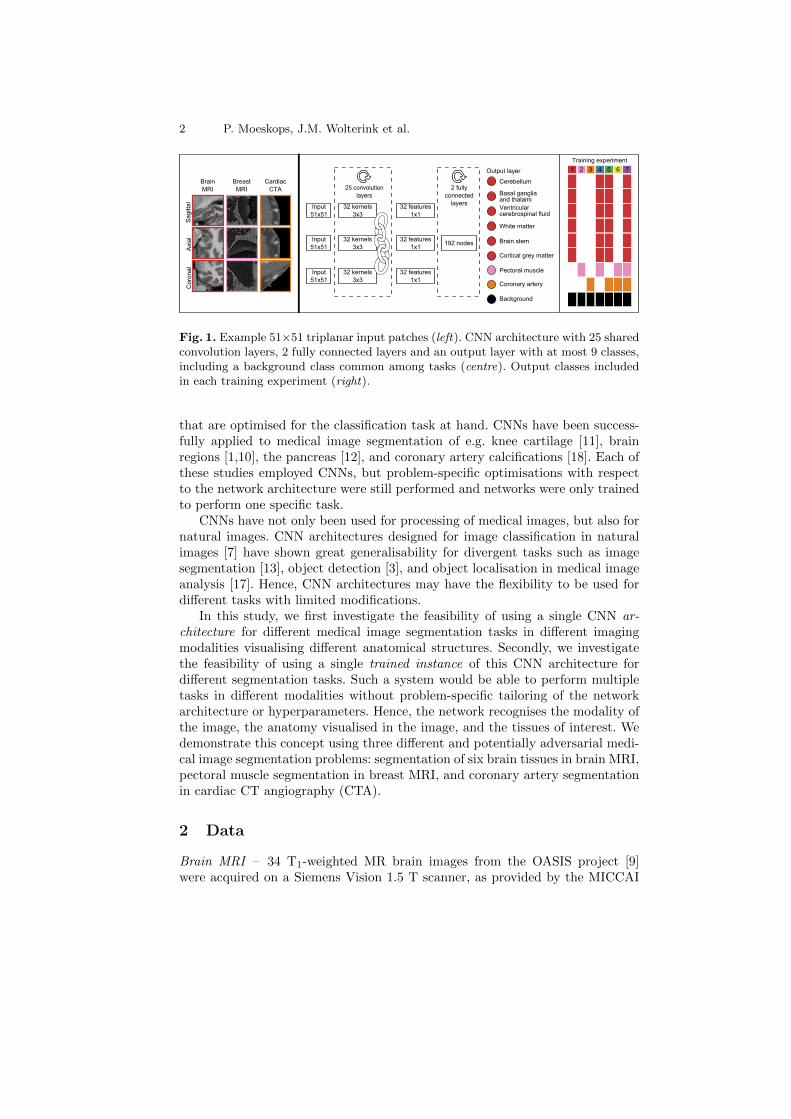
Fig. 1. Example 51×51 triplanar input patches (left). CNN architecture with 25 sharedconvolution layers, 2 fully connected layers and an output layer with at most 9 classes,including a background class common among tasks (centre). Output classes includedin each training experiment (right).
that are optimised for the classification task at hand. CNNs have been success-fully applied to medical image segmentation of e.g. knee cartilage [11], brainregions [1,10], the pancreas [12], and coronary artery calcifications [18]. Each ofthese studies employed CNNs, but problem-specific optimisations with respectto the network architecture were still performed and networks were only trainedto perform one specific task.
CNNs have not only been used for processing of medical images, but also fornatural images. CNN architectures designed for image classification in naturalimages [7] have shown great generalisability for divergent tasks such as imagesegmentation [13], object detection [3], and object localisation in medical imageanalysis [17]. Hence, CNN architectures may have the flexibility to be used fordifferent tasks with limited modifications.
In this study, we first investigate the feasibility of using a single CNN ar-chitecture for different medical image segmentation tasks in different imagingmodalities visualising different anatomical structures. Secondly, we investigatethe feasibility of using a single trained instance of this CNN architecture fordifferent segmentation tasks. Such a system would be able to perform multipletasks in different modalities without problem-specific tailoring of the networkarchitecture or hyperparameters. Hence, the network recognises the modality ofthe image, the anatomy visualised in the image, and the tissues of interest. Wedemonstrate this concept using three different and potentially adversarial medi-cal image segmentation problems: segmentation of six brain tissues in brain MRI,pectoral muscle segmentation in breast MRI, and coronary artery segmentationin cardiac CT angiography (CTA).
2 Data
Brain MRI – 34 T1-weighted MR brain images from the OASIS project [9]were acquired on a Siemens Vision 1.5 T scanner, as provided by the MICCAI
Deep Learning for Multi-Task Medical Image Segmentation 3
challenge on multi-atlas labelling [8]5. The images were acquired with voxel sizesof 1.0×1.0×1.25 mm3 and resampled to isotropic voxel sizes of 1.0×1.0×1.0 mm3.The images were manually segmented, in the coronal plane, into 134 classes thatwere, for the purpose of this paper, combined into six commonly used tissueclasses: white matter, cortical grey matter, basal ganglia and thalami, ventricularcerebrospinal fluid, cerebellum, and brain stem.
Breast MRI – 34 T1-weighted MR breast images were acquired on a SiemensMagnetom 1.5 T scanner with a dedicated double breast array coil [16]. The im-ages were acquired with in-plane voxel sizes between 1.21 and 1.35 mm and slicethicknesses between 1.35 and 1.69 mm. All images were resampled to isotropicvoxel sizes corresponding to their in-plane voxel size. The pectoral muscle wasmanually segmented in the axial plane by contour drawing.
Cardiac CTA – Ten cardiac CTA scans were acquired on a 256-detector rowPhilips Brilliance iCT scanner using 120 kVp and 200-300 mAs, with ECG-triggering and contrast enhancement. The reconstructed images had between0.4 and 0.5 mm in-plane voxel sizes and 0.45/0.90 mm slice spacing/thickness.All images were resampled to isotropic 0.45×0.45×0.45 mm3 voxel size. To seta manual reference standard, a human observer traversed the scan in the cran-iocaudal direction and painted voxels in the main coronary arteries and theirbranches in the axial plane.
3 Method
All voxels in the images were labelled by a CNN using seven different trainingexperiments (Fig. 1).
3.1 CNN Architecture
For each voxel, three orthogonal (axial, sagittal, and coronal) patches of 51×51voxels centred at the target voxel were extracted. For each of these three patches,features were determined using a deep stack of convolution layers. Each convo-lution layer contained 32 small (3×3 voxels) convolution kernels for a total of25 convolution layers [14]. To prevent over- or undersegmentation of structuresdue to translational invariance, no subsampling layers were used. To reduce thenumber of trainable parameters in the network and hence the risk of over-fitting,the same stack of convolutional layers was used for the axial, sagittal and coronalpatches.
The output of the convolution layers were 32 features for each of the threeorthogonal input patches, hence, 96 features in total. These features were inputto two subsequent fully connected layers, each with 192 nodes. The second fullyconnected layer was connected to a softmax classification layer. Depending on
5 https://masi.vuse.vanderbilt.edu/workshop2012
4 P. Moeskops, J.M. Wolterink et al.
the tasks of the network, this layer contained 2, 3, 7, 8 or 9 output nodes. Thefully connected layers were implemented as 1×1 voxel convolutions, to allow fastprocessing of arbitrarily sized images. Exponential linear units [2] were used forall non-linear activation functions. Batch normalisation [5] was used on all layersand dropout [15] was used on the fully connected layers.
3.2 Training Experiments
The same model was trained for each combination of the three tasks. In totalseven training experiments were performed (Fig. 1, right): three networks weretrained to perform one task (Experiments 1–3), three networks were trained toperform two tasks (Experiments 4–6), and one network was trained to performthree tasks (Experiment 7). The number of output nodes in the CNN was modi-fied accordingly. In each experiment, background classes of the target tasks weremerged into one class.
Each CNN was trained using mini-batch learning. A mini-batch contained210 samples, equally balanced over the tasks of the network. For each task, thetraining samples were randomly drawn from all training images, balanced overthe task-specific classes. All voxels with image intensity > 0 were consideredsamples. The network parameters were optimized using Adam stochastic opti-misation [6] with categorical cross-entropy as the cost-function.
4 Experiments and Results
The data for brain MRI, breast MRI and cardiac CTA were split into 14/20,14/20 and 6/4 training/test images, respectively. Four results were obtained foreach task: one with a network trained for only that task, two with networkstrained for that task and an additional task, and one with a network trained forall tasks together. Each network was trained with 25000 mini-batches per task.
No post-processing steps other than probability thresholding for evaluationpurposes were performed. The results are presented on the full test set. In brainMRI, the voxel class labels were determined by the highest class activation.The performance was evaluated per brain tissue type, using the Dice coefficientbetween the manual and automatic segmentations. In breast MRI and cardiacCTA, precision-recall curve analysis was performed to identify the optimal oper-ating point, defined, for each experiment, as the highest Dice coefficient over thewhole test set. The thresholds at this optimal operating point were then appliedto all images.
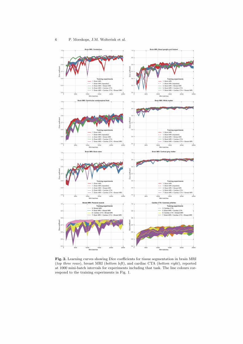
Fig. 2 shows the results of the described quantitative analysis, performed atintervals of 1000 mini-batches per task. As the networks learned, the obtainedDice coefficients increased and the stability of the results improved. For eachsegmentation task, the learning curves were similar for all experiments. Never-theless, slight differences were visible between the obtained learning curves. Toassess whether these differences were systematic or caused by the stochastic na-ture of CNN training, the training experiment using only brain MR data (Exper-iment 1) was repeated (dashed line in Fig. 2), showing similar inter-experiment
Deep Learning for Multi-Task Medical Image Segmentation 5
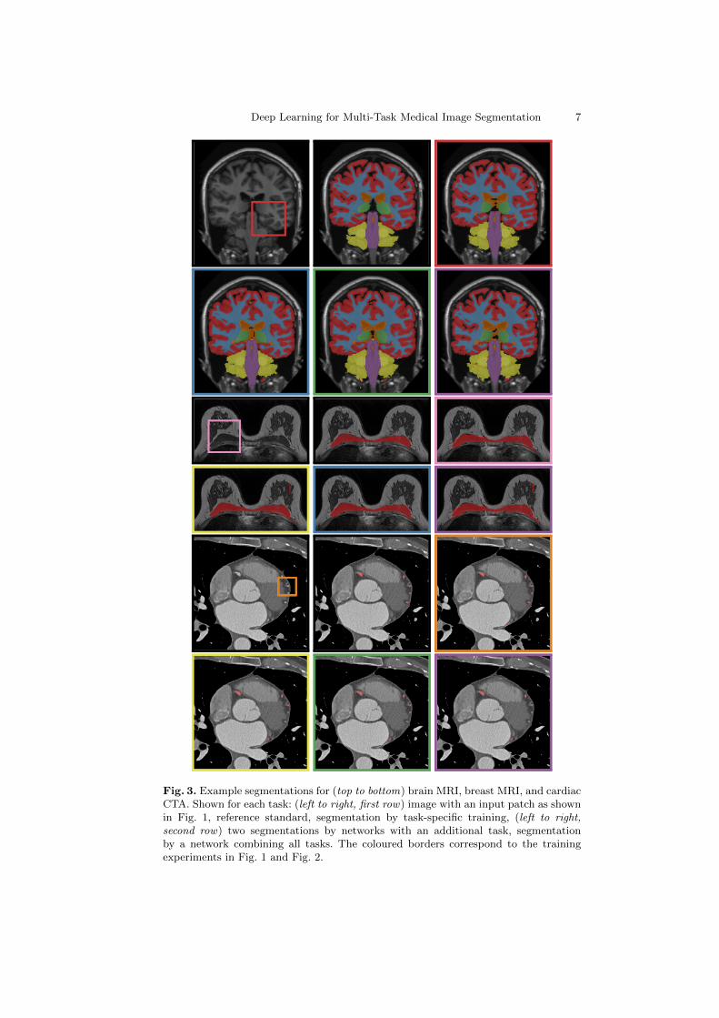
variation. Fig. 3 shows a visual comparison of results obtained for the three dif-ferent tasks. For all three tasks, all four networks were able to accurately segmentthe target tissues.
Confusion between tasks was very low. For the network trained with threetasks, the median percentage of voxels per scan labelled with a class alien to thetarget (e.g. cortical grey matter identified in breast MR) was < 0.0005% for alltasks.
5 Discussion and Conclusions
The results demonstrate that a single CNN architecture can be used to trainCNNs able to obtain accurate results in images from different modalities, visu-alising different anatomy. Moreover, it is possible to train a single CNN instancethat can not only segment multiple tissue classes in a single modality visualisinga single anatomical structure, but also multiple classes over multiple modalitiesvisualising multiple anatomical structures.
In all experiments, a fixed CNN architecture with triplanar orthogonal inputpatches was used. We have strived to utilise recent advances in deep learningsuch as batch normalisation [5], Adam stochastic optimisation [6], exponentiallinear units [2], and very deep networks with small convolution kernels [14].Furthermore, the implementation of fully connected layers as 1×1 convolutionlayers and the omission of downsampling layers allowed fast processing of wholeimages compared with more time-consuming patch-based scanning [11,12,18,10].The ability of the CNN to adapt to different tasks suggests that small architec-tural changes are unlikely to have a large effect on the performance. Volumetric3D input patches might result in increased performance, but would require a highcomputational load due to the increased size of the network parameter space.
The results for brain segmentation are comparable with previously publishedresults [10]. Due to differences in image acquisition and patient population, theobtained results for pectoral muscle segmentation and coronary artery extractioncannot be directly compared to results reported in other studies. Nevertheless,these results appear to be in line with previously published studies [4,19]. Nopost-processing other than probability thresholding for evaluation purposes wasapplied. The output probabilities may be further processed, or directly used asinput for further analysis, depending on the application.
Including multiple tasks in the training procedure resulted in a segmentationperformance equivalent to that of a network trained specifically for the task(Fig. 2). Similarities between the tasks, e.g. presence of the pectoral muscle inboth breast MR and cardiac CTA, or similar appearance of brain and breasttissue in T1-weighted MRI, led to very limited confusion. In future work, we willfurther investigate the capacity of the current architecture with more data andsegmentation tasks, and investigate to what extent the representations withinthe CNN are shared between tasks.
6 P. Moeskops, J.M. Wolterink et al.
Fig. 2. Learning curves showing Dice coefficients for tissue segmentation in brain MRI(top three rows), breast MRI (bottom left), and cardiac CTA (bottom right), reportedat 1000 mini-batch intervals for experiments including that task. The line colours cor-respond to the training experiments in Fig. 1.
Deep Learning for Multi-Task Medical Image Segmentation 7
Fig. 3. Example segmentations for (top to bottom) brain MRI, breast MRI, and cardiacCTA. Shown for each task: (left to right, first row) image with an input patch as shownin Fig. 1, reference standard, segmentation by task-specific training, (left to right,second row) two segmentations by networks with an additional task, segmentationby a network combining all tasks. The coloured borders correspond to the trainingexperiments in Fig. 1 and Fig. 2.
8 P. Moeskops, J.M. Wolterink et al.
References
1. de Brebisson, A., Montana, G.: Deep neural networks for anatomical brain seg-mentation. In: CVPR Bioimage Computing Workshop (2015)
2. Clevert, D.A., Unterthiner, T., Hochreiter, S.: Fast and accurate deep networklearning by exponential linear units (ELUs). In: ICLR (2016)
3. Girshick, R., Donahue, J., Darrell, T., Malik, J.: Region-based convolutional net-works for accurate object detection and segmentation. IEEE Trans Pattern AnalMach Intell 38(1), 142–158 (2016)
4. Gubern-Merida, A., Kallenberg, M., Martı, R., Karssemeijer, N.: Segmentation ofthe pectoral muscle in breast MRI using atlas-based approaches. In: Ayache, N.,Delingette, H., Golland, P., Mori, K. (eds.) MICCAI 2012, Part II LNCS, vol. 7511,pp. 371–378. Springer, Heidelberg (2012)
5. Ioffe, S., Szegedy, C.: Batch normalization: Accelerating deep network training byreducing internal covariate shift. In: ICML (2015)
6. Kingma, D., Ba, J.: Adam: A method for stochastic optimization. In: ICLR (2015)7. Krizhevsky, A., Sutskever, I., Hinton, G.E.: Imagenet classification with deep con-
volutional neural networks. In: NIPS. pp. 1097–1105 (2012)8. Landman, B.A., Ribbens, A., Lucas, B., Davatzikos, C., Avants, B., Ledig, C., Ma,
D., Rueckert, D., Vandermeulen, D., Maes, F., et al.: MICCAI 2012 workshop onmulti-atlas labeling. CreateSpace Independent Publishing Platform (2012)
9. Marcus, D.S., Wang, T.H., Parker, J., Csernansky, J.G., Morris, J.C., Buckner,R.L.: Open access series of imaging studies (OASIS): cross-sectional MRI data inyoung, middle aged, nondemented, and demented older adults. J Cognitive Neu-rosci 19(9), 1498–1507 (2007)
10. Moeskops, P., Viergever, M.A., Mendrik, A.M., de Vries, L.S., Benders, M.J., Is-gum, I.: Automatic segmentation of MR brain images with a convolutional neuralnetwork. IEEE Trans Med Imaging 35(5), 1252–1261 (2016)
11. Prasoon, A., Petersen, K., Igel, C., Lauze, F., Dam, E., Nielsen, M.: Deep featurelearning for knee cartilage segmentation using a triplanar convolutional neuralnetwork. In: Mori, K., Sakuma, I., Sato, Y., Barillot, C., Navab, N. (eds.) MICCAI2013, Part II. LNCS, vol. 8150, pp. 246–253. Springer, Heidelberg (2013)
12. Roth, H.R., Lu, L., Farag, A., Shin, H.C., Liu, J., Turkbey, E.B., Summers, R.M.:DeepOrgan: Multi-level deep convolutional networks for automated pancreas seg-mentation. In: Navab, N., Hornegger, J., Wells, M.W., Frangi, F.A. (eds.) MICCAI2015, Part I. LNCS, vol. 9349, pp. 556–564. Springer, Heidelberg (2015)
13. Shelhamer, E., Long, J., Darrell, T.: Fully convolutional networks for semanticsegmentation. IEEE Trans Pattern Anal Mach Intell (2016)
14. Simonyan, K., Zisserman, A.: Very deep convolutional networks for large-scaleimage recognition. In: ICLR (2015)
15. Srivastava, N., Hinton, G., Krizhevsky, A., Sutskever, I., Salakhutdinov, R.:Dropout: A simple way to prevent neural networks from overfitting. J Mach LearnRes 15(1), 1929–1958 (2014)
16. van der Velden, B.H., Dmitriev, I., Loo, C.E., Pijnappel, R.M., Gilhuijs, K.G.: As-sociation between parenchymal enhancement of the contralateral breast in dynamiccontrast-enhanced MR imaging and outcome of patients with unilateral invasivebreast cancer. Radiology 276(3), 675–685 (2015)
17. de Vos, B., Wolterink, J., de Jong, P., Viergever, M., Isgum, I.: 2D image classifi-cation for 3D anatomy localization; employing deep convolutional neural networks.In: SPIE Medical Imaging. p. 97841Y (2016)

Deep Learning for Multi-Task Medical Image Segmentation 9
18. Wolterink, J.M., Leiner, T., Viergever, M.A., Isgum, I.: Automatic coronary cal-cium scoring in cardiac CT angiography using convolutional neural networks. In:Navab, N., Hornegger, J., Wells, M.W., Frangi, F.A. (eds.) MICCAI 2015, Part I.LNCS, vol. 9349, pp. 589–596. Springer, Heidelberg (2015)
19. Zheng, Y., Loziczonek, M., Georgescu, B., Zhou, S.K., Vega-Higuera, F., Comani-ciu, D.: Machine learning based vesselness measurement for coronary artery seg-mentation in cardiac CT volumes. In: SPIE Medical Imaging. p. 79621K (2011)


![a arXiv:1907.06099v1 [cs.CV] 13 Jul 2019qdou/papers/2020/Multi-task recurrent... · Keywords: Surgical video analysis, multi-task learning, correlation loss, deep learning. Corresponding](https://img.dokumen.tips/doc/110x75/5f6a9a648fdca828eb52579a/a-arxiv190706099v1-cscv-13-jul-qdoupapers2020multi-task-recurrent-keywords.jpg)


