Embed Size (px)
Citation preview
Deep learning for cardiac imagesegmentation: A reviewChen Chen 1,∗, Chen Qin 1,∗, Huaqi Qiu 1,∗, Giacomo Tarroni1,2, Jinming Duan3,Wenjia Bai4,5, and Daniel Rueckert 1
1 Biomedical Image Analysis Group, Department of Computing, Imperial CollegeLondon, London, UK;2 Department of Computer Science, City, University of London, London, UK;3 School of Computer Science, University of Birmingham, Birmingham, UK;4 Data Science Institute, Imperial College London, London, UK;5 Division of Brain Sciences, Department of Medicine, Imperial College London,London, UKCorrespondence*:Chen [email protected]
ABSTRACT
Deep learning has become the mostwidely used approach for cardiac imagesegmentation in recent years. In this paper, weprovide a review of over 100 cardiac imagesegmentation papers using deep learning,which covers common imaging modalitiesincluding magnetic resonance imaging (MRI),computed tomography (CT), and ultrasound(US) and major anatomical structures ofinterest (ventricles, atria and vessels). Inaddition, a summary of publicly availablecardiac image datasets and code repositoriesare included to provide a base for encouragingreproducible research. Finally, we discussthe challenges and limitations with currentdeep learning-based approaches (scarcity oflabels, model generalizability across differentdomains, interpretability) and suggest potentialdirections for future research.
Keywords: Artificial intelligence, deep learning, neural networks,
cardiac image segmentation, cardiac image analysis, MRI, CT,
US
ARTICLE TYPES
Review
1 INTRODUCTION
Cardiovascular diseasess (CVDs) are the leadingcause of death globally according to World HealthOrganization (WHO). About 17.9 million peopledied from CVDs in 2016, from CVD, mainlyfrom heart disease and stroke1. The number isstill increasing annually. In recent decades, majoradvances have been made in cardiovascular researchand practice aiming to improve diagnosis andtreatment of cardiac diseases as well as reducingthe mortality of CVD. Modern medical imagingtechniques such as magnetic resonance imaging(MRI), computed tomography (CT) and ultrasound(US) are now widely used, which enable non-invasive qualitative and quantitative assessment ofcardiac anatomical structures and functions andprovide support for diagnosis, disease monitoring,treatment planning and prognosis.
Of particular interest, cardiac image segmentationis an important first step in numerous applications. Itpartitions the image into a number of semantically(i.e. anatomically) meaningful regions, based onwhich quantitative measures can be extracted,such as the myocardial mass, wall thickness, left
1 https://www.who.int/cardiovascular_diseases/about_cvd/en/
1
arX
iv:1
911.
0372
3v1
[ee
ss.I
V]
9 N
ov 2
019
Chen Chen et al. Deep learning for cardiac image segmentation: A review
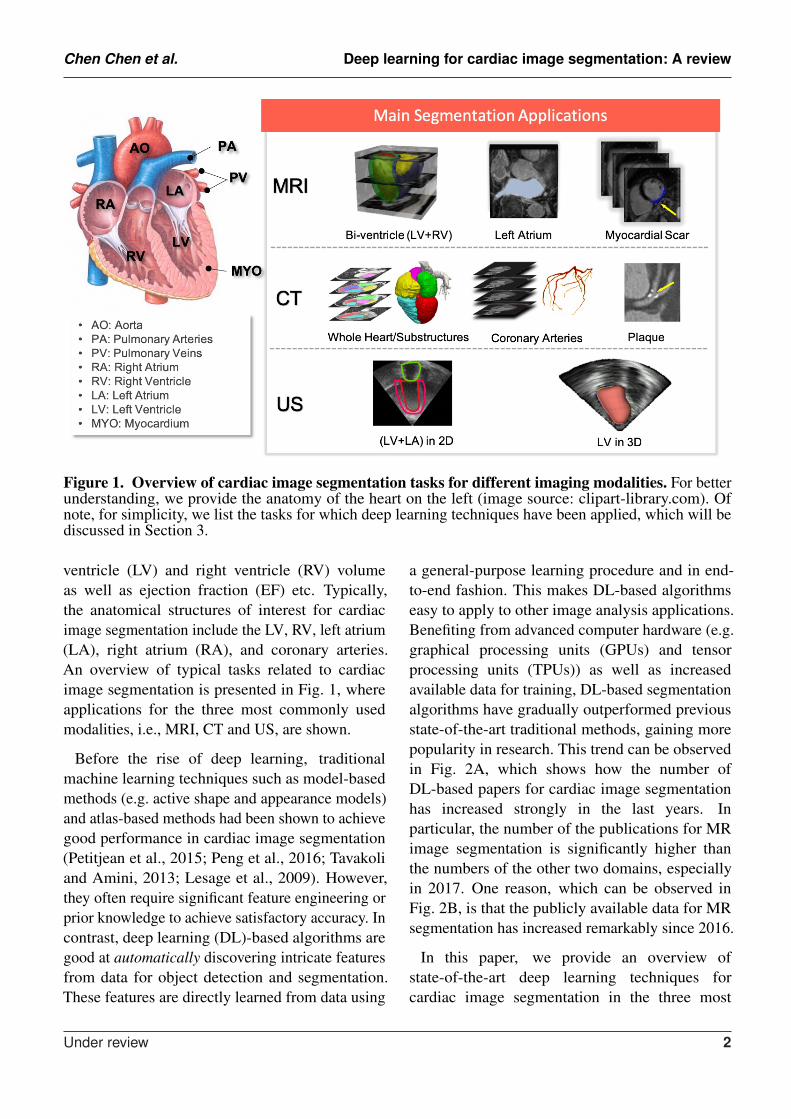
Figure 1. Overview of cardiac image segmentation tasks for different imaging modalities. For betterunderstanding, we provide the anatomy of the heart on the left (image source: clipart-library.com). Ofnote, for simplicity, we list the tasks for which deep learning techniques have been applied, which will bediscussed in Section 3.
ventricle (LV) and right ventricle (RV) volumeas well as ejection fraction (EF) etc. Typically,the anatomical structures of interest for cardiacimage segmentation include the LV, RV, left atrium(LA), right atrium (RA), and coronary arteries.An overview of typical tasks related to cardiacimage segmentation is presented in Fig. 1, whereapplications for the three most commonly usedmodalities, i.e., MRI, CT and US, are shown.
Before the rise of deep learning, traditionalmachine learning techniques such as model-basedmethods (e.g. active shape and appearance models)and atlas-based methods had been shown to achievegood performance in cardiac image segmentation(Petitjean et al., 2015; Peng et al., 2016; Tavakoliand Amini, 2013; Lesage et al., 2009). However,they often require significant feature engineering orprior knowledge to achieve satisfactory accuracy. Incontrast, deep learning (DL)-based algorithms aregood at automatically discovering intricate featuresfrom data for object detection and segmentation.These features are directly learned from data using
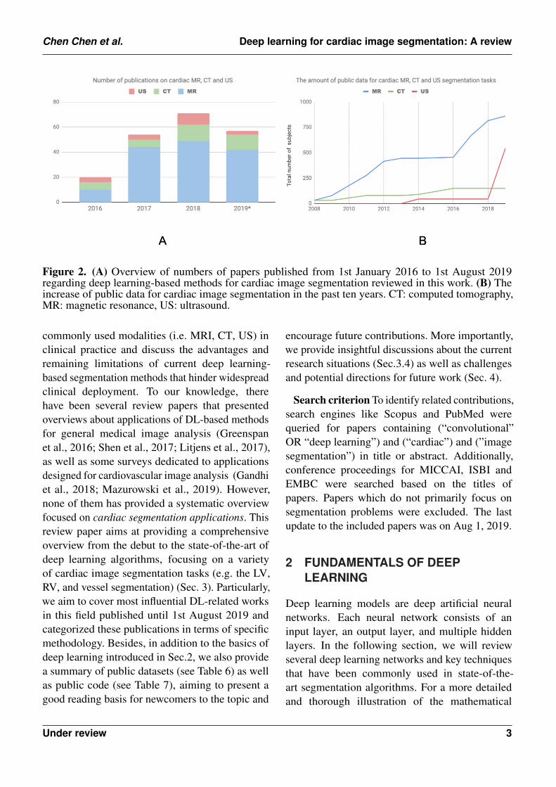
a general-purpose learning procedure and in end-to-end fashion. This makes DL-based algorithmseasy to apply to other image analysis applications.Benefiting from advanced computer hardware (e.g.graphical processing units (GPUs) and tensorprocessing units (TPUs)) as well as increasedavailable data for training, DL-based segmentationalgorithms have gradually outperformed previousstate-of-the-art traditional methods, gaining morepopularity in research. This trend can be observedin Fig. 2A, which shows how the number ofDL-based papers for cardiac image segmentationhas increased strongly in the last years. Inparticular, the number of the publications for MRimage segmentation is significantly higher thanthe numbers of the other two domains, especiallyin 2017. One reason, which can be observed inFig. 2B, is that the publicly available data for MRsegmentation has increased remarkably since 2016.
In this paper, we provide an overview ofstate-of-the-art deep learning techniques forcardiac image segmentation in the three most
Under review 2
Chen Chen et al. Deep learning for cardiac image segmentation: A review
Figure 2. (A) Overview of numbers of papers published from 1st January 2016 to 1st August 2019regarding deep learning-based methods for cardiac image segmentation reviewed in this work. (B) Theincrease of public data for cardiac image segmentation in the past ten years. CT: computed tomography,MR: magnetic resonance, US: ultrasound.
commonly used modalities (i.e. MRI, CT, US) inclinical practice and discuss the advantages andremaining limitations of current deep learning-based segmentation methods that hinder widespreadclinical deployment. To our knowledge, therehave been several review papers that presentedoverviews about applications of DL-based methodsfor general medical image analysis (Greenspanet al., 2016; Shen et al., 2017; Litjens et al., 2017),as well as some surveys dedicated to applicationsdesigned for cardiovascular image analysis (Gandhiet al., 2018; Mazurowski et al., 2019). However,none of them has provided a systematic overviewfocused on cardiac segmentation applications. Thisreview paper aims at providing a comprehensiveoverview from the debut to the state-of-the-art ofdeep learning algorithms, focusing on a varietyof cardiac image segmentation tasks (e.g. the LV,RV, and vessel segmentation) (Sec. 3). Particularly,we aim to cover most influential DL-related worksin this field published until 1st August 2019 andcategorized these publications in terms of specificmethodology. Besides, in addition to the basics ofdeep learning introduced in Sec.2, we also providea summary of public datasets (see Table 6) as wellas public code (see Table 7), aiming to present agood reading basis for newcomers to the topic and
encourage future contributions. More importantly,we provide insightful discussions about the currentresearch situations (Sec.3.4) as well as challengesand potential directions for future work (Sec. 4).
Search criterion To identify related contributions,search engines like Scopus and PubMed werequeried for papers containing (“convolutional”OR “deep learning”) and (“cardiac”) and (”imagesegmentation”) in title or abstract. Additionally,conference proceedings for MICCAI, ISBI andEMBC were searched based on the titles ofpapers. Papers which do not primarily focus onsegmentation problems were excluded. The lastupdate to the included papers was on Aug 1, 2019.
2 FUNDAMENTALS OF DEEPLEARNING
Deep learning models are deep artificial neuralnetworks. Each neural network consists of aninput layer, an output layer, and multiple hiddenlayers. In the following section, we will reviewseveral deep learning networks and key techniquesthat have been commonly used in state-of-the-art segmentation algorithms. For a more detailedand thorough illustration of the mathematical
Under review 3
Chen Chen et al. Deep learning for cardiac image segmentation: A review
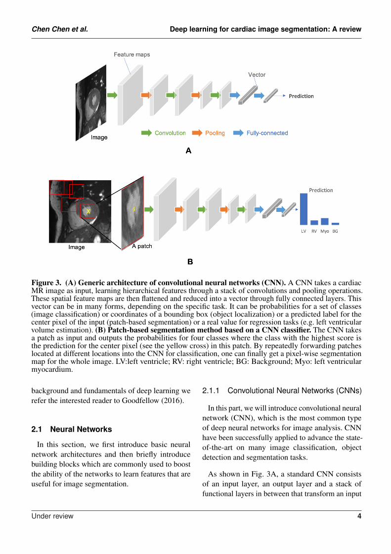
Figure 3. (A) Generic architecture of convolutional neural networks (CNN). A CNN takes a cardiacMR image as input, learning hierarchical features through a stack of convolutions and pooling operations.These spatial feature maps are then flattened and reduced into a vector through fully connected layers. Thisvector can be in many forms, depending on the specific task. It can be probabilities for a set of classes(image classification) or coordinates of a bounding box (object localization) or a predicted label for thecenter pixel of the input (patch-based segmentation) or a real value for regression tasks (e.g. left ventricularvolume estimation). (B) Patch-based segmentation method based on a CNN classifier. The CNN takesa patch as input and outputs the probabilities for four classes where the class with the highest score isthe prediction for the center pixel (see the yellow cross) in this patch. By repeatedly forwarding patcheslocated at different locations into the CNN for classification, one can finally get a pixel-wise segmentationmap for the whole image. LV:left ventricle; RV: right ventricle; BG: Background; Myo: left ventricularmyocardium.
background and fundamentals of deep learning werefer the interested reader to Goodfellow (2016).
2.1 Neural Networks
In this section, we first introduce basic neuralnetwork architectures and then briefly introducebuilding blocks which are commonly used to boostthe ability of the networks to learn features that areuseful for image segmentation.
2.1.1 Convolutional Neural Networks (CNNs)
In this part, we will introduce convolutional neuralnetwork (CNN), which is the most common typeof deep neural networks for image analysis. CNNhave been successfully applied to advance the state-of-the-art on many image classification, objectdetection and segmentation tasks.
As shown in Fig. 3A, a standard CNN consistsof an input layer, an output layer and a stack offunctional layers in between that transform an input
Under review 4
Chen Chen et al. Deep learning for cardiac image segmentation: A review
into an output in a specific form (e.g. vectors).These functional layers often contains convolutionallayers, pooling layers and/or fully-connected layers.In general, each convolution uses a n × n kernel(for 2D input) or n × n × n kernel (for 3D input)followed by batch normalization (Ioffe and Szegedy,2015) after which the output is passed through anonlinear activation function (e.g. rectified linearunit (ReLU)), which is used to extract featuremaps from an image. These feature maps are thendownsampled by pooling layers, typically by afactor of 2, which removes redundant featuresto improve the statistical efficiency and modelgeneralization. After that, fully connected layersare applied to reduce the dimension of features andfind the most task-relevant features for inference.The output of the network is a fix-sized vectorwhere each element can be a probabilistic scorefor each category (for image classification), a realvalue for a regression task (e.g. the left ventricularvolume estimation) or a set of values (e.g. thecoordinates of a bounding box for object detectionand localization).
In general, the size of convolution kernel nis chosen to be small in general, e.g. n = 3,in order to reduce computational costs. Whilethe kernels are small, one can increase thereceptive field (the area of the input image thatpotentially impacts the activation of a particularconvolutional kernel/neuron) by increasing thenumber of convolutional layers. For example, aconvolutonal layer with large 7× 7 kernels can bereplaced by three layers with small 3 × 3 kernels.The number of parameters is reduced by a factor of72/(3× (32)) ≈ 2 while the receptive field remainsthe same (7 × 7). An online resource 2 is referredhere, which illustrates and visualizes the changeof receptive field by varying the number of hiddenlayers and the size of kernels. In general, increasingthe depth of convolution neural networks (thenumber of hidden layers) to enlarge the receptivefield can lead to improved model performance, e.g.
2 https://fomoro.com/research/article/receptive-field-calculator
classification accuracy (Simonyan and Zisserman,2015).
CNNs for image classification can also beemployed for image segmentation applicationswithout major adaptations to the network architecture(Ciresan and Giusti, 2012), as shown in Fig. 3B.However, this requires to divide each image intopatches and then train a CNN to predict the classlabel of the center pixel for every patch. One majordisadvantage of this patch-based approach is that, atinference time, the network has to be deployed forevery patch individually despite the fact that thereis a lot of redundancy due to multiple overlappingpatches in the image. As a result of this inefficiency,the main application of CNNs with fully connectedlayers is object localization, which aims to estimatethe bounding box of the object of interest in animage. This bounding box is then used to cropthe image, forming an image pre-processing stepto reduce the computational cost for segmentation(Avendi et al., 2016). For efficient, end-to-endpixel-wise segmentation, a variant of CNNs calledfully convolutional neural network (FCN) is morecommonly used, which will be discussed in the nextsection.
2.1.2 Fully Convolutional Neural Networks(FCNs)
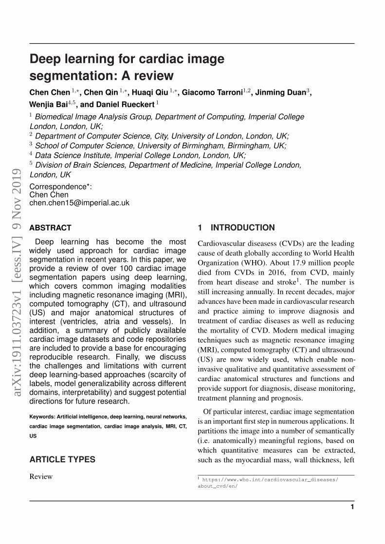
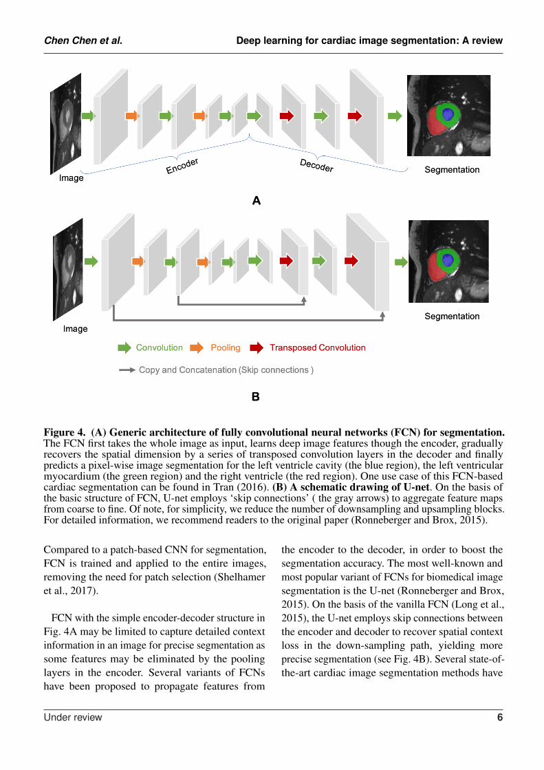
The idea of FCN was first introduced by Longet al. (2015) for image segmentation. FCNs area special type of CNNs that do not have anyfully connected layers. In general, as shown inFig. 4A, FCNs are designed to have an encoder-decoder structure such that they can take inputof arbitrary size and produce the output with thesame size. Given an input image, the encoderfirst transforms the input into high-level featurerepresentation whereas the decoder interprets thefeature maps and recovers spatial details back tothe image space for pixel-wise prediction througha series of transposed convolution and convolutionoperations. Here, transposed convolutions are usedfor up-scaling the feature maps, typically by a factorof 2. These transposed convolutions can also bereplaced by unpooling layers and upsampling layers.
Under review 5
Chen Chen et al. Deep learning for cardiac image segmentation: A review
Figure 4. (A) Generic architecture of fully convolutional neural networks (FCN) for segmentation.The FCN first takes the whole image as input, learns deep image features though the encoder, graduallyrecovers the spatial dimension by a series of transposed convolution layers in the decoder and finallypredicts a pixel-wise image segmentation for the left ventricle cavity (the blue region), the left ventricularmyocardium (the green region) and the right ventricle (the red region). One use case of this FCN-basedcardiac segmentation can be found in Tran (2016). (B) A schematic drawing of U-net. On the basis ofthe basic structure of FCN, U-net employs ‘skip connections’ ( the gray arrows) to aggregate feature mapsfrom coarse to fine. Of note, for simplicity, we reduce the number of downsampling and upsampling blocks.For detailed information, we recommend readers to the original paper (Ronneberger and Brox, 2015).
Compared to a patch-based CNN for segmentation,FCN is trained and applied to the entire images,removing the need for patch selection (Shelhameret al., 2017).
FCN with the simple encoder-decoder structure inFig. 4A may be limited to capture detailed contextinformation in an image for precise segmentation assome features may be eliminated by the poolinglayers in the encoder. Several variants of FCNshave been proposed to propagate features from
the encoder to the decoder, in order to boost thesegmentation accuracy. The most well-known andmost popular variant of FCNs for biomedical imagesegmentation is the U-net (Ronneberger and Brox,2015). On the basis of the vanilla FCN (Long et al.,2015), the U-net employs skip connections betweenthe encoder and decoder to recover spatial contextloss in the down-sampling path, yielding moreprecise segmentation (see Fig. 4B). Several state-of-the-art cardiac image segmentation methods have
Under review 6
Chen Chen et al. Deep learning for cardiac image segmentation: A review
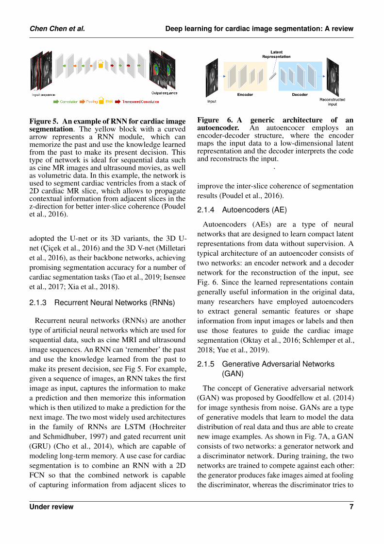
Figure 5. An example of RNN for cardiac imagesegmentation. The yellow block with a curvedarrow represents a RNN module, which canmemorize the past and use the knowledge learnedfrom the past to make its present decision. Thistype of network is ideal for sequential data suchas cine MR images and ultrasound movies, as wellas volumetric data. In this example, the network isused to segment cardiac ventricles from a stack of2D cardiac MR slice, which allows to propagatecontextual information from adjacent slices in thez-direction for better inter-slice coherence (Poudelet al., 2016).
adopted the U-net or its 3D variants, the 3D U-net (Cicek et al., 2016) and the 3D V-net (Milletariet al., 2016), as their backbone networks, achievingpromising segmentation accuracy for a number ofcardiac segmentation tasks (Tao et al., 2019; Isenseeet al., 2017; Xia et al., 2018).
2.1.3 Recurrent Neural Networks (RNNs)
Recurrent neural networks (RNNs) are anothertype of artificial neural networks which are used forsequential data, such as cine MRI and ultrasoundimage sequences. An RNN can ‘remember’ the pastand use the knowledge learned from the past tomake its present decision, see Fig 5. For example,given a sequence of images, an RNN takes the firstimage as input, captures the information to makea prediction and then memorize this informationwhich is then utilized to make a prediction for thenext image. The two most widely used architecturesin the family of RNNs are LSTM (Hochreiterand Schmidhuber, 1997) and gated recurrent unit(GRU) (Cho et al., 2014), which are capable ofmodeling long-term memory. A use case for cardiacsegmentation is to combine an RNN with a 2DFCN so that the combined network is capableof capturing information from adjacent slices to
Figure 6. A generic architecture of anautoencoder. An autoencocer employs anencoder-decoder structure, where the encodermaps the input data to a low-dimensional latentrepresentation and the decoder interprets the codeand reconstructs the input.
.
improve the inter-slice coherence of segmentationresults (Poudel et al., 2016).
2.1.4 Autoencoders (AE)
Autoencoders (AEs) are a type of neuralnetworks that are designed to learn compact latentrepresentations from data without supervision. Atypical architecture of an autoencoder consists oftwo networks: an encoder network and a decodernetwork for the reconstruction of the input, seeFig. 6. Since the learned representations containgenerally useful information in the original data,many researchers have employed autoencodersto extract general semantic features or shapeinformation from input images or labels and thenuse those features to guide the cardiac imagesegmentation (Oktay et al., 2016; Schlemper et al.,2018; Yue et al., 2019).
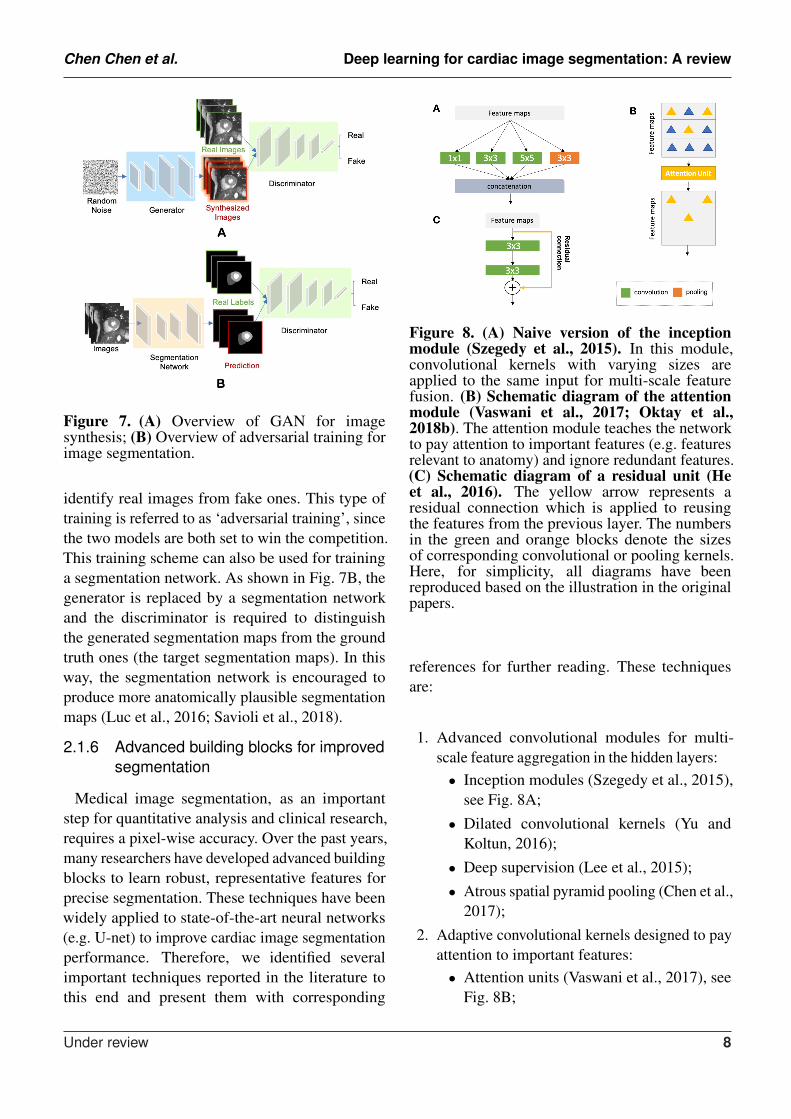
2.1.5 Generative Adversarial Networks(GAN)
The concept of Generative adversarial network(GAN) was proposed by Goodfellow et al. (2014)for image synthesis from noise. GANs are a typeof generative models that learn to model the datadistribution of real data and thus are able to createnew image examples. As shown in Fig. 7A, a GANconsists of two networks: a generator network anda discriminator network. During training, the twonetworks are trained to compete against each other:the generator produces fake images aimed at foolingthe discriminator, whereas the discriminator tries to
Under review 7
Chen Chen et al. Deep learning for cardiac image segmentation: A review
Figure 7. (A) Overview of GAN for imagesynthesis; (B) Overview of adversarial training forimage segmentation.
identify real images from fake ones. This type oftraining is referred to as ‘adversarial training’, sincethe two models are both set to win the competition.This training scheme can also be used for traininga segmentation network. As shown in Fig. 7B, thegenerator is replaced by a segmentation networkand the discriminator is required to distinguishthe generated segmentation maps from the groundtruth ones (the target segmentation maps). In thisway, the segmentation network is encouraged toproduce more anatomically plausible segmentationmaps (Luc et al., 2016; Savioli et al., 2018).
2.1.6 Advanced building blocks for improvedsegmentation
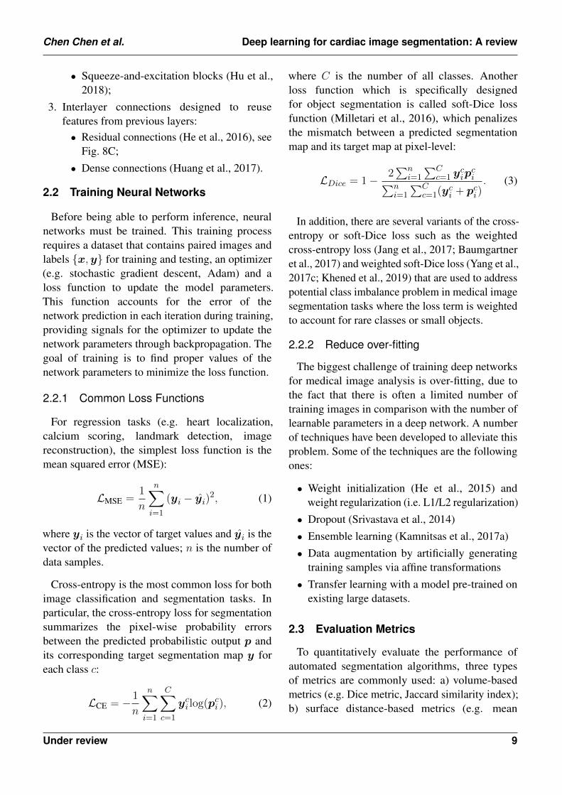
Medical image segmentation, as an importantstep for quantitative analysis and clinical research,requires a pixel-wise accuracy. Over the past years,many researchers have developed advanced buildingblocks to learn robust, representative features forprecise segmentation. These techniques have beenwidely applied to state-of-the-art neural networks(e.g. U-net) to improve cardiac image segmentationperformance. Therefore, we identified severalimportant techniques reported in the literature tothis end and present them with corresponding
Figure 8. (A) Naive version of the inceptionmodule (Szegedy et al., 2015). In this module,convolutional kernels with varying sizes areapplied to the same input for multi-scale featurefusion. (B) Schematic diagram of the attentionmodule (Vaswani et al., 2017; Oktay et al.,2018b). The attention module teaches the networkto pay attention to important features (e.g. featuresrelevant to anatomy) and ignore redundant features.(C) Schematic diagram of a residual unit (Heet al., 2016). The yellow arrow represents aresidual connection which is applied to reusingthe features from the previous layer. The numbersin the green and orange blocks denote the sizesof corresponding convolutional or pooling kernels.Here, for simplicity, all diagrams have beenreproduced based on the illustration in the originalpapers.
references for further reading. These techniquesare:
1. Advanced convolutional modules for multi-scale feature aggregation in the hidden layers:• Inception modules (Szegedy et al., 2015),
see Fig. 8A;• Dilated convolutional kernels (Yu and
Koltun, 2016);• Deep supervision (Lee et al., 2015);• Atrous spatial pyramid pooling (Chen et al.,
2017);2. Adaptive convolutional kernels designed to pay
attention to important features:• Attention units (Vaswani et al., 2017), see
Fig. 8B;
Under review 8
Chen Chen et al. Deep learning for cardiac image segmentation: A review
• Squeeze-and-excitation blocks (Hu et al.,2018);
3. Interlayer connections designed to reusefeatures from previous layers:• Residual connections (He et al., 2016), see
Fig. 8C;• Dense connections (Huang et al., 2017).
2.2 Training Neural Networks
Before being able to perform inference, neuralnetworks must be trained. This training processrequires a dataset that contains paired images andlabels x,y for training and testing, an optimizer(e.g. stochastic gradient descent, Adam) and aloss function to update the model parameters.This function accounts for the error of thenetwork prediction in each iteration during training,providing signals for the optimizer to update thenetwork parameters through backpropagation. Thegoal of training is to find proper values of thenetwork parameters to minimize the loss function.
2.2.1 Common Loss Functions
For regression tasks (e.g. heart localization,calcium scoring, landmark detection, imagereconstruction), the simplest loss function is themean squared error (MSE):
LMSE =1
n
n∑i=1
(yi − yi)2, (1)
where yi is the vector of target values and yi is thevector of the predicted values; n is the number ofdata samples.
Cross-entropy is the most common loss for bothimage classification and segmentation tasks. Inparticular, the cross-entropy loss for segmentationsummarizes the pixel-wise probability errorsbetween the predicted probabilistic output p andits corresponding target segmentation map y foreach class c:
LCE = − 1
n
n∑i=1
C∑c=1
yci log(pc
i), (2)
where C is the number of all classes. Anotherloss function which is specifically designedfor object segmentation is called soft-Dice lossfunction (Milletari et al., 2016), which penalizesthe mismatch between a predicted segmentationmap and its target map at pixel-level:
LDice = 1− 2∑n
i=1
∑Cc=1 y
cip
ci∑n
i=1
∑Cc=1(y
ci + pc
i). (3)
In addition, there are several variants of the cross-entropy or soft-Dice loss such as the weightedcross-entropy loss (Jang et al., 2017; Baumgartneret al., 2017) and weighted soft-Dice loss (Yang et al.,2017c; Khened et al., 2019) that are used to addresspotential class imbalance problem in medical imagesegmentation tasks where the loss term is weightedto account for rare classes or small objects.
2.2.2 Reduce over-fitting
The biggest challenge of training deep networksfor medical image analysis is over-fitting, due tothe fact that there is often a limited number oftraining images in comparison with the number oflearnable parameters in a deep network. A numberof techniques have been developed to alleviate thisproblem. Some of the techniques are the followingones:
• Weight initialization (He et al., 2015) andweight regularization (i.e. L1/L2 regularization)
• Dropout (Srivastava et al., 2014)• Ensemble learning (Kamnitsas et al., 2017a)• Data augmentation by artificially generating
training samples via affine transformations• Transfer learning with a model pre-trained on
existing large datasets.
2.3 Evaluation Metrics
To quantitatively evaluate the performance ofautomated segmentation algorithms, three typesof metrics are commonly used: a) volume-basedmetrics (e.g. Dice metric, Jaccard similarity index);b) surface distance-based metrics (e.g. mean
Under review 9
Chen Chen et al. Deep learning for cardiac image segmentation: A review
contour distance, Hausdorff distance); c) clinicalperformance metrics (e.g. ventricular volume andmass). For a detailed illustration of common usedclinical indices in cardiac image analysis, werecommend the review paper by Peng et al. (2016).In our paper, we mainly report the accuracy ofmethods in terms of the Dice metric for ease ofcomparison. The Dice score measures the ratioof overlap between two results (e.g. automaticsegmentation vs manual segmentation), rangingfrom 0 (mismatch) to 1 (perfect match).
3 DEEP LEARNING FOR CARDIACIMAGE SEGMENTATION
In this section, we provide a summary of deeplearning-based applications for the three mainimaging modalities: MRI, CT, and US regardingspecific applications for targeted structures. Ingeneral, these deep learning-based methods providean efficient and effective way to segmentingparticular organs or tissues (e.g. the LV, coronaryvessels, scars) in different modalities, facilitatingfollow-up quantitative analysis of cardiovascularstructure and function. Among these works, alarge portion of these methods are designed forventricle segmentation, especially in MR and USdomains. The objective of ventricle segmentationis to delineate the endocardium and epicardiumof the LV and/or RV. These segmentation mapsare important for deriving clinical indices, suchas left ventricular end-diastolic volume (LVEDV),left ventricular end-systolic volume (LVESV), rightventricular end-diastolic volume (RVEDV), rightventricular end-systolic volume (RVESV), and EF.In addition, these segmentation maps are essentialfor 3D shape analysis (Xue et al., 2018; Biffi et al.,2018), 3D+time motion analysis (Zheng et al.,2019) and survival prediction (Bello et al., 2019).
3.1 Cardiac MR Image Segmentation
Cardiac MRI is a non-invasive imaging techniquethat can visualize the structures within and aroundthe heart. Compared to CT, it does not requireionising radiation. Instead, it relies on the magnetic
field in conjunction with radio-frequency wavesto excite hydrogen nuclei in the heart, and thengenerates an image by measuring their response.By utilizing different imaging sequences, cardiacMRI allows accurate quantification of both cardiacanatomy and function (e.g. cine imaging) andpathological tissues such as scars (late gadoliniumenhancement (LGE) imaging). Accordingly, cardiacMRI is currently regarded as the gold standard forquantitative cardiac analysis (Van Der Geest andReiber, 1999).
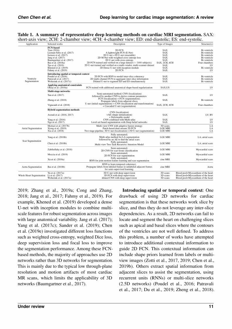
A group of representative deep learning basedcardiac MR segmentation methods are shown inTable 1. From the table, one can see that a majorityof works have focused on segmenting cardiacchambers (e.g. LV, RV, LA). In contrast, thereare relatively fewer works on segmenting abnormalcardiac tissue regions such as myocardial scarsand atrial fibrosis. This is likely due to the limitedrelevant public datasets as well as the difficulty ofthe task. In addition, to the best of our knowledge,there are very few works that apply deep learningtechniques to atrial wall segmentation, as alsosuggested by a recent survey paper (Karim et al.,2018). In the following sections, we will describeand discuss these methods regarding differentapplications in detail.
3.1.1 Ventricle Segmentation
Vanilla FCN-based Segmentation: Tran (2016)was among the first ones to apply a FCN (Shelhameret al., 2017) to segment the left ventricle,myocardium and right ventricle directly onshort-axis cardiac magnetic resonance (MR)images. Their end-to-end approach based on FCNachieved competitive segmentation performance,significantly outperforming traditional methodsin terms of both speed and accuracy. In thefollowing years, a number of works based onFCNs have been proposed, aiming at achievingfurther improvements in segmentation performance.In this regard, one stream of work focuses onoptimizing the network structure to enhance thefeature learning capacity for segmentation (Khenedet al., 2019; Li et al., 2019b; Zhou and Yang,
Under review 10
Chen Chen et al. Deep learning for cardiac image segmentation: A review
Table 1. A summary of representative deep learning methods on cardiac MRI segmentation. SAX:short-axis view; 2CH: 2-chamber view; 4CH: 4-chamber view; ED: end-diastolic; ES: end-systolic.
Application Selected works Description Type of Images Structure(s)
VentricleSegmentation
FCN-basedTran (2016) 2D FCN SAX Bi-ventricleLieman-Sifry et al. (2017) A lightweight FCN (E-Net) SAX Bi-ventricleIsensee et al. (2017) 2D U-net +3D U-net (ensemble) SAX Bi-ventricleJang et al. (2017) 2D M-Net with weighted cross entropy loss SAX Bi-ventricleBaumgartner et al. (2017) 2D U-net with cross entropy SAX Bi-ventricleBai et al. (2018a) 2D FCN trained and verified on a large dataset (∼ 5000 subjects); SAX, 2CH, 4CH Four chambersTao et al. (2019) 2D U-net trained and verified on a multi-vendor, multi-scanner dataset SAX LVKhened et al. (2019) 2D Dense U-net with inception module SAX Bi-ventricleFahmy et al. (2019) 2D FCN SAX LVIntroducing spatial or temporal contextPoudel et al. (2016) 2D FCN with RNN to model inter-slice coherency SAX Bi-ventriclePatravali et al. (2017) 2D multi-channel FCN to aggregate inter-slice information SAX Bi-ventricleWolterink et al. (2017c) Dilated U-net to segment ED and ES simultaneously SAX Bi-ventricleApplying anatomical constraintsOktay et al. (2018a) FCN trained with additional anatomical shape-based regularization SAX;US LVMulti-stage networksTan et al. (2017) Semi-automated method; CNN (localization)
followed by another CNN to derive contour parameters SAX LV
Zheng et al. (2018) FCN (localization) + FCN (segmentation);Propagate labels from adjacent slices SAX Bi-ventricle
Vigneault et al. (2018) U-net (initial segmentation) + CNN (localization and transformation)+ Cascaded U-net (segmentation) SAX, 2CH, 4CH Four chambers
Hybrid segmentation methods
Avendi et al. (2016, 2017)CNN (localization)
+AE (shape initialization)+ Deformable model
SAX LV; RV
Yang et al. (2016) CNN combined with Multi-atlas SAX LVNgo et al. (2017) Level-set based segmentation with Deep belief networks SAX LV
Atrial SegmentationMortazi et al. (2017b) Multi-view CNN with adaptive fusion strategy 3D scans LAXiong et al. (2019) Patch-based dual-stream 2D FCN LGE MRI LAXia et al. (2018) Two-stage pipeline; 3D U-net (localization) +3D U-net (segmentation) LGE MRI LA
Scar SegmentationYang et al. (2018b)
Fully automated;Multi-atlas method for LA segmentationfollowed by an AE to find the atrial scars
LGE MRI LA; atrial scars
Chen et al. (2018b) Fully automated;Multi-view Two-Task Recursive Attention Model LGE MRI LA; atrial scars
Zabihollahy et al. (2018) Semi-automated;2D CNN for scar tissue classification LGE MRI Myocardial scars
Moccia et al. (2019) Semi-automated;2D FCN for scar segmentation LGE MRI Myocardial scars
Xu et al. (2018a) Fully automated;RNN for joint motion feature learning and scar segmentaion cine MRI Myocardial scars
Aorta Segmentation Bai et al. (2018b)RNN to learn temporal coherence;
Propagate labels from labeled frames to unlabeled adjacent framesfor semi-supervised learning;
cine MRI Aorta
Whole Heart SegmentationYu et al. (2017a) 3D U-net with deep supervision 3D scans Blood pool+Myocardium of the heartLi et al. (2017) 3D FCN with deep supervision 3D scans Blood pool+Myocardium of the heartWolterink et al. (2017a) dilated CNN with deep supervision 3D scans Blood pool+Myocardium of the heart
2019; Zhang et al., 2019a; Cong and Zhang,2018; Jang et al., 2017; Fahmy et al., 2019). Forexample, Khened et al. (2019) developed a denseU-net with inception modules to combine multi-scale features for robust segmentation across imageswith large anatomical variability. Jang et al. (2017);Yang et al. (2017c); Sander et al. (2019); Chenet al. (2019e) investigated different loss functionssuch as weighted cross-entropy, weighted Dice loss,deep supervision loss and focal loss to improvethe segmentation performance. Among these FCN-based methods, the majority of approaches use 2Dnetworks rather than 3D networks for segmentation.This is mainly due to the typical low through-planeresolution and motion artifacts of most cardiacMR scans, which limits the applicability of 3Dnetworks (Baumgartner et al., 2017).
Introducing spatial or temporal context: Onedrawback of using 2D networks for cardiacsegmentation is that these networks work slice byslice, and thus they do not leverage any inter-slicedependencies. As a result, 2D networks can fail tolocate and segment the heart on challenging slicessuch as apical and basal slices where the contoursof the ventricles are not well defined. To addressthis problem, a number of works have attemptedto introduce additional contextual information toguide 2D FCN. This contextual information caninclude shape priors learned from labels or multi-view images (Zotti et al., 2017, 2019; Chen et al.,2019b). Others extract spatial information fromadjacent slices to assist the segmentation, usingrecurrent units (RNNs) or multi-slice networks(2.5D networks) (Poudel et al., 2016; Patravaliet al., 2017; Du et al., 2019; Zheng et al., 2018).
Under review 11
Chen Chen et al. Deep learning for cardiac image segmentation: A review
These networks can also be applied to leveraginginformation across different temporal frames inthe cardiac cycle to improve spatial and temporalconsistency of segmentation results (Yan et al.,2018; Savioli et al., 2018; Du et al., 2019; Qin et al.,2018a; Wolterink et al., 2017c).
Applying anatomical constraints: Anotherproblem that may limit the segmentation performanceof both 2D and 3D FCNs is that they are typicallytrained with pixel-wise loss functions only (e.g.cross-entropy or soft-Dice losses). These pixel-wise loss functions may not be sufficient to learnfeatures that represent the underlying anatomicalstructures. Several approaches therefore focus ondesigning and applying anatomical constraintsto train the network to improve its predictionaccuracy and robustness. These constraints arerepresented as regularization terms which take intoaccount the topology (Clough et al., 2019), contourand region information (Chen et al., 2019g) orshape information (Oktay et al., 2018a; Yue et al.,2019), encouraging the network to generate moreanatomically plausible segmentations. In addition toregularizing networks at training time, Painchaudet al. (2019) proposed a variational AE to correctinaccurate segmentations, in the post-processingstage.
Multi-task learning: Multi-task learning hasalso been explored to regularize FCN-basedcardiac ventricle segmentation during trainingby performing auxiliary tasks that are relevantto the main segmentation task, such as motionestimation (Qin et al., 2018b), estimation of cardiacfunction (Dangi et al., 2018b), ventricle sizeclassification (Zhang et al., 2018b) and imagereconstruction (Chartsias et al., 2018; Huanget al., 2019). Training a network for multipletasks simultaneously encourages the network toextract features which are useful across thesetasks, resulting in improved learning efficiency andprediction accuracy.
Multi-stage networks: Recently, there is agrowing interest in applying neural networks ina multi-stage pipeline which breaks down the
segmentation problem into subtasks (Vigneaultet al., 2018; Zheng et al., 2018; Li et al., 2019a; Tanet al., 2017; Liao et al., 2019). For example, Zhenget al. (2018); Li et al. (2019a) proposed a region-of-interest (ROI) localization network followed bya segmentation network. Likewise, Vigneault et al.(2018) proposed a network called Omega-Net whichconsists of a U-net for cardiac chamber localization,a learnable transformation module to normalizeimage orientation and a series of U-nets for fine-grained segmentation. By explicitly localizing theROI and by rotating the input image into a canonicalorientation, the proposed method better generalizesto images with varying sizes and orientations.
Hybrid segmentation methods: Another streamof work aims at combining neural networks withclassical segmentation approaches, e.g. level-sets (Ngo et al., 2017; Duan et al., 2018a),deformable models (Avendi et al., 2016, 2017;Medley et al., 2019), atlas-based methods (Yanget al., 2016; Rohe et al., 2017) and graph-cutbased methods (Lu et al., 2019). Here, neuralnetworks are applied in the feature extractionand model initialization stages, reducing thedependency on manual interactions and improvingthe segmentation accuracy of the conventionalsegmentation methods deployed afterwards. Forexample, Avendi et al. (2016) proposed one ofthe first DL-based methods for LV segmentationin cardiac short-axis MR images. The authors firstapplied a CNN to automatically detect the LV andthen used an AE to estimate the shape of the LV. Theestimated shape was then used to initialize follow-up deformable models for shape refinement. As aresult, the proposed integrated deformable modelconverges faster than conventional deformablemodels and the segmentation achieves higheraccuracy. In their later work, the authors extendedthis approach to segment RV (Avendi et al.,2017). While these hybrid methods demonstratedbetter segmentation accuracy than previous non-deep learning methods, most of them still requirean iterative optimization for shape refinement.Furthermore, these methods are often designed forone particular anatomical structure. As noted in
Under review 12
Chen Chen et al. Deep learning for cardiac image segmentation: A review
the recent benchmark study (Bernard et al., 2018),most state-of-the-art segmentation algorithms forbi-ventricle segmentation are based on end-to-endFCNs, which allows the simultaneous segmentationof the LV and RV.
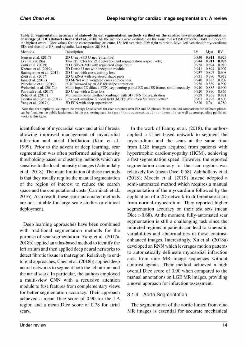
To better illustrate these developments for cardiacventricle segmentation from cardiac MR images, wecollate a list of bi-ventricle segmentation methodsthat have been trained and tested on the AutomatedCardiac Diagnosis Challenge (ACDC) dataset,reported in Table 2. For ease of comparison, weonly consider those methods which have beenevaluated on the same online test set (50 subjects).As the ACDC challenge organizers keep theonline evaluation platform open to the public, ourcomparison not only includes the methods fromthe original challenge participants (summarized inthe benchmark study paper from Bernard et al.(2018)) but also three segmentation algorithmsthat have been proposed after the challenge (i.e.Zotti et al. (2019); Li et al. (2019a); Painchaudet al. (2019)). From this comparison, one cansee that top algorithms are the ensemble methodproposed by Isensee et al. (2017) and the two-stage method proposed by Li et al. (2019a), both ofwhich are based on FCNs. In particular, comparedto the traditional level-set method (Tziritas andGrinias, 2017), both methods achieved considerablyhigher accuracy even for the more challengingsegmentation of the left ventricular myocardium(Myo), indicating the power of deep learning basedapproaches.
3.1.2 Atrial Segmentation
Atrial fibrillation (AF) is one of the most commoncardiac electrical disorders, affecting around 1million people in the UK 3. Accordingly, atrialsegmentation is of prime importance in the clinic,improving the assessment of the atrial anatomy inboth pre-operative atrial fibrillation (AF) ablationplanning and post-operative follow-up evaluations.In addition, the segmentation of atrium canbe used as a basis for scar segmentation and
3 https://www.nhs.uk/conditions/atrial-fibrillation/
atrial fibrosis quantification from LGE images.Traditional methods such as region growing (Karimet al., 2008) and methods that employ strong priors(i.e. atlas-based label fusion (Tao et al., 2016)and non-rigid registration (Zhuang et al., 2010))have been applied in the past for automated leftatrium segmentation. However, the accuracy ofthese methods highly relies on good initializationand ad-hoc pre-processing methods, which limitsthe widespread adoption in the clinic.
Recently, Bai et al. (2018a) and Vigneault et al.(2018) applied 2D FCNs to directly segment the LAand RA from standard 2D long-axis images, i.e. 2-chamber (2CH), 4-chamber (4CH) views. Notably,their networks can also be trained to segmentventricles from 2D short-axis stacks without anymodifications to the network architecture. Likewise,Xiong et al. (2019); Preetha et al. (2018); Bianet al. (2018); Chen et al. (2018a) applied 2D FCNsto segment the atrium from 3D LGE images in aslice-by-slice fashion, where they optimized thenetwork structure for enhanced feature learning.3D networks (Xia et al., 2018; Savioli et al., 2018;Jia et al., 2018; Vesal et al., 2018; Li et al., 2018)and multi-view FCN (Mortazi et al., 2017b; Yanget al., 2018a) have also been explored to capture3D global information from 3D LGE images foraccurate atrium segmentation.
In particular, Xia et al. (2018) proposed afully automatic two-stage segmentation frameworkwhich contains a first 3D U-net to roughly locate theatrial center from down-sampled images followedby a second 3D U-net to accurately segment theatrium in the cropped portions of the original imagesat full resolution. Their multi-stage approach is bothmemory-efficient and accurate, ranking first in theleft atrium segmentation challenge 2018 (LASC’18)with a mean Dice score of 0.93 evaluated on a testset of 54 cases.
3.1.3 Scar Segmentation
Scar characterization is usually performed usingLGE MR imaging, a contrast-enhanced MRimaging technique. LGE MR imaging enables the
Under review 13
Chen Chen et al. Deep learning for cardiac image segmentation: A review
Table 2. Segmentation accuracy of state-of-the-art segmentation methods verified on the cardiac bi-ventricular segmentationchallenge (ACDC) dataset (Bernard et al., 2018) All the methods were evaluated on the same test set (50 subjects). Bold numbers arethe highest overall Dice values for the corresponding structure. LV: left ventricle, RV: right ventricle, Myo: left ventricular myocardium;ED: end-diastolic; ES: end-systolic. Last update: 2019.8.1.Methods Description LV Myo RVIsensee et al. (2017) 2D U-net +3D U-net (ensemble) 0.950 0.911 0.923Li et al. (2019a) Two 2D FCNs for ROI detection and segmentation respectively; 0.944 0.911 0.926Zotti et al. (2019) 2D GridNet-MD with registered shape prior 0.938 0.894 0.910Khened et al. (2019) 2D Dense U-net with inception module 0.941 0.894 0.907Baumgartner et al. (2017) 2D U-net with cross entropy loss 0.937 0.897 0.908Zotti et al. (2017) 2D GridNet with registered shape prior 0.931 0.890 0.912Jang et al. (2017) 2D M-Net with weighted cross entropy loss 0.940 0.885 0.907Painchaud et al. (2019) FCN followed by an AE for shape correction 0.936 0.889 0.909Wolterink et al. (2017c) Multi-input 2D dilated FCN, segmenting paired ED and ES frames simultaneously 0.940 0.885 0.900Patravali et al. (2017) 2D U-net with a Dice loss 0.920 0.890 0.865Rohe et al. (2017) Multi-atlas based method combined with 3D CNN for registration 0.929 0.868 0.881Tziritas and Grinias (2017) Level-set +markov random field (MRF); Non-deep learning method 0.907 0.798 0.803Yang et al. (2017c) 3D FCN with deep supervision 0.820 N/A 0.780
Note that for simplicity, we report the average Dice scores for each structure over ED and ES phases. More detailed comparison for different phasescan be found on the public leaderboard in the post testing part (https://acdc.creatis.insa-lyon.fr) as well as corresponding publishedworks in this table.
identification of myocardial scars and atrial fibrosis,allowing improved management of myocardialinfarction and atrial fibrillation (Kim et al.,1999). Prior to the advent of deep learning, scarsegmentation was often performed using intensitythresholding-based or clustering methods which aresensitive to the local intensity changes (Zabihollahyet al., 2018). The main limitation of these methodsis that they usually require the manual segmentationof the region of interest to reduce the searchspace and the computational costs (Carminati et al.,2016). As a result, these semi-automated methodsare not suitable for large-scale studies or clinicaldeployment.
Deep learning approaches have been combinedwith traditional segmentation methods for thepurpose of scar segmentation: Yang et al. (2017a,2018b) applied an atlas-based method to identify theleft atrium and then applied deep neural networks todetect fibrotic tissue in that region. Relatively to end-to-end approaches, Chen et al. (2018b) applied deepneural networks to segment both the left atrium andthe atrial scars. In particular, the authors employeda multi-view CNN with a recursive attentionmodule to fuse features from complementary viewsfor better segmentation accuracy. Their approachachieved a mean Dice score of 0.90 for the LAregion and a mean Dice score of 0.78 for atrialscars.
In the work of Fahmy et al. (2018), the authorsapplied a U-net based network to segment themyocardium and the scars at the same timefrom LGE images acquired from patients withhypertrophic cardiomyopathy (HCM), achievinga fast segmentation speed. However, the reportedsegmentation accuracy for the scar regions wasrelatively low (mean Dice: 0.58). Zabihollahy et al.(2018); Moccia et al. (2019) instead adopted asemi-automated method which requires a manualsegmentation of the myocardium followed by theapplication of a 2D network to differentiate scarsfrom normal myocardium. They reported highersegmentation accuracy on their test sets (meanDice >0.68). At the moment, fully-automated scarsegmentation is still a challenging task since theinfarcted regions in patients can lead to kinematicvariabilities and abnormalities in those contrast-enhanced images. Interestingly, Xu et al. (2018a)developed an RNN which leverages motion patternsto automatically delineate myocardial infarctionarea from cine MR image sequences withoutcontrast agents. Their method achieved a highoverall Dice score of 0.90 when compared to themanual annotations on LGE MR images, providinga novel approach for infarction assessment.
3.1.4 Aorta Segmentation
The segmentation of the aortic lumen from cineMR images is essential for accurate mechanical
Under review 14
Chen Chen et al. Deep learning for cardiac image segmentation: A review
and hemodynamic characterization of the aorta.One common challenge for this task is the typicalsparsity of the annotations in aortic cine imagesequences, where only a few frames have beenannotated. To address the problem, Bai et al.(2018b) applied a non-rigid image registrationmethod (Rueckert et al., 1999) to propagate thelabels from the annotated frames to the unlabeledneighboring ones in the cardiac cycle, effectivelygenerating pseudo annotated frames that could beutilized for further training. This semi-supervisedmethod achieved an average Dice metric of0.96 for the ascending aorta and 0.95 for thedescending aorta over a test set of 100 subjects. Inaddition, compared to a previous approach based ondeformable models (Herment et al., 2010), theirapproach based on FCN and RNN can directlyperform the segmentation task on a whole imagesequence without requiring the explicit estimationof the ROI.
3.1.5 Whole Heart Segmentation
Apart from the above mentioned segmentationapplications which target one particular structure,deep learning can also be used to segment the mainsubstructures of the heart in 3D MR images (Yuet al., 2017a; Wolterink et al., 2017a; Li et al.,2017; Shi et al., 2018). An early work from Yuet al. (2017a) adopted a 3D dense FCN to segmentthe myocardium and blood pool in the heart from3D MR scans. Recently, more and more methodsbegan to apply deep learning pipelines to segmentmore specific substructures (incl. four chambers,myocardium (MYO), aorta, pulmonary vein (PV))in both 3D CT and MR images. This has beenfacilitated by the availability of public datasets forwhole heart segmentation (Multi-Modality WholeHeart Segmentation (MM-WHS)). In general, thesegmentation task on MR images is harder thanthe one of CT images mainly because of the largevariations in terms of image intensity distributionamong different scanners. As mentioned in a recentbenchmark study paper by Zhuang et al. (2019),deep learning methods in general achieve bettersegmentation accuracy on CT images compared
to that of MR images. We will discuss thesesegmentation methods in the next CT section infurther detail (see section 3.2.1).
3.2 Cardiac CT Image Segmentation
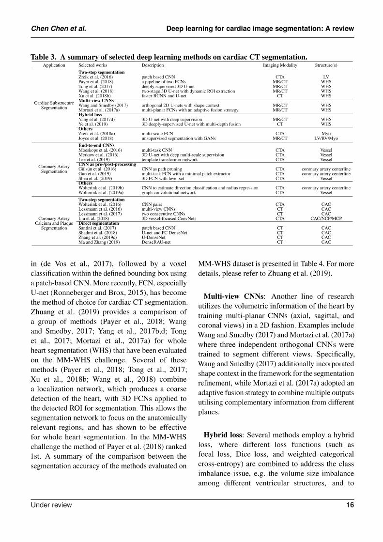
CT is a non-invasive imaging technique thatis performed routinely for disease diagnosis andtreatment planning. In particular, cardiac CT scansare used for assessment of cardiac anatomy andspecifically the coronary arteries. There are twomain imaging modalities: non-contrast CT imagingand contrast-enhanced coronary CT angiography(CTA). Typically, non-contrast CT imaging exploitsdensity of tissues to generate an image, such thatdifferent densities using various attenuation valuessuch as soft tissues, calcium, fat, and air can beeasily distinguished, and thus allows to estimatethe amount of calcium present in the coronaryarteries (Kang et al., 2012). In comparison, contrast-enhanced coronary CTA, which is acquired after theinjection of a contrast agent, can provide excellentvisualization of cardiac chambers, vessels andcoronaries, and has been shown to be effective indetecting non-calcified coronary plaques. In thefollowing sections, we will review some of themost commonly used deep learning-based cardiacCT segmentation methods. A summary of theseapproaches is presented in Table 3.
3.2.1 Cardiac Substructure Segmentation
Accurate delineation of cardiac substructuresplays a crucial role in cardiac function analysis,providing important clinical variables such as EF,myocardial mass, wall thickness etc. Typically, thecardiac substructures that are segmented include theLV, RV, LA, RA, MYO, aorta (AO) and pulmonaryartery (PA).Two-step segmentation: One group of deeplearning methods relies on a two-step segmentationprocedure, where a ROI is first extracted andthen fed into a CNN for subsequent classification(Zreik et al., 2016; Dormer et al., 2018). Forinstance, Zreik et al. (2016) proposed a two-stepLV segmentation process where a bounding box forthe LV is first detected using the method described
Under review 15
Chen Chen et al. Deep learning for cardiac image segmentation: A review
Table 3. A summary of selected deep learning methods on cardiac CT segmentation.Application Selected works Description Imaging Modality Structure(s)
Cardiac SubstructureSegmentation
Two-step segmentationZreik et al. (2016) patch based CNN CTA LVPayer et al. (2018) a pipeline of two FCNs MR/CT WHSTong et al. (2017) deeply supervised 3D U-net MR/CT WHSWang et al. (2018) two-stage 3D U-net with dynamic ROI extraction MR/CT WHSXu et al. (2018b) faster RCNN and U-net CT WHSMulti-view CNNsWang and Smedby (2017) orthogonal 2D U-nets with shape context MR/CT WHSMortazi et al. (2017a) multi-planar FCNs with an adaptive fusion strategy MR/CT WHSHybrid lossYang et al. (2017d) 3D U-net with deep supervision MR/CT WHSYe et al. (2019) 3D deeply-supervised U-net with multi-depth fusion CT WHSOthersZreik et al. (2018a) multi-scale FCN CTA MyoJoyce et al. (2018) unsupervised segmentation with GANs MR/CT LV/RV/Myo
Coronary ArterySegmentation
End-to-end CNNsMoeskops et al. (2016) multi-task CNN CTA VesselMerkow et al. (2016) 3D U-net with deep multi-scale supervision CTA VesselLee et al. (2019) template transformer network CTA VesselCNN as pre-/post-processingGulsun et al. (2016) CNN as path pruning CTA coronary artery centerlineGuo et al. (2019) multi-task FCN with a minimal patch extractor CTA coronary artery centerlineShen et al. (2019) 3D FCN with level set CTA VesselOthersWolterink et al. (2019b) CNN to estimate direction classification and radius regression CTA coronary artery centerlineWolterink et al. (2019a) graph convolutional network CTA Vessel
Coronary ArteryCalcium and Plaque
Segmentation
Two-step segmentationWolterink et al. (2016) CNN pairs CTA CACLessmann et al. (2016) multi-view CNNs CT CACLessmann et al. (2017) two consecutive CNNs CT CACLiu et al. (2018) 3D vessel-focused ConvNets CTA CAC/NCP/MCPDirect segmentationSantini et al. (2017) patch based CNN CT CACShadmi et al. (2018) U-net and FC DenseNet CT CACZhang et al. (2019c) U-DenseNet CT CACMa and Zhang (2019) DenseRAU-net CT CAC
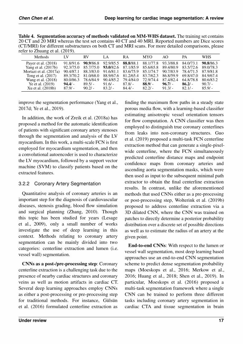
in (de Vos et al., 2017), followed by a voxelclassification within the defined bounding box usinga patch-based CNN. More recently, FCN, especiallyU-net (Ronneberger and Brox, 2015), has becomethe method of choice for cardiac CT segmentation.Zhuang et al. (2019) provides a comparison ofa group of methods (Payer et al., 2018; Wangand Smedby, 2017; Yang et al., 2017b,d; Tonget al., 2017; Mortazi et al., 2017a) for wholeheart segmentation (WHS) that have been evaluatedon the MM-WHS challenge. Several of thesemethods (Payer et al., 2018; Tong et al., 2017;Xu et al., 2018b; Wang et al., 2018) combinea localization network, which produces a coarsedetection of the heart, with 3D FCNs applied tothe detected ROI for segmentation. This allows thesegmentation network to focus on the anatomicallyrelevant regions, and has shown to be effectivefor whole heart segmentation. In the MM-WHSchallenge the method of Payer et al. (2018) ranked1st. A summary of the comparison between thesegmentation accuracy of the methods evaluated on
MM-WHS dataset is presented in Table 4. For moredetails, please refer to Zhuang et al. (2019).
Multi-view CNNs: Another line of researchutilizes the volumetric information of the heart bytraining multi-planar CNNs (axial, sagittal, andcoronal views) in a 2D fashion. Examples includeWang and Smedby (2017) and Mortazi et al. (2017a)where three independent orthogonal CNNs weretrained to segment different views. Specifically,Wang and Smedby (2017) additionally incorporatedshape context in the framework for the segmentationrefinement, while Mortazi et al. (2017a) adopted anadaptive fusion strategy to combine multiple outputsutilising complementary information from differentplanes.
Hybrid loss: Several methods employ a hybridloss, where different loss functions (such asfocal loss, Dice loss, and weighted categoricalcross-entropy) are combined to address the classimbalance issue, e.g. the volume size imbalanceamong different ventricular structures, and to
Under review 16
Chen Chen et al. Deep learning for cardiac image segmentation: A review
Table 4. Segmentation accuracy of methods validated on MM-WHS dataset. The training set contains20 CT and 20 MRI whereas the test set contains 40 CT and 40 MRI. Reported numbers are Dice scores(CT/MRI) for different substructures on both CT and MRI scans. For more detailed comparisons, pleaserefer to Zhuang et al. (2019).
Methods LV RV LA RA MYO AO PA WHSPayer et al. (2018) 91.8/91.6 90.9/86.8 92.9/85.5 88.8/88.1 88.1/77.8 93.3/88.8 84.0/73.1 90.8/86.3Yang et al. (2017b) 92.3/75.0 85.7/75.0 93.0/82.6 87.1/85.9 85.6/65.8 89.4/80.9 83.5/72.6 89.0/78.3
Mortazi et al. (2017a) 90.4/87.1 88.3/83.0 91.6/81.1 83.6/75.9 85.1/74.7 90.7/83.9 78.4/71.5 87.9/81.8Tong et al. (2017) 89.3/70.2 81.0/68.0 88.9/67.6 81.2/65.4 83.7/62.3 86.8/59.9 69.8/47.0 84.9/67.4Wang et al. (2018) 80.0/86.3 78.6/84.9 90.4/85.2 79.4/84.0 72.9/74.4 87.4/82.4 64.8/78.8 80.6/83.2
Ye et al. (2019) 94.4/ - 89.5/ - 91.6/ - 87.8/ - 88.9/ - 96.7/ - 86.2/ - 90.7/ -Xu et al. (2018b) 87.9/ - 90.2/ - 83.2/ - 84.4/ - 82.2/ - 91.3/ - 82.1/ - 85.9/ -
improve the segmentation performance (Yang et al.,2017d; Ye et al., 2019).
In addition, the work of Zreik et al. (2018a) hasproposed a method for the automatic identificationof patients with significant coronary artery stenosesthrough the segmentation and analysis of the LVmyocardium. In this work, a multi-scale FCN is firstemployed for myocardium segmentation, and thena convolutional autoencoder is used to characterizethe LV myocardium, followed by a support vectormachine (SVM) to classify patients based on theextracted features.
3.2.2 Coronary Artery Segmentation
Quantitative analysis of coronary arteries is animportant step for the diagnosis of cardiovasculardiseases, stenosis grading, blood flow simulationand surgical planning (Zhang, 2010). Thoughthis topic has been studied for years (Lesageet al., 2009), only a small number of worksinvestigate the use of deep learning in thiscontext. Methods relating to coronary arterysegmentation can be mainly divided into twocategories: centerline extraction and lumen (i.e.vessel wall) segmentation.
CNNs as a post-/pre-processing step: Coronarycenterline extraction is a challenging task due to thepresence of nearby cardiac structures and coronaryveins as well as motion artifacts in cardiac CT.Several deep learning approaches employ CNNsas either a post-processing or pre-processing stepfor traditional methods. For instance, Gulsunet al. (2016) formulated centerline extraction as
finding the maximum flow paths in a steady stateporous media flow, with a learning-based classifierestimating anisotropic vessel orientation tensorsfor flow computation. A CNN classifier was thenemployed to distinguish true coronary centerlinesfrom leaks into non-coronary structures. Guoet al. (2019) proposed a multi-task FCN centerlineextraction method that can generate a single-pixel-wide centerline, where the FCN simultaneouslypredicted centerline distance maps and endpointconfidence maps from coronary arteries andascending aorta segmentation masks, which werethen used as input to the subsequent minimal pathextractor to obtain the final centerline extractionresults. In contrast, unlike the aforementionedmethods that used CNNs either as a pre-processingor post-processing step, Wolterink et al. (2019b)proposed to address centerline extraction via a3D dilated CNN, where the CNN was trained onpatches to directly determine a posterior probabilitydistribution over a discrete set of possible directionsas well as to estimate the radius of an artery at thegiven point.
End-to-end CNNs: With respect to the lumen orvessel wall segmentation, most deep learning basedapproaches use an end-to-end CNN segmentationscheme to predict dense segmentation probabilitymaps (Moeskops et al., 2016; Merkow et al.,2016; Huang et al., 2018; Shen et al., 2019). Inparticular, Moeskops et al. (2016) proposed amulti-task segmentation framework where a singleCNN can be trained to perform three differenttasks including coronary artery segmentation incardiac CTA and tissue segmentation in brain
Under review 17
Chen Chen et al. Deep learning for cardiac image segmentation: A review
MR images. They showed that such a multi-tasksegmentation network in multiple modalities canachieve equivalent performance as a single tasknetwork. Merkow et al. (2016) introduced deepmulti-scale supervision into a 3D U-net architecture,enabling efficient multi-scale feature learning andprecise voxel-level predictions. Besides, shapepriors can also be incorporated into the network(Lee et al., 2019; Chen et al., 2019h; Duanet al., 2018b). For instance, Lee et al. (2019)explicitly enforced a roughly tubular shape priorfor the vessel segments by introducing a templatetransformer network, through which a shapetemplate can be deformed via network-basedregistration to produce an accurate segmentation ofthe input image, as well as to guarantee topologicalconstraints. More recently, graph convolutionalnetworks have also been investigated by Wolterinket al. (2019a) for coronary artery segmentationin CTA, where vertices on the coronary lumensurface mesh were considered as graph nodesand the locations of these tubular surface meshvertices were directly optimized. They showed thatsuch method significantly outperformed a baselinenetwork that used only fully-connected layers onhealthy subjects (mean Dice score: 0.75 vs 0.67). Besides, the graph convolutional network usedin their work is able to directly generate smoothsurface meshes without post-processing steps.
3.2.3 Coronary Artery Calcium and PlaqueSegmentation
Coronary artery calcium (CAC) is a direct riskfactor for cardiovascular disease. Clinically, CACis quantified using the Agatston score (Agatstonet al., 1990) which considers the lesion area and theweighted maximum density of the lesion (de Voset al., 2019). Precise detection and segmentation ofCAC are thus important for the accurate predictionof the Agatston score and disease diagnosis.
Two-step segmentation: One group of deeplearning approaches to segmentation and automaticcalcium scoring proposed to use a two-stepsegmentation scheme. For example, Wolterink et al.(2016) attempted to classify CAC in cardiac CTA
using a pair of CNNs, where the first CNN coarselyidentified voxels likely to be CAC within a ROIdetected using (de Vos et al., 2017) and then thesecond CNN further distinguished between CACand CAC-like negatives more accurately. Similarto such a two-stage scheme, Lessmann et al. (2016,2017) proposed to identify CAC in low-dose chestCT, in which a ROI of the heart or potentialcalcifications were first localized followed by aCAC classification process.
Direct segmentation: More recently, severalapproaches (Shadmi et al., 2018; Santini et al.,2017; Ma and Zhang, 2019; Zhang et al., 2019c)have been proposed for the direct segmentation ofCAC from non-contrast cardiac CT or chest CT:the majority of them employed combinations of U-net (Ronneberger and Brox, 2015) and DenseNet(Huang et al., 2017) for precise quantificationof CAC which showed that a sensitivity over90% can be achieved Santini et al. (2017). Theseaforementioned approaches all follow the sameworkflow where the CAC is first identified and thenquantified. An alternative approach is to circumventthe intermediate segmentation and to perform directquantification, such as in (Cano-Espinosa et al.,2018; de Vos et al., 2019), which have proven thatthis approach is effective and promising.
Finally, for non-calcified plaque (NCP) andmixed-calcified plaque (MCP) in coronary arteries,only a limited number of works have beenreported that investigate deep learning methods forsegmentation and quantification (Zreik et al., 2018b;Liu et al., 2018). Yet, this is a very importanttask from a clinical point of view, since theseplaques can potentially rupture and obstruct anartery, causing ischemic events and severe cardiacdamage. In contrast to CAC segmentation, NCPand MCP segmentation are more challenging dueto their similar appearances and intensities asadjacent tissues. Therefore, robust and and accurateanalysis often requires the generation of multi-planar reformatted (MPR) images that have beenstraightened along the centreline of the vesssel.Recently, Liu et al. (2018) proposed a vessel-focused 3D convolutional network with attention
Under review 18
Chen Chen et al. Deep learning for cardiac image segmentation: A review
layers to segment three types of plaques on theextracted and reformatted coronary MPR volumes.Zreik et al. (2018b) presented an automatic methodfor detection and characterization of coronaryartery plaques as well as determination of coronaryartery stenosis significance, in which a multi-taskconvolutional RNN was used to perform bothplaque and stenosis classification by analyzing thefeatures extracted along the coronary artery in anMPR image.
3.3 Cardiac Ultrasound ImageSegmentation
Cardiac ultrasound (US) imaging, also knownas echocardiography, is an indispensable clinicaltool for the assessment of cardiovascular function.It is often used clinically as the first imagingexamination owing to its portability, low costand real-time capability. While a number oftraditional methods such as active contours, level-sets and active shape models have been employed toautomate the segmentation of anatomical structuresin ultrasound images (Noble and Boukerroui,2006), the achieved accuracy is limited by variousproblems of ultrasound imaging such as low signal-to-noise ratio, varying speckle noise, low imagecontrast (especially between the myocardium andthe blood pool), edge dropout and shadows cast bystructures such as dense muscle and ribs.
As in cardiac MR and CT, several DL-basedmethods have been recently proposed to improvethe performance of cardiac ultrasound imagesegmentation in terms of both accuracy and speed.The majority of these DL-based approaches focuson LV segmentation, with only few addressing theproblem of aortic valve and LA segmentation. Asummary of the reviewed works can be found inTable 5.
3.3.1 2D LV segmentation
Deep learning combined with deformablemodels: The imaging quality of echocardiographymakes voxel-wise tissue classification highlychallenging. To address this challenge, deeplearning has been combined with deformable model
for LV segmentation in 2D images (Carneiro et al.,2010, 2012; Carneiro and Nascimento, 2010, 2013;Nascimento and Carneiro, 2014, 2019; Veni et al.,2018). Features extracted by trained deep neuralnetworks were used instead of handcrafted featuresto improve accuracy and robustness.
Several works applied deep learning in a two-stage pipeline which first localizes the targetROI via rigid transformation of a boundingbox, then segments the target structure withinthe ROI. This two-stage pipeline reduces thesearch region of the segmentation and increasesrobustness of the overall segmentation framework.Carneiro et al. (2010, 2012) first adopted thisDL framework to segment the LV in apical long-axis echocardiograms. The method uses DBN(Hinton and Salakhutdinov, 2006) to predict therigid transformation parameters for localization andthe deformable model parameters for segmentation.The results demonstrated the robustness of DBN-based feature extraction to image appearancevariations. Nascimento and Carneiro (2017) furtherreduced the training and inference complexity of theDBN-based framework by using sparse manifoldlearning in the rigid detection step.
To further reduce the computational complexity,some works perform segmentation in one stepwithout resorting to the two-stage approach.Nascimento and Carneiro (2014, 2019) appliedsparse manifold learning in segmentation, showinga reduced training and search complexity comparedto their previous version of the method, whilemaintaining the same level of segmentationaccuracy. Veni et al. (2018) applied a FCN toproduce coarse segmentation masks, which is thenfurther refined by a level-set based method.
Utilizing temporal coherence: Cardiac ultrasounddata is often recorded as a temporal sequenceof images. Several approaches aim to leveragethe coherence between temporally close framesto improve the accuracy and robustness of theLV segmentation. Carneiro and Nascimento (2010,2013) proposed a dynamic modeling method basedon a sequential monte carlo (SMC) (or particle
Under review 19
Chen Chen et al. Deep learning for cardiac image segmentation: A review
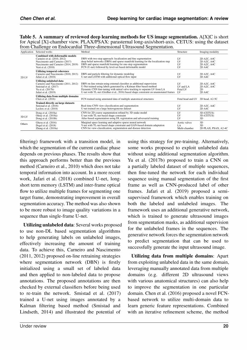
Table 5. A summary of reviewed deep learning methods for US image segmentation. A[X]C is shortfor Apical [X]-chamber view. PLAX/PSAX: parasternal long-axis/short-axis. CETUS: using the datasetfrom Challenge on Endocardial Three-dimensional Ultrasound Segmentation.Application Selected works Method Structure Imaging modality
2D LV
Combined with deformable modelsCarneiro et al. (2010, 2012) DBN with two-step approach: localization and fine segmentation LV 2D A2C, A4CNascimento and Carneiro (2017) deep belief networks (DBN) and sparse manifold learning for the localization step LV 2D A2C, A4CNascimento and Carneiro (2014, 2019) DBN and sparse manifold learning for one-step segmentation LV 2D A2C, A4CVeni et al. (2018) FCN (U-net) followed by level-set based deformable model LV 2D A4CUtilizing temporal coherenceCarneiro and Nascimento (2010, 2013) DBN and particle filtering for dynamic modeling LV 2D A2C, A4CJafari et al. (2018) U-net and LSTM with additional optical flow input LV 2D A4CUtilizing unlabeled dataCarneiro and Nascimento (2011, 2012) DBN on-line retrain using external classifier as additional supervision LV 2D A2C, A4CSmistad et al. (2017) U-Net trained using labels generated by a Kalman filter based method LV and LA 2D A2C, A4CYu et al. (2017b) Dynamic CNN fine-tuning with mitral valve tracking to separate LV from LA Fetal LV 2DJafari et al. (2019) U-net with TL-net (Girdhar et al., 2016) based shape constraint on unannotated frames LV 2D A4CUtilizing data from multiple domainsChen et al. (2016) FCN trained using annotated data of multiple anatomical structures Fetal head and LV 2D head, A2-5CTrained directly on large datasetsSmistad et al. (2018) Real time CNN view-classification and segmentation LV 2D A2C, A4CLeclerc et al. (2018) U-net trained on a large heterogeneous dataset LV 2D A4C
3D LVDong et al. (2018a) CNN for 2D coarse segmentation refined by 3D snake model LV 3D (CETUS)Oktay et al. (2018a) U-net with TL-net based shape constraint LV 3D (CETUS)Dong et al. (2018b) Atlas-based segmentation using DL registration and adversarial training LV 3D
Others Ghesu et al. (2016) Marginal space learning and adaptive sparse neural network Aortic valves 3DDegel et al. (2018) V-net with TL-net based shape constraint and GAN-based domain adaptation LA 3DZhang et al. (2018a) CNN for view-classification, segmentation and disease detection Multi-chamber 2D PLAX, PSAX, A2-4C
filtering) framework with a transition model, inwhich the segmentation of the current cardiac phasedepends on previous phases. The results show thatthis approach performs better than the previousmethod (Carneiro et al., 2010) which does not taketemporal information into account. In a more recentwork, Jafari et al. (2018) combined U-net, long-short term memory (LSTM) and inter-frame opticalflow to utilize multiple frames for segmenting onetarget frame, demonstrating improvement in overallsegmentation accuracy. The method was also shownto be more robust to image quality variations in asequence than single-frame U-net.
Utilizing unlabeled data: Several works proposedto use non-DL based segmentation algorithmsto help generating labels on unlabeled images,effectively increasing the amount of trainingdata. To achieve this, Carneiro and Nascimento(2011, 2012) proposed on-line retraining strategieswhere segmentation network (DBN) is firstlyinitialized using a small set of labeled dataand then applied to non-labeled data to proposeannotations. The proposed annotations are thenchecked by external classifiers before being usedto re-train the network. Smistad et al. (2017)trained a U-net using images annotated by aKalman filtering based method (Smistad andLindseth, 2014) and illustrated the potential of
using this strategy for pre-training. Alternatively,some works proposed to exploit unlabeled datawithout using additional segmentation algorithm.Yu et al. (2017b) proposed to train a CNN ona partially labeled dataset of multiple sequences,then fine-tuned the network for each individualsequence using manual segmentation of the firstframe as well as CNN-produced label of otherframes. Jafari et al. (2019) proposed a semi-supervised framework which enables training onboth the labeled and unlabeled images. Theframework uses an additional generative network,which is trained to generate ultrasound imagesfrom segmentation masks, as additional supervisionfor the unlabeled frames in the sequences. Thegenerative network forces the segmentation networkto predict segmentation that can be used tosuccessfully generate the input ultrasound image.
Utilizing data from multiple domains: Apartfrom exploiting unlabeled data in the same domain,leveraging manually annotated data from multipledomains (e.g. different 2D ultrasound viewswith various anatomical structures) can also helpto improve the segmentation in one particulardomain. Chen et al. (2016) proposed a novel FCN-based network to utilize multi-domain data tolearn generic feature representations. Combinedwith an iterative refinement scheme, the method
Under review 20
Chen Chen et al. Deep learning for cardiac image segmentation: A review
has shown superior performance in detectionand segmentation over traditional database-guidedmethod (Georgescu et al., 2005), FCN trainedon single-domain and other multi-domain trainingstrategies.
DL networks trained directly on large datasets:The potential of CNN in segmentation hasmotivated the collection and labeling of large-scale datasets. Several methods have sinceshown that deep learning methods, most notablyCNN-based methods, are capable of performingaccurate segmentation directly without complexpost-processing. Leclerc et al. (2018) performeda study to investigate the effect of the sizeof annotated data for the segmentation of theLV in 2D ultrasound images using a simple U-net. The authors demonstrated that the U-netapproach significantly benefits from larger amountsof training data. Furthermore, Smistad et al.(2018) demonstrated the efficiency of CNN-basedmethods by successfully performing real-time view-classification and segmentation.
3.3.2 3D LV segmentation
Segmenting cardiac structures in 3D ultrasound iseven more challenging than 2D. While having thepotential to derive more accurate volume-relatedclinical indices, 3D echocardiograms suffer fromlower temporal resolution and lower image qualitycompared to 2D echocardiograms. Moreover,3D images dramatically increase the dimensionof parameter space of neural networks, whichposes computational challenges for deep learningmethods.
One way to reduce the computational cost is toavoid direct processing of 3D data in deep learningnetworks. Dong et al. (2018a) proposed a two-stagemethod by first applying a 2D CNN to producecoarse segmentation maps on 2D slices from a 3Dvolume. The coarse 2D segmentation maps are usedto initialize a 3D shape model which is then refinedby 3D deformable model method (Kass et al.,1988). In addition, the authors used transfer learningto side-step the limited training data problem by
pre-training network on a large natural imagesegmentation dataset and then fine-tuning to theLV segmentation task.
Anatomical shape priors have been utilizedto increase the robustness of deep learning-based segmentation methods to challenging 3Dultrasound images. Oktay et al. (2018a) proposedan anatomically constrained network where a shapeconstraint-based loss is introduced to train a 3Dsegmentation network. The shape constraint isbased on the shape prior learned from segmentationmaps using auto-encoders (Girdhar et al., 2016).Dong et al. (2018b) utilized shape prior moreexplicitly by combining a neural network with aconventional atlas-based segmentation framework.Adversarial training was also applied to encouragethe method to produce more anatomically plausiblesegmentation maps, which contributes to itssuperior segmentation performance comparing to astandard voxel-wise classification 3D segmentationnetwork (Milletari et al., 2016).
3.3.3 Left-atrium segmentation
Degel et al. (2018) adopted the aforementionedanatomical constrain in 3D LA segmentation totackle the domain shift problem caused by variationof imaging device, protocol and patient condition.In addition to the anatomically constrainingnetwork, the authors applied an adversarial trainingscheme (Kamnitsas et al., 2017b) to improve thegeneralizability of the model to unseen domain.
3.3.4 Multi-chamber segmentation
Apart from LV segmentation, a few works(Zhang et al., 2018a; Smistad et al., 2017;Leclerc et al., 2019) applied deep learningmethods to perform multi-chamber (includingLV and LA) segmentation. In particular, Zhanget al. (2018a) demonstrated the applicabilityof CNNs on three tasks: view classification,multi-chamber segmentation and detection ofcardiovascular diseases. Comprehensive validationon a large (non-public) clinical dataset showedthat clinical metrics derived from automaticsegmentation are comparable or superior than
Under review 21
Chen Chen et al. Deep learning for cardiac image segmentation: A review
manual segmentation. To resemble real clinicalsituations and thus encourages the developmentand evaluation of robust and clinically effectivesegmentation methods, a large-scale dataset for2D cardiac ultrasound has been recently madepublic(Leclerc et al., 2019). The dataset andevaluation platform were released following thepreliminary data requirement investigation of deeplearning methods (Leclerc et al., 2018). The datasetis composed of apical 4-chamber view imagesannotated for LV and LA segmentation, with unevenimaging quality from 500 patients with varyingconditions. Notably, the initial benchmarking(Leclerc et al., 2019) on this dataset has shownthat modern encoder-decoder CNNs resulted inlower error than inter-observer error between humancardiologists.
3.3.5 Aortic valve segmentation
Ghesu et al. (2016) proposed a framework basedon marginal space learning (MSL), deep neuralnetworks (DNNs) and active shape model (ASM) tosegment the aortic valve in 3D cardiac ultrasoundvolumes. An adaptive sparsely-connected neuralnetwork with reduced number of parameters isused to predict a bounding box to locate the targetstructure, where the learning of the boundingbox parameters is marginalized into sub-spaces toreduce computational complexity. This frameworkshowed significant improvement over the previousnon-DL MSL (Zheng et al., 2008) method whileachieving competitive run-time.
3.4 Discussion
So far, we have presented and discussed recentprogress of deep learning-based segmentationmethods in the three modalities (i.e. MR, CT,US) that are commonly used in the assessmentof cardiovascular disease. To summarize, currentstate-of-the-art segmentation methods are mainlybased on CNNs that employ the FCN or U-net architecture. In addition, there are severalcommonalities in the FCN-based methods forcardiac segmentation which can be categorizedinto four groups: 1) enhancing network feature
learning by employing advanced building blocksin networks (e.g. inception module, dilatedconvolutions), most of which have been mentionedearlier (Sec. 2.1.5); 2) alleviating the problemof class imbalance with advanced loss functions(e.g. weighted loss functions); 3) improving thenetworks’ generalization ability and robustnessthrough a multi-stage pipeline, multi-task learning,or multi-view feature fusion; 4) forcing thenetwork to generate more anatomically-plausiblesegmentation results by incorporating shape priors,applying adversarial loss or anatomical constraintsto regularize the network during training. It isalso worthwhile to highlight that for cardiac imagesequence segmentation (e.g. cine MR images, 2DUS sequences), leveraging spatial and temporalcoherence from these sequences with advancedneural networks (e.g. RNN (Bai et al., 2018b; Jafariet al., 2018), multi-slice FCN (Zheng et al., 2018))has been explored and shown to be beneficial forimproving the segmentation accuracy and temporalconsistency of the segmentation maps.
While the results reported in the literatureshow that neural networks have become moresophisticated and powerful, it is also clear thatperformance has improved with the increase ofpublicly available training subjects. A number ofDL-based methods (especially in MRI) have beentrained and tested on public challenge datasets,which not only provide large amounts of datato exploit the capabilities of deep learning inthis domain, but also a platform for transparentevaluation and comparison. In addition, many ofthe participants in these challenges have sharedtheir code with other researchers via open-sourcecommunity websites (e.g. Github). Transparent andfair benchmarking and sharing of code are bothessential for continued progress in this domain. Wesummarize the existing public datasets in Table 6and public code repositories in Table 7 for reference.
2D Networks vs 3D Networks: An interestingconclusion supported by Table 7 is that the targetimage type can affect the choice of networkstructures (i.e. 2D networks, 3D networks). For3D imaging acquisitions such as LGE-MRI and
Under review 22
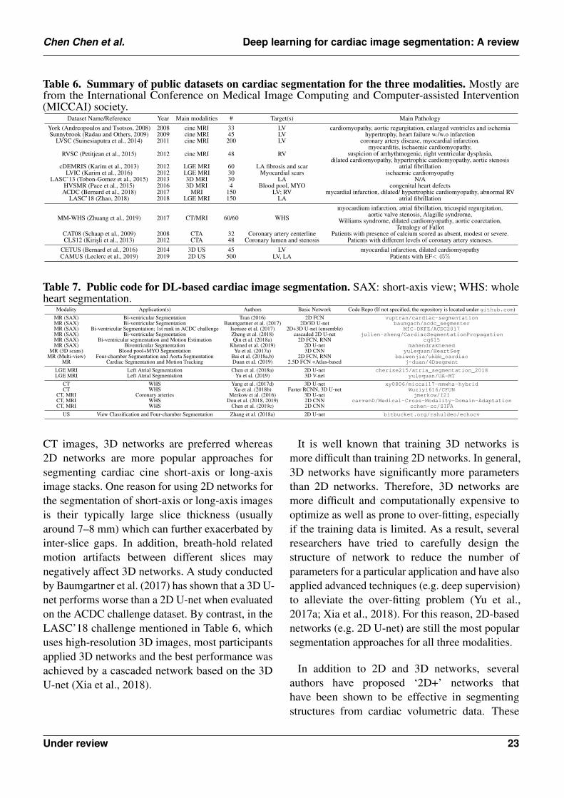
Chen Chen et al. Deep learning for cardiac image segmentation: A review
Table 6. Summary of public datasets on cardiac segmentation for the three modalities. Mostly arefrom the International Conference on Medical Image Computing and Computer-assisted Intervention(MICCAI) society.
Dataset Name/Reference Year Main modalities # Target(s) Main PathologyYork (Andreopoulos and Tsotsos, 2008) 2008 cine MRI 33 LV cardiomyopathy, aortic regurgitation, enlarged ventricles and ischemiaSunnybrook (Radau and Others, 2009) 2009 cine MRI 45 LV hypertrophy, heart failure w./w.o infarction
LVSC (Suinesiaputra et al., 2014) 2011 cine MRI 200 LV coronary artery disease, myocardial infarction.
RVSC (Petitjean et al., 2015) 2012 cine MRI 48 RVmyocarditis, ischaemic cardiomyopathy,
suspicion of arrhythmogenic, right ventricular dysplasia,dilated cardiomyopathy, hypertrophic cardiomyopathy, aortic stenosis
cDEMRIS (Karim et al., 2013) 2012 LGE MRI 60 LA fibrosis and scar atrial fibrillationLVIC (Karim et al., 2016) 2012 LGE MRI 30 Myocardial scars ischaemic cardiomyopathy
LASC’13 (Tobon-Gomez et al., 2015) 2013 3D MRI 30 LA N/AHVSMR (Pace et al., 2015) 2016 3D MRI 4 Blood pool, MYO congenital heart defects
ACDC (Bernard et al., 2018) 2017 MRI 150 LV; RV mycardial infarction, dilated/ hypertrophic cardiomyopathy, abnormal RVLASC’18 (Zhao, 2018) 2018 LGE MRI 150 LA atrial fibrillation
MM-WHS (Zhuang et al., 2019) 2017 CT/MRI 60/60 WHS
myocardium infarction, atrial fibrillation, tricuspid regurgitation,aortic valve stenosis, Alagille syndrome,
Williams syndrome, dilated cardiomyopathy, aortic coarctation,Tetralogy of Fallot
CAT08 (Schaap et al., 2009) 2008 CTA 32 Coronary artery centerline Patients with presence of calcium scored as absent, modest or severe.CLS12 (Kirisli et al., 2013) 2012 CTA 48 Coronary lumen and stenosis Patients with different levels of coronary artery stenoses.
CETUS (Bernard et al., 2016) 2014 3D US 45 LV myocardial infarction, dilated cardiomyopathyCAMUS (Leclerc et al., 2019) 2019 2D US 500 LV, LA Patients with EF< 45%
Table 7. Public code for DL-based cardiac image segmentation. SAX: short-axis view; WHS: wholeheart segmentation.
Modality Application(s) Authors Basic Network Code Repo (If not specified, the repository is located under github.com)MR (SAX) Bi-ventricular Segmentation Tran (2016) 2D FCN vuptran/cardiac-segmentationMR (SAX) Bi-ventricular Segmentation Baumgartner et al. (2017) 2D/3D U-net baumgach/acdc_segmenterMR (SAX) Bi-ventricular Segmentation; 1st rank in ACDC challenge Isensee et al. (2017) 2D+3D U-net (ensemble) MIC-DKFZ/ACDC2017MR (SAX) Bi-ventricular Segmentation Zheng et al. (2018) cascaded 2D U-net julien-zheng/CardiacSegmentationPropagationMR (SAX) Bi-ventricular segmentation and Motion Estimation Qin et al. (2018a) 2D FCN, RNN cq615MR (SAX) Biventricular Segmentation Khened et al. (2019) 2D U-net mahendrakhened
MR (3D scans) Blood pool+MYO Segmentation Yu et al. (2017a) 3D CNN yulequan/HeartSegMR (Multi-view) Four-chamber Segmentation and Aorta Segmentation Bai et al. (2018a,b) 2D FCN, RNN baiwenjia/ukbb_cardiac
MR Cardiac Segmentation and Motion Tracking Duan et al. (2019) 2.5D FCN +Atlas-based j-duan/4Dsegment
LGE MRI Left Atrial Segmentation Chen et al. (2018a) 2D U-net cherise215/atria_segmentation_2018LGE MRI Left Atrial Segmentation Yu et al. (2019) 3D V-net yulequan/UA-MT
CT WHS Yang et al. (2017d) 3D U-net xy0806/miccai17-mmwhs-hybridCT WHS Xu et al. (2018b) Faster RCNN, 3D U-net Wuziyi616/CFUN
CT, MRI Coronary arteries Merkow et al. (2016) 3D U-net jmerkow/I2ICT, MRI WHS Dou et al. (2018, 2019) 2D CNN carrenD/Medical-Cross-Modality-Domain-AdaptationCT, MRI WHS Chen et al. (2019c) 2D CNN cchen-cc/SIFA
US View Classification and Four-chamber Segmentation Zhang et al. (2018a) 2D U-net bitbucket.org/rahuldeo/echocv
CT images, 3D networks are preferred whereas2D networks are more popular approaches forsegmenting cardiac cine short-axis or long-axisimage stacks. One reason for using 2D networks forthe segmentation of short-axis or long-axis imagesis their typically large slice thickness (usuallyaround 7–8 mm) which can further exacerbated byinter-slice gaps. In addition, breath-hold relatedmotion artifacts between different slices maynegatively affect 3D networks. A study conductedby Baumgartner et al. (2017) has shown that a 3D U-net performs worse than a 2D U-net when evaluatedon the ACDC challenge dataset. By contrast, in theLASC’18 challenge mentioned in Table 6, whichuses high-resolution 3D images, most participantsapplied 3D networks and the best performance wasachieved by a cascaded network based on the 3DU-net (Xia et al., 2018).
It is well known that training 3D networks ismore difficult than training 2D networks. In general,3D networks have significantly more parametersthan 2D networks. Therefore, 3D networks aremore difficult and computationally expensive tooptimize as well as prone to over-fitting, especiallyif the training data is limited. As a result, severalresearchers have tried to carefully design thestructure of network to reduce the number ofparameters for a particular application and have alsoapplied advanced techniques (e.g. deep supervision)to alleviate the over-fitting problem (Yu et al.,2017a; Xia et al., 2018). For this reason, 2D-basednetworks (e.g. 2D U-net) are still the most popularsegmentation approaches for all three modalities.
In addition to 2D and 3D networks, severalauthors have proposed ‘2D+’ networks thathave been shown to be effective in segmentingstructures from cardiac volumetric data. These
Under review 23
Chen Chen et al. Deep learning for cardiac image segmentation: A review
‘2D+’ networks are mainly based on 2D networks,but are adapted with increased capacity to utilize3D context. These networks include multi-viewnetworks which leverage multi-planar information(i.e. coronal, sagittal, axial views) (Mortazi et al.,2017b; Wang and Smedby, 2017), multi-slicenetworks, and 2D FCNs combined with RNNswhich incorporate context across multiple slices(Duan et al., 2019; Patravali et al., 2017; Poudelet al., 2016; Du et al., 2019). These ‘2D+’networksinherit the advantages of 2D networks while stillbeing capable of leveraging through-plane spatialcontext for more robust segmentation with strong3D consistency.
4 CHALLENGES AND FUTURE WORK
It is evident from the literature that deep learningmethods have matched or surpassed the previousstate of the art in a various cardiac segmentationapplications, mainly benefiting from the increasedsize of public datasets and the emergence ofadvanced network architectures as well as powerfulhardware for computing. Given this rapid process,one may wonder if deep learning methods canbe directly deployed to real-world applications toreduce the workload of clinicians. The currentliterature suggests that there is still a longway to go. In the following paragraphs, wesummarize several major challenges in the field ofcardiac segmentation and some recently proposedapproaches that attempt to address them. Thesechallenges and related works also provide potentialresearch directions for future work in this field.
4.1 Scarcity of Labels
One of the biggest challenges for deep learningapproaches is the scarcity of annotated data.In this review, we found that the majorityof studies uses a fully supervised approach totrain their networks, which requires a largenumber of annotated images. In fact, annotatingcardiac images is time consuming and oftenrequires significant amounts of expertise. Whiledata augmentation techniques such as cropping,
padding, and geometric transformations (e.g. affinetransformations) can be used to increase the sizeof training samples, their diversity may still belimited, failing to reflect the spectrum of real-world data distributions. Several methods havebeen proposed to overcome this challenge. Thesemethods can be categorized into four classes:transfer learning with fine-tuning, weakly and semi-supervised learning, self-supervised learning, andunsupervised learning.
• Transfer learning with fine-tuning. Transferlearning aims at reusing a model pre-trainedon one task as a starting point to train fora second task. The key of transfer learningis to learn features in the first task thatare related to the second task such that thenetwork can quickly converge even with limiteddata. Several researchers have successfullydemonstrated the use of transfer learning toimprove the model generalization ability forcardiac ventricle segmentation across differentscanners, where they first trained a model on alarge dataset and then fine-tuned it on a smalldataset (Bai et al., 2018a; Khened et al., 2019;Cong and Zhang, 2018; Fahmy et al., 2019;Chen et al., 2019f).
• Weakly and semi-supervised learning. Weaklyand semi-supervised learning methods aim atimproving the learning accuracy by makinguse of both labeled and unlabeled or weakly-labeled data (e.g annotations in forms ofscribbles or bounding boxes). In this context,several works have been proposed for cardiacventricle segmentation in MR images. Oneapproach is to estimate full labels onunlabeled or weakly labeled images for furthertraining. For example, Bai et al. (2018b);Qin et al. (2018a) utilized motion informationto propagate labels from labeled frames tounlabeled frames in a cardiac cycle whereasBai et al. (2017); Can et al. (2018) appliedthe expectation maximization (EM) algorithmto predict and refine the estimated labelsrecursively. Others have explored differentapproaches to regularize the network when
Under review 24
Chen Chen et al. Deep learning for cardiac image segmentation: A review
training on unlabeled images, applying multi-task learning (Chartsias et al., 2018), or globalconstraints (Kervadec et al., 2019).
• Self-supervised learning. Another approach isself-supervised learning which aims at utilizinglabels that are generated automatically withouthuman intervention. These labels, designedto encode some properties or semantics ofthe object, can provide strong supervisorysignals to pre-train a network before fine-tuningfor a given task. A very recent work fromBai et al. (2019) has shown the effectivenessof self-supervised learning for cardiac MRimage segmentation where the authors usedauto-generated anatomical position labels topre-train a segmentation network. Compared toa network trained from scratch, networks pre-trained on the self-supervised task performedbetter, especially when the training data wasextremely limited.
• Unsupervised learning. Unsupervised learningaims at learning without paired labeled data.Compared to the former three classes, there islimited literature about unsupervised learningmethods for cardiac image segmentation,perhaps because of the difficulty of thetask. An early attempt has been made whichapplied adversarial training to train a networksegmenting LV and RV from CT and MRimages without requiring a training set of pairedimages and labels (Joyce et al., 2018).
Apart from utilizing unlabeled images for trainingneural networks, another interesting direction isactive learning (Mahapatra et al., 2018), whichtries to select the most representative images froma large-scale dataset, reducing redundant labelingworkload and training cost. This technique is alsorelated to incremental learning, which aims toimprove the model performance with new classesadded incrementally while avoiding a dramaticdecrease in overall performance (Castro et al.,2018). Given the increasing size of the availablemedical datasets, and the practical challengesof labeling and storing large amounts of images
from various sources, it is of great interest todevelop algorithms capable of distilling a large-scale cardiac dataset into a small one containing themost representative cases for labeling and training.
4.2 Model Generalization Across VariousImaging Modalities, Scanners andPathologies.
Another common limitation in DL-based methodsis that they still lack generalization capabilitieswhen presented with previously unseen samples(e.g. data from a new scanner, abnormal andpathological cases that have not been included inthe training set). In other words, deep learningmodels tend to be biased by their respective trainingdatasets. This limitation prevents models to bedeployed in the real world and therefore diminishestheir impact for improving clinical workflows.To improve the model performance across MRimages acquired from multiple vendors and multiplescanners, Tao et al. (2019) collected a large multi-vendor, multi-center, heterogeneous labeled trainingset from patients with cardiovascular diseases.However, this approach may not scale to the realworld, as it implies the collection of a vastly largedataset covering all possible cases. Moreover, it stillfaces the aforementioned collecting and labelingchallenge.
Unsupervised domain adaptation. Severalresearchers have recently started to investigate theuse of unsupervised domain adaptation techniquesthat aim at optimizing the model performance onunseen datasets without additional labeling costs.Several works have successfully applied adversarialtraining for cross-modality segmentation tasks,adapting a cardiac segmentation model learned fromMR images to CT images and vice versa (Douet al., 2018, 2019; Ouyang et al., 2019; Chen et al.,2019c). These type of approaches can also beadopted for semi-supervised learning, where thetarget domain is a new set of unlabeled data of thesame modality (Chen et al., 2019d).
Data augmentation. An alternative yet simpleand effective approach is data augmentation. The
Under review 25
Chen Chen et al. Deep learning for cardiac image segmentation: A review
main idea is to increase the variety of trainingimages so that the training set distribution ismore close to the one of a test set in the realworld. In general, this type of augmentation isachieved by applying a stack of geometric orphotometric transformations to existing image-labelpairs. Recently, Chen et al. (2019a) have proposeda data normalization and augmentation pipelinewhich enables a neural network for cardiac MRimage segmentation trained from a single-scannerdataset to generalize well across multi-scanner andmulti-site datasets. Zhang et al. (2019b) applieda similar data augmentation approach to improvethe model generalization ability on unseen datasets.Their method has been verified on three tasksincluding left atrial segmentation from 3D MRIand left ventricle segmentation from 3D ultrasoundimages. However, effectively designing such apipeline requires expertise, which may not be easyto be extended to other applications. Most recently,several researchers have began to investigate theuse of generative models (e.g. GANs, variationalAE (Kingma and Welling, 2013)), reinforcementlearning (Cubuk et al., 2019) and adversarialexample generation (Volpi et al., 2018) that aimat directly learning data augmentation strategiesfrom existing data. In particular, the generativemodel-based approach has been proven to beeffective for one-shot brain segmentation (Zhaoet al., 2019) and few-shot cardiac MR imagesegmentation (Chaitanya et al., 2019) and is thusworth to be explored for more applications in thefuture.
4.3 Lack of Model Interpretability
Unlike symbolic artificial intelligence systems,deep learning systems are difficult to interpret andnot transparent. Once a network has been trained,it behaves like a ‘black box’, providing predictionswhich are not directly interpretable. This issuemakes the model unpredictable, intractable formodel verification, and ultimately untrustworthy.Recent studies have shown that deep learning-based vision recognition systems can be attackedby images modified with nearly imperceptible
perturbations (Szegedy et al., 2014; Kurakin et al.,2017; Goodfellow et al., 2015). These attacks canalso happen in medical scenarios, e.g. a DL-basedsystem may make a wrong diagnosis given an imagewith adversarial noise or even just small rotation,as demonstrated in a very recent paper (Finlaysonet al., 2019). Although there is no denying thatdeep learning has become a very powerful toolfor image analysis, building resilient algorithmsrobust to potential attacks remains an unsolvedproblem. One potential solution, instead of buildingthe resilience into the model, is raising failureawareness of the deployed networks. This can beachieved by providing users with segmentationquality scores (Robinson et al., 2019) or confidencemaps such as uncertainty maps (Sander et al., 2019)and attention maps (Heo et al., 2018). These scoresor maps can be used as evidence to alert users whenfailure happens. For example, Sander et al. (2019)built a network that is able to simultaneously predictthe segmentation mask over cardiac structures andits associated spatial uncertainty map, where thelatter one could be used to highlight potentialincorrect regions. Such uncertainty informationcould alert human experts for further justificationand refinement in a human-in-the-loop setting.
4.4 Future work
Smart imaging. We have shown that deeplearning-based methods are able to segment imagesin real-time with good accuracy. However, thesealgorithms can still fail on those image acquisitionswith low image quality or significant artifacts.Although there have been several algorithmsdeveloped to avoid this problem by either checkingthe image quality before follow-up studies (Ruijsinket al., 2019; Tarroni et al., 2019), or predicting thesegmentation quality to detect failures (Peng andZhang, 2012; Robinson et al., 2019; Zhou et al.,2019), the development of algorithms that can giveinstant feedback to correct and optimize the imageacquisition process is also important despite lessexplored. Improving the imaging quality can greatlyimprove the effectiveness of medical imaging aswell as the accuracy of imaging-based diagnosis.
Under review 26
Chen Chen et al. Deep learning for cardiac image segmentation: A review
For radiologists, however, finding the optimalimaging and reconstruction parameters to scan eachpatient can take a great amount of time. Therefore, aDL-based system that has the potential of efficientlyand effectively improving the image quality withless noise is of great need. Some researchers haveutilized learning-based methods (mostly are deeplearning-based) for better image resolution (Oktayet al., 2016), view planning (Alansary et al., 2018),motion correction (Dangi et al., 2018a; Tarroniet al., 2018), artifacts reduction (Oksuz et al., 2019),shadow detection (Meng et al., 2019) and noisereduction (Wolterink et al., 2017b) after imageacquisition. However, combining these algorithmswith segmentation algorithms and seamlesslyintegrating them into an efficient, patient-specificimaging system for high-quality image analysis anddiagnosis is still an open challenge. An alternativeapproach is to directly predict cardiac segmentationmaps from undersampled k-space data to acceleratethe whole procedure, which bypasses the imagereconstruction stage (Schlemper et al., 2018).
Data harmonization. A number of works havereported the existence of missing labels andinconsistent labeling protocols among differentcardiac image datasets (Zheng et al., 2018;Chen et al., 2019a). Variations have beenfound in defining the end of basal slices aswell as the endocardial wall of myocardium(some include papillary muscles as part of theendocardial contours whereas others do not).These inconsistencies can be a major obstaclefor transferring, evaluating and deploying deeplearning models trained from one domain (e.g.hospital) to another. Therefore, building a standardbenchmark dataset like CheXpert (Irvin et al.,2019) that 1) is large enough to have substantialdata diversity that reflects the spectrum of real-world diversity; 2) has a standard labeling protocolapproved by experts, is indeed a need. However,directly building such a dataset from scratch is time-consuming and expensive. A more promising waymight be developing an automated tool to combineexisting datasets from multiple sources and then toharmonize them to a unified, high-quality dataset.
This tool can not only open the door for crowd-sourcing but also enable the rapid deployment ofthose DL-based segmentation models.
Data privacy. As deep learning is a data-drivenapproach, an unavoidable and rife concern isabout the data privacy. Regulations such as TheGeneral Data Protection Regulation (GDPR) nowplay an important role to protect users’ privacy andhave forced organizations to treat data ownershipseriously. On the other hand, from a technical pointof view, how to store, query, and process datasuch that there is no privacy concerns for buildingdeep learning systems has now become an evenmore difficult but interesting challenge. Building aprivacy-preserving algorithm requires to combinecryptography and deep learning together and tomix techniques from a wide range of subjects suchas data analysis, distributed computing, federatedlearning, differential privacy, in order to achievemodels with strong security, fast run time, and greatgeneralizability (Dwork and Roth, 2014; Abadiet al., 2016; Bonawitz et al., 2017; Ryffel et al.,2018). In this respect, Papernot (2018) published areport for guidance, which summarized a set of bestpractices for improving the privacy and security ofmachine learning systems. Yet, this field is still inits infancy.
5 CONCLUSION
In this review paper, we provided a comprehensiveoverview of these deep learning techniques usedin three common imaging modalities (MRI, CT,US), covering a wide range of existing deeplearning approaches (mostly are CNN-based) thatare designed for segmenting different cardiacanatomical structures (e.g. cardiac ventricle,atria, vessel). In particular, we presented anddiscussed recent progress of deep learning-basedsegmentation methods in the three modalities,outlined future potential and the remaininglimitations of these deep learning-based cardiacsegmentation methods that may hinder widespreadclinical deployment. We hope that this reviewcan provide an intuitive understanding of those
Under review 27
Chen Chen et al. Deep learning for cardiac image segmentation: A review
deep learning-based techniques that have madea significant contribution to cardiac imagesegmentation and also increase the awareness ofcommon challenges in this field that call for futurecontribution.
ABBREVIATIONS
Imaging-related terminology: CT: computedtomography; CTA: computed tomography angiography;LAX: long-axis; MPR: multi-planar reformatted;MR: magnetic resonance; MRI: magnetic resonanceimaging; LGE: late gadolinium enhancement;RFCA: radio-frequency catheter ablation; SAX:short-axis; US: ultrasound; 2CH: 2-chamber; 3CH:3-chamber; 4CH: 4-chamber.Cardiac structures and indexes: AF: atrialfibrillation; AS: aortic stenosis; AO: aorta; CVD:cardiovascular diseases; CAC: coronary arterycalcium; DCM: dilated cardiomyopathy; ED: end-diastole; ES: end-systole; EF: ejection fraction;HCM: hypertrophic cardiomyopathy; LA: leftatrium; LV: left ventricle; LVEDV: left ventricularend-diastolic volume; LVESV: left ventricular end-systolic volume; MCP: mixed-calcified plaque;MI: myocardial infarction; MYO: myocardium;NCP: non-calcified plaque; PA: pulmonary artery;PV: pulmonary vein; RA: right atrium; RV: rightventricle; RVEDV: right ventricular end-diastolicvolume; RVESV: right ventricular end-systolicvolume; RVEF: right ventricular ejection fraction;WHS: whole heart segmentation.Machine learning terminology: AE: autoencoder;ASM: active shape model; BN: batch normalization;CNN: convolutional neural network; CRF: conditionalrandom field; DBN: deep belief network; DL:deep learning; DNN: deep neural network;EM: expectation maximization; FCN: fullyconvolutional neural network; GAN: generativeadversarial network; GRU: gated recurrent units;MSE: mean squared error; MSL: marginal spacelearning; MRF: markov random field; LSTM:Long-short term memory; ReLU: rectified linearunit; RNN: recurrent neural network; ROI: region-of-interest; SMC: sequential monte carlo; SRF:structured random forest; SVM: support vector
machine.Cardiac image segmentation datasets: ACDC:Automated Cardiac Diagnosis Challenge; CETUS:Challenge on Endocardial Three-dimensionalUltrasound Segmentation; MM-WHS: Multi-Modality Whole Heart Segmentation; LASC: LeftAtrium Segmentation Challenge; LVSC: LeftVentricle Segmentation Challenge; RVSC: RightVentricle Segmentation Challenge.Others: EMBC: The International Engineeringin Medicine and Biology Conference; GDPR:The General Data Protection Regulation; GPU:graphic processing unit; FDA: United StatesFood and Drug Administration; ISBI: TheIEEE International Symposium on BiomedicalImaging; MICCAI: International Conference onMedical Image Computing and Computer-assistedIntervention; TPU: tensor processing unit; WHO:World Health Organization.
CONFLICT OF INTEREST STATEMENT
The authors declare that the research was conductedin the absence of any commercial or financialrelationships that could be construed as a potentialconflict of interest.
AUTHOR CONTRIBUTIONS
CC, WB, DR conceived and designed the work;CC, CQ, HQ searched and read the MR, CT, USliterature, respectively; CC, CQ, HQ drafted themanuscript together; WB, DR, GT, JD providedcritical revision with insightful and constructivecomments to improve the manuscript; All authorsread and approved the manuscript.
FUNDING
This work is supported by the SmartHeart EPSRCProgramme Grant (EP/P001009/1). Huaqi Qiuis supported by the EPSRC Programme Grant(EP/R005982/1).
Under review 28
Chen Chen et al. Deep learning for cardiac image segmentation: A review
DATA AVAILABILITY STATEMENT
The datasets summarized in Table 6 can be found in their corresponding websites listed below:
1. York: http://www.cse.yorku.ca/˜mridataset/2. Sunnybrook:http://www.cardiacatlas.org/studies/sunnybrook-cardiac-data/
3. LVSC:http://www.cardiacatlas.org/challenges/lv-segmentation-challenge/
4. RVSC:http://www.litislab.fr/?projet=1rvsc
5. cDEMRIS:https://www.doc.ic.ac.uk/˜rkarim/la_lv_framework/fibrosis
6. LVIC:https://www.doc.ic.ac.uk/˜rkarim/la_lv_framework/lv_infarct
7. LASC’13:www.cardiacatlas.org/challenges/left-atrium-segmentation-challenge/
8. HVSMR:http://segchd.csail.mit.edu/
9. ACDC:https://acdc.creatis.insa-lyon.fr/
10. LASC’18:http://atriaseg2018.cardiacatlas.org/data/
11. MM-WHS:http://www.sdspeople.fudan.edu.cn/zhuangxiahai/0/mmwhs17/
12. CAT08:http://coronary.bigr.nl/centerlines/
13. CLS12:http://coronary.bigr.nl/stenoses
14. CETUS:https://www.creatis.insa-lyon.fr/Challenge/CETUS
15. CAMUS:https://www.creatis.insa-lyon.fr/Challenge/camus
ACKNOWLEDGMENTS
We would like to thank our colleagues: Karl Hahn, Qingjie Meng, James Batten, and Jonathan Passerat-Palmbach who provided insight and expertise that greatly assisted the work, and also constructive andthoughtful comments from Turkay Kart that greatly improved the manuscript.
REFERENCES
Abadi, M., Chu, A., Goodfellow, I. J., McMahan, H. B., Mironov, I., Talwar, K., et al. (2016). Deep learningwith differential privacy. In the 2016 ACM SIGSAC Conference on Computer and CommunicationsSecurity, Vienna, Austria, October 24-28, 2016. 308–318
Agatston, A. S., Janowitz, W. R., Hildner, F. J., Zusmer, N. R., Viamonte, M., and Detrano, R. (1990).Quantification of coronary artery calcium using ultrafast computed tomography. Journal of the AmericanCollege of Cardiology 15, 827–832
Alansary, A., Folgoc, L. L., Vaillant, G., Oktay, O., Li, Y., Bai, W., et al. (2018). Automatic view planningwith multi-scale deep reinforcement learning agents. In Medical Image Computing and ComputerAssisted Intervention. 277–285. doi:10.1007/978-3-030-00928-1\ 32
Andreopoulos, A. and Tsotsos, J. K. (2008). Efficient and generalizable statistical models of shape andappearance for analysis of cardiac MRI. Medical image analysis 12, 335–357. doi:10.1016/j.media.2007.12.003
Avendi, M. R., Kheradvar, A., and Jafarkhani, H. (2016). A combined deep-learning and deformable-modelapproach to fully automatic segmentation of the left ventricle in cardiac mri. Medical Image Analysis 30,108–119
Avendi, M. R., Kheradvar, A., and Jafarkhani, H. (2017). Automatic segmentation of the right ventriclefrom cardiac MRI using a learning-based approach. Magnetic resonance in medicine: official journal
Under review 29
Chen Chen et al. Deep learning for cardiac image segmentation: A review
of the Society of Magnetic Resonance in Medicine / Society of Magnetic Resonance in Medicine 78,2439–2448. doi:10.1002/mrm.26631
Bai, W., Chen, C., Tarroni, G., Duan, J., Guitton, F., Petersen, S. E., et al. (2019). Self-Supervised learningfor cardiac MR image segmentation by anatomical position prediction. In Medical Image Computingand Computer Assisted Intervention. 541–549. doi:10.1007/978-3-030-32245-8\ 60
Bai, W., Oktay, O., Sinclair, M., Suzuki, H., Rajchl, M., Tarroni, G., et al. (2017). Semi-supervised learningfor Network-Based cardiac MR image segmentation. In Medical Image Computing and ComputerAssisted Intervention, eds. M. Descoteaux, L. Maier-Hein, A. Franz, P. Jannin, D. L. Collins, andS. Duchesne (Springer International Publishing), vol. 10434, 253–260
Bai, W., Sinclair, M., Tarroni, G., Oktay, O., Rajchl, M., Vaillant, G., et al. (2018a). Automatedcardiovascular magnetic resonance image analysis with fully convolutional networks. Journal ofCardiovascular Magnetic Resonance 20, 65
Bai, W., Suzuki, H., Qin, C., Tarroni, G., Oktay, O., Matthews, P. M., et al. (2018b). Recurrent neuralnetworks for aortic image sequence segmentation with sparse annotations. In Medical Image Computingand Computer Assisted Intervention. 586–594. doi:10.1007/978-3-030-00937-3\ 67
Baumgartner, C. F., Koch, L. M., Pollefeys, M., and Konukoglu, E. (2017). An exploration of 2D and 3Ddeep learning techniques for cardiac MR image segmentation. In International Workshop on StatisticalAtlases and Computational Models of the Heart. 1–8
Bello, G. A., Dawes, T. J. W., Duan, J., Biffi, C., de Marvao, A., Howard, L. S. G. E., et al. (2019). Deeplearning cardiac motion analysis for human survival prediction. Nature machine intelligence 1, 95–104.doi:10.1038/s42256-019-0019-2
Bernard, O., Bosch, J. G., Heyde, B., Alessandrini, M., Barbosa, D., Camarasu-Pop, S., et al. (2016).Standardized evaluation system for left ventricular segmentation algorithms in 3D echocardiography.IEEE transactions on medical imaging 35, 967–977. doi:10.1109/TMI.2015.2503890
Bernard, O., Lalande, A., Zotti, C., Cervenansky, F., Yang, X., Heng, P.-A., et al. (2018). Deep learningtechniques for automatic MRI cardiac Multi-Structures segmentation and diagnosis: Is the problemsolved? IEEE Transactions on Medical Imaging 37, 2514–2525. doi:10.1109/TMI.2018.2837502.https://acdc.creatis.insa-lyon.fr/
Bian, C., Yang, X., Ma, J., Zheng, S., Liu, Y.-A., Nezafat, R., et al. (2018). Pyramid network with onlinehard example mining for accurate left atrium segmentation. In Statistical Atlases and ComputationalModels of the Heart. Atrial Segmentation and LV Quantification Challenges 2018 (Springer InternationalPublishing), 237–245. doi:10.1007/978-3-030-12029-0\ 26
Biffi, C., Oktay, O., Tarroni, G., Bai, W., De Marvao, A., Doumou, G., et al. (2018). Learning interpretableanatomical features through deep generative models: Application to cardiac remodeling. In MedicalImage Computing and Computer Assisted Intervention. vol. 11071 LNCS, 464–471. doi:10.1007/978-3-030-00934-2\ 52
Bonawitz, K., Ivanov, V., Kreuter, B., Marcedone, A., McMahan, H. B., Patel, S., et al. (2017). Practicalsecure aggregation for privacy-preserving machine learning. In the 2017 ACM SIGSAC Conference onComputer and Communications Security, CCS 2017, Dallas, TX, USA, October 30 - November 03, 2017.1175–1191. doi:10.1145/3133956.3133982
Can, Y. B., Chaitanya, K., Mustafa, B., Koch, L. M., Konukoglu, E., and Baumgartner, C. F. (2018).Learning to segment medical images with Scribble-Supervision alone. In Deep Learning in MedicalImage Analysis and Multimodal Learning for Clinical Decision Support (Springer InternationalPublishing), 236–244
Under review 30
Chen Chen et al. Deep learning for cardiac image segmentation: A review
Cano-Espinosa, C., Gonzalez, G., Washko, G. R., Cazorla, M., and Estepar, R. S. J. (2018). AutomatedAgatston score computation in non-ECG gated CT scans using deep learning. In Medical Imaging 2018:Image Processing (International Society for Optics and Photonics), vol. 10574, 105742K
Carminati, M. C., Boniotti, C., Fusini, L., Andreini, D., Pontone, G., Pepi, M., et al. (2016). Comparisonof image processing techniques for nonviable tissue quantification in late gadolinium enhancementcardiac magnetic resonance images. Journal of thoracic imaging 31, 168–176. doi:10.1097/RTI.0000000000000206
Carneiro, G., Nascimento, J., and Freitas, A. (2010). Robust left ventricle segmentation from ultrasounddata using deep neural networks and efficient search methods. In 2010 IEEE International Symposiumon Biomedical Imaging: From Nano to Macro. 1085–1088
Carneiro, G. and Nascimento, J. C. (2010). Multiple dynamic models for tracking the left ventricle ofthe heart from ultrasound data using particle filters and deep learning architectures. In Conference onComputer Vision and Pattern Recognition (IEEE), 2815–2822
Carneiro, G. and Nascimento, J. C. (2011). Incremental on-line semi-supervised learning for segmentingthe left ventricle of the heart from ultrasound data. In 2011 International Conference on ComputerVision (IEEE), 1700–1707
Carneiro, G. and Nascimento, J. C. (2012). The use of on-line co-training to reduce the training set size inpattern recognition methods: Application to left ventricle segmentation in ultrasound. In Conference onComputer Vision and Pattern Recognition (IEEE), 948–955
Carneiro, G. and Nascimento, J. C. (2013). Combining multiple dynamic models and deep learningarchitectures for tracking the left ventricle endocardium in ultrasound data. IEEE transactions on patternanalysis and machine intelligence 35, 2592–2607
Carneiro, G., Nascimento, J. C., and Freitas, A. (2012). The segmentation of the left ventricle of theheart from ultrasound data using deep learning architectures and derivative-based search methods. IEEETransactions on Image Processing 21, 968–982. doi:10.1109/TIP.2011.2169273
Castro, F. M., Marın-Jimenez, M. J., Guil, N., Schmid, C., and Alahari, K. (2018). End-to-end incrementallearning. In European Conference on Computer Vision. 241–257. doi:10.1007/978-3-030-01258-8\ 15
Chaitanya, K., Karani, N., Baumgartner, C., Donati, O., Becker, A., and Konukoglu, E. (2019). Semi-Supervised and Task-Driven data augmentation. In International Conference on Information Processingin Medical Imaging. 29–41. doi:10.1007/978-3-030-20351-1\ 3
Chartsias, A., Joyce, T., Papanastasiou, G., Semple, S., Williams, M., Newby, D., et al. (2018). Factorisedspatial representation learning: Application in semi-supervised myocardial segmentation. In MedicalImage Computing and Computer Assisted Intervention. vol. 11071 LNCS, 490–498
Chen, C., Bai, W., Davies, R. H., Bhuva, A. N., Manisty, C., Moon, J. C., et al. (2019a). Improving thegeneralizability of convolutional neural network-based segmentation on CMR images. Arxiv Preprintabs/1907.01268. Available at http://arxiv.org/abs/1907.01268 (Accessed September 1,2019)
Chen, C., Bai, W., and Rueckert, D. (2018a). Multi-task learning for left atrial segmentation on GE-MRI. In9th International Workshop, STACOM 2018, Held in Conjunction with MICCAI 2018, Granada, Spain,September 16, 2018, (Springer International Publishing), 292–301. doi:10.1007/978-3-030-12029-0\ 32
Chen, C., Biffi, C., Tarroni, G., Petersen, S., Bai, W., and Rueckert, D. (2019b). Learning shape priors forrobust cardiac MR segmentation from multi-view images. In Medical Image Computing and ComputerAssisted Intervention. 523–531
Under review 31
Chen Chen et al. Deep learning for cardiac image segmentation: A review
Chen, C., Dou, Q., Zhou, J., Qin, J., and Heng, P. A. (2019c). Synergistic image and feature adaptation:Towards Cross-Modality domain adaptation for medical image segmentation. Conference on ArtificialIntelligence , 865–872doi:10.1609/aaai.v33i01.3301865
Chen, H., Zheng, Y., Park, J.-H., Heng, P.-A., and Zhou, S. K. (2016). Iterative multi-domainregularized deep learning for anatomical structure detection and segmentation from ultrasound images. InMedical Image Computing and Computer-Assisted Intervention – MICCAI 2016 (Springer InternationalPublishing), 487–495
Chen, J., Yang, G., Gao, Z., Ni, H., Firmin, D., and others (2018b). Multiview two-task recursive attentionmodel for left atrium and atrial scars segmentation. In Medical Image Computing and Computer AssistedIntervention. 455–463. doi:10.1007/978-3-030-00934-2\ 51
Chen, J., Zhang, H., Zhang, Y., Zhao, S., Mohiaddin, R., Wong, T., et al. (2019d). Discriminative consistentdomain generation for semi-supervised learning. In Medical Image Computing and Computer AssistedIntervention. 595–604. doi:10.1007/978-3-030-32245-8\ 66
Chen, L.-C., Papandreou, G., Schroff, F., and Adam, H. (2017). Rethinking atrous convolution for semanticimage segmentation. Arxiv Preprint abs/1706.05587. Available at http://arxiv.org/abs/1706.05587(Accessed September 1, 2019)
Chen, M., Fang, L., and Liu, H. (2019e). FR-NET: Focal loss constrained deep residual networksfor segmentation of cardiac MRI. In International Symposium on Biomedical Imaging. 764–767.doi:10.1109/ISBI.2019.8759556
Chen, S., Ma, K., and Zheng, Y. (2019f). Med3d: Transfer learning for 3d medical image analysis.Arxiv Preprint abs/1904.00625. Available at http://arxiv.org/abs/1904.00625 (AccessedSeptember 1, 2019)
Chen, X., Williams, B. M., Vallabhaneni, S. R., Czanner, G., Williams, R., and Zheng, Y. (2019g). Learningactive contour models for medical image segmentation. In Conference on Computer Vision and PatternRecognition. 11632–11640
Chen, Y.-C., Lin, Y.-C., Wang, C.-P., Lee, C.-Y., Lee, W.-J., Wang, T.-D., et al. (2019h). Coronary arterysegmentation in cardiac CT angiography using 3D multi-channel U-net. In Medical Imaging with DeepLearning. 1907.12246
Cho, K., van Merrienboer, B., Gulcehre, C., Bahdanau, D., Bougares, F., Schwenk, H., et al. (2014).Learning phrase representations using RNN Encoder-Decoder for statistical machine translation. InConference on Empirical Methods in Natural Language Processing (ACL), 1724–1734
Cicek, O., Abdulkadir, A., Lienkamp, S. S., Brox, T., and Ronneberger, O. (2016). 3D U-Net: Learningdense volumetric segmentation from sparse annotation. In Medical Image Computing and ComputerAssisted Intervention. 424–432. doi:10.1007/978-3-319-46723-8\ 49
Ciresan, D. C. and Giusti, A. (2012). Deep neural networks segment neuronal membranes in electronmicroscopy images. In Conference on Neural Information Processing Systems. 2852–2860
Clough, J. R., Oksuz, I., Byrne, N., Schnabel, J. A., and King, A. P. (2019). Explicit topological priorsfor deep-learning based image segmentation using persistent homology. In Information Processing inMedical Imaging. vol. 11492 LNCS, 16–28. doi:10.1007/978-3-030-20351-1\ 2
Cong, C. and Zhang, H. (2018). Invert-U-Net DNN segmentation model for MRI cardiac left ventriclesegmentation. The Journal of Engineering 2018, 1463–1467. doi:10.1049/joe.2018.8302
Cubuk, E. D., Zoph, B., Mane, D., Vasudevan, V., and Le, Q. V. (2019). Autoaugment: Learningaugmentation policies from data. In Conference on Computer Vision and Pattern Recognition. 1–14
Under review 32
Chen Chen et al. Deep learning for cardiac image segmentation: A review
Dangi, S., Linte, C. A., and Yaniv, Z. (2018a). Cine cardiac MRI slice misalignment correction towardsfull 3D left ventricle segmentation. In Proceedings of SPIE The International Society for OpticalEngineering. 1057607. doi:10.1117/12.2294936
Dangi, S., Yaniv, Z., and Linte, C. A. (2018b). Left ventricle segmentation and quantification from cardiaccine MR images via multi-task learning. In Statistical Atlases and Computational Models of the Heart.Atrial Segmentation and LV Quantification Challenges - 9th International Workshop, STACOM 2018,Held in Conjunction with MICCAI 2018, Granada, Spain, September 16, 2018, Revised Selected Papers.21–31. doi:10.1007/978-3-030-12029-0\ 3
de Vos, B. D., Wolterink, J. M., De Jong, P. A., Leiner, T., Viergever, M. A., and Isgum, I. (2017).Convnet-based localization of anatomical structures in 3-D medical images. IEEE Transactions onMedical Imaging 36, 1470–1481
de Vos, B. D., Wolterink, J. M., Leiner, T., de Jong, P. A., Lessmann, N., and Isgum, I. (2019). Directautomatic coronary calcium scoring in cardiac and chest CT. IEEE Transactions on Medical Imaging38, 2127–2138
Degel, M. A., Navab, N., and Albarqouni, S. (2018). Domain and geometry agnostic CNNs for left atriumsegmentation in 3D ultrasound. In Medical Image Computing and Computer Assisted Intervention –MICCAI 2018 (Springer International Publishing), 630–637
Dong, S., Luo, G., Wang, K., Cao, S., Li, Q., and Zhang, H. (2018a). A combined fully convolutionalnetworks and deformable model for automatic left ventricle segmentation based on 3D echocardiography.Biomed Res. Int. 2018, 5682365. doi:10.1155/2018/5682365
Dong, S., Luo, G., Wang, K., Cao, S., Mercado, A., Shmuilovich, O., et al. (2018b). VoxelAtlasGAN:3D left ventricle segmentation on echocardiography with atlas guided generation and Voxel-to-Voxeldiscrimination. In Medical Image Computing and Computer Assisted Intervention – MICCAI 2018(Springer International Publishing), 622–629
Dormer, J. D., Ma, L., Halicek, M., Reilly, C. M., Schreibmann, E., and Fei, B. (2018). Heart chambersegmentation from CT using convolutional neural networks. In Medical Imaging 2018: BiomedicalApplications in Molecular, Structural, and Functional Imaging, Houston, Texas, United States, 10-15February 2018. 105782S
Dou, Q., Ouyang, C., Chen, C., Chen, H., Glocker, B., Zhuang, X., et al. (2019). Pnp-adanet: Plug-and-playadversarial domain adaptation network at unpaired cross-modality cardiac segmentation. IEEE Access 7,99065–99076. doi:10.1109/ACCESS.2019.2929258
Dou, Q., Ouyang, C., Chen, C., Chen, H., and Heng, P.-A. (2018). Unsupervised Cross-Modality domainadaptation of ConvNets for biomedical image segmentations with adversarial loss. In International JointConferences on Artificial Intelligence. 691–697
Du, X., Yin, S., Tang, R., Zhang, Y., and Li, S. (2019). Cardiac-DeepIED: Automatic pixel-level deepsegmentation for cardiac bi-ventricle using improved end-to-end encoder-decoder network. IEEE journalof translational engineering in health and medicine 7, 1–10. doi:10.1109/JTEHM.2019.2900628
Duan, J., Bello, G., Schlemper, J., Bai, W., Dawes, T. J. W., Biffi, C., et al. (2019). Automatic 3Dbi-ventricular segmentation of cardiac images by a shape-constrained multi-task deep learning approach.IEEE Transactions on Medical Imaging PP, 1. doi:10.1109/TMI.2019.2894322
Duan, J., Schlemper, J., Bai, W., Dawes, T. J. W., Bello, G., Doumou, G., et al. (2018a). Deep nested levelsets: Fully automated segmentation of cardiac MR images in patients with pulmonary hypertension. InMedical Image Computing and Computer Assisted Intervention. 595–603
Under review 33
Chen Chen et al. Deep learning for cardiac image segmentation: A review
Duan, Y., Feng, J., Lu, J., and Zhou, J. (2018b). Context aware 3D fully convolutional networks forcoronary artery segmentation. In International Workshop on Statistical Atlases and ComputationalModels of the Heart (Springer), 85–93
Dwork, C. and Roth, A. (2014). The algorithmic foundations of differential privacy. Foundations andTrends in Theoretical Computer Science 9, 211–407
Fahmy, A. S., El-Rewaidy, H., Nezafat, M., Nakamori, S., and Nezafat, R. (2019). Automatedanalysis of cardiovascular magnetic resonance myocardial native T1 mapping images using fullyconvolutional neural networks. Journal of Cardiovascular Magnetic Resonance 21, 1–12. doi:10.1186/s12968-018-0516-1
Fahmy, A. S., Rausch, J., Neisius, U., Chan, R. H., Maron, M. S., Appelbaum, E., et al. (2018). Automatedcardiac MR scar quantification in hypertrophic cardiomyopathy using deep convolutional neural networks.JACC. Cardiovascular imaging 11, 1917–1918. doi:10.1016/j.jcmg.2018.04.030
Finlayson, S. G., Bowers, J. D., Ito, J., Zittrain, J. L., Beam, A. L., and Kohane, I. S. (2019). Adversarialattacks on medical machine learning. Science 363, 1287–1289. doi:10.1126/science.aaw4399
Gandhi, S., Mosleh, W., Shen, J., and Chow, C.-M. (2018). Automation, machine learning, and artificialintelligence in echocardiography: A brave new world. Echocardiography 35, 1402–1418. doi:10.1111/echo.14086
Georgescu, B., Zhou, X. S., Comaniciu, D., and Gupta, A. (2005). Database-guided segmentationof anatomical structures with complex appearance. In 2005 IEEE Computer Society Conference onComputer Vision and Pattern Recognition (CVPR’05) (IEEE), vol. 2, 429–436
Ghesu, F. C., Krubasik, E., Georgescu, B., Singh, V., Yefeng Zheng, Hornegger, J., et al. (2016). Marginalspace deep learning: Efficient architecture for volumetric image parsing. IEEE Transactions on MedicalImaging 35, 1217–1228
Girdhar, R., Fouhey, D. F., Rodriguez, M., and Gupta, A. (2016). Learning a predictable and generativevector representation for objects. In European Conference on Computer Vision (Springer InternationalPublishing), 484–499
Goodfellow, I. (2016). Deep learning. Adaptive computation and machine learning (Cambridge,Massachusetts ; London, England: The MIT Press)
Goodfellow, I. J., Pouget-Abadie, J., Mirza, M., Xu, B., Warde-Farley, D., Ozair, S., et al. (2014).Generative adversarial networks. In Conference on Neural Information Processing Systems (CurranAssociates, Inc.), 2672–2680
Goodfellow, I. J., Shlens, J., and Szegedy, C. (2015). Explaining and harnessing adversarial examples. InInternational Conference on Learning Representations. 43405
Greenspan, H., Van Ginneken, B., and Summers, R. M. (2016). Guest editorial deep learning in medicalimaging: Overview and future promise of an exciting new technique. IEEE Transactions on MedicalImaging 35, 1153–1159
Gulsun, M. A., Funka-Lea, G., Sharma, P., Rapaka, S., and Zheng, Y. (2016). Coronary centerlineextraction via optimal flow paths and CNN path pruning. In Medical Image Computing and ComputerAssisted Intervention (Springer), 317–325
Guo, Z., Bai, J., Lu, Y., Wang, X., Cao, K., Song, Q., et al. (2019). Deepcenterline: A multi-task fullyconvolutional network for centerline extraction. In International Conference on Information Processingin Medical Imaging (Springer), 441–453
He, K., Zhang, X., Ren, S., and Sun, J. (2015). Delving deep into rectifiers: Surpassing Human-Levelperformance on ImageNet classification. In International Conference on Computer Vision (IEEEComputer Society), 1026–1034
Under review 34
Chen Chen et al. Deep learning for cardiac image segmentation: A review
He, K., Zhang, X., Ren, S., and Sun, J. (2016). Deep residual learning for image recognition. In Conferenceon Computer Vision and Pattern Recognition. 770–778
Heo, J., Lee, H. B., Kim, S., Lee, J., Kim, K. J., Yang, E., et al. (2018). Uncertainty-Aware attention forreliable interpretation and prediction. In Advances in Neural Information Processing Systems 31, eds.S. Bengio, H. Wallach, H. Larochelle, K. Grauman, N. Cesa-Bianchi, and R. Garnett (Curran Associates,Inc.). 909–918
Herment, A., Kachenoura, N., Lefort, M., Bensalah, M., Dogui, A., Frouin, F., et al. (2010). Automatedsegmentation of the aorta from phase contrast MR images: validation against expert tracing in healthyvolunteers and in patients with a dilated aorta. Journal of magnetic resonance imaging 31, 881–888.doi:10.1002/jmri.22124
Hinton, G. E. and Salakhutdinov, R. R. (2006). Reducing the dimensionality of data with neural networks.Science 313, 504–507
Hochreiter, S. and Schmidhuber, J. (1997). Long short-term memory. Neural computation 9, 1735–1780.doi:10.1162/neco.1997.9.8.1735
Hu, J., Shen, L., and Sun, G. (2018). Squeeze-and-Excitation networks. In Conference on Computer Visionand Pattern Recognition. 7132–7141. doi:10.1109/CVPR.2018.00745
Huang, G., Liu, Z., van der Maaten, L., and Weinberger, K. Q. (2017). Densely connected convolutionalnetworks. In Conference on Computer Vision and Pattern Recognition. 2261–2269
Huang, Q., Yang, D., Yi, J., Axel, L., and Metaxas, D. (2019). FR-Net: Joint reconstruction andsegmentation in compressed sensing cardiac MRI. In Functional Imaging and Modelling of the Heart(Springer International Publishing), 352–360. doi:10.1007/978-3-030-21949-9\ 38
Huang, W., Huang, L., Lin, Z., Huang, S., Chi, Y., Zhou, J., et al. (2018). Coronary artery segmentation bydeep learning neural networks on computed tomographic coronary angiographic images. In 2018 40thAnnual International Conference of the IEEE Engineering in Medicine and Biology Society (EMBC)(IEEE), 608–611
Ioffe, S. and Szegedy, C. (2015). Batch normalization: accelerating deep network training by reducinginternal covariate shift. In ICML (JMLR.org), 448–456
Irvin, J., Rajpurkar, P., Ko, M., Yu, Y., Ciurea-Ilcus, S., Chute, C., et al. (2019). CheXpert: A largechest radiograph dataset with uncertainty labels and expert comparison. In Conference on ArtificialIntelligence. 590–597
Isensee, F., Jaeger, P. F., Full, P. M., Wolf, I., Engelhardt, S., and Maier-Hein, K. H. (2017). Automaticcardiac disease assessment on cine-MRI via Time-Series segmentation and domain specific features. InStatistical Atlases and Computational Models of the Heart. ACDC and MMWHS Challenges (SpringerInternational Publishing), 120–129. doi:10.1007/978-3-319-75541-0\ 13
Jafari, M. H., Girgis, H., Abdi, A. H., Liao, Z., Pesteie, M., Rohling, R., et al. (2019). Semi-Supervisedlearning for cardiac left ventricle segmentation using conditional deep generative models as prior. In2019 IEEE 16th International Symposium on Biomedical Imaging (ISBI 2019) (IEEE), 649–652
Jafari, M. H., Girgis, H., Liao, Z., Behnami, D., Abdi, A., Vaseli, H., et al. (2018). A unified frameworkintegrating recurrent Fully-Convolutional networks and optical flow for segmentation of the left ventriclein echocardiography data. In Deep Learning in Medical Image Analysis and Multimodal Learning forClinical Decision Support (Springer International Publishing), 29–37
Jang, Y., Hong, Y., Ha, S., Kim, S., and Chang, H.-J. (2017). Automatic segmentation of LV and RV incardiac MRI. In International Workshop on Statistical Atlases and Computational Models of the Heart(Springer), 161–169
Under review 35
Chen Chen et al. Deep learning for cardiac image segmentation: A review
Jia, S., Despinasse, A., Wang, Z., Delingette, H., Pennec, X., Jaıs, P., et al. (2018). Automaticallysegmenting the left atrium from cardiac images using successive 3D U-Nets and a contour loss. InInternational Workshop on Statistical Atlases and Computational Models of the Heart. 221–229
Joyce, T., Chartsias, A., and Tsaftaris, S. A. (2018). Deep Multi-Class segmentation without Ground-Truthlabels. In Medical Imaging with Deep Learning. 1–9
Kamnitsas, K., Bai, W., Ferrante, E., Mcdonagh, S., and Sinclair, M. (2017a). Ensembles of multiplemodels and architectures for robust brain tumour segmentation. In Brainlesion: Glioma, MultipleSclerosis, Stroke and Traumatic Brain Injuries - Third International Workshop, BrainLes 2017, Heldin Conjunction with MICCAI 2017, Quebec City, QC, Canada, September 14, 2017, Revised SelectedPapers. 450–462
Kamnitsas, K., Baumgartner, C., Ledig, C., Newcombe, V., Simpson, J., Kane, A., et al. (2017b).Unsupervised domain adaptation in brain lesion segmentation with adversarial networks. In InternationalConference on Information Processing in Medical Imaging (Springer International Publishing), 597–609
Kang, D., Woo, J., Kuo, C. J., Slomka, P. J., Dey, D., and Germano, G. (2012). Heart chambers and wholeheart segmentation techniques. Journal of Electronic Imaging 21, 010901
Karim, R., Bhagirath, P., Claus, P., James Housden, R., Chen, Z., Karimaghaloo, Z., et al. (2016).Evaluation of state-of-the-art segmentation algorithms for left ventricle infarct from late gadoliniumenhancement MR images. Medical image analysis 30, 95–107. doi:10.1016/j.media.2016.01.004
Karim, R., Blake, L.-E., Inoue, J., Tao, Q., Jia, S., James Housden, R., et al. (2018). Algorithms for leftatrial wall segmentation and thickness – evaluation on an open-source CT and MRI image database.Medical Image Analysis 50, 36–53. doi:10.1016/j.media.2018.08.004
Karim, R., Housden, R. J., Balasubramaniam, M., Chen, Z., Perry, D., Uddin, A., et al. (2013). Evaluationof current algorithms for segmentation of scar tissue from late gadolinium enhancement cardiovascularmagnetic resonance of the left atrium: an open-access grand challenge. Journal of cardiovascularmagnetic resonance: official journal of the Society for Cardiovascular Magnetic Resonance 15, 105.doi:10.1186/1532-429X-15-105
Karim, R., Mohiaddin, R., and Rueckert, D. (2008). Left atrium segmentation for atrial fibrillationablation. In Medical Imaging 2008: Visualization, Image-Guided Procedures, and Modeling, San Diego,California, United States, 16-21 February 2008 (SPIE), March 2008, 69182U. doi:10.1117/12.771023
Kass, M., Witkin, A., and Terzopoulos, D. (1988). Snakes: Active contour models. Int. J. Comput. Vis. 1,321–331
Kervadec, H., Dolz, J., Tang, M., Granger, E., Boykov, Y., and Ben Ayed, I. (2019). Constrained-CNNlosses for weakly supervised segmentation. In Medical Image Analysis. vol. 54, 88–99. doi:10.1016/j.media.2019.02.009
Khened, M., Kollerathu, V. A., and Krishnamurthi, G. (2019). Fully convolutional multi-scale residualDenseNets for cardiac segmentation and automated cardiac diagnosis using ensemble of classifiers.Medical Image Analysis 51, 21–45. doi:10.1016/j.media.2018.10.004
Kim, R. J., Fieno, D. S., Parrish, T. B., Harris, K., Chen, E. L., Simonetti, O., et al. (1999). Relationship ofMRI delayed contrast enhancement to irreversible injury, infarct age, and contractile function. Circulation100, 1992–2002. doi:10.1161/01.cir.100.19.1992
Kingma, D. P. and Welling, M. (2013). Auto-Encoding variational bayes. In International Conference onLearning Representations. 1–14
Kirisli, H. A., Schaap, M., Metz, C. T., Dharampal, A. S., Meijboom, W. B., Papadopoulou, S. L., et al.(2013). Standardized evaluation framework for evaluating coronary artery stenosis detection, stenosisquantification and lumen segmentation algorithms in computed tomography angiography. Medical
Under review 36
Chen Chen et al. Deep learning for cardiac image segmentation: A review
Image Analysis 17, 859–876. doi:10.1016/j.media.2013.05.007. http://coronary.bigr.nl/stenoses
Kurakin, A., Goodfellow, I., and Bengio, S. (2017). Adversarial examples in the physical world. In 5thInternational Conference on Learning Representations, ICLR 2017,Toulon, France, April 24-26, 2017,Workshop Track Proceedings. 1–14
Leclerc, S., Smistad, E., Grenier, T., Lartizien, C., Ostvik, A., Espinosa, F., et al. (2018). Deep learningapplied to Multi-Structure segmentation in 2D echocardiography: A preliminary investigation of therequired database size. In 2018 IEEE International Ultrasonics Symposium (IUS) (IEEE), 1–4
Leclerc, S., Smistad, E., Pedrosa, J., Ostvik, A., Cervenansky, F., Espinosa, F., et al. (2019). Deep learningfor segmentation using an open Large-Scale dataset in 2D echocardiography. IEEE Trans. Med. Imaginghttps://www.creatis.insa-lyon.fr/Challenge/camus
Lee, C.-Y., Xie, S., Gallagher, P., Zhang, Z., and Tu, Z. (2015). Deeply-Supervised nets. In ArtificialIntelligence and Statistics. 562–570
Lee, M. C. H., Petersen, K., Pawlowski, N., Glocker, B., and Schaap, M. (2019). TETRIS: Templatetransformer networks for image segmentation with shape priors. IEEE transactions on medical imaging
Lesage, D., Angelini, E. D., Bloch, I., and Funka-Lea, G. (2009). A review of 3D vessel lumen segmentationtechniques: Models, features and extraction schemes. Medical Image Analysis 13, 819–845
Lessmann, N., Isgum, I., Setio, A. A., de Vos, B. D., Ciompi, F., de Jong, P. A., et al. (2016). Deepconvolutional neural networks for automatic coronary calcium scoring in a screening study with low-dosechest CT. In Medical Imaging 2016: Computer-Aided Diagnosis (International Society for Optics andPhotonics), vol. 9785, 978511
Lessmann, N., van Ginneken, B., Zreik, M., de Jong, P. A., de Vos, B. D., Viergever, M. A., et al. (2017).Automatic calcium scoring in low-dose chest CT using deep neural networks with dilated convolutions.IEEE Transactions on Medical Imaging 37, 615–625
Li, C., Tong, Q., Liao, X., Si, W., Chen, S., Wang, Q., et al. (2019a). APCP-NET: Aggregated parallelCross-Scale pyramid network for CMR segmentation. In 2019 IEEE 16th International Symposium onBiomedical Imaging (ISBI 2019). 784–788. doi:10.1109/ISBI.2019.8759147
Li, C., Tong, Q., Liao, X., Si, W., Sun, Y., Wang, Q., et al. (2018). Attention based hierarchicalaggregation network for 3D left atrial segmentation: 9th international workshop, STACOM 2018, heldin conjunction with MICCAI 2018, granada, spain, september 16, 2018, revised selected papers. InStatistical Atlases and Computational Models of the Heart. Atrial Segmentation and LV QuantificationChallenges, eds. M. Pop, M. Sermesant, J. Zhao, S. Li, K. McLeod, A. Young, K. Rhode, and T. Mansi(Cham: Springer International Publishing), vol. 11395 of Lecture Notes in Computer Science. 255–264.doi:10.1007/978-3-030-12029-0\ 28
Li, J., Yu, Z., Gu, Z., Liu, H., and Li, Y. (2019b). Dilated-Inception net: Multi-Scale feature aggregationfor cardiac right ventricle segmentation. IEEE transactions on bio-medical engineering , 1–1doi:10.1109/TBME.2019.2906667
Li, J., Zhang, R., Shi, L., and Wang, D. (2017). Automatic Whole-Heart segmentation in congenital heartdisease using Deeply-Supervised 3D FCN. In Reconstruction, Segmentation, and Analysis of MedicalImages (Springer International Publishing), 111–118. doi:10.1007/978-3-319-52280-7\ 11
Liao, F., Chen, X., Hu, X., and Song, S. (2019). Estimation of the volume of the left ventricle from MRIimages using deep neural networks. IEEE transactions on cybernetics 49, 495–504. doi:10.1109/TCYB.2017.2778799
Lieman-Sifry, J., Le, M., Lau, F., Sall, S., and Golden, D. (2017). FastVentricle: Cardiac segmentationwith ENet. In Functional Imaging and Modelling of the Heart, eds. M. Pop and G. A. Wright (Cham:
Under review 37
Chen Chen et al. Deep learning for cardiac image segmentation: A review
Springer International Publishing), vol. 10263 LNCS of Lecture Notes in Computer Science, 127–138.doi:10.1007/978-3-319-59448-4\ 13
Litjens, G., Kooi, T., Bejnordi, B. E., Setio, A. A. A., Ciompi, F., Ghafoorian, M., et al. (2017). A surveyon deep learning in medical image analysis. Medical Image Analysis 42, 60 – 88
Liu, J., Jin, C., Feng, J., Du, Y., Lu, J., and Zhou, J. (2018). A vessel-focused 3D convolutional network forautomatic segmentation and classification of coronary artery plaques in cardiac CTA. In InternationalWorkshop on Statistical Atlases and Computational Models of the Heart (Springer), 131–141
Long, J., Shelhamer, E., and Darrell, T. (2015). Fully convolutional networks for semantic segmentation.In Conference on Computer Vision and Pattern Recognition. 3431–3440
Lu, X., Chen, X., Li, W., and Qiao, Y. (2019). Graph cut segmentation of the right ventricle in cardiac MRIusing multi-scale feature learning. In Proceedings of the 3rd International Conference on Cryptography,Security and Privacy (ACM), 231–235. doi:10.1145/3309074.3309117
Luc, P., Couprie, C., Chintala, S., and Verbeek, J. (2016). Semantic segmentation using adversarialnetworks. In NIPS Workshop on Adversarial Training. 1–12
Ma, J. and Zhang, R. (2019). Automatic calcium scoring in cardiac and chest CT using DenseRAUnet.Arxiv Preprint abs/1907.11392. Available at http://arxiv.org/abs/1907.11392 (AccessedSeptember 1, 2019)
Mahapatra, D., Bozorgtabar, B., Thiran, J.-P., and Reyes, M. (2018). Efficient active learning for imageclassification and segmentation using a sample selection and conditional generative adversarial network.In Medical Image Computing and Computer Assisted Intervention (Springer International Publishing),580–588. doi:10.1007/978-3-030-00934-2\ 65
Mazurowski, M. A., Buda, M., Saha, A., and Bashir, M. R. (2019). Deep learning in radiology: Anoverview of the concepts and a survey of the state of the art with focus on MRI. Journal of magneticresonance imaging 49, 939–954. doi:10.1002/jmri.26534
Medley, D. O., Santiago, C., and Nascimento, J. C. (2019). Segmenting the left ventricle in cardiac incardiac MRI: From handcrafted to deep region based descriptors. In 2019 IEEE 16th InternationalSymposium on Biomedical Imaging (ISBI 2019). 644–648. doi:10.1109/ISBI.2019.8759179
Meng, Q., Sinclair, M., Zimmer, V., Hou, B., Rajchl, M., Toussaint, N., et al. (2019). Weakly supervisedestimation of shadow confidence maps in fetal ultrasound imaging. IEEE Transactions on MedicalImaging
Merkow, J., Marsden, A., Kriegman, D., and Tu, Z. (2016). Dense volume-to-volume vascular boundarydetection. In Medical Image Computing and Computer Assisted Intervention (Springer), 371–379
Milletari, F., Navab, N., and Ahmadi, S. (2016). V-Net: Fully convolutional neural networks for volumetricmedical image segmentation. In 2016 Fourth International Conference on 3D Vision (3DV). 565–571.doi:10.1109/3DV.2016.79
Moccia, S., Banali, R., Martini, C., Muscogiuri, G., Pontone, G., Pepi, M., et al. (2019). Developmentand testing of a deep learning-based strategy for scar segmentation on CMR-LGE images. MagneticResonance Materials in Physics, Biology and Medicine 32, 187–195. doi:10.1007/s10334-018-0718-4
Moeskops, P., Wolterink, J. M., van der Velden, B. H., Gilhuijs, K. G., Leiner, T., Viergever, M. A., et al.(2016). Deep learning for multi-task medical image segmentation in multiple modalities. In MedicalImage Computing and Computer Assisted Intervention (Springer), 478–486
Mortazi, A., Burt, J., and Bagci, U. (2017a). Multi-planar deep segmentation networks for cardiacsubstructures from MRI and CT. In International Workshop on Statistical Atlases and ComputationalModels of the Heart (Springer), 199–206
Under review 38
Chen Chen et al. Deep learning for cardiac image segmentation: A review
Mortazi, A., Karim, R., Rhode, K., Burt, J., and Bagci, U. (2017b). CardiacNET: Segmentation of leftatrium and proximal pulmonary veins from MRI using multi-view CNN. In Lecture Notes in ComputerScience (including subseries Lecture Notes in Artificial Intelligence and Lecture Notes in Bioinformatics).377–385. doi:10.1007/978-3-319-66185-8\ 43
Nascimento, J. C. and Carneiro, G. (2014). Non-rigid segmentation using sparse low dimensional manifoldsand deep belief networks. In Proceedings of the IEEE Conference on Computer Vision and PatternRecognition (cv-foundation.org), 288–295
Nascimento, J. C. and Carneiro, G. (2017). Deep learning on sparse manifolds for faster object segmentation.IEEE Transactions on Image Processing 26, 4978–4990
Nascimento, J. C. and Carneiro, G. (2019). One shot segmentation: unifying rigid detection and non-rigidsegmentation using elastic regularization. IEEE Trans. Pattern Anal. Mach. Intell. doi:10.1109/TPAMI.2019.2922959
Ngo, T. A., Lu, Z., and Carneiro, G. (2017). Combining deep learning and level set for the automatedsegmentation of the left ventricle of the heart from cardiac cine magnetic resonance. Medical ImageAnalysis 35, 159–171
Noble, J. A. and Boukerroui, D. (2006). Ultrasound image segmentation: a survey. IEEE Transactions onMedical Imaging 25, 987–1010. doi:10.1109/TMI.2006.877092
Oksuz, I., Clough, J., Bai, W., Ruijsink, B., Puyol-Anton, E., Cruz, G., et al. (2019). High-qualitysegmentation of low quality cardiac MR images using k-space artefact correction. In Medical Imagingwith Deep Learning, eds. M. J. Cardoso, A. Feragen, B. Glocker, E. Konukoglu, I. Oguz, G. Unal,and T. Vercauteren (London, United Kingdom: PMLR), vol. 102 of Proceedings of Machine LearningResearch, 380–389
Oktay, O., Bai, W., Lee, M., Guerrero, R., Kamnitsas, K., Caballero, J., et al. (2016). Multi-inputcardiac image super-resolution using convolutional neural networks. In Medical Image Computing andComputer Assisted Intervention. vol. 9902 LNCS, 246–254. doi:10.1007/978-3-319-46726-9\ 29
Oktay, O., Ferrante, E., Kamnitsas, K., Heinrich, M., Bai, W., Caballero, J., et al. (2018a). Anatomicallyconstrained neural networks (acnns): Application to cardiac image enhancement and segmentation. IEEETransactions on Medical Imaging 37, 384–395
Oktay, O., Schlemper, J., Folgoc, L. L., Lee, M., Heinrich, M., Misawa, K., et al. (2018b). Attention U-Net:Learning where to look for the pancreas. In Medical Imaging with Deep Learning. 1804.03999
Ouyang, C., Kamnitsas, K., Biffi, C., Duan, J., and Rueckert, D. (2019). Data efficient unsuperviseddomain adaptation for Cross-Modality image segmentation. In Medical Image Computing and ComputerAssisted Intervention. 669–677
Pace, D. F., Dalca, A. V., Geva, T., Powell, A. J., Moghari, M. H., and Golland, P. (2015). InteractiveWhole-Heart segmentation in congenital heart disease. Medical image computing and computer-assistedintervention: MICCAI ... International Conference on Medical Image Computing and Computer-AssistedIntervention 9351, 80–88. doi:10.1007/978-3-319-24574-4\ 10. http://segchd.csail.mit.edu/
Painchaud, N., Skandarani, Y., Judge, T., Bernard, O., Lalande, A., and Jodoin, P.-M. (2019). Cardiac MRIsegmentation with strong anatomical guarantees. In Medical Image Computing and Computer AssistedIntervention. 632–640
Papernot, N. (2018). A marauder’s map of security and privacy in machine learning: An overview ofcurrent and future research directions for making machine learning secure and private. In the 11th ACMWorkshop on Artificial Intelligence and Security, CCS 2018, Toronto, ON, Canada, October 19, 2018. 1.doi:10.1145/3270101.3270102
Under review 39
Chen Chen et al. Deep learning for cardiac image segmentation: A review
Patravali, J., Jain, S., and Chilamkurthy, S. (2017). 2d-3d fully convolutional neural networks for cardiacmr segmentation. In Statistical Atlases and Computational Models of the Heart. ACDC and MMWHSChallenges - 8th International Workshop, STACOM 2017, Held in Conjunction with MICCAI 2017,Quebec City, Canada, September 10-14, 2017, Revised Selected Papers (Springer), 130–139
Payer, C., Stern, D., Bischof, H., and Urschler, M. (2018). Multi-label whole heart segmentation usingCNNs and anatomical label configurations. In International Workshop on Statistical Atlases andComputational Models of the Heart (Springer International Publishing), 190–198
Peng, B. and Zhang, L. (2012). Evaluation of image segmentation quality by adaptive ground truthcomposition. In European Conference on Computer Vision (Springer Berlin Heidelberg), 287–300.doi:10.1007/978-3-642-33712-3\ 21
Peng, P., Lekadir, K., Gooya, A., Shao, L., Petersen, S. E., and Frangi, A. F. (2016). A review ofheart chamber segmentation for structural and functional analysis using cardiac magnetic resonanceimaging. Magnetic Resonance Materials in Physics, Biology and Medicine 29, 155—195. doi:10.1007/s10334-015-0521-4
Petitjean, C., Zuluaga, M. A., Bai, W., Dacher, J.-N., Grosgeorge, D., Caudron, J., et al. (2015). Rightventricle segmentation from cardiac MRI: a collation study. Medical image analysis 19, 187–202.doi:10.1016/j.media.2014.10.004
Poudel, R. P. K., Lamata, P., and Montana, G. (2016). Recurrent fully convolutional neural networks formulti-slice MRI cardiac segmentation. In 1st International Workshops on Reconstruction and Analysisof Moving Body Organs, RAMBO 2016 and 1st International Workshops on Whole-Heart and GreatVessel Segmentation from 3D Cardiovascular MRI in Congenital Heart Disease, HVSMR 2016. 83–94
Preetha, C. J., Haridasan, S., Abdi, V., and Engelhardt, S. (2018). Segmentation of the left atrium from3D Gadolinium-Enhanced MR images with convolutional neural networks. In Statistical Atlases andComputational Models of the Heart. Atrial Segmentation and LV Quantification Challenges (SpringerInternational Publishing), 265–272. doi:10.1007/978-3-030-12029-0\ 29
Qin, C., Bai, W., Schlemper, J., Petersen, S. E., Piechnik, S. K., Neubauer, S., et al. (2018a). Joint learningof motion estimation and segmentation for cardiac mr image sequences. In Medical Image Computingand Computer Assisted Intervention. 472–480
Qin, C., Bai, W., Schlemper, J., Petersen, S. E., Piechnik, S. K., Neubauer, S., et al. (2018b). Joint motionestimation and segmentation from undersampled cardiac MR image. In Machine Learning for MedicalImage Reconstruction (Springer International Publishing), 55–63. doi:10.1007/978-3-030-00129-2\ 7
Radau, P. and Others (2009). Evaluation framework for algorithms segmenting short axis cardiac MRI. TheMIDAS Journal - Cardiac MR Left Ventricle Segmentation Challenge http://www.cardiacatlas.org/studies/sunnybrook-cardiac-data/
Robinson, R., Valindria, V. V., Bai, W., Oktay, O., Kainz, B., Suzuki, H., et al. (2019). Automatedquality control in image segmentation: application to the UK biobank cardiovascular magnetic resonanceimaging study. Journal of Cardiovascular Magnetic Resonance 21, 18. doi:10.1186/s12968-019-0523-x
Rohe, M.-M., Sermesant, M., and Pennec, X. (2017). Automatic Multi-Atlas segmentation of myocardiumwith SVF-Net. In Statistical Atlases and Computational Models of the Heart. ACDC and MMWHSChallenges (Springer International Publishing), 170–177. doi:10.1007/978-3-319-75541-0\ 18
Ronneberger, F. P., Olaf and Brox, T. (2015). U-Net: Convolutional networks for biomedical imagesegmentation. In Medical Image Computing and Computer Assisted Intervention (Springer), 234–241
Rueckert, D., Sonoda, L. I., Hayes, C., Hill, D. L., Leach, M. O., and Hawkes, D. J. (1999). Nonrigidregistration using free-form deformations: application to breast MR images. IEEE Transactions onMedical Imaging 18, 712–721. doi:10.1109/42.796284
Under review 40
Chen Chen et al. Deep learning for cardiac image segmentation: A review
Ruijsink, B., Puyol-Anton, E., Oksuz, I., Sinclair, M., Bai, W., Schnabel, J. A., et al. (2019). Fullyautomated, Quality-Controlled cardiac analysis from CMR: Validation and Large-Scale application tocharacterize cardiac function. Journal of the American College of Cardiology doi:10.1016/j.jcmg.2019.05.030
Ryffel, T., Trask, A., Dahl, M., Wagner, B., Mancuso, J., Rueckert, D., et al. (2018). A generic frameworkfor privacy preserving deep learning. In Privacy preserving machine learning. 1–8
Sander, J., de Vos, B. D., Wolterink, J. M., and Isgum, I. (2019). Towards increased trustworthiness ofdeep learning segmentation methods on cardiac MRI. In Medical Imaging 2019: Image Processing(International Society for Optics and Photonics), vol. 10949, 1094919. doi:10.1117/12.2511699
Santini, G., Della Latta, D., Martini, N., Valvano, G., Gori, A., Ripoli, A., et al. (2017). An automatic deeplearning approach for coronary artery calcium segmentation. In International Federation for Medicaland Biological Engineering. vol. 65, 374–377
Savioli, N., Montana, G., and Lamata, P. (2018). V-FCNN: Volumetric fully convolution neural networkfor automatic atrial segmentation. In Statistical Atlases and Computational Models of the Heart.Atrial Segmentation and LV Quantification Challenges (Springer International Publishing), 273–281.doi:10.1007/978-3-030-12029-0\ 30
Savioli, N., Vieira, M. S., Lamata, P., and Montana, G. (2018). Automated segmentation on the entirecardiac cycle using a deep learning work - flow. In 2018 Fifth International Conference on SocialNetworks Analysis, Management and Security (SNAMS). 153–158. doi:10.1109/SNAMS.2018.8554962
Savioli, N., Vieira, M. S., Lamata, P., and Montana, G. (2018). A generative adversarial model for rightventricle segmentation. Arxiv Preprint abs/1810.03969. Available at http://arxiv.org/abs/1810.03969 (Accessed September 1, 2019)
Schaap, M., Metz, C. T., van Walsum, T., van der Giessen, A. G., Weustink, A. C., Mollet, N. R., et al.(2009). Standardized evaluation methodology and reference database for evaluating coronary arterycenterline extraction algorithms. Medical image analysis 13, 701–714. doi:10.1016/j.media.2009.06.003.:http://coronary.bigr.nl/centerlines/
Schlemper, J., Oktay, O., Bai, W., Castro, D. C., Duan, J., Qin, C., et al. (2018). Cardiac MRsegmentation from undersampled k-space using deep latent representation learning. In MedicalImage Computing and Computer Assisted Intervention (Springer International Publishing), 259–267.doi:10.1007/978-3-030-00928-1\ 30
Shadmi, R., Mazo, V., Bregman-Amitai, O., and Elnekave, E. (2018). Fully-convolutional deep-learningbased system for coronary calcium score prediction from non-contrast chest CT. In InternationalSymposium on Biomedical Imaging (IEEE), 24–28
Shelhamer, E., Long, J., and Darrell, T. (2017). Fully convolutional networks for semantic segmentation.IEEE transactions on pattern analysis and machine intelligence 39, 640–651. doi:10.1109/TPAMI.2016.2572683
Shen, D., Wu, G., and Suk, H.-I. (2017). Deep learning in medical image analysis. Annual review ofbiomedical engineering 19, 221–248
Shen, Y., Fang, Z., Gao, Y., Xiong, N., Zhong, C., and Tang, X. (2019). Coronary arteries segmentationbased on 3D FCN with attention gate and level set function. IEEE Access 7, 42826–42835
Shi, Z., Zeng, G., Zhang, L., Zhuang, X., Li, L., Yang, G., et al. (2018). Bayesian VoxDRN: A probabilisticdeep voxelwise dilated residual network for whole heart segmentation from 3D MR images. In MedicalImage Computing and Computer Assisted Intervention (Springer International Publishing), 569–577.doi:10.1007/978-3-030-00937-3\ 65
Under review 41
Chen Chen et al. Deep learning for cardiac image segmentation: A review
Simonyan, K. and Zisserman, A. (2015). Very deep convolutional networks for large-scale imagerecognition. In International Conference on Learning Representations. 14. doi:10.1016/j.infsof.2008.09.005
Smistad, E. and Lindseth, F. (2014). Real-time tracking of the left ventricle in 3D ultrasound using kalmanfilter and mean value coordinates. Medical Image Segmentation for Improved Surgical Navigation , 189
Smistad, E., Ostvik, A., Haugen, B. O., and Lovstakken, L. (2017). 2D left ventricle segmentation usingdeep learning. In 2017 IEEE International Ultrasonics Symposium (IUS) (IEEE), 1–4
Smistad, E., Østvik, A., Mjal Salte, I., Leclerc, S., Bernard, O., and Lovstakken, L. (2018). Fully automaticReal-Time ejection fraction and MAPSE measurements in 2D echocardiography using deep neuralnetworks. In 2018 IEEE International Ultrasonics Symposium (IUS) (IEEE), 1–4
Srivastava, N., Hinton, G., Krizhevsky, A., Sutskever, I., and Salakhutdinov, R. (2014). Dropout: A simpleway to prevent neural networks from overfitting. Journal of machine learning research: JMLR 15,1929–1958
Suinesiaputra, A., Cowan, B. R., Al-Agamy, A. O., Elattar, M. A., Ayache, N., Fahmy, A. S., et al.(2014). A collaborative resource to build consensus for automated left ventricular segmentation ofcardiac MR images. Medical image analysis 18, 50–62. doi:10.1016/j.media.2013.09.001. http://www.cardiacatlas.org/challenges/lv-segmentation-challenge/
Szegedy, C., Liu, W., Jia, Y., Sermanet, P., Reed, S., Anguelov, D., et al. (2015). Going deeper withconvolutions. In Conference on Computer Vision and Pattern Recognition. 1–9
Szegedy, C., Zaremba, W., Sutskever, I., Bruna, J., Erhan, D., Goodfellow, I., et al. (2014). Intriguingproperties of neural networks. In 2nd International Conference on Learning Representations, ICLR2014, Banff, AB, Canada, April 14-16, 2014, Conference Track Proceedings. 1–10
Tan, L. K., Liew, Y. M., Lim, E., and McLaughlin, R. A. (2017). Convolutional neural network regressionfor short-axis left ventricle segmentation in cardiac cine mr sequences. Medical Image Analysis 39,78–86
Tao, Q., Ipek, E. G., Shahzad, R., Berendsen, F. F., Nazarian, S., and van der Geest, R. J. (2016). Fullyautomatic segmentation of left atrium and pulmonary veins in late gadolinium-enhanced MRI: Towardsobjective atrial scar assessment. Journal of magnetic resonance imaging 44, 346–354. doi:10.1002/jmri.25148
Tao, Q., Yan, W., Wang, Y., Paiman, E. H. M., Shamonin, D. P., Garg, P., et al. (2019). Deep learning–basedmethod for fully automatic quantification of left ventricle function from cine MR images: A multivendor,multicenter study. Radiology 290, 180513. doi:10.1148/radiol.2018180513
Tarroni, G., Oktay, O., Bai, W., Schuh, A., Suzuki, H., Passerat-Palmbach, J., et al. (2019). Learning-Based quality control for cardiac MR images. IEEE Transactions on Medical Imaging 38, 1127–1138.doi:10.1109/TMI.2018.2878509
Tarroni, G., Oktay, O., Sinclair, M., Bai, W., Schuh, A., Suzuki, H., et al. (2018). A comprehensiveapproach for learning-based fully-automated inter-slice motion correction for short-axis cine cardiacMR image stacks. In Medical Image Computing and Computer Assisted Intervention. 268–276
Tavakoli, V. and Amini, A. A. (2013). A survey of shaped-based registration and segmentation techniquesfor cardiac images. Computer Vision and Image Understanding 117, 966–989. doi:10.1016/j.cviu.2012.11.017
Tobon-Gomez, C., Geers, A. J., Peters, J., Weese, J., Pinto, K., Karim, R., et al. (2015). Benchmarkfor algorithms segmenting the left atrium from 3D CT and MRI datasets. IEEE transactions onmedical imaging 34, 1460–1473. doi:10.1109/TMI.2015.2398818. www.cardiacatlas.org/challenges/left-atrium-segmentation-challenge/
Under review 42
Chen Chen et al. Deep learning for cardiac image segmentation: A review
Tong, Q., Ning, M., Si, W., Liao, X., and Qin, J. (2017). 3D deeply-supervised U-net based whole heartsegmentation. In International Workshop on Statistical Atlases and Computational Models of the Heart(Springer), 224–232
Tran, P. V. (2016). A fully convolutional neural network for cardiac segmentation in Short-Axis MRI.Arxiv Preprint abs/1604.00494. Available at http://arxiv.org/abs/1604.00494 (AccessedSeptember 1, 2019)
Tziritas, G. and Grinias, E. (2017). Fast fully-automatic localization of left ventricle and myocardiumin mri using mrf model optimization, substructures tracking and b-spline smoothing. In InternationalWorkshop on Statistical Atlases and Computational Models of the Heart. 91–100
Van Der Geest, R. J. and Reiber, J. H. (1999). Quantification in cardiac MRI. Journal of magneticresonance imaging 10, 602–608
Vaswani, A., Shazeer, N., Parmar, N., Uszkoreit, J., Jones, L., Gomez, A. N., et al. (2017). Attention isall you need. In Conference on Neural Information Processing Systems, eds. I. Guyon, U. V. Luxburg,S. Bengio, H. Wallach, R. Fergus, S. Vishwanathan, and R. Garnett (Curran Associates, Inc.), 5998–6008
Veni, G., Moradi, M., Bulu, H., Narayan, G., and Syeda-Mahmood, T. (2018). Echocardiographysegmentation based on a shape-guided deformable model driven by a fully convolutional network prior.In 2018 IEEE 15th International Symposium on Biomedical Imaging (ISBI 2018) (IEEE), 898–902
Vesal, S., Ravikumar, N., and Maier, A. (2018). Dilated convolutions in neural networks for left atrialsegmentation in 3D gadolinium Enhanced-MRI. In Statistical Atlases and Computational Models of theHeart. Atrial Segmentation and LV Quantification Challenges - 9th International Workshop, STACOM2018, Held in Conjunction with MICCAI 2018, Granada, Spain, September 16, 2018, Revised SelectedPapers. 319–328
Vigneault, D. M., Xie, W., Ho, C. Y., Bluemke, D. A., and Noble, J. A. (2018). Ω-Net (Omega-Net): Fullyautomatic, multi-view cardiac MR detection, orientation, and segmentation with deep neural networks.Medical Image Analysis 48, 95–106. doi:10.1016/j.media.2018.05.008
Volpi, R., Namkoong, H., Sener, O., Duchi, J. C., Murino, V., and Savarese, S. (2018). Generalizing tounseen domains via adversarial data augmentation. In Conference on Neural Information ProcessingSystems. 5339–5349
Wang, C., MacGillivray, T., Macnaught, G., Yang, G., and Newby, D. (2018). A two-stage 3D unetframework for multi-class segmentation on full resolution image. Arxiv Preprint , 1–10Available athttp://arxiv.org/abs/1804.04341 (Accessed September 1, 2019)
Wang, C. and Smedby, O. (2017). Automatic whole heart segmentation using deep learning and shapecontext. In Statistical Atlases and Computational Models of the Heart. ACDC and MMWHS Challenges- 8th International Workshop, STACOM 2017, Held in Conjunction with MICCAI 2017, Quebec City,Canada, September 10-14, 2017, Revised Selected Papers (Springer), 242–249
Wolterink, J. M., Leiner, T., de Vos, B. D., van Hamersvelt, R. W., Viergever, M. A., and Isgum, I. (2016).Automatic coronary artery calcium scoring in cardiac CT angiography using paired convolutional neuralnetworks. Medical Image Analysis 34, 123–136
Wolterink, J. M., Leiner, T., and Isgum, I. (2019a). Graph convolutional networks for coronary arterysegmentation in cardiac CT angiography. Arxiv Preprint abs/1908.05343. Available at http://arxiv.org/abs/1908.05343 (Accessed September 1, 2019)
Wolterink, J. M., Leiner, T., Viergever, M. A., and Isgum, I. (2017a). Dilated convolutional neural networksfor cardiovascular MR segmentation in congenital heart disease. In Reconstruction, Segmentation, andAnalysis of Medical Images. 95–102
Under review 43

Chen Chen et al. Deep learning for cardiac image segmentation: A review
Wolterink, J. M., Leiner, T., Viergever, M. A., and Isgum, I. (2017b). Generative adversarial networks fornoise reduction in Low-Dose CT. IEEE Transactions on Medical Imaging 36, 2536–2545. doi:10.1109/TMI.2017.2708987
Wolterink, J. M., Leiner, T., Viergever, M. A., and Isgum, I. (2017c). Automatic segmentation and diseaseclassification using cardiac cine mr images. In Statistical Atlases and Computational Models of the Heart.ACDC and MMWHS Challenges - 8th International Workshop, STACOM 2017, Held in Conjunctionwith MICCAI 2017, Quebec City, Canada, September 10-14, 2017, Revised Selected Papers. vol. 10663LNCS of Lecture Notes in Computer Science (including subseries Lecture Notes in Artificial Intelligenceand Lecture Notes in Bioinformatics), 101–110
Wolterink, J. M., van Hamersvelt, R. W., Viergever, M. A., Leiner, T., and Isgum, I. (2019b). Coronaryartery centerline extraction in cardiac CT angiography using a CNN-based orientation classifier. MedicalImage Analysis 51, 46–60
Xia, Q., Yao, Y., Hu, Z., and Hao, A. (2018). Automatic 3D atrial segmentation from GE-MRIs usingvolumetric fully convolutional networks. In Statistical Atlases and Computational Models of the Heart.Atrial Segmentation and LV Quantification Challenges (Springer International Publishing), 211–220.doi:10.1007/978-3-030-12029-0\ 23
Xiong, Z., Fedorov, V. V., Fu, X., Cheng, E., Macleod, R., and Zhao, J. (2019). Fully automatic leftatrium segmentation from late gadolinium enhanced magnetic resonance imaging using a dual fullyconvolutional neural network. IEEE Transactions on Medical Imaging 38, 515–524. doi:10.1109/TMI.2018.2866845
Xu, C., Xu, L., Gao, Z., Zhao, S., Zhang, H., Zhang, Y., et al. (2018a). Direct delineation of myocardialinfarction without contrast agents using a joint motion feature learning architecture. Medical imageanalysis 50, 82–94. doi:10.1016/j.media.2018.09.001
Xu, Z., Wu, Z., and Feng, J. (2018b). CFUN: Combining faster R-CNN and U-net network for efficientwhole heart segmentation. Arxiv Preprint abs/1812.04914. Available at http://arxiv.org/abs/1812.04914 (Accessed September 1, 2019)
Xue, W., Brahm, G., Pandey, S., Leung, S., and Li, S. (2018). Full left ventricle quantification via deepmultitask relationships learning. Medical Image Analysis 43, 54–65. doi:10.1016/j.media.2017.09.005
Yan, W., Wang, Y., Li, Z., van der Geest, R. J., and Tao, Q. (2018). Left ventricle segmentation viaOptical-Flow- net from Short-Axis cine MRI : Preserving the temporal coherence of cardiac motion. InMedical Image Computing and Computer Assisted Intervention (Springer International Publishing), vol.11073 LNCS, 613–621. doi:10.1007/978-3-030-00937-3\ 70
Yang, G., Chen, J., Gao, Z., Zhang, H., Firmin, D., and others (2018a). Multiview sequential learningand dilated residual learning for a fully automatic delineation of the left atrium and pulmonary veinsfrom late Gadolinium-Enhanced cardiac MRI images. In Conf Proc IEEE Eng Med Biol Soc. vol. 2018,1123–1127. doi:10.1109/EMBC.2018.8512550
Yang, G., Zhuang, X., Khan, H., Haldar, S., Nyktari, E., Li, L., et al. (2018b). Fully automatic segmentationand objective assessment of atrial scars for long-standing persistent atrial fibrillation patients using lategadolinium-enhanced MRI. Medical physics 45, 1562–1576. doi:10.1002/mp.12832
Yang, G., Zhuang, X., Khan, H., Haldar, S., Nyktari, E., Ye, X., et al. (2017a). Segmenting atrialfibrosis from late Gadolinium-Enhanced cardiac MRI by Deep-Learned features with stacked sparseAuto-Encoders. In Medical Image Understanding and Analysis (Springer International Publishing),195–206. doi:10.1007/978-3-319-60964-5\ 17
Under review 44
Chen Chen et al. Deep learning for cardiac image segmentation: A review
Yang, H., Sun, J., Li, H., Wang, L., and Xu, Z. (2016). Deep fusion net for multi-atlas segmentation:Application to cardiac MR images. In Medical Image Computing and Computer Assisted Intervention(Springer International Publishing), 521–528. doi:10.1007/978-3-319-46723-8\ 60
Yang, X., Bian, C., Yu, L., Ni, D., and Heng, P.-A. (2017b). 3D convolutional networks for fully automaticfine-grained whole heart partition. In International Workshop on Statistical Atlases and ComputationalModels of the Heart (Springer), 181–189
Yang, X., Bian, C., Yu, L., Ni, D., and Heng, P.-A. (2017c). Class-Balanced deep neural network forautomatic ventricular structure segmentation. In Statistical Atlases and Computational Models of theHeart. ACDC and MMWHS Challenges (Springer International Publishing), 152–160. doi:10.1007/978-3-319-75541-0\ 16
Yang, X., Bian, C., Yu, L., Ni, D., and Heng, P.-A. (2017d). Hybrid loss guided convolutional networks forwhole heart parsing. In International Workshop on Statistical Atlases and Computational Models of theHeart (Springer), 215–223
Ye, C., Wang, W., Zhang, S., and Wang, K. (2019). Multi-depth fusion network for whole-heart CT imagesegmentation. IEEE Access 7, 23421–23429
Yu, F. and Koltun, V. (2016). Multi-Scale context aggregation by dilated convolutions. In InternationalConference on Learning Representations. 1–13
Yu, L., Cheng, J.-Z., Dou, Q., Yang, X., Chen, H., Qin, J., et al. (2017a). Automatic 3D cardiovascularMR segmentation with Densely-Connected volumetric ConvNets. In Medical Image Computingand Computer Assisted Intervention (Springer International Publishing), 287–295. doi:10.1007/978-3-319-66185-8\ 33
Yu, L., Guo, Y., Wang, Y., Yu, J., and Chen, P. (2017b). Segmentation of fetal left ventricle inechocardiographic sequences based on dynamic convolutional neural networks. IEEE Trans. Biomed.Eng. 64, 1886–1895
Yu, L., Wang, S., Li, X., Fu, C.-W., and Heng, P.-A. (2019). Uncertainty-aware self-ensembling modelfor semi-supervised 3D left atrium segmentation. In Medical Image Computing and Computer AssistedIntervention. 605–613
Yue, Q., Luo, X., Ye, Q., Xu, L., and Zhuang, X. (2019). Cardiac segmentation from LGE MRI usingdeep neural network incorporating shape and spatial priors. In Medical Image Computing and ComputerAssisted Intervention. 559–567
Zabihollahy, F., White, J. A., and Ukwatta, E. (2018). Myocardial scar segmentation from magneticresonance images using convolutional neural network. In Medical Imaging 2018: Computer-AidedDiagnosis (International Society for Optics and Photonics), vol. 10575, 105752Z. doi:10.1117/12.2293518
Zhang, D. P. (2010). Coronary artery segmentation and motion modelling. Ph.D. thesis, Imperial CollegeLondon
Zhang, J., Du, J., Liu, H., Hou, X., Zhao, Y., and Ding, M. (2019a). LU-NET: An improved U-Net forventricular segmentation. IEEE Access 7, 92539–92546. doi:10.1109/ACCESS.2019.2925060
Zhang, J., Gajjala, S., Agrawal, P., Tison, G. H., Hallock, L. A., Beussink-Nelson, L., et al. (2018a).Fully automated echocardiogram interpretation in clinical practice: feasibility and diagnostic accuracy.Circulation 138, 1623–1635
Zhang, L., Karanikolas, G. V., Akcakaya, M., and Giannakis, G. B. (2018b). Fully automatic segmentationof the right ventricle via Multi-Task deep neural networks. In IEEE International Conference onAcoustics, Speech and Signal Processing. 6677–6681. doi:10.1109/ICASSP.2018.8461556
Under review 45
Chen Chen et al. Deep learning for cardiac image segmentation: A review
Zhang, L., Wang, X., Yang, D., Sanford, T., Harmon, S., Turkbey, B., et al. (2019b). When unseen domaingeneralization is unnecessary? rethinking data augmentation. Arxiv Preprint abs/1906.03347. Availableat http://arxiv.org/abs/1906.03347 (Accessed September 1, 2019)
Zhang, W., Zhang, J., Du, X., Zhang, Y., and Li, S. (2019c). An end-to-end joint learning framework ofartery-specific coronary calcium scoring in non-contrast cardiac CT. Computing 101, 667–678
Zhao, A., Balakrishnan, G., Durand, F., Guttag, J. V., and Dalca, A. V. (2019). Data augmentation usinglearned transformations for one-shot medical image segmentation. In Conference on Computer Visionand Pattern Recognition. 8543–8553
[Dataset] Zhao, X. Z., J. (2018). 2018 left atrial segmentation challenge dataset. http://atriaseg2018.cardiacatlas.org/
Zheng, Q., Delingette, H., and Ayache, N. (2019). Explainable cardiac pathology classification on cineMRI with motion characterization by semi-supervised learning of apparent flow. Medical Image Analysis56, 80–95. doi:10.1016/j.media.2019.06.001
Zheng, Q., Delingette, H., Duchateau, N., and Ayache, N. (2018). 3-D consistent and robust segmentationof cardiac images by deep learning with spatial propagation. IEEE Transactions on Medical Imaging 37,2137–2148. doi:10.1109/TMI.2018.2820742
Zheng, Y., Barbu, A., Georgescu, B., Scheuering, M., and Comaniciu, D. (2008). Four-chamber heartmodeling and automatic segmentation for 3-D cardiac CT volumes using marginal space learning andsteerable features. IEEE Transactions on Medical Imaging 27, 1668–1681
Zhou, L., Deng, W., and Wu, X. (2019). Robust image segmentation quality assessment without groundtruth. Arxiv Preprint abs/1903.08773. Available at http://arxiv.org/abs/1903.08773(Accessed September 1, 2019)
Zhou, X.-Y. and Yang, G.-Z. (2019). Normalization in training U-Net for 2D biomedical semanticsegmentation. IEEE Robotics and Automation Letters PP, 1–1. doi:10.1109/LRA.2019.2896518
Zhuang, X., Li, L., Payer, C., Stern, D., Urschler, M., Heinrich, M. P., et al. (2019). Evaluation of algorithmsfor Multi-Modality whole heart segmentation: An Open-Access grand challenge. Medical Image Analysis58, 101537. doi:https://doi.org/10.1016/j.media.2019.101537. http://www.sdspeople.fudan.edu.cn/zhuangxiahai/0/mmwhs17/
Zhuang, X., Rhode, K. S., Razavi, R. S., Hawkes, D. J., and Ourselin, S. (2010). A registration-basedpropagation framework for automatic whole heart segmentation of cardiac MRI. IEEE Transactions onMedical Imaging 29, 1612–1625. doi:10.1109/TMI.2010.2047112
Zotti, C., Luo, Z., Lalande, A., Humbert, O., and Jodoin, P.-M. (2017). GridNet with automatic shape priorregistration for automatic MRI cardiac segmentation. In International Workshop on Statistical Atlasesand Computational Models of the Heart. 73–81
Zotti, C., Luo, Z., Lalande, A., and Jodoin, P.-M. (2019). Convolutional neural network with shapeprior applied to cardiac MRI segmentation. IEEE Journal of Biomedical and Health Informatics 23,1119–1128. doi:10.1109/JBHI.2018.2865450
Zreik, M., Leiner, T., De Vos, B. D., Van Hamersvelt, R. W., Viergever, M. A., and Isgum, I. (2016).Automatic segmentation of the left ventricle in cardiac CT angiography using convolutional neuralnetworks. In International Symposium on Biomedical Imaging. 40–43
Zreik, M., Lessmann, N., van Hamersvelt, R. W., Wolterink, J. M., Voskuil, M., Viergever, M. A., et al.(2018a). Deep learning analysis of the myocardium in coronary CT angiography for identification ofpatients with functionally significant coronary artery stenosis. Medical Image Analysis 44, 72–85
Zreik, M., van Hamersvelt, R. W., Wolterink, J. M., Leiner, T., Viergever, M. A., and Isgum, I. (2018b).A recurrent CNN for automatic detection and classification of coronary artery plaque and stenosis in
Under review 46
Chen Chen et al. Deep learning for cardiac image segmentation: A review
coronary CT angiography. IEEE Transactions on Medical Imaging 38, 1588–1598. doi:10.1109/TMI.2018.2883807
Under review 47