Embed Size (px)
Citation preview
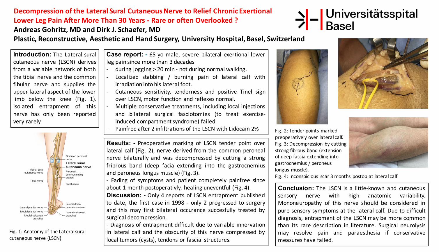
Introduction: The Lateral suralcutaneous nerve (LSCN) derivesfrom a variable network of boththe tibial nerve and the commonfibular nerve and supplies theupper lateral aspect of the lowerlimb below the knee (Fig. 1).Isolated entrapment of thisnerve has only been reportedvery rarely.
Decompression of the Lateral Sural CutaneousNerve to Relief ChronicExertionalLower Leg Pain After More Than 30 Years -‐ Rare or often Overlooked ?Andreas Gohritz, MD and Dirk J. Schaefer, MDPlastic, Reconstructive, Aesthetic and Hand Surgery, University Hospital, Basel, Switzerland
Results: - Preoperative marking of LSCN tender point overlateral calf (Fig. 2), nerve derived from the common peronealnerve bilaterally and was decompressed by cutting a strongfribrous band (deep facia extending into the gastrocnemiusand peroneus longus muscle) (Fig. 3).-‐ Fading of symptoms and patient completely painfree sinceabout 1 month postoperativly, healing uneventful (Fig. 4).Discussion: - Only 4 reports of LSCN entrapment publishedto date, the first case in 1998 -‐ only 2 progressed to surgeryand this may first bilateral occurance succesfully treated bysurgical decompression.-‐ Diagnosis of entrapment difficult due to variable innervationin lateral calf and the obscurity of this nerve compressed bylocal tumors (cysts), tendons or fascial structures.
Conclusion: The LSCN is a little-‐known and cutaneoussensory nerve with high anatomic variability.Mononeuropathy of this nerve should be considered inpure sensory symptoms at the lateral calf. Due to difficultdiagnosis, entrapment of the LSCN may be more commonthan its rare description in literature. Surgical neurolysismay resolve pain and paraesthesia if conservativemeasures have failed.
Case report: - 65-‐yo male, severe bilateral exertional lowerleg pain since more than 3 decades-‐ during jogging > 20 min -‐ not during normal walking.-‐ Localized stabbing / burning pain of lateral calf with
irradiation into his lateral foot.-‐ Cutaneous sensitivity, tenderness and positive Tinel sign
over LSCN, motor function and reflexes normal.-‐ Multiple conservative treatments, including local injections
and bilateral surgical fasciotomies (to treat exercise-‐induced compartment syndrome) failed
-‐ Painfree after 2 infiltrations of the LSCN with Lidocain 2%
Fig. 1: Anatomy of the Lateral suralcutaneous nerve (LSCN)
813
• There is complete heterogeneity of the location of the anastomosis in terms of the level of the calf.
Entrapment
Only three reports of entrapment of the LSCN were found in the literature, and all three referred to the nerve as the lateral cutaneous nerve of the calf. The fi rst report, in 1998, described entrapment of the nerve as it pierced the tendon of the biceps femoris muscle [ 11 ]. The second report, in 2006, described entrapment of the nerve by a peri-popliteal cystic
bursitis [ 2 ]. In the most recent report, in 2013, Khalil et al. [ 1 ] reported a patient with an isolated LCNC entrapment from a fi brous band of tissue. The patient had temporary relief from local anesthetic and steroid injections; he opted for surgery (see below) with good relief 1 year later.
Physical Exam
Cutaneous sensitivity or hyperalgesia may be seen in the dis-tribution of the LSCN. Careful palpation or Tinel’s test over the course of the nerve from the popliteal fossa to the fi bular head may occasionally reveal sensitivity of an entrapped or otherwise infl amed LSCN (Fig. 72.5 ). Motor function (spe-cifi cally the tibialis anterior, tibialis posterior, and extensor hallucis longus muscles) and refl exes should be normal.
Differential Diagnosis (Table 72.3 )
The most common cause of lateral calf pain is an L5 radicu-lopathy [ 1 ]; however, if the symptoms are purely sensory in nature, LSCN pathology may be the etiology. Given the sig-nifi cant heterogeneity in origin and course of all of the nerves of the sural complex, cutaneous pain in the posterolateral lower leg must be thought of in terms of all of the potential contributors to the region.
The superior border of the LSCN territory abuts that of the lateral femoral cutaneous nerve (see Chap. 61 ) anteriorly and the posterior femoral cutaneous nerve posteriorly (see Chap. 62 ). The anteromedial border of the LSCN territory abuts that of the intermediate cutaneous nerve of the thigh (and the patellar plexus ) proximally and that of the saphe-nous nerve (see Chap. 59 ) distally. The posteromedial border of the LSCN territory overlaps signifi cantly with that of the medial sural cutaneous nerve and the sural nerve itself (see Chap. 71 ). The inferior border of the LSCN abuts that of the superfi cial fi bular/peroneal nerve (see Chap. 68 ). Pain emanating from a lesion of one of these neighbors should present with a distribution more typical of the nerve in question, but when confi ned to a “border zone,” the diagno-sis may become more elusive.
Diagnostic Tests (Table 72.4 )
The diagnosis is usually confi rmed by electrodiagnostics. Needle EMG should be normal, and there should be no delay in the sensory nerve action potential (SNAP) of the sural nerve or the common peroneal nerve across the fi bular head. Campagnolo et al. [ 12 ] described a technique of NCV, plac-ing the stimulating electrodes 2 cm posteriomedial and 4 cm proximal to the center of the fi bular head, with the recording electrode placed 12 cm distally to the stimulation site.
Posterior femoralcutaneous nerve
Sciatic nerve
Common peronealnerve
Lateral suralcutaneous nervePeronealcommunicatingbranch
Lateral dorsalcutaneous nerve
Lateral calcanealbranches
Inferior clunealnerves
Tibial nerve
Medial suralcutaneous nerve
Sural nerve
Medial calcanealbranches
Medial plantar nerve
Lateral plantar nerve
Fig. 72.3 Nerves of the lower extremity (Image courtesy of Springer)
72 Lateral Sural Cutaneous Nerve Entrapment
Fig. 2: Tender points marked preoperatively over lateral calf.Fig. 3: Decompression by cuttingstrong fibrous band (extension of deep fascia extending into gastrocnemius / peroneus longus muscle).Fig. 4: Inconspicious scar 3 months postop at lateral calf