Embed Size (px)
Citation preview

Declaration of Conflict of Interest or Relationship
I have no conflicts of interest to disclose with regard to the subject matter of this presentation.
SENSITIVE DETECTION OF MYELINATION CHANGE IN MULTIPLE SCLEROSIS BY MCDESPOT
J.Su1, H.H.Kitzler2, M.Zeineh1, S.C.Deoni3, C.Harper-Little2, A.Leung2, M.Kremenchutzky2, and B.K.Rutt1
1Stanford U, CA, USA, 2TU Dresden, SN, Germany, 2U of Western Ontario, ON, Canada, 3Brown U, RI, USA
ISMRM 2011 E-POSTER #7224

Background
• There is a growing demand for metrics that can provide more detailed, spatially localized, and sensitive tracking of disease progression in multiple sclerosis (MS) than common clinical scoring methods such as EDSS
• Quantitative MR techniques are promising in this regard because they offer the possibility of detecting changes in normal-appearing white matter
SENSITIVE DETECTION OF MYELINATION CHANGE IN MULTIPLE SCLEROSIS BY MCDESPOT ISMRM 2011 #7224

Purpose
• To apply mcDESPOT, a whole-brain, myelin-selective, multi-component relaxometric imaging method, in a 1-year longitudinal pilot MS study
• Assess the ability of the method to sense different rates of demyelination for different MS courses and compare it to changes in EDSS
SENSITIVE DETECTION OF MYELINATION CHANGE IN MULTIPLE SCLEROSIS BY MCDESPOT ISMRM 2011 #7224
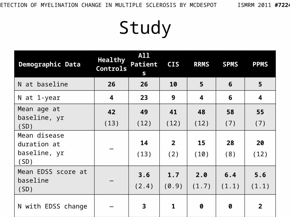
Study
Demographic Data Healthy Controls
All Patients CIS RRMS SPMS PPMS
N at baseline 26 26 10 5 6 5
N at 1-year 4 23 9 4 6 4
Mean age at baseline, yr(SD)
42
(13)
49
(12)
41
(12)
48
(12)
58
(7)
55
(7)
Mean disease duration at baseline, yr(SD)
—14
(13)
2
(2)
15
(10)
28
(8)
20
(12)
Mean EDSS score at baseline(SD)
—3.6
(2.4)
1.7
(0.9)
2.0
(1.7)
6.4
(1.1)
5.6
(1.1)
N with EDSS change — 3 1 0 0 2
SENSITIVE DETECTION OF MYELINATION CHANGE IN MULTIPLE SCLEROSIS BY MCDESPOT ISMRM 2011 #7224

Scanning Methods
• 1.5T GE Signa HDx, 8-channel head RF coil
• mcDESPOT: 2mm3 isotropic covering whole brain, about 15 min.– SPGR: TE/TR = 2.1/6.7ms, α = {3,4,5,6,7,8,11,13,18}°– bSSFP: TE/TR = 1.8/3.6ms, α = {11,14,20,24,28,34,41,51,67}°
• 2D T2 FLAIR: 0.86 mm2 in-plane and 3mm slice resolution
• 3D T1 IR-SPGR: 1mm3 resolution with pre/post Gd contrast
SENSITIVE DETECTION OF MYELINATION CHANGE IN MULTIPLE SCLEROSIS BY MCDESPOT ISMRM 2011 #7224

Processing Methods: MWF
• Linearly coregister and brain extract mcDESPOT SPGR and SSFP images with FSL1
• Find myelin water fraction maps using the established mcDESPOT fitting algorithm2
Myelin Water Fraction
SENSITIVE DETECTION OF MYELINATION CHANGE IN MULTIPLE SCLEROSIS BY MCDESPOT ISMRM 2011 #7224
1FMRIB Software Library. 2Deoni et al., Magn Reson Med. 2008 Dec;60(6):1372-87

Processing Methods: Demyelination
• Non-linearly register mcDESPOT MWF maps to MNI152 standard space
• Combine normals together to form mean and standard deviation MWF volumes
• For each subject, calculate a z-score ([x – μ]/σ) at every voxel to determine if it is significantly demyelinated, i.e. MWF < -4σ below the mean
Demyelinated Voxels
SENSITIVE DETECTION OF MYELINATION CHANGE IN MULTIPLE SCLEROSIS BY MCDESPOT ISMRM 2011 #7224

Processing Methods: 1-year & DVF
• At 1-year, demyelinated voxels are based on z-scores with respect to the baseline normal distribution
• Find demyelinated volume fraction (DVF)– Sum the volume of demyelinated voxels and
normalize by brain mask volume– # demy. voxels in compartment * voxel volume /
compartment volume
SENSITIVE DETECTION OF MYELINATION CHANGE IN MULTIPLE SCLEROSIS BY MCDESPOT ISMRM 2011 #7224

Results: Mean MWF in Whole Brain
• Dotted line shows mean MWF for normals. Rank sum testing was done for each bar against this value
• Testing was also done for RRMS vs. SPMS and CIS vs. RRMS, any significant differences are shown with a connecting bracket
• Significance levels:– * p < 0.05– ** p < 0.01– *** p < 0.001.
SENSITIVE DETECTION OF MYELINATION CHANGE IN MULTIPLE SCLEROSIS BY MCDESPOT ISMRM 2011 #7224

Results: DVF Change
• Colors denote subject type
• Arrowheads indicate the direction of change and the DVF at 1-year
• Dashed lines show subjects who also had a change in EDSS
SENSITIVE DETECTION OF MYELINATION CHANGE IN MULTIPLE SCLEROSIS BY MCDESPOT ISMRM 2011 #7224
Normals
CIS
RRMS
SPMS
PPMS

Results: DVF in Whole Brain
• Dotted line shows mean demyelinated volume fraction change for normals
• Definite MS patients are losing significantly more myelin than normals
• Progressive patients have a greater rate of demyelination
SENSITIVE DETECTION OF MYELINATION CHANGE IN MULTIPLE SCLEROSIS BY MCDESPOT ISMRM 2011 #7224

Discussion & Conclusions• The normal pool at 1-year is currently too small to show
significance for the changes in mean MWF
• DVF, however, is sensitive enough to show statistically significant changes in brain myelination over the study period
• Progressive patients show greater disease decline that are not reflected in their EDSS disability score
• EDSS and DVF measure different aspects of the disease. Patients with changes in EDSS did not actually have the largest demyelination changes
SENSITIVE DETECTION OF MYELINATION CHANGE IN MULTIPLE SCLEROSIS BY MCDESPOT ISMRM 2011 #7224