Embed Size (px)
Citation preview

LUND UNIVERSITY
PO Box 117221 00 Lund+46 46-222 00 00
Deciphering the Pathogenesis of Acute Myeloid Leukemia
Reckzeh, Kristian
Published: 2012-01-01
Link to publication
Citation for published version (APA):Reckzeh, K. (2012). Deciphering the Pathogenesis of Acute Myeloid Leukemia Division of Molecular Medicineand Gene Therapy, Dept of Laboratory Medicine
General rightsCopyright and moral rights for the publications made accessible in the public portal are retained by the authorsand/or other copyright owners and it is a condition of accessing publications that users recognise and abide by thelegal requirements associated with these rights.
• Users may download and print one copy of any publication from the public portal for the purpose of privatestudy or research. • You may not further distribute the material or use it for any profit-making activity or commercial gain • You may freely distribute the URL identifying the publication in the public portalTake down policyIf you believe that this document breaches copyright please contact us providing details, and we will removeaccess to the work immediately and investigate your claim.

DECIPHERING THE PATHOGENESIS OF ACUTE MYELOID LEUKEMIA
Kristian Reckzeh
2012

2
Copyright © Kristian Reckzeh, 2012
On the cover: Wright-Giemsa staining of AML. © AFIP Atlas of Tumor Pathology, with permission.
Lund University, Faculty of Medicine, Doctoral Dissertation Series 2012:37 ISBN 978-91-86871-99-4 ISSN 1652-8220

3
Table of Contents
ABSTRACT 5
ABBREVATIONS 6
LIST OF PUBLICATIONS 9
PREFACE 11
INTRODUCTION 12
Stem Cells 12
Hematopoiesis 14
Hematopoietic Stem Cells 14
Self-Renewal and HSC Fates 18
Regulation of Hematopoietic Stem Cells 19
The Role of C/EBPα in Hematopoiesis 22
FLT3 Signaling in Hematopoiesis 30
Acute Myeloid Leukemia 35
FOCUS OF THE PRESENT INVESTIGATION 42
Specific Aims and Questions 42
SUMMARY OF RESULTS 43
Paper I Molecular and cellular effects of oncogene cooperation in a genetically accurate AML mouse model 43
Paper II Cytokine requirements and oncogene addiction in a mouse model for AML with FLT3-ITD 43
Paper III Impact of gene dosage and loss of the wild type allele on FLT3 internal tandem duplication induced expansion of myeloid primed multipotent progenitor cells 44
GENERAL DISCUSSION 45
SVENSK SAMMANFATTNING 47

4
DEUTSCHE ZUSAMMENFASSUNG 48
ACKNOWLEDGEMENTS 49
REFERENCES 51
APPENDIX 63
Paper I
Paper II
Paper III

5
Abstract
Acute myeloid leukemia (AML) is a malignant disorder of the blood system. Hematopoietic stem cells (HSC) supply and maintain this system by differentiating via intermediates into lineage-restricted progenitors that strongly proliferate to keep up with the high turn-over of mature blood cells. In AML, the mechanisms controlling differentiation and proliferation of myeloid cells are disturbed leading to the accumulation of undifferentiated cells that interfere with the production of normal blood cells.
Mutations of the transcription factor C/EBPα have been observed in 10 percent in AML with normal cytogenetics. In addition, internal tandem duplications (ITD) of FLT3 are frequently observed alterations in AML and coincide with mutations of C/EBPα.
The effects of FLT3-ITD cooperation with C/EBPα mutations in AML are not fully understood. To address this, knockin mouse strains harboring different Cebpa mutations and Flt3-ITD were used to generate an AML mouse model. This model demonstrated a block at the transition from pre-granulocyte-macrophage progenitor (pGMP) to GMP due to disrupted C/EBPα function. The cooperative effect of FLT3-ITD is composed of enhancing the generation of leukemia-initiating GMPs and activation of STAT5 targets. In in vitro studies it was demonstrated that FLT3-ITD reduces the cytokine-requirements for cell growth and that leukemic cells harboring FLT3-ITD are more sensitive to inhibition of the FLT3 pathway in vitro. To address the impact of FLT3-ITD gene dosage and loss of Flt3 wild type allele in vivo the Flt3-ITD knockin mouse was crossed to the Flt3 receptor knockout mouse. These studies demonstrated that the myeloproliferative phenotype was FLT3-ITD dosage-dependent and independent of FL. In summary, the data presented provide deeper insights into oncogene cooperation and FLT3-ITD dosage in AML.

6
Abbrevations
AGM Aorta-gonad-mesonephros ALL Acute lymphoblastic leukemia AML Acute myeloid leukemia AML1 Acute myeloid leukemia 1 (RUNX1) ANG-1 Angiopoietin 1 ATP Adenosine triphosphate bHLH basic helix-loop-helix motif bZIP basic region leucine zipper BM Bone marrow BMI-1 B-lymphoma Mo-MLV insertion region 1 BRM Basic region mutation CB Cord blood CBF Core-binding factor CDDO 2-Cyano-3,12-dioxooleana-1,9-dien-28-oic acid CDK cyclin-dependent kinase C/EBPα CCAAT/enhancer binding protein alpha CFC Colony-forming cell CFU-S Colony-forming unit spleen CLP Common lymphoid progenitor CML Chronic myeloid leukemia CMP Common myeloid progenitor CN Copy number DNA Desoxyribonucleic acid E2F E2-binding factor eIF eukaryotic translation initiation factor ERK Extracellular signal-regulated kinase ESC Embryonic stem cell FAB French-American-British FACS Fluorescence activated cell sorting FL FLT3 ligand FLT3 FMS-like tyrosine kinase receptor 3 GATA-1 GATA binding protein 1 GMP Granulocyte-macrophage progenitor G-CSF Granulocyte-colony stimulating factor G-CSFR G-CSF receptor GM-CSF Granulocyte-macrophage-colony stimulating factor

7
GM-CSFR GM-CSF receptor GTP Guanosine triphosphate GvHD Graft-versus-host disease GvL Graft-versus-leukemia HLA Human leukocyte antigen HOXB4 Homeobox gene B4 HSC Hematopoietic stem cell HSCT Hematopoietic stem cell transplantation ICM Inner cell mass IL Interleukin IPSC Induced pluripotent stem cell ITD Internal tandem duplication JAK Janus kinase JM Juxtamembrane domain KIT SCF receptor LEF-1 lymphoid enhancer binding factor 1 LMO2 Lim-domain only 2 LMPP Lymphoid-primed multipotent progenitor LOH Loss of heterozygosity LT Long-term MAPK Mitogen-activated protein kinase MASI Mutant allele-specific imbalance M-CSF Macrophage-colony stimulating factor M-CSFR M-CSF receptor MDS Myelodysplastic syndrome MEP Megakaryocyte-erythrocyte progenitor MLL Mixed lineage-leukemia MPN Myeloproliferative neoplasm MPP Multipotent progenitor MSC Mesenchymal stem cell MYC Myelocytomatosis oncogene NK cell Natural killer cell ORF Open reading frame PB Peripheral blood PDGFR Platelet-derived growth factor receptor PI3K Phosphatidylinositol 3-kinase PU.1 SPI1 RAS Rat sarcoma RNA Ribonucleic acid RTK Receptor tyrosine kinase RUNX1 Runt-related transcription factor 1

8
SCF Stem cell factor SCL Stem cell leukemia SCN Severe congenital neutropenia SDF-1 Stromal cell-derived factor 1 SFFV Spleen focus forming virus SH2 Src-homology 2 SHIP SH2-domain-containing inositol phosphatase SLAM Signaling lymphocyte activation molecule SPI1 SFFV proviral integration 1 ST Short-term STAT5 Signal transducer and activator of transcription 5 SWI/SNF Switch/sucrose nonfermentable TAD1 Transactivation domain 1 TAL-1 T-cell acute lymphocytic leukemia 1 TBP TATA-box binding protein TF Transcription factor TFIIB Transcription initiation factor IIB TGF-β Transforming growth factor beta TI Tyrosine kinase inhibitory domain TKD Tyrosine kinase domain TNF-α Tumor necrosis factor alpha TPO Thrombopoietin TRIB2 Tribbles homolog 2 VCAM1 Vascular cell adhesion molecule 1 WBC White blood cell count WHO World Health Organization wt wild-type

9
List of Publications
This thesis is based on the articles listed below and are referred to in the text by their roman numerals (I-III).
I Reckzeh K, Bereshchenko O, Mead AJ, Rehn M, Kharazi S, Jacobsen SE, Nerlov C, Cammenga J. Molecular and cellular effects of oncogene cooperation in a genetically accurate AML mouse model. Leukemia 2012. Feb 9. doi: 10.1038/leu.2012.37. [Epub ahead of print].
II Reckzeh K, Ohlsson E, Fock V, Cammenga J. Cytokine requirements and oncogene addiction in a mouse model for AML with FLT3-ITD. Manuscript.
III Kharazi S, Mead A, Mansour A, Hultquist A, Boiers C, Luc S, Buza-Vidas N, Ma Z, Ferry H, Atkinson D, Reckzeh K, Masson K, Cammenga J, Rönnstrand L, Arai F, Suda T, Nerlov C, Sitnicka E, Jacobsen SE. Impact of gene dosage and loss of the wild type allele on FLT3 internal tandem duplication induced expansion of myeloid primed multipotent progenitor cells. Blood 2011; 118(13): 3613-3621.
Paper I and III are reproduced with permission from Nature Publishing Group and The American Society of Hematology.

10
Additional publications during PhD training:
Reckzeh K, Cammenga J. Molecular mechanisms underlying deregulation of C/EBPalpha in acute myeloid leukemia. Int J Hematol . 2010; 91, 557-568. Review.
Rehn M, Olsson A, Reckzeh K, Diffner E, Carmeliet P, Landberg G, Cammenga J. Hypoxic induction of vascular endothelial growth factor regulates murine hematopoietic stem cell function in the low-oxygenic niche. Blood. 2011; 118(6):1534-1543.

11
Preface
This is my thesis! It took six years of studying, reading, experimenting and commuting to get to this point. I am happy and proud to finally have come so far. I was very privileged to work at this institution, the Lund Stem Cell Center, where scientists from different research areas come together and perform research of impact and importance. Particularly, the Division of Molecular Medicine and Gene Therapy has been a dynamic place of creative interaction and scientific inspiration. I was very fortunate to receive so much invaluable help and support from colleagues, friends and family during this time. With this thesis I want to summarize my work on the role of two factors involved in regulation of normal and malignant hematopoiesis which is the process of blood formation in adult bone marrow. The proteins I have studied contain important functions in pathways controlling proliferation and differentiation and appear to be frequently altered in malignant hematopoiesis.
This thesis also wants to introduce this field to a broader audience, since many people who are not scientists have followed me with interest over the years. During this time I have always tried to explain what I was doing, but usually I failed. So, it is my last attempt. Listen carefully, this time!
The thesis gives you an overview on the hematopoietic system, will briefly discuss the concept of hematopoietic stem cells and introduce acute myeloid leukemia (AML), one of many malignant hematopoietic disorders. Finally, it presents a summary of my studies and a discussion of the results.
The above mentioned proteins C/EBPα and FLT3 are frequently altered by mutations, which change their function and eventually lead to development of AML. To learn more about the mechanisms involved in leukemia initiation we designed a mouse model that allows us to study the molecular and cellular background of this disease. Ultimately, our findings may help discovering better therapy and cure for AML.
Parts of the text and figures in this thesis have previously been published in a review article about the transcription factor C/EBPα in the International Journal of Hematology (Reckzeh and Cammenga, 2010).

12
Introduction
Stem Cells
The term stem cell defines a cell of undifferentiated state with the properties 1) to indefinitely self-renew by dividing into cells of identical state and properties and 2) to differentiate into all mature, specialized cells existent in a specific tissue.
Stem cells are distinguished developmentally into embryonic stem cells (ESC) and somatic or adult stem cells (Figure 1). ESCs are derived from the inner cell mass (ICM) of an early embryo and have the potential to differentiate into all tissues of the organism (pluripotent). While adult stem cells are virtually found in all tissues of an adult organism, their potential to differentiate is restricted to the specific tissue or organ. An example of a tissue-specific stem cell is the hematopoietic stem cell (HSC) which supplies the blood system and differentiates into multiple cell types found in blood (multipotent).
Because of their potential to differentiate into multiple tissues and capacity to self-renew, stem cell therapies have been proposed for regenerative medicine and tissue replacement after injury or disease. However, the isolation of human ESCs includes destruction of the embryo which raises ethical controversies utilizing these cells for research or treatment of diseases.
The generation of induced pluripotent stem cells (IPSC) from somatic cells has been an important advance in stem cell research because it does not involve the ‘use’ of embryos and creates cells with practically the same properties as ESCs (Takahashi and Yamanaka, 2006; Yu et al., 2007). Before ESCs or IPSCs can effectively be used in the treatment of patients a couple of drawbacks need to be resolved. The indefinite growth of stem cells carries the risk of cancer. Embryonic stem cells transplanted to a recipient would form teratomas.
The use of adult stem cells is limited by the low number that can be isolated from the specific tissue and stem cell expansion in vitro is still stuck at a preliminary stage. Moreover, unless the stem cells are patient-derived, immunological differences between donor and recipient would always require lifelong immuno-suppressive medication. Hence, the discovery to induce pluripotency in a somatic cell from a patient opened new opportunities. However, recipes to safely guide an unspecialized, replicating cell towards functional tissue are not yet available.

13
Figure 1. Differentiation potential of embryonic and somatic stem cells. Pluripotent embryonic stem cells derived from the inner cell mass of the blastocyst can give rise to all cell types of the primary germ layers including ectoderm (e.g. spine, peripheral nerves and brain), mesoderm (e.g. muscles, blood) and endoderm (e.g. thymus, lungs, digestive organs). Restricted to a specific tissue, somatic stem cells are able to replenish dying cells and repair damages. © Mike David Jones, with permission.
Stem cell therapy has been proposed for the treatment of many degenerative disorders such as Alzheimer and Parkinson, cardiovascular diseases, diabetes and various forms of cancer. So far however, only hematopoietic stem cell transplantation (HSCT) is used routinely in the clinic. Here, transplantation of HSCs is supposed to replace the defective hematopoietic system of the host. However, HSCT also comes with risks and side effects.
Hence, to make stem cell therapy more applicable, it requires a better understanding of the underlying mechanisms and factors controlling differentiation and self-renewal of stem cells.
All experiments in this thesis are carried out in mice and therefore all data and discussion refers to murine hematopoiesis unless otherwise indicated.

14
Hematopoiesis
The hematopoietic system is probably one of the best studied tissues and the HSC serves in many ways as a model for adult stem cells of other tissues.
The process of blood formation to continuously replace short-lived mature blood cells is referred to as hematopoiesis (Orkin and Zon, 2008). Mature blood cells carry out a variety of tasks and can be categorized into specific cell lineages according to their function (Table 1). Red blood cells or erythrocytes are specialized to transport oxygen. Platelets derived from megakaryocytes prevent excessive bleeding from injury by clotting. White blood cells (WBC) include lymphocytes (B- and T-cells), natural killer (NK) cells, granulocytes (neutrophils, eosinophils and basophils), macrophages which protect from infections, and pathogens and build up the immune system of the individual.
Most cell types of these blood lineages are short-lived compared to the lifespan of the individual and need to be replenished constantly. In human adults the hematopoietic system produces therefore around 1012 new blood cells per day at steady state (Ogawa, 1993). To maintain a balanced output of mature cells and to react to increased demands for specific cell types under stress like bleedings or disease, proliferation and differentiation processes are subject to strict control mechanisms. Defects in these processes causing an imbalance result in various hematopoietic disorders.
Table 1. Cell types, their function and cell count in human peripheral blood.
Cell type Function Count per µl Blood
Red cells (erythrocytes) Transport of O2 and CO2 4-6 x10⁶
Platelets (thrombocytes) Blood clotting 0.15-0.4 x 10⁶
B-cells (B lymphocytes) Antibody production 0.14-0.8 x 10³
T-cells (T lymphocytes) Elimination of virus-infected cells and regulation of the immune system
0.5-1.8 x 10³
NK cells Elimination of tumor and virus-infected cells 0.08-0.4 x 10³
Macrophages Phagocytosis of bacteria, fungi and neutrophils 0.2-0.9 x 10³
Neutrophils Phagocytosis of microorganisms and pathogens 2.1-6 x10³
Eosinophils Parasite destruction 0.03-0.3 x 10³
Basophils Histamine release 0.01-0.1 x10³
Hematopoietic Stem Cells
The hematopoietic system is supplied and replenished by rare, self-renewing HSCs capable of maintaining homeostasis throughout life residing in the bone marrow (BM) of adult mammals (Morrison et al., 1995). The definition of an HSC includes the following

15
properties: (1) HSCs are multipotent and should therefore give rise to all hematopoietic lineages; (2) HSCs are able to reconstitute hematopoiesis in an ablated recipient; (3) HSCs are able to give rise to HSCs, i.e. they possess self-renewal capacity.
History
First evidence for cells with these properties was provided in 1961 when it was demonstrated that colonies of hematopoietic cells emerged in the spleen of lethally irradiated recipient mice after BM transplantation (Till and McCulloch, 1961). These nodules of proliferating hematopoietic cells were termed CFU-S (colony-forming unit spleen). Following experiments demonstrated that CFU-S contain rare cells able to produce different other hematopoietic cells and to give rise to new colonies (Becker et al., 1963; Siminovitch et al., 1963), which are considered properties of a stem cell. However, later it was suggested that an even more primitive HSC (preCFU-S) colonizes the BM to give rise to cells capable of colonizing the spleen (Hodgson and Bradley, 1979; Schofield, 1978). These experiments were just the beginning of the identification of HSCs, and the HSC-defining criteria proposed by Till and McCulloch still remain valid.
Later, by tracking myeloid and lymphoid cells in primary and secondary recipients carrying identical retroviral markers different groups provided evidence for the existence of a cell in BM that has multi-lineage potential, can self-renew and functionally repopulate the blood system of an irradiated recipient (Dick et al., 1985; Keller et al., 1985; Keller and Snodgrass, 1990). However, isolation and functional examination of an untouched HSC required the identification of characteristic HSC markers and the development of techniques such as fluorescence activated cell sorting (FACS). Enormous efforts have been taken to find single markers specific for HSCs. However, prospective isolation of HSCs still involves several cell surface markers. Major work in this regard was carried out by Osawa and colleagues who demonstrated the clonal nature of HSCs by serial transplantation of single cells, identified as a part of a rare population in the BM, which gave sustained multi-lineage engraftment (Osawa et al., 1996). The identification and employment of cell surface receptors of the SLAM family has improved and simplified the ability to rigorously isolate HSCs from progenitors (Kiel et al., 2005).
Hierarchy
Hematopoiesis is described as a process where rare HSCs constitute the top of a hierarchy of intermediate subpopulations that successively develop into large numbers of mature cells of all lineages (Figure 2) (Weissman, 2000).
The HSC compartment is a heterogeneous population containing cells with long-term (lifespan) and short-term (8 weeks) self-renewal capacity and multipotent progenitors

16
(LT-, ST-HSCs and MPPs) without self-renewal. However, all these subpopulations carry the potential to give rise to all blood lineages (Morrison and Weissman, 1994).
It is generally believed that in a first pivotal step, differentiation of the HSC results in lineage-committed progenitors with either lymphoid or myeloid potential defined as common lymphoid progenitor (CLP) and common myeloid progenitor (CMP). In murine hematopoiesis two distinct progenitor populations with exclusive myeloid or lymphoid potential have been identified (Akashi et al., 2000; Kondo et al., 1997).
B-, T- and NK-cells are derived from the CLP through intermediate generation of lineage restricted progenitors. The CMP generates the granulocyte-macrophage progenitor (GMP) and megakaryocyte/ erythroid progenitor (MEP) through which mature granulocytes, macrophages, megakaryocytes/platelets and erythrocytes are formed. The development from lineage-committed progenitors to mature cells progresses through a number of intermediates which potential have not been completely identified. However, with being more and more lineage-restricted these intermediates become increasingly proliferating to produce the amount of blood cells needed.
Figure 2. Schematic overview of the hematopoietic system. Self-renewing hematopoietic stem cells give rise to subsets of intermediates becoming more and more lineage-restricted. At the same time, proliferation of these progenitors increases to meet the immense needs of mature cells.
While it is widely accepted that a single HSC has the capacity to differentiate into all other hematopoietic cell types, the differentiation paths towards mature cells are not completely resolved and remain controversial. Adolfsson et al. (2005) questioned the view of MPPs differentiating into CMP and CLP subpopulations by postulating that a first lineage commitment step would already take place directly after the HSC level
LT-HSC
MPP/LMPP
plateletserythrocytes
granulocytesmacrophages
ST-HSC
CMP CLP
MEP GMP
Pro-NKPro-B Pro-T
B-cells T-cells NK-cells
Multipotentprogenitors
Lineage-commitedprogenitors
Lineage-restrictedprogenitors
Mature cells
Prolife
ratio
n
17
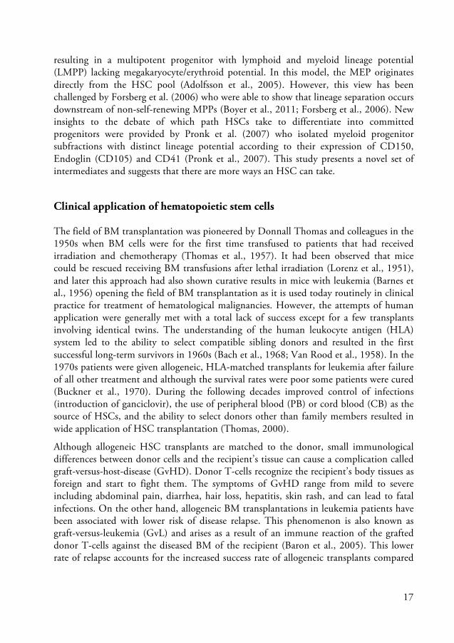
resulting in a multipotent progenitor with lymphoid and myeloid lineage potential (LMPP) lacking megakaryocyte/erythroid potential. In this model, the MEP originates directly from the HSC pool (Adolfsson et al., 2005). However, this view has been challenged by Forsberg et al. (2006) who were able to show that lineage separation occurs downstream of non-self-renewing MPPs (Boyer et al., 2011; Forsberg et al., 2006). New insights to the debate of which path HSCs take to differentiate into committed progenitors were provided by Pronk et al. (2007) who isolated myeloid progenitor subfractions with distinct lineage potential according to their expression of CD150, Endoglin (CD105) and CD41 (Pronk et al., 2007). This study presents a novel set of intermediates and suggests that there are more ways an HSC can take.
Clinical application of hematopoietic stem cells
The field of BM transplantation was pioneered by Donnall Thomas and colleagues in the 1950s when BM cells were for the first time transfused to patients that had received irradiation and chemotherapy (Thomas et al., 1957). It had been observed that mice could be rescued receiving BM transfusions after lethal irradiation (Lorenz et al., 1951), and later this approach had also shown curative results in mice with leukemia (Barnes et al., 1956) opening the field of BM transplantation as it is used today routinely in clinical practice for treatment of hematological malignancies. However, the attempts of human application were generally met with a total lack of success except for a few transplants involving identical twins. The understanding of the human leukocyte antigen (HLA) system led to the ability to select compatible sibling donors and resulted in the first successful long-term survivors in 1960s (Bach et al., 1968; Van Rood et al., 1958). In the 1970s patients were given allogeneic, HLA-matched transplants for leukemia after failure of all other treatment and although the survival rates were poor some patients were cured (Buckner et al., 1970). During the following decades improved control of infections (introduction of ganciclovir), the use of peripheral blood (PB) or cord blood (CB) as the source of HSCs, and the ability to select donors other than family members resulted in wide application of HSC transplantation (Thomas, 2000).
Although allogeneic HSC transplants are matched to the donor, small immunological differences between donor cells and the recipient’s tissue can cause a complication called graft-versus-host-disease (GvHD). Donor T-cells recognize the recipient’s body tissues as foreign and start to fight them. The symptoms of GvHD range from mild to severe including abdominal pain, diarrhea, hair loss, hepatitis, skin rash, and can lead to fatal infections. On the other hand, allogeneic BM transplantations in leukemia patients have been associated with lower risk of disease relapse. This phenomenon is also known as graft-versus-leukemia (GvL) and arises as a result of an immune reaction of the grafted donor T-cells against the diseased BM of the recipient (Baron et al., 2005). This lower rate of relapse accounts for the increased success rate of allogeneic transplants compared

18
to transplants from identical twins, and indicates that allogeneic BM transplantation is a form of immunotherapy. GvL is the major benefit of transplants that do not employ the highest immuno-suppressive regimens. However, GvL is mainly beneficial in hematopoietic malignancies with slow progress such as chronic leukemia and less effective in rapidly growing acute leukemias.
The use of HSC transplantation is not limited to hematological malignancies but also widely applicable to treat other hematological disorders such as immune deficiencies and hemoglobin disorders. In addition, the application of HSCs in gene therapy has been proposed to correct monogenetic hematopoietic disorders where HSCs are collected from a patient and the deficient gene is corrected by retroviral gene transfer followed by replacing the host’s hematopoietic system with those transduced HSCs (Karlsson, 1991; Woods et al., 2002). Although the principals of this approach have been proven and patients with severe combined immuno-deficiencies have been successfully treated, major concerns exist regarding retroviral insertion mutagenesis and require future improvements in vector design and gene transfer strategies. However, in many cases gene therapy is the final approach for the patient due to lack of alternative treatment (Baum et al., 2004).
From the beginning, aspirated BM was the source of HSCs, but today HSCs are less invasively harvested from PB after mobilization with growth factors such as granulocyte-stimulating factor (G-CSF) or a combination of cyclophosphamide and G-CSF, and from umbilical CB which has been shown to be less immuno-responsive to HLA-mismatch and therefore associated with a lower risk of GvHD. The amount of harvested HSCs from cord blood is however often too low for successful treatment of adults, and the ex vivo expansion of HSCs prior transplantation has therefore been a major goal in stem cell research over the last two decades. However, attempts to expand HSCs in vitro while maintaining self-renewal and long-term repopulating capacity have met with limited success. Recent studies have identified potential in Notch ligand, aryl hydrocarbon receptor antagonists or pleiotrophin to expand human HSCs in vitro (Boitano et al., 2010; Delaney et al., 2010; Himburg et al., 2010).
Self-Renewal and HSC Fates
The HSC’s ability to self-renew is a prerequisite in order to meet the immense needs of mature blood cells over a lifespan period. Self-renewal of the HSC occurs by either undergoing symmetric cell division giving rise to two new identical daughter cells with the same properties as the original HSC (resulting in HSC expansion) or dividing asymmetrically into one new daughter cell sustaining HSC properties and another daughter cell that starts to differentiate into more restricted, multipotent progenitors (HSC maintenance). Herein, asymmetry could either be achieved by intrinsic cell-fate

19
determinants (divisional asymmetry) or by exposure of the two daughter cells to different extrinsic signals (environmental asymmetry).
While populating the fetal liver during embryonic development HSCs divide symmetrically and expand the HSC pool, whereas during steady-state adult hematopoiesis the number of HSCs remains relatively constant. It is believed that HSCs are widely non-dividing or quiescent under steady-state conditions to maintain the HSC pool and life-lasting hematopoietic homeostasis (Cheshier et al., 1999; Domen and Weissman, 1999; Wagers et al., 2002). In addition, as every other cell, HSCs can go into programmed cell-death (apoptosis). Finally, migrating to other anatomical sites in or outside the BM is also considered a fate option of HSCs (Figure 3).
Figure 3. HSC fate options. HSCs divide asymmetrically (a) giving rise to a new HSC and a differentiating cell or symmetrically (b) yielding either two HSCs or two differentiating daughter cells. HSCs can furthermore undergo apoptosis or become quiescent.
Regulation of Hematopoietic Stem Cells
Because of the high turnover of cells in the hematopoietic system and to be able to act rapidly in response to acute blood loss or infection the equilibrium of HSC self-renewal, quiescence and differentiation must be strictly regulated. The mechanisms regulating HSCs and their fate options involve multiple, highly orchestrated pathways that determine the cell cycle status and gene expression profiles. In general, cell-extrinsic and cell-intrinsic factors may be distinguished. Extrinsic factors in the surrounding environment called the hematopoietic niche signaling through receptors on the cell surface can influence HSC behavior as well as transcription factors regulating gene expression inside the HSC.

20
The hematopoietic stem cell niche
HSCs reside in specialized anatomical sites (niches) in the BM of adult mammals. These sites include the surface along the endosteum and areas nearby vascular sinusoids found throughout the marrow (Ehninger and Trumpp, 2011). This microenvironment involves interaction with other cell types such as osteoblasts, stromal fibroblasts and endothelial cells, which provide cytokines and growth factors. The hematopoietic niches are believed to be less oxygenated (hypoxic), a condition promoting HSC maintenance by protecting vital HSCs from damaging effects of reactive oxygen species (Suda et al., 2011). The concept of stem cell niches as highly organized places controlling HSC function was firstly proposed more than 30 years ago (Schofield, 1978), and today it is clear that the niche plays a pivotal role in regulating HSC fate together with cell autonomous mechanisms (Lymperi et al., 2010).
Extrinsic factors
The different cell types found in the hematopoietic niche provide growth factors and cytokines that affect HSC and their behavior. Cells contained in the niche express several factors important for HSC function including membrane-bound stem cell factor (SCF; binds to the KIT receptor), thrombopoietin (TPO), a ligand for the MPL receptor, erythropoietin (EPO) and G-CSF (Arai et al., 2009; Buza-Vidas et al., 2006; Krantz, 1991; Yang et al., 1998). The fms-like tyrosine kinase receptor (FLT3) ligand is expressed in stromal fibroblasts of the BM microenvironment and synergizes with other hematopoietic growth factors to promote proliferation of primitive hematopoietic precursors (Lyman et al., 1995; Stirewalt and Radich, 2003).
An important role has been recently addressed to nestin-positive mesenchymal stem cells (MSC) which were found in close proximity to HSCs (Mendez-Ferrer et al., 2010). These cells express factors crucial for HSC maintenance including stromal cell-derived factor 1 (SDF-1 also named CXCL12), SCF, angiopoietin-1 (ANG-1), IL-7, vascular cell adhesion molecule 1 (VCAM1), and osteopontin (OPN)(Ehninger and Trumpp, 2011).
There are several other factors that have been demonstrated to act negatively on HSCs. The secreted transforming growth factor beta (TGF-β) protein has been shown to repress HSC activity by signaling through the SMAD pathway and subsequent regulation of the cell cycle (Blank and Karlsson, 2011). The tumor necrosis factor alpha (TNF-α) was demonstrated to negatively regulate HSCs in vitro and recently a suppressive role for TNF-α on HSCs in vivo was identified in TNF-α-deficient mice (Bryder et al., 2001; Pronk et al., 2011).

21
Intrinsic factors
Transcription factors (TF) are internal signals that govern gene expression and regulate HSC fate. Whether gene expression is directly determined by the influence of extrinsic factors (instructive model) or through stochastic variation of internal signals in an environment with extrinsic factors allowing certain key decisions (permissive model) remains not fully understood. However, it is now believed that HSC fate decisions rely more on stochastic mechanisms while committed progenitors may be higher influenced by extrinsic signals (Enver et al., 1998).
The number of TFs involved in specification of HSCs during ontogeny and fate regulation during adult hematopoiesis has grown large and only a few can be discussed here.
In embryonic hematopoiesis, it has been shown that the bHLH factor SCL/tal-1 (Stem cell leukemia/T-cell acute lymphocytic leukemia 1) and its associated partner, the Lim-domain only 2 (LMO2) are essential for specification of a HSC precursor from the hemangioblast (Kim and Bresnick, 2007). HSCs are initially found in the aorta-gonad-mesonephros (AGM) region and dorsal arteries of the mid-gestation mouse embryo (de Bruijn et al., 2000). At this stage, the runt domain containing DNA-binding subunit of the core-binding factor (CBF) RUNX1 (AML1) is required for the emergence and function of HSCs (Okuda et al., 1996; Wang et al., 1996). The mixed lineage-leukemia gene (MLL) and HOXB4 have been identified as regulators of HSC maturation during early hematopoiesis (Ernst et al., 2004). HOXB4, a member of the homeobox domain TFs was shown to have a dramatic effect on HSC self-renewal. Retroviral overexpression of HOXB4 demonstrated great expansion of HSCs ex vivo (Antonchuk et al., 2002).
Once adult HSCs have arisen and moved to the bone marrow several of the TFs previously required for emergence and formation of HSCs may no longer be required for continuous survival and self-renewal. However, there are a few factors known to be involved in regulation of adult HSC self-renewal and quiescence. BMI-1, a member of the Polycomb group proteins that regulate HOX proteins, was identified as an important transcriptional regulator of self-renewal (Iwama et al., 2004; Lessard and Sauvageau, 2003). Another important factor for HSC integrity is Gfi-1, a zinc-finger transcriptional repressor that regulates the cell cycle inhibitor p21 (Hock et al., 2004; Zeng et al., 2004).
In addition to TFs like SCL and RUNX1 that are involved in almost all hematopoietic lineages there are factors that have lineage-specific expression patterns. Their disruption does usually only affect a single or a small number of related lineages. Important TFs for the development and differentiation of myeloid lineages are PU.1, C/EBPα and GATA-1. The latter is highly expressed in MEPs and to a lesser extent in HSCs and CMPs while GATA-1 expression is required for definitive erythropoiesis (McDevitt et al., 1997).
22
The level of PU.1 expression is relatively low in HSCs, CMPs and CLPs compared to B-cells whereas its expression is high in mature myeloid cells (Scott et al., 1994). Pu.1 knock-out mice lack mature myeloid and B-cells probably due to an early block of transition from multipotent progenitors (MPPs) to lineage-restricted CMPs and CLPs. Interestingly, PU.1 deficient mice (PU.1 level at 10% of wt mice) develop erythroid cells and have a normal MEP compartment supporting the model of MEPs being the direct progeny of HSCs (Rosenbauer et al., 2006).
The transcription factor C/EBPα is an example of a lineage-specific TF. It is expressed by HSCs, myeloid progenitors, and granulocytes. It is indispensible for the maturation of neutrophil and eosinophil granulocytes as C/EBPα knockout mice lack mature granulocytes without affecting other hematopoietic lineages.
Because TFs regulate the specification and emergence of HSCs and subsequently govern hematopoiesis by controlling production and differentiation of HSCs, it is conceivable that disorders of the hematopoietic system often involve alterations of the regulatory network of TFs. In hematopoietic malignancies TFs are often involved in chromosomal translocations or become targets of somatic mutations that disrupt their normal function (Orkin and Zon, 2008).
The Role of C/EBPα in Hematopoiesis
The CCAAT/enhancer binding protein alpha (C/EBPα) is one of six members of a subgroup of basic region leucine zipper (bZIP) TF proteins (Landschulz et al., 1988). They share structurally related N-terminal transactivation domains whereas the basic DNA-binding region and C-terminal leucine rich dimerization domain are highly conserved throughout all members (Ramji and Foka, 2002).
C/EBPs are able to form homo- and heterodimers with other C/EBPs as well as with transcription factors of other families via their leucine zipper domain and can subsequently bind to DNA via the adjacent located basic amino acid residues. The exact distance between the basic region and leucine zipper domain is crucial for proper binding to DNA. Alteration by nucleotide insertions or deletions in the DNA-binding domain therefore impairs or limits the function of C/EBPs (Miller et al., 2003). Several C/EBPs appear in functionally different isoforms by expression from multiple translational initiation sites and alternative splicing.

23
Structure and function of C/EBPα.
The intronless CEBPA gene is located on chromosome 19q13.1 and encodes 2783 base pairs. C/EBPα is expressed in various cell types including hepatocytes, adipocytes, enterocytes, keratinocytes, lung epithelial cells, mammary gland cells and hematopoietic cells. In these cells, C/EBPα protein-dimers directly activate transcription from lineage-specific gene promoters. The C/EBPα protein was originally isolated from rat liver nuclei capable of interacting with the CCAAT homology present in many gene promoters and viral enhancers (Landschulz et al., 1988). Deletion of the Cebpa gene in mice leads to insufficient storage of hepatic glycogen and death from hypoglycemia shortly after birth. Expression of enzymes important in glycogen synthesis are decreased or delayed, and hepatocytes fail to accumulate lipids properly demonstrating the need of C/EBPα for the establishment and maintenance of energy homeostasis in neonates (Wang et al., 1995). At the same time C/EBPα knock-out mice lack white adipose tissue, mature neutrophil and eosinophil granulocytes indicating a role in differentiation of hematopoietic and non-hematopoietic tissues (Zhang et al., 1997).
The mRNA encoding C/EBPα contains alternative translation initiation sites resulting in two major protein isoforms: the fully translated protein C/EBPα (42 kDa; p42), and an N-terminal truncated protein C/EBPα (30 kDa; p30), lacking a transactivation domain important for anti-mitotic activity (Lin et al., 1993). The open reading frame (ORF) used for translation of the two isoforms harbors three AUG (AUG1,2 and AUG3) start codons. Additionally, there is another translation start site further upstream in a different reading frame. Translation initiation from AUG1,2 results in full-length translation of p42 whereas translation from the out of frame start site is prematurely terminated making the ribosome read over AUG1,2 and re-initiate at AUG3 to translate a truncated p30 protein (Kaufman, 2004).
The shorter p30 protein lacks the first 117 amino-acids of full-length C/EBPα (Figure 4). This transactivation domain (TAD1) is important for direct interaction with the TATA-box binding protein (TBP) and another transcription initiation factor (TFIIB), both essential components of the RNA polymerase II transcription complex (Nerlov and Ziff, 1995). Notably, the transactivation domain is also crucial for growth arrest by E2F-mediated regulation of the level of c-myc expression (Johansen et al., 2001; Lin et al., 1993; Porse et al., 2001). The truncated p30 isoform inhibits p42 C/EBPα-mediated transactivation of target genes. This inhibition occurs by formation of heterodimers that have impaired DNA-binding and transactivation ability compared to p42 C/EBPα homodimers (Cleaves et al., 2004).
The protein isoforms share another transactivation domain (TAD2) that has been shown to interact with the cyclin-dependent kinases CDK2/4, the SWI/SNF chromatin remodeling complex and the CDK inhibitor 1 (p21) (Wang et al., 2001). Both proteins contain a basic DNA-binding domain and a leucine zipper (bZIP) required for

24
dimerization and interaction with other TFs in the C-terminus. Highly conserved, non-DNA-binding residues in this region interact with E2F. Mutation of these residues results in loss of C/EBPα-E2F interaction leading to dysplasia of neutrophil granulocytes and hypoplasia of white adipose tissues (Porse et al., 2001).
Figure 4. Functional distinct C/EBPα isoforms. Two major proteins (42 kDa, p42 and 30 kDa, p30) with specific functional properties are naturally translated from C/EBPα mRNA containing three AUG start codons (AUG1,2 and AUG3) within the main open reading frame (ORF) and a weak AUG start site in a small upstream ORF (uORF). Translation initiation is modulated by activity of eIF2α / eIF4E.The full-length protein consists of 358 amino acids while the N-terminal truncated protein lacks the first 117 amino acids.The region that is unique for p42 contains a transactivation domain (TAD1). Both isoforms share another transactivation domain (TAD2) and the basic leucine zipper (bZIP) DNA binding domain.
C/EBPα in normal and malignant hematopoiesis.
The TF C/EBPα plays a crucial role in the process of myeloid lineage commitment and granulocyte differentiation. Its expression is required throughout granulopoiesis, and mature neutrophil and eosinophil granulocytes are absent in mice deficient of C/EBPα while other hematopoietic lineages including monocytes remain unaffected. C/EBPα regulates the expression of many myeloid genes, including genes encoding growth factor receptors (granulocyte colony-stimulating factor receptor, G-CSFR; macrophage colony-stimulating factor receptor, M-CSFR; granulocyte-macrophage colony-stimulating factor receptor, GM-CSFR) (Ramji and Foka, 2002).

25
C/EBPα is expressed in hematopoietic stem and progenitor cells (Figure 5) and is involved in HSC regulation as C/EBPα deficient HSCs demonstrate enhanced reconstitution abilities and self-renewal compared to wt HSCs (Zhang et al., 2004). However, C/EBPα deficient HSCs have lost the potential to form GMP and its progeny whereas enforced expression of C/EBPα in MPPs induces myeloid differentiation at the cost of erythroid development demonstrating the ability to take action in cell fate decisions and hence the importance of C/EBPα at developmental stages before CMP and GMP level (Heath et al., 2004; Suh et al., 2006).
An essential prerequisite of C/EBPα is coordinating growth arrest and cell differentiation. Overexpression of p42 is strongly inhibiting cell growth in vitro. Several mechanisms have been proposed how C/EBPα mediates growth arrest and differentiation [for review see (Nerlov, 2004)]. Widely accepted is the ability of C/EBPα to repress c-myc via protein-protein interaction with E2F. However, C/EBPα promotes growth arrest in a cell-specific manner; therefore different mechanisms may apply in different tissues.
Figure 5. Relative expression of Cebpa in myeloid progenitors and HSCs. Q-PCR was carried out on sorted cell populations of B6SJL mice using Fluidigm. Expression of Cebpa is shown relatively to expression of Hprt. (LT-HSC as LSKCD150+CD48-; MPP as LSKCD150-CD48+; preGM as LKCD41-CD16/32low/-; CD150-CD105-; GMP as LKCD41-CD16/32+CD150- (n=4-8).
Due to its function in cell cycle arrest and neutrophil differentiation CEBPA mutations were proposed to be involved in leukemogenesis and today it is clear that CEBPA mutations are present in 7%–10% of human AML and are mostly associated with a normal karyotype. The mutation patterns observed are generally biallelic and almost always combine an N-terminal frameshift mutation on one allele with a C-terminal in-frame insertion/deletion mutation on the other allele (Pabst and Mueller, 2007). N-terminal mutations prevent expression of the p42 protein isoform and favor the expression of the p30 isoform, whereas C-terminal mutations disrupt DNA binding and
Cebpa
LT-HSC
MPP
preGM
GMP0
1
2
3
4
5
rela
tive
expr
essi
on

26
dimerization of both isoforms (Figure 6). The mouse models that studied C/EBPα function in normal and malignant hematopoiesis are summarized in Table 2.
Figure 6. Mutations of the CEBPA gene in AML. C/EBPα mutations observed in acute myeloid leukemia (AML) frequently occur as biallelic mutations and cluster predominantly in the N- and C-terminal region of either allele. Mutations within the first 300 base pairs lead to a frameshift or premature stop codon (yellow bar) eliminating p42 expression without affecting p30 translation. C-terminal insertions or deletions (red letters) disrupt the bZIP domain and affect both p42 and p30 C/EBPα. Mutations in the bZIP domain have been studied in the BRM2 (I294A, K297A) and K313KK mouse models.
Pabst and colleagues firstly identified mutations leading to N-terminal truncation and expression of the p30 C/EBPα protein (Pabst et al., 2001). Initially, as it was thought to be the most frequent leukemogenic event the effect of p30 C/EBPα was studied. Overexpression of p30 C/EBPα in human CD34+ cord blood cells showed a remarkable inhibition of myeloid and erythroid differentiation (Schwieger et al., 2004).
Kirstetter and colleagues studied mice heterozygous or homozygous for a similar mutation in the Cebpa locus resulting in exclusive expression of p30 C/EBPα from the targeted allele (L allele; CebpaL/+) (Kirstetter et al., 2008). Strikingly, CebpaL/+ exhibit 4-fold more p30 than p42 C/EBPα, a finding also observed in AML patients with the N-terminal mutation. However, the mice did not display alterations in hematopoietic tissues or granulocyte differentiation. Hence, although reduced DNA-binding activity by p30/p42 heterodimers may be involved in disturbed granulocytic differentiation, further decreased C/EBPα function is required for leukemogenesis. Solely, total abrogation of p42 C/EBPα translation in CebpaL/L mice uncoupled cell-cycle control and terminal

27
differentiation resulting in aberrant granulopoiesis and progression to AML with full penetrance. In summary, p30 C/EBPα accounts for loss of growth control but sustain crucial myeloid commitment to the GMP level. The importance of GMP formation for AML development is supported by the finding that C/EBPα deficient hematopoietic chimera lack GMPs due to a block in transition from CMP to GMP and do not develop AML. Also, retroviral expression of BCR-ABL in C/EBPα null background resulted in erythroid leukemia rather than AML/myeloid blast-crisis of CML (Wagner et al., 2006; Zhang et al., 2004).
Table 2. Models investigating C/EBPα function in normal and malignant hematopoiesis. Model C/EBPα function Hematopoietic phenotype Reference
Cebpa-/- Null No GMPs or mature granulocytes; enhanced HSC self-renewal; no AML
(Zhang et al., 1997)
C/EBPα-BRM2*
Binding to E2F disrupted, otherwise normal DNA-binding
Transplantable MPN; transformation of myeloid cells; no AML
(Porse et al., 2001)
C/EBPα-p30*
Enforced p30 expression, normal bZIP domain
Myeloid lineage commitment; transplantable AML; gene expression profile shared with MLL-AF9 AML
(Kirstetter et al., 2008)
C/EBPα-K313KK* Disrupted bZIP domain
Expansion of pre-leukemic HSCs; impaired myeloid lineage commitment; no GMPs; transplantable AML
(Bereshchenko et al., 2009)
C/EBPα-K313KK/p30
Enforced p30 expression / disrupted bZIP domain
Expansion of pre-leukemic HSCs; myeloid lineage commitment; accelerated AML development
(Bereshchenko et al., 2009)
*no phenotype in heterozygous mice.
Porse and colleagues studied repression of E2F-mediated transcription by deleting the region in the bZIP domain of C/EBPα (BRM2) that interacts with E2F in vivo. However, this mutation does not affect DNA-binding or induction of C/EBPα target genes. BRM2 homozygous mice developed a transplantable disorder of the granulocytic lineage with expansion of myeloid progenitor populations indicating a role of C/EBPα mediated E2F repression in controlling cell cycle and proliferation of myeloid progenitors (Porse et al., 2005). However, although transformation of myeloid cells

28
occurred in this mouse model, clinical symptoms of AML such as thrombocytopenia, anemia and elevated white blood count were absent.
These observations support the role of C/EBPα as a myeloid specific tumor suppressor but also demonstrate that E2F repression alone is not sufficient to induce AML. Therefore, targeting DNA-binding function of C/EBPα was the next step to investigate. An in-frame insertion of lysine at position 314 (K313KK; K allele; CebpaK/+) in the Cebpa locus was engineered by Bereshchenko et al. mimicking a common C-terminal mutation in AML (Bereshchenko et al., 2009). Combining different hypomorphic Cebpa alleles (L/L, K/K and K/L), this study investigated the potential of CEBPA mutations to initiate AML. The presence of the K allele promoted expansion and proliferation of HSCs and MPPs prior to development of AML as K/K and K/L mice exhibited less quiescent and increased numbers of HSCs and MPPs. Myeloid differentiation was more profoundly blocked in K/K and K/L mice (no GMPs) than in L/L mice (normal GMP levels). However, K/K mice in some cases developed even erythroleukemia instead of AML. This phenotype has otherwise been observed in C/EBPα deficient hematopoietic chimera. Further, whereas all combinations led to AML the K/L combination of both mutations accelerated disease progression and mice of that setup developed AML fastest. In conclusion, the predominant biallelic CEBPA mutations affecting the C-terminus and the N-terminus in human AML obviously meet exactly the level of C/EBPα function that is needed for myeloid lineage commitment but prevents terminal granulocyte differentiation leading to the development of AML.
Regulation of C/EBPα
C/EBPα has been found to be regulated at a number of levels, including gene transcription, translation, protein-protein interactions and phosphorylation mediated changes in DNA-binding activity, activation potential and nuclear localization.
Repression of C/EBPα transcription by CEBPA promoter silencing has been demonstrated in AML patients without mutations in the CEBPA gene (Wouters et al., 2007). The transcription factor LEF-1 (lymphoid enhancer binding factor 1) has been reported to be suppressed in myeloid cells exhibiting a block in differentiation at a promyelocytic stage in patients with severe congenital neutropenia (SCN). LEF-1 activates the CEBPA promoter and its expression in SCN patient cells restored C/EBPα levels and induced myeloid differentiation (Skokowa et al., 2006).
Leukemic fusion genes involving CBF family members such as AML1-MDS-EVI1 in t(3;21) AML or CBFB-MYH11 in inv(16) are involved in repression of C/EBPα at the post-transcriptional level (Helbling et al., 2004; Helbling et al., 2005). The RNA-binding protein calreticulin is highly activated in t(3;21) AML. Calreticulin binds GC-rich structures of C/EBPα mRNA and inhibits its translation. The same inhibitory

29
mechanism of C/EBPα mRNA translation was shown for the CBFB-MYH11 fusion protein resulting from inv(16). Similarly, in blasts from patients with CML myeloid blast crisis, inhibition of translation of C/EBPα mRNA is caused by hnRNP-E2, another RNA-binding protein. HnRNP-E2 is induced by BCR-ABL via a MAPK-dependent mechanism. Down-regulation of hnRNP-E2 by the BCR-ABL inhibitor imatinib restores both C/EBPα protein expression and granulocytic differentiation of those blasts (Chang et al., 2007; Perrotti et al., 2002).
Ribosomal scanning for translation initiation sites depends on activity of the alpha subunit of the eukaryotic translation initiation factor 2 (eIF-2α) that is regulated by eIF2 kinases (Calkhoven et al., 2000). The eIF-2α acts as an attenuator of protein synthesis initiation. Interestingly, mice with a homozygous mutation at the eIF-2α phosphorylation site (S51A) die after birth due to defective gluconeogenesis. In addition, homozygous mutant embryos and neonates display a deficiency in pancreatic beta cells (Scheuner et al., 2001). This phenotype is surprisingly similar to that of C/EBPα null mice. Hence, by regulating the ratio between p42 and p30 C/EBPα activity of eIF-2α and the process of translation initiation [for review see (Kaufman, 2004)] may be involved in leukemogenesis. Experimental evidence supporting this hypothesis is limited although it has been demonstrated that eIF-4E is aberrantly expressed in AML (Assouline et al., 2009). CDDO (2-Cyano-3,12-dioxooleana-1,9-dien-28-oic acid), increased the ratio between p42 and p30 C/EBPα isoforms by dephosphorylation of eIF-2α and phosphorylation of eIF-4E. In addition, CDDO had the potential to differentiate primary blasts from patients with AML (Koschmieder et al., 2007).
There is growing evidence for deregulation of C/EBPα function at the protein level also being involved in leukemogenesis. Various mechanisms affecting phosphorylation, ubiquitination and sumoylation of C/EBPα protein have been described. Tribbles homolog 2 (TRIB2), a downstream target of NOTCH1 has been identified in AML. It associates with and inhibits C/EBPα by enhancing proteasomal degradation of C/EBPα protein (Keeshan et al., 2006). Phosphorylation of C/EBPα at Serine 21 led to a conformational change in C/EBPα protein dimers turning their transactivation domains further apart. Mediated by extracellular signal-regulated kinase 1/2 (ERK-1/2), phosphorylation of C/EBPα inhibited granulocytic differentiation and favors monocytic development (Ross et al., 2004). Activated FLT3 signaling affecting ERK1/2 in human AML was shown to inhibit C/EBPα function by the same mechanism (Radomska et al., 2006).

30
FLT3 Signaling in Hematopoiesis
The FLT3 (CD135) receptor, also known as fetal liver kinase 2 (FLK2) (Rosnet et al., 1991) belongs to the receptor tyrosine kinase (RTK) type III family which also includes the M-CSFR (CD115) (Coussens et al., 1986), KIT (CD117) (Yarden et al., 1987) as well as the platelet-derived growth factor receptors alpha (PDGFRα) and beta (PDGFRβ) (Claesson-Welsh et al., 1989; Yarden et al., 1986). All members of this RTK group play a crucial rule in proliferation, survival and differentiation of hematopoietic cells: KIT and FLT3 are both required for the survival, proliferation and differentiation of early hematopoietic progenitors, M-CSFR is important for growth and differentiation of macrophages and PDGFRβ is believed to play a role in megakaryopoiesis (Reilly, 2003).
The receptor FLT3 is expressed on early myeloid and lymphoid progenitors, but not in erythroid cells, megakaryocytes or mast cells (Stirewalt and Radich, 2003). In fact, FLT3 surface expression is believed to distinguish an early commitment step from self-renewing, uncommitted murine HSCs to MPPs which have started to differentiate (Adolfsson et al., 2001).
Structure and function of FLT3
FLT3 is the receptor for the FLT3 ligand (FL). It contains five immunoglobulin-like domains in the extracellular ligand-binding domain (LBD). The human FLT3 gene is located on chromosome 13q12 and encodes a protein of 993 amino acid with 85% sequence-homology compared to mouse Flt3 (Rosnet et al., 1993). A transmembrane domain spans the region through the cell membrane while the intracellular domain contains the juxtamembrane domain (JM), the tyrosine kinase 1 domain (TKD1), tyrosine kinase inhibitory domain (TI) and the tyrosine kinase 2 domain (TKD2) (Figure 7). The receptor appears in two forms: the mature and membrane-bound N-glycosylated form (150 kDa) and the immature, mannose-rich form of 130 kDa (Stirewalt and Radich, 2003).
The FLT3 receptor remains as an unphosphorylated inactivated protein monomer until binding of FL induces a conformational change allowing receptor dimerization and subsequently activation of the intracellular tyrosine kinase that phosphorylates several sites of the intracellular domain. This leads ultimately to activation of downstream effectors (Stirewalt and Radich, 2003). The activated and phosphorylated FLT3-FL complex is rapidly internalized and degraded similarly to the rapid internalization and turnover of other RTK type III receptors like KIT. Serum levels of FL are normally low in homeostasis. However, FL can be released upon low white-blood-cell (WBC) counts as a result of hematopoietic disease or chemotherapy.

31
FL is expressed in many hematopoietic cell types as soluble form or membrane-bound, which are both biologically active (Lyman et al., 1995). Whether these two isoforms contain distinct functions remains to be investigated (Lyman and Jacobsen, 1998). Disruption of the Fl gene reduced the number of LMPPs, and myeloid and lymphoid progenitors (Boiers et al., 2010; McKenna et al., 2000; Sitnicka et al., 2002) highlighting the important role of FLT3 signaling in hematopoiesis. Moreover, complementary signaling of FLT3 and IL-7 has been demonstrated to be indispensable for fetal and adult lymphopoiesis (Jensen et al., 2008; Sitnicka et al., 2003).
Stimulation of early myeloid progenitors in vitro by FL without the addition of other growth factors promoted monocytic differentiation without a distinct proliferative effect. However, FL stimulation in combination with IL-3, SCF and G-CSF resulted in a more vigorous proliferation (Rusten et al., 1996).
Figure 7. FLT3 receptor domains and sites frequently mutated. About 70 percent of mutations observed in AML are located in the juxtamembrane domain (JM), whereas the remaining 30 percent target the tyrosine kinase 1 domain (TK1).
The FLT3 signaling pathway
The FLT3 signaling cascade has not been completely described. However, it has been shown that binding of the FL triggers the phosphatidylinositol 3-kinase (PI3K), a regulatory protein of phospholipid metabolism and the rat sarcoma (RAS) pathway which is involved in cell proliferation and apoptosis (Figure 8). The activated receptor can bind to the Src-homology 2 (SH2) domain of the p85 subunit of PI3K and with SH2-containing sequence proteins (SHC) that are involved in phospolipid and proliferative pathways (Zhang and Broxmeyer, 1999). In addition, FLT3 binds to GTPase-activating

32
protein, important regulatory protein of the RAS pathway (Dosil et al., 1993; Rottapel et al., 1994).
The FLT3 receptor has also been found to be associated with SH2-domain-containing inositol phosphatase (SHIP), which might act as a negative regulator of proliferation by competitively binding SHCs that otherwise activate RAS (Marchetto et al., 1999; Zhang et al., 1999). The activation of FLT3 involves also modulation of STAT5A indicating the involvement of the Janus kinase / signal transducer and activator of transcription (JAK/STAT) pathway in FLT3 signaling (Zhang et al., 2000).
The signaling pathway data must be interpreted with caution since most of these experiments were carried out in either mouse models or human cell lines artificially expressing FLT3. In addition, he expression pattern of FLT3 differs in mouse and human. Although being species cross-reactive, FL exists in different isoforms in mouse and human, whose biological relevance is unknown. Further, it is unclear whether the different isoforms are tissue-specifically expressed (Stirewalt and Radich, 2003). In conclusion, FLT3 pathways seem to be highly species and tissue specific, which indicates that it might be difficult to generalize the above findings to normal human hematopoietic cells (Dosil et al., 1993; Zhang et al., 1999).
FLT3 mutations in acute myeloid leukemia
Due to the fact of being a strong promoter of cell proliferation and survival the implication of FLT3 signaling in malignant hematopoiesis appears obvious. In fact, FLT3 is highly expressed in almost all AML subtypes, B-cell acute lymphoblastic leukemia (ALL), a few T-cell ALLs and CMLs (Gilliland and Griffin, 2002). In addition, genomic alterations of the FLT3 gene occur in up to 30 percent of all AMLs making it one of the most frequently targeted genes in AML.
The most common form is an internal tandem duplication (ITD) in exon 14 and 15 due to in-frame insertions or deletions varying in size between 3 and more than 400 base pairs (Nakao et al., 1996). Until recently, it was believed that ITD mutations occur exclusively in the JM domain but newer investigations show that approximately 70 percent of ITD mutations localize in this region whereas the remaining 30 percent are in fact found in the TKD1 domain (Breitenbuecher et al., 2009). Cell line studies utilizing retroviral transduction have shown that FLT3-ITD promote ligand-independent dimerization and constitutive activation of the FLT3 receptor leading to cytokine-independent cell growth (Kiyoi et al., 1998). Whether FL can further stimulate the activity of FLT3 is likely dependent on the ratio of wt versus activated receptor. This ratio is important because FLT3-ITD can form homodimers or heterodimers with wt FLT3 and may affect the degree of autophosphorylation (Kiyoi et al., 2002). Experiments have not fully elucidated this question (Gilliland and Griffin, 2002).

33
The second most common type of FLT3 mutations is a missense point mutation in exon 20 of the TKD substituting an aspartic acid to tyrosine (D835Y). Other mutations in codon 835, such as deletions, have also been described, but these are far less common (Schnittger et al., 2002). The kinase domain is inactive until dimerization and a conformational change allows access of adenosine triphosphate (ATP) and substrate. The D835Y mutation targets the so-called activation-loop leading to phosphorylation of a specific tyrosine residue within the loop and causing the loop to change to an activated configuration which in term allows access to the kinase (Yamamoto et al., 2001). Activation loop mutations have also been reported for other RTKs (Gilliland and Griffin, 2002).
Figure 8. Cellular localization and signaling of wt FLT3 and FLT3-ITD. The wt FLT3 receptor is expressed on the cell surface after intracellular maturation and glycosylation. Upon FLT3 ligand binding the receptor dimerizes and autophosphorylates activating its kinase domains which in term activate downstream targets. The activated receptor is rapidly internalized and subsequently degraded. The properties of the wt/ITD FLT3 receptor in regards to cellular localization and signaling are much less defined. Mutant and wt FLT3 mRNA is transported to the endoplasmatic reticulum (ER) and translated into protein. The immature glycosylated 130 kDa form of FLT3-ITD is retained in the ER and activates the STAT5 pathway while wt FLT3 is transported to the Golgi apparatus where complex glycosylation occurs followed by surface expression. Activation of STAT5 by FLT3-ITD leads to transcription of genes like BAD etc via Pim-1 while wt FLT3 signals through PI3K and MAPK pathways.

34
Both ITD and activation loop mutations result in constitutive activation of the FLT3 kinase and downstream targets including STAT5, RAS/MAPK, and PI3K/AKT (Fenski et al., 2000; Yamamoto et al., 2001). FLT3-ITD was shown to confer factor-independent growth to the murine hematopoietic cell lines 32D and Ba/F3 that are dependent on IL-3 for growth. In contrast, wt FLT3, even in the presence of FL, did not confer factor-independent growth to these cells (Hayakawa et al., 2000; Mizuki et al., 2000; Mizuki et al., 2003).
The molecular mechanisms responsible for different signaling of mutated FLT3 receptor versus wt receptor are not completely understood (see also Figure 8). In addition, whether ITD and TKD mutations of FLT3 result in activation of the same signaling pathways is still in dispute. However, recent attention has been drawn to the fact that wt FLT3 is predominantly occurring as the mature, complexly glycosylated 150 kDa form whereas FLT3-ITD is retained in the endoplasmatic reticulum (ER) and therefore not as much glycoslylated (Koch et al., 2008). Recent studies have demonstrated that aberrant localization of the mutated FLT3 receptor in the ER led to activated STAT5 while the wt receptor located in the cell membrane signals mainly via the ERK and PI3K pathway (Choudhary et al., 2009; Schmidt-Arras et al., 2009). In accordance with the experimental data showing FLT3-ITD entrapment in the ER is the observation that surface expression of FLT3-ITD is decreased or absent depending on the mutational status (Hu et al., 2004).
Different mouse models using various approaches have been generated to study the molecular background of mutant FLT3 in hematopoietic malignancies. Kelly et al. (2002) could show that expression of FLT3-ITD using a murine transduction/transplantation model leads to an oligoclonal MPN with a relatively short latency (Kelly et al., 2002). Interestingly, this disease was not transplantable into secondary recipients. Using the same experimental system, Grundler et al. (2005) showed that FLT3-ITD and FLT3-TKD mutations induce different diseases with different latency and that this is due to a difference in the ability of the two mutations to phosphorylate STAT5 (Grundler et al., 2005).
More genetically accurate models were generated by using knockin and conditional knockin of a human ITD sequence into the normal Flt3 locus guaranteeing correct timing and level of expression of FLT3-ITD. Li et al. concentrated mainly on the heterozygous mice while Lee et al. analyzed FLT3-ITD homozygous mice (Lee et al., 2007; Li et al., 2008). Both models showed a myeloproliferative disease and as expected, the myeloproliferative phenotype was more pronounced in mice having two copies of FLT3-ITD.

35
Acute Myeloid Leukemia
The term leukemia (from ancient greek: leukós haima, “white blood”) was originally described by Rudolf Virchow (Virchow, 1856). Leukemia is characterized by high numbers of non-functional, immature white blood cells in peripheral blood and bone marrow.
Deregulated hematopoiesis ultimately leading to leukemia is a consequence of acquisition of genetic and epigenetic changes in blood stem and progenitor cells, which otherwise produce the huge numbers of mature, red and white blood cells. These so-called blast cells have lost their ability to differentiate and response to normal regulation of proliferation and survival, and progressively displace normal blood cells in the BM with the result of fatal infections, bleedings, and infiltration of other organs (Tenen, 2003). Depending on which blood cell lineage is affected, leukemia is subdivided into lymphoid or myeloid leukemia. Furthermore, chronic and acute leukemia are distinguished by the amount of blast cells in the BM. The occurrence of more than 20 percent blasts in the BM is considered as acute leukemia.
Epidemiology
Acute myeloid leukemia (AML) is a complex malignant disease and its great genetic heterogeneity makes this disorder exceptionally challenging in the clinic. AML is the most common acute leukemia with a prevalence of 3.8 per 100000 adults and increasing to 18 in 100000 adults older than 65 years (Deschler and Lubbert, 2006).
During the past decades, refinements in diagnosis of AML subtypes and advances in therapeutic approaches have improved the situation for patients with AML. However, still most people suffering from AML will die of their disease or complications related to therapy.
Clinical aspects
AML is diagnosed as a result of a complete blood count showing decreased numbers of red blood cells (anemia), platelets (thrombocytopenia) and neutrophil granulocytes (neutropenia) whereas the total white blood count are most commonly increased (leukocytosis) due to an accumulation of leukemic blast cells. For identification of blasts an examination of peripheral blood analysis can be carried out, but definitive diagnosis requires a BM sample, which is analyzed morphologically by microscopy and flow cytometry to diagnose the presence of leukemic blasts and to differentiate from other

36
types of leukemia. In addition, cytogenetic analysis is carried out to identify chromosomal abnormalities and translocations which can have prognostic significance.
The early signs of AML are often vague and non-specific including fever, loss of appetite and weight loss. However, the patient shows symptoms due to the replacement of mature, functional blood cells by leukemic blasts: lack of neutrophils increases the susceptibility to infections; anemia causes fatigue and short breath while low platelet numbers lead to bleedings and easy bruising.
Treatment of AML consists primarily of chemotherapy and can be divided into induction therapy and consolidation therapy. Induction therapy aims at reducing the number of leukemic cells to a minimum (complete remission) while the goal of consolidation therapy is to eliminate any residual, undetectable disease. The standard induction chemotherapy regimen consists of cytarabine for seven consecutive days and anthracycline for three days (Abeloff, 2004). Despite the variety of underlying cytogenetics of the disease, this protocol induces complete remission in up to 70 percent of all patients (Bishop, 1997).
After complete remission, leukemic cells still remain in numbers too small to be detected and would cause a relapse in short time unless consolidation therapy is given. Consolidation therapy is individualized based on the patient's cytogenetic status and prognostic factors as well as general health and age. For inv(16), t(8;21), and t(15;17) AMLs (good prognosis), patients will typically undergo an additional 3–5 courses of intensive chemotherapy (Abeloff, 2004). For patients with high-risk cytogenetics, underlying myelodysplastic syndrome (MDS), or therapy-related AML (bad prognosis, high risk of relapse), allogeneic stem cell transplantation is usually recommended if the patient has a suitable donor. The best consolidation therapy for intermediate-risk AML (normal cytogenetics) is less clear and depends on the specific situation, including the age and overall health of the patient, the patient's personal values, and whether a suitable stem cell donor is available (Appelbaum et al., 2000). Hematopoietic stem cell transplantation is becoming the standard care for young patients with intermediate risk AML and is considered if a patient relapses.
Classification
The diversity of myeloid malignancies and in particular acute myeloid leukemia in terms of phenotype and prognostic outcome required a classification to select the right therapeutic approach and improve treatment. Originally, AML was classified into groups depending on the maturation status of the blast cells. The so-called French-American-British (FAB) nomenclature (Bennett et al., 1976) divided AML into 8 subtypes M0 to M7. However this classification did not incorporate underlying genetic abnormalities or clinical outcome. Recent advances in the understanding of the genetic basis of AML have provided new insights with regards to genetics and clinical features of AML and the
37
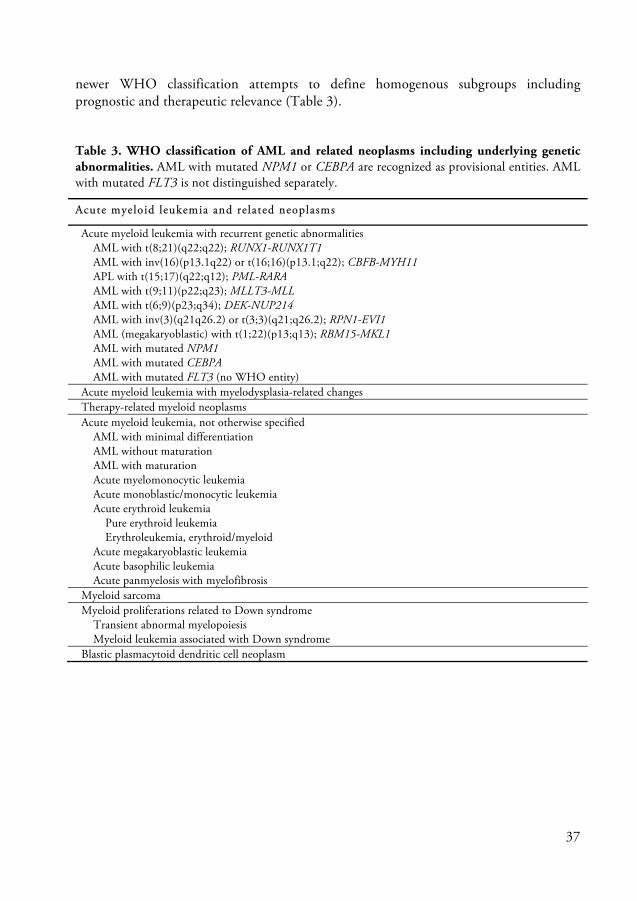
newer WHO classification attempts to define homogenous subgroups including prognostic and therapeutic relevance (Table 3).
Table 3. WHO classification of AML and related neoplasms including underlying genetic abnormalities. AML with mutated NPM1 or CEBPA are recognized as provisional entities. AML with mutated FLT3 is not distinguished separately.
Acute myeloid leukemia and related neoplasms
Acute myeloid leukemia with recurrent genetic abnormalities AML with t(8;21)(q22;q22); RUNX1-RUNX1T1 AML with inv(16)(p13.1q22) or t(16;16)(p13.1;q22); CBFB-MYH11 APL with t(15;17)(q22;q12); PML-RARA AML with t(9;11)(p22;q23); MLLT3-MLL AML with t(6;9)(p23;q34); DEK-NUP214 AML with inv(3)(q21q26.2) or t(3;3)(q21;q26.2); RPN1-EVI1 AML (megakaryoblastic) with t(1;22)(p13;q13); RBM15-MKL1 AML with mutated NPM1 AML with mutated CEBPA AML with mutated FLT3 (no WHO entity) Acute myeloid leukemia with myelodysplasia-related changes Therapy-related myeloid neoplasms Acute myeloid leukemia, not otherwise specified AML with minimal differentiation AML without maturation AML with maturation Acute myelomonocytic leukemia Acute monoblastic/monocytic leukemia Acute erythroid leukemia Pure erythroid leukemia Erythroleukemia, erythroid/myeloid Acute megakaryoblastic leukemia Acute basophilic leukemia Acute panmyelosis with myelofibrosis Myeloid sarcoma Myeloid proliferations related to Down syndrome Transient abnormal myelopoiesis Myeloid leukemia associated with Down syndrome Blastic plasmacytoid dendritic cell neoplasm

38
Molecular pathology of AML
The fundamental features of AML include inappropriate proliferation in the absence of normal growth signals, acquired self-renewal, escape from programmed cell death, block of differentiation, aberrant cell cycle control, genomic instability and multi-organ dissemination of leukemic cells. These properties (Table 4) may be directly linked to genetic lesions and individually amenable to therapy, but it is clear that many of them result from integration of multiple alterations in the leukemic cell (Licht and Sternberg, 2005).
Table 4. Malignant properties of AML and the underlying lesions (Licht and Sternberg, 2005). Property Molecular les ion Differentiation block Fusion transcription factors;
Retinoic acid receptor α: PML-RARα, PLZF-RARα, Core binding factor: RUNX1-MTG8, CBFβ-MYH11, RUNX1-EVI1, MLL-fusions, Hox gene fusions and overexpression, Point mutation of transcription factors PU.1, C/EBPα, RUNX1, GATA-1.
Autonomous cell proliferation Activating mutations: FLT3, RAS, c-KIT, c-FMS.
Escape from apoptosis AKT pathway activation following RTK activation leads to Bad deactivation, p53 mutations in AML of the elderly, p53 dysregulation by fusion proteins, NPM1 mutations, Bcl2 overexrepssion, Survivin (IAP) overexpression.
Increased self-renewal β−catenin mutations, activation of Wnt-catenin pathway by fusion transcription factors, Activated RTK pathways cooperate to induce self-renewal.
Loss of cell cycle control p53 dysfunction, loss of Rb, p15 and p16 cyclin-dependent kinase gene methylation.
Dissemination TNF secretion by leukemic blasts stimulates endothelium. Increased selectin, cadherin and integrin expression encourage adhesion and egress through vessels.
However, although oncogenes like receptor tyrosine kinases and classical tumor-suppressor genes (like RB and p53) are also mutated in AML, experimental evidence suggests that hematopoiesis-specific TFs are the gatekeepers of AML (Cammenga, 2005). Disruption of these gatekeepers interferes with hematopoietic differentiation and subsequent apoptosis of cells. The disrupted TFs in AML can be classified into three
39
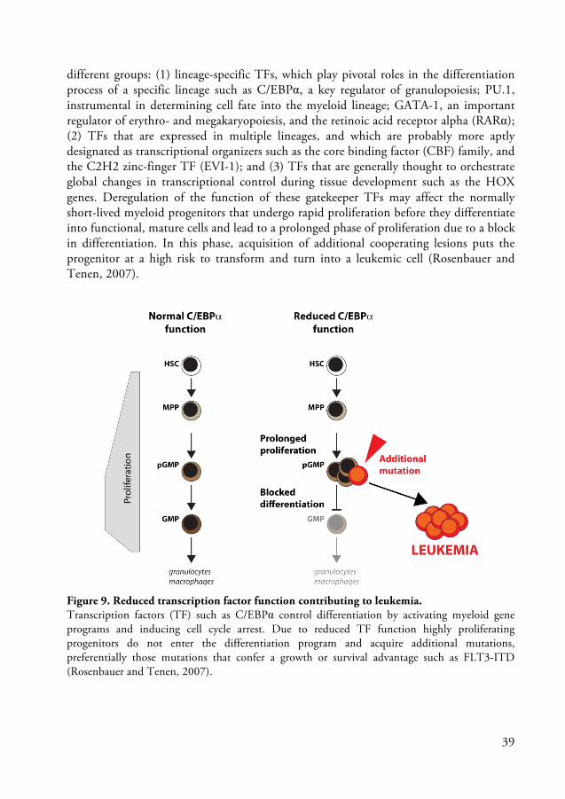
different groups: (1) lineage-specific TFs, which play pivotal roles in the differentiation process of a specific lineage such as C/EBPα, a key regulator of granulopoiesis; PU.1, instrumental in determining cell fate into the myeloid lineage; GATA-1, an important regulator of erythro- and megakaryopoiesis, and the retinoic acid receptor alpha (RARα); (2) TFs that are expressed in multiple lineages, and which are probably more aptly designated as transcriptional organizers such as the core binding factor (CBF) family, and the C2H2 zinc-finger TF (EVI-1); and (3) TFs that are generally thought to orchestrate global changes in transcriptional control during tissue development such as the HOX genes. Deregulation of the function of these gatekeeper TFs may affect the normally short-lived myeloid progenitors that undergo rapid proliferation before they differentiate into functional, mature cells and lead to a prolonged phase of proliferation due to a block in differentiation. In this phase, acquisition of additional cooperating lesions puts the progenitor at a high risk to transform and turn into a leukemic cell (Rosenbauer and Tenen, 2007).
Figure 9. Reduced transcription factor function contributing to leukemia. Transcription factors (TF) such as C/EBPα control differentiation by activating myeloid gene programs and inducing cell cycle arrest. Due to reduced TF function highly proliferating progenitors do not enter the differentiation program and acquire additional mutations, preferentially those mutations that confer a growth or survival advantage such as FLT3-ITD (Rosenbauer and Tenen, 2007).

40
In the model of reduced TF function contributing to leukemia (Figure 9) additional alterations occur affecting normal proliferation and survival to ultimately transform leukemic cells (Rosenbauer and Tenen, 2007). In term, Gilliland and Griffin (2002) postulated a two-hit model in leukemogenesis where at least two genetic changes take place: one initial mutation affecting differentiation, and secondary, mutations conferring a proliferative or survival advantage. This idea has arisen from the observation that FLT3-ITD causes an MPN but is not capable of inducing AML (Castilla et al., 1999; Gilliland and Griffin, 2002; Lee et al., 2007; Li et al., 2008).
Figure 10. Oncogene cooperation in AML. Growing body of evidence suggests that signaling mutations cooperate with specific partners in leukemogenesis. Alterations of the same class are absent/rarely observed in AML. In addition, translocations inculding t(8;21) and inv(16) cluster together with KIT alterations whereas t(15;17), point mutations in AML1 and CEBPA coincide with FLT3 activating mutations (Haferlach, 2008).
However, FLT3 mutations frequently occur in conjunction with other gene rearrangements. For example, FLT3-ITDs have been infrequently reported in patients with t(8;21) and inv(16), but are most commonly associated with mutant NPM1 and t(15;17). The translocations result in expression of fusion genes including the AML1-ETO, CBFB-SMMHC, and PML-RARα, respectively. There are convincing data that these fusion genes are not sufficient to cause leukemia, but do contribute to impaired differentiation of hematopoietic cells (Gilliland and Griffin, 2002).
Growing experimental evidence and clinical observations point to specific association of signaling mutations cooperating with initiating gatekeeper mutations (Figure 10) (Castilla et al., 1999; Haferlach, 2008; Okuda et al., 1998; Westendorf et al., 1998). The hypothesis has important implications in approach to novel therapies of AML, in that molecular targeting of both FLT3-ITD and mutations involving transcription factors

41
may improve outcome in AML. Therefore, it is of great interest to design mouse models that as accurate as possible reflect the genetic background of AML. These models would allow us to study the molecular and cellular effects of oncogene cooperation during onset and progression of AML and subsequently help to identify new approaches in AML treatment.

42
Focus of the Present Investigation
The pathogenesis of AML is a complex process involving the disruption of multiple regulatory mechanisms due to acquisition of genetic alterations in hematopoietic stem and progenitor cells.
The role of the transcription factor C/EBPα in normal and malignant hematopoiesis has been intensively studied and specific mutations of the CEBPA gene modulating its function have been causally linked to development of AML. In addition, ITD mutations of the FLT3 receptor tyrosine kinase are a common scheme in hematopoietic malignancies, especially in AML. It has been proposed that the consecutively acquired genetic changes collaborate, but how the functional changes of these factors cooperate in leukemogenesis is still poorly understood.
The studies presented in this thesis have been aimed at deciphering the pathogenesis of AML by investigating the molecular and cellular effects of C/EBPα and FLT3 mutations before onset and towards progression to AML in a mouse model.
Specific Aims and Questions
Do FLT3-ITD and C/EBPα cooperate in AML pathogenesis? What are the molecular and cellular effects of cooperation of biallelic C/EBPα mutations and FLT3-ITD in a genetically accurate mouse model for human AML?
Does FLT3-ITD reduce the cytokine requirements of leukemic cells and substitute for cytokine dependent growth in vitro? Does FLT3 inhibition limit leukemic cell growth in vitro and in vivo? Do increased levels of FLT3 ligand impair the efficacy of FLT3 inhibition?
What are the hematopoietic consequences of acquisition of Flt3-ITD homozygosity/hemizygosity in adult mice in vivo? Which role plays FLT3 ligand in the myeloproliferative phenotype of Flt3ITD/ITD mice?

43
Summary of Results
Paper I Molecular and cellular effects of oncogene cooperation in a genetically accurate AML mouse model
Based on three knock-in mouse strains a genetically accurate AML mouse model was designed to investigate recurrent C/EBPα mutations and FLT3-ITD collaborating in leukemogenesis. In opposite to biallelic Cebpa mutations, monoallelic Cebpa mutations in cooperation with Flt3-ITD were not sufficient to initiate AML. Hematopoietic chimeras with biallelic Cebpa mutations showed a profound block in differentiation of granulocytes regardless of the mutational status of Flt3 and developed AML with full penetrance. However, additional Flt3-ITD shortened the latency of AML development. Leukemic mice showed an expansion of phenotypic GMPs in the BM which were able to initiate leukemia upon transplantation into secondary recipients confirming LIC potential in a myeloid committed progenitor cell.
As a result of disrupted C/EBPα function, myeloid differentiation was halted at the transition from pGMP to GMP. In addition, a higher frequency of MegE progenitors (MkP, pMegE, pCFU-E and CFU-E) and an increased pool of hematopoietic stem and progenitor cells (LSK cells including LT-HSCs and MPPs) was observed prior onset of AML. In hematopoietic chimeras harboring the additional Flt3-ITD the effect of biallelic Cebpa mutations on MegE progenitors was reduced and led to higher frequency of MPPs, pGMPs and GMPs. Gene expression analysis revealed a deregulated GMP population that had partially or fully left the myeloid differentiation path and acquired HSC-like self-renewal and enhanced survival. The cooperative effect of Flt3-ITD is composed of an additional proliferative signal via activation of STAT5 target genes and enhancing the generation of leukemia-initiating GMPs.
Paper II Cytokine requirements and oncogene addiction in a mouse model for AML with FLT3-ITD
The previously described AML mouse model was utilized in this study to investigate the effect of FLT3 inhibitors on leukemic cell growth and more generally clarify whether leukemic cells are growth dependent of the activated FLT3 pathway. Continuous leukemic cell growth in vitro required colony-formation in semisolid medium supplemented with SCF and IL-3. Further, in suboptimal IL-3 concentration leukemic

44
cells heterozygous for FLT3-ITD demonstrated higher colony-forming cell (CFC) capacity compared to leukemic cells with undisturbed FLT3 signaling. In the presence of the FLT3 inhibitors CEP-701 (Lestaurtinib, Cephalon, Inc.) or AC-220 (Quizartinib, Ambit Bioscience, Inc.), CFC capacity was reduced in an inhibitor dosage-dependent manner. However, a higher sensitivity of leukemic cells harboring FLT3-ITD could be demonstrated. To test the FLT3-inhibitory effect in vivo recipients transplanted with leukemic cells were treated with CEP-701 by subcutaneous injections once daily over a period of 2 weeks and monitored for AML development. There was no difference in survival observed between treated and untreated recipients. However, it remained unclear if plasma levels of CEP-701 were high enough to inhibit FLT3 activation.
Paper III Impact of gene dosage and loss of the wild type allele on FLT3 internal tandem duplication induced expansion of myeloid primed multipotent progenitor cells
Based on cross-breeding of Flt3ITD/ITD knock-in mice with Flt3+/+, Flt3-/- and Fl-/- (FLT3 ligand knockout) mice, this study aimed to specifically address the impact of FLT3-ITD gene dosage, loss of Flt3 wild type allele and the role of FLT3 ligand on the myeloproliferative disease induced by FLT3-ITD in adult mice. It has been previously shown that mice develop an MPN-like disorder and that the phenotype was FLT3-ITD dosage-dependent (Lee et al., 2007). In comparison to Flt3ITD/+ mice it was demonstrated that the MPN phenotype in Flt3ITD/ITD mice was paralleled by an enhanced cellularity, expansion of myeloid progenitors as well as monomyelocytic cells and overexpression of STAT5 target genes. This phenotype was also found in hemizygous Flt3ITD/- mice, although at less pronounced levels. Moreover, the manifestation of the MPN in Flt3ITD/ITD mice was FLT3 ligand independent. Further, the stronger myeloid phenotype in Flt3ITD/- mice compared to Flt3ITD/+ mice could not be linked to the lack of wt FLT3 interaction with FLT3 ligand as Flt3ITD/+ mice did not show enhanced MPN in the absence of FLT3 ligand. In summary, this study demonstrated that the enhanced MPN phenotype of Flt3ITD/ITD mice compared to Flt3ITD/+ mice is attributable to a FL-independent FLT3-ITD dosage effect.

45
General Discussion
The pathology of AML is of great molecular heterogeneity. These molecular alterations disrupt the regulation of cell proliferation, differentiation, self-renewal, survival, cell cycle checkpoint control, DNA repair and chromatin stability, and cell dissemination (see Table 4). Insights into the pathways that are affected in AML has come from three different approaches: cytogenetic identification and molecular cloning of recurrent chromosomal rearrangements, identification of retroviral integration sites in murine models of AML and screening for mutations in genes known to be important regulators of hematopoiesis (Cammenga, 2005). Subsequently, the identified factors were studied in a variety of in vitro and in vivo models including retroviral transduction in cell lines or primary cells followed by transplantation, transgenic mice, knockin and knockout models. However, apart from making the disease accessible to experimentation otherwise impossible in human patients the main contribution of these models has been to our understanding of leukemogenesis as a multistep process. Nevertheless, the clinical relevance was quite limited (McCormack et al., 2005).
Genetically accurate mouse models of AML investigating the relationship between two or more cooperating oncogenes are missing. The model presented here utilized knockin mouse strains and demonstrated cooperation of a FLT3-ITD mutation with commonly observed C/EBPα mutations (Paper I). While disruption of the C/EBPα TF function introduced a myeloid differentiation block at the transition from pGMP to GMP, FLT3-ITD enhanced the generation of leukemia-initiating GMPs and conferred an additional proliferative signal via activation of STAT5 target genes which accelerated progression to AML. Due to expression from the original promoter preserving accurate levels and regulation of oncogene expression the design of this model is as genetically accurate as possible and allows furthermore the investigation of molecular and cellular processes prior transformation and onset of leukemia.
The inhibition of FLT3 signaling has been proposed to be a potential target in the fight against AML. However, the identification of RTK inhibitors was previously based on IL-3 dependent cell lines transfected with FLT3-ITD or AML cell lines over expressing FLT3-ITD (Levis et al., 2002; Pratz et al., 2009; Smith et al., 2004). None of these studies actually resemble the normal levels and ratios between wt and mutant FLT3 observed in primary cells in mouse or human AML. This discrepancy presumably accounts partly for the failure of significant numbers of new drugs late in clinical trials and points out how essential it is that accurate murine AML models are developed to exploit more specific, targeted therapeutics (Fathi and Levis, 2011; McCormack et al., 2005). Although, as every mouse model has to deal with pathway discrepancies (proven

46
for FLT3 and ligand) between mouse and human species (Kikushige et al., 2008), the model presented here could serve as a new in vivo platform to test the potential of new drugs before they enter expensive clinical trials. It was used in the ongoing study (Paper II) that tested Lestaurtinib (CEP-701) and Quizartinib (AC-220) for their potential to inhibit activated FLT3 signaling and growth of leukemic cells in vitro and in vivo. Although final data is still missing this study demonstrated that much higher concentrations of CEP-701 and AC-220 are needed to achieve similar FLT3-ITD inhibition as in the originally studies with cytokine-dependent cell lines.
The second part of this study was focused on clinical observations regarding the FLT3 receptor (Paper III). FLT3-ITD is associated with high relapse rate and poor overall survival in AML. The clinical outcome is furthermore determined by the allelic ratio of wt FLT3 and FLT3-ITD. Variations in the allelic ratio are a result of mutant allele-specific imbalance (MASI) either due to copy number-neutral loss of heterozygosity (CN-LOH) or mutant allele amplification. While the impact of Flt3-ITD dosage on disease phenotype has been modeled in vivo, less is known about the effect when the second wt allele is lost (Lee et al., 2007). However, loss of wt FLT3 and acquisition of FLT3-ITD homozygosity upon disease progression was shown to be associated with a worse outcome (Gale et al., 2008). Whether this outcome is impaired by an FLT3-ITD dosage effect or loss of the wt allele remains unclear. Previous studies demonstrated hetero-complexes between wt FLT3 and FLT3-ITD resulting in tyrosine phosphorylation of the wt receptor (Kiyoi et al., 2002) suggesting aberrant activation of wt FLT3 by FLT3-ITD.
To address this complex field and the hematopoietic consequences homo- and hemizygous Flt3-ITD mice were generated and bred against Fl knockout mice (McKenna et al., 2000). Using these models it was demonstrated that the MPN phenotype in Flt3-ITD homozygous mice was paralleled by an enhanced cellularity, expansion of myeloid progenitors as well as monomyelocytic cells and overexpression of STAT5 target genes. This phenotype was also found in hemizygous Flt3ITD/- mice, although at less pronounced levels. The data presented here suggests that both gene dosage of FLT3-ITD and the loss of wt FLT3 contributes to the phenotype in the respective mouse strains. In addition, the degree of the myeloproliferative disease in Flt3ITD/ITD mice was independent of FL. The FLT3-ITD knockin model used here closely reflects the event of acquiring an activating FLT3 mutation, expressed under the control of the endogenous promoter. However, the impact of FLT3-ITD dosage, loss of wt allele and impact of FL would be more relevant, if tested in an AML model setup presented in Paper I as FLT3-ITD mutations are typically occurring secondary to mutations disrupting a hematopoietic gatekeeper.

47
Svensk Sammanfattning
Leukemi är en cancersjukdom i blod och benmärg som karakteriseras av en störd blodbildning. Vid normal blodbildning delar sig ett begränsat antal omogna blodceller så kallade blod stamceller (eller hematopoietiska stamceller) och mognar till funktionella blodceller. Genom att dela sig utan att mogna har stamcellerna även potential till at förnya sig själva. När nya stamceller och mogna celler samtidigt bildas kan en kontinuerlig blodproduktion upprätthållas genom livstiden. Då leukemi uppstår mognar blodcellerna inte fullt ut, vilket leder till en anhopning av omogna celler med brist av funktionella blodceller som följd. Ett fåtal leukemiska stamceller tros ligga bakom den maligna expansionen. Konventionell leukemibehandling minskar mängden omogna celler men förefaller inte vara verksam på de leukemiska stamcellerna. De leukemiska stamceller som blir kvar efter behandling förmodas vara den bakomliggande orsaken till återfall. Akut myeloisk leukemi (AML) är en typ av leukemi där man känner till flera genetiska förandringar (mutationer) som förekommer i de maligna blodcellerna. För att bättre kunna forstå uppkomst av AML har vi upprättat en musmodell för human AML genom att föra in tre vanligt förekommande mutationer i det genetiska materialet hos musens blodceller. Vår modell tillåter molekylära och cellulära studier både före och under manifestationen av sjukdomen. Jämfört med tidigare modeller representerar vår modell orsaken till leukemien på ett bättre sätt.
Våra resultat och de framtida studier de kommer att leda till kan bidra till nya framtida terapier i kampen mot AML. Dessutom kan musmodellen användas för att studera innovativa och oprövade läkemedel.

48
Deutsche Zusammenfassung
Leukämie ist eine Krebserkrankung im Blut und Knochenmark, die durch eine gestörte Blutbildung charakterisiert ist. Bei normaler Blutbildung teilt sich eine Anzahl weniger unreifer Blutzellen, sogenannter Blutstammzellen und reift zu funktionellen Blutzellen heran. Stammzellen können sich selbst erneuern indem sie sich teilen ohne dabei zu reifen. Durch die kontinuierliche Neubildung von unreifen Stammzellen und reifen Blutzellen ist es möglich die Blutproduktion ein Leben lang aufrechtzuerhalten. Bei einer Leukämieerkrankung reifen die Blutzellen nicht vollständig, wodurch es zu einer Anhäufung von unreifen Zellen und einem Mangel an funktionellen, gereiften Blutzellen kommt. Man nimmt an, dass eine geringe Zahl leukämischer Stammzellen die maligne Vermehrung unreifer Zellen verursacht. Konventionelle Behandlungsmethoden versuchen diese Zellen zu vernichten und im besten Fall ganz zu beseitigen. Sie haben jedoch häufig keine oder geringe Wirkung auf die leukämischen Stammzellen. Die Zellen, welche durch die Therapy nicht beseitigt werden, verursachen häufig einen Rückfall. Akute myeloische Leukämie (AML) ist eine akute Form von Leukämie, in der eine Anzahl genetischer Veränderungen (Mutationen) identifiziert sind. Um die Kausalität zwischen genetischen Veränderungen und der Leukämieentstehung zu zeigen sowie die Entstehung von AML besser zu verstehen, haben wir ein Mausmodell für humane AML entwickelt, indem wir drei häufig vorkommende Mutationen in das genetische Material von Blutzellen der Maus eingeführt haben. Dieses Modell ermöglicht es uns, die molekulären und zellulären Zusammenhänge vor und während der Manifestation der Krankheit zu studieren. Im Vergleich zu früheren Modellen spiegelt unser Modell die leukemie-verursachenden Bedingungen besser wider.
Diese Ergebnisse und weiterführende Studien helfen möglicherweise neue Therapieansätze im Kampf gegen AML zu finden. Darüber hinaus erlaubt uns das Mausmodell neue und bisher ungeprüfte Medikamente zu testen.

49
Acknowledgements
The past six years at the Lund Stem Cell Center have been exciting for me with all the up and downs, but also really a pleasure, and I think it has been a tremendous booster for my scientific career. I have enjoyed these years a lot which is probably also a reason why it took me a little longer… It has been a privilege to study and do research in such an inspiring scientific environment with all the opportunities given. Whether it was visiting Toshio Suda’s laboratory in Tokyo, attending international conferences or just all the possibilities in the lab allowing extensive research; all this support, which is not a matter of course, I greatly appreciated.
However, every workplace is as good as the people working there. In fact, I have really enjoyed the relaxed and stimulating atmosphere as well as the company with all past and present members of the Divison of Molecular Medicine and Gene Therapy. I would like to express my gratitude to those people and everyone else who has been involved in my thesis.
First and foremost I am thankful to my supervisor Jörg Cammenga for your confidence in me eventually developing the skills it takes to become a PhD even when it took a little longer J, for the independence I have had carrying out my projects, for sharing your scientific knowledge and enthusiasm (and for the ambitious projects). I really enjoyed your encouragement and unlimited support. Thank you!
I am really grateful to my co-supervisor Stefan Karlsson who offered shelter to the little Cammenga group from B10 and in this way let us benefit from an excellent scientific environment. Thank you for your interest and encouragement.
The person who really helped me through these years is Matilda Rehn and I would like to thank you especially! From the beginning you were on top of things and I was always one step behind. You cut through the jungle and I could follow your path laidback. But sometimes we fought together and this has been an unforgettable time for me!
I would also like to warmly thank Christine and Göran Karlsson for your friendship and caring about my career as well as for the many enjoyable hours with your lovely family. Special thanks to the G-Star for sharing the Wilsede experience with me as well as London and San Diego, and for letting me borrow the office linebacker outfit to kick some ass.
Thanks also to my students Ewa Ohlsson and Valerie Fock who never took me too seriously. I appreciated that you did not mention my confusion or hopeless experiment plans to Jörg. Special thanks also for the clown corner.

50
The entire crew on A12 which has grown so big nowadays deserves my deep appreciation, especially Karin Olsson (and in the old days Eva Nilsson) who runs the lab and was an invaluable help solving all kinds of situations. In particular I would like say thank you to Emma, Matilda NH and Roksana for taking me through skiing in the Rocky Mountains, seizures in N.Y. and superb scientific meetings. Definitely, a memorable experience! I thank my gym and sauna buddies Alex, Jens, Pekka (you three compensated well for the G-Star loss), Maria D, Roger and others. Kavitha should be thanked for scientific discussions, encouragement during hard times and teaching me lessons in table tennis. She is the one and only I have ever lost to! I have had a blast with Stuart and the tiger tights on MS Stubnitz. A special thanks also to the Wannabe-Poker-Stars Lina, Valli and Praveen. Also, thanks to Roman, for an unforgettable night at the nation that never happened and Timotej.
Talia and Zsuzsanna, for taking good care of Jörg from now on, and Sofie and Maria A, for teaching me the important Swedish words as well as Evan and Carmen for having a break out in the sun when it was not yet politically incorrect.
Camilla Björklöv, Eva Gynnstam, Lena Persson-Feld and everybody else in the animal facilities have my gratitude for taking good care of all my mice.
I would also like to express my gratitude to my collaborators; Shabnam Kharazi and Adam Mead for your tremendous help with FACS and Fluidigm. Claus Nerlov and Sten Eirik Jacobsen for scientific discussions and help with my projects. Special thanks also to Kentaro Hosokawa, Fumio Arai and Toshio Suda for your generous hospitality and making my visit in Japan such a memorable experience.
Above all, I would like to thank my wife Rikke for your love and endless support during these tough years and for putting your own career on hold while I went to Sweden. You and Selma and Villads make me happy every day.

51
References
Abeloff, M. D. (2004). Clinical oncology. In, (Philadelphia, Pa.: Elsevier Churchill Livingstone), pp. xxiv, 3205 p.
Adolfsson, J., Borge, O. J., Bryder, D., Theilgaard-Monch, K., Astrand-Grundstrom, I., Sitnicka, E., Sasaki, Y., and Jacobsen, S. E. (2001). Upregulation of Flt3 expression within the bone marrow Lin(-)Sca1(+)c-kit(+) stem cell compartment is accompanied by loss of self-renewal capacity. Immunity 15, 659-669.
Adolfsson, J., Mansson, R., Buza-Vidas, N., Hultquist, A., Liuba, K., Jensen, C. T., Bryder, D., Yang, L., Borge, O. J., Thoren, L. A., et al. (2005). Identification of Flt3+ lympho-myeloid stem cells lacking erythro-megakaryocytic potential a revised road map for adult blood lineage commitment. Cell 121, 295-306.
Akashi, K., Traver, D., Miyamoto, T., and Weissman, I. L. (2000). A clonogenic common myeloid progenitor that gives rise to all myeloid lineages. Nature 404, 193-197.
Antonchuk, J., Sauvageau, G., and Humphries, R. K. (2002). HOXB4-induced expansion of adult hematopoietic stem cells ex vivo. Cell 109, 39-45.
Appelbaum, F. R., Baer, M. R., Carabasi, M. H., Coutre, S. E., Erba, H. P., Estey, E., Glenn, M. J., Kraut, E. H., Maslak, P., Millenson, M., et al. (2000). NCCN Practice Guidelines for Acute Myelogenous Leukemia. Oncology (Williston Park) 14, 53-61.
Arai, F., Yoshihara, H., Hosokawa, K., Nakamura, Y., Gomei, Y., Iwasaki, H., and Suda, T. (2009). Niche regulation of hematopoietic stem cells in the endosteum. Ann N Y Acad Sci 1176, 36-46.
Assouline, S., Culjkovic, B., Cocolakis, E., Rousseau, C., Beslu, N., Amri, A., Caplan, S., Leber, B., Roy, D. C., Miller, W. H., Jr., and Borden, K. L. (2009). Molecular targeting of the oncogene eIF4E in acute myeloid leukemia (AML): a proof-of-principle clinical trial with ribavirin. Blood 114, 257-260.
Bach, F. H., Albertini, R. J., Joo, P., Anderson, J. L., and Bortin, M. M. (1968). Bone-marrow transplantation in a patient with the Wiskott-Aldrich syndrome. Lancet 2, 1364-1366.
Barnes, D. W., Corp, M. J., Loutit, J. F., and Neal, F. E. (1956). Treatment of murine leukaemia with X rays and homologous bone marrow; preliminary communication. Br Med J 2, 626-627.
Baron, F., Maris, M. B., Sandmaier, B. M., Storer, B. E., Sorror, M., Diaconescu, R., Woolfrey, A. E., Chauncey, T. R., Flowers, M. E. D., Mielcarek, M., et al. (2005). Graft-Versus-Tumor Effects After Allogeneic Hematopoietic Cell Transplantation With Nonmyeloablative Conditioning. Journal of Clinical Oncology 23, 1993-2003.
Baum, C., von Kalle, C., Staal, F. J., Li, Z., Fehse, B., Schmidt, M., Weerkamp, F., Karlsson, S., Wagemaker, G., and Williams, D. A. (2004). Chance or necessity? Insertional mutagenesis in gene therapy and its consequences. Mol Ther 9, 5-13.

52
Becker, A. J., Mc, C. E., and Till, J. E. (1963). Cytological demonstration of the clonal nature of spleen colonies derived from transplanted mouse marrow cells. Nature 197, 452-454.
Bennett, J. M., Catovsky, D., Daniel, M. T., Flandrin, G., Galton, D. A., Gralnick, H. R., and Sultan, C. (1976). Proposals for the classification of the acute leukaemias. French-American-British (FAB) co-operative group. Br J Haematol 33, 451-458.
Bereshchenko, O., Mancini, E., Moore, S., Bilbao, D., Mansson, R., Luc, S., Grover, A., Jacobsen, S. E., Bryder, D., and Nerlov, C. (2009). Hematopoietic stem cell expansion precedes the generation of committed myeloid leukemia-initiating cells in C/EBPalpha mutant AML. Cancer Cell 16, 390-400.
Bishop, J. F. (1997). The treatment of adult acute myeloid leukemia. Semin Oncol 24, 57-69. Blank, U., and Karlsson, S. (2011). The role of Smad signaling in hematopoiesis and translational
hematology. Leukemia 25, 1379-1388. Boiers, C., Buza-Vidas, N., Jensen, C. T., Pronk, C. J., Kharazi, S., Wittmann, L., Sitnicka, E.,
Hultquist, A., and Jacobsen, S. E. (2010). Expression and role of FLT3 in regulation of the earliest stage of normal granulocyte-monocyte progenitor development. Blood 115, 5061-5068.
Boitano, A. E., Wang, J., Romeo, R., Bouchez, L. C., Parker, A. E., Sutton, S. E., Walker, J. R., Flaveny, C. A., Perdew, G. H., Denison, M. S., et al. (2010). Aryl hydrocarbon receptor antagonists promote the expansion of human hematopoietic stem cells. Science 329, 1345-1348.
Boyer, S. W., Schroeder, A. V., Smith-Berdan, S., and Forsberg, E. C. (2011). All hematopoietic cells develop from hematopoietic stem cells through Flk2/Flt3-positive progenitor cells. Cell Stem Cell 9, 64-73.
Breitenbuecher, F., Schnittger, S., Grundler, R., Markova, B., Carius, B., Brecht, A., Duyster, J., Haferlach, T., Huber, C., and Fischer, T. (2009). Identification of a novel type of ITD mutations located in nonjuxtamembrane domains of the FLT3 tyrosine kinase receptor. Blood 113, 4074-4077.
Bryder, D., Ramsfjell, V., Dybedal, I., Theilgaard-Monch, K., Hogerkorp, C. M., Adolfsson, J., Borge, O. J., and Jacobsen, S. E. (2001). Self-renewal of multipotent long-term repopulating hematopoietic stem cells is negatively regulated by Fas and tumor necrosis factor receptor activation. J Exp Med 194, 941-952.
Buckner, C. D., Epstein, R. B., Rudolph, R. H., Clift, R. A., Storb, R., and Thomas, E. D. (1970). Allogeneic marrow engraftment following whole body irradiation in a patient with leukemia. Blood 35, 741-750.
Buza-Vidas, N., Antonchuk, J., Qian, H., Mansson, R., Luc, S., Zandi, S., Anderson, K., Takaki, S., Nygren, J. M., Jensen, C. T., and Jacobsen, S. E. (2006). Cytokines regulate postnatal hematopoietic stem cell expansion: opposing roles of thrombopoietin and LNK. Genes Dev 20, 2018-2023.
Calkhoven, C. F., Muller, C., and Leutz, A. (2000). Translational control of C/EBPalpha and C/EBPbeta isoform expression. Genes Dev 14, 1920-1932.
Cammenga, J. (2005). Gatekeeper pathways and cellular background in the pathogenesis and therapy of AML. Leukemia 19, 1719-1728.
53
Castilla, L. H., Garrett, L., Adya, N., Orlic, D., Dutra, A., Anderson, S., Owens, J., Eckhaus, M., Bodine, D., and Liu, P. P. (1999). The fusion gene Cbfb-MYH11 blocks myeloid differentiation and predisposes mice to acute myelomonocytic leukaemia. Nat Genet 23, 144-146.
Chang, J. S., Santhanam, R., Trotta, R., Neviani, P., Eiring, A. M., Briercheck, E., Ronchetti, M., Roy, D. C., Calabretta, B., Caligiuri, M. A., and Perrotti, D. (2007). High levels of the BCR/ABL oncoprotein are required for the MAPK-hnRNP-E2 dependent suppression of C/EBPalpha-driven myeloid differentiation. Blood 110, 994-1003.
Cheshier, S. H., Morrison, S. J., Liao, X., and Weissman, I. L. (1999). In vivo proliferation and cell cycle kinetics of long-term self-renewing hematopoietic stem cells. Proc Natl Acad Sci U S A 96, 3120-3125.
Choudhary, C., Olsen, J. V., Brandts, C., Cox, J., Reddy, P. N., Bohmer, F. D., Gerke, V., Schmidt-Arras, D. E., Berdel, W. E., Muller-Tidow, C., et al. (2009). Mislocalized activation of oncogenic RTKs switches downstream signaling outcomes. Mol Cell 36, 326-339.
Claesson-Welsh, L., Hammacher, A., Westermark, B., Heldin, C. H., and Nister, M. (1989). Identification and structural analysis of the A type receptor for platelet-derived growth factor. Similarities with the B type receptor. J Biol Chem 264, 1742-1747.
Cleaves, R., Wang, Q. F., and Friedman, A. D. (2004). C/EBPalphap30, a myeloid leukemia oncoprotein, limits G-CSF receptor expression but not terminal granulopoiesis via site-selective inhibition of C/EBP DNA binding. Oncogene 23, 716-725.
Coussens, L., Van Beveren, C., Smith, D., Chen, E., Mitchell, R. L., Isacke, C. M., Verma, I. M., and Ullrich, A. (1986). Structural alteration of viral homologue of receptor proto-oncogene fms at carboxyl terminus. Nature 320, 277-280.
de Bruijn, M. F., Speck, N. A., Peeters, M. C., and Dzierzak, E. (2000). Definitive hematopoietic stem cells first develop within the major arterial regions of the mouse embryo. EMBO J 19, 2465-2474.
Delaney, C., Heimfeld, S., Brashem-Stein, C., Voorhies, H., Manger, R. L., and Bernstein, I. D. (2010). Notch-mediated expansion of human cord blood progenitor cells capable of rapid myeloid reconstitution. Nat Med 16, 232-236.
Deschler, B., and Lubbert, M. (2006). Acute myeloid leukemia: epidemiology and etiology. Cancer 107, 2099-2107.
Dick, J. E., Magli, M. C., Huszar, D., Phillips, R. A., and Bernstein, A. (1985). Introduction of a selectable gene into primitive stem cells capable of long-term reconstitution of the hemopoietic system of W/Wv mice. Cell 42, 71-79.
Domen, J., and Weissman, I. L. (1999). Self-renewal, differentiation or death: regulation and manipulation of hematopoietic stem cell fate. Mol Med Today 5, 201-208.
Dosil, M., Wang, S., and Lemischka, I. R. (1993). Mitogenic signalling and substrate specificity of the Flk2/Flt3 receptor tyrosine kinase in fibroblasts and interleukin 3-dependent hematopoietic cells. Mol Cell Biol 13, 6572-6585.
Ehninger, A., and Trumpp, A. (2011). The bone marrow stem cell niche grows up: mesenchymal stem cells and macrophages move in. J Exp Med 208, 421-428.

54
Enver, T., Heyworth, C. M., and Dexter, T. M. (1998). Do stem cells play dice? Blood 92, 348-351; discussion 352.
Ernst, P., Fisher, J. K., Avery, W., Wade, S., Foy, D., and Korsmeyer, S. J. (2004). Definitive hematopoiesis requires the mixed-lineage leukemia gene. Dev Cell 6, 437-443.
Fathi, A., and Levis, M. (2011). FLT3 inhibitors: a story of the old and the new. Curr Opin Hematol 18, 71-76.
Fenski, R., Flesch, K., Serve, S., Mizuki, M., Oelmann, E., Kratz-Albers, K., Kienast, J., Leo, R., Schwartz, S., Berdel, W. E., and Serve, H. (2000). Constitutive activation of FLT3 in acute myeloid leukaemia and its consequences for growth of 32D cells. Br J Haematol 108, 322-330.
Forsberg, E. C., Serwold, T., Kogan, S., Weissman, I. L., and Passegue, E. (2006). New evidence supporting megakaryocyte-erythrocyte potential of flk2/flt3+ multipotent hematopoietic progenitors. Cell 126, 415-426.
Gale, R. E., Green, C., Allen, C., Mead, A. J., Burnett, A. K., Hills, R. K., and Linch, D. C. (2008). The impact of FLT3 internal tandem duplication mutant level, number, size, and interaction with NPM1 mutations in a large cohort of young adult patients with acute myeloid leukemia. Blood 111, 2776-2784.
Gilliland, D. G., and Griffin, J. D. (2002). The roles of FLT3 in hematopoiesis and leukemia. Blood 100, 1532-1542.
Grundler, R., Miething, C., Thiede, C., Peschel, C., and Duyster, J. (2005). FLT3-ITD and tyrosine kinase domain mutants induce 2 distinct phenotypes in a murine bone marrow transplantation model. Blood 105, 4792-4799.
Haferlach, T. (2008). Molecular genetic pathways as therapeutic targets in acute myeloid leukemia. Hematology Am Soc Hematol Educ Program, 400-411.
Hayakawa, F., Towatari, M., Kiyoi, H., Tanimoto, M., Kitamura, T., Saito, H., and Naoe, T. (2000). Tandem-duplicated Flt3 constitutively activates STAT5 and MAP kinase and introduces autonomous cell growth in IL-3-dependent cell lines. Oncogene 19, 624-631.
Heath, V., Suh, H. C., Holman, M., Renn, K., Gooya, J. M., Parkin, S., Klarmann, K. D., Ortiz, M., Johnson, P., and Keller, J. (2004). C/EBPalpha deficiency results in hyperproliferation of hematopoietic progenitor cells and disrupts macrophage development in vitro and in vivo. Blood 104, 1639-1647.
Helbling, D., Mueller, B. U., Timchenko, N. A., Hagemeijer, A., Jotterand, M., Meyer-Monard, S., Lister, A., Rowley, J. D., Huegli, B., Fey, M. F., and Pabst, T. (2004). The leukemic fusion gene AML1-MDS1-EVI1 suppresses CEBPA in acute myeloid leukemia by activation of Calreticulin. Proc Natl Acad Sci U S A 101, 13312-13317.
Helbling, D., Mueller, B. U., Timchenko, N. A., Schardt, J., Eyer, M., Betts, D. R., Jotterand, M., Meyer-Monard, S., Fey, M. F., and Pabst, T. (2005). CBFB-SMMHC is correlated with increased calreticulin expression and suppresses the granulocytic differentiation factor CEBPA in AML with inv(16). Blood 106, 1369-1375.
Himburg, H. A., Muramoto, G. G., Daher, P., Meadows, S. K., Russell, J. L., Doan, P., Chi, J. T., Salter, A. B., Lento, W. E., Reya, T., et al. (2010). Pleiotrophin regulates the expansion and regeneration of hematopoietic stem cells. Nat Med 16, 475-482.

55
Hock, H., Hamblen, M. J., Rooke, H. M., Schindler, J. W., Saleque, S., Fujiwara, Y., and Orkin, S. H. (2004). Gfi-1 restricts proliferation and preserves functional integrity of haematopoietic stem cells. Nature 431, 1002-1007.
Hodgson, G. S., and Bradley, T. R. (1979). Properties of haematopoietic stem cells surviving 5-fluorouracil treatment: evidence for a pre-CFU-S cell? Nature 281, 381-382.
Hu, Z. B., Quentmeier, H., Meyer, C., Kaufmann, M., MacLeod, R. A., and Drexler, H. G. (2004). New cytokine-dependent acute myeloid leukemia cell line MUTZ-11 with disomic chromosome rearrangement t(16;17). Leuk Res 28, 509-515.
Iwama, A., Oguro, H., Negishi, M., Kato, Y., Morita, Y., Tsukui, H., Ema, H., Kamijo, T., Katoh-Fukui, Y., Koseki, H., et al. (2004). Enhanced self-renewal of hematopoietic stem cells mediated by the polycomb gene product Bmi-1. Immunity 21, 843-851.
Jensen, C. T., Kharazi, S., Boiers, C., Cheng, M., Lubking, A., Sitnicka, E., and Jacobsen, S. E. (2008). FLT3 ligand and not TSLP is the key regulator of IL-7-independent B-1 and B-2 B lymphopoiesis. Blood 112, 2297-2304.
Johansen, L. M., Iwama, A., Lodie, T. A., Sasaki, K., Felsher, D. W., Golub, T. R., and Tenen, D. G. (2001). c-Myc is a critical target for c/EBPalpha in granulopoiesis. Mol Cell Biol 21, 3789-3806.
Karlsson, S. (1991). Treatment of genetic defects in hematopoietic cell function by gene transfer. Blood 78, 2481-2492.
Kaufman, R. J. (2004). Regulation of mRNA translation by protein folding in the endoplasmic reticulum. Trends Biochem Sci 29, 152-158.
Keeshan, K., He, Y., Wouters, B. J., Shestova, O., Xu, L., Sai, H., Rodriguez, C. G., Maillard, I., Tobias, J. W., Valk, P., et al. (2006). Tribbles homolog 2 inactivates C/EBPalpha and causes acute myelogenous leukemia. Cancer Cell 10, 401-411.
Keller, G., Paige, C., Gilboa, E., and Wagner, E. F. (1985). Expression of a foreign gene in myeloid and lymphoid cells derived from multipotent haematopoietic precursors. Nature 318, 149-154.
Keller, G., and Snodgrass, R. (1990). Life span of multipotential hematopoietic stem cells in vivo. J Exp Med 171, 1407-1418.
Kelly, L. M., Liu, Q., Kutok, J. L., Williams, I. R., Boulton, C. L., and Gilliland, D. G. (2002). FLT3 internal tandem duplication mutations associated with human acute myeloid leukemias induce myeloproliferative disease in a murine bone marrow transplant model. Blood 99, 310-318.
Kiel, M. J., Yilmaz, O. H., Iwashita, T., Terhorst, C., and Morrison, S. J. (2005). SLAM family receptors distinguish hematopoietic stem and progenitor cells and reveal endothelial niches for stem cells. Cell 121, 1109-1121.
Kikushige, Y., Yoshimoto, G., Miyamoto, T., Iino, T., Mori, Y., Iwasaki, H., Niiro, H., Takenaka, K., Nagafuji, K., Harada, M., et al. (2008). Human Flt3 is expressed at the hematopoietic stem cell and the granulocyte/macrophage progenitor stages to maintain cell survival. J Immunol 180, 7358-7367.
Kim, S. I., and Bresnick, E. H. (2007). Transcriptional control of erythropoiesis: emerging mechanisms and principles. Oncogene 26, 6777-6794.

56
Kirstetter, P., Schuster, M. B., Bereshchenko, O., Moore, S., Dvinge, H., Kurz, E., Theilgaard-Monch, K., Mansson, R., Pedersen, T. A., Pabst, T., et al. (2008). Modeling of C/EBPalpha mutant acute myeloid leukemia reveals a common expression signature of committed myeloid leukemia-initiating cells. Cancer Cell 13, 299-310.
Kiyoi, H., Ohno, R., Ueda, R., Saito, H., and Naoe, T. (2002). Mechanism of constitutive activation of FLT3 with internal tandem duplication in the juxtamembrane domain. Oncogene 21, 2555-2563.
Kiyoi, H., Towatari, M., Yokota, S., Hamaguchi, M., Ohno, R., Saito, H., and Naoe, T. (1998). Internal tandem duplication of the FLT3 gene is a novel modality of elongation mutation which causes constitutive activation of the product. Leukemia 12, 1333-1337.
Koch, S., Jacobi, A., Ryser, M., Ehninger, G., and Thiede, C. (2008). Abnormal localization and accumulation of FLT3-ITD, a mutant receptor tyrosine kinase involved in leukemogenesis. Cells Tissues Organs 188, 225-235.
Kondo, M., Weissman, I. L., and Akashi, K. (1997). Identification of clonogenic common lymphoid progenitors in mouse bone marrow. Cell 91, 661-672.
Koschmieder, S., D'Alo, F., Radomska, H., Schoneich, C., Chang, J. S., Konopleva, M., Kobayashi, S., Levantini, E., Suh, N., Di Ruscio, A., et al. (2007). CDDO induces granulocytic differentiation of myeloid leukemic blasts through translational up-regulation of p42 CCAAT enhancer binding protein alpha. Blood 110, 3695-3705.
Krantz, S. B. (1991). Erythropoietin. Blood 77, 419-434. Landschulz, W. H., Johnson, P. F., Adashi, E. Y., Graves, B. J., and McKnight, S. L. (1988).
Isolation of a recombinant copy of the gene encoding C/EBP. Genes Dev 2, 786-800. Lee, B. H., Tothova, Z., Levine, R. L., Anderson, K., Buza-Vidas, N., Cullen, D. E., McDowell,
E. P., Adelsperger, J., Frohling, S., Huntly, B. J., et al. (2007). FLT3 mutations confer enhanced proliferation and survival properties to multipotent progenitors in a murine model of chronic myelomonocytic leukemia. Cancer Cell 12, 367-380.
Lessard, J., and Sauvageau, G. (2003). Bmi-1 determines the proliferative capacity of normal and leukaemic stem cells. Nature 423, 255-260.
Levis, M., Allebach, J., Tse, K. F., Zheng, R., Baldwin, B. R., Smith, B. D., Jones-Bolin, S., Ruggeri, B., Dionne, C., and Small, D. (2002). A FLT3-targeted tyrosine kinase inhibitor is cytotoxic to leukemia cells in vitro and in vivo. Blood 99, 3885-3891.
Li, L., Piloto, O., Nguyen, H. B., Greenberg, K., Takamiya, K., Racke, F., Huso, D., and Small, D. (2008). Knock-in of an internal tandem duplication mutation into murine FLT3 confers myeloproliferative disease in a mouse model. Blood 111, 3849-3858.
Licht, J. D., and Sternberg, D. W. (2005). The Molecular Pathology of Acute Myeloid Leukemia. ASH Education Program Book 2005, 137-142.
Lin, F. T., MacDougald, O. A., Diehl, A. M., and Lane, M. D. (1993). A 30-kDa alternative translation product of the CCAAT/enhancer binding protein alpha message: transcriptional activator lacking antimitotic activity. Proc Natl Acad Sci U S A 90, 9606-9610.
Lorenz, E., Uphoff, D., Reid, T. R., and Shelton, E. (1951). Modification of irradiation injury in mice and guinea pigs by bone marrow injections. J Natl Cancer Inst 12, 197-201.

57
Lyman, S. D., and Jacobsen, S. E. (1998). c-kit ligand and Flt3 ligand: stem/progenitor cell factors with overlapping yet distinct activities. Blood 91, 1101-1134.
Lyman, S. D., James, L., Escobar, S., Downey, H., de Vries, P., Brasel, K., Stocking, K., Beckmann, M. P., Copeland, N. G., Cleveland, L. S., and et al. (1995). Identification of soluble and membrane-bound isoforms of the murine flt3 ligand generated by alternative splicing of mRNAs. Oncogene 10, 149-157.
Lymperi, S., Ferraro, F., and Scadden, D. T. (2010). The HSC niche concept has turned 31. Has our knowledge matured? Ann N Y Acad Sci 1192, 12-18.
Marchetto, S., Fournier, E., Beslu, N., Aurran-Schleinitz, T., Dubreuil, P., Borg, J. P., Birnbaum, D., and Rosnet, O. (1999). SHC and SHIP phosphorylation and interaction in response to activation of the FLT3 receptor. Leukemia 13, 1374-1382.
McCormack, E., Bruserud, O., and Gjertsen, B. T. (2005). Animal models of acute myelogenous leukaemia - development, application and future perspectives. Leukemia 19, 687-706.
McDevitt, M. A., Shivdasani, R. A., Fujiwara, Y., Yang, H., and Orkin, S. H. (1997). A "knockdown" mutation created by cis-element gene targeting reveals the dependence of erythroid cell maturation on the level of transcription factor GATA-1. Proc Natl Acad Sci U S A 94, 6781-6785.
McKenna, H. J., Stocking, K. L., Miller, R. E., Brasel, K., De Smedt, T., Maraskovsky, E., Maliszewski, C. R., Lynch, D. H., Smith, J., Pulendran, B., et al. (2000). Mice lacking flt3 ligand have deficient hematopoiesis affecting hematopoietic progenitor cells, dendritic cells, and natural killer cells. Blood 95, 3489-3497.
Mendez-Ferrer, S., Michurina, T. V., Ferraro, F., Mazloom, A. R., Macarthur, B. D., Lira, S. A., Scadden, D. T., Ma'ayan, A., Enikolopov, G. N., and Frenette, P. S. (2010). Mesenchymal and haematopoietic stem cells form a unique bone marrow niche. Nature 466, 829-834.
Miller, M., Shuman, J. D., Sebastian, T., Dauter, Z., and Johnson, P. F. (2003). Structural basis for DNA recognition by the basic region leucine zipper transcription factor CCAAT/enhancer-binding protein alpha. J Biol Chem 278, 15178-15184.
Mizuki, M., Fenski, R., Halfter, H., Matsumura, I., Schmidt, R., Muller, C., Gruning, W., Kratz-Albers, K., Serve, S., Steur, C., et al. (2000). Flt3 mutations from patients with acute myeloid leukemia induce transformation of 32D cells mediated by the Ras and STAT5 pathways. Blood 96, 3907-3914.
Mizuki, M., Schwable, J., Steur, C., Choudhary, C., Agrawal, S., Sargin, B., Steffen, B., Matsumura, I., Kanakura, Y., Bohmer, F. D., et al. (2003). Suppression of myeloid transcription factors and induction of STAT response genes by AML-specific Flt3 mutations. Blood 101, 3164-3173.
Morrison, S. J., Uchida, N., and Weissman, I. L. (1995). The biology of hematopoietic stem cells. Annu Rev Cell Dev Biol 11, 35-71.
Morrison, S. J., and Weissman, I. L. (1994). The long-term repopulating subset of hematopoietic stem cells is deterministic and isolatable by phenotype. Immunity 1, 661-673.
Nakao, M., Yokota, S., Iwai, T., Kaneko, H., Horiike, S., Kashima, K., Sonoda, Y., Fujimoto, T., and Misawa, S. (1996). Internal tandem duplication of the flt3 gene found in acute myeloid leukemia. Leukemia 10, 1911-1918.

58
Nerlov, C. (2004). C/EBPalpha mutations in acute myeloid leukaemias. Nat Rev Cancer 4, 394-400.
Nerlov, C., and Ziff, E. B. (1995). CCAAT/enhancer binding protein-alpha amino acid motifs with dual TBP and TFIIB binding ability co-operate to activate transcription in both yeast and mammalian cells. EMBO J 14, 4318-4328.
Ogawa, M. (1993). Differentiation and proliferation of hematopoietic stem cells. Blood 81, 2844-2853.
Okuda, T., Cai, Z., Yang, S., Lenny, N., Lyu, C. J., van Deursen, J. M., Harada, H., and Downing, J. R. (1998). Expression of a knocked-in AML1-ETO leukemia gene inhibits the establishment of normal definitive hematopoiesis and directly generates dysplastic hematopoietic progenitors. Blood 91, 3134-3143.
Okuda, T., van Deursen, J., Hiebert, S. W., Grosveld, G., and Downing, J. R. (1996). AML1, the target of multiple chromosomal translocations in human leukemia, is essential for normal fetal liver hematopoiesis. Cell 84, 321-330.
Orkin, S. H., and Zon, L. I. (2008). Hematopoiesis: an evolving paradigm for stem cell biology. Cell 132, 631-644.
Osawa, M., Hanada, K., Hamada, H., and Nakauchi, H. (1996). Long-term lymphohematopoietic reconstitution by a single CD34-low/negative hematopoietic stem cell. Science 273, 242-245.
Pabst, T., and Mueller, B. U. (2007). Transcriptional dysregulation during myeloid transformation in AML. Oncogene 26, 6829-6837.
Pabst, T., Mueller, B. U., Zhang, P., Radomska, H. S., Narravula, S., Schnittger, S., Behre, G., Hiddemann, W., and Tenen, D. G. (2001). Dominant-negative mutations of CEBPA, encoding CCAAT/enhancer binding protein-alpha (C/EBPalpha), in acute myeloid leukemia. Nat Genet 27, 263-270.
Perrotti, D., Cesi, V., Trotta, R., Guerzoni, C., Santilli, G., Campbell, K., Iervolino, A., Condorelli, F., Gambacorti-Passerini, C., Caligiuri, M. A., and Calabretta, B. (2002). BCR-ABL suppresses C/EBPalpha expression through inhibitory action of hnRNP E2. Nat Genet 30, 48-58.
Porse, B. T., Bryder, D., Theilgaard-Monch, K., Hasemann, M. S., Anderson, K., Damgaard, I., Jacobsen, S. E., and Nerlov, C. (2005). Loss of C/EBP alpha cell cycle control increases myeloid progenitor proliferation and transforms the neutrophil granulocyte lineage. J Exp Med 202, 85-96.
Porse, B. T., Pedersen, T. A., Xu, X., Lindberg, B., Wewer, U. M., Friis-Hansen, L., and Nerlov, C. (2001). E2F repression by C/EBPalpha is required for adipogenesis and granulopoiesis in vivo. Cell 107, 247-258.
Pratz, K. W., Cortes, J., Roboz, G. J., Rao, N., Arowojolu, O., Stine, A., Shiotsu, Y., Shudo, A., Akinaga, S., Small, D., et al. (2009). A pharmacodynamic study of the FLT3 inhibitor KW-2449 yields insight into the basis for clinical response. Blood 113, 3938-3946.
Pronk, C. J., Rossi, D. J., Mansson, R., Attema, J. L., Norddahl, G. L., Chan, C. K., Sigvardsson, M., Weissman, I. L., and Bryder, D. (2007). Elucidation of the phenotypic, functional, and

59
molecular topography of a myeloerythroid progenitor cell hierarchy. Cell Stem Cell 1, 428-442.
Pronk, C. J., Veiby, O. P., Bryder, D., and Jacobsen, S. E. (2011). Tumor necrosis factor restricts hematopoietic stem cell activity in mice: involvement of two distinct receptors. J Exp Med 208, 1563-1570.
Radomska, H. S., Basseres, D. S., Zheng, R., Zhang, P., Dayaram, T., Yamamoto, Y., Sternberg, D. W., Lokker, N., Giese, N. A., Bohlander, S. K., et al. (2006). Block of C/EBP alpha function by phosphorylation in acute myeloid leukemia with FLT3 activating mutations. J Exp Med 203, 371-381.
Ramji, D. P., and Foka, P. (2002). CCAAT/enhancer-binding proteins: structure, function and regulation. Biochem J 365, 561-575.
Reckzeh, K., and Cammenga, J. (2010). Molecular mechanisms underlying deregulation of C/EBPalpha in acute myeloid leukemia. Int J Hematol 91, 557-568.
Reilly, J. T. (2003). Receptor tyrosine kinases in normal and malignant haematopoiesis. Blood Rev 17, 241-248.
Rosenbauer, F., Owens, B. M., Yu, L., Tumang, J. R., Steidl, U., Kutok, J. L., Clayton, L. K., Wagner, K., Scheller, M., Iwasaki, H., et al. (2006). Lymphoid cell growth and transformation are suppressed by a key regulatory element of the gene encoding PU.1. Nat Genet 38, 27-37.
Rosenbauer, F., and Tenen, D. G. (2007). Transcription factors in myeloid development: balancing differentiation with transformation. Nat Rev Immunol 7, 105-117.
Rosnet, O., Mattei, M. G., Marchetto, S., and Birnbaum, D. (1991). Isolation and chromosomal localization of a novel FMS-like tyrosine kinase gene. Genomics 9, 380-385.
Rosnet, O., Stephenson, D., Mattei, M. G., Marchetto, S., Shibuya, M., Chapman, V. M., and Birnbaum, D. (1993). Close physical linkage of the FLT1 and FLT3 genes on chromosome 13 in man and chromosome 5 in mouse. Oncogene 8, 173-179.
Ross, S. E., Radomska, H. S., Wu, B., Zhang, P., Winnay, J. N., Bajnok, L., Wright, W. S., Schaufele, F., Tenen, D. G., and MacDougald, O. A. (2004). Phosphorylation of C/EBPalpha inhibits granulopoiesis. Mol Cell Biol 24, 675-686.
Rottapel, R., Turck, C. W., Casteran, N., Liu, X., Birnbaum, D., Pawson, T., and Dubreuil, P. (1994). Substrate specificities and identification of a putative binding site for PI3K in the carboxy tail of the murine Flt3 receptor tyrosine kinase. Oncogene 9, 1755-1765.
Rusten, L. S., Lyman, S. D., Veiby, O. P., and Jacobsen, S. E. (1996). The FLT3 ligand is a direct and potent stimulator of the growth of primitive and committed human CD34+ bone marrow progenitor cells in vitro. Blood 87, 1317-1325.
Scheuner, D., Song, B., McEwen, E., Liu, C., Laybutt, R., Gillespie, P., Saunders, T., Bonner-Weir, S., and Kaufman, R. J. (2001). Translational control is required for the unfolded protein response and in vivo glucose homeostasis. Mol Cell 7, 1165-1176.
Schmidt-Arras, D., Bohmer, S. A., Koch, S., Muller, J. P., Blei, L., Cornils, H., Bauer, R., Korasikha, S., Thiede, C., and Bohmer, F. D. (2009). Anchoring of FLT3 in the endoplasmic reticulum alters signaling quality. Blood 113, 3568-3576.

60
Schnittger, S., Schoch, C., Dugas, M., Kern, W., Staib, P., Wuchter, C., Loffler, H., Sauerland, C. M., Serve, H., Buchner, T., et al. (2002). Analysis of FLT3 length mutations in 1003 patients with acute myeloid leukemia: correlation to cytogenetics, FAB subtype, and prognosis in the AMLCG study and usefulness as a marker for the detection of minimal residual disease. Blood 100, 59-66.
Schofield, R. (1978). The relationship between the spleen colony-forming cell and the haemopoietic stem cell. Blood Cells 4, 7-25.
Schwieger, M., Lohler, J., Fischer, M., Herwig, U., Tenen, D. G., and Stocking, C. (2004). A dominant-negative mutant of C/EBPalpha, associated with acute myeloid leukemias, inhibits differentiation of myeloid and erythroid progenitors of man but not mouse. Blood 103, 2744-2752.
Scott, E. W., Simon, M. C., Anastasi, J., and Singh, H. (1994). Requirement of transcription factor PU.1 in the development of multiple hematopoietic lineages. Science 265, 1573-1577.
Siminovitch, L., McCulloch, E. A., and Till, J. E. (1963). The Distribution of Colony-Forming Cells among Spleen Colonies. J Cell Physiol 62, 327-336.
Sitnicka, E., Brakebusch, C., Martensson, I. L., Svensson, M., Agace, W. W., Sigvardsson, M., Buza-Vidas, N., Bryder, D., Cilio, C. M., Ahlenius, H., et al. (2003). Complementary signaling through flt3 and interleukin-7 receptor alpha is indispensable for fetal and adult B cell genesis. J Exp Med 198, 1495-1506.
Sitnicka, E., Bryder, D., Theilgaard-Monch, K., Buza-Vidas, N., Adolfsson, J., and Jacobsen, S. E. (2002). Key role of flt3 ligand in regulation of the common lymphoid progenitor but not in maintenance of the hematopoietic stem cell pool. Immunity 17, 463-472.
Skokowa, J., Cario, G., Uenalan, M., Schambach, A., Germeshausen, M., Battmer, K., Zeidler, C., Lehmann, U., Eder, M., Baum, C., et al. (2006). LEF-1 is crucial for neutrophil granulocytopoiesis and its expression is severely reduced in congenital neutropenia. Nat Med 12, 1191-1197.
Smith, B. D., Levis, M., Beran, M., Giles, F., Kantarjian, H., Berg, K., Murphy, K. M., Dauses, T., Allebach, J., and Small, D. (2004). Single-agent CEP-701, a novel FLT3 inhibitor, shows biologic and clinical activity in patients with relapsed or refractory acute myeloid leukemia. Blood 103, 3669-3676.
Stirewalt, D. L., and Radich, J. P. (2003). The role of FLT3 in haematopoietic malignancies. Nat Rev Cancer 3, 650-665.
Suda, T., Takubo, K., and Semenza, G. L. (2011). Metabolic regulation of hematopoietic stem cells in the hypoxic niche. Cell Stem Cell 9, 298-310.
Suh, H. C., Gooya, J., Renn, K., Friedman, A. D., Johnson, P. F., and Keller, J. R. (2006). C/EBPalpha determines hematopoietic cell fate in multipotential progenitor cells by inhibiting erythroid differentiation and inducing myeloid differentiation. Blood 107, 4308-4316.
Takahashi, K., and Yamanaka, S. (2006). Induction of pluripotent stem cells from mouse embryonic and adult fibroblast cultures by defined factors. Cell 126, 663-676.

61
Tenen, D. G. (2003). Disruption of differentiation in human cancer: AML shows the way. Nat Rev Cancer 3, 89-101.
Thomas, E. D. (2000). Landmarks in the development of hematopoietic cell transplantation. World J Surg 24, 815-818.
Thomas, E. D., Lochte, H. L., Jr., Lu, W. C., and Ferrebee, J. W. (1957). Intravenous infusion of bone marrow in patients receiving radiation and chemotherapy. N Engl J Med 257, 491-496.
Till, J. E., and McCulloch, E. A. (1961). A direct measurement of the radiation sensitivity of normal mouse bone marrow cells. Radiat Res 14, 213-222.
Wagers, A. J., Christensen, J. L., and Weissman, I. L. (2002). Cell fate determination from stem cells. Gene Ther 9, 606-612.
Van Rood, J. J., Eernisse, J. G., and Van Leeuwen, A. (1958). Leucocyte antibodies in sera from pregnant women. Nature 181, 1735-1736.
Wang, H., Iakova, P., Wilde, M., Welm, A., Goode, T., Roesler, W. J., and Timchenko, N. A. (2001). C/EBPalpha arrests cell proliferation through direct inhibition of Cdk2 and Cdk4. Mol Cell 8, 817-828.
Wang, N. D., Finegold, M. J., Bradley, A., Ou, C. N., Abdelsayed, S. V., Wilde, M. D., Taylor, L. R., Wilson, D. R., and Darlington, G. J. (1995). Impaired energy homeostasis in C/EBP alpha knockout mice. Science 269, 1108-1112.
Wang, Q., Stacy, T., Binder, M., Marin-Padilla, M., Sharpe, A. H., and Speck, N. A. (1996). Disruption of the Cbfa2 gene causes necrosis and hemorrhaging in the central nervous system and blocks definitive hematopoiesis. Proc Natl Acad Sci U S A 93, 3444-3449.
Weissman, I. L. (2000). Stem cells: units of development, units of regeneration, and units in evolution. Cell 100, 157-168.
Westendorf, J. J., Yamamoto, C. M., Lenny, N., Downing, J. R., Selsted, M. E., and Hiebert, S. W. (1998). The t(8;21) fusion product, AML-1-ETO, associates with C/EBP-alpha, inhibits C/EBP-alpha-dependent transcription, and blocks granulocytic differentiation. Mol Cell Biol 18, 322-333.
Virchow, R. (1856). Die Leukämie. Gesammelte Abhandlungen zur Wissenschaftlichen Medizin, 190.
Woods, N. B., Ooka, A., and Karlsson, S. (2002). Development of gene therapy for hematopoietic stem cells using lentiviral vectors. Leukemia 16, 563-569.
Wouters, B. J., Jorda, M. A., Keeshan, K., Louwers, I., Erpelinck-Verschueren, C. A., Tielemans, D., Langerak, A. W., He, Y., Yashiro-Ohtani, Y., Zhang, P., et al. (2007). Distinct gene expression profiles of acute myeloid/T-lymphoid leukemia with silenced CEBPA and mutations in NOTCH1. Blood 110, 3706-3714.
Yamamoto, Y., Kiyoi, H., Nakano, Y., Suzuki, R., Kodera, Y., Miyawaki, S., Asou, N., Kuriyama, K., Yagasaki, F., Shimazaki, C., et al. (2001). Activating mutation of D835 within the activation loop of FLT3 in human hematologic malignancies. Blood 97, 2434-2439.
Yang, F. C., Watanabe, S., Tsuji, K., Xu, M. J., Kaneko, A., Ebihara, Y., and Nakahata, T. (1998). Human granulocyte colony-stimulating factor (G-CSF) stimulates the in vitro and

62
in vivo development but not commitment of primitive multipotential progenitors from transgenic mice expressing the human G-CSF receptor. Blood 92, 4632-4640.
Yarden, Y., Escobedo, J. A., Kuang, W. J., Yang-Feng, T. L., Daniel, T. O., Tremble, P. M., Chen, E. Y., Ando, M. E., Harkins, R. N., Francke, U., and et al. (1986). Structure of the receptor for platelet-derived growth factor helps define a family of closely related growth factor receptors. Nature 323, 226-232.
Yarden, Y., Kuang, W. J., Yang-Feng, T., Coussens, L., Munemitsu, S., Dull, T. J., Chen, E., Schlessinger, J., Francke, U., and Ullrich, A. (1987). Human proto-oncogene c-kit: a new cell surface receptor tyrosine kinase for an unidentified ligand. EMBO J 6, 3341-3351.
Yu, J., Vodyanik, M. A., Smuga-Otto, K., Antosiewicz-Bourget, J., Frane, J. L., Tian, S., Nie, J., Jonsdottir, G. A., Ruotti, V., Stewart, R., et al. (2007). Induced pluripotent stem cell lines derived from human somatic cells. Science 318, 1917-1920.
Zeng, H., Yucel, R., Kosan, C., Klein-Hitpass, L., and Moroy, T. (2004). Transcription factor Gfi1 regulates self-renewal and engraftment of hematopoietic stem cells. EMBO J 23, 4116-4125.
Zhang, D. E., Zhang, P., Wang, N. D., Hetherington, C. J., Darlington, G. J., and Tenen, D. G. (1997). Absence of granulocyte colony-stimulating factor signaling and neutrophil development in CCAAT enhancer binding protein alpha-deficient mice. Proc Natl Acad Sci U S A 94, 569-574.
Zhang, P., Iwasaki-Arai, J., Iwasaki, H., Fenyus, M. L., Dayaram, T., Owens, B. M., Shigematsu, H., Levantini, E., Huettner, C. S., Lekstrom-Himes, J. A., et al. (2004). Enhancement of hematopoietic stem cell repopulating capacity and self-renewal in the absence of the transcription factor C/EBP alpha. Immunity 21, 853-863.
Zhang, S., and Broxmeyer, H. E. (1999). p85 subunit of PI3 kinase does not bind to human Flt3 receptor, but associates with SHP2, SHIP, and a tyrosine-phosphorylated 100-kDa protein in Flt3 ligand-stimulated hematopoietic cells. Biochem Biophys Res Commun 254, 440-445.
Zhang, S., Fukuda, S., Lee, Y., Hangoc, G., Cooper, S., Spolski, R., Leonard, W. J., and Broxmeyer, H. E. (2000). Essential role of signal transducer and activator of transcription (Stat)5a but not Stat5b for Flt3-dependent signaling. J Exp Med 192, 719-728.
Zhang, S., Mantel, C., and Broxmeyer, H. E. (1999). Flt3 signaling involves tyrosyl-phosphorylation of SHP-2 and SHIP and their association with Grb2 and Shc in Baf3/Flt3 cells. J Leukoc Biol 65, 372-380.

63
Appendix
Paper I
Paper II
Paper III