Embed Size (px)
Citation preview

Benign tumors of melanocytes: Pattern recognition
Deba P Sarma, MD., Omaha
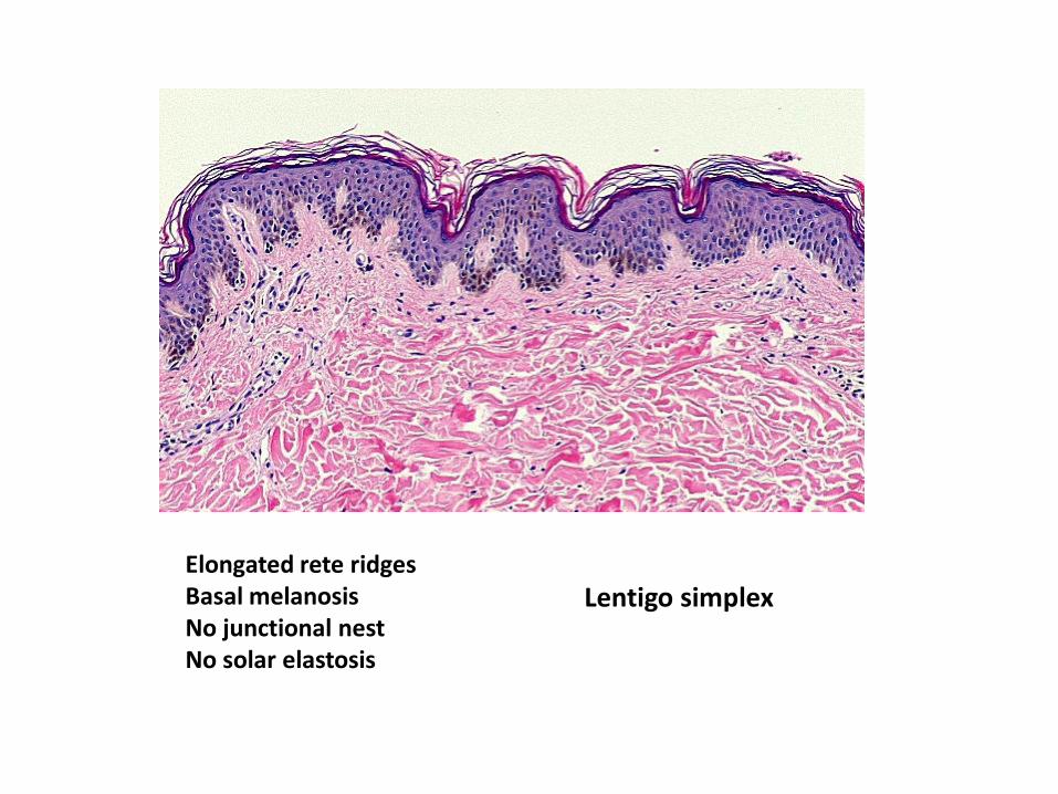
Lentigo simplex Elongated rete ridges Basal melanosis No junctional nest No solar elastosis

Solar lentigo Elongated rete ridges Basal melanosis No junctional nest Dermal solar elastosis

Junctional Nevus
Flat epidermal lesion Junctional nests with clefts

Raised lesion Dermal nests No junctional nests
Dermal melanocytic nevus

Compound melanocytic nevus Raised or flat lesion Junctional clefted nests and dermal nests

70% occurs in pts < 20 yrs Raised, junctional or compound nevus Epithelioid clefted nests oriented vertically Cytologic and nuclear pleomorphism
Spitz (Epithelioid) nevus

Dysplastic nevus All ages Compound nevus (or junctional nevus) Junctional clefted nests with transverse growth pattern (bridging) Cytologic and nuclear pleomorphism

Flat lesion No junctional nest Dermal pigmented spindled melanocytes
Blue nevus

Lentiginous nevus Flat lesion Common melanocytic nevus + lentigo simplex
Now , let me show you examples of several variation of melanocytic nevi

Markedly pigmented compound melanocytic nevus
Pigmented melanocytic nevus

Halo nevus
• Halo nevus is a common melanocytic nevus with a chronic inflammatory infiltrate resulting in a zone of depigmentation surrounding the nevus.
• The infiltrating cells are predominantly T-lymphocytes, and cytotoxic (CD8) lymphocytes outnumber helper (CD4) lymphocytes.
• Occurs most commonly in children (average age of onset is 15 years).
• Histology of halo nevus varies depending on the age of the lesion; a dense, somewhat bandlike lymphocytic infiltrate is present in the papillary and reticular dermis with nests of nevus cells located centrally. The lesion is dome-shaped architecture similar to other dermal or compound nevi. Identifying residual nevus cells may be difficult in some cases. S-100 stains the nevus cells.
Within the lymphocytic infiltrates, several ill-defined nests of epitheliod cells

Neurotized dermal melanocytic nevus (Neuronevus) Old dermal nevi may show degenerative change and the nevus nests may resemble nerves and Meissner corpuscle-like structures as seen in this case. Presence of nevocellular nests is helpful in diagnosis. In the first glance, you may think that it is a neurifibroma.

Let me show you a few more cases of Spitz nevus
Case 1. M 11 yrs, right upper arm, 8 mm lesion
Symmetrical, raised compound nevus

Vertically oriented spindle and epitheliod junctional nests with peripheral clefts
Kamino body
Kamino bodies are intraepidermal eosinophilic hyaline globules and are seen in 80% of cases of Spitz nevi . Kamino bodies are composed of laminin, type IV collagen and fibronectin.

Vertically oriented spindle and epitheliod junctional nests with peripheral clefts

Nevus nest in the keratin layer (transepidermal elimination)

Intraepidermal Kamino bodies
Kamino bodies are intraepidermal eosinophilic hyaline globules and are seen in 80% of cases of Spitz nevi . Kamino bodies are composed of laminin, type IV collagen and fibronectin.

Deeper dermal nests show mature nevus cells and lymphocytic infiltates

Comment
• The lesion is a raised , symmetrical compound nevus composed of predominently epitheliod and spindle nevocellular junctional nests and some dermal nests showing maturation with lymphocytic infiltrates towards the deeper dermis.
• The junctional nests are vertically oriented, many with peripheral clefts.
• Numerous round eosinophilic Kamino bodies are noted in the suprapalillary epidermis.
• Nevus cells show some pleomorphism with prominent nucleoli and rare mitosis.
• Pagetoid spread in the epidermis is not prominent.
• Features of dysplastic nevus or melanoma are not present.
• Overall picture is that of a classic benign Spitz nevus.

Case 2. M 9 yrs, right cheek


Case 3. M 2 yrs, right cheek


You will need to see quite a few cases of Spitz nevus and dysplastic nevus before you feel
confident about your diagnosis. More cases you see, better you will get. Books are helpful,
but real slides from real patients are your best teacher.
Experience cannot be taught, it has to be acquired over time !
Deba P Sarma, MD
Omaha


![Sarma Customer Service Surveys]](https://img.dokumen.tips/doc/110x75/58a3ad4d1a28ab9e6a8b6017/sarma-customer-service-surveys.jpg)