Embed Size (px)
Citation preview

1594 JAW Vol. 15, No. 7 June 1990: 1594-607
JACQUES M. T. DE BAKKER, PHD, RU%EN CORONEL, MD, SARA TASSERON, ARTHUR A. M. WILDE, MD, TOBIAS HOF, PHD, MICHIEL J. JANSE, FRANS J. L. VAN CAPELLE, PHD, AN E. BECKER, MD, FACC, GEORGE ;A
Amsterdam and Utrecht, The Netherlands
Electropbysiologic and histologic studies were ~~orrn~ on I,angendorffqterfused human hearts from patients who underwent heart transplantation far&on. In nine bearts, 15 sustai dias could be induced by program hearts, mapping of epicardial and e activity dm4ng tachycardia was carried out. examination of the h&u&d area between the site of latest activation of one cycle aad the site of earliest activation of the next cycle revealed zones of viable myocardial tissue.
In two hearts in which the time gap between latest and earliest activatioa was small, sr~lving myocardial tissue constituted a continuous tract that traversed the infarct. In Wee other hearts in wtdch the time gap was large, surviv-
Only a small percent of patients who survive myocardial infarction develop sustained ventricular tachycardia 224 h after the onset of infarction (1). Sustained ventricular tachy cardia can, however, be induced in 48% of patients with healed myocardial infarction but without documented ar- rhythmias (2). This finding suggests that in at least half of the patients who survive myocardial infarction, the substrate for sustained ventricular tachycardia is present, but in most cases, the trigger for starting a tachycardia never occurs. We therefore studied the mechanism of ventricular tachycardias associated with chronic infarction in Langendorff-perfused hearts from patients in whom extensive infarction dictated heart transplantation.
From the Interuniversity Cardiology Institute. The Netherlands, the Departments of Experimental Cardiology and Pathology. Academic Medical Center, Amsterdam and the Heart-J&g Institute.-University Hospital, Utrecht, The Netherlands.
Manuscript received July 31. 1989: revised manuscript received Decem- ber 20, 1969. accepted January iO,l9!IO.
Address Jacques M. T. de Bakker, PhD, Department of Experimental Cardiology, Academic Medical Center, Meibergdreef 9, I105 AZ Amsterdam, The Netherlands.
ing tissue consisted of parallel
the fibers witbiin the bundles was
removed from one of the tiissue bath during basic Lion of this preparation revealed by a zigzag route of activation over bra bundles of surviving myocytes separ tissue.
Convincing evidence from clinical and experimlental stud- ies (3-12) shows that reentry is the underlying mechanism of these arrhythmias. In experimental models of chronic infarc- tion, both complete occlusion of a coronary artery (6-9,121 and occlusion followed by reperfusion (13-15) have been effected. The infarcts produced in these two animal models are different, but the resulting tachycardias usually originate in a surviving epicardial zone that overlies the infarcted area, although intramural reentry has also been reported (16). In contrast, tachycardias in the infarcted human heart usually arise in a subendocardial region (17-21); however, tachycar- dias that originate epicardially aud those in which activation revolves around the infarction scar have also been reported (22,231. Although the location of the arrhythmogenic area may differ, histologic studies (12-15,17,24,25) suggest that the arrangement of surviviug cardiac fibers in and around the infarct zone plays an important role in the genesis of such tachycardias.
The purpose of our study was to investigate the role of the arrangement of surviving cardiac fibers in the infarct zone, constituting the substrate for such tachycardias. For this, we used Langendoti-perfused human hearts from patients who
Ql990 by the American College of Cardiology 073%1097/90/$3.50

JACC Vol. 15. No. -I June 1990: 1.594-607
DE BAKKER ET AL. t595
cumented in one
(Ca*+) 1.5. phosphate
same solution. The y in the cold Tyrode
d 5% carbon dioxi
arteries were c
elsewhere (26). Th 50% human blood
into the left and the other into the right cavity. The heart started to beat spontaneously or to fibrillate within minutes of perfusion. If fibrillation occurred, the heart was defibril- lated by direct current countershock after approximately 5 min. Perfusion was maintained at a temperature of 37 + O.YC until either the measurements were completed or contraction noticeably decreased (after 4 to 5 h).
~~~uetio~ of tachyea ia. A bipolar hook electrode was used to St eart with a §t~~~~t~ of twit diastolic t premature stimuli were use to induce tachycardia. The site of stimulation was epicardial and was altered if no sustained tachycardia could be evoked. In nine hearts, one or more moaomo~hic sustained (lasting >30 s) ventricular tachycardias could be induced. The cycle length of the induced tachycardias varied from 260 to 560 ms (mean 340). This is close to the mean cycle length of 330 ms of tachycardias that were induced in patients during antiar- rhythmic surgery. One of the explanted hearts came from a patient in whom spontaneously occurring ventricular tachy-
ely. In four of the
iiity. Signals recorded with bi-
rence, and were used to
rdias were selected for
of the grid electrodes, the terminals were arranged in an 8 by atrix with interelectrode distances of 6 mm. In the other.
e terminals were arranged in a 4 by 16 matrix with interelectrode distances of 10 mm. Pour to eight successive positions of the grid electrode were used to scan the epicar- dial surface. These positions were recorded phottigraphi- tally.
Tissue bath recordings. The posterior papillary muscle of one heart was resected and studied in a tissue bath. In this heart, only nonsustained tachycardias could be induced. The preparation was ~o~~te~ in a tissue bath in which a modi~ed Tyrode solution circulated at a rate of 28 ml/mi volume of the tissue bath was 9.5 ml. The compositio solution was identical to that used for the isolated hearts. The solution was aerated with a mixture of 95% O2 and 5% CO*. Temperature was kept constant at 37 ? 0. superfusion with an oxygenated solution, a bo approximately 600 pm exists in which transmernbrane PO- tential characteristics are relatively well preserved (27). The surviving subendocardial rim of the papillary muscle was approximately 400 pm thick. Stimulation was performed at a

basic cycle length of 800 ms at 1 cm from the base of the papillary muscle. Recordings were made from three sites located 0.5, 1 and i.5 mm, respectively, from the site of stimulation, atong a line perpendicular to the long axis of the papillary muscle. Additional recordings were made from six sites located along a line from the bas2 to the top of the papillary muscle. This line ran through the 1 mm point from the site of stimulation, and the interelectrode distance was 2 or 6 mm. After electrophysiologic study, the preparation was fixed with formalin and prepared for histologic investigation.
histologic methods. Five hearts in which monomorphic sustained ventricular tachycardias could be induced and in which the sites of earliest and latest activity were s2.5 cm apart were selected for detailed histologic study. After fixation with formalin, the heart was cut into seven sections: five slices with a thickness of 1.2 cm and a basal and an apicsl slice. The cutting planes corresponded to the six planes through the parallels. On the basis of the electrical recordings, the area between the sites of earliest and latest activity was determined and cut from the slice or slices in tissue blocks, with a length of approximately 3 cm. Sections of 10 pm thickness were produced with use of conventional histologic techniques. Both elastin-van Gieson and hematox- ylin-eosin stains were employed. Elastin-van Gieson-stained sections were used to trace fibrotic cardiac tissue in the infarct zone. Hematoxylin-eosin-stained sections were used to map surviving cells. Schematic drawings, highlighting viable cardiac tissue in contrast to fibrotic tissue, were made of selected sections by using a projection microscope.
To determine whether a surviving area in one section made contact with a surviving area in the next section, transparent sheets of successive schematic drawings were made. Surviving areas that showed an overlap when stacking the sheets of adjacent sections were considered to belong to the same bundle. In this way, it was possible to demonstrate the existence of a continuous tract of surviving myocytes in subsequent sections. Natural landmarks such as the cross sections of venules and arterioles were used to orient suc- cessive sections above each other. These markers were always selected close to the site of interest in order to limit inaccurate orientation.
Activation sequence . fifteen mono- morphic sustained ventricular tachycardias of different con-
figurations were induced in nine isolated hearts. Cycle length of the tachycardias ranged from 260 to 560 ms (mean 340). Endocardial as well as epicardial recordings were obtained in 10 tachycardias. In seven tachycardias, earliest epicardial activation appeared 220 ms after earliest endocardial acti- vation. In three tachycardias, earliest epicardial and en- docardial activation was almost simultaneous (difference <20 ms). Epicardial maps usually showed a larger area of
early activation (area within the 20 ms isochrone) than did endocardial maps. In all cases, the endocardial activation pattern showed a focal area of earliest activity from which activation spread more or less centrifugally and was blocked toward the infarcted zone. The site of earliest activation was always located within 2 cm of the border of the infarct. The characteristics of the tachycardias were similar to those recorded in the majority of patients during a~tia~hythmic surgery (17).
e infarct. Histologic ex- ed between the site of
latest activation of one cycle and earliest activation of the next cycle invariably showed that the infarct was interwoven with strands of surviving muscle fibers. The fibers within this infarcted area usually showed shifts in direction. Bundles of viable myocardial fibers surrounded by fibrous tissue ranged in diameter from that of a single cell to a few millimeters. These bundles could run individually over a few hundred micrometers or less, then merge and divide again. Bundles were located subendocardially, subepicardially or intramu- rally.
Tachycardias th Short Diastolic intervals In two tachycardias recorded in two different hearts, the
distance between the latest and earliest activated site was C2.5 cm.
Activation ~~~en~~ of heart 1. (Fi 2). The en- docardial activation pattern of the left is shown in the upper left panel of Figure 1. An area of damaged tissue extended from base to apex in the posterior wall. Activation started at the right border of the infarct, and main activation spread out toward the lateral wall. Activation toward the septum was blocked after 80 to 100 ms; 240 ms after the onset of endocardial activation, activity reached the septal side of the infarct (site e). The delay between activation at this site and the onset of the next cycle (at site a) was only 30 ms, suggesting reexcitation at the “origin” by means of surviving cardiac tissue between sites e and a.
In the righr panel of Figure I, endocardial electrograms recorded at sites a to fare shown. Signal f is recorded at a position between the site of latest activation of one cycle and earliest activation of the next. The signal has no sharp intrinsic deflection, but e consists of several components (arrows). C the signal recorder at site f with recordings made at neighboring sites (sites e and b) reveals the origin of two of the components. The deflec- tion indicated by the first (leftmost) of the three arrows coincides with the intrinsic deflection of the signal at site e at the border of the infarct and the surviving septum. The deflection indicated by the third (rightmost) arrow coincides with activation in the healthy lateral wall (site b). The deflection of the second (middle) arrow, however, does not coincide with documented activity anywhere else in the

JACC Vol. IS, No. 7 June I : 1594407
~ndoca~dia~ activation patterns of an& and right ~~~~~~
ventricle during sustained ventricular
ctrograms recorded with a 44 point balloon electrode inserted into the cavity.
al activation of the
arrived at the left side 240 MS later. There was a time gap of 30 ms between the latest activa-
signals, correlated well with the infarct zone. ant./lat. = anterior and iateral walls; right ventricie; t = time.
icates that viable tissu underneath the ret between activation
ardial activation pattern of the same tachycardia as in the position of a4 by 16 point grid electrode shown in
the lower panel. lsochrones are in ms and timed with respect to earliest endocardial activation 0 = 0 ms in Fig. I). Earliest epicar- dial activation arose at de of the infarct simultaneously with the endocardial o indicate in spread of activa- tion. LAD = :eft anteri artery; = left ventricle: RV = right ventricle.
damaged 91Ssue
grid eleclrode
postenor descendmg artery
ity. The slow downstroke of the de ection indicates that ust be located at some
In the lower ieft pun of Figure f. the endocardial activation pattern of the ri t ventr ic!e indicates that earliest
rs in the posterior wall I60 ms set in the left ve cle.
ctrical activity in the same tachycardiu wus performed with a 4 by 16 grid electrode (Fig. 2). The lower panel shows that in this position, the grid electrode covered the epicardial area that faced the site of earliest endocardial activity. The u per panel shows the epicardial activation pattern of the area covered by the grid electrode. The earliest epicardial activation was virtually simultaneous with the earliest endocardial activation. Epi- cardial activation began at the posterior wall of the left ventricle, just to the left of the posterior descending artery. As in the endocardial activation maps, there was only a smail
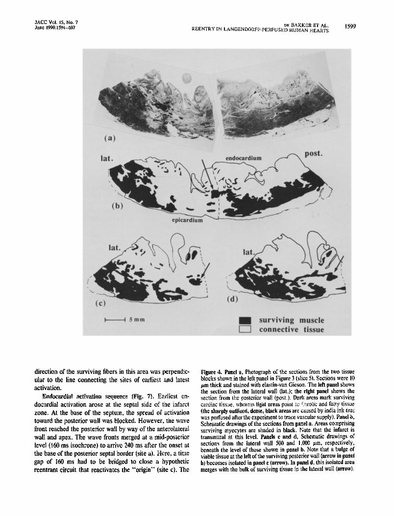
pm thick histologic sections from the lateral and posterior tissue blocks. At this level, the infarct is clearly transmural. In panel b, there is a bulge of surviving tissue (arrow)

1598 DE BAKKER ET AL. REENTRY IN LANGENDORFF-PERFUSED HUMAN HEARTS
JACC Vol. IS. No. 7 June 1990: 1594-607
Figure 3. Rig@, Schematic draw& of the post,erior view of the heart in Figure I. The numbers indicate slices in which the heart was cut (basal part not shown). The upper levels of the slices correspond to the planes through the six parallels that subdivided the balloon electrode used for ehdocardial mapping. The hatched &a Of slice 5 is the area in which the return path for reentrant activation within the infarct was expected. Left. Schem&ic drawing of slice 5 indi- cating the lateral and posterior segments that were selected for detailid histologic study. PDA = posterior descending artery; dther abbreviations as in Figure I.
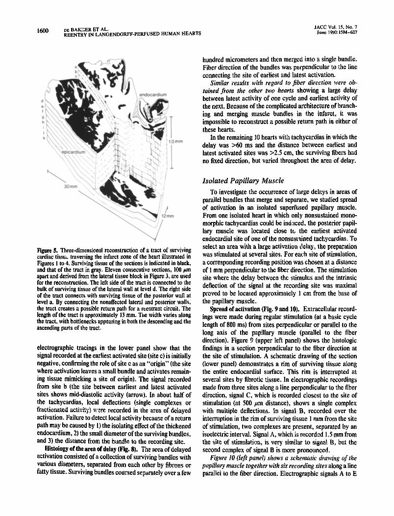
protruding into the infarct z&e. Panel c, deiived from a Section 500 m more apical in the latelr: tissue block, indicates that the bulge of surviving &sue his become isolated (arrow). Finally, in panel d (1,000 r;in underneath section b), the island of surviving tissue m&es with the healthy lateral tiall. This observation suggests that there was a surviving butidle connecting viable tissue at either side of the infarct zone and possibly forming a return path for reedtry. To prove that the tract wab indeed continuous, we made schematic drawings of series of,seciions 100 pm apart throughout the lateral tissue block of Figure 3 and compared each section with its success&. A three-dimensional recon- strtiction of the tract is shown in Figure 5. The schematic drawings of 10 successive sections (a to k), each 100 pm apart, show that the tract (gray area) fuses with remaining healthy tissue of the lateral Wall at level d. From here, the tract runs ddwn toward level k, and remains horizontal over a distance of about 6 mm in level j before it ascends and finally merges with remaining healthy tissue bf the posterior wall at level a. The uppel- sectidn of the reconstruction (level a) lies iO0 ,~ii~ beneath the lateral se&on in Figure 4a. Although the tract appears to be continuous, there are sonic narrow,passages, especialli in the descending and ascending parts of the tract. The sm&%t width of these bottlenecks was approximately 250 j~ni.
One might speculate that unidirectional block could occur preferentiilly at these sites. To demonstrate that continuity was maintained in the narrow corridors, we iinalyzed all the intervening sections (Fik. 5) at Id m intervals. At this resolution, too, the surviving tract appeared to be contin& ous. The tract was located at some distance from the endocardial surface, which may account for the shallotiness of the endocardial signals during the iresumed return of the activition wave through the tract (see Fig. lf).
icardial tissue in hea ). The subendocardial tract did not constitute the only possibility for reentry. The epicardial activation map (Fig. 2) suggested the presence of a surviving tract in the subepicardial part of the infarct. An anatomic substantiation for such a tract could be demonstrated by using the same procedure as described above. The course of the tract is schematica1l.y shown in the lower part of the middle panel (heavy line) in Figure 6. In sections a and c of the lateral wall, surviving tissue is interrupted, preventing activation from passing to the pos-
ese levels. Section b shows continuity of surviving tissue from the left to the right, and represents the part of the tract in the lateral wall.
Sections d and e are taken from the posterior wall and are 100 pm apart. Each section alone is not continuous with respect to surviving tissue, but by bverlapping the two, the interrupted areas in one section (ovals) are bridged by viable tissue of the other section. Continuity in this part of the tract was maintained in the intervening 10 pm thick sections. The smallest width of the tract in these areas is approximately 280 m. The lateral and posterior parts of the tract run at different levels (middle panel) put are connected by slirviving tissue at the border between the tissue blocks (the vertical part of the heavy line in the lower pati of the middle panel).
Dime&o& ofsurviving tracts in heart 1. The length of the endocardiai tract within the infarct z&e was approximately 13 mm. In both the endocardially and subepicardially l&ted tracts, fiber orientation was mainly in the direction of the spread of activation. Only in the vertical part of the subepi- cardial tract was the fiber orientation perpendicular to the spread of activation. However, fibers in this area formed a single cdmp%ct bundle and were not isolated from each other by fibrous tissue. The short length of the tracts together with a fib& orientation mainly in the direction of the tract may account for the small delay of activation in this area.
Findings in he& 2. The second heart with a small delay between earliest and latest activation exhibited similar re- sults. A tract of surviving tissue, located 1.5 mm from the epicardial s&ace of the anterior wall, was traced in this heart. Its smallest width was approximately 350 pm. Two morphologic&y different tachycardias could be induced. In both, the area between the site of latest activation of one cycle and die site of earliest activation of the next was the same. Because we could trace only one surviving tract in this area, it is probable that this tract was used as a return path for activation in both tachycardias. The tachycardias rc- volved in opposite directions, indicating that unidirectional block in this area could occur in either direction.
Tachycardias With Long Diastolic Intervals
In three hearts, the sites of latest activation of one cycle of the tachycardia and earliest activation of the next were c2.5 cm apart, and the delay was >I50 ms. In all three, the
JACC Vol. IS. No. 7 June 1990:1594-607
direction of the surviving fibers in this area was perpendic- ular to the line connecting the sites of earliest and latest activation.
ion arose a zone. At the base of the septum, the ad of activation
wall and apex. The wave fronts merged at a mid-posterior level (160 ms isocbrone) to arrive 240 ms after the base of the posterior septal border (site a). gap of 160 ms had to be bridged to close a hypothetic reentrant circuit that reactivates the “origin” (site c). The
Figure 4. el a, Photograph of the sections from the two tissue blocks sh in the left panel in Figure 3 (slice 9. Sections were 10 hrn thick and stained with elastin-van Gieson. The left panel shows
he lateral wall (lat.); right panel shows the h areas mark surviving ilrrotic and fatty tissue
Schematic drawings of the sections from el a. Areas comprising surviving myocytes are shaded in black. Note that the infarct is transmural at this level. Schematic drawings of sections from the lateral wall 500 and ‘1,000 plm, respectively, beneath the level of those shown in panel b. Note that a bu viable tissue at the left of the surviving posterior wall (arrow in b) becomes isolated in panel c (arrow). In panel d, this isolated area merges with the bulk of surviving tissue in the lateral wall (arrow).
1600 DE BAK!:ER ET AL. REENTRY IN LANGENDORFF-PERFUSED HUMAN HEARTS
JACC Vu!. 15, No. 7 June 1990:1594-607
Figure 5. Threedimensional reconstruction of a tract of surviving cardiac tissui traversing the infarct zone of the heart illustrated in Figures 1 to 4. Surviving tissue of the sections is indicated in black, and that of the tract in gray. Eleven consecutive sections, 100 pm apart and derived from the lateral tissue block in Figure 3, are used for the reconstruction. The left side of the tract is connected to the bulk of surviving tissue of the lateral wall at level d. The right side of the tract connects with surviving tissue of the posterior wall at level a. By connecting the nonaffected lateral and posterior walls, the tract creates a possible return path for a reentrant circuit. The length of the tract is approximately 13 mm. The width varies along the tract, with bottlenecks appearing in both the descending and the ascending parts of the tract.
electrographic tracings in the lower panel show that the signal recorded at the earliest activated site (site c) is initially negative, confirming the role of site c as an “origin” (the site where activation leaves a small bundle and activates remain- ing tissue mimicking a site of origin). The signal recorded from site b (the site between earliest and latest activated sites shows mid-diastolic activity (arrow). In about half of the tachycardias, local deflections (single complexes or fractionated activity) wze recorded in the area of delayed activation. Failure to detect local activity because of a return path may be caused by I) the isolating effect of the thickened endocardium, 2) the small diameter of the surviving bundles, and 3) the distance from the bund!e to the recording site.
Histology of the area of delay (Fig. g). The area of delayed activation consisted of a collection of surviving bundles with various diameters, separated from each other by fibrous or fatty tissue. Surviving bundles coursed separately over a few
hundred micrometers aud then merged into a single bundle. Fiber direction of the bundles was perpendicular to the line connecting tbe site of earliest and latest activation.
Similar results with regard to fiber direction were ob- tained from the other two hearts showing a large delay between latest activity of one cycle and earliest activity of the next. Because of the complicated architecture of branch- ing and merging muscle bundles in the infarct, it was impossible to reconstruct a possible return path in either of these hearts.
In the remaining 10 hearts with tachyc delay was >60 ms and the distance be latest activated sites was >2.5 cm, the no fixed direction, but varied throughout the area of delay.
Isolated Papillary Muscle To investigate the occurrence of large delays in areas of
parallel bundles that merge and separate, we studied spread of activation in an isolated superfused papillary muscle. From one isolated heart in which only nonsustained mono- morphic tachycardias could be indl.tced, the posterior papil- lary muscle was located close to the earliest activated endocardial site of one of the nonsustained tachycardias. To select an area with a large activation delay, the preparation was stimulated at several sites. For each site of stimulation, a corresponding recording position was chosen at a distance of 1 mm perpendicular to the fiber direction. The stimulation site where the delay between the stimulus and the intrinsic deflection of the signal at the recording site was maximal proved to be located approximately 1 cm from the base of the papillary muscle.
Spread of activation (Fig. 9 and 10). Extracellular record- ings were made during regular stimulation (at a basic cycle length of 800 ms) from sites perpendicular or parallel to the long axis of the papillary muscle (parallel to the fiber direction). Figure 3 (upper left panel) shows the histologic findings in a section perpendicular to the fiber direction at the site of stimulation. A schematic drawing of the section (lower panel) demonstrates a rim of surviving tissue along the entire endocardial surface. This rim is interrupted at several sites by fibrotic tissue. In electrographic recordings made from three sites along a line perpendicular to the fiber direction, signal C, which is recorded closest to the site of stimulation (at 500 pm distance), shows a single complex with multiple deflections. In signal B, recorded over the interruption in the rim of surviving tissue 1 mm from the site of stimulation, two complexes are present, separated by an isoelectric interval. Signal A, which is recorded 1.5 mm from the site of stimulation, is very similar to signal B, but the second complex of signal B is more pronounced.
Figure 10 (left panel) shows a schematic drawing of the pa.pillary muscle together with six recording sites along a line para!lei to the fiber direction. Electrographic signals A to E

JACC Vol. IS, No. 7 June 199&1594-607
these sites show two co plexes separated by an isoelectric interval. becomes shorter as the recording site is closer ere, the two complexes merge, yielding a signal that consists of a single complex with multiple deflections. Aitb~~gh the site of stimulation was close 10 sites B a earliest depolarization is found in signal D. The first de ns in signals E an are later in succession, indicating t activation proceeds from site toward site F. The set deflection comes earliest in F a progressively later toward A, indicating that a second wave appears to travel somewhat later from sites F to A, The main activation at site A (second deflection) appears to reach it by a rambling route by way of site F.
istologic correlation (Fig. This observation is supported by the sections taken the recording sites (Fig. 10). In the surviving rim of sections B to E, there is an interruption that disappears in section F. the number of sections at site F, we found that the bundles merged near site F and then divided once more.
The architecture of the papillary muscle and its spread of activation is shown schematically in Figure I I. Delay of the activation wave is due to a lengthening of the route. The apparent conduction velocity in the area around sire A perpendicular to the fiber orientation would be approxi- mately 0.03 m/s (distance 1.5 mm, delay 46 ms), but the real conduction velocity is much faster. The distance from the site of stimulation to site A by way of site F is approximately 2 cm. The delay between the stimulus and activation at site A is 46 ms, resulting in a mean conduction velocity of 0.45 m/s, a value close to normal for cardiac muscle.
igure 6. Schematic drawings illustrating an epicardial tract of surviving tissue traversing f the heart described in Figures I to 5. In the lower panel, the entire course of the tract is shown sche line). On the left, three
rawings of the lateral tissue block (set type +I of the I) are shown. Sections were 300 plm apart. Se&ans a and co~t~~~ity of surviving tissue is ~~ter~~~ted at the sites
indicated by the arrows. In section b, there is continuity of surviving myocardium from the Left Lo rig t. The surviving areas in this section constitute the lateral part of th . In the pb,;*:rior tissue block. continuity is absent in sections e (oval areas). Only if sections
de stacked is continuity of viable tissue present. The ged between sections d and e show the critical areas were
an interruption of the continuitv in one section is overlapped by surviving tissue in the other se&on. The lateral and posterior parts of the tract are connected at the border between !he tissue blocks. Abbreviations as in Figures 1 and 4.
A substantial body of evidence (3-12) supports the con- cept that ventricular tachycardias in the chronic phase of myocardial infarction are based on reentry. Reentrant ar- rhythmias may be caused by abnorm embrane variables, abnormal geometric arrangement of cardial fibers or a combination of the two. Transmembrane potentials obtained from superfused resected endocardial preparations from patients with ventricular tachycardia in the chronic phase of myocardial infarction were only mildly depressed (17). This observation indicates that ac!ion potential characteristics of

1602 DE BAKKER ET AL. REENTRY IN LANGENDORFF-PERFUSED HUMAN HEARTS
JACC Vol. IS, No. 7 June 199&1594-607
Figure 7. Endocardial activation pattern of sustained ventricular tachycardia with a large time gap between latest activation of one cycle and earliest activation of the next cycle. The distance between the site of latest and earliest activation was ~2.5 cm. Isochrones are in ms and timed with respect to earliest endocardial activation. Activation starts at the left side of the infarct and arrives at the right side of the infarct 240 ms later. Electrographic tracings (below) are the signals recorded from the earliest activated site (c), the latest activated site (a) and an intermediate site (b). The signal at site b shows mid-diastolic activation (arrow). Abbreviations as in Figure 1.
surviving myocardial fibers within an infarct may be close to normal. This view is supported by findings in animal exper- iments (12,28), where action potentials recorded from cells in the epicardial border zone became normal with the passage of time. Therefore, in the chronic phase of myocar- dial infarction, reentrant tachycardias may be caused pre- dominantly by abnormalities in the geometric arrangement of surviving fibers, and not by abnormalities in transmem- brane potentials.
Recordmg technlqse. Despite certain disadvantages, uni- polar recording is, in our opinion, best suited for mapping studies such as those undertaken in this study. The fast downstroke of the unipolar complex reflects the moment of activation. Areas where activation arises are characterized by an initial negative deflection, whereas recordings made from sites where activation is passing exhibit initial “R
waves.” Interpretation of unipolar sigaals remains possible in the case of polyphasic deflections. When a signal t reveals polyphasic deflections is compared with signals recorded at neighboring electrode terminals, the nature of the deflections can usually be clarified. This is usually not possible for bipolar signals with multiple components. Even the selection of that part of the signal that corresponds to t intrinsic deflection is often problematic in bipolar reco ings. A disadvantage of unipolar recordings may be encoun- tered when signals are reco d in areas of injured tissue. In that case, the intrinsic d obscured by larger remote components from neighboring healthy tissue. Although in bipolar recordings, distant events are canceled, sometimes permitting better estimation of the intrinsic deflection, the steepness of unipolar complexes provides information about the vitality of underlying tissue, allowing discrimination between healthy and damaged myo- cardium. It is because of the facility of interpretation of uoipolar complexes under different conditions that we prefer the unipolar recording mode.
The spatial resolution of the balloon electrode and one of the grid electrodes (4 by 16 matrix) was 1.4 and 1 cm*, respectively. The spatial resolution of the other grid elec- trode was 0.4 cm’. Detection of a complete reentrant circuit, in which small bundles are part of the circuit, would require the use of an electrode system with very small interelectrode distances. Even then, however, it is impossible to follow activation in all parts of the circuit because of the rambling course of the tracts. In addition, the large number of plunge electrodes that would be required, especially in the area of the tract, would certainly affect the spread of activation. Our experience (17) with intraoperative mapping during antiar- rhythmic surgery has shown that the earliest activated site during tachycardia can reliably be identified. In addition, in a large number of tachycardias, presystolic ar late potentials were found at several sites near the origin, permitting a tentative reconstruction of the route followed by the im- pulse. Thus, the resolution we used is sufficient to select areas where a possible return path for activation can be expected. Histologic examination of these areas showed that surviving bundles were present and, in some cases, the tracts were continuous, connecting remaining healthy parts of the heart on either side of the infarct. It is not possible with our methods to follow activation within the tracts.
Conduction in surviving tissue within the infarct. Bundles of viable myocardial fibers embedded in fibrous tissue are a common finding in chronic myocardial infarction (24). In the human heart, cardiac tissue preferentially survives immedi- ately beneath the endocardium (24,29). The reason for this may be the presence of a subendocardial vascular plexus and diffusion of oxygen and substrate from the cavitary blood (30,31). The subendocardial location of surviving bundles may account for the subendocardial origin of the majority of lachycardias (18-21). The surviving subendocardial layers

JAW Vol. 15. No. 7 June 1990:1594-607
ane!, photograph of a sec-
areas consist of survivin
ing bundles (bright areas) separated from each other by fibrous tissue (dar areas). The photomi-
t shows histologic features in a section 100 pm beneath the left section. separated bundles have merged into a single bun- dle. Fiber direction was perpendicular to the line connecting the earliest and the latest activated site.
nldo = e~docardia~ wall; other abbreviations as in igure 1.
resemble the surviving epicardial layers found in experimen- tal chronic infarction after complete occlusion of a coronary artery. In these surviving epicardia! sheets that overly the infarct zone, the anisotropic structure of cardiac fibers is the major determinant for reentry (11,121. Our observations showed that the surviving s~be~docardial fayer in human infarcts is not homogeneous, but is ir.teirtipted at many sites, similar to the histologic structure of the papillary muscle in Figure 9. We also observed that surviving bundles were not confined to the subendocardial layers but were found intra- murally and subepicardial~y as well. Iln cases in which the time gap between latest activation of one cycle and the earliest activation of the next cycle was small, a continuous bundle of surviving myocytes that traversed the infarct was
found. This observation does not imply that we also proved the unique path followed by the activation; there are small areas of fibrous tissue within the tract that hamper the selection of this path of activation (Fig. 5).
r’n closes with large time gaps over short distances, clusters of surviving muscle bundles separated by fibrous tissue were present, and the repeated fusion and bifurcation of these bundles resembled the inhomogeneous anisotropy found in the papillary muscle. An important factor that determines conduction is the coupling resistance between cells. Even under normal conditions, tissue anisotropy causes a difference in coupling resistance perpendicular and parallel to the fiber direction, resulting in differences in conduction velocity along these two axes (32-35).

1604 DE BAKKER ET AL. REENTRY IN LANGENDORFPPERFUSED HUMAN HEARTS
JACC Vol. 15. No. 7 June 199@:lS94-607
-r-
w 1. lmm _. ,s-
I I I
0.5mV 20ms
An increase in coupling resistance may influence conduc- tion in two ways. 1) Local delays may occur as a result of impulse transmission across a high resistance junction. The occurrence of an electrotonic foot that precedes the upstroke of the action potentials would be expected in this case. Such aberrant action potentials have been found in experimental infarction (28). 2) Proliferation of connective tissue may reduce or abolish the side to side electrical coupling between cells and increase the distance to be traveled between poi& (that is, a winding, circuitous route for activation may be followed). Thus, the tract is lengthened and a zigzag course of propagation results. In our example of the papillary muscle, the delay can be explained entirely by an increase in the distance to be covered, which is caused by merging and diverging bundles. A repetition of this phenomenon could cause large delays. Because the structure of the surviving bundles in the areas of large delays resembles that of the
C
el, Section of muscle from
the left ventricle of a heart in which only nonsustained ventricular tachy- cardias could be induced. 5ri~~t ar- eas represent surviving cardiac tis- sue; dark areas mainly consist of fibrotic tissue. Along the entire sur- face, a rim of surviving tissue is present. Lower lest panel, Schematic drawing of the section depicted in the upper panel. Survivitig tissue * shown in black. Tracings on the ri are signals recorded from sites in cated in the lower I. The prepa- ration was stimulated at the site marked by the arrow, using a basic cycle length of 800 ms. stim = site of stimulation.
papillary muscle, delays based on a lengthening of a conduc- tion pathway could play a role in these tachycardias.
block. Unidirectional block, which is nec- essary to initiate a reentrant arrhythmia, may result from regional differences in recovery of excitability or from the asymmetric anatomic structure of some regions. The geom- etry of the surviving bundles that merge and divide indicates that geometric factors may play an important role in the occurrence of unidirectional block in the hearts we studied. It could occur at sites where a thin bundle suddenly enters a larger one because current output of the small bundle is too low to drive the larger one (35,36). Conduction in the reverse direction may still be possible if the large bundle delivers enough current to depolarize the small one. Also, branching sites may give rise to a directive-dc~ende~t electrical load and, as a consequence, may cause Bsidirectional block (37). A three-dimensional reconstruction of a return path for

JACC Vol. 15. No. 7 June 1990: 1594407
2mm
I
0.5mV
activation (Fig. 5) demonstrates that there are several sites where the width of the tract changes, thus yielding possible sites for unidirectional block due to a change in diameter of a bundle. Figure 8 shows that the area of delay consists of merging and bifurcating bundles with different diameters. Thus, unidirectional block based both on an abrupt change in the diameter of a bundle and on branching sites of bundles may be expected to occur in such areas.
0try ntry? Surviving bundles within an area ’ ~~~stitnte the return path of a macroreentrant circuit. ik~ever, it is also possible that they constitute a reentrant circuit o dimensions of the circuit can conduction velocity in the circuit. promised area of the heart is not activated by means of an exit from the tract. In the heart shown in Figure I, two tracts were found, and it is possible that microreentry through these two pathways occurred. However, the following observations argue against this
20ms I -I 5OOpm
0. Left panel, Schematic drawing of the papillary muscle from the heart in Figure 9. ts indicate recording sites. The site of stimulation was located at the signals recorded at the black panel. Schematic drawings of the histologic findings in the surviving rim at the recording sites. Sections from which these schematic drawings were obtained were cut perpendicular to the long axis of the papillary muscle. Surviving areas are shaded black. See text for further discussion.
concept and support the occurrence of macroreentry. 1) A microcircuit requires an area of slow conduction, but from histologic results, it is unlikely that large delays occur in the tracts. 2) The activation pattern in Figure 1 the delay in one of the tracts was equal to th an activation front to return to its exit from the tract throngh the noncompromised part of the heart. If the delay shorter, two sites of exit would be expected. In the heart in which we could prove that noncompromised parts of the heart were connected by surviving tissue that traversed

1606 DE BAKKER ET AL. REENTRY IN LANGENDORFF-PERFUSED HUMAN HEARTS
JACC Vol. 15. No. 7 June 1990: 1.594-607
Figure 11. Possible spread of activation through the papillary mus- cle from the heart in Figure 10. The heavy arrows indicate the direction of the activation waves. Horiz&tal arrows indicate the recording sites shown in Figure lil. Delay of activation in the direction perpendicular to the long axis of the papillary muscle at level A occurs because of a winding route of activation through site F.
the infarct, only one tract was found. Thus, at least for some of the tachycardias that occur in the chronic phase of myocardial infarction, macroreentry takes place through surviving areas within the infarct and the nonaffected parts of the heart.
We‘ are aware that tachycardias in which the time gap between earliest and latest activation is small represent onl) a pinority of tachycardias (this observation is compatible with our findings during antiarrhythmic surgery). In cases in which the time gap is large, macroreentry through the infarct is less clear. In several tachycardias we found local activa- tion at several sites in the zone of delay. In all these cases, spread of activation was from the last activated site toward the earliest activated site, a finding compatible with the concept that the area represents the return path of ti large circuit.
Consequences for therapy. Our data show that more than one tract of surviving tissue may traverse an ‘infarct zone, connecting the remaining healthy tissue at either side of the infarct. In addition, it appears that the location of the tracts is not confined to the subendocardial surface, but may run intramurally or even subepicardially. Both the mimber of tracts and their location h&e consequences for the mapping procedure that is usu#y carried out to support surgical therapy. The different locations of the tracts indicate that
mapping of only the endocardial surface is not sufficient in all cases and should be extended to epicardial or even intramu- ral areas. The presence of several tracts implies that a resection procedure should not be restricted to removal of a small area at the “origin” (that is, the exit to the bulk of surviving tissue), even if the !&origin” can be located with high precision. It cannot be excluded that only one tract is preferentially involved in the main@ance of a tachycardia and that it masks the existence of other tracts. In a few mapping studies performed during antiarrhythmic surgery, we found multiple sites of “origin,” suggesting that several pathways were involved in than tachycardia at the same time. The existence of several tracts may also be one reason for less than optimal results obtained with catheter ablation, even though the “origin” can be located with precision. The relatively small area destroyed by ablation may damage one circuit while leaving another intact. A second reason might be the location of the tracts. Tracts are more diflicult to attack by catheter ablation if they are located intramurally and are inaccessible if their locqtion is epicardial.
We are grateful to J. R. Lapohr, MD and S. R. Wooley. MD for careful preparation of the explanted hearts. We thank Charles Belterman for expert technical assistance, Ernst Heeren for excellent histologic support and Ruud Verhoeven for skillful photography.
I.
2.
3.
4.
5.
6.
7.
8.
9.
Wellens HJJ, Bar FWHM, van Agt EJDM, Brugada P. Medical treatmeni of ventricular tachycardia: considerations in the selection of patients for surgical treatment. Am J Cardiol 1982;49:186-93.
Brugada P, Waldecker B, Kersschot Y, Zehender M, Wellens HJJ. Ventricular arrhythmias initiated by programmed stimulation in four grpups of patients with healed myocardial infarction. J Am Coil Cardiol 198618: 1035-40.
Josephson ME, Horowitz LN. Farshidi A, Kastor JA. Recurrent sus- tained ventricular tachycardia. 1. Mechanisms. Circulation 1978;57:433-9.
Josephson ME, Marchlinski FE, Buxton AE, et al. Electrophysiologic basis for susiained ventricular tachycardia-role of reentry. In: Joseph- son ME, Wellens HJJ.‘eds. Tachycardias: Mechanisms, Diagnosis, Treat- ment. Philadelphia: Lea & Febiger. 1984:305-23.
Josephson ME, Buxton AE, Marchlipski FE, et al. Sustained ventricular tachycardia in coronary artery disease-evidence for reentrant mecha- nism. In: Zipes DP, Jalife I, eds. Cardiac Electrophysiology and Arrhyth- mias. Orlando, FL: Grune & Stratton, 1985:409:18.
El-Sherif N, Smith RA. Evans K. Canine ventricular arrhythmias in the late myocardial infarction period: epicardial mapping of reentrant circuits. Circ Res 1981:49:255-71.
El-Sherif N. Mehra R, Gough WB, Zeiler RH. Reentrant ventricular arrhythmias in the late myocardial infarction period: interruption of reentrant circuits by cryotherma! techniques. Circulation 1983;68:644-56.
Karagueuzian HS, Fenoglio JJ, Weiss MB, Wit AL. Protracted ventric- ular tachycardia induced by premature stimulation in the canine heart after coronary avery occlusion and reperfusion. Circ Res 1979;44:833-46.
Mehra R. Zeiler R, Gough WB. El-Sherif N. Reentrant ventricular arrhythmias in the late myocardial infarction period. 9. Electrophysio- logic-anatomic correlation of reentrant circuits. Circulation 1983:67: I I- 24.

IO. Gessman LJ. Agarwal J Endo T, Helfant RH. Localization and mechanism of ventricular bycardia by ice mapping 1 week after the onset of myocardial infarction in dogs. Circulation 1983;68:657-66.
IL Wit AL, Ailessie MA, Bonke FIM, Lammers W, Smeets 9, Fenoglio 99. Electrophysiologic mapping to determine the mechanism of ex~e~me~ta~ ventricular tachycardia initiated by premature impulse: experimental approach and initial results demonstrating reentrant excitation. Am J Cardiol 1982;49: 166-85.
12. Gardner PI, Ursell PC, Due Pham T, Fenoglio JJ, Wit CkiOfiiC veniricular tacbycardia: anatomic and elect stances. In: Ref 4:29-60.
Experimental ysiologic sub-
13. Michelson EL, Spear JF, Moore EN. Electrophysiologic and anatomic correlation of sustained ventricular tacbya~byt~mias in a model of chronic myocardial infarction. Am J Cardiol 1980;45:583-90.
14. Michelson EL, Spear JF. Moore EN. Further ~~ectr~~pbysiologj~ and anatomic correlates in a canine model of chronic m”*ocardiall infarction susceptible to the initiation of sustained ventricular tacbycardias. Anal Ret 1981;201:55-65.
15. Gang ES, Bigger JT Jr, Livelli FD Jr. A mod” for chronic ischemic arrhythmias: the relation between electrically :nducible ventricular tachy cardia, ventricular ~b~~lat~oi~ threshold and infarct size. Am J Cardiol 19X2$0:469-77.
16. Kramer JB. Saffitz Je‘, Witkowski FX, Corn PB. bttramural FKeIIdPy as LI
mechanism of ventricular tacbycardia during evolving canine myocardial infarction. Circ Res 1985;56:736-54.
17. de Bakker JMT. van Capelle FJL, Janse MJ, et al. Reentry as a cause of ventricular tachycardia br patients with chronic iscbemic-heart disease: electrophysiologic and anatomic correlation. Circulation 19X&77:589- 606.
18.
19.
20.
21.
22.
23.
Wittig JH, Boineau JP. Surgical trearment of ventricular arrhythmias using epicardial. transmural and endocardial mapping. Ann Thorac Surg 1975;20:117-26.
Josephson ME, Horowitz LN. Farshidi A, Spear JF. Kastor 3A. Moore EN. Recurrent sustained ventricular tachycardia. 2. Endocardial map- ping. Circulation 1978;57:440-7.
Boineau JP, Cox JL. Rationale for a direct surgical approach to control ventricular arrhythmias: relation to specific intraoperative technique to mechanism and location of arrhythmic circuit. Am J Cardiol 1982:49:381- %.
Horowitz LN, Harken AH, Kastor JA, Josephson ME. Ventricular resection guided by epicardial and endocardial mapping for treatment of recurrent ventricular tachycardia. N En@ J Med 1980;302:589-93.
Downar E, Parson ID. Mickleborough LL. Cameron DA, Yao LC. Waxman MB. On-line epicardial mapping of intraoperative ventricular arrhythmias: initial clinical experience. J Am Colt Cardiol 1984;4:703-14.
Mason JW, Stioson EB, Oter PE. et al. The mechanisms of ventricular tachycardia in humans determined by intraoperative recording of tbe electrical activation sequence. Int J Cardiol 1985:8:163-72.
24. Fenoglio JJ, Due Pham T, Harken AM, Horowitz LN, Josephson Wit AL. Recurrent sustained ventricular tachycardia: structure and ultrastructure of subendocardial regions in which tachycardia originates, Circulation 1983;68:518-33.
electrophysiology of human myocardial infarction. 1. Abnormalities of cellular activation. Circulation 1979;59:247-56.
26. Downar E, Janse MJ, Durrer D. The effect of acute coronary artery occlusion of subepicardial transmembrane potentials in the intact porcine heart. Circulation 1977;56:217-24.
27. Wiiensky RL, TranumJensen J. Coronel R, Wilde AAM, Fiolet JWT, Janse MJ. The subendocardial border zone durine acute ischemia of the rabbit heart: an electrophysiologic, metabolic and morphologic correla- tive study. Circulation 1986;74: 1137-46.
28. Gardner PI, UrseDl PC, Fenoglio JJ, Wit AL. Electrophysidogic and anatomic basis for fractionated electrograms recorded from healed myo- cardial infarcts. Circulation 1985;72:596-61 I.
29.
30.
31.
32.
33.
34.
35.
36.
37.
Friedman PL. Fenoglio JJ Jr, Wit AL. Time course for reversal of electrophysiological and ultrastructural abnormalities in subendocardial Purkinje fibers surviving extensive myocardial infarction in dogs. Circ Res 1975:36: 127-44.
F&on WFM. The dynamic factor in enlargement of coronary arterial anastomoses. and paradoxical change in the subendocardial plexus. Br Heart J 1964;26339-50.
Estes HE Jr, Entman ML, Dixon HB. Hackel DB. The vascular supply of the left ventricular wall: anatomic observations, plus a hypothesis regard- ing acute events in coronary artery disease. Am Heart J 1966;71:58-67.
Corbin LV, Scher AM. The canine heart as an electrocardiographic generator: dependence on cardiac cell orientation. Circ Res 1977;41:58- 67.
Roberts DE. Hersb LT. Scber AM Influence of cardiac fiber orientation on wavefront voltage, conduction velocity and tissue resistivity in the dog. Circ Res 1979;44:701-12.
Spa& MS, Miller WT Jr, Ceselowitz DB. Barr RC, Kootsey JM, Johnson EA. The discontinuous nature of propagation in normal canine cardiac muscle: evidence for recurrent discontinuities of intracellular resistance that affect the membrane currents. Circ Res 19X1:48:39-54.
Joyner RW. Mechanisms of unidirectional block in cardiac tissue. Biophys J 1981;35:113-25.
Spach MS. Dolber PC. Relating extracellular potentials and their deriva- tives to anisotropic propagation at a microscopic level in human cardiac muscle: evidence for electrical uncoupling of side-to-side fiber connec- tions with increasing age. Circ Res 1986;58:356-71.
Spach MS, Miller WT Jr, Dolber PC, Kootsey JM, Sommer JR, Mosher CE. The functional role of structural complexities in the propagation Of depolarization in the atrium of the dog: cardiac conduction disturbances due to discontinuities of effective axial resistivity. Circ Res 1982;50:t;j- 91.