Embed Size (px)
Citation preview
© 2013 Gado and Aldahmash. This work is published by Dove Medical Press Limited, and licensed under Creative Commons Attribution – Non Commercial (unported, v3.0) License. The full terms of the License are available at http://creativecommons.org/licenses/by-nc/3.0/. Non-commercial uses of the work are permitted without any further
permission from Dove Medical Press Limited, provided the work is properly attributed. Permissions beyond the scope of the License are administered by Dove Medical Press Limited. Information on how to request permission may be found at: http://www.dovepress.com/permissions.php
Drug Design, Development and Therapy 2013:7 1245–1252
Drug Design, Development and Therapy Dovepress
submit your manuscript | www.dovepress.com
Dovepress 1245
O r i g i n a l r e s e a r c h
open access to scientific and medical research
Open access Full Text article
http://dx.doi.org/10.2147/DDDT.S50928
antioxidant effect of arabic gum against mercuric chloride-induced nephrotoxicity
ali M gado1
Badr a aldahmash2
1Forensic Medicine and clinical Toxicology Department, college of Medicine, Tanta University, Tanta, egypt; 2Medical laboratory Department, college of health sciences, King saud University, riyadh, saudi arabia
correspondence: ali M gado Forensic Medicine and clinical Toxicology Department, college of Medicine, Tanta University, Tanta, egypt email [email protected]
Abstract: The effects of Arabic gum (AG) against nephrotoxicity of mercury (Hg), an oxidative-
stress inducing substance, in rats were investigated. A single dose of mercuric chloride (5 mg/kg
intraperitoneal injection) induced renal toxicity, manifested biochemically by a significant
increase in serum creatinine, blood urea nitrogen, thiobarbituric acid reactive substances, and
total nitrate/nitrite production in kidney tissues. In addition, reduced glutathione, glutathione
peroxidase, and catalase enzymes in renal tissues were significantly decreased. Pretreatment of
rats with AG (7.5 g/kg/day per oral administration), starting 5 days before mercuric chloride
injection and continuing through the experimental period, resulted in a complete reversal of
Hg-induced increase in creatinine, blood urea nitrogen, thiobarbituric acid reactive substances,
and total nitrate/nitrite to control values. Histopathologic examination of kidney tissues confirmed
the biochemical data; pretreatment of AG prevented Hg-induced degenerative changes of kidney
tissues. These results indicate that AG is an efficient cytoprotective agent against Hg-induced
nephrotoxicity by a mechanism related at least in part to its ability to decrease oxidative and
nitrosative stress and preserve the activity of antioxidant enzymes in kidney tissues.
Keywords: mercury, acacia gum, oxidative stress, lipid per oxidation, kidney toxicity
IntroductionMercury (Hg) is a hazardous environmental and industrial pollutant which induces
severe alterations in the body tissues of both humans and animals.1,2 The toxicity of
Hg depends on the form of the Hg compounds (elemental, inorganic, and organic).
Inorganic Hg accumulates predominantly in the kidneys, causing acute renal failure.3,4
The uptake, accumulation, and toxicity of inorganic Hg in the kidney have been related
to it binding to endogenous thiol-containing molecules.5 Thiol-containing enzymes have
been recognized as the targets of inorganic Hg.5,6 Moreover, binding of mercuric ions
to thiol groups may cause decreased glutathione (GSH) levels, leading to increases in
levels of reactive oxygen species (ROS), such as superoxide anion radicals, hydrogen
peroxide, and hydroxyl radicals, which provoke lipid, protein, deoxyribonucleic acid
(DNA), and ribonucleic acid (RNA) oxidation.7,8 Considering that oxidative stress and
endogenous thiol depletion are involved in inorganic Hg toxicity, it has been suggested
that antioxidants could contribute to the treatment of Hg poisoning.9,10 In this way,
melatonin, curcumin, and vitamin E have been found to play a protective effect against
mercuric chloride (HgCl2)-induced acute renal toxicity.2,11–13 Similarly, a number of
plant extracts with antioxidant properties have been shown to inhibit HgCl2-induced
renal toxicity.14–16

Drug Design, Development and Therapy 2013:7submit your manuscript | www.dovepress.com
Dovepress
Dovepress
1246
gado and aldahmash
Arabic gum (AG) is a dried, gummy exudate from the
stems and branches of Acacia senegal (Leguminosae),
composed of calcium, magnesium, and potassium salts of
the polysaccharide Arabic gum acid.17 AG has been used in
Arabic folk medicine to reduce both the frequency and the
need for hemodialysis in chronic renal failure patients.18 AG
also has been shown to reduce urinary nitrogen excretion by
increasing urea disposal in the cecum and lowering serum
urea concentration in rats and humans.19,20 Additionally, we
have recently reported that AG prevented gentamicin-induced
nephrotoxicity. Co-treatment of AG significantly prevented
gentamicin-induced lipid peroxidation in the kidney tissue,
which was closely associated with protection of renal func-
tion and histological changes.18
To the best of our knowledge, there are no studies concern-
ing the nephroprotective effect of AG against Hg intoxication.
Therefore, the present study was carried out to investigate:
1) the adverse effect of acute Hg intoxication on the kidneys
based on serum biochemical parameters, oxidative stress, and
histopathologic alterations; and 2) the possible mitigating
effect of AG against acute Hg intoxication in rats.
Materials and methodschemicalsHg in the form of HgCl
2 was purchased from CHEMA TEC
CO (Alexandria, Egypt). AG was purchased from Sigma-
Aldrich (St Louis, MO, USA), and thiobarbituric acid was a
product of Sigma-Aldrich. All other chemicals were of the
highest grade commercially available.
animalsMale Swiss albino rats (Animal house of College of Phar-
macy, King Saud University, Riyadh, Saudi Arabia) weighing
150–200 g were used in all experiments. Animals were main-
tained under standard conditions of temperature and humid-
ity with regular light/dark cycles and allowed free access to
food (Purina Chow, Gray Summit, MO, USA) and water. All
animal experiments were conducted according to the regula-
tions of the Committee on Bioethics for Animal Experiments
of Riyadh Colleges of Dentistry and Pharmacy.
animal treatmentThe animals were divided at random into four groups of ten
animals each. The first group (control) received vehicles
used for Hg (physiological saline solution, intraperitoneal
injection [IP]). The second group received AG by oral gavage
(7.5 g/kg/day) for 1 week.21 The third group was injected
with HgCl2 (5 mg/kg IP).22 The fourth group, received Ag
per oral route (os) (7.5 g/kg/day) for 5 days, then injected
with HgCl2 (5 mg/kg IP) and continued on Ag daily until the
end of the experiment (1 week). Blood samples were taken
by cardiac puncture, under light ether anesthesia, into non-
heparinized tubes. Serum was separated by centrifugation
for 5 minutes at 1,000 xg and stored at -20°C until analysis.
Animals were sacrificed by cervical dislocation and the
kidneys were quickly isolated, washed with saline, blotted
dry on filter paper, and weighed, and 10% (% weight per
volume [w/v]) homogenate of the left kidney was made in
ice-cold saline.
Measurement of serum biochemical parametersSerum creatinine and blood urea nitrogen (BUN) concen-
trations were determined colorimetrically as described by
Bonsnes and Taussky, and Hallet and Cook, respectively,
using commercially available diagnostic kits (bioMérieux-
RCS, Lyon, France).23,24
Determination of lipid peroxides, gsh content, and enzyme activities of gsh peroxidase and catalase in kidney homogenateGSH content and lipid peroxidation (malondialdehyde pro-
duction) in the kidney tissues were determined according
to Ellman, and Ohkawa et al, respectively.25,26 The enzyme
activity of glutathione peroxidase (GSH-Px) and catalase
were measured in the kidney homogenates according to Kraus
and Ganther, and Higgins et al, respectively.27,28
Determination of total nitrate/nitrite concentrations in renal tissuesTotal nitrate/nitrite (NOx) was measured as stable end
product, nitrite, according to the method of Miranda et al.29
The assay is based on the reduction of nitrate by vanadium
trichloride combined with detection by the acidic Griess
reaction. The diazotization of sulfanilic acid with nitrite at
acidic pH and subsequent coupling with N-(10 naphthyl)-
ethylenediamine produced an intensely colored product that
is measured spectrophotometrically at 540 nm. The levels of
total NOx were expressed as mol g-1 wet tissue.
histopathologyHistopathologic examination was performed on the animals of
each group. Right kidney samples were taken. The tissue samples
were fixed for at least 48 hours in 10% formalin in phosphate
buffer (pH 7). The samples were then embedded in paraffin

Drug Design, Development and Therapy 2013:7 submit your manuscript | www.dovepress.com
Dovepress
Dovepress
1247
arabic gum and mercuric chloride–induced nephrotoxicity
wax, cut into 5 µm sections, and stained with hematoxylin
and eosin. The slides were coded and were examined by a
histopathologist who was unaware of the treated groups.
statistical analysisData are expressed as mean ± standard error. Statistical
comparison between different groups was conducted using
one-way analysis of variance (ANOVA) followed by a
Tukey–Kramer multiple comparison test to judge the dif-
ference between various groups. Significance was accepted
at P,0.05.
Resultseffects of ag on hg-induced changes in serum biochemical parametersSerum creatinine and blood urea nitrogen (BUN) were sig-
nificantly increased after injection of Hg as compared with
the control group (P,0.001) (Figures 1 and 2). Pretreatment
of animals with AG (7.5 g/kg/day per os) 5 days before and
concomitantly with Hg significantly reduced the rise in the
level of BUN and creatinine.
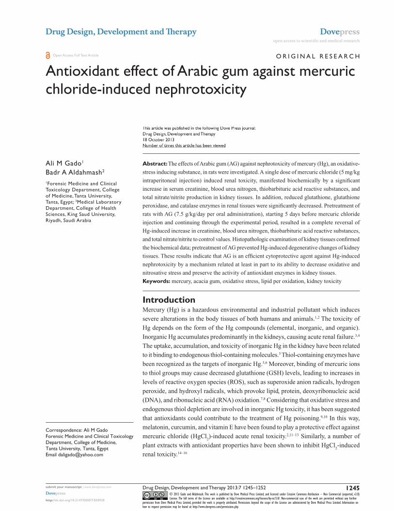
Oxidative and nitrosative stress biomarkersFigures 3 and 4 show the effects of AG, Hg, and their combi-
nation on oxidative stress biomarkers in renal tissues, namely
thiobarbituric acid reactive substance (TBARS) and reduced
GSH, respectively. Hg resulted in a significant decrease in
GSH content to reach only 75% of control group. Also, it
leads to a significant 73% increase in TBARS as compared
to the control group. Combined AG treatment with Hg
significantly decreased TBARS (P,0.001) and restored
GSH level in renal tissues compared to the control values.
Figure 5 shows the effects of AG, Hg, and their combination
on the level of NOx levels in rat renal tissues. Hg resulted in
a significant 91% increase of NOx in renal tissues as com-
pared to the control group. Combined AG treatment with
Hg significantly decreased NOx in renal tissues (P,0.05)
compared to the control values.
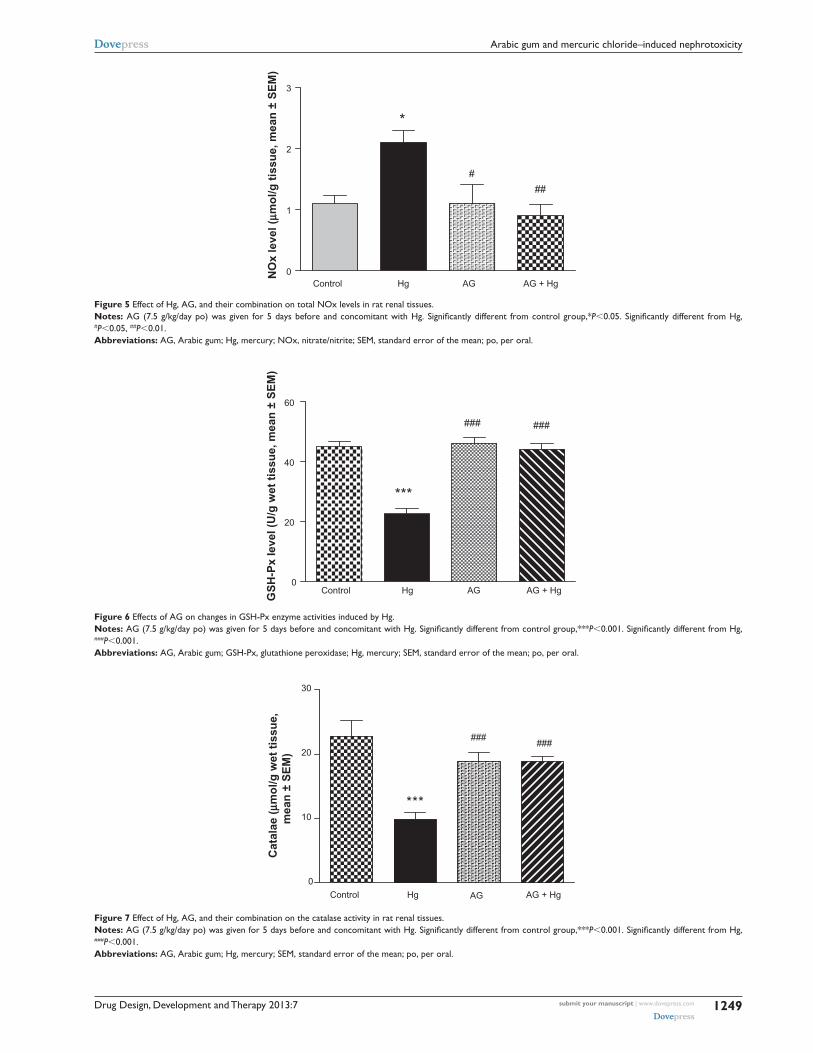
antioxidant enzymes activitiesFigures 6 and 7 show the effects of AG, Hg, and their com-
bination on the activity of antioxidant enzymes GSH-Px
and catalase in renal tissues, respectively. Hg resulted in a
significant decrease in both GSH-Px and catalase enzyme
activities as compared to the control group (P,0.001 and
P,0.001, respectively). Combined AG treatment with Hg
significantly improved both enzymes’ activity (P,0.001) in
renal tissues compared to the control values.
Kidney pathologyPathological examination of the kidneys of control and AG
groups showed normal morphology of the renal parenchyma
Control0.0
0.5
1.0
1.5
**
###
#
Cre
atin
ine
leve
l (m
g%
, mea
n ±
SE
M)
Hg AG AG + Hg
Figure 1 effects of ag on elevated levels of serum creatinine induced by hg. Notes: ag (7.5 g/kg/day po) was given for 5 days before and concomitant with Hg. Significantly different from control group,**P,0.01. Significantly different from Hg, #P,0.05, ###P,0.001.Abbreviations: ag, arabic gum; hg, mercury; seM, standard error of the mean; po, per oral.

Drug Design, Development and Therapy 2013:7submit your manuscript | www.dovepress.com
Dovepress
Dovepress
1248
gado and aldahmash
0
50
100
150
200 ***
### ###
TB
AR
S (
nm
ol/g
kid
ney
, mea
n ±
SE
M)
Control Hg AG AG + Hg
Figure 3 effect of hg, ag, and their combination on the levels of TBars in rat renal tissues. Notes: ag (7.5 g/kg/day po) was given for 5 days before and concomitant with Hg. Significantly different from control group,***P,0.001. Significantly different from Hg, ###P,0.001. Abbreviations: ag, arabic gum; hg, mercury; seM, standard error of the mean; TBars, thiobarbituric acid reactive substances; po, per oral.
0
1
2
3
4
***
# #
Control Hg AG AG + Hg
GS
H (
µmo
l/g k
idn
ey, m
ean
± S
EM
)
Figure 4 effect of hg, ag, and their combination on the levels of reduced gsh in rat renal tissues. Notes: AG (7.5 g/kg/day po) was given for 5 days before and concomitant with Hg. Significantly different from control group,***P,0.001. Significantly different from Hg, #P,0.05. Abbreviations: ag, arabic gum; gsh, glutathione; hg, mercury; seM, standard error of the mean; po, per oral.
0
100
200
300
######
***
Control Hg AG AG + Hg
BU
N le
vel (
mg
%, m
ean
± S
EM
)
Figure 2 effects of ag on elevated levels of BUn induced by hg. Notes: ag (7.5 g/kg/day po) was given for 5 days before and concomitant with Hg. Significantly different from control group,***P,0.001. Significantly different from Hg, ###P,0.001. Abbreviations: ag, arabic gum; BUn, blood urea nitrogen; hg, mercury; seM, standard error of the mean; po, per oral.
Drug Design, Development and Therapy 2013:7 submit your manuscript | www.dovepress.com
Dovepress
Dovepress
1249
arabic gum and mercuric chloride–induced nephrotoxicity
0
1
2
3
*
###
Control Hg AG AG + HgN
Ox
leve
l (µm
ol/g
tis
sue,
mea
n ±
SE
M)
Figure 5 effect of hg, ag, and their combination on total nOx levels in rat renal tissues. Notes: AG (7.5 g/kg/day po) was given for 5 days before and concomitant with Hg. Significantly different from control group,*P,0.05. Significantly different from Hg, #P,0.05, ##P,0.01. Abbreviations: ag, arabic gum; hg, mercury; nOx, nitrate/nitrite; seM, standard error of the mean; po, per oral.
0
20
40
60
***
### ###
Control Hg AG AG + Hg
GS
H-P
x le
vel (
U/g
wet
tis
sue,
mea
n ±
SE
M)
Figure 6 effects of ag on changes in gsh-Px enzyme activities induced by hg. Notes: AG (7.5 g/kg/day po) was given for 5 days before and concomitant with Hg. Significantly different from control group,***P,0.001. Significantly different from Hg, ###P,0.001. Abbreviations: ag, arabic gum; gsh-Px, glutathione peroxidase; hg, mercury; seM, standard error of the mean; po, per oral.
0
10
20
30
### ###
***
Control Hg AG AG + Hg
Cat
alae
(µm
ol/g
wet
tis
sue,
mea
n ±
SE
M)
Figure 7 effect of hg, ag, and their combination on the catalase activity in rat renal tissues. Notes: ag (7.5 g/kg/day po) was given for 5 days before and concomitant with Hg. Significantly different from control group,***P,0.001. Significantly different from Hg, ###P,0.001. Abbreviations: ag, arabic gum; hg, mercury; seM, standard error of the mean; po, per oral.

Drug Design, Development and Therapy 2013:7submit your manuscript | www.dovepress.com
Dovepress
Dovepress
1250
gado and aldahmash
with well-defined glomeruli and tubules with non-significant
changes (Figures 8 and 9). However, animals treated with
Hg showed clear signs of glomerular and tubular necrosis,
interstitial nephritis, and desquamation of the tubular epi-
thelial cells in the renal cortex (Figure 10). Interestingly,
kidney specimens from rats treated with AG and Hg revealed
significant improvement in glomeruli and renal tubules,
evidenced by less vacuolization and more preservation of
tubular histology (Figure 11).
DiscussionMercuric ion, one of strongest thiol-binding agents, increases
the intracellular levels of ROS and induces oxidative stress,
resulting in tissue damage.30–32 Hg toxicity is associated with
superoxide radical generation and GSH reduction.33,34 Our study
demonstrates that the treatment of rats with HgCl2 revealed a
significant enhancement in TBARS levels, indicative of the
generation of lipid peroxides. Enhanced lipid peroxidation
levels were also reported in Hg toxicity by Agarwal et al and
Sener et al.13,35 HgCl2 is known to increase the production of
many ROS, such as superoxide and H2O
2, which cause lipid
peroxidation and subsequently oxidative tissue damage.36–38
Endogenous GSH has a specific role in protecting the body from
Hg toxicity due to its function as a carrier of Hg and its antioxi-
dant properties. GSH binds with Hg and forms a complex that
prevents Hg from binding to cellular proteins and subsequently
causing damage to both enzymes and tissue.39 Hg poisoning
leads to a reduction of intracellular GSH content and decreases
the antioxidant potential of the cells. The present study revealed
that Hg-treated rats showed a significant depletion of serum
GSH levels. Agarwal et al reported a significant reduction of
GSH levels in liver, kidney and brain tissues.12,13
Alterations observed in the activity of GSH-Px and cata-
lase in kidney tissues of Hg-exposed animals indicate the
generation of ROS (O-2 or H
2O
2). Inhibition in the activity
of renal antioxidant enzymes, such as superoxide dismutase
(SOD), GSH-Px, and catalase, in addition to depletion of
GSH levels was also reported earlier.40,41 Enhanced crea-
tinine and BUN levels indicate nephrotoxicity, as reported
by Rumbeiha et al.42 Histopathologic alterations in kidney
tissues after Hg exposure were revealed. Rumbeiha et al,
Al-Saleh et al, Sarwar Alam et al, and Augusti et al have also
reported similar histopathologic alterations in Hg-induced
nephrotoxicity.42–45
Pretreatment with AG attenuated the Hg-induced oxi-
dative damage. Hence, pretreatment with AG significantly
restored the increased TBARS and decreased GSH levels to
the normal values. This could be attributed to the excellent
antioxidant properties of AG.46 These properties seems to
Figure 10 a photomicrograph of the kidney of an mercury-treated rat. The red arrows showing necrotic changes of the rental tubular cells and some tubles contain casts, ×400 magnification.
Figure 8 a photomicrograph of the renal cortex of a control rat. The red arrows showing parenchyma with normal glomeruli and tubules, ×200 magnification.
Figure 9 a photomicrograph of kidney of an arabic gum-treated rat. The red arrows showing cortical tubules and peritubular capillaries with no pathogenic changes, ×200 magnification.

Drug Design, Development and Therapy 2013:7 submit your manuscript | www.dovepress.com
Dovepress
Dovepress
1251
arabic gum and mercuric chloride–induced nephrotoxicity
be due to its ability to scavenge free radicals. The kidneys
are the primary target organ for accumulation and toxic-
ity of inorganic Hg.5 In fact, in as little as l hour, 50% of
an administered dose of inorganic Hg is present in the
kidney.47 Within the kidney, the majority of mercuric ions
were detected in the cortex and outer stripe of the outer
medulla. This finding was expected considering that the
proximal tubule, which spans these two renal zones, is
the primary site of accumulation of mercuric ions.5 The
histopathologic findings in the kidney tissue of Hg-treated
rats include severe diffuse acute necrosis of the tubular epi-
thelium, fragmentation and shedding of tubular epithelium
in the lumina of the renal tubules, and interstitial edema
as a result of tubular leakage. The interaction of Hg with
protein thiol groups is thought to play an important role
in nephrotoxicity induced by Hg at the cellular level.5 The
results of this study indicate that AG improved Hg-induced
nephrotoxicity, manifested by a decrease in both serum
creatinine and urea levels, and minimized the intensity of
the renal lesions. The nephroprotective effect of AG against
many nephrotoxic agents was noted in several reports.18,21,
48–53 The antioxidation induced by AG might be one of the
most likely mechanisms contributing to its beneficial effect
against renal injury. This antioxidant effect of AG was
confirmed previously by in vitro studies, which showed
that AG had a dose-dependent scavenging of superoxide
radicals generated enzymatically and nonenzymatically.54
It could be suggested that AG scavenges Hg free-radical
generation and, in turn, inhibits lipid peroxidation–induced
injury in renal tissues, which has been suggested to protect
renal structure and function. Therefore, the protective effect
is provided by AG on renal tissue through antioxidants as
well as by scavenging free radicals in vivo.
ConclusionIn summary, our data indicate that Hg-induced nephrotoxic-
ity is related to lipid peroxidation. Coadministration of AG
provided protection against HG-induced nephrotoxicity, pos-
sibly by inhibiting the free radical mediated process. These
protective effects of AG on renal injury induced by Hg might
have a considerable impact on developing clinically feasible
strategies to treat patients with toxin-induced renal failure.
DisclosureThe authors report no conflicts of interest in this work.
References 1. Mahboob M, Shireen KF, Atkinson A, Khan AT. Lipid peroxidation
and antioxidant enzyme activity in different organs of mice exposed to low level of mercury. J Environ Sci Health B. 2001;36(5):687–697.
2. Sener G, Sehirli AO, Avanoglu-Dülger G. Melatonin protects against mercury (ll)-induced oxidative tissue damage in rats. Pharmacol Toxicol. 2003;93(6):290–296.
3. Emanuelli T, Rocha JB, Pereira ME, et al. Effect of mercuric chloride intoxication and dimercaprol treatment on delta-aminolevulinate dehy-dratase from brain, liver and kidney of adult mice. Pharmacol Toxicol. 1996;79(3):136–143.
4. Tanaka-Kagawa T, Suzuki M, Naganuma A, Yamanaka N, Imura N. Strain difference in sensitivity of mice to renal toxicity of inorganic mercury. J Pharmacol Exp Ther. 1998;285(1):335–341.
5. Zalups RK. Molecular interactions with mercury in the kidney. Pharmacol Rev. 2000;52(1):113–143.
6. Nogueira CW, Soares FA, Nascimento PC, Muller D, Rocha JB. 2,3- Dimercaptopropane-1-sulfonic acid and meso-2,3-dimercaptosuccinic acid increase mercury- and cadmium-induced inhibition of delta- aminolevulinate dehydratase. Toxicology. 2003;184(2–3):85–95.
7. Li Z, Wu J, Deleo CJ. RNA damage and surveillance under oxidative stress. IUBMB Life. 2006;58(10):581–588.
8. Clarkson TW. The toxicology of mercury. Crit Rev Clin Lab Sci. 1997;34(4):369–403.
9. Patrick L. Mercury toxicity and antioxidants. Part 1: Role of glutathione and alpha-lipoic acid in the treatment of mercury toxicity. Altern Med Rev. 2002;7(6):456–471.
10. Pillai A, Gupta S. Antioxidant enzyme activity and lipid peroxidation in liver of female rats co-exposed to lead and cadmium: effects of vitamin E and Mn2+. Free Radic Res. 2005;39(7):707–712.
11. Nava M, Romero F, Quiroz Y, Parra G, Bonet L, Rodríguez-Iturbe B. Melatonin attenuates acute renal failure and oxidative stress induced by mercuric chloride in rats. Am J Physiol Renal Physiol. 2000;279(5): F910–F918.
12. Agarwal R, Goel SK, Behari JR. Detoxification and antioxidant effects of curcumin in rats experimentally exposed to mercury. J Appl Toxicol. 2010;30(5):457–468.
13. Agarwal R, Goel SK, Chandra R, Behari JR. Role of vitamin E in preventing acute mercury toxicity in rat. Environ Toxicol Pharmacol. 2010;29(1):70–78.
14. Ahn CB, Song CH, Kim WH, Kim YK. Effects of Juglans sinensis Dode extract and antioxidant on mercury chloride-induced acute renal failure in rabbits. J Ethnopharmacol. 2002;82(1):45–49.
15. Oda SS, El-Ashmawy IM. Adverse effects of the anabolic steroid, boldenone undecylenate, on reproductive functions of male rabbits. Int J Exp Pathol. 2012;93(3):172–178.
16. Sarwar Alam M, Kaur G, Jabbar Z, Javed K, Athar M. Eruca sativa seeds possess antioxidant activity and exert a protective effect on mercuric chlo-ride induced renal toxicity. Food Chem Toxicol. 2007;45(6):910–920.
Figure 11 a photomicrograph of the kidney of an arabic gum- and mercury-treated rat. The red arrows showing insignificant tubular epithelial changes in the form of cloudy swelling, ×200 magnification.

Drug Design, Development and Therapy
Publish your work in this journal
Submit your manuscript here: http://www.dovepress.com/drug-design-development-and-therapy-journal
Drug Design, Development and Therapy is an international, peer-reviewed open-access journal that spans the spectrum of drug design and development through to clinical applications. Clinical outcomes, patient safety, and programs for the development and effective, safe, and sustained use of medicines are a feature of the journal, which
has also been accepted for indexing on PubMed Central. The manu-script management system is completely online and includes a very quick and fair peer-review system, which is all easy to use. Visit http://www.dovepress.com/testimonials.php to read real quotes from published authors.
Drug Design, Development and Therapy 2013:7submit your manuscript | www.dovepress.com
Dovepress
Dovepress
Dovepress
1252
gado and aldahmash
17. Rehan A, Johnson KJ, Kunkel RG, Wiggins RC. Role of oxygen radicals in phorbol myristate acetate-induced glomerular injury. Kidney Int. 1985;27(3):503–511.
18. Al-Majed AA, Mostafa AM, Al-Rikabi AC, Al-Shabanah OA. Protective effects of oral Arabic gum administration on gentamicin-induced nephrotoxicity in rats. Pharmacol Res. 2002;46(5):445–451.
19. Ramsammy L, Ling KY, Josepovitz C, Levine R, Kaloyanides GJ. Effect of gentamicin on lipid peroxidation in rat renal cortex. Biochem Pharmacol. 1985;34(21):3895–3900.
20. Salahudeen AK, Clark EC, Nath KA. Hydrogen peroxide-induced renal injury. A protective role for pyruvate in vitro and in vivo. J Clin Invest. 1991;88(6):1886–1893.
21. Al-Majed AA, Abd-Allah AR, Al-Rikabi AC, Al-Shabanah OA, Mostafa AM. Effect of oral administration of Arabic gum on cisplatin-induced nephrotoxicity in rats. J Biochem Mol Toxicol. 2003;17(3): 146–153.
22. Augusti PR, Conterato GM, Somacal S, et al. Effect of lycopene on nephrotoxicity induced by mercuric chloride in rats. Basic Clin Pharmacol Toxicol. 2007;100(6):398–402.
23. Bonsnes RW, Taussky HN. On the colorimetric determination of creatinine by the Jaffe reaction. J Biol Chem. 1945;158:581–591.
24. Hallett CJ, Cook JG. Reduced nicotinamide adenine dinucleotide-coupled reaction for emergency blood urea estimation. Clin Chim Acta. 1971;35(1):33–37.
25. Ellman GL. Tissue sulfahydryl groups. Arch Biochem Biophys. 1959; 82(1):70–77.
26. Ohkawa H, Ohish N, Yagi K. Assay for lipid peroxides in animal tissues by thiobarbituric acid. Anal Biochem. 1979;95(2):351–358.
27. Kraus RJ, Ganther HE. Reaction of cyanide with glutathione peroxidase. Biochem Biophys Res Commun. 1980;96(3):1116–1122.
28. Higgins CP, Baehner RL, McCallister J, Boxer LA. Polymorphonuclear leukocyte species differences in the disposal of hydrogen peroxide (H2O2). Proc Soc Exp Biol Med. 1978;158(3):478–481.
29. Miranda KM, Espey MG, Wink DA. A rapid, simple spectrophotometric method for simultaneous detection of nitrate and nitrite. Nitric Oxide. 2001;5(1):62–71.
30. Zahir F, Rizwi SJ, Haq SK, Khan RH. Low dose mercury toxicity and human health. Environ Toxicol Pharmacol. 2005;20(2):351–360.
31. Hussain S, Atkinson A, Thompson SJ, Khan AT. Accumulation of mercury and its effect on antioxidant enzymes in brain, liver, and kidneys of mice. J Environ Sci Health B. 1999;34(4):645–660.
32. Reus IS, Bando I, Andrés D, Cascales M. Relationship between expression of HSP70 and metallothionein and oxidative stress during mercury chloride induced acute liver injury in rats. J Biochem Mol Toxicol. 2003;17(3):161–168.
33. Girardi G, Elías MM. Mercuric chloride effects on rat renal redox enzymes activities: SOD protection. Free Radic Biol Med. 1995;18(1):61–66.
34. Miura K, Naganuma A, Himeno S, Imura N. Mercury toxicity. In: Goyer RA, Cherian MG, editors. Toxicology of Metals: Biochemical Aspects. Berlin: Springer-Verlag; 1995:163–187.
35. Sener G, Sehirli O, Tozan A, Velioğlu-Ovunç A, Gedik N, Omurtag GZ. Ginkgo biloba extract protects against mercury(II)-induced oxidative tissue damage in rats. Food Chem Toxicol. 2007;45(4):543–550.
36. Miller DM, Lund BO, Woods JS. Reactivity of Hg(II) with superoxide: evidence for the catalytic dismutation of superoxide by Hg(II). J Biochem Toxicol. 1991;6(4):293–298.
37. Huang YL, Cheng SL, Lin TH. Lipid peroxidation in rats administrated with mercuric chloride. Biol Trace Elem Res. 1996;52(2):193–206.
38. Linden A, Gülden M, Martin HJ, Maser E, Seibert H. Peroxide-induced cell death and lipid peroxidation in C6 glioma cells. Toxicol In Vitro. 2008;22(5):1371–1376.
39. Kromidas L, Trombetta LD, Jamall IS. The protective effects of glutathione against methylmercury cytotoxicity. Toxicol Lett. 1990;51(1):67–80.
40. Agarwal R, Raisuddin S, Tewari S, Goel SK, Raizada RB, Behari JR. Evaluation of comparative effect of pre- and posttreatment of selenium on mercury-induced oxidative stress, histological alterations, and metallothionein mRNA expression in rats. J Biochem Mol Toxicol. 2010;24(2):123–135.
41. Gstraunthaler G, Pfaller W, Kotanko P. Glutathione depletion and in vitro lipid peroxidation in mercury or maleate induced acute renal failure. Biochem Pharmacol. 1983;32(19):2969–2972.
42. Rumbeiha WK, Fitzgerald SD, Braselton WE, Roth RA, Kaneene JB. Potentiation of mercury-induced nephrotoxicity by endotoxin in the Sprague-Dawley rat. Toxicology. 2000;149(2–3):75–87.
43. Al-Saleh I, El-Doush I, Shinwari N, Al-Baradei R, Khogali F, Al-Amodi M. Does low mercury containing skin-lightening cream (fair and lovely) affect the kidney, liver, and brain of female mice? Cutan Ocul Toxicol. 2005;24(1):11–29.
44. Sarwar Alam M, Kaur G, Jabbar Z, Javed K, Athar M. Eruca sativa seeds possess antioxidant activity and exert a protective effect on mercuric chlo-ride induced renal toxicity. Food Chem Toxicol. 2007;45(6):910–920.
45. Augusti PR, Conterato GM, Somacal S, et al. Effect of lycopene on nephrotoxicity induced by mercuric chloride in rats. Basic Clin Pharmacol Toxicol. 2007;100(6):398–402.
46. Ali BH. Does gum Arabic have an antioxidant action in rat kidney? Ren Fail. 2004;26(1):1–3.
47. Zalups RK. Early aspects of the intrarenal distribution of mercury after the intravenous administration of mercuric chloride. Toxicology. 1993;79(3):215–228.
48. Mahmoud MF, Diaai AA, Ahmed F. Evaluation of the efficacy of ginger, Arabic gum, and Boswellia in acute and chronic renal failure. Ren Fail. 2012;34(1):73–82.
49. Ali BH, Al-Husseni I, Beegam S, et al. Effect of gum Arabic on oxida-tive stress and inflammation in adenine-induced chronic renal failure in rats. PLoS ONE. 2013;8(2):e55242.
50. Ali BH, Ziada A, Al Husseni I, Beegam S, Al-Ruqaishi B, Nemmar A. Effect of Acacia gum on blood pressure in rats with adenine-induced chronic renal failure. Phytomedicine. 2011;18(13):1176–1180.
51. Ali BH, Al-Salam S, Al Husseni I, et al. Effects of Gum Arabic in rats with adenine-induced chronic renal failure. Exp Biol Med (Maywood). 2010;235(3):373–382.
52. Ali AA, Ali KE, Fadlalla AE, Khalid KE. The effects of gum Arabic oral treatment on the metabolic profile of chronic renal failure patients under regular haemodialysis in Central Sudan. Nat Prod Res. 2008;22(1): 12–21.
53. Nasir O, Umbach AT, Rexhepaj R, et al. Effects of gum Arabic (Acacia senegal) on renal function in diabetic mice. Kidney Blood Press Res. 2012;35(5):365–372.
54. Abd-Allah AR, Al-Majed AA, Mostafa AM, Al-Shabanah OA, Din AG, Nagi MN. Protective effect of Arabic gum against cardiotoxicity induced by doxorubicin in mice: a possible mechanism of protection. J Biochem Mol Toxicol. 2002;16(5):254–259.


![Appendix M. Included Studies - IUPUIScholarWorks …327(7414):523-8. [Rec#: 50928] INCLUDE: considered for evidence table Abratt R and Viljoen G. Assessment of quality of life by clinicians—experience](https://img.dokumen.tips/doc/110x75/5b2bae347f8b9aa6198b80cb/appendix-m-included-studies-iupuischolarworks-3277414523-8-rec-50928.jpg)


![xr20m1172 120 101813 - Exar 4 B y te T X F IF O 6 4 B yte R X F IF O C h a n n e l 1 I2 C / S P I In te rfa ce G P IO [7:0] R X A R T S A # C T S A # C rysta l O sc / B u ffe r A 1](https://img.dokumen.tips/doc/110x75/5b1976717f8b9a46258c9789/xr20m1172-120-101813-exar-4-b-y-te-t-x-f-if-o-6-4-b-yte-r-x-f-if-o-c-h-a-n-n-e.jpg)