Embed Size (px)
Citation preview

Received: 13 August 2017 Revised: 8 October 2017 Accepted: 11October 2017
DOI: 10.1002/humu.23361
MUTAT I ON UPDAT E
DCCmutation update: Congenital mirrormovements, isolatedagenesis of the corpus callosum, and developmental split brainsyndrome
Ashley P. L. Marsh1,2 Timothy J. Edwards3,4 Charles Galea5 HelenM. Cooper3
Elizabeth C. Engle6,7,8,9,10,11 Saumya S. Jamuar6,7,8,12,13 AurélieMéneret14,15
Marie-LaureMoutard16,17,18 Caroline Nava14,19 Agnès Rastetter14
Gail Robinson20 Guy Rouleau21,22 Emmanuel Roze14,15 Megan Spencer-Smith23,24
Oriane Trouillard14 Thierry Billette de Villemeur16,17,25,26
Christopher A.Walsh6,7,8,9,11,12 TimothyW. Yu6,11,12 IRC5 Consortium∗
Delphine Heron17,19 Elliott H. Sherr27 Linda J. Richards3,28
Christel Depienne14,19,29,30 Richard J. Leventer2,31,32 Paul J. Lockhart1,2
1Bruce LefroyCentre forGeneticHealth Research,MurdochChildren's Research Institute, Royal Children'sHospital, Parkville, Victoria, Australia
2Department of Paediatrics, University ofMelbourne, Parkville, Victoria, Australia
3QueenslandBrain Institute, TheUniversity ofQueensland, St Lucia, Brisbane, Australia
4Faculty ofMedicine, TheUniversity ofQueensland, Herston, Brisbane, Australia
5DrugDelivery, Disposition andDynamics (D4),Monash Institute of Pharmaceutical Sciences,MonashUniversity, Parkville, Victoria, Australia
6Division ofGenetics andGenomics, BostonChildren'sHospital, Boston,Massachusetts
7MantonCenter forOrphanDisease Research, BostonChildren'sHospital, Boston,Massachusetts
8HowardHughesMedical Institute, BostonChildren'sHospital, Boston,Massachusetts
9Department ofNeurology, BostonChildren'sHospital andHarvardMedical School, Boston,Massachusetts
10Department ofOphthalmology, BostonChildren'sHospital andHarvardMedical School, Boston,Massachusetts
11Program inMedical and PopulationGenetics, Broad Institute ofMassachusetts Institute of Technology (MIT) andHarvard, Cambridge,Massachusetts
12Department of Pediatrics, HarvardMedical School, Boston,Massachusetts
13Department of Paediatrics, KKWomen's andChildren'sHospital, Paediatric AcademicClinical Programme,Duke–NUSMedical School, Singapore, Singapore
14INSERM,U1127, CNRSUMR7225, SorbonneUniversités, UPMCUniv Paris 06UMRS1127, Institut duCerveau et de laMoelle épinière, ICM, Paris, France
15Département deNeurologie, AP-HP,Hôpital de la Pitié-Salpêtrière, Paris, France
16Service deNeuropédiatrie, AP-HP,Hôpital Trousseau, Paris, France
17UPMC,GRCConCer-LD, SorbonneUniversité, Paris, France
18Centre de référence “Neurogénétique”, Paris, France
19Département deGénétique, AP-HP,Hôpital de la Pitié-Salpêtrière, Paris, France
20NeuropsychologyResearchUnit, School of Psychology, TheUniversity ofQueensland, Brisbane,Queensland, Australia
21Department ofNeurology andNeurosurgery,McGill UniversityHealthCenter,Montreal, Quebec, Canada
22Montreal Neurological Institute andHospital,McGill University,Montréal, Quebec, Canada
23Clinical Sciences,MurdochChildren's Research Institute, Parkville, Victoria, Australia
24School of Psychological Sciences andMonash Institute of Cognitive andClinical Neurosciences,MonashUniversity, ClaytonCampus, Clayton, Victoria, Australia
25Centre deRéférence “déficiences intellectuelles de causes rares”, Paris, France
26INSERMU1141, Paris, France
27Department ofNeurology, UCSFBenioff Children'sHospital, San Francisco, California
28TheUniversity ofQueensland, School of Biomedical Sciences, St Lucia, Brisbane, Australia
HumanMutation. 2018;39:23–39. c© 2017Wiley Periodicals, Inc. 23wileyonlinelibrary.com/journal/humu

24 MARSH ET AL.
29Département deMédicine translationnelle etNeurogénétique, IGBMC,CNRSUMR7104, INSERMU964, Université de Strasbourg, Illkirch, France
30Laboratoires de génétique, Institut de génétiquemédicale d'Alsace, HôpitauxUniversitaires de Strasbourg, Strasbourg, France
31NeuroscienceResearchGroup,MurdochChildren's Research Institute, Parkville, Victoria, Australia
32Department ofNeurology, University ofMelbourne, Royal Children'sHospital, Parkville, Victoria, Australia
Correspondence
AshleyP.L.Marsh,BruceLefroyCentre for
GeneticHealthResearch,MurdochChildren's
Research Institute, RoyalChildren'sHospital,
Parkville, Victoria,Australia.
Email: [email protected]
∗Membersof the InternationalResearch
Consortium for theCorpusCallosumand
CerebralConnectivity (IRC5) are listed in the
Appendix.
Funding information
ContractGrant Sponsors:NationalHealth and
MedicalResearchCouncil (NHMRC)Australia
(GNT1059666,GNT1126153,GNT1032364);
Campbell EdwardsTrust;Victorian
Government'sOperational InfrastructureSup-
portProgram;AustralianGovernmentNHMRC
IRIISS;BostonChildren'sHospital;National
InstitutesofHealth IDDRC (1U54HD090255);
AustralianPostgraduateAward;University of
QueenslandResearchScholarship; Thierry and
AnnickDesmarest Foundation.
AbstractThe deleted in colorectal cancer (DCC) gene encodes the netrin-1 (NTN1) receptor DCC, a trans-
membrane protein required for the guidance of commissural axons. GermlineDCCmutations dis-
rupt the development of predominantly commissural tracts in the central nervous system (CNS)
and cause a spectrum of neurological disorders. Monoallelic, missense, and predicted loss-of-
functionDCCmutations cause congenitalmirrormovements, isolated agenesis of the corpus callo-
sum (ACC), or both. Biallelic, predicted loss-of-functionDCCmutations cause developmental split
brain syndrome (DSBS). Although the underlyingmolecularmechanisms leading to disease remain
poorly understood, they are thought to stem from reduced or perturbedNTN1 signaling. Here, we
review the 26 reported DCC mutations associated with abnormal CNS development in humans,
including 14 missense and 12 predicted loss-of-function mutations, and discuss their associated
clinical characteristics and diagnostic features.We provide an update on the observed genotype–
phenotype relationships of congenital mirror movements, isolated ACC and DSBS, and correlate
this to our current understanding of the biological function of DCC in the development of the
CNS. All mutations and their associated phenotypes were deposited into a locus-specific LOVD
(https://databases.lovd.nl/shared/genes/DCC).
K EYWORDS
ACC, agenesis of the corpus callosum, axon guidance, DCC, developmental split brain syndrome,
horizontal gaze palsy with progressive scoliosis, mirror movements, mutation, Netrin-1, NTN1
1 BACKGROUND
Deleted in colorectal cancer (DCC) (MIM# 120470) encodes the DCC
netrin-1 (NTN1) receptor protein, an evolutionarily conserved, single-
pass transmembrane glycoprotein belonging to the immunoglobu-
lin (Ig) superfamily of cell adhesion molecules (Fearon et al., 1990;
Keino-Masu et al., 1996). DCC is located at 18q21.2 and spans
chr18:52,340,172–53,535,903 (GRCh38/hg38) (Hedrick et al., 1994).
The canonical DCC transcript consists of 29 exons that encode a
159 kDa (1,447 amino acid) protein. DCC is a putative dependence
receptor originally thought to function as a tumor suppressor gene,
although this role has not been proven in vivo and remains contro-
versial (Bin et al., 2015; Fearon et al., 1990; Llambi, Causeret, Bloch-
Gallego, &Mehlen, 2001;Williams et al., 2006).More recently, the role
of DCC in the development of the central nervous system (CNS) was
established (Fazeli et al., 1997; Finger et al., 2002; Jamuar et al., 2017;
Keino-Masu et al., 1996;Marsh, et al., 2017; Srour et al., 2010).
DCC is expressed by commissural axons and bindsNTN1, a secreted
protein encoded by the NTN1 gene (MIM# 601614), which functions
both locally and diffusely as a bifunctional guidance cue (Blasiak,
Kilinc, & Lee, 2017; Keino-Masu et al., 1996; Kennedy, Serafini, de la
Torre, & Tessier-Lavigne, 1994). Both DCC and NTN1 are expressed
throughout the developing mouse and human brain, with distinct but
complementary spatial and temporal expression patterns (Harter
et al., 2010; Jamuar et al., 2017; Ren et al., 2006; Shu, Valentino,
Seaman, Cooper, & Richards, 2000). DCC is required for the transduc-
tion of NTN1-induced attractive and long-range repulsive signaling in
the coordinated outgrowth and guidance of commissural axons that
cross the anatomical midline of the body (Chan et al., 1996; Hong et al.,
1999; Keino-Masu et al., 1996).
The development of commissural tracts is dependent on neurons
forming synaptic connections with their target cells located on the
opposite side of the CNS. To achieve this, neurons extend commissural
axons tipped with a specialized, motile sensing device called a growth
cone (Tessier-Lavigne & Goodman, 1996). Growth cones express an
array of axon guidance receptors such asDCC. The binding of a chemo-
tactic (diffusible) or haptotactic (substrate-bound) guidance cue to
an axon guidance receptor generates a permissive or non-permissive
axonal outgrowth signal, or an attractive or repulsive directional
response (Raper & Mason, 2010; Tessier-Lavigne & Goodman, 1996).
The signals transduced by axon guidance receptors converge to mod-
ulate the assembly of growth cone filamentous actin and cytoskeletal
microtubules required for directing axon outgrowth and guidance
toward or away from a guiding cell population (Kahn & Baas, 2016;
Luo, 2002).
Monoallelic and biallelic germline DCC mutations disrupt commis-
sural axon guidance (Jamuar et al., 2017; Marsh, et al., 2017; Srour
et al., 2010; Welniarz et al., 2017b). These disruptions impair the nor-
mal development and function of tracts such as the corticospinal tract
(CST) and corpus callosum (CC). Monoallelic DCC mutations cause
congenital mirror movements (MMs; MIM# 157600) in association
with abnormal midline crossing of the CST, isolated agenesis of the
MARSH ET AL. 25
CC (iACC; MIM# 217990), or both (Marsh et al., 2017; Srour et al.,
2010). Alternatively, biallelicDCCmutations leading to predicted loss-
of-function (LoF) cause developmental split brain syndrome (DSBS;
MIM#617542), amore complex syndrome associatedwith agenesis of
the corpus callosum (ACC) aswell aswidespread failureof commissural
tracts throughout the rest of the CNS, with or without MMs (Jamuar
et al., 2017).
2 VARIANT DATABASE
We created a new repository for all reported disease-associated
DCC sequence variants using the Leiden Open Variation Database
(LOVD 3.0: https://databases.lovd.nl/shared/genes/DCC) (Fokkema
et al., 2011). The International Research Consortium for the Cor-
pus Callosum and Cerebral Connectivity (IRC5, https://www.irc5.org)
encourages users to register and submit data, including clinical and
neuroimaging phenotypic data.
3 VARIANT NOMENCLATURE
The nomenclature for DNA and protein sequence variants adheres
to the guidelines of the Human Genome Variation Society (den
Dunnen & Antonarakis, 2000). Variant descriptions were based
on the following Genbank reference sequences: NG_013341.2,
NM_005215.3, and NP_005206.2. We utilized theMutalyzer program
(https://mutalyzer.nl/) to validate variant descriptions. At the time of
publication, the DCC Locus Reference Genomic sequence (LRG_1107)
was under curation and pending approval (MacArthur et al., 2014).
4 VARIANTS
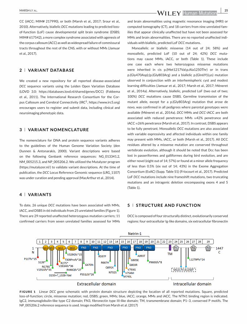
To date, 26 unique DCC mutations have been associated with MMs,
iACC, andDSBS in 66 individuals from25 unrelated families (Figure 1).
There are 29 reported unaffected heterozygous mutation carriers; 11
confirmed carriers from seven unrelated families assessed for MMs
and brain abnormalities using magnetic resonance imaging (MRI) or
computed tomography (CT), and 18 carriers from nine unrelated fam-
ilies that appear clinically unaffected but have not been assessed for
MMs and brain abnormalities. There are no reported unaffected indi-
viduals with biallelic, predicted LoFDCCmutations.
Monoallelic or biallelic missense (14 out of 24; 58%) and
monoallelic, predicted LoF (10 out of 24; 42%) DCC muta-
tions may cause MMs, iACC, or both (Table 1). These include
one case each where two heterozygous missense mutations
were inherited in cis p.(Met1217Val;p.Ala1250Thr) or in trans
p.(Gly470Asp);(p.(Gly803Arg) and a biallelic p.(Gln691Lys) mutation
observed in conjunction with an interhemispheric cyst and modest
learning difficulties (Jamuar et al., 2017; Marsh et al., 2017; Méneret
et al., 2014a). Alternatively, biallelic, predicted LoF (two out of two;
100%) DCC mutations cause DSBS. Germline transmission of the
mutant allele, except for a p.(Gly803Arg) mutation that arose de
novo, was confirmed in all pedigrees where parental genotypes were
available (Méneret et al., 2014a). DCC-MMs and DCC-iACC are both
associated with reduced penetrance: MMs ≈42% penetrance and
iACC≈26%penetrance (Marsh et al., 2017). In contrast, DSBS appears
to be fully penetrant. Monoallelic DCC mutations are also associated
with variable expressivity and affected individuals within one family
may present with MMs, iACC, or both (Marsh et al., 2017). All DCC
residues altered by a missense mutation are conserved throughout
vertebrate evolution, although it should be noted that Dcc has been
lost in passeriformes and galliformes during bird evolution, and are
either novel (eight out of 14; 57%) or found at a minor allele frequency
of less than 0.5% (six out of 14; 43%) in the Exome Aggregation
Consortium (ExAC) (Supp. Table S1) (Friocourt et al., 2017). Predicted
LoF DCCmutations include nine frameshift mutations, two truncating
mutations and an intragenic deletion encompassing exons 4 and 5
(Table 1).
5 STRUCTURE AND FUNCTION
DCC is composedof four structurally distinct, evolutionarily conserved
regions: four extracellular Ig-like domains, six extracellular fibronectin
F IGURE 1 Linear DCC gene schematic with protein domain structure depicting the location of all reported mutations. Square, predictedloss-of-function; circle, missense mutation; red, DSBS; green, MMs; blue, iACC; orange, MMs and iACC. The NTN1 binding region is indicated.IgC2, immunoglobulin-like type C2 domain; FN3, fibronectin type III-like domain; TM, transmembrane domain; P1–3, conserved P motifs. TheNP_005206.2 reference sequence is used. Imagemodified fromMarsh et al. (2017)

26 MARSH ET AL.
TABLE1
Overviewofallreported
DCCmutationslin
kedto
MMs,iACC,andDSB
S
Fam
ilyPhen
otype
No.of
affected
Sexof
affected
Variant
iden
tifica-
tion
Inheritance
(transm
ission)
Allele
Exon/
intron
cDNA
Protein
Protein
domain
ExA
CdbSN
PReferen
ce
1DSB
S∆2
2M
Hom.+
SEQ
Germlin
e(both)*
Bi.
1c.31_91
+7622del
p.(P
ro11
Thrfs*15)
––
–Jamuar
etal.(2017)
2MMs
22M
SEQ
Germlin
e(paternal)
Mono.
2c.377C>A
p.(Ser126* )
IgC2-1
––
Mén
eret
etal.(2014a)
3MMs†
11M
SEQ
Germlin
e(m
aternal)
Mono.
3c.527A>G
p.(A
sn176Ser)
IgC2-2
5/121228
rs138724679
Mén
eret
etal.(2014a)
4MMs†
44M
SEQ
Germlin
e(either)*
Mono.
3c.571dupG
p.(V
al191Glyfs*35)
IgC2-2
––
Sharafad
dinzadeh
etal.
(2008)
Srouret
al.(2010)
5MMs
11F
SEQ
Unkn
own
(unkn
own)
Mono.
3i_5i
c.(697+1_698-1)_
(985+1_986-1)
del
p.(A
sp233+?_
Leu328-?del)
IgC2-3
––
Borgheresietal.(2010)
Mén
eret
etal.(2014a)
6DSB
S∆1
1F
SEQ
Germlin
e(both)
Bi.
4c.788_794del
p.(V
al263Alafs*36)
IgC2-3
––
Jamuar
etal.(2017)
7cA
CCor
MMs±
pACC†
73M:4F
SEQ
Germlin
e(either)*
Mono.
4c.823C>T
p.(A
rg275* )
IgC2-3
––
Mén
eret
etal.(2014a)
Marsh
etal.(2017)
Welniarzet
al.(2017b)
8MM
±pACC
31M:2F
SEQ
Germlin
e(either)
Mono.
4c.823C>T
p.(A
rg275* )
IgC2-3
––
Mén
eret
etal.(2014a)
Men
eret
etal.(2015)
Welniarzet
al.(2017b)
9cA
CCor
pACC±
MMs†
55F
Linkage
+ES+
SEQ
Germlin
e(either)
Mono.
5c.925delA
p.(T
hr309
Profs*26)
IgC2-3
––
Marsh
etal.(2017)
10
MMs†
11
9M:2F
Linkage
+SE
QGermlin
e(either)
Mono.
6i
c.1140+1G>A
p.(V
al329
Glyfs*15)
IgC2-3-IgC
2-4
linker
––
Srouret
al.(2009)
Srouret
al.(2010)
11
MMs†
11M
SEQ
Germlin
e(m
aternal)
Mono.
8c.1336_1337
insA
GCC
p.(A
rg446
Glnfs*27)
FN3-1
––
Mén
eret
etal.(2014a)
12
MMs†
11F
SEQ
Germlin
e(m
aternal);
denovo(in
trans)
Bi.
8;16
c.1409G>A;
c.2407G>A
p.(G
ly470Asp);
p.(G
ly803Arg)
FN3-1;FN3-4
385/121006;-
rs141813053/-
Mén
eret
etal.(2014a)
13
MMs
43M:1F
Linkage
+ES+
SEQ
Germlin
e(m
aternal)*
Mono.
10
c.1652delC
p.(P
ro551
Leufs* 26)
FN3-2
––
Fran
zet
al.(2015)
(Continues)

MARSH ET AL. 27
TABLE1
(Continued
)
Fam
ilyPhen
otype
No.of
affected
Sexof
affected
Variant
iden
tifica-
tion
Inheritance
(transm
ission)
Allele
Exon/
intron
cDNA
Protein
Protein
domain
ExA
CdbSN
PReferen
ce
14
cACC†
11F
SEQ
Germlin
e(paternal)
Mono.
11
c.1790G>C
p.(A
rg597Pro)
FN3-2
––
Marsh
etal.(2017)
15
MMs†
11M
SEQ
Germlin
e(unkn
own)
Mono.
13
c.2000G>A
p.(A
rg667His)
FN3-3
194/121316
rs200099519
Mén
eret
etal.(2014a)
16
cACC
11M
SEQ
Unkn
own
(unkn
own)
Bi.
14
c.2071C>A
p.(G
ln691Lys)
FN3-3
––
Grieb
elet
al.(1995)
Jamuar
etal.(2017)
17
MMs
11F
SEQ
Unkn
own
(unkn
own)
Mono.
14
c.2105A>G
p.(A
sn702Ser)
FN3-3
372/121354
rs35691189
Djarm
ati-Westenberger
etal.(2011)
Mén
eret
etal.(2014a)
18
cACC†
11M
SEQ
Germlin
e(paternal)
Mono.
15
c.2227A>T
p.(M
et743Leu)
FN3-4
–rs199651452
Marsh
etal.(2017)
19
cACC†
21M:1F
SEQ
Germlin
e(paternal)
Mono.
15
c.2260G>A
p.(V
al754Met)
FN3-4
19/121284
–Marsh
etal.(2017)
20
cACCan
dMMs†
43M:1F
Linkage
+ES+
SEQ
Germlin
e(m
aternal)*
Mono.
16
c.2378T>G
p.(V
al793Gly)
FN3-4
––
Marsh
etal.(2017)
21
pACC
and/or
MMs†
31M:2F
SEQ
Germlin
e(m
aternal)
Mono.
16
c.2414G>A
p.(G
ly805Glu)
FN3-4
––
Marsh
etal.(2017)
22
cACC
11F
SEQ
Unkn
own
(unkn
own)
Mono.
17
c.2677G>A
p.(A
la893Thr)
FN3-5
––
Marsh
etal.(2017)
23
MMs
33F
SEQ
Germlin
e(m
aternal)*
Mono.
19
c.2873
_2877dup
p.(P
ro960
Glyfs*8)
FN3-6
––
Mén
eret
etal.(2014a)
24
cACC†
11F
SEQ
Germlin
e(in
cis)
(maternal)*
Mono.
25;26
c.3649A>G;
c.3748G>A
p.(M
et1217Val);
p.(A
la1250Thr)
P1-P2lin
ker
-;2/121388
–Marsh
etal.(2017)
25
MMs
42M:2F
SEQ
Germlin
e(either)*
Mono.
26
c.3836_3837del
p.(Leu
1279
Profs*24)
P1-P2lin
ker
––
Cincottaet
al.(2002)
Dep
ienneet
al.(2012)
DSB
S,developmen
talsplit
brainsyndrome;cA
CC,complete
isolatedagen
esisofthecorpuscallo
sum;pACC,partialisolatedagen
esisofthecorpuscallo
sum;M
Ms,mirrormovem
ents;∆
,unaffected
carrier;†,incom-
plete
pen
etrance;E
S,exomesequen
cing;SE
Q,directsequen
cingofDCC;H
om.;homozygo
sity
mapping;*,inferred
tran
smissionofmutantallele;B
i.,biallelic;M
ono.,monoallelic;IgC
2,immunoglobulin
-liketypeC2
domain;FN3,fibronectintypeIII-likedomain;ExA
C,ExomeAggregationConsortium;dbSN
P,dbSN
Preference
SNPiden
tificationnumber.TheNM_005215.3an
dNP_005206.2reference
sequen
cesareused.

28 MARSH ET AL.
F IGURE 2 A: Schematic drawing of DCCwith protein domain struc-ture depicting the location of the NTN1 binding sites (BS) 0, 1, and 2.B: Structure of NTN1 protein depicting the location of its DCC bind-ing sites BS0, BS1, and BS2. C: NTN1 residues (colored spheres) thatbind the 4th and 5th FN3 domains of DCC (yellow and green rib-bons, respectively) are conserved throughout evolution. Conservedresidues are colored red, non-conserved residues are colored blue.IgC2, immunoglobulin-like type C2 domain; FN3, fibronectin type III-like domain; P1–3, conserved P motifs; NTR, netrin-like domain; LE,laminin-type epidermal growth factor-like domain; LN, laminin-likedomain
type III-like (FN3) domains, a transmembrane domain, and an intracel-
lular domainwith three conservedPmotifs (Supp. Figure S1) (Kolodziej
et al., 1996). Each region of DCC appears to have a unique functional
role required for the transduction of NTN1 signaling.
The distal N-terminal region of DCC contains four Ig-like domains
and three inter-domain linkers that together comprise 371amino acids
(residues 46–417). These domains fold into a horseshoe-like configu-
ration that appears necessary for DCC to transduce NTN1-signaling
(Chen et al., 2013). The proximal N-terminal region of DCC contains
six FN3 domains and five inter-domain linkers that together comprise
612 amino acids (residues 429–1041). Regionswithin the 4th, 5th, and
6th FN3 domains bind NTN1 (Bennett et al., 2002; Finci et al., 2014;
Geisbrecht, Dowd, Barfield, Longo, & Leahy, 2003; Xu, et al., 2014).
This extracellular binding brings two DCC proteins into close prox-
imity, enabling their intracellular domains to homodimerize and initi-
ate the recruitment of multi-protein complexes required to transduce
NTN1 signaling (Mille et al., 2009; Stein, Zou, Poo, & Tessier-Lavigne,
2001).
The DCC ligand, NTN1, is an extracellular protein belonging to
the laminin superfamily and is composed of three structurally distinct
regions: a laminin-like domain (LN), three laminin-type epidermal
growth factor-like domains (LE) and a netrin-like domain (NTR)
(Serafini et al., 1994). NTN1 has three distinct DCC-binding sites
(binding site 1, 2, and 0; BS1, BS2, and BS0) (Figure 2). BS1 is located
on the third LE of NTN1 and interacts exclusively with the 5th FN3
domain of DCC. This site functions as a DCC-specific binding site
and is stabilized by several hydrophobic interactions supported by
a surrounding network of hydrogen bonds (Figure 3A) (Finci et al.,
2014). BS2 is located on the first and second LE domains of NTN1 and
interacts with a distinct region of the 5th FN3 domain as well as the
N-terminal region of the 6th FN3domain ofDCC. This site functions as
a generic binding site that can interact with receptors other than DCC
(Finci et al., 2014). BS2 is predominantly stabilized by a group of sul-
fate and chloride anions that neutralize positively charged patches on
NTN1 and DCC residues located at the binding interface (Figure 3B)
(Finci et al., 2014). The adaptable nature of BS2 allows binding to the
1st and 2nd Ig-like domains of members of the unc-5 netrin receptor
(UNC5; MIM# 603610) family of repulsive axon guidance proteins,
which appear to have a higher affinity for BS2 relative to DCC (Finci
et al., 2014; Geisbrecht et al., 2003; Hong et al., 1999; Leonardo
et al., 1997). As a result, UNC5 are able to outcompete DCC at BS2
and switch an attractive growth cone response (mediated via NTN1-
induced DCC homodimerization) to a repulsive response (mediated
via NTN1-induced DCC and UNC5 heterodimerization) (Finci et al.,
2014; Grandin et al., 2016; Hong et al., 1999). In contrast to BS1 and
BS2, BS0 is located on the LN of NTN1 and interacts with the 4th FN3
domain of DCC (Xu et al., 2014). This NTN1 protein is different to the
one simultaneously engaged at BS1 and BS2, but binds the same DCC
protein occupied at BS1 (Figure 3C). The binding of DCC by a second
NTN1 protein at BS0 is thought to generate a signaling cluster impor-
tant for the transduction of a directional response toward or away
from a guiding cell population (Finci, Zhang,Meijers, &Wang, 2015).
Taken together, these studies indicate that NTN1 forms two dis-
tinct binding clusters through BS0, BS1, and BS2 and that these clus-
ters underpin its bifunctionality as an attractive and repulsive guid-
ance cue. The first cluster involves the 4th and 5th FN3 domains of
one DCC protein at BS0 and BS1, while the second cluster involves
the 5th and 6th FN3 domains of a second DCC protein or the 1st
and 2nd Ig-like domains of an UNC5 family member protein at BS2.
However, it should be noted that the actual in vivo binding struc-
ture may differ as these studies utilized a recombinant NTN1 pro-
tein lacking its NTR domain and different truncated DCC fragments,
both lacking the NTN1 binding region in its entirety (Finci et al., 2014;
Xu et al., 2014).
The importance of the 4th, 5th, and 6th FN3 domains are high-
lighted by the significant enrichment of DCC missense mutations
linked to MMs and/or iACC within these NTN1 binding regions
compared with missense variants located in these domains in the
ExAC (Figure 4A and B) (Marsh et al., 2017). This enrichment is most
significant formonoallelic, missensemutations linked to iACC (five out
of nine; 56%) and suggests that missensemutations may cause disease
through different, perhaps more complex mechanisms compared with
those mutations leading to predicted LoF and haploinsufficiency. This
may be due to reduced or perturbed NTN1-induced axon repulsion
and/or attraction caused by dysfunction of the mutant DCC protein in
both a mutant-mutant homodimer and wild-type–mutant homodimer
complex. Additional studies are required to determine whether these
missense alterations cause complete or partial LoF or perhaps gain-
of-function as antimorphic, hypermorphic, or neomorphic mutations.
For instance, it remains to be determined whether mutant DCC
proteins are stably expressed and trafficked to the plasma membrane
or aberrantly sequestered in the cytoplasm likemutant NTN1 proteins

MARSH ET AL. 29
F IGURE 3 A: Binding site 1 (BS1) andBS2 of theNTN1/DCC complex (PDB ID: 4URT).B: BS2 of theNTN1/DCCcomplex (PDB ID: 4URT).C: BS0of the NTN1/DCC complex (PDB ID: 4PLO). Structure of NTN1 (transparent molecular surface) bound to DCC FN3 domains (orange ribbons). Theexpansion of the red dotted box region shows the NTN1 (magenta sticks) and DCC (gray or white sticks) involved in binding at BS1, BS2, or BS0.Hydrogen bonds are represented by dotted black lines. FN3, fibronectin type III-like domain; PDB ID, Protein Data Bank identification

30 MARSH ET AL.
F IGURE 4 The fibronectin type III-like (FN3) domains are required for NTN1 binding and DCC function. A: Enrichment of DCCmissense muta-tions linked toMMsand/or iACCwithin theNTN1binding region. Structure ofNTN1/DCCcomplex.DCC is depicted as anorange ribbonandNTN1as awhite solvent accessible surface.DCCmissensemutations locatedwithin the FN3-4 and FN3-5 domains are represented as blue spheres (PDBID: 4PLO). B: Expansion of the binding interface (red dotted box in A) with NTN1 residues colored blue and critical DCC residues representedas purple sticks. Mutation of V793 and G805 to Gly and Glu, respectively, is associated with both MMs and iACC. C: Sequence alignment of theDCC FN3 domains. Residues highlighted in green are predicted to be directly involved in NTN1 binding by Finci et al. 2014 and/or Xu et al. (2014).Residues in yellowaremissensemutations associatedwithMMsand/or iACC.Residues in blue aremissensemutations associatedwithMMsand/oriACC that are also predicted to be directly involved in NTN1 binding. The NP_005206.2 reference sequence is used. PDB ID, Protein Data Bankidentification. Imagemodified fromMarsh et al. (2017)
(Méneret et al., 2017). However, absence of the DSBS phenotype in an
individual with a biallelic p.(Gln691Lys) missense mutation suggests
that these mutant proteins may retain some residual function (Jamuar
et al., 2017).
The DCC transmembrane domain comprises 24 amino acids
(residues 1,098–1,122). To bind NTN1, DCC must correctly parti-
tion its transmembrane domain within the cell plasma membrane.
The palmitoylation and localization of the transmembrane domain
to lipid rafts (cholesterol- and sphingolipid-enriched membrane sub-
domains of the plasma membrane) has been demonstrated to be
necessary for DCC to transduce NTN1-induced axon guidance and
outgrowth (Guirland, Suzuki, Kojima, Lu, & Zheng, 2004; Herincs
et al., 2005).
The intracellular, C-terminal region of DCC contains 324 amino
acids (residues 1,123–1,447) and is required to transduce NTN1
signals into appropriate growth cone responses.Within theC-terminal
region there are three conserved P motifs referred to as P1 (encoded
by residues 1,150−1,167), P2 (encoded by residues 1,330−1,365),and P3 (encoded by residues 1,424−1,447) (Kolodziej et al., 1996).These P motifs are involved in numerous protein–protein interactions
and phosphorylation cascades required for the intracellular trans-
duction of NTN1 signaling (Fothergill et al., 2014; Hong et al., 1999;
Li et al., 2004; Ren et al., 2004; Stein & Tessier-Lavigne, 2001). For
example, the NTN1-induced, intracellular homodimerization of the
DCC P3 motifs initiates downstream signaling events that generate
an attractive growth cone response (Li et al., 2002; Li et al., 2004; Liu
et al., 2004; Meriane et al., 2004; Ren et al., 2004; Ren et al., 2008;
Stein et al., 2001). Alternatively, the NTN1-induced, intracellular
heterodimerization of the DCC P1 motif and the DCC binding (DB)
motif of UNC5 family members initiates downstream signaling events
that generate a long-range, repulsive growth cone response (Hong
et al., 1999; Keleman & Dickson, 2001; Li, Aurandt, Jürgensen, Rao,
& Guan, 2006; Norris, Sundararajan, Morgan, Roberts, & Lundquist,
2014).
6 GENOTYPE–PHENOTYPE CORRELATION
Monoallelic frameshift and nonsense DCC mutations are predicted to
result in haploinsufficiency via nonsense mediated mRNA decay or
rapid turnover of the truncated protein, resulting in reduced NTN1
binding (Srour et al., 2010). This is in contrast to DCC missense muta-
tions that likely generate a full-length protein within the cell (Finci
et al., 2014; Meriane et al., 2004). DCC proteins carrying a missense
mutationmay be non-functional, hypomorphic, or may have an altered
function that imparts adominant effect throughperturbedNTN1bind-
ing, receptormultimerization and/or downstream signaling (Finci et al.,
2014; Meriane et al., 2004). Monoallelic, truncating DCC mutations

MARSH ET AL. 31
predicted to result in haploinsufficiency are frequently associatedwith
MMs (eight out of 10; 80%) or MMs with iACC (two out of 10; 20%).
DCCmissensemutations are less frequently associatedwithMMs (five
out of 14; 36%). Instead, the majority of these mutations are associ-
ated with iACC (seven out of 14; 50%) or MMs with iACC (two out
of 14; 14%). This disparity may be due to developmental differences
between the CC and the subcerebrally projecting neurons that com-
prise the CST. The neuronal populations that comprise each of these
tracts are molecularly distinct and uniquely employ DCC and NTN1
signaling to reach their contralateral targets (Fothergill et al., 2014;
Molyneaux, Arlotta, Menezes, &Macklis, 2007). Therefore, a missense
or predicated LoFDCCmutation may differentially affect commissural
versus subcerebral axon trajectories, thus leading to iACC, MMs or
both (Marsh et al., 2017). Although these observations do support the
hypothesis that distinct disease mechanisms contribute to DCC-MMs
or DCC-iACC, additional functional investigations are required to con-
firm this correlation. These investigations could utilize in vivo tech-
niques such as in utero electroporation or conditional gene knockout
to study the effect loss or dysfunction of Dcc has on the outgrowth
and guidance of axons extending from specific populations of projec-
tion neurons during development.
Monoallelic, missense mutations located within the NTN1 binding
sites of DCC appear to be strongly associated with iACC (Marsh et al.,
2017). Indeed, the majority of DCC mutations located within the 4th
and 5th FN3 domains are associated with iACC (five out of six; 83%).
Interestingly, the p.(Val793Gly) and p.(Gly805Glu)missensemutations
located directly within theNTN1 binding interface are associatedwith
a more severe phenotype that features both iACC and MMs (Marsh
et al., 2017). Previous studies have shown a differential disruption to
NTN1-induced axon attraction and repulsion dependent on the loca-
tion of the mutated DCC residue within the NTN1 binding interface
(Figure 4C) (Finci et al., 2014). Consequently, DCC mutations located
within the NTN1 binding interface may cause more severe disrup-
tions to commissural axon outgrowth and guidance that result in a
phenotype that features both iACC and MMs. Overall, it appears that
the majority of missense iACC mutations localize to the FN3 domains
(seven out of nine; 78%), highlighting the importance of these domains
to DCC function and callosal development.
The observed structure of pedigrees with monoallelic, predicted
LoF DCC mutations has suggested a sex-bias in MMs and iACC phe-
notype expression (Marsh et al., 2017; Sharafaddinzadeh, Bavarsad,
Yousefkhah, & Aleali, 2008; Srour et al., 2009). Indeed, within these
pedigrees a significant proportion of males displayed MMs, while
iACC was almost exclusively detected in females (Marsh et al., 2017).
Sex differences in callosal morphology have previously been linked
with testosterone levels during prenatal brain development (Chura
et al., 2010; Moffat, Hampson, Wickett, Vernon, & Lee, 1997). Con-
sistent with these observations, RNAseq and RT-qPCR analyses have
detected a significant dose-dependent increase in DCC expression in
testosterone-treated neural stem cells derived fromhuman embryonic
stem cells (Marsh et al., 2017). These findings suggest that iACC may
occur when the expression of DCC falls below a threshold level during
CCdevelopment, aswould occurmore commonly in females. However,
given the incomplete penetrance observed in both sexes, the pheno-
type must also be influenced by additional genetic, epigenetic, and/or
environment factors (Marsh et al., 2017). Furthermore, the sex-specific
nature of this phenotype imbalance remains to be verified as reciprocal
analyses utilizing estrogens have not been reported. Additional in vivo
investigations are required to evaluate the potential impact the hor-
monal context during brain development has on DCC expression and
whether it influences the expressivity of the MMs and iACC pheno-
type.
7 BIOLOGICAL SIGNIFICANCE
Commissural axons form connections between the left and right
sides of the brain that are required for the transfer and integration
of information generated by sensory, motor, and associative neurons.
These connections are defined anatomically as either commissures
or decussations. While commissures cross the midline to form pre-
dominantly homotopic connections, decussations descend or ascend
along the neuraxis before crossing the midline to form connections
with different neuronal populations. For instance, axons within the
CC arch over the lateral ventricles in a segregated, orderly manner
to form predominantly homotopic connections with their target cells
in the contralateral cerebral hemisphere (Zhou et al., 2013). These
connections function by mediating higher-order brain processes and
facilitating the integration of sensory and motor information between
the two cerebral hemispheres (Paul et al., 2007).Within the CST, axons
cross the midline at the medulla to form the pyramidal decussation
between the brainstem and the spinal cord (Nathan, Smith, & Deacon,
1990). The connections formed by axons within the crossed CST
facilitate the voluntary and unilateral movement of the contralateral
distal limbs (Welniarz,Dusart, &Roze, 2017a). Several additional tracts
within thebrainstemaredisrupted inDSBS (Jamuar et al., 2017). These
appear to include the decussation of the superior cerebellar peduncles
(also referred to as the commissure of Wernekinck) that connects
the cerebellum to the midbrain, and the commissural tract linking
the lateral abducens and medial occulomotor nerves, required for
conjugate horizontal eyemovement (Jamuar et al., 2017; Jen, 2008).
The function of each commissural tract is underpinned by the
transcriptionally distinct neuronal subtypes that comprise it (Harada,
Sato, & Nakamura, 2016; Molyneaux et al., 2007). For instance, the
CC is formed from pioneer axons extending from neurons in the
cingulate cortex and follower axons extending from callosal projection
neurons locatedwithin layers II/III andVaof the cerebral cortex (Fame,
MacDonald, & Macklis, 2011). In comparison, the CST is comprised
of an array of axons extending from subcerebrally projecting neurons
situated within layer Vb of the cortex (Dum & Strick, 1991; Harwell
et al., 2012). Notably, the tracts associated with the brainstem origi-
nate from diverse nuclei. For example, the transverse pontocerebellar
projections arise from pontine nuclei while cranial nerves originate
from nuclei located throughout the midbrain, pons and medulla. To
reach their contralateral targets, these neuronal subtypes extend
axons that express a unique repertoire of receptors, including DCC, on
their growth cones (Dickson & Zou, 2010; Evans and Bashaw, 2010).
The tightly regulated, spatiotemporal expression of axon guidance

32 MARSH ET AL.
receptors and their ligands control the development of tracts such as
theCCandCST (Chedotal &Richards, 2010). As highlighted below, our
understanding of the development of these tracts and the associated
role of DCC and NTN1 signaling in humans has been predominantly
informed by the study of these biological processes inmice.
The formation of theCC is complex anddependent on the execution
of several developmental steps. Briefly, these include midline pattern-
ing and remodeling of the interhemispheric fissure by astroglia to
create a permissible substrate for commissural axons to cross themid-
line (Gobius et al., 2016;Hayhurst,Gore, Tessier-Lavigne,&McConnell,
2008; Okada, et al., 2008; Silver, Lorenz, Wahlsten, & Coughlin, 1982);
secretion of guidance cues by populations of glia and neurons situated
around the midline (Shu & Richards, 2001; Shu, Li, Keller, & Richards,
2003; Unni et al., 2012); pioneering of the callosal tract by axons
extending from neurons in the cingulate cortex (Rash & Richards,
2001); and fasciculation of callosal axons with pioneer axons as they
grow toward and across the midline (Koester & O'Leary, 1994; Rash
& Richards, 2001). Dcc and Ntn1 are required for the normal develop-
ment of the CC and their loss or dysfunction in mice is associated with
complete ACC (Fazeli et al., 1997; Finger et al., 2002; Serafini et al.,
1996).While both pre-crossing pioneer and callosal axons expressDcc,
only the former axonal population appears to be attracted toward the
midline by Ntn1 (Fothergill et al., 2014; Shu et al., 2000). Instead, it has
been shown that pre-crossing callosal axons utilize Dcc and Ntn1 sig-
naling to attenuate the repulsive signaling of Robo1 and Slit2 (another
axon guidance receptor-ligand pair) to approach and cross the midline
(Fothergill et al., 2014; Stein & Tessier-Lavigne, 2001). The expression
of Dcc in post-crossing callosal axons is subsequently downregulated,
thereby restoring the repulsive effect of Robo1 and Slit2 signaling
to direct axons away from the midline and toward their targets in
the contralateral cerebral hemisphere (Fothergill et al., 2014; Shu
et al., 2000).
The subcerebrally projecting neurons that comprise theCSTmainly
originate from theprimarymotor andpremotor areas of the cortex and
follow a stereotyped route to innervate their targets in the spinal cord
(Dum & Strick, 1991). During development, axons from these projec-
tion neurons converge and descend through the internal capsule and
cerebral peduncles of the midbrain before entering the ventral brain-
stem where they form the medullary pyramids. Most CST projections
then cross the midline in the caudal region of the medulla to form the
pyramidal decussation, before projecting inferiorly in the spinal cord
to synapse with lower motor neurons or interneurons in the ventral
spinal cord to facilitate voluntary movement of the limbs (Welniarz
et al., 2017a). Dcc is required for the normal development of the CST
in mice and Dcc dysfunction is associated with a failure of the CST
to cross the midline at the level of the pyramidal decussation (Finger
et al., 2002; Welniarz et al., 2017b). However, Dcc does not appear to
be expressed in the brainstem CST of mice as it is significantly down-
regulated in the distal portion of the axon that grows beyond the inter-
nal capsule (Finger et al., 2002; Shu et al., 2000). Interestingly, the role
of Dcc in the development of the CST at themidlinewas reported to be
non-cell autonomous as conditional knockout ofDcc in the cortex (and
therefore theCST) causedACCbut not a failure of theCST to cross the
midline (Welniarz et al., 2017b).
In the brainstem and spinal cord, Dcc is present on commissural
axons, which are guided circumferentially from the dorsal roof plate
toward the ventral floor plate (where midline crossing occurs) in
response to Ntn1 (Dominici et al., 2017; Holley, 1982; Tessier-Lavigne,
Placzek, Lumsden, Dodd, & Jessell, 1988; Varadarajan, et al., 2017;
Yee, Simon, Tessier-Lavigne, & O'Leary, 1999). The attractive response
of these commissural axons to Ntn1 is regulated by Dcc in partner-
ship with Robo3, another axon guidance receptor required for normal
development of the hindbrain and spinal cord (Chen, Gore, Long,Ma, &
Tessier-Lavigne, 2008;Marillat et al., 2004; Sabatier et al., 2004; Zelina
et al., 2014). Pioneering studies utilizing embryonic chick spinal cord
demonstrated that this axonal navigation is reliant on a ventral–dorsal
gradient of Ntn1 diffused from the floor plate (Kennedy et al., 1994).
Subsequent studies in mice also detected a similar graded distribution
ofNtn1extending fromthe floor plate, supporting theobservation that
Ntn1 functions as a long-range diffusible chemoattractant (Kennedy,
Wang, Marshall, & Tessier-Lavigne, 2006; Serafini et al., 1996). How-
ever, recent reports have revised this model by demonstrating that
Ntn1 produced by neural progenitors in the ventral ventricular zone
of hindbrain and spinal cord neuroepithelium are essential for com-
missural axon extension toward the ventral midline in embryonic mice
(Dominici et al., 2017; Varadarajan, et al., 2017; Yamauchi et al., 2017).
These reports propose that, in mice, commissural axons are directed
viaDcc-dependent haptotaxis toward the ventral midline byNtn1 pro-
duced and deposited at the pial surface of the hindbrain and spinal
cord, not via a diffusible gradient of Ntn1 emanating from the floor
plate (Dominici et al., 2017; Varadarajan & Butler, 2017; Varadara-
jan et al., 2017; Yamauchi et al., 2017). Therefore, regardless of how
Ntn1 is presented to axons, it is clear that it functions to direct Dcc-
expressing commissural axons toward the ventral midline of the hind-
brain and spinal cord during development.
8 CLINICAL RELEVANCE
MMs are involuntary movements on one side of the body that mirror
voluntarymovements made on the opposite side (Cincotta & Ziemann,
2008). DCC-MMs commonly present in infancy or early childhood and
persist stably into adulthood (Depienne et al., 2011; Franz et al., 2015;
Marsh et al., 2017;Méneret et al., 2014a; Srour et al., 2009; Srour et al.,
2010). DCC-MMs manifest in the fingers and hands but may also be
present in the forearms, toes, and feet in a subset of affected individ-
uals (Supp. Table S2) (Marsh et al., 2017; Méneret et al., 2014a; Srour
et al., 2010).With effort, some affected individuals can partly suppress
these involuntary movements (Srour et al., 2009). Individuals with
DCC-MMs exhibit a range of functional disabilities besides difficulties
in fine bimanual activities. These include fatigue, spontaneous muscle
contractions, and pain in the upper limbs during extended manual
activities such as writing, as well as general clumsiness and compen-
satory maneuvers to inhibit involuntary movements (Depienne et al.,
2011; Marsh et al., 2017; Méneret et al., 2014a; Meneret, Trouillard,
Brochard, & Roze, 2015). As a result,DCC-MMsmay preclude affected
individuals from professions and social activities that demand sus-
tained or complex bimanual coordination. Individuals with DCC-MMs

MARSH ET AL. 33
do not appear to exhibit any additional clinical manifestations and
have a normal developmental outcome (Depienne et al., 2011; Franz
et al., 2015; Marsh et al., 2017; Méneret et al., 2014a; Meneret, et al.,
2015; Sharafaddinzadeh et al., 2008; Srour et al., 2009; Srour et al.,
2010; Welniarz et al., 2017b). However, only a subset of individuals
diagnosed with DCC-MMs are reported to have undergone brain
imaging (12 out of 42; 29%) and formal neuropsychological evaluation
(two out 42; 5%) and therefore the phenotypic spectrum ofDCC-MMs
remains to be defined (Marsh et al., 2017).
DCC-MMs are observed in association with midline axon guidance
defects, evidenced by decreased crossing of descending corticospinal
motor projections at the pyramidal decussation (Marsh et al., 2017;
Welniarz et al., 2017b). This reduction of crossed projections occurs
in conjunction with a relative, reciprocal increase of uncrossed pro-
jections (Marsh et al., 2017; Welniarz et al., 2017b). Concordantly,
unilateral transcortical stimulation of the primary hand motor area
elicits both normal, contralateral and abnormal, ipsilateral motor
evoked potentials in individuals with DCC-MMs (Cincotta et al., 1994;
Srour et al., 2010; Welniarz et al., 2017b). As a result, DCC-MMs
are thought to originate from the bilateral transmission of motor
commands through normally crossed and abnormally uncrossed,
fast-conducting CST projections in the spinal cord (Srour et al., 2010;
Welniarz et al., 2017b;Welniarz, Dusart, Gallea, & Roze, 2015).
Individuals with monoallelic, missense DCC mutations may also
present with iACC, with or without MMs (Marsh et al., 2017). ACC
describes the partial or complete absence of the CC and is character-
ized by the failure of callosal axons to cross themidline. Apart from the
abnormalities typically expected to be associated with ACC, such as
the absence of the hippocampal commissure and cingulate gyrus and
dysmorphic lateral ventricles or colpocephaly, individuals with iACC
do not present with additional brain abnormalities (Cesaretti et al.,
2016; Kuker, Mayrhofer, Mader, Nagele, & Krageloh-Mann, 2003).
Individuals with DCC-iACC may present with complete iACC (14 out
of 19; 74%) or partial iACC (five out of 19; 26%) (Marsh et al., 2017).
Approximately half of these individuals also present with MMs (nine
out of 19; 47%),which suggests thatDCC-iACCmay frequently present
as part of a global disorder of midline crossing (Marsh et al., 2017; Par-
rish, Roessmann, & Levinsohn, 1979; Paul et al., 2007). Other callosal
abnormalities such as hypoplasia (a uniformly thin CC) and/or dyspla-
sia (abnormal CC shape) have not been observed in individuals with
DCC-iACC (Marsh et al., 2017). Individuals with DCC-iACC show no
consistent additional gross brain abnormalities in the posterior com-
missure, ventricular system, cerebral cortex,whitematter, hippocampi,
brainstem, basal ganglia, cerebral and cerebellar peduncles, cerebellar
vermis and hemispheres, optic chiasm, and pituitary gland (Figure 5)
(Marsh et al., 2017). However, the anterior commissure of some DCC-
iACC individuals may be enlarged (Marsh et al., 2017). Enlargement of
the anterior commissure is also observed in a minority of individuals
withACCandhasbeen suggested to represent a compensatorymecha-
nism tomaintain connectivity between the cerebral hemispheres (Barr
and Corballis, 2002; Hetts, Sherr, Chao, Gobuty, & Barkovich, 2006).
The clinical manifestations of DCC-iACC vary, but they are gener-
ally associated with a favorable development outcome with no intel-
lectual disability (Marsh et al., 2017) (Supp. Table S2). Still, individuals
with DCC-iACC do commonly exhibit typical neurobehavioral conse-
quences associated with ACC, such as impairments in emotional and
social functioning in addition to attention, language, visuospatial, and
literacy and numeracy deficits (Marsh et al., 2017; Paul et al., 2007;
Sotiriadis &Makrydimas, 2012).
Biallelic, DCC mutations leading to predicted LoF are associated
with DSBS, a complex syndrome associated with a broad disorganiza-
tion of white-matter tracts throughout the CNS (Jamuar et al., 2017).
The features ofDSBS include absence of all commissures (including the
CC, anterior, and posterior), brainstemdefects (including hypoplasia of
the pons andmidbrain), horizontal gaze palsy and progressive scoliosis
with a variable age of onset (Figure 5).
Several features of DSBS overlap with HGPPS1 (MIM# 607313), a
congenital syndrome caused by biallelic mutations in the axon guid-
ance receptor ROBO3 (MIM# 608630) (Chan et al., 2006; Jen et al.,
2004; Sicotte et al., 2006). Robo3 is a functional, intracellular bind-
ing partner of Dcc, expressed by commissural axons in the brainstem
and spinal cord (Marillat et al., 2004; Sabatier et al., 2004; Zelina et al.,
2014). The overlapping feature of horizontal gaze palsy in DSBS and
HGPPS1 affected individuals appears to originate from hindbrain mid-
line axon guidance defects in tracts that control conjugate horizon-
tal eye movement (Chan et al., 2006; Jamuar et al., 2017; Jen et al.,
2004; Renier et al., 2010; Sicotte et al., 2006). The pathogenesis of pro-
gressive scoliosis in both these disorders is unknown, but may stem
from defective spinal commissural interneurons or abnormal devel-
opment of extrapyramidal projections (Jamuar et al., 2017; Jen et al.,
2004; RabeBernhardt et al., 2012). DiffusionMRI of oneDSBS individ-
ual with a biallelic p.(Val263Alafs*36) mutation revealed several tract
defects, including absenceof thedecussationof the superior cerebellar
peduncles and transverse pontocerebellar projections (Jamuar et al.,
2017). These commissural defects are also observed in individualswith
HGPPS1 (Sicotte et al., 2006). Individuals with DSBS may also present
with MMs, which are associated with the reduced midline crossing
of descending CST projections at the pyramidal decussation (Jamuar
et al., 2017). This is in contrast to individuals with HGPPS1 who do
not display MMs, a feature attributed to an uncrossed CST leading
to reversed lateralization of motor control (Jen et al., 2004). It is cur-
rently unknown why some individuals with DSBS and complete loss of
DCC do not present with MMs. However, like HGPPS1, this may be
due to a complete, rather than partial, failure of the descending CST
to cross the midline. The other major clinical manifestations of DSBS
include intellectual disability, global developmental delay, and hypoto-
nia (Supp. Table S2) (Jamuar et al., 2017). Individuals with DSBS have a
poor developmental outcome compared to individualswithDCC-iACC,
likely attributed to additional brain abnormalities affecting the for-
mation of other commissural tracts (Jamuar et al., 2017; Marsh et al.,
2017).
9 DIAGNOSTIC STRATEGIES
The clinical assessment and diagnosis of MMs is commonly based on
the Woods and Teuber severity scale developed in 1978 (Woods &
Teuber, 1978). The assessment typically consists of three different

34 MARSH ET AL.
F IGURE 5 Axial MRI of control (A) and individuals with complete DCC-iACC (D), partial DCC-iACC (G) and DSBS (J). Midsagittal MRI of con-trol (B) and individuals with complete DCC-iACC (E), partial DCC-iACC with absence of the rostrum and genu (H) and DSBS (K). Coronal MRI ofcontrol (C) and individuals with complete DCC-iACC (F), partial DCC-iACC (I), and DSBS (L). iACC, isolated agenesis of the corpus callosum; DSBS,developmental split brain syndrome. Images A–I adapted fromMarsh et al. (2017). Images J–L adapted from Jamuar et al. (2017)
handmotor tasks. The level ofMMs visible in themirror hand is scored
on a scale between 1—barely discernible repetitive movement and 4—
movement equal to that expected for the intendedhand (Woods&Teu-
ber, 1978). The majority of individuals with DCC-MMs score between
2 (slight but unsustained or stronger, but briefer, repetitive move-
ment) and 3 (strong and sustained repetitive movement). Recently,
accelerometer gloves were utilized to quantitatively evaluate MMs in
a multiplex MMs family with a monoallelic, p.(Pro551Leufs*26) DCC
mutation (Franz et al., 2015). These gloves detect subtle movement,
in contrast to electromyography that measures the electrical activity
produced by skeletal muscle (Franz et al., 2015). Interestingly, the
accelerometer gloves led to the diagnosis of subclinical MMs in two
additional family members initially diagnosed as unaffected carriers
following standardized neurological assessment. The current pen-
etrance of DCC-MMs is approximately 42% (Marsh et al., 2017). In
light of these findings, it appears likely that a proportion of clinically
unaffected mutation carriers have subclinical MMs and that the true
prevalence of DCC-MMs may be higher. It remains to be determined
whether subclinicalDCC-MMsare associatedwith a similar, or perhaps
less severe, failure of the CST to cross themidline.
Brain abnormalities such as ACC can be readily identified utilizing
ultrasonography, CT, and MRI. MRI is considered the gold standard
for the diagnosis of ACC because of its superior sensitivity and ability
to differentiate iACC and complex ACC (ACC with additional brain
abnormalities) (Rapp et al., 2002; Tercanli & Prufer, 2016; Wright,
Sibley, & Baker, 2010). The distinction of the latter is important in a

MARSH ET AL. 35
prenatal setting as individuals with iACC generally have a favorable
developmental outcome compared with those with complex ACC
(D'Antonio et al., 2016; des Portes et al., 2017; Edwards, Sherr,
Barkovich, & Richards, 2014; Sotiriadis & Makrydimas, 2012). Like-
wise, individuals with DCC-iACC have a favorable developmental
outcome while individuals with DSBS have a poor developmental
outcome. The current penetrance of DCC-iACC is approximately 26%
(Marsh et al., 2017). However, brain imaging studies have not been
completed for the majority of affected individuals originating from
DCC-MMs-only families (29 out of 36; 81%). Given itsmild clinical phe-
notype and concomitance with DCC-MMs, it is probable that further
brain imaging studies will identify DCC-iACC in a proportion of these
mutation-positive individuals. Like DCC-MMs, the true prevalence of
DCC-iACC is likely to be higher than current estimates.
Genetic screening for monoallelic DCCmutations is recommended
for individuals presenting with MMs, iACC or both, in the absence of
intellectual disability. Alternatively, screening for biallelic DCC muta-
tions with predicted LoF is recommended for individuals presenting
with characteristic features of DSBS, including horizontal gaze palsy,
ACC, brainstem defects, and progressive scoliosis. Direct sequencing
of coding exons and flanking intronic regions using germline DNA is
advised when screening for DCC mutations. Given the mild clinical
phenotype and high rates of incomplete penetrance associated with
DCC-MMs andDCC-iACC, cascade testing should be considered for all
extended familymembers at risk of inheriting amutant allele.Differen-
tial genetic diagnoses for DCC-MMs include NTN1 and RAD51 (MIM#
179617) (Depienne et al., 2012; Méneret et al., 2014a; Méneret et al.,
2017); for DCC-iACC include CDK5RAP2 (MIM# 608201, although
only one family has been described to date (Jouan et al., 2016)); and
for DSBS include ROBO3 (note: individuals with HGPPS1 do not suf-
fer from intellectual disability and have normal forebrain commissures,
consistentwith the predominant role ofROBO3 in the developing hind-
brain and spinal cord) (Chan et al., 2006; Jen et al., 2004; Sicotte
et al., 2006). However, the low diagnostic yield of genetic screening
studies indicates that additional MMs and iACC disease genes remain
to be identified and therefore mutations in DCC, NTN1, RAD51, and
CDK5RAP2 will not always be identified in individuals with these phe-
notypes (Franz et al., 2015; Jamuar et al., 2017; Marsh et al., 2017;
Méneret et al., 2014a;Méneret et al., 2017;Méneret, et al., 2014b).
10 VARIANTS OF UNKNOWN
SIGNIFICANCE
The classification and clinical interpretation of aDCC sequence variant
is challengedby the variable expressivity ofmonoallelicDCCmutations
and the incomplete penetrance associated with DCC-MMs and DCC-
iACC. It may also be complicated by phenotypic heterogeneity and the
identification of other potential pathogenic variants in an individual.
In this context and in the absence of sufficient evidence to classify a
DCC variant as pathogenic or not, it is recommended to report such a
genetic alteration as a variant of unknown significance (VUS).
To illustrate these issues, we describe individual N-0083-01
with two novel, monoallelic VUS predicted to be damaging by
most in silico tools: a maternally inherited NM_005215.3:c.916G >
A:p.(Gly306Arg) DCC alteration and a de novo NM_001270399.1:c.
1246G > A:p.(Gly416Ser) TUBA1A (MIM# 602529) alteration (Supp.
Table S3) (Jamuar et al., 2017). The individual has complete ACC with
a small pons and small inferior aspect of the midbrain, but no def-
inite cortical malformation as would be expected to result from a
pathogenic TUBA1A mutation (Keays et al., 2007). However, the indi-
vidual does not exhibit themild clinical manifestations associated with
DCC-iACC. Instead, she has global developmental delay and severe
cognitive impairments with significant emotional and behavioral
problems in addition to spastic diplegia, strabismus and cortical visual
impairment (Supp. Table S4). As exemplified by this case, additional
disease modeling and physiologically relevant functional assays are
required to better inform the classification and clinical interpreta-
tion ofDCC sequence variants, particularly for those causing missense
alterations.
11 FUTURE PROSPECTS
Mutations in DCC disrupt the development of predominantly commis-
sural tracts in the CNS and cause a spectrum of neurological disorders
ranging fromMMsand iACCwith a normal or favorable developmental
outcome, to DSBSwith a poor developmental outcome. These findings
are consistentwith previously reported animalmodels and support the
notion that DCC, in partnership with NTN1, functions as a master reg-
ulator of commissural axon guidance at the midline (Fazeli et al., 1997;
Finger et al., 2002; Varadarajan et al., 2017; Welniarz et al., 2017b).
Future investigations into the underlying molecular mechanisms lead-
ing to these disorders will strengthen current genotype–phenotype
correlations. However, insightful correlationswill be dependent on the
multidisciplinary phenotyping of affected individuals, employing stan-
dardized neurological assessment, formal neuropsychological evalua-
tion, and brain imaging studies. Utilization of advanced neuroimaging
modalities, such as diffusion MRI-based tractography and functional
MRI,will also aid our understanding of how the brains of these affected
individuals is wired in the context of DCC dysfunction or LoF during
development. We anticipate the DCC locus-specific LOVD will func-
tion as a central data repository thatwill assist researchers to establish
these important genotype-phenotype correlations.
ACKNOWLEDGMENTS
The authors gratefully acknowledge the participation of the patients
and their families in these studies and the support of the Australian
Disorders of the Corpus Callosum (AusDoCC) organization, as well as
the generous financial support of the Lefroy and Handbury families.
The authors also acknowledge and thank IvoFokkemaand JohanT. den
Dunnen from theLeidenUniversityMedicalCenter for their assistance
in establishing the DCC LOVD.
DISCLOSURE STATEMENT
A.M. received travel funding from Zambon Company and AbbVie
Inc. E.R. has received research support from Merz-Pharma, Orkyn,

36 MARSH ET AL.
Aguettant, IP Santé, Ultragenix, IPSEN, Association Française pour
l'Hémiplégie Alternante, AMADYS; served on scientific advisory
boards for Orkyn, Ultragenix, Retrophin, and Merz Pharma; received
speech honoraria from Orkyn, Aguettant, Merz Pharma, and Ultra-
genix; and received travel funding from the Dystonia Coalition, the
Dystonia Medical Research Foundation, the Movement Disorders
Society, and the European Academy of Neurology. S.S.J. is co-founder
of Global Gene Corporation. T.W.Y. is co-founder of and part-time con-
sultant to Claritas Genomics.
ORCID
Ashley P. L. Marsh http://orcid.org/0000-0001-6049-6931
REFERENCES
Barr, M. S., & Corballis, M. C. (2002). The role of the anterior commissure in
callosal agenesis.Neuropsychology, 16(4), 459–471.
Bennett, K. L., Bradshaw, J., Youngman, T., Rodgers, J., Greenfield, B., Aruffo,
A., & Linsley, P. S. (1997). Deleted in colorectal carcinoma (DCC) binds
heparin via its fifth fibronectin type III domain. Journal of BiologicalChemistry, 272(43), 26940–26946.
Bin, J. M., Han, D., Lai Wing Sun, K., Croteau, L. P., Dumontier, E., Cloutier, J.
F.,… Kennedy, T. E. (2015). Complete loss of netrin-1 results in embry-
onic lethality and severe axon guidance defects without increased neu-
ral cell death. Cell Reports, 12(7), 1099–1106.
Blasiak, A., Kilinc, D., & Lee, G. U. (2017). Neuronal cell bodies remotely reg-
ulate axonal growth response to localizednetrin-1 treatment via second
messenger andDCCdynamics. Frontiers in CellularNeuroscience,10(298)
Cesaretti, C., Nanni, M., Ghi, T., Parazzini, C., Conte, G., Contro, E.,… Righ-
ini, A. (2016). Variability of forebrain commissures in callosal agenesis:
A prenatal MR imaging study. American Journal of Neuroradiology, 37(3),521–527.
Chan, S. S., Zheng, H., Su, M. W., Wilk, R., Killeen, M. T., Hedgecock, E. M.,
& Culotti, J. G. (1996). UNC-40, a C. elegans homolog of DCC (Deleted
in Colorectal Cancer), is required in motile cells responding to UNC-6
netrin cues. Cell, 87(2), 187–195.
Chan, W. M., Traboulsi, E. I., Arthur, B., Friedman, N., Andrews, C., & Engle,
E. C. (2006). Horizontal gaze palsy with progressive scoliosis can result
from compound heterozygous mutations in ROBO3. Journal of MedicalGenetics, 43(3), e11.
Chedotal, A., & Richards, L. J. (2010). Wiring the brain: The biology of
neuronal guidance. Cold Spring Harbor Perspectives in Biology, 2(6),a001917. https://doi.org/10.1101/cshperspect.a001917
Chen, Q., Sun, X., Zhou, X-hH., J-hH, Liu, Wu, J., Zhang, Y., & Wang, J-
hH. (2013). N-terminal horseshoe conformation of DCC is functionally
required for axon guidance and might be shared by other neural recep-
tors. Journal of Cell Science, 126(Pt 1), 186–195.
Chen, Z., Gore, B. B., Long, H., Ma, L., & Tessier-Lavigne, M. (2008). Alterna-
tive splicing of the Robo3 axon guidance receptor governs the midline
switch from attraction to repulsion.Neuron, 58(3), 325–332.
Chura, L. R., Lombardo, M. V., Ashwin, E., Auyeung, B., Chakrabarti, B., Bull-
more, E. T., & Baron-Cohen, S. (2010). Organizational effects of fetal
testosteroneonhumancorpus callosumsizeandasymmetry.Psychoneu-roendocrinology, 35(1), 122–132.
Cincotta, M., Ragazzoni, A., de Scisciolo, G., Pinto, F., Maurri, S., & Barontini,
F. (1994). Abnormal projection of corticospinal tracts in a patient with
congenital mirror movements. Neurophysiologie Clinique, 24(6), 427–434.
Cincotta, M., & Ziemann, U. (2008). Neurophysiology of unimanual motor
control and mirror movements. Clinical Neurophysiology, 119(4), 744–762.
D'Antonio, F., Pagani, G., Familiari, A., Khalil, A., Sagies, T. L., Malinger, G.,
…5 Prefumo, F. (2016). Outcomes associated with isolated agenesis of
the corpus callosum: Ameta-analysis. Pediatrics, 138(3)
den Dunnen, J. T., & Antonarakis, S. E. (2000). Mutation nomenclature
extensions and suggestions to describe complex mutations: A discus-
sion.Human000Mutation, 15(1), 7–12.
Depienne, C., Bouteiller, D., Méneret, A., Billot, S., Groppa, S., Klebe, S.,
… Roze, E. (2012). RAD51 haploinsufficiency causes congenital mirror
movements in humans.American Journal Of HumanGenetics, 90(2), 301–307.
Depienne, C., Cincotta, M., Billot, S., Bouteiller, D., Groppa, S., Brochard, V.,
… Roze, E. (2011). A novel DCC mutation and genetic heterogeneity in
congenital mirrormovements.Neurology, 76(3), 260–264.
des Portes, V., Rolland, A., Velazquez-Dominguez, J., Peyric, E.,
Cordier, M. P., Gaucherand, P., … Guibaud, L. (2017). Out-
come of isolated agenesis of the corpus callosum: A population-
based prospective study. European Journal of Paediatric Neurology.https://doi.org/10.1016/j.ejpn.2017.08.003
Dickson, B. J., & Zou, Y. (2010). Navigating intermediate targets: The ner-
vous system midline. Cold Spring Harbor Perspectives in Biology, 2(8),a002055. https://doi.org/10.1101/cshperspect.a002055
Dominici, C., Moreno-Bravo, J. A., Puiggros, S. R., Rappeneau, Q., Rama, N.,
Vieugue, P., … Chédotal, A. (2017). Floor-plate-derived netrin-1 is dis-
pensable for commissural axon guidance.Nature, 545(7654), 350–354.
Dum, R. P., & Strick, P. L. (1991). The origin of corticospinal projections from
the premotor areas in the frontal lobe. Journal of Neuroscience, 11(3),667–689.
Edwards, T. J., Sherr, E. H., Barkovich, A. J., & Richards, L. J. (2014). Clini-
cal, genetic and imaging findings identify new causes for corpus callo-
sum development syndromes. Brain, 137(Pt 6), 1579–1613.
Evans, T. A., & Bashaw, G. J. (2010). Axon guidance at the midline: Of mice
and flies. Current Opinion in Neurobiology, 20(1), 79–85.
Fame, R. M., MacDonald, J. L., & Macklis, J. D. (2011). Development, spec-
ification, and diversity of callosal projection neurons. Trends in Neuro-sciences, 34(1), 41–50.
Fazeli, A., Dickinson, S. L., Hermiston, M. L., Tighe, R. V., Steen, R. G., Small,
C. G., … Weinberg, R. A. (1997). Phenotype of mice lacking functional
Deleted in colorectal cancer (Dcc) gene.Nature, 386(6627), 796–804.
Fearon, E. R., Cho, K. R., Nigro, J. M., Kern, S. E., Simons, J.W., Ruppert, J. M.,
…, Vogelstein, B. (1990). Identification of a chromosome 18q gene that
is altered in colorectal cancers. Science, 247(4938), 49–56.
Finci, L., Zhang, Y., Meijers, R., &Wang, J. H. (2015). Signaling mechanism of
the netrin-1 receptor DCC in axon guidance. Progress in Biophysics andMolecular Biology, 118(3), 153–160.
Finci, L. I., Kruger, N., Sun, X., Zhang, J., Chegkazi, M., Wu, Y.,…Meijers, R.
(2014). The crystal structure of netrin-1 in complex with DCC reveals
the bifunctionality of netrin-1 as a guidance cue. Neuron, 83(4), 839–849.
Finger, J. H., Bronson, R. T., Harris, B., Johnson, K., Przyborski, S. A., &Acker-
man, S. L. (2002). The netrin 1 receptors Unc5h3 andDcc are necessary
at multiple choice points for the guidance of corticospinal tract axons.
Journal of Neuroscience, 22(23), 10346–10356.
Fokkema, I. F. A.C., Taschner, P. E. M., Schaafsma, G. C. P., Celli, J., Laros, J. F.
J., & den Dunnen, J. T. (2011). LOVD v.2.0: The next generation in gene
variant databases.HumanMutation, 32(5), 557–563.
Fothergill, T., Donahoo, A-L. S., Douglass, A., Zalucki, O., Yuan, J., Shu,
T., … Richards, L. J. (2014). Netrin-DCC signaling regulates corpus

MARSH ET AL. 37
callosum formation through attraction of pioneering axons and bymod-
ulating slit2-mediated repulsion. Cerebral Cortex, 24(5), 1138–1151.
Franz, E. A., Chiaroni-Clarke, R., Woodrow, S., Glendining, K. A., Jasoni, C.
L., Robertson, S. P.,…Markie, D. (2015). Congenital mirror movements:
Phenotypes associated with DCC and RAD51 mutations. Journal of theNeurological Sciences, 351, 140–145.
Friocourt, F., Lafont, A-G., Kress, C., Pain, B.,Manceau,M., Dufour, S., &Ché-
dotal, A. (2017). Recurrent DCC gene losses during bird evolution. Sci-entific Reports, 7, 37569. https://doi.org/10.1038/srep37569
Geisbrecht, B. V., Dowd, K. A., Barfield, R. W., Longo, P. A., & Leahy, D. J.
(2003). Netrin binds discrete subdomains of DCC and UNC5 and medi-
ates interactions between DCC and heparin. Journal of Biological Chem-istry, 278(35), 32561–32568.
Gobius, I., Morcom, L., Suárez, R., Bunt, J., Bukshpun, P., Reardon, W., …Richards Linda, J. (2016). Astroglial-mediated remodeling of the inter-
hemispheric Midline is required for the formation of the corpus callo-
sum. Cell Reports, 17(3), 735–747.
Grandin, M., Meier, M., Delcros, J. G., Nikodemus, D., Reuten, R., Patel, T. R.,
… Stetefeld, J. (2016). Structural decoding of the Netrin-1/UNC5 inter-
action and its therapeutical implications in cancers. Cancer Cell, 29(2),173–185.
Guirland, C., Suzuki, S., Kojima, M., Lu, B., & Zheng, J. Q. (2004). Lipid rafts
mediate chemotropic guidance of nerve growth cones. Neuron, 42(1),51–62.
Harada, H., Sato, T., & Nakamura, H. (2016). Fgf8 signaling for development
of themidbrain and hindbrain. Development,Growth and Differentiation,58(5), 437–445.
Harter, P. N., Bunz, B., Dietz, K., Hoffmann, K., Meyermann, R., & Mittel-
bronn, M. (2010). Spatio-temporal deleted in colorectal cancer (DCC)
and netrin-1 expression in human foetal brain development. Neu-ropathology and Applied Neurobiology, 36(7), 623–635.
Harwell, C. C., Parker, P. R., Gee, S. M., Okada, A., McConnell, S. K., Kreitzer,
A. C., & Kriegstein, A. R. (2012). Sonic hedgehog expression in corticofu-
gal projection neurons directs cortical microcircuit formation. Neuron,73(6), 1116–1126.
Hayhurst, M., Gore, B. B., Tessier-Lavigne, M., & McConnell, S. K. (2008).
Ongoing sonic hedgehog signaling is required for dorsal midline forma-
tion in the developing forebrain.Developmental Neurobiology, 68(1), 83–100.
Hedrick, L., Cho, K. R., Fearon, E. R.,Wu, T. C., Kinzler, K.W., & Vogelstein, B.
(1994). The DCC gene product in cellular differentiation and colorectal
tumorigenesis.Genes and Development, 8(10), 1174–1183.
Herincs, Z., Corset, V., Cahuzac, N., Furne, C., Castellani, V., Hueber, A.
O., & Mehlen, P. (2005). DCC association with lipid rafts is required
for netrin-1-mediated axon guidance. Journal of Cell Science, 118(Pt 8),1687–1692.
Hetts, S. W., Sherr, E. H., Chao, S., Gobuty, S., & Barkovich, A. J. (2006).
Anomalies of the corpus callosum: An MR analysis of the phenotypic
spectrum of associated malformations. American Journal of Roentgenol-ogy, 187(5), 1343–1348.
Holley, J. A. (1982). Early development of the circumferential axonal path-
way in mouse and chick spinal cord. Journal of Comparative Neurology,205(4), 371–382.
Hong, K., Hinck, L., Nishiyama, M., Poo, M. M., Tessier-Lavigne, M., & Stein,
E. (1999). A ligand-gated association between cytoplasmic domains of
UNC5 and DCC family receptors converts netrin-induced growth cone
attraction to repulsion. Cell, 97(7), 927–941.
Jamuar, S. S., Schmitz-Abe, K., D'Gama, A. M., Drottar, M., Chan, W. M.,
Peeva, M., … Yu, T. W. (2017). Biallelic mutations in human DCC
cause developmental split-brain syndrome.Nature Genetics, 49(4), 606–612.
Jen, J. C. (2008). Effects of failure of development of crossing brainstem
pathways on ocular motor control. Progress in Brain Research, 171, 137–141.
Jen, J. C., Chan, W. M., Bosley, T. M., Wan, J., Carr, J. R., Rub, U., …Engle, E. C. (2004). Mutations in a human ROBO gene disrupt hindbrain
axon pathway crossing and morphogenesis. Science, 304(5676), 1509–1513.
Jouan, L., Ouled Amar Bencheikh, B., Daoud, H., Dionne-Laporte, A.,
Dobrzeniecka, S., Spiegelman, D., … Rouleau, G. A. (2016). Exome
sequencing identifies recessive CDK5RAP2 variants in patients with
isolated agenesis of corpus callosum. European Journal of Human Genet-ics, 24(4), 607–610.
Kahn, O. I., & Baas, P. W. (2016). Microtubules and Growth Cones: Motors
Drive the Turn. Trends in Neurosciences, 39(7), 433–440.
Keays, D. A., Tian, G., Poirier, K., Huang, G. J., Siebold, C., Cleak, J.,… Flint, J.
(2007). Mutations in alpha-tubulin cause abnormal neuronal migration
inmice and lissencephaly in humans. Cell, 128(1), 45–57.
Keino-Masu, K., Masu, M., Hinck, L., Leonardo, E. D., Chan, S. S., Culotti, J.
G., & Tessier-Lavigne, M. (1996). Deleted in Colorectal Cancer (DCC)
encodes a netrin receptor. Cell, 87(2), 175–185.
Keleman, K., & Dickson, B. J. (2001). Short- and long-range repulsion by the
Drosophila Unc5 netrin receptor.Neuron, 32(4), 605–617.
Kennedy, T. E., Serafini, T., de la Torre, J. R., & Tessier-Lavigne, M. (1994).
Netrins are diffusible chemotropic factors for commissural axons in the
embryonic spinal cord. Cell, 78(3), 425–435.
Kennedy, T. E., Wang, H., Marshall, W., & Tessier-Lavigne, M. (2006). Axon
guidance by diffusible chemoattractants: A gradient of netrin protein in
the developing spinal cord. The Journal of Neuroscience, 26(34), 8866–8874.
Koester, S. E., & O'Leary, D. D. (1994). Axons of early generated neurons
in cingulate cortex pioneer the corpus callosum. Journal of Neuroscience,14(11 Pt 1), 6608–6620.
Kolodziej, P. A., Timpe, L. C., Mitchell, K. J., Fried, S. R., Goodman, C. S., Jan,
L. Y., & Jan, Y. N. (1996). frazzled encodes a Drosophila member of the
DCC immunoglobulin subfamily and is required forCNSandmotor axon
guidance. Cell, 87(2), 197–204.
Kuker, W., Mayrhofer, H., Mader, I., Nagele, T., & Krageloh-Mann, I.
(2003). Malformations of the midline commissures: MRI findings in dif-
ferent forms of callosal dysgenesis. European Radiology, 13(3), 598–604.
Leonardo, E. D., Hinck, L., Masu, M., Keino-Masu, K., Ackerman, S. L., &
Tessier-Lavigne,M. (1997). VertebratehomologuesofC. elegansUNC-5
are candidate netrin receptors.Nature, 386(6627), 833–838.
Li, W., Aurandt, J., Jürgensen, C., Rao, Y., & Guan, K-L. (2006). FAK and
Src kinases are required for netrin-induced tyrosine phosphorylation of
UNC5. Journal of Cell Science, 119(1), 47–55.
Li, W., Lee, J., Vikis, H. G., Lee, S-H., Liu, G., Aurandt, J.,…Guan, K-L. (2004).
Activation of FAK and Src are receptor-proximal events required for
netrin signaling.Nature Neuroscience, 7(11), 1213–1221.
Li, X., Meriane, M., Triki, I., Shekarabi, M., Kennedy, T. E., Larose, L., &
Lamarche-Vane, N. (2002). The adaptor protein Nck-1 couples the
netrin-1 receptor DCC (deleted in colorectal cancer) to the activation
of the small GTPase Rac1 through an atypical mechanism. The JournalOf Biological Chemistry, 277(40), 37788–37797.
Liu, G., Beggs, H., Jürgensen, C., Park, H-T., Tang, H., Gorski, J., … Rao, Y.
(2004). Netrin requires focal adhesion kinase and Src family kinases
for axon outgrowth and attraction. Nature Neuroscience, 7(11), 1222–1232.
Llambi, F., Causeret, F., Bloch-Gallego, E., &Mehlen, P. (2001). Netrin-1 acts
as a survival factor via its receptors UNC5H and DCC. EMBO Journal,20(11), 2715–2722.

38 MARSH ET AL.
Luo, L. (2002). Actin cytoskeleton regulation in neuronal morphogenesis
and structural plasticity.Annual Review of Cell andDevelopmental Biology,18, 601–635.
MacArthur, J. A., Morales, J., Tully, R. E., Astashyn, A., Gil, L., Bruford, E.
A., … Cunningham, F. (2014). Locus Reference Genomic: Reference
sequences for the reporting of clinically relevant sequence variants.
Nucleic Acids Research, 42(Database issue), D873–8.
Marillat, V., Sabatier, C., Failli, V., Matsunaga, E., Sotelo, C., Tessier-Lavigne,
M., & Chedotal, A. (2004). The slit receptor Rig-1/Robo3 controls mid-
line crossing by hindbrain precerebellar neurons and axons. Neuron,43(1), 69–79.
Marsh, A. P., Heron, D., Edwards, T. J., Quartier, A., Galea, C., Nava, C., …Depienne C (2017). Mutations in DCC cause isolated agenesis of the
corpus callosum with incomplete penetrance. Nature Genetics, 49(4),511–514.
Méneret, A., Depienne, C., Riant, F., Trouillard, O., Bouteiller, D., Cincotta,
M.,…Roze, E. (2014a). Congenital mirrormovements:Mutational anal-
ysis of RAD51 andDCC in 26 cases.Neurology, 82(22), 1999–2002.
Méneret, A., Franz, E. A., Trouillard, O., Oliver, T. C., Zagar, Y., Robertson, S.
P.,…Markie, D. (2017).Mutations in the netrin-1 gene cause congenital
mirror movements. The Journal of Clinical Investigation, 127(11), 3923–3936
Meneret, A., Trouillard, O., Brochard, V., & Roze, E. (2015). Congenital mir-
ror movements caused by a mutation in the DCC gene. Developmen-tal Medicine and Child Neurology, 57(8), 776. https://doi.org/10.1111/dmcn.12810
Méneret, A., Trouillard, O., Vidailhet, M., Depienne, C., & Roze, E. 2014b.
Congenitalmirrormovements:Nomutation inDNAL4 in17 index cases.
Journal of Neurology, 261(10), 2030.
Meriane, M., Tcherkezian, J., Webber, C. A., Danek, E. I., Triki, I., McFarlane,
S.,… Lamarche-Vane, N. (2004). Phosphorylation of DCC by Fyn medi-
ates Netrin-1 signaling in growth cone guidance. The Journal of Cell Biol-ogy, 167(4), 687–698.
Mille, F., Llambi, F., Guix, C., Delloye-Bourgeois, C., Guenebeaud, C., Castro-
Obregon, S., … Mehlen, P. (2009). Interfering with multimerization of
netrin-1 receptors triggers tumor cell death. Cell Death and Differentia-tion, 16(10), 1344–1351.
Moffat, S. D., Hampson, E., Wickett, J. C., Vernon, P. A., & Lee, D. H. (1997).
Testosterone is correlated with regional morphology of the human cor-
pus callosum. Brain Research, 767(2), 297–304.
Molyneaux, B. J., Arlotta, P., Menezes, J. R. L., & Macklis, J. D. (2007). Neu-
ronal subtype specification in the cerebral cortex.Nature ReviewsNeuro-science, 8(6), 427–437.
Nathan, P. W., Smith, M. C., & Deacon, P. (1990). The corticospinal tracts in
man. Course and location of fibres at different segmental levels. Brain,113(Pt 2), 303–324.
Norris, A. D., Sundararajan, L., Morgan, D. E., Roberts, Z. J., & Lundquist,
E. A. (2014). The UNC-6/Netrin receptors UNC-40/DCC and UNC-5
inhibit growth cone filopodial protrusion via UNC-73/Trio, Rac-
like GTPases and UNC-33/CRMP. Development, 141(22), 4395–
4405.
Okada, T., Okumura, Y., Motoyama, J., & Ogawa, M. (2008). FGF8 signaling
patterns the telencephalic midline by regulating putative key factors of
midline development.Developmental Biology, 320(1), 92–101.
Parrish, M. L., Roessmann, U., & Levinsohn, M. W. (1979). Agenesis of the
corpus callosum: A study of the frequency of associatedmalformations.
Annals in Neurology, 6(4), 349–354.
Paul, L. K., Brown,W. S., Adolphs, R., Tyszka, J.M., Richards, L. J., Mukherjee,
P., & Sherr, E. H. (2007). Agenesis of the corpus callosum:Genetic, devel-
opmental and functional aspects of connectivity. Nature Reviews Neuro-science, 8(4), 287–299.
Rabe Bernhardt, N., Memic, F., Gezelius, H., Thiebes, A. L., Vallstedt, A., &
Kullander, K. (2012). DCC mediated axon guidance of spinal interneu-
rons is essential for normal locomotor central pattern generator func-
tion.Developmental Biology, 366(2), 279–289.
Raper, J., & Mason, C. (2010). Cellular strategies of axonal pathfind-
ing. Cold Spring Harbor Perspectives in Biology, 2(9), a001933.
https://doi.org/10.1101/cshperspect.a001933
Rapp, B., Perrotin, F., Marret, H., Sembely-Taveau, C., Lansac, J., & Body, G.
(2002). [Value of fetal cerebral magnetic resonance imaging for the pre-
natal diagnosis and prognosis of corpus callosum agenesis). Journal deGynécologie, Obstétrique et Biologie de la Reproduction (Paris), 31(2 Pt 1),
173–182.
Rash, B. G., & Richards, L. J. (2001). A role for cingulate pioneering axons in
the development of the corpus callosum. Journal of Comparative Neurol-ogy, 434(2), 147–157.
Ren, T., Anderson, A., Shen, W. B., Huang, H., Plachez, C., Zhang, J., …Richards, L. J. (2006). Imaging, anatomical, and molecular analysis of
callosal formation in the developing human fetal brain. The AnatomicalRecord. Part A, Discoveries in Molecular, Cellular, and Evolutionary Biology,288(2), 191–204.
Ren, X. R., Hong, Y., Feng, Z., Yang, H.M.,Mei, L., & Xiong,W. C. (2008). Tyro-
sine phosphorylation of netrin receptors in netrin-1 signaling.Neurosig-nals, 16(2-3), 235–245.
Ren, X. R., Ming, G. L., Xie, Y., Hong, Y., Sun, D.M., Zhao, Z. Q.,…Xiong,W. C.
(2004). Focal adhesion kinase in netrin-1 signaling.Nature Neuroscience,7(11), 1204–1212.
Renier, N., Schonewille, M., Giraudet, F., Badura, A., Tessier-Lavigne, M.,
Avan, P., … Chedotal, A. (2010). Genetic dissection of the func-
tion of hindbrain axonal commissures. PLoS Biology, 8(3), e1000325.https://doi.org/10.1371/journal.pbio.1000325
Sabatier, C., Plump, A. S., Le, M., Brose, K., Tamada, A., Murakami, F., …Tessier-Lavigne, M. (2004). The divergent Robo family protein Rig-
1/Robo3 is a negative regulator of slit responsiveness required for mid-
line crossing by commissural axons. Cell, 117(2), 157–169.
Serafini, T., Colamarino, S. A., Leonardo, E. D., Wang, H., Beddington, R.,
Skarnes, W. C., & Tessier-Lavigne, M. (1996). Netrin-1 is required for
commissural axon guidance in the developing vertebrate nervous sys-
tem. Cell, 87(6), 1001–1014.
Serafini, T., Kennedy, T. E., Galko,M. J., Mirzayan, C., Jessell, T. M., & Tessier-
Lavigne, M. (1994). The netrins define a family of axon outgrowth-
promoting proteins homologous to C. elegans UNC-6. Cell, 78(3), 409–424.
Sharafaddinzadeh, N., Bavarsad, R., Yousefkhah, M., & Aleali, A. (2008).
Familial mirrormovements over five generations.Neurology India, 56(4),482–483.
Shu, T., Li, Y., Keller, A., & Richards, L. J. (2003). The glial sling is a migra-
tory population of developing neurons. Development (Cambridge, Eng-land), 130(13), 2929–2937.
Shu, T., & Richards, L. J. (2001). Cortical axon guidance by the glial wedge
during the development of the corpus callosum. The Journal of Neuro-science, 21(8), 2749–2758.
Shu, T., Valentino, K. M., Seaman, C., Cooper, H. M., & Richards, L. J.
(2000). Expression of the Netrin-1 receptor, deleted in colorectal can-
cer (DCC), is largely confined to projecting neurons in the developing
forebrain. The Journal of Comparative Neurology, 416(2), 201–212.
Sicotte, N. L., Salamon, G., Shattuck, D. W., Hageman, N., Rub, U., Sala-
mon, N., … Jen, J. C. (2006). Diffusion tensor MRI shows abnormal
brainstem crossing fibers associatedwithROBO3mutations.Neurology,67(3), 519–521.
Silver, J., Lorenz, S. E., Wahlsten, D., & Coughlin, J. (1982). Axonal guidance
during development of the great cerebral commissures:Descriptive and

MARSH ET AL. 39
experimental studies, in vivo, on the role of preformed glial pathways.
Journal of Comparative Neurology, 210(1), 10–29.
Sotiriadis, A., & Makrydimas, G. (2012). Neurodevelopment after prena-
tal diagnosis of isolated agenesis of the corpus callosum: An integra-
tive review. American Journal of Obstetrics and Gynecology, 206(4), 337.e1–5.
Srour,M., Philibert,M., Dion,M.H., Duquette, A., Richer, F., Rouleau, G. A., &
Chouinard, S. (2009). Familial congenital mirror movements: Report of
a large 4-generation family.Neurology, 73(9), 729–731.
Srour, M., Rivière, J-B. B., Pham, J. M., Dubé, M-P. P., Girard, S., Morin,
S., … Rouleau, G. A. (2010). Mutations in DCC cause congenital mir-
rormovements. Science (New York, N.Y.), 328(5978), 592. https://doi.org/10.1126/science.1186463
Stein, E., &Tessier-Lavigne,M. (2001).Hierarchical organizationof guidance
receptors: Silencing of netrin attraction by slit through a Robo/DCC
receptor complex. Science, 291(5510), 1928–1938.
Stein, E., Zou, Y., Poo, M., & Tessier-Lavigne, M. (2001). Binding of DCC
by netrin-1 to mediate axon guidance independent of adenosine A2B
receptor activation. Science, 291(5510), 1976–1982.
Tercanli, S., & Prufer, F. (2016). Fetal neurosonogaphy: Ultrasound andmag-
netic resonance imaging in competition.Ultraschall in der Medizin, 37(6),555–557.
Tessier-Lavigne,M., &Goodman, C. S. (1996). Themolecular biology of axon
guidance. Science, 274(5290), 1123–1133.
Tessier-Lavigne, M., Placzek, M., Lumsden, A. G., Dodd, J., & Jessell, T. M.
(1988). Chemotropic guidance of developing axons in the mammalian
central nervous system.Nature, 336(6201), 775–778.
Unni, D. K., Piper, M., Moldrich, R. X., Gobius, I., Liu, S., Fothergill, T., …Richards, L. J. (2012).Multiple Slits regulate the development ofmidline
glial populations and the corpus callosum.Developmental Biology,365(1),36–49.
Varadarajan, S. G., &Butler, S. J. (2017). Netrin1 establishesmultiple bound-
aries for axon growth in the developing spinal cord. Developmental Biol-ogy, 430(1), 177–187.
Varadarajan, S. G., Kong, J. H., Phan, K. D., Kao, T. J., Panaitof, S. C., Cardin,
J., … Butler, S. J. (2017). Netrin1 produced by neural progenitors, not
floor plate cells, is required for axon guidance in the spinal cord.Neuron,94(4), 790–799.e3.
Welniarz, Q., Dusart, I., Gallea, C., & Roze, E. (2015). One hand clap-
ping: Lateralization of motor control. Frontiers in Neuroanatomy, 9(75).https://doi.org/10.3389/fnana.2015.00075
Welniarz, Q., Dusart, I., & Roze, E. (2017a). The corticospinal tract: Evolu-
tion, development, and human disorders. Developmental Neurobiology,77(7), 810–829.
Welniarz, Q., Morel, M. P., Pourchet, O., Gallea, C., Lamy, J. C., Cincotta,
M., … Roze, E. (2017b). Non cell-autonomous role of DCC in the guid-
ance of the corticospinal tract at the midline. Science Reports, 7(1), 410.https://doi.org/10.1038/s41598-017-00514-z
Williams, M. E., Lu, X., McKenna, W. L., Washington, R., Boyette, A., Strick-
land, P.,…Hinck, L. (2006).UNC5Apromotesneuronal apoptosis during
spinal cord development independent of netrin-1. Nature Neuroscience,9(8), 996–998.
Woods, B. T., & Teuber, H. L. (1978). Mirror movements after childhood
hemiparesis.Neurology, 28(11), 1152–1157.
Wright, C., Sibley, C. P., & Baker, P. N. (2010). The role of fetal magnetic res-
onance imaging. Arch Dis Child Fetal Neonatal Ed, 95(2), F137–41.
Xu, K., Wu, Z., Renier, N., Antipenko, A., Tzvetkova-Robev, D., Xu, Y., …Nikolov,D.B. (2014). Structuresof netrin-1bound to two receptors pro-
vide insight into its axon guidance mechanism. Science (New York, N.Y.),344(6189), 1275–1279.
Yamauchi, K., Yamazaki, M., Abe, M., Sakimura, K., Lickert, H., Kawasaki,
T., … Hirata, T. (2017). Netrin-1 derived from the ventricular zone,
but not the floor plate, directs hindbrain commissural axons to
the ventral midline. Scientific Reports, 7(1), 11992. https://doi.org/10.1038/s41598-017-12269-8
Yee, K. T., Simon, H. H., Tessier-Lavigne, M., & O'Leary, D. M. (1999). Exten-
sion of long leading processes and neuronalmigration in themammalian
brain directed by the chemoattractant netrin-1. Neuron, 24(3), 607–622.
Zelina, P., Blockus, H., Zagar, Y., Peres, A., Friocourt, F., Wu, Z.,… Chedotal,
A. (2014). Signaling switch of the axon guidance receptor Robo3 during
vertebrate evolution.Neuron, 84(6), 1258–1272.
Zhou, J., Wen, Y., She, L., Sui, Y-n, Liu, L., Richards, L. J., & Poo, M-m (2013).
Axon position within the corpus callosum determines contralateral cor-
tical projection. Proceedings of the National Academy of Sciences of theUnited States of America, 110(29), E2714–E2723.
SUPPORTING INFORMATION
Additional Supporting Informationmay be found online in the support-
ing information tab for this article.
How to cite this article: Marsh APL, Edwards TJ, Galea C,
et al. DCC mutation update: Congenital mirror movements,
isolated agenesis of the corpus callosum and developmental
split brain syndrome.HumanMutation. 2018;39:23–39. https://
doi.org/10.1002/humu.23361
APPENDIX
Members of the International Research Consortium for the Cor-
pus Callosum and Cerebral Connectivity (IRC5, https://www.irc5.org)
are listed as follows: Vicki Anderson (Murdoch Children's Research
Institute, Melbourne, Australia); Tania Attié-Bitach (Hospital Necker-
Enfants Malades and Université Paris Descartes, Paris, France); War-
ren Brown (Travis Research Fuller Institute, Fuller Graduate School
of Psychology, Pasadena, CA); Christel Depienne (Université Paris
Descartes, Paris, France); Delphine Heron (Université Paris Descartes,
Paris, France); Roberto Lent (Federal University of Rio de Janeiro,
Brazil); Richard J. Leventer (Murdoch Children's Research Institute,
Melbourne, Australia); Paul J. Lockhart (Murdoch Children's Research
Institute, Melbourne, Australia); Simone Mandelstam (Florey Neuro-
sciences, Melbourne, Australia); George McGillivray (Murdoch Chil-
dren's Research Institute, Melbourne, Australia); Lynn K. Paul (Cali-
fornia Institute of Technology, Pasadena, CA); Linda J. Richards (The
University of Queensland, Queensland Brain Institute and School of
Biomedical Sciences, Brisbane, Australia); Gail Robinson (The Univer-
sity of Queensland, School of Psychology, Brisbane, Australia); Elliott
H. Sherr (University of California, San Francisco, CA); Fernanda Tovar-
Moll (Federal University of Rio de Janeiro and D'Or Institute for
Research and Education).