Embed Size (px)
Citation preview

RESEARCH ARTICLE 3997
Development 140, 3997-4007 (2013) doi:10.1242/dev.091934© 2013. Published by The Company of Biologists Ltd
INTRODUCTIONNeuroanatomical and functional differences between the left and rightsides of the nervous system are conserved and crucial features thatoffer selective advantages for neuronal computation and behaviouralperformance (Concha et al., 2012). One central question that hasengaged the attention of investigators for years is how asymmetriccircuits develop in the embryonic nervous system. In vertebrates, thisquestion has begun to be systematically addressed using zebrafish, amodel organism that besides offering general advantages for geneticmanipulation and direct visualisation of developmental processes,also reveals conspicuous asymmetries in the diencephalic epithalamus(Concha, 2004; Concha et al., 2009; Roussigne et al., 2011). Theepithalamic region is formed by two main neuronal groups: the pinealcomplex (composed of pineal and parapineal nuclei) and the habenula(Hb) (Concha and Wilson, 2001). Among these nuclei, the Hbfunctions as a relay station that links limbic and striatal forebrainnuclei to monoaminergic systems of the ventral midbrain (Bianco andWilson, 2009; Aizawa et al., 2011). In zebrafish, the Hb comprisesdorsal (d-Hb) and ventral (v-Hb) domains that are homologous to the
medial and lateral Hb of mammals, respectively (Aizawa et al., 2005;Aizawa et al., 2011). In contrast to mammals, the zebrafish d-Hbdevelops pronounced cellular and molecular asymmetries from about36 hours post-fertilisation (hpf) that appear to rely on lateraliseddifferences in the timing of neurogenesis (Aizawa et al., 2007). Suchgenetically encoded asymmetries are proposed to depend, at least inpart, on early interactions established between the Hb and the left-sided-positioned parapineal nucleus (Concha et al., 2003; Gamse etal., 2003; Bianco et al., 2008), and results in the asymmetricorganisation of molecular markers that define lateral (d-HbL) andmedial (d-HbM) cellular subdomains of the d-Hb (Gamse et al., 2003;Aizawa et al., 2005; Agetsuma et al., 2010). One important additionalfeature exhibited by unipolar neurons of the d-HbL and d-HbM is theirdistinct axonal terminal morphology and target selectivity for dorsaland ventral domains of the interpeduncular nucleus (IPN) (Gamse etal., 2003; Aizawa et al., 2005; Bianco et al., 2008). As a consequenceof such selectivity and the asymmetric organisation of d-HbL and d-HbM domains, neuronal projections from the left (primarily d-HbL)and right (primarily d-HbM) are largely segregated within dorsal andventral domains of the IPN, respectively (Aizawa et al., 2005; Gamseet al., 2005), and this distinctive neuronal substrate appears to haveimportant roles in the modulation of behavioural responses to fear,anxiety and stress-inducing stimuli (Facchin et al., 2009; Agetsumaet al., 2010; Lee et al., 2010; Jesuthasan, 2011).
Besides the observed cellular and molecular asymmetries, thezebrafish d-Hb also develops structural asymmetries in the size andorganisation of neuropil domains (Concha et al., 2000) within whichdendritic extensions from habenular neurons mix and synapse withaxonal afferents arriving from other regions of the forebrain throughthe stria medullaris (Hendricks and Jesuthasan, 2007a; Bianco etal., 2008; Miyasaka et al., 2009). Habenular neuropil formationbegins at ~48 hpf, subsequent to the specification of d-HbL and d-HbM cellular domains, and becomes progressively enlarged on the
1Institute of Biomedical Sciences, Facultad de Medicina, Universidad de Chile,Independencia 1027, Santiago 8380453, Chile. 2Biomedical Neuroscience Institute,Universidad de Chile, Independencia 1027, Santiago 8380453, Chile. 3KarlsruheInstitute of Technology, Hermann-von-Helmholtz-Platz 1, 76344 Eggenstein-Leopoldshafen, Germany. 4Department of Nutrition, Diabetes and Metabolism,Facultad de Medicina, Pontificia Universidad Católica de Chile, Santiago 8331150,Chile. 5Department of Cell and Developmental Biology, University College London,Gower Street, London WC1E 6BT, UK.
*Authors for correspondence ([email protected]; [email protected])
This is an Open Access article distributed under the terms of the Creative Commons AttributionLicense (http://creativecommons.org/licenses/by/3.0), which permits unrestricted use, distributionand reproduction in any medium provided that the original work is properly attributed.
Accepted 8 July 2013
SUMMARYAlthough progress has been made in resolving the genetic pathways that specify neuronal asymmetries in the brain, little is knownabout genes that mediate the development of structural asymmetries between neurons on left and right. In this study, we identifydaam1a as an asymmetric component of the signalling pathways leading to asymmetric morphogenesis of the habenulae in zebrafish.Daam1a is a member of the Formin family of actin-binding proteins and the extent of Daam1a expression in habenular neurondendrites mirrors the asymmetric growth of habenular neuropil between left and right. Local loss and gain of Daam1a functionaffects neither cell number nor subtype organisation but leads to a decrease or increase of neuropil, respectively. Daam1a thereforeplays a key role in the asymmetric growth of habenular neuropil downstream of the pathways that specify asymmetric cellulardomains in the habenulae. In addition, Daam1a mediates the development of habenular efferent connectivity as local loss and gainof Daam1a function impairs or enhances, respectively, the growth of habenular neuron terminals in the interpeduncular nucleus.Abrogation of Daam1a disrupts the growth of both dendritic and axonal processes and results in disorganised filamentous actin andα-tubulin. Our results indicate that Daam1a plays a key role in asymmetric habenular morphogenesis mediating the growth ofdendritic and axonal processes in dorsal habenular neurons.
KEY WORDS: Asymmetry, Nervous system, Morphogenesis, Daam1, Habenula, Zebrafish
Daam1a mediates asymmetric habenular morphogenesis byregulating dendritic and axonal outgrowthAlicia Colombo1,*, Karina Palma1,2, Lorena Armijo1,2, Marina Mione3, Iskra A. Signore1,2, Camila Morales1,Néstor Guerrero1,2, Margarita M. Meynard1,2, Ramón Pérez1,2, José Suazo4, Katherine Marcelain1, Luis Briones1,2, Steffen Härtel1,2, Stephen W. Wilson5 and Miguel L. Concha1,2,*
Dev
elop
men
t

3998
left side reaching a conspicuous asymmetric array by 4 days ofdevelopment (Concha et al., 2000; Concha et al., 2003).Importantly, organisation of neuropil asymmetry resembles theasymmetric distribution of kctd12.1 transcripts (Gamse et al., 2003),suggesting a causative link between neuropil organisation and theasymmetric distribution of cell subtypes in the d-Hb. Consistentwith this idea, loss or gain of Kctd12.1 function affects habenularneuropil formation (Taylor et al., 2011), and the asymmetry ofhabenular neuropil becomes disrupted in conditions that affect theasymmetric expression of kctd12.1, such as after ablation of theparapineal nucleus (Concha et al., 2003; Bianco et al., 2008) and inmutants with defective Fibroblast growth factor 8 (Fgf8)(acerebellar−/−, ace−/−) (Regan et al., 2009) or Wnt/β-catenin(masterblind−/−, mbl−/−) (Carl et al., 2007) signalling. In these cases,the d-Hb develops into an almost bilaterally symmetric structure.
Despite our growing understanding of the signalling events thatspecify neuronal asymmetries in the d-Hb, we still know very littleabout how structural asymmetries in neuropil distribution arise. Inthis study, we identify a novel genetic component of the pathwaysthat control asymmetric neuropil formation in the d-Hb of zebrafish.dishevelled associated activator of morphogenesis 1a (daam1a) wasisolated from a reverse genetic screen searching for genesdifferentially expressed in left and right sides of the zebrafish brain.daam1a encodes a Diaphanous-related formin (Drf) protein thatbelongs to the phylogenetically conserved Formin family of actinassembly factors (Wallar and Alberts, 2003). The extent of Daam1aexpression matches the asymmetric growth of habenular neuropilduring embryonic and larval stages of zebrafish. Local loss and gainof Daam1a function in the left Hb prior to the onset of neuropilformation results in decreased or increased left habenular neuropil,respectively, without affecting neurogenesis or cell subtypespecification. At the level of single habenular neurons, knockdownof Daam1a results in impaired growth of both dendritic and axonalextensions. Our results indicate that Daam1a is a key modulator ofasymmetric habenular morphogenesis, mediating the outgrowth ofdendritic and axonal processes in dorsal habenular neurons.
MATERIALS AND METHODSZebrafish linesEmbryos of zebrafish (Danio rerio) were obtained by natural spawning,raised at 28°C in embryo medium and staged according to morphology(Kimmel et al., 1995) and age (hours and days post-fertilisation; hpf anddpf). Zebrafish lines used were: wild-type Tübingen, Tg(foxD3:GFP)(Gilmour et al., 2002), Tg(pou4f1-hsp70l:GFP) (Aizawa et al., 2005),Tg(ET16:GFP) (Parinov et al., 2004), acerebellarti282 (Reifers et al., 1998)and masterblindtm213 (Heisenberg et al., 2001). All animal protocols wereapproved by the Bioethics Committee of the Faculty of Medicine,University of Chile.
Suppression subtractive hybridisation and screening ofdifferentially expressed clonesRight (R) and left (L) halves of juvenile (1-month-old) zebrafish brains weremicrodissected, mRNA isolated with Oligotex Direct mRNA Kit(QIAGEN) and cDNA synthesised by the Gubler–Hoffman method (Gublerand Hoffman, 1983). Suppression subtractive hybridisation (SSH) wasperformed using the PCR-Select cDNA Subtraction Kit (BD Biosciences-Clontech). For direct SSH, R cDNA was used as tester with L cDNA asdriver (R-L) and the opposite was used for reverse SSH (L-R). AmplifiedR and L cDNAs were labelled with [α32P] dCTP by random priming (Prime-a-Gene Labelling System, Promega) and used as probes to hybridisezebrafish cDNA commercial libraries: 611 Zebrafish Brain cDNA pt.2330.1.545, 611 Zebrafish Brain cDNA pt.1 330.1.521 and 609 ZebrafishEST pt.1.357.1.512 (RZPD). Differential clones were sequenced andanalysed in silico (www.ncbi.nlm.nih.gov, www.ensembl.org).
Quantitative real-time PCRTotal RNA was isolated from three independent pools of ten L and ten Rhalves of zebrafish brains, and cDNA generated using SuperScript IIReverse Transcriptase (Invitrogen). Primers used were: 5�-GGAGGT-CATGGCGCGTCC-3� (sense) and 5�-CCTCCC GAAGACGGTAGGTG-3� (antisense) for daam1a; 5�-AGTTCTTTCAGCTGCGGGACCTTA-3�(sense) and 5�-GCGACGGACAGTGTGCGAGAG-3� (antisense) forkctd12.1; 5�-AGCTGTTTTAGATGGGGTGTTGTC-3� (sense) and 5�-AATGTCCTGGTTCTGCCTTAC-3� (antisense) for cpd2 (cadps2); and5�-GGCACCGGTTCTGGCTTCAC-3� (sense) and 5�-CGGAGAT-CACTGGGGCATAGGTA-3� (antisense) for α-tubulin (control). Quantitative PCR was carried out on a 7300 real-time PCR system(Applied Biosystems, Carlsbad, CA, USA) with Platinum SYBR GreenqPCR Supermix-UDG (Invitrogen). Data were compiled and collectedusing MxPro QPCR (Agilent Technologies), and presented as the foldchange in gene expression normalised to α-tubulin and expressed as Lrelative to R or R relative to L. Relative fold changes were calculated by acomparative C (T) method (Schmittgen and Livak, 2008).
Whole-mount in situ hybridisation, immunofluorescence andnuclear stainingWhole-mount in situ hybridisation was performed as described (Thisse andThisse, 2008) using antisense probes for kctd12.1, pou4f1 (Aizawa et al.,2005), lefty1 (Bisgrove et al., 1999), southpaw (Long et al., 2003) and pitx2(Essner et al., 2000). Antisense daam1a was synthesised from the partialcoding sequence contained in a commercial clone (UCDMp611A02132Q14,RZPD). The anti-Daam1 antibody (Abnova, 1:50) (Liu et al., 2008)recognised 111 amino acids of the N-terminal region of the human DAAM1,which shares 84% identity with the zebrafish Daam1a protein. For anti-Daam1 and anti-GFP (Invitrogen, 1:1000), fixed embryos were treated with1 mg/ml collagenase (Sigma) (Burgess et al., 2009). Immunostaining for anti-acetylated α-tubulin (Sigma-Aldrich, 1:1000) was performed as described(Concha et al., 2003). For anti-α-tubulin (Sigma, 1:50) and Alexa Fluor 568phalloidin (Invitrogen, 1:200), embryos were fixed in 3.7% formaldehyde(Merck) in 0.8% MeOH/PBS, or in 4% paraformaldehyde (PFA)(Polysciences). Alexa Fluor 488-, 568- and 647-conjugated secondaryantibodies were used (Invitrogen, 1:200). For nuclear counterstaining,embryos were incubated at 4°C with NeuroTrace 488 green Nissl (MolecularProbes, 1:250) (Signore et al., 2009) and TO-PRO-3 iodide stain (642/661)(Molecular Probes, 1:1000) (Regan et al., 2009).
Morpholino antisense oligonucleotide injection and rescue assaysAntisense morpholino oligonucleotides (MOs) against southpaw (spaw-MO, 10 ng/embryo) (Long et al., 2003), no tail (ntl-MO, 1.8 ng/embryo)(Feldman and Stemple, 2001), ulk2 (ulk2spl-MO, 2 ng/embryo) (Taylor et al.,2011) and daam1a (daam1a-MO; 8 ng/embryo; 5�-AGCTATGACCCCCTCTCAAAATGGC-3�, tagged with lissamine at the3�) (Kida et al., 2007), and control morpholino (Co-MO, 8 ng/embryo; 5�-AGCTATGACCCCCTCTCAAAATGGC-3�, tagged with lissamine at the3�) were injected in one- to four-cell zebrafish embryos using standardprocedures. Efficiency of gene knockdown was assessed by phenotype,epithalamic lefty1 expression (absent in spaw-MO and bilateral in ntl-MO),or Daam1a immunostaining (reduced in daam1a-MO). pCS2-GFP-hDaam1and unc-51-like kinase (ulk2) [gifts from Drs R. Habas (College of Scienceand Technology, Temple University, Philadelphia, PA, USA) and J. Gamse(Medical Center, Vanderbilt University, Nashville, TN, USA), respectively]were used for standard mRNA in vitro synthesis. For rescue experiments,embryos were co-injected with one of the following combinations: 200 pgGFP-hDAAM1 mRNA + 8 ng daam1a-MO; 200 pg GFP-hDAAM1mRNA + 8 ng Co-MO; 200 pg GFP-hDAAM1 mRNA + 2 ng ulk2spl-MO;150 pg ulk2 mRNA + 2 ng ulk2spl-MO; or 150 pg ulk2 mRNA + 8 ngdaam1a-MO.
Local and focal electroporationFocal electroporation was performed as described (Tawk et al., 2009) usingpCS2-GAP43-mCherry or pCS2-GAP43-GFP plasmids. Localelectroporation was adapted from a previous report (Hendricks andJesuthasan, 2007b). In brief, glass injection needles were filled withdaam1a-MO (1 mM), Co-MO (1 mM), pcDNA-C-DAAM1-HA (1.5-2
RESEARCH ARTICLE Development 140 (19)
Dev
elop
men
t
μg/μl) or pCS2-GAP43-GFP (1.5-2 μg/μl). MO or DNA was injected intothe third ventricle using a Picospritzer III (Parker Hannifin). Five 25-voltpulses, each lasting 1 msecond, were manually applied at 20 Hz using aGrass DS9 stimulator.
Labelling of habenular efferent projectionsLarvae were fixed in 4% PFA in PBS and the skin covering the dorsaldiencephalon and eyes was removed. Crystals of the lipophilic dyes DiDand DiO (Molecular Probes) were applied to L and R Hb, respectively, usingtungsten needles connected to a micromanipulator (Aizawa et al., 2005;Signore et al., 2009). Labelled larvae were incubated in 0.5% PFA in PBSat 48°C for 2 days in the darkness, to allow lipophilic dyes to reach the IPN.
Confocal image acquisition and processingEmbryos were anaesthetised (0.003% tricaine), mounted in 1% low meltingagarose and imaged as described (Concha et al., 2003; Signore et al., 2009)using UltraView RS spinning disc microscope (PerkinElmer) with C-Apochromat 40×/1.2 W or 63×/1.2 W, or Fluoview1000 Spectral confocalmicroscope (Olympus) with Plan-Apochromat 40×/1.0 W. Images weredeconvolved to reduce blurring and noise (Huygens Professional, SVI) andoptimised for brightness/contrast keeping the dynamic range of raw images.3D image projections were obtained using Volocity (PerkinElmer), ImageJ(http://rsbweb.nih.gov/ij/) and SCIAN-Soft (http://www.scian.cl). Finalfigure panels were selected according to both consensus criteria andrepresentative phenotype.
Quantification of cell number, neuropil and IPN volumeThe number of HuC (Elavl3)- and HuD (Elavl4)-positive (+) nuclei in theHb labelled by anti-HuC/D immunostaining (Invitrogen, 1:500) wasdetermined from confocal image z-stacks as described (Roussigné et al.,2009). Volume was measured in 3D reconstructions using custom-madeimage-processing routines written in SCIAN-Soft, which involvedthresholding, manual and automated segmentation, and generation of binarymasks of the habenular neuropil and IPN.
StatisticsStatistical analyses were performed using Origin Pro8 software.Comparison of individual datasets from different conditions was performedusing the Kolmogorov–Smirnov (KS) test. A t-test was used to comparedifferences in IPN volume. The Mann–Whitney non-parametric test wasused to compare differences in neuropil volume and values of L-R geneexpression obtained by qPCR. Significance was set at P<0.05.
RESULTSA subtractive hybridisation approach identifieddaam1a as a gene enriched on the left side of thezebrafish brainTo identify genes with asymmetric expression in the zebrafish brain,cDNAs obtained through reverse transcription of mRNA samples
3999RESEARCH ARTICLEDaam1a mediates brain asymmetry
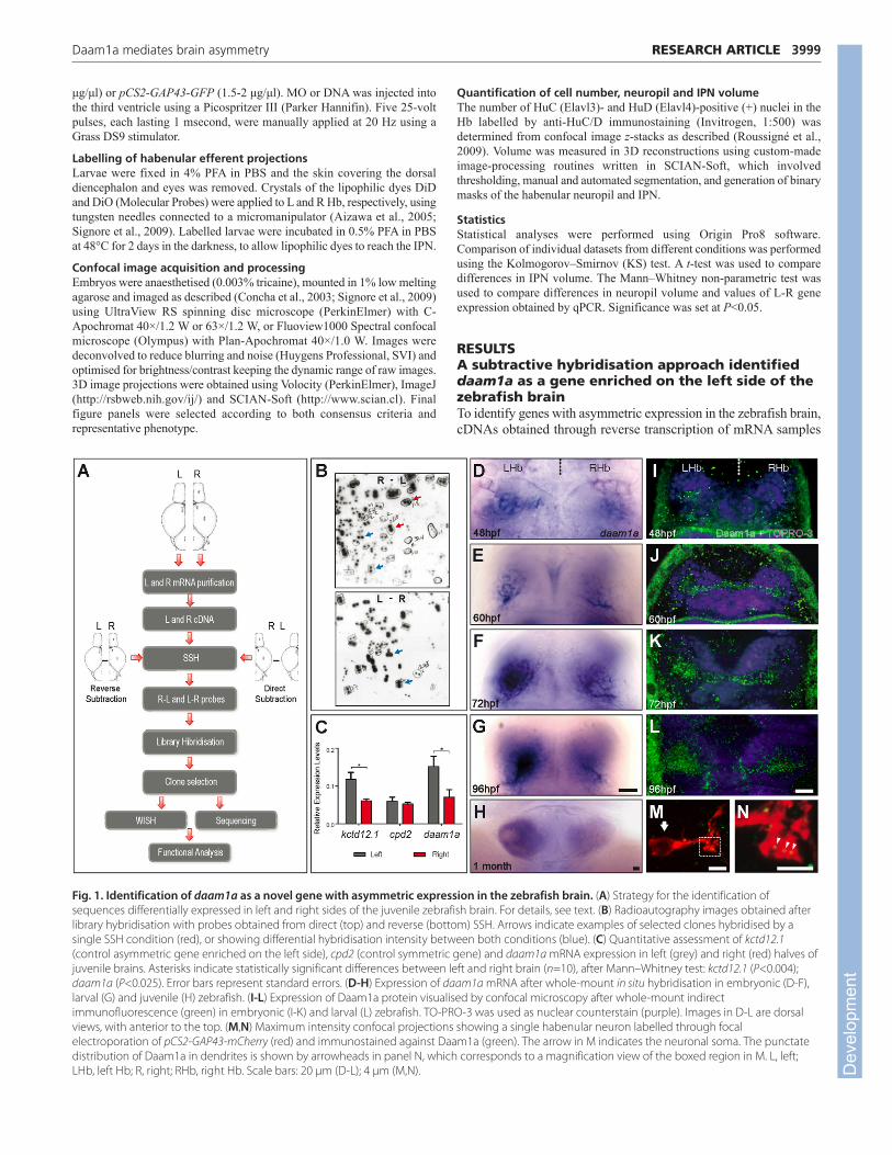
Fig. 1. Identification of daam1a as a novel gene with asymmetric expression in the zebrafish brain. (A) Strategy for the identification ofsequences differentially expressed in left and right sides of the juvenile zebrafish brain. For details, see text. (B) Radioautography images obtained afterlibrary hybridisation with probes obtained from direct (top) and reverse (bottom) SSH. Arrows indicate examples of selected clones hybridised by asingle SSH condition (red), or showing differential hybridisation intensity between both conditions (blue). (C) Quantitative assessment of kctd12.1(control asymmetric gene enriched on the left side), cpd2 (control symmetric gene) and daam1a mRNA expression in left (grey) and right (red) halves ofjuvenile brains. Asterisks indicate statistically significant differences between left and right brain (n=10), after Mann–Whitney test: kctd12.1 (P<0.004);daam1a (P<0.025). Error bars represent standard errors. (D-H) Expression of daam1a mRNA after whole-mount in situ hybridisation in embryonic (D-F),larval (G) and juvenile (H) zebrafish. (I-L) Expression of Daam1a protein visualised by confocal microscopy after whole-mount indirectimmunofluorescence (green) in embryonic (I-K) and larval (L) zebrafish. TO-PRO-3 was used as nuclear counterstain (purple). Images in D-L are dorsalviews, with anterior to the top. (M,N) Maximum intensity confocal projections showing a single habenular neuron labelled through focalelectroporation of pCS2-GAP43-mCherry (red) and immunostained against Daam1a (green). The arrow in M indicates the neuronal soma. The punctatedistribution of Daam1a in dendrites is shown by arrowheads in panel N, which corresponds to a magnification view of the boxed region in M. L, left;LHb, left Hb; R, right; RHb, right Hb. Scale bars: 20 μm (D-L); 4 μm (M,N). D
evel
opm
ent

4000
isolated from L and R halves of juvenile brains were used for direct(R-L) and reverse (L-R) SSH (Fig. 1A). Differential cDNA productswere used as probes to screen zebrafish cDNA libraries. Libraryclones that were differentially hybridised by probes from eitherdirect or reverse SSH but not both, and those for which strength ofhybridisation significantly differed between the two SSH conditions(Fig. 1B), were chosen for further in silico analysis and assessmentof mRNA expression through whole-mount in situ hybridisation.We identified daam1a as a gene enriched on the left side of thezebrafish brain. qPCR analysis corroborated the presence of higherlevels of daam1a mRNA in the L compared with the R side ofjuvenile brains (Mann–Whitney test, P<0.025) (Fig. 1C).
Daam1a is expressed in the asymmetric neuropilof the developing dorsal habenuladaam1a mRNA was detected in a distinct domain of the d-Hb that islarger on the L than on the R. Asymmetric expression was detectedfrom ~48 hpf (Fig. 1D,E), became conspicuous at 72 hpf (Fig. 1F),and continued throughout larval (Fig. 1G) and juvenile (Fig. 1H)stages into adulthood (data not shown). In the larvae, daam1a mRNAlocalised to the asymmetric habenular neuropil (Fig. 1G). Whole-mount immunofluorescence (using an antibody directed againsthuman DAAM1 that also detects zebrafish Daam1a) (Liu et al., 2008)revealed a modest asymmetric expression of Daam1 at the outset ofhabenular neuropil formation (Fig. 1I) that progressively increasedfollowing the changes in neuropil growth (Fig. 1J-L). At thesubcellular level, Daam1a was detected in a punctate pattern that washighly enriched in the habenular neuropil (Fig. 1L) and could bedetected in the soma (Fig. 1M, arrow) and dendritic processes(Fig. 1N, arrowheads) of single habenular neurons. Consistent withDaam1a levels mirroring the growth of habenular neuropil,
expression of daam1a was symmetric in the Hb of ace−/− and mbl−/−
mutants, which develop symmetric habenular neuropil (Carl et al.,2007; Regan et al., 2009) (supplementary material Fig. S1; Table 1).Conditions in which Nodal signalling in the brain is either absent (e.g.after southpaw morpholino injection, spaw-MO) (Long et al., 2003)or bilateral (e.g. after no tail morpholino injection, ntl-MO) (Feldmanand Stemple, 2001), and which result in randomisation of habenularneuropil asymmetry (Concha et al., 2000; Gamse et al., 2005), alsoresulted in randomised directionality of daam1a asymmetricexpression in the Hb (supplementary material Fig. S1; Table 1).Altogether, these results indicate that expression levels of Daam1amirror the asymmetric growth of habenular neuropil in the Hb,suggesting that Daam1a is a readout of asymmetric habenularmorphogenesis.
Global loss of Daam1a function disrupts thedevelopment of habenular neuropil and efferentconnectivity to the IPNTo investigate a possible role of Daam1a in asymmetric habenularmorphogenesis, we initially performed global Daam1a knockdownby injection at the one- to four-cell stage of a morpholino antisenseoligonucleotide designed to block daam1a translation (daam1a-MO) (Kida et al., 2007). Embryos injected with control morpholino(Co-MO) developed a characteristic left-sided enlarged habenularneuropil as assessed by anti-acetylated α-tubulin immunostaining(Fig. 2A, brackets) (Concha et al., 2000; Concha et al., 2003). Inaddition, axons from habenular neurons labelled with the pou4f1-hsp70l:GFP transgene (Aizawa et al., 2005) projected through thefasciculi retroflexus to the IPN, where they organised in acharacteristic ring-shape pattern (Fig. 2K). Global loss of Daam1afunction induced two main classes of phenotypes in the Hb. First,
RESEARCH ARTICLE Development 140 (19)
Table 1. Expression of daam1a and kctd12.1 in embryos with symmetric and heterotaxic habenular development
Symmetric (%)
L>R (%) R>L (%) L R Absent (%) Total (n)
southpaw-MO (spaw-MO)daam1a
Co-MO 93 7 – – – 46spaw-MO 53 45 – – 2 38
kctd12.1Co-MO 98 2 – – – 44spaw-MO 53 47 – – – 30
no tail-MO (ntl-MO)daam1a
Co-MO 94 6 – – – 31ntl-MO 42 43 5 – 10 70
kctd12.1Co-MO 100 – – – – 44ntl-MO 55 41 2 – 2 83
acerebellar (aceti282–/–)daam1a
sibling 96 2 – - 2 120aceti282–/– 20 3 – 39 35 68
kctd12.1sibling 88 6 – 1 5 158aceti282–/– – – – 55 45 130
masterblind (mbltm213–/–)daam1a
sibling 73 12 – 10 5 61mbltm213–/– 5 3 – 72 20 54
kctd12.1sibling 98 – – 1 – 41mbltm213–/– 6 15 – 68 3 33
L, left; R, right; symmetric L, both sides of the Hb showing left characteristics; symmetric R, both sides of the Hb showing right characteristics. Dev
elop
men
t

some daam1a-MO embryos (32%; n=324) displayed reverseddirectionality of habenular asymmetry with neuropil in the R Hbshowing L-side characteristics and L Hb displaying neuropilproperties of the R Hb (Table 2). We found that such completereversal of neuropil asymmetry was due to an early role of Daam1ain the control of embryo asymmetry, which resulted in disruption ofNodal signalling in both the lateral plate mesoderm and theepithalamus, and subsequently in randomised directionality ofepithalamic asymmetry (data not shown).
The second class of habenular phenotype involved a reduction inboth the extent of habenular neuropil (Fig. 2B-D, brackets) and thenumber of habenular axons reaching the IPN (Fig. 2L-N). Mostdaam1a-MO embryos displayed a moderate phenotype in both theHb (54%; Fig. 2C,P) and the IPN (62%; Fig. 2M,Q). In these cases,which comprised left-sided and reversed phenotypes with mild ormoderate neuropil reduction, the pattern of neuropil still remainedasymmetric (Fig. 2C); this is in contrast to the most severephenotypes (26%) in which both sides of the Hb developed a
4001RESEARCH ARTICLEDaam1a mediates brain asymmetry
Fig. 2. Global knockdown of Daam1a affects the formation of habenular neuropil and axonal efferent connectivity to the IPN. (A-O) Dorsalviews of the habenular region (A-J) and IPN (K-O) of Tg(pou4f1-hsp70l:GFP) zebrafish at 4.5 dpf, with anterior to the top. The habenular neuropil wasimmunostained against acetylated α-tubulin (A-E, white), whereas the soma (F-J) and efferent projections (K-O) of dorsal habenular neurons expressingpou4f1-hsp70l:GFP were labelled in vivo (green). Defects in the Hb and IPN induced by injection of daam1a-MO were classified as ‘normal’, ‘mild’,‘moderate’ or ‘severe’ phenotypes (columns 1-4). Column 5 (rescue) corresponds to the phenotypes observed after co-expression of daam1a-MO andhDAAM1 mRNA. All images are maximum intensity z-stack confocal projections. Vertical dotted brackets in A-E indicate the extent of habenular neuropilin the left Hb. Asterisks in F-J indicate the enlarged cellular domain of the Hb expressing pou4f1-hsp70l:GFP. (P,Q) Distribution of the differentphenotypes of habenular neuropil (P) and efferent connectivity to the IPN (Q) in Co-MO, daam1a-MO and rescue conditions. Data are presented as thepercentage of total larvae showing the indicated phenotype. L, left; LHb, left Hb; R, right; RHb, right Hb. Scale bars: 20 μm.
Table 2. Effects of global knock-down of Daam1a in habenular neuropil asymmetry and in the distribution of genetic markers ofthe habenulae
L>R (%) R>L (%) Symmetric R (%) Absent (%) Total (n)
NeuropilCo-MO 96 4 – – 357daam1a-MO 37 32 31 – 324
pou4f1-hsp70l:GFPCo-MO 4 96 – – 138daam1a-MO 35 65 – – 109
ET16:GFPCo-MO 99 1 – – 54daam1a-MO 69 30 – – 59
daam1aCo-MO 100 – - – 129daam1a-MO 33 35 32 – 138
kctd12.1Co-MO 100 – – – 253daam1a-MO 39 33 28 – 170
L, left; R, right; symmetric R, both sides of the Hb showing right characteristics. Dev
elop
men
t

4002
minimal amount of neuropil (Fig. 2D). Co-injection of daam1a-MOand human DAAM1 (hDAAM1) mRNA resulted in habenularneuropil and IPN connectivity similar to controls (Fig. 2E,O-Q),indicating that abrogation of Daam1a function was directlyresponsible for the observed phenotypes. Indeed, functional loss ofDaam1a did not disrupt habenular neurogenesis as the number ofHuC (+) post-mitotic habenular neurons was similar in control anddaam1a-MO embryos at 48 hpf (daam1a-MO=230±22; Co-MO=234±17; mean±s.d.; P>0.05; n=9). Specification of habenularcell subtypes was also unaffected as revealed by the normalasymmetric organisation of GFP (+) domains after Daam1aknockdown in two transgenic lines labelling distinct asymmetricallydistributed neuronal subtypes of the d-Hb, Tg(pou4f1-hsp70l:GFP)(Aizawa et al., 2005) and Tg(ET16::GFP) (Parinov et al., 2004)(Fig. 2G-I; Table 2). Furthermore, the parapineal nucleus waslateralised towards the side of enlarged habenular neuropil, and inthe most severe neuropil phenotypes (Fig. 2D) the parapineal stilladopted a lateral position (data not shown), suggesting that Daam1aknockdown affected events downstream of the pathways that
control the lateral positioning of the parapineal nucleus. The onlyasymmetric habenular marker that was reduced after Daam1aknockdown, other than daam1a, was kctd12.1 (supplementarymaterial Fig. S2; Table 2), which also localises to the habenularneuropil (Gamse et al., 2003). In conclusion, these results indicatethat Daam1a is required for habenular asymmetric morphogenesis,in particular for the development of asymmetric neuropil and theestablishment of connectivity from the Hb to the IPN.
Habenular knockdown of Daam1a decreases theformation of neuropil and efferent connectivityto the IPNTo verify that Daam1a plays a direct role in neuronalmorphogenesis of the Hb, we performed spatio-temporallyrestricted knockdown of Daam1a through local electroporation ofdaam1a-MO directly into the habenular region at a developmentalstage that followed cell subtype specification and preceded theonset of habenular morphogenesis (44 hpf) (Fig. 3A). We focusedon targeting the L Hb, as this was the primary site of endogenous
RESEARCH ARTICLE Development 140 (19)
Fig. 3. Local loss and gain of Daam1a functioninduces opposite modulatory effects in thegrowth of habenular neuropil and axonalefferent connectivity to the IPN. (A) Schematics ofthe local electroporation procedure (a), daam1a-MOdesign (b) and constitutively active form of C-Daam1a (c). (B-J) Dorsal views of the habenularregion (B-G) and IPN (H-J) of Tg(pou4f1-hsp70l:GFP)zebrafish at 4.5 dpf, with anterior to the top. Thehabenular neuropil was immunostained againstacetylated α-tubulin (B-D, white), whereas the soma(E-G) and efferent projections (H-J) of dorsalhabenular neurons expressing pou4f1-hsp70l:GFPwere labelled in vivo (green). Vertical dotted bracketsin B-D indicate the extent of neuropil developmentin the left Hb. Asterisks in E-G indicate the enlargedcellular domain of the Hb expressing pou4f1-hsp70l:GFP. (K-M) Dorsal (left) and lateral (right) viewsof the IPN at 4.5 dpf, with anterior to the top, afterlabelling the left (red) and right (green) Hb with DiDand DiO, respectively. Images in B-M are maximumintensity z-stack confocal projections, organised inthree columns that correspond to differentelectroporation conditions: control-MO (left),daam1a-MO (middle) and pcDNA-C-DAAM1-HA(right). (N-P) Dorsal views of three-dimensionalvolumetric reconstructions of the left habenularneuropil in the three electroporation conditions. (Q) Quantification of left (grey) and right (red)habenular neuropil volumes. Asterisks indicatestatistically significant decrease (daam1a-MO, n=5) orincrease (pcDNA-C-DAAM1-HA, n=5) of left habenularneuropil volume with respect to controls (Co-MO,n=5; control plasmid, n=6), after Mann–Whitney test(P<0.05). Error bars represent s.d. D, dorsal; L, left;LHb, left Hb; R, right; RHb, right Hb; V, ventral. Scalebars: 20 μm.
Dev
elop
men
t

daam1a expression, although we obtained similar results afterbilateral electroporation of the Hb (Fig. 3; supplementary materialFig. S5). Local electroporation of daam1a-MO efficiently led toboth morpholino incorporation (supplementary material Fig. S3)and reduced levels of Daam1a protein expression (supplementarymaterial Fig. S4). Importantly, the extent of neuropil was markedlyreduced in the L Hb of daam1a-MO-electroporated embryos(Fig. 3C, brackets) compared with controls (Fig. 3B, brackets),similar to the severe phenotypes observed after global knockdownof Daam1a (compare Fig. 3C with Fig. 2D). Indeed, quantificationof habenular neuropil volume revealed a significant decrease inthe L Hb of electroporated embryos compared with controls (L-Hb-daam1a-MO=6752±442 μm3; L-Hb-Co-MO=10,042±747μm3; P<0.05; n=5) (Fig. 3N,O,Q) whereas neuropil of the R Hbremained unaffected (Fig. 3Q). In addition to changes in habenularneuropil, left-sided local electroporation of daam1a-MO alsoinduced a decrease in the extent of pou4f1-hsp70l:GFP (+) axonallabelling in the IPN (Fig. 3I; supplementary material Fig. S5) andin the volume of L Hb projections reaching the dorsal IPN (dIPN)compared with controls (L-Hb-dIPN-daam1-MO=13,001±502μm3; L-Hb-dIPN-Co-MO=29,820±1595.4 μm3; P<0.05; n=4),whereas projections to the ventral IPN (vIPN) from either R or LHb appeared unaltered (data not shown, P>0.05; n=4) (Fig. 3K,L).As we did not observe axons projecting to ectopic brain regions(data not shown) and the number and organisation of habenularcell subtypes appeared unaffected as revealed by the normalexpression of the pou4f1-hsp70l:GFP transgene (Fig. 3C,F), weinterpreted the loss of IPN connectivity as a primary defect inhabenular axonal extension. In summary, local knockdownexperiments indicate that Daam1a is required after 44 hpf in thehabenular region for the growth of asymmetric neuropil andhabenular efferent connectivity to the IPN.
Habenular overexpression of an activated form ofDAAM1 increases the formation of neuropil andefferent connectivity to the IPNTo determine whether increased levels of Daam1a activity couldpromote habenular neuropil formation and axon extension, weperformed local electroporation of a plasmid coding for a truncatedform of DAAM1 that lacks the N-terminal regulatory domains (C-
Daam1) and works as a dominant-positive (constitutively active) form(Fig. 3C) (Habas et al., 2001; Matusek et al., 2006; Ju et al., 2010).Local electroporation of pcDNA-C-DAAM1-HA in the habenularregion at 44 hpf resulted in an ipsilateral increase of neuropil whendirected to the L (Fig. 3D; n=8) but not to the R (supplementarymaterial Fig. S6; n=9), compared with embryos electroporated witha control plasmid (data not shown; n=6), a finding that wascorroborated by quantification of neuropil volume (L-Hb-C-DAAM1=14,473±119 μm3; L-Hb-Co-plasmid=10,239±824 μm3;P<0.05; n=5) (Fig. 3P,Q). By contrast to the differential effect inhabenular neuropil growth, local electroporation of pcDNA-DAAM1-HA led to increase of pou4f1-hsp70l:GFP (+) habenular axons in theIPN when directed to either L (Fig. 3J,M) or R (supplementarymaterial Fig. S6). In left-side-electroporated embryos, the volume ofL Hb projections in the dIPN increased significantly (L-Hb-dIPN-C-DAAM1=36,147±818 μm3; L-Hb-dIPN-Co-plasmid=27,603±1600μm3; P<0.05; n=3) whereas projections to the vIPN from either L orR Hb appeared unaffected (data not shown, P>0.05; n=3)(Fig. 3K,M). Habenular expression of pou4f1-hsp70l:GFP remainedunchanged irrespective of the side (L, R or bilateral) of pcDNA-C-DAAM1-HA electroporation (Fig. 3G; supplementary material Fig.S6), indicating that habenular cell subtype organisation wasunaffected. We thus conclude that over-activation of Daam1 in thehabenular region, prior to and/or during the onset of neuropil andaxonal morphogenesis, is sufficient to promote the growth of bothhabenular neuropil (when directed to the L) and axonal efferents to theIPN (when directed to either L or R).
Daam1a regulates dendritic and axonaloutgrowth in individual habenular neuronsTo assess directly whether dendritic and axonal outgrowth wassensitive to the modulation of Daam1a, we analysed the patternof dendritic arborisation and shape of axonal terminals ofindividual habenular neurons labelled through focalelectroporation of pCS2:GAP43-GFP. In control embryos, werecognised the stereotypical unipolar morphology of habenularneurons (Bianco et al., 2008), with a dense dendritic arbour(Fig. 4A, arrowheads) and a single unbranched axon extendingfrom the soma (Fig. 4A, arrow; n=6). In addition, we observed thecharacteristic complex pattern of axon terminal morphology in the
4003RESEARCH ARTICLEDaam1a mediates brain asymmetry
Fig. 4. Loss and gain of Daam1a function modulates dendritic growth and axonal terminal arborisation of dorsal habenular neurons.(A,E) Single habenular neuron from a control embryo showing the characteristic dense dendritic arborisation (A, arrowheads) and a single unbranchedaxon (A, arrow) that terminates in the IPN with complex bilateral spiral branches (E). (B,F) Single habenular neuron from a daam1a-MO embryo showinga pruned dendritic arbour (B, arrowheads) and a single axon (B, arrow) with incomplete and less complex organisation of axonal terminals in the IPN (F).(C,G) Single habenular neuron from an embryo co-injected with daam1a-MO and hDAAM1, showing normal dendritic arborisation (C, arrowheads) andpartial rescue of axonal terminal morphology in the IPN (G). (D,H) Single habenular neuron from an embryo locally electroporated with pcDNA-C-DAAM1-HA at 44 hpf, showing increased dendritic arborisation (D, arrowheads) and axonal extension in the IPN (H). Images correspond to dorsal viewsof maximum intensity z-stack confocal projections of single neurons at 4.5 dpf, labelled through focal electroporation of pCS2:GAP43-GFP, with anteriorto the top. Scale bars: 10 μm. D
evel
opm
ent

4004
IPN, with bilateral and spiral branches (Fig. 4E) (Bianco et al.,2008). By contrast, habenular neurons from daam1a-MO embryosshowed a greatly pruned dendritic arbour (Fig. 4B, arrowheads)and an incomplete and less complex pattern of axonal terminals inthe IPN (Fig. 4F; n=5), compared with controls. Electroporatedneurons from embryos co-injected with daam1a-MO andhDAAM1 mRNA at the one- to two-cell stage developed a patternof dendritic arborisation similar to controls (Fig. 4C, arrowheads)and showed a partial rescue of axonal terminal morphology in theIPN (Fig. 4G). Finally, habenular neurons from embryos locallyelectroporated with pcDNA-C-DAAM1-HA at 44 hpf showedincreased dendritic arborisation (Fig. 4D, arrowheads) and axonalextension (Fig. 4H) in the IPN. Altogether, these findingscorroborate that Daam1a regulates dendritic and axonal outgrowthin dorsal habenular neurons.
Daam1a is required for normal organisation ofthe cytoskeleton in the habenulaeAs members of the Drf subfamily of Formin proteins act as actinassembly factors (Wallar and Alberts, 2003) and also play a lesscommon role in regulating the stability of microtubules (Ju et al.,2010), we investigated whether the global loss of Daam1a functionaffected the organisation of F-actin and α-tubulin in the Hb.Embryos injected with Co-MO and co-stained with fluorescentphalloidin (to label F-actin) and Nissl (to delineate the cellularcontext of the Hb) showed a distinct smooth fibrillar organisation ofF-actin in the Hb, with increased density of labelling in theasymmetric neuropil domains (Fig. 5A,D,G; n=12). By contrast, F-actin staining in the Hb of daam1a-MO embryos was irregular inshape and showed thicker and shorter bundles compared with
controls (Fig. 5B,E,H; n=10). In these embryos, α-tubulin was alsodecreased and exhibited a less elaborate distribution than in controls(Fig. 5K,N,Q; n=10). Co-injection of daam1a-MO and hDAAM1mRNA resulted in partial rescue of the normal architecture of F-actin organisation in the Hb (Fig. 5C,F,I; n=10) and the strength ofα-tubulin staining was increased (Fig. 5L,O,R; n=10). Altogether,these results indicate that Daam1a is required for the normalassembly and organisation of actin filaments in the developingzebrafish Hb, and also suggest a possible additional role for Daam1ain regulating microtubules.
Daam1a and Ulk2 collaborate in habenularneuropil morphogenesisAs the zebrafish Unc-51-like 2 (Ulk2) kinase has been recentlyimplicated in the control of habenular neuropil morphogenesisdownstream of Kctd proteins (Taylor et al., 2011), and the functionalloss of Ulk2 induces a reduced habenular neuropil phenotype thatresembles Daam1a knockdown (compare Fig. 6B and 6C), weinvestigated whether Daam1a and Ulk2 interact in the promotionof neuropil growth. Co-injection of morpholino directed againstdaam1a and ulk2 (ulk2spl-MO) at concentrations that in the contextof single knockdowns induce only a modest reduction of habenularneuropil (Fig. 6D,E), led to severe reduction of habenular neuropil(Fig. 6F). Furthermore, hDAAM1 mRNA rescued the habenularneuropil phenotype induced by loss of Ulk2 (Fig. 6H) whereas ulk2mRNA retained the habenular neuropil phenotype caused byknockdown of Daam1a (Fig. 6I). Altogether, these findings indicatethat Daam1a and Ulk2 play a synergistic role in the promotion ofneuropil growth, and that Daam1a is likely to work downstream ofUlk2 in the process.
RESEARCH ARTICLE Development 140 (19)
Fig. 5. Knockdown of Daam1a affects the organisation offilamentous actin and microtubules in the Hb. (A,D,G) The Hb ofcontrol embryos labelled with fluorescent phalloidin (F-actin, white)and counterstained with Nissl (to delineate the cellular context,purple) show a distinct smooth fibrillar organisation of F-actin, withincreased compaction in the neuropil domains. (B,E,H) The Hb ofdaam1a-MO embryos show an irregular pattern of F-actin, withshorter and thicker bundles. (C,F,I) The Hb of embryos co-injectedwith daam1a-MO and hDAAM1, show a smooth fibrillar pattern of F-actin similar to controls. (J,M,P) The Hb of control embryosimmunostained against α-tubulin (white) and counterstained withNissl showed a distinct asymmetric distribution of α-tubulincorresponding to the neuropil domains. (K,N,Q) The Hb of daam1a-MO embryos show decreased and less elaborate α-tubulin staining.(L,O,R) The Hb of embryos co-injected with daam1a-MO andhDAAM1, show increased levels and thickness of α-tubulin (+)bundles than controls. G-I and P-R correspond to magnificationviews of the boxed regions in D-F and M-O, respectively. Imagescorrespond to dorsal views of maximum intensity z-stack confocalprojections, with anterior to the top. LHb, left Hb; RHb, right Hb. Scalebars: 20 μm.
Dev
elop
men
t

DISCUSSIONIn this study, we used a reverse genetic approach to identify daam1aas a new genetic component of the pathways that controlasymmetric habenular morphogenesis. Expression of daam1a in thedeveloping Hb follows the specification of cell subtypes and mirrorsthe asymmetric changes in neuropil growth at embryonic and larvalstages. Consistent with a role of Daam1a downstream of the mainpathways that control the development of brain asymmetry (e.g. Fgfand Wnt) and its directionality (Nodal) in the brain, habenulardaam1a expression becomes symmetric in ace−/− and mbl−/−
mutants, and randomised after loss of Spaw or Ntl function.Importantly, loss and gain of Daam1a function decreases andincreases, respectively, the formation of habenular neuropil andefferent axonal connectivity to the IPN without affecting habenularneurogenesis or cell subtype specification. These findings, and thefact that Daam1a promotes neuropil growth downstream of Ulk2,indicate that Daam1a plays a key role in asymmetric habenularmorphogenesis, mediating the growth of habenular neuropil andefferent axonal connectivity to the IPN.
The opposite modulatory effect shown by loss and gain ofDaam1a function in the ability to promote dendritic and axonalgrowth provides strong evidence in support of a role of Daam1a asa key modulator of neuronal morphogenesis in the Hb. The punctatedistribution of Daam1a expression in the developing habenularneuropil, and more specifically in dendrites of habenular neurons,is also consistent with Daam1a participating in neuronal processoutgrowth. Indeed, functional abrogation of Daam1a markedlydisrupts the formation of dendritic processes examined in singlehabenular neurons, also leading to an incomplete and less complexpattern of axonal terminals in the IPN. Consistent with this,Drosophila Daam (dDaam) and mouse Daam1 (mDaam1) bothregulate filopodial formation and neurite outgrowth in developingneurons (Matusek et al., 2008; Salomon et al., 2008; Gonçalves-Pimentel et al., 2011). dDaam shows a punctate distribution in F-actin-rich structures in the growth cones of cultured primary neurons(Matusek et al., 2008), whereas puncta of mDaam1 expression areobserved in the dendritic shaft of pyramidal cells of thehippocampus, and in synaptic regions where Daam1 colocaliseswith pre-synaptic (Sv2) and post-synaptic (Psd95; also known asDlg4) markers (Salomon et al., 2008). Importantly, dDaam isrequired for filopodial extension at axonal growth cones (Matuseket al., 2008; Gonçalves-Pimentel et al., 2011), whereas mDaam1
appears to play a role in dendritic extension (Salomon et al., 2008).In zebrafish, habenular neurons require Daam1a for both dendriticand axonal morphogenesis, although these two events appear toshow differential Daam1a requirements, as they are affected todifferent extents after Daam1a knockdown (dendritic>axonal) andcan be rescued to distinct degrees by overexpression of hDAAM1(again, dendritic>axonal). In addition, the extension of dendriticprocesses appears to be subject to local growth control dependingon the side of the Hb. For example, local electroporation of C-DAAM1 induces habenular neuropil growth only when directed tothe L side, which suggests a mechanism that moderates theinfluence of Daam1 on neuropil growth in the R Hb, withoutaffecting its ability to promote axonal extensions to the IPN. Theseresults appear to be consistent with a recently proposed model ofasymmetric local modulation of habenular neuropil growthmediated by Kctd proteins in zebrafish (Taylor et al., 2011). In thismodel, Kctd12.1 and Kctd12.2 are expressed primarily in the L andR Hb, respectively, and exert a stronger inhibition of neuropilgrowth on the R than the L Hb through interacting with Ulk2, akinase that is expressed bilaterally in the Hb from 48 hpf (Taylor etal., 2011). Whether Kctd proteins inhibit the ability of Daam1a topromote neuropil growth in the R Hb is yet to be determined.However, the onset of asymmetric daam1a expression coincideswith the onset of ulk2 expression and both are concurrent withneuropil growth in the Hb, suggesting a link between these events.Indeed, Daam1a and Ulk2 play a synergistic role in the promotionof neuropil growth. Furthermore, hDAAM1 overexpression rescuesthe impaired neuropil phenotype induced by functional loss of Ulk2(whereas Ulk2 appears to be unable to rescue the phenotype inducedby Daam1a knockdown) suggesting that Daam1a worksdownstream of Ulk2. Altogether, these findings are consistent witha collaborative role of Daam1a and Ulk2 in the promotion ofneuropil growth downstream of the pathways that establish cellsubtype specification in the Hb. Where and how such collaborationoccurs remains to be determined. An intriguing possibility is theendocytic pathway, as Ulk1 and Ulk2 localise to vesicular structuresin growth cones and regulate neuronal process extension throughearly endosome trafficking at nascent neurites (Tomoda et al., 2004;Zhou et al., 2007; Toda et al., 2008), whereas zebrafish Daam1a isactively transported to endocytic vesicles and its asymmetricsubcellular localisation appears to be relevant for the convergent-extension movements of notochordal cells (Kida et al., 2007).
4005RESEARCH ARTICLEDaam1a mediates brain asymmetry
Fig. 6. Daam1a cooperates with Ulk2 in habenular neuropil formation. (A-I) The organisation of habenular neuropil at 4.5 hpf as revealed by antiacetylated α-tubulin immunostaining, after injection at the one- to four-cell stage with Co-MO (A), optimal doses of either ulk2spl-MO (B, 2 ng) ordaam1a-MO (C, 8 ng), suboptimal doses of either ulk2spl-MO (D, 1 ng) or daam1a-MO (E, 4 ng), a combination of suboptimal doses of ulk2spl-MO anddaam1a-MO (F), optimal doses of ulk2spl-MO (2 ng) combined with either dUlk2 (G) or hDAAM1 (H) mRNAs, or optimal dose of daam1a-MO (8 ng)combined with ulk2 mRNA (I). Images correspond to dorsal views of maximum intensity confocal z-stack projections, with anterior to the top. LHb, leftHb; RHb, right Hb. Scale bar: 20 μm.
Dev
elop
men
t

4006
At the effector level, Daam1a regulates both F-actin andmicrotubule assembly during habenular morphogenesis. The formerrole is consistent with previous reports indicating that Daam1 worksas an F-actin nucleation and elongation factor induced by RhoGTPases (Prokop et al., 2011), and that dDaam cooperates withArp2 and Arp3 in a common mechanism of filopodia formation thatdepends on Enabled and is regulated through Profilin (Chickadee –FlyBase) activity (Gonçalves-Pimentel et al., 2011). Reports linkingDaam1 to microtubule assembly and stabilisation are, however,scarce (Ju et al., 2010). The observed increase in intensity of α-tubulin (+) labelling after expression of hDAAM1 suggests thatzebrafish Daam1a regulates microtubule dynamics in the Hb.Regulation of F-actin and microtubule dynamics might result fromDaam1a interacting with other signalling pathways, such asWnt/PCP signalling, which is known to modulate the growth ofaxons and dendrites (Wang and Nathans, 2007; Tissir and Goffinet,2010). Daam1 works as an intermediary of Wnt/PCP signalling thatleads to Rho activation and subsequent remodelling of thecytoskeleton during Xenopus and zebrafish gastrulation (Habas etal., 2001; Kida et al., 2007), and some ligands and core componentsof Wnt/PCP are expressed in the developing Hb at the time ofneuropil formation (data not shown).
Here, we demonstrate that Daam1a plays a key role inasymmetric habenular morphogenesis by regulating dendritic andaxonal outgrowth in dorsal habenular neurons, possibly through themodulation of cytoskeletal dynamics. Although Daam1 and Ulk2have a synergistic role in the promotion of habenular neuropilgrowth, downstream of the pathways that establish asymmetric cellsubtype specification, further studies are needed to address howthese two factors interact and become differentially modulated on Land R sides of the Hb.
AcknowledgementsWe thank Ricardo Armisen for critical reading of the manuscript; Drs RaymondHabas and Joshua Gamse for providing plasmids; members of the Laboratoryof Experimental Ontogeny Fish Facility for fish care; Erika Labbe and Jorge Jarafor technical help with image processing; Carmen Gloria Lemus for advice inthe statistical approach; and Luis Michea for sharing the use of the qPCRmachine.
FundingThis work was supported by the Howard Hughes Medical Institute (to M.L.C.),The National Fund for Scientific and Technological Development (FONDECYT)[1109324 to A.C., 1120579 to S.H. and 1120558 to M.L.C.]; the Commissionof the European Communities [FP6-2004-NEST-PATH EDCBNL to M.L.C. andS.W.W.]; the Scientific Millennium Initiative [P09-015-F to M.L.C. and S.H.];and the Wellcome Trust [S.W.W. and M.C.]. Deposited in PMC for immediaterelease.
Competing interests statementThe authors declare no competing financial interests.
Author contributionsA.C. participated in the design of the study; performed SSH, libraryhybridisation, in situ hybridisation screening, clone selection, and morpholinoand mRNA injections; helped in the supervision of the study; wrote early draftsof the manuscript; and provided comments and revisions on further versions.K.P. carried out most immunostaining and prepared most figures. L.A. andM.M.M. performed local and focal electroporation, and processed the imagesof these experiments. M.M. designed the SSH protocol and helped inperforming SHH. I.S. carried out actin/tubulin immunostaining, preparedfigures and provided comments on the manuscript. C.M. carried out in situhybridisation, anti-HuC/D inmunostaining and the determination of habenularcell number and neuropil volume. N.G. performed lipophilic labelling ofhabenular efferent connectivity. R.P. carried out qPCR experiments. J.S andK.M. helped with data analysis and provided comments on the writting. S.H.and L.B. performed neuropil and IPN volume data analyses. S.W.W. provided
lab support at early stages of the reverse genetics approach and revisons/comments on later versions of the manuscript. M.L.C. conceived andsupervised the project, and wrote the manuscript.
Supplementary materialSupplementary material available online athttp://dev.biologists.org/lookup/suppl/doi:10.1242/dev.091934/-/DC1
ReferencesAgetsuma, M., Aizawa, H., Aoki, T., Nakayama, R., Takahoko, M., Goto, M.,
Sassa, T., Amo, R., Shiraki, T., Kawakami, K. et al. (2010). The habenula iscrucial for experience-dependent modification of fear responses in zebrafish.Nat. Neurosci. 13, 1354-1356.
Aizawa, H., Bianco, I. H., Hamaoka, T., Miyashita, T., Uemura, O., Concha, M.L., Russell, C., Wilson, S. W. and Okamoto, H. (2005). Laterotopicrepresentation of left-right information onto the dorso-ventral axis of azebrafish midbrain target nucleus. Curr. Biol. 15, 238-243.
Aizawa, H., Goto, M., Sato, T. and Okamoto, H. (2007). Temporally regulatedasymmetric neurogenesis causes left-right difference in the zebrafishhabenular structures. Dev. Cell 12, 87-98.
Aizawa, H., Amo, R. and Okamoto, H. (2011). Phylogeny and ontogeny of thehabenular structure. Front Neurosci 5, 138.
Bianco, I. H. and Wilson, S. W. (2009). The habenular nuclei: a conservedasymmetric relay station in the vertebrate brain. Philos. Trans. R. Soc. B 364,1005-1020.
Bianco, I. H., Carl, M., Russell, C., Clarke, J. D. and Wilson, S. W. (2008). Brainasymmetry is encoded at the level of axon terminal morphology. Neural Dev. 3,9.
Bisgrove, B. W., Essner, J. J. and Yost, H. J. (1999). Regulation of midlinedevelopment by antagonism of lefty and nodal signaling. Development 126,3253-3262.
Burgess, H. A., Johnson, S. L. and Granato, M. (2009). Unidirectional startleresponses and disrupted left-right co-ordination of motor behaviors in robo3mutant zebrafish. Genes Brain Behav. 8, 500-511.
Carl, M., Bianco, I. H., Bajoghli, B., Aghaallaei, N., Czerny, T. and Wilson, S.W. (2007). Wnt/Axin1/beta-catenin signaling regulates asymmetric nodalactivation, elaboration, and concordance of CNS asymmetries. Neuron 55, 393-405.
Concha, M. L. (2004). The dorsal diencephalic conduction system of zebrafish asa model of vertebrate brain lateralisation. Neuroreport 15, 1843-1846.
Concha, M. L. and Wilson, S. W. (2001). Asymmetry in the epithalamus ofvertebrates. J. Anat. 199, 63-84.
Concha, M. L., Burdine, R. D., Russell, C., Schier, A. F. and Wilson, S. W.(2000). A nodal signaling pathway regulates the laterality of neuroanatomicalasymmetries in the zebrafish forebrain. Neuron 28, 399-409.
Concha, M. L., Russell, C., Regan, J. C., Tawk, M., Sidi, S., Gilmour, D. T.,Kapsimali, M., Sumoy, L., Goldstone, K., Amaya, E. et al. (2003). Local tissueinteractions across the dorsal midline of the forebrain establish CNS laterality.Neuron 39, 423-438.
Concha, M. L., Signore, I. A. and Colombo, A. (2009). Mechanisms of directionalasymmetry in the zebrafish epithalamus. Semin. Cell Dev. Biol. 20, 498-509.
Concha, M. L., Bianco, I. H. and Wilson, S. W. (2012). Encoding asymmetrywithin neural circuits. Nat. Rev. Neurosci. 13, 832-843.
Essner, J. J., Branford, W. W., Zhang, J. and Yost, H. J. (2000). Mesendodermand left-right brain, heart and gut development are differentially regulated bypitx2 isoforms. Development 127, 1081-1093.
Facchin, L., Burgess, H. A., Siddiqi, M., Granato, M. and Halpern, M. E. (2009).Determining the function of zebrafish epithalamic asymmetry. Philos. Trans. R.Soc. B 364, 1021-1032.
Feldman, B. and Stemple, D. L. (2001). Morpholino phenocopies of sqt, oep,and ntl mutations. Genesis 30, 175-177.
Gamse, J. T., Thisse, C., Thisse, B. and Halpern, M. E. (2003). The parapinealmediates left-right asymmetry in the zebrafish diencephalon. Development130, 1059-1068.
Gamse, J. T., Kuan, Y. S., Macurak, M., Brösamle, C., Thisse, B., Thisse, C. andHalpern, M. E. (2005). Directional asymmetry of the zebrafish epithalamusguides dorsoventral innervation of the midbrain target. Development 132,4869-4881.
Gilmour, D. T., Maischein, H. M. and Nüsslein-Volhard, C. (2002). Migrationand function of a glial subtype in the vertebrate peripheral nervous system.Neuron 34, 577-588.
Gonçalves-Pimentel, C., Gombos, R., Mihály, J., Sánchez-Soriano, N. andProkop, A. (2011). Dissecting regulatory networks of filopodia formation in aDrosophila growth cone model. PLoS ONE 6, e18340.
Gubler, U. and Hoffman, B. J. (1983). A simple and very efficient method forgenerating cDNA libraries. Gene 25, 263-269.
Habas, R., Kato, Y. and He, X. (2001). Wnt/Frizzled activation of Rho regulatesvertebrate gastrulation and requires a novel Formin homology protein Daam1.Cell 107, 843-854.
RESEARCH ARTICLE Development 140 (19)
Dev
elop
men
t

Heisenberg, C. P., Houart, C., Take-Uchi, M., Rauch, G. J., Young, N.,Coutinho, P., Masai, I., Caneparo, L., Concha, M. L., Geisler, R. et al. (2001).A mutation in the Gsk3-binding domain of zebrafish Masterblind/Axin1 leadsto a fate transformation of telencephalon and eyes to diencephalon. GenesDev. 15, 1427-1434.
Hendricks, M. and Jesuthasan, S. (2007a). Asymmetric innervation of thehabenula in zebrafish. J. Comp. Neurol. 502, 611-619.
Hendricks, M. and Jesuthasan, S. (2007b). Electroporation-based methods forin vivo, whole mount and primary culture analysis of zebrafish braindevelopment. Neural Dev. 2, 6.
Jesuthasan, S. (2011). Fear, anxiety and control in the zebrafish. Dev. Neurobiol.Ju, R., Cirone, P., Lin, S., Griesbach, H., Slusarski, D. C. and Crews, C. M.
(2010). Activation of the planar cell polarity formin DAAM1 leads to inhibitionof endothelial cell proliferation, migration, and angiogenesis. Proc. Natl. Acad.Sci. USA 107, 6906-6911.
Kida, Y. S., Sato, T., Miyasaka, K. Y., Suto, A. and Ogura, T. (2007). Daam1regulates the endocytosis of EphB during the convergent extension of thezebrafish notochord. Proc. Natl. Acad. Sci. USA 104, 6708-6713.
Kimmel, C. B., Ballard, W. W., Kimmel, S. R., Ullmann, B. and Schilling, T. F.(1995). Stages of embryonic development of the zebrafish. Dev. Dyn. 203, 253-310.
Lee, A., Mathuru, A. S., Teh, C., Kibat, C., Korzh, V., Penney, T. B. andJesuthasan, S. (2010). The habenula prevents helpless behavior in larvalzebrafish. Curr. Biol. 20, 2211-2216.
Liu, W., Sato, A., Khadka, D., Bharti, R., Diaz, H., Runnels, L. W. and Habas, R.(2008). Mechanism of activation of the Formin protein Daam1. Proc. Natl. Acad.Sci. USA 105, 210-215.
Long, S., Ahmad, N. and Rebagliati, M. (2003). The zebrafish nodal-relatedgene southpaw is required for visceral and diencephalic left-right asymmetry.Development 130, 2303-2316.
Matusek, T., Djiane, A., Jankovics, F., Brunner, D., Mlodzik, M. and Mihály, J.(2006). The Drosophila formin DAAM regulates the tracheal cuticle patternthrough organizing the actin cytoskeleton. Development 133, 957-966.
Matusek, T., Gombos, R., Szécsényi, A., Sánchez-Soriano, N., Czibula, A.,Pataki, C., Gedai, A., Prokop, A., Raskó, I. and Mihály, J. (2008). Forminproteins of the DAAM subfamily play a role during axon growth. J. Neurosci. 28,13310-13319.
Miyasaka, N., Morimoto, K., Tsubokawa, T., Higashijima, S., Okamoto, H.and Yoshihara, Y. (2009). From the olfactory bulb to higher brain centers:genetic visualization of secondary olfactory pathways in zebrafish. J. Neurosci.29, 4756-4767.
Parinov, S., Kondrichin, I., Korzh, V. and Emelyanov, A. (2004). Tol2transposon-mediated enhancer trap to identify developmentally regulatedzebrafish genes in vivo. Dev. Dyn. 231, 449-459.
Prokop, A., Sánchez-Soriano, N., Gonçalves-Pimentel, C., Molnár, I., Kalmár,T. and Mihály, J. (2011). DAAM family members leading a novel path intoformin research. Commun. Integr. Biol. 4, 538-542.
Regan, J. C., Concha, M. L., Roussigne, M., Russell, C. and Wilson, S. W.(2009). An Fgf8-dependent bistable cell migratory event establishes CNSasymmetry. Neuron 61, 27-34.
Reifers, F., Böhli, H., Walsh, E. C., Crossley, P. H., Stainier, D. Y. and Brand, M.(1998). Fgf8 is mutated in zebrafish acerebellar (ace) mutants and is requiredfor maintenance of midbrain-hindbrain boundary development andsomitogenesis. Development 125, 2381-2395.
Roussigné, M., Bianco, I. H., Wilson, S. W. and Blader, P. (2009). Nodalsignalling imposes left-right asymmetry upon neurogenesis in the habenularnuclei. Development 136, 1549-1557.
Roussigné, M., Blader, P. and Wilson, S. W. (2011). The zebrafish epithalamusclears a path through the complexity of brain lateralization. Dev. Neurobiol. 72,269-281.
Salomon, S. N., Haber, M., Murai, K. K. and Dunn, R. J. (2008). Localization ofthe Diaphanous-related formin Daam1 to neuronal dendrites. Neurosci. Lett.447, 62-67.
Schmittgen, T. D. and Livak, K. J. (2008). Analyzing real-time PCR data by thecomparative C(T) method. Nat. Protoc. 3, 1101-1108.
Signore, I. A., Guerrero, N., Loosli, F., Colombo, A., Villalón, A., Wittbrodt, J.and Concha, M. L. (2009). Zebrafish and medaka: model organisms for acomparative developmental approach of brain asymmetry. Philos. Trans. R. Soc.B 364, 991-1003.
Tawk, M., Bianco, I. H. and Clarke, J. D. (2009). Focal electroporation inzebrafish embryos and larvae. Methods Mol. Biol. 546, 145-151.
Taylor, R. W., Qi, J. Y., Talaga, A. K., Ma, T. P., Pan, L., Bartholomew, C. R.,Klionsky, D. J., Moens, C. B. and Gamse, J. T. (2011). Asymmetric inhibitionof Ulk2 causes left-right differences in habenular neuropil formation. J.Neurosci. 31, 9869-9878.
Thisse, C. and Thisse, B. (2008). High-resolution in situ hybridization to whole-mount zebrafish embryos. Nat. Protoc. 3, 59-69.
Tissir, F. and Goffinet, A. M. (2010). Planar cell polarity signaling in neuraldevelopment. Curr. Opin. Neurobiol. 20, 572-577.
Toda, H., Mochizuki, H., Flores, R., 3rd, Josowitz, R., Krasieva, T. B., Lamorte,V. J., Suzuki, E., Gindhart, J. G., Furukubo-Tokunaga, K. and Tomoda, T.(2008). UNC-51/ATG1 kinase regulates axonal transport by mediating motor-cargo assembly. Genes Dev. 22, 3292-3307.
Tomoda, T., Kim, J. H., Zhan, C. and Hatten, M. E. (2004). Role of Unc51.1 andits binding partners in CNS axon outgrowth. Genes Dev. 18, 541-558.
Wallar, B. J. and Alberts, A. S. (2003). The formins: active scaffolds that remodelthe cytoskeleton. Trends Cell Biol. 13, 435-446.
Wang, Y. and Nathans, J. (2007). Tissue/planar cell polarity in vertebrates: newinsights and new questions. Development 134, 647-658.
Zhou, X., Babu, J. R., da Silva, S., Shu, Q., Graef, I. A., Oliver, T., Tomoda, T.,Tani, T., Wooten, M. W. and Wang, F. (2007). Unc-51-like kinase 1/2-mediated endocytic processes regulate filopodia extension and branching ofsensory axons. Proc. Natl. Acad. Sci. USA 104, 5842-5847.
4007RESEARCH ARTICLEDaam1a mediates brain asymmetry
Dev
elop
men
t