Embed Size (px)
Citation preview

BACTERIOLOGICAL REVIEWSVol. 28, No. 3, p. 330-366 September, 1964Copyright @ 1964 American Society for Microbiology
Printed in U.S.A.
THE GROUP D STREPTOCOCCI'R. H. DEIBEL2
American Meat Institute Foundation, The University of Chicago, Chicago, Illinois
INTRODUCTION .................................................. 331EARLY TAXONOMY AND NOMENCLATURE .................................................. 332PHYSIOLOGY AND METABOLISM .................................................. 333
Proteolytic and Peptidase Activity................................................... 333Amino Acid Catabolism................................................... 334Arginine................................................... 334Agmatine ................................................... 334Canavanine ................................................... 335Allantoin ................................................... 335Serine ................................................... 335Tyrosine ................................................... 335Phenylalanine................................................... 336
Carbohydrate Catabolism................................................... 336Pyruvate ................................................... 336Citrate................................................... 337Malate................................................... 338Succinoxidase system................................................... 338Aerobic diversion of glucose metabolism ................................................... 339Glycerol................................................... 340Gluconate ................................................... 340Pentose ................................................... 340Galactose................................................... 341
Hydrogen Transport................................................... 341Catalase ................................................... 341
Slime Formation ................................................... 342Dextran formation by S. bovis................................................... 342Slime production by S. faecalis................................................... 343
Unusual Characteristics of Enterococci................................................... 344NUTRITION................................................. 345
Vitamins and Cofactors................................................... 345Lipoate requirements of enterococci ................................................... 346
Amino Acids................................................. 348Growth requirements ................................................... 348Transaminase activity and D-amino acid utilization.......................................... 349
SEROLOGY ...................................................................................... 349Group Antigen ................... ............................................. 349Type Antigens .................. .............................................. 351
S. faecalis ................ ................................................ 351S. faecium ................ ................................................ 351S. bovis................................................... 351
CELL STRUCTURE .................................................. 351Cell-Wall Chemistry................................................... 351Cytoplasmic Membrane Chemistry ................................................... 352Nutritional Aspects of Wall Synthesis ................................................... 352Neuraminidase Sensitivity ................................................... 352Bacteriophage ................................................... 353
CURRENT CONCEPTS OF GROUP D STREPTOCOCCUS TAXONOMY................................... 353
1 Publication no. 266, American Meat Institute Foundation.2 Present address: Division of Bacteriology, College of Agriculture, Cornell University, Ithaca,
N.Y.
330
on Septem
ber 6, 2018 by guesthttp://m
mbr.asm
.org/D
ownloaded from

GROUP D STREPTOCOCCI
Enterococci................................................ 353S. bovis and S. equinus ................................................ 355
COMMENTS REGARDING ISOLATION................................................ 355ECOLOGY................................................ 356
Species Distribution in the Human Host ................................................ 356Species Distribution in Domestic Animals................................................ 356Occurrence of Enterococci in Wild Animals................................................ 357Incidence of Enterococci in Insects, Plants, and Soil........................................... 357
PUBLIC HEALTH SIGNIFICANCE................................................ 357Food Poisoning ................................................ 357Pathogenicity ................................................ 358Group D Streptococci as Indicators of Fecal Contamination ..................................... 358
LITERATURE CITED .......................................................................... 359
INTRODUCTION
The keen interest in the metabolism, nutrition,and taxonomy of the group D streptococci isreflected in the plethora of literature associatedwith these subjects. These microorganisms havebeen involved in numerous studies dealing withclinical and food microbiology, and their utilityas biochemical tools in studying fundamentalmetabolic processes cannot be overlooked.Within the past decade, an improved taxo-
nomic scheme has emerged which facilitates amore definitive speciation within the enterococcusdivision as defined by Sherman (170). Previ-ously, this division consisted of Streptococcusdurans, and S. faecalis and its varieties liquefa-ciens and zymogenes. However, recent studiesindicate that two metabolically distinct speciesexist within the previously considered S. faecalisspecies, and the revised scheme now recognizesS. faecalis and S. faecium. This speciation hasevolved as a result of a spectrum of differentialcharacteristics including nutritional patterns,serological properties, metabolic activities, andpossibly cell structure as reflected by neur-aminidase (lysozyme) sensitivity. S. durans isphysiologically similar to S. faecium, and avarietal status (S. faecium var. durans) has beensuggested (44). The observation that enterococ-cal proteolytic ability varies in degree amongdifferent strains places doubt on the establish-ment of S. faecalis varieties. These taxonomicconsiderations pervade both fundamental andapplied disciplines with which these microorgan-isms are associated. In the immediate past, therecent nature of this improved classificationscheme has not allowed its widespread utilization.The possible utility of enterococci as indices
of fecal contamination or as a means of deter-mining the sanitary history of food products,
their association with food-poisoning outbreaks,and their occurrence in human pathologicalconditions attest to their importance in publichealth considerations. In each of these areas,isolation, quantitation, and precise identificationare requisites for proper interpretation of results.Moreover, the relative incidence of the respectivespecies, as well as the influence of a spectrum ofdifferent selective media upon their incidence,have not been realized and merit continued study.The employment of enterococci as indicator
organisms of fecal contamination has receivedrenewed impetus recently. However, the use ofthese bacteria in this capacity has not beenaccepted generally, because their significance andthe environmental factors relating to their oc-currence remain to be elucidated fully. A factorwhich tends to discount their employment as fecalindicators in some food products is their abilityto grow in environments far removed from theoriginal source of fecal contamination. Thus,once introduced into a food-processing plant,these bacteria can become established, and theirsubsequent introduction and growth in a foodproduct does not infer necessarily fecal con-tamination.
In the past, enterococci have been implicatedas the etiological agent of food-poisoning inci-dents. This association has been questioned (46),and definitive evidence affirming or negatingtheir involvement in food-borne gastroenteritis islacking. Despite the stigma associated with theirfecal origin and doubtful food-poisoning pro-pensity, these bacteria possess potential valuein some food fermentations. Indeed, one strainhas been proposed as a starter culture for therapid ripening of Cheddar cheese (36), and thisprocess has enjoyed some commercial success.There is no reason to doubt that enterococci may
331VOL. 28, 1964
on Septem
ber 6, 2018 by guesthttp://m
mbr.asm
.org/D
ownloaded from

BACTERIOL. REV.
be employed similarly in other food fermenta-tions.
In the study of fundamental metabolic proc-esses, the use of enterococci without regard totheir exact taxonomic position may lead to con-fusion. Although S. faecalis and S. faecium possessa number of common characteristics, theirmetabolic dichotomy is apparent. Establishmentof the precise taxonomic position of the strain(s)under study will not only avoid conflicting results,but it may ultimately aid in a more definitiveclassification of these streptococci.
It is the aim of this communication to relatesome bacteriological and biochemical charac-teristics of the group D streptococci with theirtaxonomy, and to discuss certain aspects ofmetabolism which are peculiar to these micro-organisms. Finally, a consideration of theirecology and public health significance will bepresented.
EARLY TAXONOMY AND NOMENCLATURE
The first definitive streptococcal classificationscheme dates back to 1906, when Andrewes andHorder (2) suggested seven groups of streptococcibased primarily on morphology, fermentativeability, and growth characteristics in milk. Thepredominant Streptococcus, isolated from humanfeces, was termed S. faecalis, and it was definedby its active fermentation of several carbohy-drates and its abundant growth at 20 C. Incontrast to S. faecalis, isolates from horse fecesdid not ferment lactose, and required relativelyhigh temperatures for growth. These strains werenamed S. equinus.The next significant contribution to strepto-
coccal classification was made by Orla-Jensen(139), who based his divisions on fermentationcharacteristics, tolerance to heat and sodiumchloride, and temperature limits of growth. Theheat-resistant streptococci of fecal origin thatinitiated growth at 10 and 45 C were dividedinto two physiological types: (i) S. faecium and(ii) S. glycerinaceus and S. liquefaciens. S. liquefa-ciens differed from S. glycerinaceus only in itsability to liquify gelatin. In contrast to the S.glycerinaceus type, S. faecium fermented arabi-nose and seldom fermented glycerol or sorbitol.In addition, Orla-Jensen described the occur-rence of streptococci from bovine feces thatpossessed unique physiological characteristicswhich afforded their differentiation from other
fecal streptococci. For these strains, he proposedthe name S. bovis.
It was not until 1937 when Sherman (170)published his classification scheme that a trulydefinitive separation of the genus Streptococcuswas made. This scheme divided the genus intofour divisions based on the ability to initiategrowth at 10 and 45 C. Some of the fecal strepto-cocci were included in the enterococcus groupwhich was characterized by growth at both 10and 45 C. The term enterococcus was employedto commemorate its use by Thiercelin (193), whofirst employed it in his early description of a fecalStreptococcus. Sherman's enterococcus divisionexcluded S. bovis and S. equinus, as these organ-isms did not initiate growth at 10 C or grow inmedia containing 6.5% sodium chloride. Thesestreptococci were grouped in his "viridans" divi-sion.
Aside from the ability to grow at 10 and 45 Cand sodium chloride tolerance, Sherman's entero-coccus division was characterized further by itsresistance to heat (60 C for 30 min) and its initia-tion of growth at pH 9.6. At first, four species wereincluded in this division; however, in a later study(171), the close relationship of S. faecalis, S.liquefaciens, and S. zymogenes was noted, and avarietal status for S. liquefaciens and S. zymogeneswas suggested because these organisms differedonly in their proteolytic and hemolytic abilities.S. durans possessed a number of differing physio-logical characteristics, and it was consideredthat this Streptococcus was distinctive at thespecies level (172, 173). In the following years,Sherman (174, 185) endeavored to relate Orla-Jensen's S. glycerinaceus species to Andrewesand Horder's S. faecalis, and discounted the S.faecium species because all enterococci fermentedglycerol. Subsequently, Orla-Jensen (140) sub-stantiated and extended the validity of S. faeciumby associating the fermentation of melibiose andthe inability to ferment melezitose and inositolwith this species. In addition, S. faecium grewcharacteristically at 50 C. Both S. glycerinaceusand S. liquefaciens evidenced opposite fermenta-tion reactions and seldom initiated growth above45 C.
In 1943 Gunsalus and Sherman (74) observedthat all enterococci tested fermented glycerolaerobically. However, two types of enterococcicould be discerned by the ability to fermentthis polyol under anaerobic conditions. Later,
332 DEIBEL
on Septem
ber 6, 2018 by guesthttp://m
mbr.asm
.org/D
ownloaded from

GROUP D STREPTOCOCCI
Gunsalus (69) reported that those strains fer-menting glycerol anaerobically required anexogenous hydrogen acceptor which occurrednaturally in yeast extract. This hydrogen acceptorcould be replaced by fumarate. In view of thedifferential activity with glycerol under anaerobicconditions, Gunsalus suggested a reconsiderationof the speciation proposed by Orla-Jensen.Skadhauge (181) further observed that strains
corresponding to S. glycerinaceus and S. liquefa-ciens (both correspond to Sherman's S. faecalis)grew in media containing 0.04% tellurite incontrast to S. faecium and S. durans. In addition,he confirmed Orla-Jensen's speciation based onfermentative characteristics and temperaturelimits of growth. Shattock (167) substantiatedthe distinction between S. faecium and S. faecalisin her study of physiological characteristics of 350strains of fecal streptococci, and Barnes (9) addedanother differentiating characteristic by notingthe ability of S. faecalis to reduce tetrazoliumsalts, in contrast to the inability of S. faeciumto reduce these compounds. Deibel, Lake, andNiven (44) further reported that S. faecalis wasdistinctive in that it utilized citrate, gluconate,and glycerol (anaerobically) as sources of energy,and its growth in a semisynthetic medium didnot require supplementation with folic acid.S. faecium strains gave opposite reactions. Inaddition, it was noted that S. durans was similarphysiologically to S. faecium, and a varietalstatus for this organism was suggested.
Serologically, all streptococci adhering toSherman's enterococcus criteria contain thegroup D antigen (171, 184), and the employmentof special techniques has afforded the demonstra-tion of this antigen in S. bovis and S. equinus(92, 166, 183). Group D serological characteris-tics will be discussed in detail in another section.However, for clarity of presentation, it is deemednecessary to point out that all of the enterococcias defined by Sherman, as well as S. bovis andS. equinus, possess the group D antigen.With the advantage of time, it would appear
that another step toward a rational and systema-tic scheme for the classification of fecal strep-tococci presents itself. The group D streptococcimay now be considered to embrace S. bovis, S.equinus, and Sherman's enterococcus division,which consists of S. faecalis and S. faecium (andits variety durans). In this review, the termenterococcus will be employed to connote specifi-
cally those group D streptococci which follow theSherman criteria (i.e., S. faecalis and S. faecium).In contrast to the enterococci, S. bovis and S.equinus have not been studied to the extent thata definitive speciation exists; further studieswith these organisms may result in a somewhataltered speciation.
PHYSIOLOGY AND METABOLISM
Proteolytic and Peptidase Activity
Proteolytic activity has been associated chieflywith S. faecalis; S. bovis, S. equinus, and S.faecium are generally considered to be devoid ofthis activity. Classically, streptococcal proteoly-tic activity has been detected in test-tube cul-tures. However, it has been observed (44) thatthis method is less sensitive than the agar platemethod described by Burnett, Pelezar, and Conn(20). Some strains of S. faecalis and S. faeciumthat were previously considered to be nonproteo-lytic were found to possess relatively weak ac-tivity when tested by the plate method. As aresult of these studies and the frequent loss ofhemolytic activity observed in enterococcusstrains, it is suggested that the designation ofS. faecalis varieties (i.e., liquefaciens and zymog-enes) be discontinued.
Unlike the proteolytic activity of the group Astreptococci (40), the activity of enterococci isnot enhanced by anaerobiosis. Moreover, incu-bation under increased CO2 tension does notenhance proteolysis. Heat-denatured pepsin,casein, gelatin, protamine, and wheat glutenare usually hydrolyzed by the actively proteoly-tic strains. However, only activity with gelatinis demonstrable in plate cultures with the lessactive strains (Deibel, unpublished data).An optimal pH of 7.4 has been reported for
enterococcal proteolytic activity (51), but Rabinand Zimmerman (153) demonstrated an optimalpH of 6.3 for enzyme synthesis. The amino acidrequirements for proteinase synthesis parallelthose necessary for growth. In contrast, purinesand pyrimidines are not required for growth butappear to be necessary for optimal proteinasebiosynthesis. The vitamin requirements alsodiffer, in that only riboflavine and pyridoxalare necessary for proteinase synthesis (77).Dudani (51) observed the peculiar require-
ment of vitamin B12 for enzyme production inone of nine proteolytic strains investigated.
VOL. 28, 1964 333
on Septem
ber 6, 2018 by guesthttp://m
mbr.asm
.org/D
ownloaded from

BACTERIOL. REV.
None of the enterococci has been reported torequire this cofactor for growth.The relationship of proteolytic enterococci to
food poisoning and certain disease conditions hasreceived attention in the past. Guthof (76)associated increased numbers of proteolyticenterococci in the feces of individuals withvarious pathological conditions, but Deibeland Silliker (46) discounted the possible associa-tion of gelatin-hydrolyzing enterococci or the endproducts of their activity with food-poisoningpropensity.
Peptidase activity of the enterococci has re-ceived little attention, and aside from the workof Dudani (51) the literature is sparse in thisarea. The latter investigation demonstratedpeptidase activity with glycyl-L-leucine andD,L-alanylglycine, and both alkaline and acidicpH optima were reported for these enzymes.Depending upon the substrate involved and thepH value of the test system, magnesium ormanganese was required for activation of theenzyme.The majority of the enterococci hydrolyze
hippurate at the peptide bond, forming glycineand benzoic acid. The characteristics of thisreaction have not been studied, and the relation-ship to peptidase activity remains to be eluci-dated. Hokoma and Salle (84) described thehydrolysis of glycocholic acid by S. faecalis,and related this activity to the peptidases charac-teristic of the mammalian gastrointestinal tract.In each instance in which peptidase activity wasassociated with the enterococci, the amino acidglycine was involved in the formation of thepeptide. However, definitive studies regardingthe specificity of the enzyme(s) are lacking.
Amino Acid CatabolismThe degradation of amino acids by group D
streptpcocci is limited, although recent observa-tions indicate that this activity is somewhatgreater than considered previously. It has beendemonstrated that degradative activity is notconfined to the amino acids arginine and tyrosinebut includes serine, agmatine, phenylalanine,and canavanine. S. bovis and S. equinus do nothydrolyze arginine (134) or decarboxylate tyro-sine (119), and their degradative activity withother amino acids has not been investigatedextensively.
Arginine. Since Hills (82) first described the
hydrolysis of arginine by streptococci, thedistribution of this characteristic among thestreptococci and the hydrolytic sequence havebeen the subject of numerous investigations.Niven, Smiley, and Sherman (134) reported thedeamination of arginine by all enterococci.Gale (64) observed the ability of this amino acidto serve as an energy source in the active uptakeof certain amino acids into the amino acid pool.Subsequently, the mechanistic aspects of deg-radation were investigated [see reference ma-terial in (41)], and it was demonstrated that thearginine dihydrolase enzyme of Hills consisted,in reality, of a series of enzymes. The first stepinvolves the hydrolytic deamination of arginineto citrulline. In the hydrolysis of citrulline toornithine, adenosine triphosphate (ATP) isformed. This reaction involves the generation ofcarbamyl phosphate, as first described by Jones,Spector, and Lipmann (95) in work with S.faecalis R (S. faecium in the current classificationscheme). The characteristics and reaction spec-trum of carbamyl phosphate were reviewedrecently (94). Bauchop and Elsden (14) relatedthe ATP generated to the total cell crop produced,and demonstrated the utilization of the resultantenergy in growth processes. A molar relationshipwas established between the arginine hydrolyzedand the ATP generated.
In our laboratory (41), it has been observedthat, although all enterococci hydrolyze arginine,only S. faecalis can couple the energy liberatedwith growth processes. This observation hastaxonomic utility and aids in the differentiationof S. faecalis from S. faecium (and var. durans).The inability of S. faecium to utilize the energyreleased in the hydrolytic process is an enigma,and certainly represents a loss in the overalleconomy of the cell. Whether or not the detailedmechanism of hydrolysis differs in these twoenterococcal species, or factors other than thoseassociated with its degradation are operative,will remain a subject for further study.
Agmatine. The utilization of agmatine as asource of energy for growth processes parallelsarginine utilization (41). Again, S. faecium can-not utilize this compound as an energy source,and only approximately 50% of the strains arecapable of hydrolyzing it with the production ofammonia (41).
Moller (124) first observed agmatine hydrolysisin a strain of S. faecalis, and by analogy formu-
334 DEIBEL
on Septem
ber 6, 2018 by guesthttp://m
mbr.asm
.org/D
ownloaded from

GROUP D STREPTOCOCCI
lated an agmatine dihydrolase system. In thissequential reaction, monocarbinylputrescine andputrescine correspond to citrulline and ornithine,respectively, in the arginine dihydrolase system.The observation that S. faecalis utilizes agmatineas an energy source and the structural similarityof this compound to arginine lead to the sup-position that the energy-yielding mechanism inboth systems is similar, if not identical.
Canavanine. Canavanine is a competitiveinhibitor of arginine desimidation, and severalpathways have been reported by which it ismetabolized and thus detoxified by enterococci.In a reaction requiring glucose as an energysource, canavanine is cleaved to form guanidineand homoserine (98). The various strains ofenterococci employed in the latter study evi-denced differential degradative activity. Canava-nine can also be hydrolyzed to O-ureido-L-homoserine and ammonia (97). Although the cellsuspensions employed in this investigation wereconsiderably more active in arginine desimida-tion, these reactions may reflect the activity ofseparate and distinct enzymes. Growth of thestrain used in these studies (S. faecalis R) wasnot inhibited by canavanine (198). Moreover,canavanine could not replace the growth re-quirement for arginine. Recently, Himmel andZimmerman (83) observed a stimulation of S.faecalis growth in the presence of canavanineand arginine. It was assumed that the argininedihydrolase system was inhibited by canavanine,thus affording a greater concentration of argi-nine for synthetic reactions. Additional studieswith this arginine analogue in relation to itsinhibition of growth, inhibition of argininehydrolysis, and possible differential degradationby S. faecalis and S. faecium may furnish aninsight to the previously noted difference inarginine metabolism by the two species.
Allantoin. The analogy between degradation ofthe cyclic ureide allantoin by S. allantoicus anddecomposition of the ureido group of citrullineby enterococci deserves comment. S. allantoicuswas isolated and described by Barker (8). Thetaxonomic position of this organism has not beenestablished specifically, although it does re-semble the enterococci in its overall physio-logical characteristics. This organism fermentsallantoin, and the primary attack has beenidentified as a hydrolytic cleavage of the ring toyield allantoic acid. The subsequent steps are
unknown, but the major end products are urea,ammonia, oxamic acid, and carbon dioxide.Lesser amounts of lactic, acetic, formic, andglycolic acids are produced. Valentine and Wolfe(195, 196) demonstrated the phosphorolyticcleavage of the ureido group in carbamyl oxa-mate, resulting in the formation of carbamylphosphate and oxamic acid. Apparently, this isthe energy-yielding reaction in the allantoinfermentation.
It is not known whether or not the entero-cocci can utilize allantoin or, more specifically,carbamyl oxamate as an energy source. Furtherstudies as well as a more complete descriptionof S. allantoicus (including its serological rela-tionships) are indicated.
Serine. This amino acid is degraded by S.faecalis but not by S. faecium (42). The observa-tion has taxonomic utility in that S. faecaliscan utilize serine as a source of energy for growthpurposes, in contrast to S. faecium. A molarrelationship between the serine utilized andammonia produced has been established forS. faecalis. The most probable pathway for thisdegradative process is the deamination to pyru-vate, which may be mediated by a serine de-hydrase. This enzyme has been reported to occurin certain strains of lactobacilli (102, 103).In a subsequent section, the utilization of
pyruvate as an energy source for the growth ofS. faecalis (in contrast to S. faecium) will bepresented. As will be discussed, this fermentationrequires an exogenous source of the cofactorlipoic acid for growth in a semisynthetic medium.Similarly, when S. faecalis is cultured in thesemisynthetic medium containing serine as theenergy source, lipoic acid is required. Thus, itwould appear that serine is deaminated topyruvate, and the energy-yielding step occursin the lipoate-linked metabolism of this com-pound.Only L-serine is utilized by S. faecalis; D-serine
as well as D, L-homoserine and L-seramine areneither utilized as an energy source nor de-aminated extensively. A large number of otheramino acids, including both optical forms ofalanine and some of its structural analogues, weretested. However, none was found to supportgrowth or give rise to ammonia production(Deibel and Niven, unpublished data).
Tyrosine. The majority of the enterococcidecarboxylate tyrosine to form the correspond-
VOL. 28, 1964 335
on Septem
ber 6, 2018 by guesthttp://m
mbr.asm
.org/D
ownloaded from
BACTERIOL. REV.
ing amine tyramine; this reaction was the sub-ject of a previous review (63). This activity issomewhat variable among the enterococcusspecies, particularly the S. faecium strains (10).A possible role of tyramine-producing entero-cocci in neonatal diarrhea was suggested (62).However, a subsequent study failed to relate thesebacteria to the disease condition (162). In addi-tion, the involvement of tyramine-producingstrains in outbreaks of food poisoning was dis-counted, since large amounts of the amine failedto produce illness when consumed by humanvolunteers (35).
Phenylalanine. Although Gale (61) associateda high degree of substrate specificity with histyrosine decarboxylase preparations, it has beendemonstrated that phenylalanine is also de-carboxylated (117). With acetone powders ofS. fascalis R (S. faecium), sufficient decarboxylaseactivity with phenylalanine was observed suchthat the assay for tyrosine gave somewhathigher values than the actual amount of tyrosinepresent. The end product of phenylalaninedecarboxylation was determined and identifiedas phenylethylamine by the formation of itsderivatives. In another study (4), phenylalaninedecarboxylase activity was observed with S.faecalis, and the inhibition of this activity bytetracycline was considerably higher than thatobtained with the tyrosine decarboxylase.Phenylethylamine was also identified as the endproduct of the reaction in this study. Morerecently, Lestrovaya and Mardashev (108)demonstrated phenylalanine decarboxylase ac-tivity with S. faecalis, and noted its inhibition byhalogen-substituted arylamines. In addition,these investigators observed the decarboxyla-tion of dihydroxyphenylalanine-a reactionnot previously associated with the enterococci.
In this laboratory, the decarboxylase activityof phenylalanine was compared with tyrosinedecarboxylase activity in growing cultures. Tenrepresentative enterococcus strains were cul-tured in two complex media: one containing 1.0%L-tyrosine; the other, 1.0% L-phenylalanine.The modified Eldrige technique of Williams andCampbell (207) was used to detect the evolvedcarbon dioxide. After 1 week of incubation,tyrosine decarboxylase activity was detectedvisually in nine of the cultures; however, noneof the strains evidenced demonstrable activitywith phenylalanine. Thus, it would appear that
decarboxylase activity with phenylalanine islimited, and relatively sensitive methods mustbe employed to detect this activity.
Carbohydrate CatabolismThe enterococci are generally considered to be
catalase-negative microorganisms, not containingcytochrome and lacking a tricarboxylic acidcycle. The anaerobic fermentation of glucose isa homofermentative process resulting in theformation of lactic acid, and the dextrorotatoryisomer of lactate is formed. Until recently, thehexose diphosphate pathway was considered theprimary, if not the sole, mechanism for carbo-hydrate degradation and energy production.However, the fermentation of gluconate, arabi-nose, pyruvate, citrate, and malate serves toillustrate the metabolic diversity of carbohydratedegradation by the enterococci, and suggests acertain degree of independence from the classicalhexose diphosphate pathway.
Pyruvate. In relation to the hexose diphosphatesystem, pyruvate serves as a "hydrogen sink,"allowing the regeneration of oxidized nicotin-amide adenine dinucleotide (NAD) through theagency of lactic dehydrogenase. Thus, the addi-tion of pyruvate in substrate quantities would notbe expected to alter radically the total cell crop,because energy generation is not associated withthe reduction of pyruvate to lactate. However,the observation that pyruvate can serve as anenergy source for the growth of S. faecalis re-quires a departure from concepts associated withits role in the hexose diphosphate pathway, andopens a heretofore unexpected avenue of entero-coccal carbohydrate metabolism with a spectrumof ramifications (43).
Pyruvate is utilized as an energy source onlyby S. faecalis (43). The fermentation is adaptive(38) and occurs under anaerobic and aerobicconditions of growth. The utilization of pyruvateas an energy source has taxonomic value, andmay be employed as an aid in the differentiationof S. faecalis from S. faecium. Only a few strainsof S. bovis have been tested for this characteristic(with negative results), but whether or not thisspecies or S. equinus has the ability to fermentpyruvate is unknown.When S. faecalis is cultured in a semisynthetic,
casein-hydrolysate medium with pyruvate asthe energy source, the cofactor lipoic acid isrequired for growth. Culture of the organism
336 DEIBEL
on Septem
ber 6, 2018 by guesthttp://m
mbr.asm
.org/D
ownloaded from

GROUP D STREPTOCOCCI
with other carbohydrate-energy sources such ashexoses, ribose, glycerol, and gluconic acid doesnot manifest a lipoate requirement. As previouslydiscussed, growth with serine also evidenced alipoate requirement, thus implicating the me-tabolism of pyruvate in the degradation of theamino acid.Although pyruvate could be metabolized by a
number of mechanisms, only the phosphoro-clastic and dismutation pathways require con-sideration in relation to anaerobic energy me-tabolism. The dismutation pathway has re-ceived considerable attention relative to thephosphoroclastic pathway, and the overallmechanisms involved in dismutative activityhave been described and reviewed by Gunsalus(71). The key oxidation associated with energyproduction has not been identified, but is be-lieved to involve an aldehyde level of oxidationwhich ultimately results in the formation ofacetyl-coenzyme A (CoA). In this system,lipoate serves as an acyl carrier and undergoes acyclic oxidation and reduction. Hydroxyethylthiamine diphosphate has been identified as theprimary reaction product resulting from theinitial decarboxylation of pyruvate, and theinvolvement of this compound in the oxidationand generation of energy in the dismutationpathway has been suggested (101).The requirement for lipoate in the fermenta-
tion of pyruvate by S. faecalis suggests a signifi-cant role of dismutative activity in pyruvatecatabolism. However, fermentation balancesindicate that the phosphoroclastic pathway isalso operative, as evidenced by the production ofsignificant quantities of formate (43). Themechanistic aspects of the phosphoroclasticreaction have not been elucidated, and theinvolvement of lipoate has not been demon-strated. Although good carbon recoveries andO/R balances were not obtained with pyruvateas the energy source (due to the formation of aviscous slime), formate was consistently foundas an end product, and its concentration variedfrom 0.31 to 0.42 mmole per mmole of pyruvatefermented. The absolute requirement for lipoatein growth studies afforded the speculation thatlipoate is involved also in the phosphoroclasticreaction. However, studies with cell-free systemsfailed to demonstrate the involvement of thiscofactor. An absolute requirement for thiaminediphosphate was observed (43), and Wood
and O'Kane (211) demonstrated significantstimulation with tetrahydrofolic acid and yeastextract. The demonstration of energy generationin the phosphoroclastic reaction (109) and theapparent lipoate-nonlinked nature of the reac-tion would suggest the ability of S. faecalis toutilize pyruvate in the absence of exogenouslipoate. The relationship of this reaction to theoverall fermentation of pyruvate by S. faecalisis not understood, and presents an interestingand presently paradoxical accumulation of data.
Citrate. A detailed study of citrate fermentationby the enterococci finds its origin in the investi-gations of Campbell and Gunsalus (23, 73).At alkaline pH values, resting-cell suspensionsaccumulated pyruvate, acetate, and carbondioxide. From the data presented with growingcultures, a phosphoroclastic breakdown of thepyruvate occurred at alkaline pH values, amixed phosphoroclastic-dismutation breakdownoccurred at neutrality, and an almost completedismutative metabolism took place under acidicconditions. A variation in the end products wasnoted also as a function of the strain employed.These results indicate that the pH value of themedium, as well as the genetic potential of thestrain, determine the relative activity of phos-phoroclastic and dismutation reactions.
Previously, Gunsalus and Niven (72) noted theradical effect of pH on the enterococcal glucosefermentation. At pH 9.0, the yield of lactatesubstantially decreased, and large amounts ofacetate, formate, and ethanol accumulated. Only88% of the carbon was recovered, owing to theformation of a viscous polysaccharide under theseconditions. The results indicated an alteredpyruvate metabolism and an enhancement ofphosphoroclastic activity, as judged by theincreased formate concentrations.The primary reaction involved in enterococcal
citrate metabolism is a C2-C4 cleavage, and theseproducts have been identified as acetate andoxalacetate, respectively (65). Acetyl-CoA wasnot formed in the reaction, and the enzymeassociated with this activity was termed citrides-molase. If the extracts were freed from oxalace-tate carboxylase, acetate and oxalacetate accumu-lated. Thus, the overall degradation of citritemay be summarized as follows: citrate is cleavedto acetate and oxalacetate, the latter is decar-boxylated to pyruvate, and the metabolic fateof this compound is pH- and strain-dependent.
337V:OL. 28, 1964
on Septem
ber 6, 2018 by guesthttp://m
mbr.asm
.org/D
ownloaded from

BACTERIOL. REV.
The utilization of citrate as an energy source
is characteristic of S. faecalis; S. faecium isunable to grow with this compound as the energy
source (44). The related compounds, isocitrateand cis-aconitic acid, are not utilized as energy
sources by either species. In addition, whenS. faecalis is grown with citrate as the energy
source in a casein-hydrolysate medium, lipoateis required (43). Consequently, it was inferredthat the energy-yielding step in citrate fermenta-tion by the enterococei is a lipoate-linked system,and most probably occurs in the fermentation ofpyruvate.
Malate. The fermentation of malate by entero-cocci was reported in thesis form by Whittenbury(204) and Jones (90). While a large number ofvarious compounds were screened for theirability to serve as energy substrates, it was alsoobserved in this laboratory that malate is utilizedby S. faecalis but not by S. faecium (42). Cul-tures incubated under anaerobic conditions on
complex media with malate as the energy source
evidenced a slow rate of growth as well as a sub-maximal growth response. These results were
comparable to those obtained with glycerol or
mannitol as the energy source. Generally, malatedegradation in other biological systems involvesan initial dehydrogenation. These considerationsprompted experiments utilizing fumarate as an
external hydrogen acceptor in the system. Asnoted in Table 1, growth was stimulated withfumarate under anaerobic conditions of incuba-tion. Aerobic incubation also afforded enhancedgrowth, and it was surmised that oxygen can
also serve as a hydrogen acceptor in malatedegradation.
Pyruvate was implicated in the fermentationof serine and citrate by demonstrating a lipoaterequirement in cultures grown with these sub-strates. However, when this method was extendedto the malate fermentation, it was observed thatgrowth took place in the absence of exogenous
lipoate, and repeated attempts failed to demon-strate a lipoate requirement. The addition oflipoate stimulated growth, and a combination oflipoate and fumarate produced a maximal cellcrop (Table 2). These results indicate that anotherand heretofore unsuspected mechanism of energy
generation is associated with malate degradation.Apparently, pyruvate is not involved in this
energy-generating step, and further studies with
this reaction merit consideration.
Succinoxidase system. Gunsalus (69) demon-strated the ability of fumarate to serve as ahydrogen acceptor in the glycerol fermentation.In this laboratory, it was observed that culturescontaining glucose and fumarate evidenced car-bon dioxide formation and an increased produc-tion of volatile acids. When growth responseswere compared in a glucose versus a glucose-fumarate medium, a superior response was notedin cultures of the latter. These characteristics werepeculiar to the majority of the S. faecalis strainsin the collection and were not demonstrable withS. faecium.
TABLE 1. Effect of fumarate on the growth responseof Streptococcus faecalis strain FB82 to malatea
Condition of incubationAddition to basal mediunmb Anaer- Atmos- Aerobicd
obic phericc
None.................... 21 33 48Fumarate (1.0%) .......... 26 39 60Fumarate + malate ....... 135 140 152Malate (1.0%) ............. 96 116 140
a Growth is expressed as optical density times100.
bTryptone-yeast extract medium, 24-hr incuba-tion.
c Incubated with 10 ml of medium in 18-mm testtubes.
d Cultured in 125-ml flasks and incubated on areciprocating shaking apparatus.
The increased growth response and produc-tion of carbon dioxide and volatile acids in thecultures grown in glucose-fumarate medium ledto the hypothesis that fumarate diverted thenormal glucose fermentation, and the associa-tion of this activity with S. faecalis implicatedpyruvate metabolism. It was postulated thatfumarate reacted with the reduced NAD gene-rated in the oxidation of glyceraldehyde-3-phosphate, and thus altered the role of pyruvatein the regeneration of oxidized NAD. The me-tabolism of pyruvate via the dismutation andphosphoroclastic pathways would then accountfor the increased growth response and the volatileacid and carbon dioxide production. Alterna-tively, the hydration of fumarate by a fumarase,leading to malate formation and its metabolism,would also account for the observed results. Totest this hypothesis, S. faecalis was cultured in amedium containing glucose and fumarate. Each
338 DEIBEL
on Septem
ber 6, 2018 by guesthttp://m
mbr.asm
.org/D
ownloaded from

GROUP D STREPTOCOCCI
50 ml of the complex medium contained 4.4mmoles of fumarate and 2.8 mmoles of glucose;5 jxc of 1,4-C'4-fumarate were added to oneflask, and another received 5 isc of 2,3-C'4-fumarate. After overnight incubation, theevolved carbon dioxide was trapped in bariumhydroxide tubes, and, after zinc sulfate clarifica-tion by the method of Neish (129), sampleswere steam distilled for volatile acid content.No significant amounts of radioactivity weredetected in either the volatile acid fractions orthe precipitated barium carbonate samples
TABLE 2. Stimulatory effect of lipoate and fumarateon malate fermentation by Streptococcus
faecalis FB82a
Addition to basal mediumbPer cent malate in medium
0 0.2 0.5 1.0
None............... 3 38 60 116Lipoatec............. 2 40 75 122Fumarated........... 3 48 100 135Lipoate + fumarate. 6 54 116 140
a Results are expressed as optical density times100.
bSemisynthetic medium (44) incubated 20 hrunder atmospheric conditions in 18-mm test tubecultures.
, Amount added was 20 mug per 10 ml of me-dium.
d Amount added was 0.1 g per 10 ml of medium.
(Table 3), indicating the inability of the organismto degrade extensively the added fumarate.To substantiate this conclusion, a carbon
balance was performed. If fumarate were furthermetabolized to carbon dioxide and volatileacids, then a significant increase in recoveredcarbon would be manifested if the added fumaratehad not been taken into account in the carbonbalance. However, if fumarate were reduced tosuccinate and not further metabolized, then acarbon recovery of approximately 100% wouldbe expected. The experiment was performed withglucose and fumarate as substrates and, forcomparative purposes, with glucose alone. Asseen in Table 4, the recoveries approached 100%.Perhaps a higher value could have been obtainedif ethanol had been quantitated, since smallamounts of this alcohol are normally produced(149). These results tend to negate the possibleinvolvement of a fumarase and strengthen the
role of fumarate as a hydrogen acceptor in thefermentation reactions of S. faecalis.The recent studies of Sanadi and Fluharty
(159) indicate that an energy-generating reac-tion occurs in beef heart mitochondria in the
TABLE 3. Fate of radioactive fumarate in theglucose-fumarate fermentation effected by
Streptococcus faecalis FB82
Radioactivity (counts per min)*in culture containing
Sample1,4-C14- 2,3-C'4-fumarate fumarate
Culture mediumBefore inoculation 6,450,000 6,210,00After incubation,
clarified .......... 6,380,000 6,180,000CO2................ 1,500 200Volatile acids. . 65,000 55,000Volatile acid residue.. 6,610,000 6,390,000
* All values are corrected for background count.
TABLE 4. Carbon recoveries with cultures ofStreptococcus faecalis FB82 grown in
glucose and glucose-fumarate
Substrate orproduct
Glucose.Lactate .....
Acetoin......CO2 .........Formate .....Acetate ....
Carbon recov-ery (%).....
Condition of culture
Glucose Glucose and fumarate
C6(mmoles/
100mmoles)
100.0180.0
1.08.60.63.9
c(mmoles)
600.0540.04.08.62.47.8
93.7
C6(mmoles/
100mmoles)
100.0119.05.6
54.115.251.2
c(mmoles)
600.0357.022.454.115.2
102.4
92.0
oxidation of reduced NAD and concurrent re-duction of fumarate. Conceivably, the reductionof fumarate could also be coupled to high-energy phosphate generation in the fermenta-tion reactions of the enterococci, thus indicatingthat further studies with enterococcal prepara-tions are desirable.
Aerobic diversion of glucose metabolism. Seeleyand VanDemark (161) observed a 30% increase
339VOL. 28, 1964
on Septem
ber 6, 2018 by guesthttp://m
mbr.asm
.org/D
ownloaded from

BACTERIOL. REV.
in cell crop when S. faecalis B33A was incubatedaerobically, as compared with anaerobic incuba-tion, with glucose as the substrate. Previously,O'Kane (137) demonstrated the effect of lipoate(pyruvate oxidation factor) on the aerobic oxida-tion of glucose in cell suspensions of S. faecalislOCi. Glucose was degraded to pyruvate bothaerobically and anaerobically. Aerobically, inthe absence of lipoate, pyruvate accumulated,or it was metabolized to acetoin. However, in thepresence of lipoate, pyruvate was oxidized toacetate and CO2. In toto, these results suggesta diversion of aerobic glucose metabolism inanalogy to that effected by fumarate. In thisinstance, oxygen rather than fumarate is servingas the final hydrogen acceptor, and the additionalgrowth response under aerobic conditions ofincubation may reflect the additional energygenerated by pyruvate oxidation. More recently,London and Appleman (110) reported significantdifferences in the aerobic metabolism of glucoseby S. faecium and S. faecalis. The acetate-acetoin ratio for S. faecalis was 1:1 in contrastto a 35:1 ratio for S. faecium. In addition, a 40%increase in aerobic growth was observed withS. faecalis, and a 12% increase was noted withS. faecium. These investigators ascribed thesuperior aerobic growth response of S. faecalisto a higher pH value, resulting from decreasedacid formation. This concept is diametric to thediversion scheme, and additional studies areindicated prior to resolving the effect of aerobiosison the increased growth response.
Glycerol. Glycerol fermentation has occupied acentral role in the taxonomy of the enterococci,and the details of glycerol degradation have beenelucidated. Gunsalus and Sherman (74) notedthe "fermentation" of glycerol by all enterococciunder aerobic conditions and the anaerobicutilization by only certain strains. End-productanalyses (69) indicated that the metabolism ofglycerol proceeded through the hexose diphos-phate pathway, and lactate accumulated. Aspreviously mentioned, fumarate or other uni-dentified substances in the complex mediumcould serve as hydrogen acceptors in the fermen-tation of this reduced substrate. The strainsthat did not ferment glycerol anaerobically weredependent upon a primary phosphorylation toa-glycerophosphate. This phosphorylated inter-mediate was oxidized to lactate, and hydrogenperoxide accumulated (75).
Jacobs and VanDemark (89) demonstrated afundamental difference in aerobic and anaerobicglycerol metabolism by S. faecalis lOC1. Cellcrops were grown aerobically and anaerobically,and the hydrogen transport was followed in thetwo systems. Aerobically, glycerol was firstphosphorylated (glycerol kinase), forming a-glyc-erophosphate. a-Glycerophosphate oxidase medi-ated the oxidation of this compound (ultimatelyforming lactate), and this enzyme was flavin-linked. Hydrogen peroxide accumulated, and theabsence of a NAD linkage was observed.
In sharp contrast to the aerobic system,glycerol is first dehydrogenated anaerobically,probably forming dihydroxyacetone, which issubsequently phosphorylated and metabolizedto lactate (89). The glycerol dehydrogenase wasNAD-linked, and could be coupled either toreduce fumarate directly or with the intermediatereduction of exogenous flavin as demonstratedwith crude cell-free preparations. The reductionof fumarate with NAD (which may involve aflavin intermediate) merits stressing, as defini-tive evidence was obtained in this study relatingthe regeneration of oxidized NAD through theagency of fumarate.
Recently, it was demonstrated that the anaero-bic utilization of glycerol is characteristic ofS. faecalis (44). In general, S. faecium does notgrow under these conditions, and a differentialtest utilizing a glycerol-fumarate medium wasproposed.
Gluconate. Sokatch and Gunsalus (189) demon-strated an inducible fermentation of gluconate inS. faecalis lOCL. End-product analyses and themetabolism of labeled substrates indicated thatthe degradation occurred by the combined ac-tion of the pentose phosphate and Entner-Doudoroff pathways. The distribution of thischaracteristic was investigated (44), and onlyS. faecalis utilized gluconate as an energy source.The test has differential value, and may beemployed in distinguishing among the entero-coccus species.
Pentose. S. faecium ferments L-arabinose incontrast to S. faecalis, and only an occasionalenterococcus strain ferments D(+)xylose. Noneof ten representative enterococci tested in ourlaboratory fermented L(-)xylose, D(-)arabi-nose, D(+)arabitol, L(-)arabitol, D(-)xylitol,or D(-)lyxose. All enterococcus strains fermentD (-)ribose. Thus, aside from ribose, pentose
340 DEIBEL
on Septem
ber 6, 2018 by guesthttp://m
mbr.asm
.org/D
ownloaded from

GROUP D STREPTOCOCCI
fermentation by the enterococci appears to berather limited, and confined to arabinose fer-mentation by S. faecium.A reinvestigation of arabinose fermentation by
S. faecalis (most probably S. faecium) gaveunexpected results (58). Resting-cell suspensionsconverted 95% of the substrate to lactate, andgrowing cultures converted 80 to 85% to lactate.No carbon dioxide or acetate was produced, andonly small amounts of formate were detected.These yields of lactate are far in excess of the60% theoretical yield if the pentose were cleavedto lactate and acetate, as observed in other lacticacid bacteria. The ability to ferment sedoheptu-lose led to the conclusion that a new mechanismof pentose fermentation was operative, involvingthe pentose phosphate pathway. Confirmationand extension of these results appear to be in orderand indicate, as the authors suggest, a newmechanism of pentose degradation.
Galactose. The end products of galactose me-tabolism have been shown to differ radically fromthose of glucose (148, 190, 202). In S. pyogenescultures, approximately 50%O of the substrate isconverted to formate, acetate, and ethanol in a2:1:1 ratio. Although the same end products andratios were observed by Gunsalus and Niven(72) in the alkaline fermentation of glucose, asimilar effect of pH on the galactose fermenta-tion by S. pyogenes could not be demonstrated.S. faecalis converted 67c% of the galactosefermented to lactic acid. Subsequently, Fuku-yama and O'Kane (59) extended this observa-tion, and in experiments with S. faecalis lOC1the fermentation of labeled substrates gaveresults indicating galactose was dissimilated viathe hexose diphosphate pathway. The alterationof end products presumably occurs at the pyru-vate level (59, 148). However, the exact mecha-nism is yet to be resolved. The fermentation ofgalactose by both S. faecalis and S. faeciumand the inability of S. faecium to ferment pyru-vate afford a speculation regarding a differencein end products of galactose catabolism bythese enterococcus species.
Hydrogen TransportThe group D streptococci lack cytochrome
pigments, and the flavin coenzymes occupy acentral role in aerobic hydrogen transport.Generally, these enzymes are NAD-linked to thespecific substrate dehydrogenases and mediate
hydrogen transfer to oxygen, artificial hydrogenacceptors, and possibly fumarate (89). Dolin (50)reviewed the characteristics of five distinctflavoprotein enzymes that are associated withreduced NAD oxidation in S. faecalis. Theseactivities include a reduced NAD oxidase andperoxidase, a cytochrome c reductase (which isperhaps of vestigial significance), a diaphorase,and a menadione reductase. The enterococcalfumarate reductase has not been studied. Theevidence obtained to date indicates the absenceof an energy-yielding reaction that is coupledwith the oxidation of reduced NAD (49).
Seeley and VanDemark (161) described theformation of a peroxidase in S. faecalis B33A.Anaerobically grown cells accumulated hydrogenperoxide when exposed to air, in contrast toaerobically grown cells. The enzyme was formedadaptively and differed from the classical peroxi-dase in that it lacked hemin. As yet, the distribu-tion of the enzyme among the group D strepto-cocci is not known. A concomitant 30% increasein cell crop was observed in the aerobicallygrown, peroxidase-producing cultures with glu-cose as the energy source. Thus, an additionalenergy-yielding reaction presumably takes placewhen the organism is incubated aerobically.The slow growth rate and submaximal growth
response of the enterococei, when culturedanaerobically with glycerol or malate, and thestimulation of growth activity under these condi-tions by the addition of fumarate, reflect thedifficulty of the organism in regenerating oxidizedNAD. Similarly, fumarate may serve to regene-rate oxidized NAD in the glucose fermentationand, thus, afford the generation of additionalenergy in the subsequent metabolism of pyru-vate. In an analogous manner, the oxidation ofglucose under aerobic conditions of growth maybe diverted by the regeneration of oxidized NAD,with oxygen as the terminal hydrogen acceptor.The ability of S. faecalis to couple the energy-yielding reactions of pyruvate dissimilation togrowth processes would suggest the restrictionof this activity to S. faecalis strains, and a differ-ence in end products as well as growth responsemay be manifested in a comparison of the S.faecalis and S. faecium activities. These consid-erations merit further study, and may aid inexplaining the observed increase of cell cropunder aerobic conditions of growth.
Catalase. The lack of catalase activity is one
VOL . 28, 1964 341
on Septem
ber 6, 2018 by guesthttp://m
mbr.asm
.org/D
ownloaded from

BACTERIOL. REV.
of the classical physiological tests employed todifferentiate group D streptococci from morpho-logically similar staphylococci. Only a rareenterococcus strain possesses this activity, andthese strains, like the typical strains withoutcatalase activity, also lack iron-porphyrin com-pounds (45). The only enterococci which havebeen reported to possess catalase activity wereisolated from silage (106). A study of two ofthese strains (both S. faecalis) in this laboratoryby Jones (91) revealed a gradual loss of activitywhen the strains were maintained by dailytransfer in broth media. The activity could notbe reactivated by growth under a variety ofconditions in the strains which had lost thecharacteristic, although high activity could bemaintained by daily transfer on agar slopes.Cationic supplementation of the growth mediumwith ferric, manganese, and, to a lesser extent,zinc ions, coupled with aerobiosis, enhancedsignificantly the catalase activity. However, theactivity in cell suspensions and cell-free extracts(all cell crops grown in the absence of addedcations) failed to respond to added cations andindicated that an active metabolism was a per-requisite for cationic stimulation. The exactrole of the cations in catalase activity is notunderstood. As is demonstrable with the classicalenzyme, quantitative oxygen evolution wasobserved; however, the enterococcal activity wasnot inhibited by cyanide or azide. In addition,iron-porphyrin compounds were not detectedeither chemically or spectrophotometrically.
In the lactic acid bacteria, catalase activity isnot restricted to the enterococci, and a largenumber of pediococci and lactobacilli have beenreported to possess this enzyme. [See references(45)]. Some properties of the Lactobacillus andPediococcus enzymes were described by Delwiche(47, 48), and iron-porphyrin compounds are alsolacking in these bacteria (45). The enterococcalactivity is not inhibited by Atebrin (91), and,as yet, the functional group of the enzyme hasnot been identified in any of the lactic acidbacteria possessing the activity.
Slime Formation
Slime formation is not a constant characteristicof any group D Streptococcus species. However,when cultured under appropriate conditions, themajority of S. bovis strains elaborate a polysac-charide when cultured with sucrose. This sub-
stance has been characterized as a dextran. Theenterococci do not form a polysaccharide underthese conditions, but some S. faecalis strainsproduce a viscous slime in broth cultures whenthe pH of the medium is maintained at neutralityor at slightly alkaline conditions. The majorcomponent of this material has been identifiedtentatively as a ribonucleic acid (RNA) nucleo-protein.
Dextran formation by S. bovis. While studyingthe ability of S. salivarius to form slime fromsucrose, it was observed that an occasionalstrain of S. bovis also formed a polysaccharideunder these cultural conditions (133). Unlikethe water-soluble levan produced by S. salivarius,S. bovis elaborated an insoluble dextran. Later,carbon dioxide was demonstrated to have aprofound effect on dextran production by S.bovis (37). Only 5 of 87 strains formed grosslydetectable polysaccharide when sucrose-gelatin,agar-plate cultures were incubated aerobically.On the other hand, incubation under increasedcarbon dioxide tension resulted in dextran pro-duction by 71 of the strains. In another study(25), this effect of carbon dioxide was not demon-strable. The agar stab method employed todetect slime formation, however, is open tocriticism.The enhancement of slime production by car-
bon dioxide was substantiated and investigatedfurther (5, 6, 7, 142, 212). Under anaerobicconditions of incubation, Tween 80 replaced thecarbon dioxide enhancement of dextran syn-thesis (37). Studies on the quantitative produc-tion of slime stressed the adequacy of thebuffering system employed in the medium (142).Acetate was more effective than phosphate in thisrespect, as higher acetate concentrations couldbe used. Although bicarbonate could replacecarbon dioxide, maximal dextran synthesis wasshown to involve the dissolved gas and not thebicarbonate ion.The most probable function of carbon dioxide
in the production of dextran by S. bovis centerson its relationship to enzyme synthesis, ratherthan actual incorporation in the polysaccharidemolecule. Growth of the organism with C1402results primarily in the labeling of aspartic acidand, to a lesser extent, threonine, glutamate, andseveral purines and pyrimidines (212). Tween 80may function by altering the cell's permeability,thus facilitating a higher intracellular aspartate
342 DEIBEL
on Septem
ber 6, 2018 by guesthttp://m
mbr.asm
.org/D
ownloaded from
GROUP D STREPTOCOCCI
concentration (212). In this respect, the Tweenhas a similar effect to that noted in the nutritionof the "minute" streptococci (115). The observa-tion that dextransucrase is a constitutive enzymein S. bovis (7), coupled with the nutritional andradioactive carbon dioxide studies, indicates thatan overall enhancement of enzyme synthesis,including dextransucrase, is effected by increasedcarbon dioxide tensions (12).The properties of the dextransucrase isolated
from S. bovis are sufficiently similar to the Leuco-nostoc enzyme to suggest identity (5). The chiefdifferences lie in the temperature required foroptimal activity (37 to 44 C for the S. bovisenzyme and 25 to 29 C for the Leuconostoc en-zyme) and the degree of branching in the dextranpolymer produced. Usually, the Leuconostocpolymers are highly branched, in contrast to theunbranched dextran elaborated by S. bovis (5).
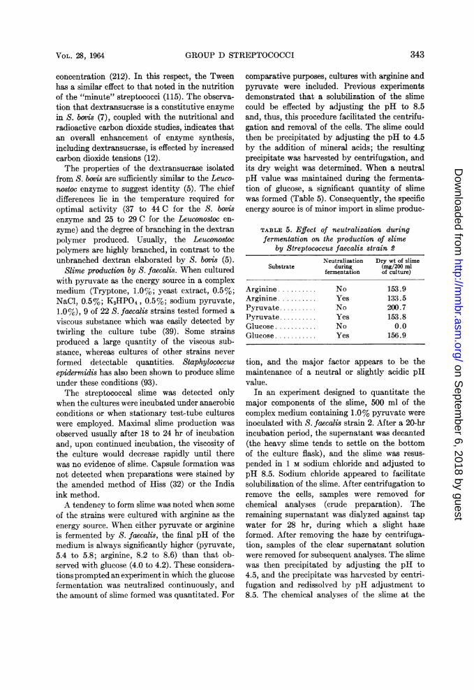
Slime production by S. faecalis. When culturedwith pyruvate as the energy source in a complexmedium (Tryptone, 1.0%; yeast extract, 0.5%;NaCl, 0.5%; K2HPO4, 0.5%; sodium pyruvate,1.0%), 9 of 22 S. faecalis strains tested formed aviscous substance which was easily detected bytwirling the culture tube (39). Some strainsproduced a large quantity of the viscous sub-stance, whereas cultures of other strains neverformed detectable quantities. Staphylococcusepidermidis has also been shown to produce slimeunder these conditions (93).The streptococcal slime was detected only
when the cultures were incubated under anaerobicconditions or when stationary test-tube cultureswere employed. Maximal slime production wasobserved usually after 18 to 24 hr of incubationand, upon continued incubation, the viscosity ofthe culture would decrease rapidly until therewas no evidence of slime. Capsule formation wasnot detected when preparations were stained bythe amended method of Hiss (32) or the Indiaink method.A tendency to form slime was noted when some
of the strains were cultured with arginine as theenergy source. When either pyruvate or arginineis fermented by S. faecalis, the final pH of themedium is always significantly higher (pyruvate,5.4 to 5.8; arginine, 8.2 to 8.6) than that ob-served with glucose (4.0 to 4.2). These considera-tions prompted an experiment in which the glucosefermentation was neutralized continuously, andthe amount of slime formed was quantitated. For
comparative purposes, cultures with arginine andpyruvate were included. Previous experimentsdemonstrated that a solubilization of the slimecould be effected by adjusting the pH to 8.5and, thus, this procedure facilitated the centrifu-gation and removal of the cells. The slime couldthen be precipitated by adjusting the pH to 4.5by the addition of mineral acids; the resultingprecipitate was harvested by centrifugation, andits dry weight was determined. When a neutralpH value was maintained during the fermenta-tion of glucose, a significant quantity of slimewas formed (Table 5). Consequently, the specificenergy source is of minor import in slime produc-
TABLE 5. Effect of neutralization duringfermentation on the production of slime
by Streptococcus faecalis strain 2
Neutralization Dry wt of slimeSubstrate during (mg/200 ml
fermentation of culture)
Arginine .......... No 153.9Arginine .......... Yes 133.5Pyruvate .......... No 200.7Pyruvate.......... Yes 153.8Glucose ........... No 0.0Glucose ........... Yes 156.9
tion, and the major factor appears to be themaintenance of a neutral or slightly acidic pHvalue.
In an experiment designed to quantitate themajor components of the slime, 500 ml of thecomplex medium containing 1.0% pyruvate wereinoculated with S. faecalis strain 2. After a 20-hrincubation period, the supernatant was decanted(the heavy slime tends to settle on the bottomof the culture flask), and the slime was resus-pended in 1 M sodium chloride and adjusted topH 8.5. Sodium chloride appeared to facilitatesolubilization of the slime. After centrifugation toremove the cells, samples were removed forchemical analyses (crude preparation). Theremaining supernatant was dialyzed against tapwater for 28 hr, during which a slight hazeformed. After removing the haze by centrifuga-tion, samples of the clear supernatant solutionwere removed for subsequent analyses. The slimewas then precipitated by adjusting the pH to4.5, and the precipitate was harvested by centri-fugation and redissolved by pH adjustment to8.5. The chemical analyses of the slime at the
343VOL. 28, 1964
on Septem
ber 6, 2018 by guesthttp://m
mbr.asm
.org/D
ownloaded from

BACTERIOL. REV.
three stages of purification are presented inTable 6. Acid precipitation of the slime alteredsignificantly its chemical composition. From theanalyses of the preparation obtained after dialy-sis, it appears that the slime consists predomi-nantly of protein (54%) and RNA (35%).Samples taken after dialysis and after acid
precipitation (the latter sample was dissolved atan alkaline pH) were placed on a Sephadexcolumn and eluted with 1 M NaCl; the fractionsobtained were analyzed spectrophotometrically.Acid precipitation of the dialyzed preparationaltered its fractional-elution diagram, as com-
TABLE 6. Comparison of analyses of Streptococcusfaecalis slime in different stages of purification
Analysis* Crudet Prepn after Aciddialysis precipitated
mg/mi mg/mi mg/mi
Carbohydrate. 0.125 0.08 0.03DNA.......... o.4 0.06 (6.9)t 0.06 (9.5)RNA .......... 0.52 0.30 (34.5) 0.15 (23.8)Protein ........ 1.24 0.51 (54.2) 0.40 (63.5)Dry weight.. 0.94 0.63
* The following references state the method em-ployed in the respective analyses: total carbohy-drate (26); DNA (199); RNA (122); and protein(112).
t Total volume of crude preparation was 140 ml.t Figures in parentheses represent per cent of
dry weight.
pared with the dialyzed preparation's pattern(Fig. 1). The acid precipitation not only de-creased the elution range, but it also increasedthe concentration of the fractions. These results,coupled with the altered chemical composition ofthe acid-precipitated preparation, tend to negatethis step in the purification procedure. The be-havior of the slime material in Sephadex columns,its precipitability at acidic pH values and solu-bility at an alkaline pH (123), as well as itschemical composition, favor the probability thatthe major slime component consists of a RNAnucleoprotein.A review of the literature has yielded no infor-
mation on the production of slime, as distinctfrom capsule formation, by the enterococci.Reports on the formation of an RNA slime by anymicroorganism are few, and the elaboration of aRNA nucleoprotein slime has not been reported.
Three theories have been advanced to account
for the production of an extracellular slime ofnucleic acid nature. The formation of DNA(deoxyribonucleic acid) slime by staphylococcimay result from an inhibition of deoxyribonu-clease production (27). A deranged metabolism,involving the accumulation of slime, has alsobeen suggested (24). Another possibility involvesan altered permeability of the cells, such as thatobserved in DNA slime production by Micro-coccus halodenitrificans; however, the same wasnot true with another halophile, Vibrio costicolus(188).
Dialyzed Acid-Precipitated1.8 - Sample Sample
1.4-
E 1.0 X Protein1U0-C
E~ 0.6
Acid0.2 L
10 12 14 16 18 20 22 10 12 14 16 18Fraction Number
FIG. 1. Comparison of Sephadex G-50 elutiondiagrams of two slime preparations. Each fractioncontained 4.2 ml. Nucleic acid and protein wereestimated spectrophotometrically by optical-densitydeterminations at 260 and 280,, respectively.
The chemical composition of the slime isolatedfrom the enterococci, namely, an RNA-proteincomplex with small concentrations of DNA,does not favor a concept of lysis with the produc-tion of slime. With the meager evidence ob-tained, it would appear that alkaline culture ofS. faecalis may result in a deranged metabolismwith, perhaps, an attending altered permeability.
Unusual Characteristics of EnterococciIn recent years, a number of reports have
appeared in which physiological characteristicsnot associated with classical descriptions ofenterococci are related to specific strains. This,perhaps, is to be expected as more detailed in-vestigations are undertaken and additionalstudies on large culture collections are performedto detect these variants. The observations donot detract from the classical descriptions of the
344 DEIBEL
on Septem
ber 6, 2018 by guesthttp://m
mbr.asm
.org/D
ownloaded from

GROUP D STREPTOCOCCI
more commonly occurring strains, but serve toillustrate the flexibility that is incorporated in allbacterial taxa.
Motility in streptococci other than the group
D organisms is a rarity. The nonmotile nature isoften extended to include all streptococci, butthis generalization is contrary to past and recentstudies with the enterococci. Accounts of mo-
tility in enterococcus strains published prior to1957 were considered by Graudal (66, 67). Thisinvestigator studied the characteristics of 129motile strains, predominantly of fecal origin,and found them all to be enterococci, includingboth S. faecalis and S. faecium. All but one
strain possessed the same flagellar antigen.Subsequent studies of the motile variety indi-cated that these organisms are widely distributedand not characteristic of any given environment(87, 105). Aside from their motility, these strainsare typical enterococci, and the establishment of avarietal status (i.e., mobilis) has little taxonomicvalue and does not appear to be warranted.
S. faecalis has been reported to clot citratedrabbit plasma (56). Only citrate-fermentingstrains evidenced this property, and culturesnot adapted to citrate required a 7- to 8-hrincubation period in the test system, in contrastto the 3- to 4-hr period required by adaptedstrains. None of the strains clotted plasma con-
taining other anticoagulants, such as fluoride,heparin, or oxalate, whereas staphylococciclotted plasma regardless of the anticoagulantsemployed. It was concluded that plasma coagula-tion was due to metabolic decomposition of citraterather than the possession of a coagulase enzyme.
In another study (17), Aerobacter aerogenesdemonstrated similar activity, and 4 of 13enterococcus strains also clotted citrated plasma.The activity of both the Aerobacter and entero-coccus strains was inhibited by sodium fluoro-acetate. Peculiarly, 0.1 M potassium cyanidehastened clotting by the enterococci. In a subse-quent study, Wood (210) refuted the correlationbetween citrate utilization and clotting activity.A nutrient broth was prepared in which theculture had grown within a cellophane container.It was reasoned that neither bacteria nor extra-cellular enzymes would diffuse into the testbroth. After incubation, the sterile broth coagu-
lated plasma in less than 5 hr. The diametricresults obtained in these two studies (56, 210)have not been resolved, and the "coagulase
activity" of the enterococci cannot be statedwith certainty.
Langston and Williams (107) described thereduction of nitrate by two strains of S. faecium.The reduction was inhibited by oxygen and cy-anide, and the optimal pH for activity was 8.0.Although a number of reports have appearedassociating this activity with lactobacilli (156),this is the only study in which nitrate reductaseactivity has been implicated in enterococcalphysiology.The production of L( +)lactic acid is generally
considered to be characteristic of all streptococci.However, in two studies, the production of in-active lactic acid by enterococci has been re-ported (104, 111). Evidently, these strains, un-like the typical enterococci, possess a lactic acidracemase.
NUTRITION
In the following discussion, no attempt willbe made to cover exhaustively the nutritionalaspects of the group D streptococci, as much ofthis material has been incorporated in previousreviews (68, 125). Some general aspects will bepresented and specific topics which are peculiarto this group of streptococci will be entertained.
Vitamins and CofactorsThe uniquely simple requirements of S. bovis
for exogenous amino acids and vitamins serveto differentiate this organism from all otherstreptococci. Most S. bovis strains require biotinand are stimulated by thiamine, pantothenate,and nicotinate when the cultures are incubatedunder atmospheric conditions (135). When culti-vated under anaerobic conditions with an in-creased carbon dioxide tension in a mediumcontaining Tween 80 (thus obviating a biotinrequirement), no vitamin requirements weredemonstrable (57). Strain variation exists;some require pantothenate, and others arestimulated by thiamin (12). Generally, S. bovisdoes not require exogenous purines or pyrimi-dines; however, here again, strain variationsdo occur (12).
Unlike S. bovis, the nutritional requirements ofthe enterococci are complex. The majority of thestrains require biotin, nicotinate, pantothenate,riboflavine, and pyridoxine (132). Characteristi-cally, S. faecium requires folic acid in contrast toS. faecalis, and this requirement has taxonomic
345VOL. 28, 1964
on Septem
ber 6, 2018 by guesthttp://m
mbr.asm
.org/D
ownloaded from

BACTERIOL. REV.
utility in differentiating these species (44).Folate can be replaced by thymine in the nutri-tion of S. faecium (68). This observation, coupledwith the known role of folate in purine synthesis,is indicative of its function in the metabolism ofS. faecium. The absence of a folate requirementby S. faecalis suggests not only its synthesis butthat of thymine as well.
Lipoate requirements of enterococci. O'Kaneand Gunsalus (138) first observed that cellsuspensions of S. faecalis required an unknownfactor present in yeast extract (pyruvate oxida-tion factor, POF) which was necessary for theaerobic oxidation of pyruvate. In subsequentstudies, the factor was purified, crystallized, andfinally synthesized, and it is commonly referredto as lipoic acid. Several reviews have appearedcovering its chemistry, function, assay, andbiological spectrum of activity (70, 71, 191).
S. faecalis requires lipoate when grown in a
semidefined, casein-hydrolysate medium con-
taining pyruvate as the energy source. Thisrequirement appears to be linked specificallywith pyruvate metabolism, as it is not demon-strable when hexoses, hexitols, ribose, gluconate,or glycerol is supplied as the energy source. Theadaptive nature of the pyruvate fermentationnecessitates growth of the inoculum culture in a
complex medium with pyruvate as the energysource prior to its growth in the casein-hydroly-sate medium. Glucose-grown inocula give erraticand variable responses when tested in the semi-defined medium (energy source, pyruvate) withvarious concentrations of lipoate (38).When a pyruvate-adapted inoculum is used, a
dose-response curve is obtained with 0.8 to 4.0m/ug of D, ilipoate per 10 ml of medium. It hasbeen demonstrated previously that only theL form of lipoate is active for S. faecalis (86).Thus, these concentrations represent one-half ofthe utilizable lipoate. In our laboratory, a test-tube bioassay method (similar to that employedin the B-vitamin assays with other lactic acidbacteria) has been used to quantitate lipoate withS. faecalis strain FB82. The assay is performedwith 10 ml of the casein-hydrolysate medium(energy source, pyruvate) in 18-mm test tubesand varying concentrations of lipoate (0.8 to4.0 mjug per 10 ml of medium). The pyruvate-adapted inoculum is washed once with water anddiluted tenfold, and one drop is used to inoculatethe assay tubes. After 18 to 20 hr of incubation
at 37 C, the turbidity is measured, and a dose-response curve is plotted. The method comparesfavorably with other assay methods and offersthe advantage of simplicity. Moreover, the re-quirement is specific in that it cannot be replacedby acetate, nor does acetate exert a sparingeffect.
Initial attempts to grow S. faecalis in a lipoate-supplemented, casein-hydrolysate medium withpyruvate as the energy source under conditionsof strict aerobiosis (vigorous shaking in air)met with failure. Regardless of the methodemployed to grow the inoculum (aerobically oranaerobically), no growth was manifested in
TABLE 7. Effect of oxygen on the lipoate requirementof Streptococcus faecalis strain FB82*
Concn of DL-lipoate neces-Condition of incubation sary for half-maximal growth
(myg/10 ml of medium)
Anaerobic ............. 0.55Static-atmospherict ..... 1.15Aerobict ............. 20.0
* Casein-hydrolysate medium; 20 hr. Energysource, 1.0% pyruvate.
t Cultured in 18-mm test tubes (10 ml).t Cultured in 125-ml flasks (10 ml) and incu-
bated on a reciprocating shaking apparatus.
aerobically incubated cultures. Subsequent ex-periments revealed that the optimal concentra-tion of lipoate under aerobic conditions wassignificantly higher than that under anaerobicconditions, and thereby offered an explanationfor the failures encountered in the initial experi-ments. Thus, the amount of lipoate required forgrowth may be expressed as a function of theoxygen tension under which the culture is incu-bated (Table 7). No explanation is available toaccount for this increased lipoate requirement,although the results may indicate either anoxidation or partial destruction of the lipoate oran increased requirement by the organism whencultured aerobically.The addition of reducing compounds, such as
thioglycolate and ascorbate, decreased thelipoate requirement, when cultures were incu-bated under static-atmospheric conditions, to alevel approximating that observed when thecultures were incubated anaerobically. However,when cultures were incubated aerobically withthese compounds in concentrations as high as
346 DEIBEL
on Septem
ber 6, 2018 by guesthttp://m
mbr.asm
.org/D
ownloaded from

GROUP D STREPTOCOCCI
1.0%, no sparing effect on the lipoate require-ment was demonstrable.When S. faeium is cultured with any fermenta-
ble carbohydrate under anaerobic or static-atmospheric conditions of incubation, lipoate isnot required. In contrast, 11 of 20 strains testedrequired lipoate when cultured aerobically withglucose as the energy source. Additional experi-ments with these 11 strains indicated a lipoaterequirement for aerobic growth with all fermenta-ble substrates. Included in the 52 compoundstested were pentoses, hexoses, polyols, di- andtri-saccharides, and organic acids. Conversely,lipoate was not required for growth of the re-
dentified factor were required (197). Recently, areinvestigation of the process established theidentity of the unknown factor as lipoic acid(31). However, the requirement of this strain forlipoate, regardless of the substrate employedunder aerobic conditions, may indicate its in-volvement not only in glycerol oxidation but,more generally, in aerobic metabolism.
In view of the varied reports regarding lipoaterequirements of the enterococci, an attempt willbe made to collate the independent studies andperhaps clarify the results so that orientation forfurther study may be enhanced. To recapitulate,studies in our laboratory have demonstrated an
TABLE 8. Comparison of the effect of energy source and oxygen on the lipoaterequirement of Streptococcus faecalis and S. faecium
Condition of incubation*
Organism Energy source Aerobic Anaerobic
+ Lipoate - Lipoate + Lipoate - Lipoate
S. faecalis Glucose 105 97 110 105FB82 Pyruvate 110 12 126 9
S. faecium Glucose 72 1 105 110F24 Ribose 80 0 110 110
S. faecium Glucose 62 90 100 105Igau Ribose 68 95 110 95
* Optical density X 100; 48-hr incubation period.
maining nine S. faecium strains, regardless of theenergy source or condition of incubation. Growthresponses of S. faecalis, and lipoate-requiringand nonrequiring strains of S. faecium, are com-pared in Table 8. Neither S. faecalis FB82 nornine other representative S. faecalis strains re-quire lipoate when cultured aerobically withglucose as the energy source.The lipoate requirement for one-half maximal
growth of S. faecium (energy source, glucose)under aerobic conditions is approximately 0.45mjug per 10 ml of medium. Some strain variationwas encountered, and growth with concentra-tions of lipoate producing submaximal responseswas erratic. As noted in Table 7, this concentra-tion is significantly lower than that required byS. faecalis under aerobic conditions, and approxi-mates the level required by S. faecalis underanaerobic conditions.
In a study of the cofactor requirements forglycerol oxidation by cell suspensions of S.faecalis F24 (S. faecium), it was observed thatthiamine, nicotinate, riboflavine, and an uni-
absolute requirement for lipoate when S. faecalisis grown with pyruvate as the energy source.This requirement is specific and cannot be re-placed with acetate. The amount of lipoate re-quired for growth may be expressed as a func-tion of the oxygen tension under which theculture is incubated, and significantly higher con-centrations are required under highly aerobicconditions. S. faecium, on the other hand, doesnot utilize pyruvate as an energy source and doesnot require lipoate or acetate when culturedanaerobically with any energy source. However,when cultured aerobically, approximately 50%of the strains require lipoate regardless of theenergy source. In contrast, S. faecalis does notrequire lipoate when grown aerobically withglucose as the energy source.
In a study employing S. faecalis 734, the or-ganism was incubated under static-atmosphericconditions with glucose as the energy source,and it was observed that acetate or a preparationof lipoate (POF) was not required for growth(113). However, cultures incubated with glu-
347VOL. 28, 1964
on Septem
ber 6, 2018 by guesthttp://m
mbr.asm
.org/D
ownloaded from

BACTERIOL. REV.
conate as the energy source manifested an acetateor POF requirement. Carbon dioxide (bicar-bonate), or compounds whose metabolismresulted in carbon dioxide production, replacedthe acetate or POF requirement in gluconate-grown cultures. The fermentation of gluconateby this strain suggests its classification (in thecurrent scheme) as S. faecalis. However, studiesin our laboratory have failed to associate alipoate requirement with the gluconate fermenta-tion by other S. faecalis strains. The relationshipof the lipoate requirement (as well as the acetateand carbon dioxide effect) with the gluconatefermentation merits further study and mayinvolve strain variation.Shockman (175) observed that acetate is re-
quired only under aerobic conditions when S.faecalis R (S. faecium) and another strain (S.faecalis ATCC 9790, not yet identified in termsof current classification) are cultured with glucoseas the energy source. A combination of lipoateand thiamine replaced the acetate requirement.Under anaerobic conditions of incubation, neitheracetate nor lipoate was required. Thus, the re-sults obtained in our laboratory with the lipoate-requiring S. faecium strains are identical tothose observed previously by Shockman. Thesestudies suggest that the aerobic requirement ofS. faecium for lipoate can be replaced by acetate.
It has been demonstrated (192) that relativelylarge amounts of propionate (1 to 3 mg per mlof medium) will inhibit the growth of S. faecalisR (S. faecium) and that this inhibition can bereversed competitively with acetate or protogen(lipoate). Oleate reversed the inhibition non-competitively. An assay method was proposedsubsequently (191) for acetate or lipoate withpropionate-inhibited cultures of S. faecalis ATCC8043 (S. faecium). Hill (81) first observed aninhibition of growth with propionate and S.faecalis 8043 (S. faecium) and postulated that theinhibition resulted from the combination ofpropionate with CoA.
Collectively, the results obtained with the S.faecium strains indicate that acetate is an essen-tial metabolite regardless of the oxygen tension.Anaerobically, this requirement can be demon-strated by propionate inhibition, and the in-ference can be made that a sufficient quantity ofacetate is metabolically produced to satisfygrowth requirements. The aerobic requirementfor exogenous acetate or lipoate may reflect either
an inhibition of acetate generation or an increasedphysiological requirement under aerobic growthconditions. Although these considerations mayaid in an explanation of the results obtained ingrowth studies, the lipoate requirement in glyc-erol oxidation by cell suspensions may involveother functions, such as electron transfer. Thisrole of lipoate has been established in the catabo-lism of lactate by Butyribacterium rettgeri (100).The possible inhibition effected by propionateeither aerobically or anaerobically with glucoseor pyruvate as the energy source has not beeninvestigated in the metabolism of S. faecalis.In summation, it would appear that progresshas been made in elucidating the role of lipoatein enterococcus nutrition; however, completeresolution of the problem must await furtherstudy.
Amino Acids
Growth requirements. In the first report de-scribing the amino acid requirements for thegrowth of S. bovis, the absence of a requirementfor any specific amino acid was noted (135).In a medium containing only arginine and gluta-mate, satisfactory serial growth occurred withthe majority of the strains studied. Supplementa,tion of a medium containing arginine with leucine,isoleucine, threonine, and equal amounts ofphenylalanine and tyrosine produces an antago-nistic effect presumbly by blocking the synthesisof other amino acids (200). Subsequently, anenhanced growth response was observed in thearginine-containing medium when bicarbonatewas added (150). Bicarbonate also reversed theinhibitory effect of isoleucine, leucine, and otheramino acids, and the stimulatory effect was asso-ciated with growth in the lag phase (151). Asdiscussed previously, Wright (212) studied theincorporation of carbon dioxide, associated itsfixation in asparate, and noted the generalizedstimulation of growth which was ascribed to anincreased concentration of intermediates neces-sary for protein and nucleic acid synthesis. Paul(143) observed a requirement for numerous aminoacids and a peptide factor for satisfactory growthof strains isolated from sheep and bovine sources.These results indicate a more complex nutritionthan that described by previous investigators.Included in the study were strains of S. equinus,and their requirements paralleled those of S.bovis. In this study, no attempt to grow S. bovis
348 DEIBEL
on Septem
ber 6, 2018 by guesthttp://m
mbr.asm
.org/D
ownloaded from

GROUP D STREPTOCOCCI
in a simple medium with arginine and glutamatewas reported. The complex requirements observedmay reflect amino acid antagonisms, as demon-strated previously.Almost simultaneously, reports from two
laboratories indicated that S. bovis was capable ofutilizing ammonium salts as a sole source ofnitrogen (152, 208). Growth with ammoniumsalts was stimulated by acetate (152) or bi-carbonate, and glutamine also served as a solesource of nitrogen (208). Growth with ammoniumsalts, but not with glutamine, required an un-known substance formed by autoclaving phos-phate with glucose (209). The addition of poten-tial electron acceptors or a lowering of the pHvalue of the medium replaced this requirement.Asparagine also satisfies the nitrogen require-ment (18), and enzymatic studies indicate thatglutamine and asparagine are the most likelyprimary products of ammonia incorporation(18).The unique ability of S. bovis to utilize am-
monium salts as a sole source of nitrogen is notshared by other Streptococcus species. In com-parison, the enterococci possess relatively com-plex requirements in that at least ten aminoacids are necessary for growth, and still othersare significantly stimulatory (116, 132). Varia-tions in the specific requirements occur and mayrepresent strain differences. Also, other factorssuch as the composition of the medium and themethod of sterilization employed may be im-plicated (116). As yet, the requirements of S.faecium and S. faecalis have not been compared.
In our laboratory, it has been observed thatreplacement of an acid and enzymatic hydrolysateof casein with amino acids results in an inferiorgrowth response. The possible involvement ofpeptide stimulation was considered. However,Langston (personal communication) demon-strated a doubling of the growth response of S.faecalis with 0.25% sodium carbonate in anadequately buffered synthetic medium. Theseresults parallel the carbon dioxide stimulationobtained with S. bovis, and indicate the desira-bility of additional studies.
Transaminase activity and D-amino acid utiliza-tion. In a study of the transaminase activity ofS. faecalis R (S. faecium), the a-keto acids cor-responding to histidine, leucine, isoleucine,tryptophan, tyrosine, and valine were trans-aminated, and the activity was pyridoxal-linked
(68). The inactivity of a-ketoglutarate and oxal-acetate was noted, and deemed peculiar in viewof the known glutamic and aspartic transaminaseactivity. The impermeable nature of these com-pounds was suggested to account for their in-activity. Only the a-hydroxy acids correspond-ing to leucine and valine were utilized, and adehydrogenation to the a-keto acid and subse-quent transamination were shown to be involved.The pyridoxal-linked, alanine racemase ac-
tivity of the enterococci, and the reciprocity ofD-alanine and pyridoxal have been consideredpreviously (68). A methionine racemase, whichis not pyridoxal-linked, has been reported (176).This racemase is active in the post-log phase ofgrowth and affords the utilization of D-methioninebut only in the presence of the L isomer. Unlikeother amino acids, L-methionine is not assimi-lated quantitatively by S. faecalis 9790, andapproximately one-fifth of it is metabolicallyconverted to extracellular D-methionine whichis subsequently oxidized nonbiologically to thesulfoxide (177).
SEROLOGYGroup Antigen
The early literature concerning the serology ofthe group D streptococci was considered exten-sively by Skadhauge (181). As mentioned previ-ously, all enterococci fulfilling the Shermancriteria possess the group D antigen. In addition,the presence of this antigen has been demon-strated in S. boyts and S. equinus. The intracellu-lar location of the antigen in all group D strepto-cocci, as well as its chemical identity, meritdetailed consideration.
Initial work pointed out the variability of re-sponse and the difficulty of producing a potentgroup D antiserum in rabbits (166). Strain varia-tion was observed, and repeated immunizing doseswere necessary over protracted periods of time.Quite often, commercially produced, group anti-sera are encountered that possess extremely lowlevels of group-specific antibody (168). In recentyears, studies by Shattock and others have offeredan explanation for these peculiar results. In con-trast to the cell-wall location of the group antigenin other streptococci, the group D antigen occursintracellularly (54, 92). Previously, Shattock(166) observed the ease with which the antigencould be demonstrated in strains of S. bovis whenthe cell preparation employed for immunization
VOL. 28, 1964 349
on Septem
ber 6, 2018 by guesthttp://m
mbr.asm
.org/D
ownloaded from

BACTERIOL. REV.
was first mechanically shaken to disrupt the cells.Another factor enhancing serological demonstra-tion of the antigen, and also associated with itsintracellular nature, is its concentration by alco-holic precipitation prior to use in the precipitintest (166).The intracellular location of the antigen in all
group D streptococci (92) offers an explanationfor the refractory nature of most strains to theserological demonstration of the group antigen.Shattock (168) surmised that the antigen is notreadily available at the site of antibody forma-tion, and that disruption of the cell afforded lib-eration of the antigen and thus indirectly con-tributed to its antigenicity. Likewise, extractionand precipitation effects concentration and de-tection of the antigen in extracts.The procedures employed to prepare the anti-
gen for immunization and the extracts for sero-logical testing were considered by Shattock (166).Medrek and Barnes (118) demonstrated highyields of the antigen when the culture mediumcontained 0.5 to 1.0% glucose and poor yields inmedia containing low glucose concentrations.Characteristically, growth of S. bovis, S. equinus,and S. faecium in the Todd-Hewitt medium re-sulted in an inferior group reaction in contrast tothe results obtained with S. faecalis. No explana-tion was available for this difference, althoughgrowth in glucose-containing media resolved thesedifferences, and group reactions were enhanced inall species. The possible involvement of nutri-tional conditions affecting the synthesis of thegroup antigen has been suggested (92), sincemedia which support excellent growth of the or-ganism do not always produce cells with sub-stantial quantities of antigen.
Equivocal results obtained in cross-reactions ofS. bovis extracts with enterococcal group D serahave been noted (130, 165, 171), and Niven (130)observed the failure of absorption tests to resolvethese cross-reactions. These results led to Shat-tock's demonstration of the group D antigen inS. bovis (166). Subsequently, Smith and Shattock(183) observed variable results with intact cells ofboth S. bovis and S. equinus. However, in disinte-grated preparations the antigen was demonstrablein both species. The results with S. equinus wereconfirmed in another laboratory (60), and Jonesand Shattock established the identity of the groupD antigen in the enterococci and S. bovis (92).The peculiar location of the group antigen in
the group D organisms serves to differentiatethem from all other streptococci. The exact loca-tion of the antigen has been investigated, andHijmans (79) observed the absence of the antigenin L forms of enterococci, thus obviating itspossible cytoplasmic location. Shattock andSmith (169) further reported the occurrence ofthe antigen in cultures of one L form after con-tinual subculture in a medium containing an in-creased glucose concentration. However, approxi-mately 97% of the antigen was found in themedium and not within the L form, and 85% ofthe group antigen was released during digestionof intact cells with a phage-associated, cell-walllysin. These results indicated the absence of theantigen in the cytoplasm and, when associatedwith previous studies regarding its absence in thecell wall, led to the conclusion that the antigen islocated between the cytoplasmic membrane andthe cell wall. These conclusions have been con-firmed in another study (206).
Recently, the chemical nature of the group Dantigen has been elucidated (205, 206) and foundto be a peculiar type of glycerol-teichoic acid.Unlike other teichoic acids in which the D-alanineis linked to the polyol, the group D antigen con-tains D-alanine in ester linkage with the hydroxylgroups of glucose, and the glucose is esterified tothe C2 group of glycerol. The glycerol moleculesare joined through phosphodiester linkages in the1, 3 positions to form the polymer. The high sugarcontent and amino acid attachment to the glucoseresidues were considered peculiar to the group Dantigen (205).Some variation was noted in the teichoic acids
from S. faecalis 39 and S. faecium 8191 (205). InS. faecalis 39, the side chain consisted of a glucosedisaccharide (kojibiose) in contrast with theglucose trisaccharide (kojitriose) of S. faecium8191. In further contrast, the S. faecium polymercontained ilysine as well as D-alanine, whereasthe S. faecalis complex contained only D-alanine.Evidence was presented indicating that the im-munological specificity resided in the carbohy-drate side chains, since no reaction occurred witha poly-glycerol phosphate preparation obtainedfrom a group A Streptococcus. Indirect results andthe analogous lack of amino acid participation inthe immunological specificity of Staphylococcusteichoic acids led to the conclusion that aminoacids were not involved as immunological de-terminants in the group D polymer.
350 DEIBEL
on Septem
ber 6, 2018 by guesthttp://m
mbr.asm
.org/D
ownloaded from

GROUP D STREPTOCOCCI
In the isolation of the group D teichoic acid, theformation of complexes with cellular RNA was
noted. It was concluded, however, that teichoicacid did not occur in the cell as an RNA-teichoicacid complex and that, in the course of cellulardisruption and purification, the complex was
formed (205).In the traditional method of preparing extracts
for serological studies, the antigen is acid-ex-tracted from the cells with heating. The labilityof glucosyl residues on the side chain of the poly-mer was suggested as a possible source of error inthis procedure (205), which may account for theserological identity of the group D antigen in theenterococci and S. bovis, as reported previously(92). The possibility of subtle differences in thecomposition of the antigen as a function of speciesexists; however, further studies are necessary foraffirmation or negation of this speculation.
Type Antigens
S. faecalis. Skadhauge (181) observed boththermostable 0 antigens and thermolabile Kantigens in S. faecalis strains. The K antigenswere associated with the surface of the cell (en-velope antigen), and were deemed unsuitable forserological studies because of variability incurredby storage and repeated subculture of the strains.Six types were established on the basis of the 0antigens. In his study of 350 enterococcus strains,Nyman (136) reported that 90% of the typablestrains belonged to three types, and one of thethree types was associated principally withstrains from pathological material. The unpub-lished studies of Sharpe, as quoted by Shattock(168), indicate that approximately 20 types ofS. faecalis exist, and that these antigens are dis-tinctive and not shared by other members of thegroup D streptococci.
Elliott (54) examined the chemical compositionof the type-specific antigens in three differentserological types of S. faecalis and one S. faeciumvar. durans type. In each instance, the antigenwas composed primarily of glucosamine, rham-nose, and glucose. The antigens formed an
integral part of the cell wall, and this situation isanalogous to the group antigens of other strepto-cocci. It is assumed that these type antigens are
identical with the 0 antigens of Skadhauge.S. faecium. The cell-wall, type-specific antigens
of S. faecium and its variety durans are also dis-tinctive within this physiological group (no cross-
reactions with S. faecalis), and a sharing ofcommon type antigens exists between strains ofS. faecium and its variety durans (164). A com-mon type-specific antigen has been reported tooccur in two strains of S. faecium (163) and astrain of S. lactis (group N). Previously, Niven(130) also noted the existence of a common typeantigen in certain enterococcus and S. lactisstrains.
S. bovis. In an extensive study of the typespecificity of S. bovis, Medrek and Barnes (119)observed that 75 of 152 strains were typable, andthese 75 strains fell into 12 types. In one type,represented by one strain, the antigen was lo-cated in the cell wall; in the remaining types, itoccurred in the capsule. Heterogeneity was indi-cated by the inability of a number of S. bovisstrains, isolated in other studies, to react withthe 12 type-specific antisera prepared in thisstudy. In addition, none of the 12 type sera reactedwith other group D organisms, indicating sero-logical distinction from these streptococci. How-ever, in other studies it was observed that S. bovisshares a common type antigen with streptococciof group E (145) and group N (146). Clearly, thetype antigens of S. bovis are somewhat hetero-geneous. They may differ in their location, but,unlike the enterococci, they occur generally in thecapsular material. The type-specific antigens ofS. equinus have not been studied.
CELL STRUCTURE
Cell-Wall Chemistry
Since a comprehensive review of cell-wall me-tabolism and composition appeared recently(144), only specific topics related to the group Dstreptococci will be included in this discussion. Asstated previously, the type-specific antigens, lo-cated in the cell wall, are species specific in theenterococci, and on this basis one might expectchemical variation in the wall composition ofthese two species. However, in the limited studiesconducted to date, no significant differences havebeen observed. This may reflect either subtledifferences in composition or variance in chemicalstructure within the realm of similar composition.Further, the taxonomic dichotomy of S. faecalisand S. faecium is not, as yet, recognized widely,and minor variations in cell walls associated withindividual species may have been overlooked.The overall composition of the cell walls of the
351VOL. 28, 1964
on Septem
ber 6, 2018 by guesthttp://m
mbr.asm
.org/D
ownloaded from

BACTERIOL. REV.
group D streptococci is similar. In the only com-parative study of group D wall composition (threeS. faecalis strains and one each of S. faecium, S.bovis, and S. faecium var. durans were examined),lysine, glutamate, alanine, rhamnose, glucose, andglucosamine were the principal compounds de-tected, and each was common to all species (92).Galactosamine was also a principal component inall species except S. bovis. Mannose (a minorcomponent) has been reported to occur only inS. faecalis; however, in one strain (S. faecalis370) its presence was detected in two laboratories(34, 92) and its absence was reported from an-other laboratory (182). Also, Cummins andHarris (34) detected mannose in S. faecalis 6782,in contrast to the results obtained by Salton(157). Considerable variation has been observedin the occurrence of galactose. Two of three S.faecalis strains contained this sugar, as did astrain of S. faecium var. durans and S. bovis; theabsence of galactose was noted in S. faecium4MEC (92).As in other lactic acid bacteria, four major
fractions of polymeric compounds (mucopeptide,"protein," and ribitol and glycerol teichoic acids)have been identified (88) in S. faecalis R (S.faecium). D-Alanine occurs in the mucopeptide,and both rhamnose and D-alanine are common tothe two types of teichoic acid. Unlike the ribitolteichoic acid, the glycerol teichoic acid does notcontain an amino sugar. The occurrence of bothtypes of teichoic acid in the wall of S. faecalis wasobserved in another study (3). As yet, the rela-tionship between these teichoic acids and typespecificity has not been studied.
Cytoplasmic Membrane ChemistryThe chemical composition of the membrane in
S. faecalis 9790 was found to differ in preparationsof cells taken from the log and stationary phasesof the growth cycle (178). Log-phase membranescontained more protein (49 to 55%) and less lipid(28 %) than did stationary phase membranes(42% protein, 36% lipid). A large number ofvarious amino acids occurred in the protein frac-tion, and the relative absence of D-amino acidswas noted. The lipid-phosphorus concentration(3%), coupled with the low nitrogen content,indicated that the lipid consists primarily of phos-phatidic acids. In another study (15), the relativeabsence of rhamnose in membrane preparationsof S. faecalis R (S. faecium), as compared with its
occurrence in the cell wall, was noted and couldbe employed to indicate the degree of wall con-tamination in membrane preparations.
Nutritional Aspects of Wall Synthesis
Studies in Shockman's laboratory, concernedprimarily with S. faecalis 9790, divorced cell-wallsynthesis from active growth (179, 180). Wallsynthesis continued after growth cessation in theabsence of amino acids required for protoplasmicprotein synthesis but in the presence of aminoacids necessary for wall synthesis (i.e., lysine,alanine). Depletion of wall-required amino acidsin the medium resulted in rapid lysis.
Hydroxylysine does not replace the lysinegrowth requirement for S. faecalis R (S. faecium),but it does exert a sparing effect (147) and pre-vents lysine-depletion lysis (186). The absence ofhydroxylysine conversion from lysine has beendemonstrated, and cells grown with lysine containno hydroxylysine (194). The marked stimulationof growth by hydroxylysine observed in lysine-limiting media has been associated with activehydroxylysine incorporation in the cell wall. Cellscontaining hydroxylysine in their walls were moreresistant to autolysis, the lytic effects of neu-raminidase and penicillin, and the disruption bysonic oscillation, in comparison with cells grownsolely with lysine (187).
Neuraminidase SensitivityThe differential ability of neuraminidase to lyse
various living enterococcal species has not beenstudied extensively. Generally, some strains arerefractory to the lytic effect of this enzyme, andothers are lysed rapidly. Chesbro (28) observedsome indication of species variation within theenterococci. S. faecalis appeared to be relativelyresistant in comparison with most S. faeciumstrains. Lysis of S. faecium was enhanced bygrowth in a carbonate-containing medium andaccentuated by growth at pH 9.4. In contrast,supplementation of the medium with potassiumor sodium ions decreased the neuraminidase sensi-tivity. Abrams (1), working with S. faecalis 9790,associated the neuraminidase sensitivity of hisstrain with the absence of wall galactose, sinceSalton's neuraminidase-resistant strain containedthis sugar in the cell wall (158). This relationshiphas been questioned by Perkins (144). Utilizingthe action of neuraminidase, Bibb and Straughn(15) prepared protoplasts with S. faecalis 9790
352 DEIBEL
on Septem
ber 6, 2018 by guesthttp://m
mbr.asm
.org/D
ownloaded from

GROUP D STREPTOCOCCI
(178), S. faecium HF8AG (28), and S. faecalisF24 (S. faecium). Bleiweis and Zimmerman (16)observed that spheroplast formation could beinvoked by the action of neuraminidase on cellsgrown previously in the presence of penicillin.The employment of penicillin in the production ofenterococcal L forms was studied by Hijmans andKastelein (80).Using heat-treated cells (70 C for 15 min) and
the synergistic action of neuraminidase and tryp-sin, Hartsell and Caldwell (78) proposed a lyticscheme for the differentiation of the group Dstreptococci. The excellent agreement of thisscheme with the physiological classification sug-gested its employment as a rapid method for theidentification of these streptococci. In the pro-cedure, S. bovis can be separated from the entero-cocci because it is resistant to the combinedenzymes (neuraminidase and trypsin) while S.faecalis and S. faecium are lysed. S. equinus wasnot included in the study. When the procedurewas repeated with another sample of heated cells,an increase in the neuraminidase content, theomission of trypsin, and the addition of base atthe end of the reaction period resulted in the lysisof S. faecium and its variety durans but not of S.faecalis. S. faecium could be differentiated fromits variety durans in that the latter agglutinatedwhen acidified cell suspensions were heated. Con-firmation and extension of these results to includeS. equinus are lacking, and additional study isdesirable, especially in view of the possible easeand rapidity of species identification within thegroup D streptococci.
BacteriophagePublications prior to 1947 dealing with the re-
lationship between bacteriophage and group Dstreptococci were considered by Evans and Chinn(55). These investigators observed the lysis ofmost enterococci by a single phage strain; lessthan 50% of the strains were lysed by anothertype. No correlation between phage-induced lysisand the classification scheme employed at thattime could be made. Recently, the possiblespecificity of phages for the various group Dspecies has been investigated and two serologic-ally distinct phages either separately or in com-bination lysed 91% of the S. faecalis strains (30).A third serologically distinct phage lysed S.faecium, S. bovis, and atypical S. faecalis strains.As yet, a definite separation of group D species
by lysotyping has not been accomplished. Somespecific characteristics of group D bacteriophageshave been reported (155). Bleiweis and Zimmer-man (16) employed a lytic enzyme, obtained froma phage-infected culture of S. faccalis, to induceprotoplast formation, and this enzyme was usedby Shattock and Smith (169) in their study on thelocation of the group D antigen.
CURRENT CONCEPTS OF GROUP DSTREPTOcoccUs TAXONOMY
Two characteristics, common to all group Dstreptococci, are the ability to grow at 45 C andin media containing 40% bile. S. bovis and S.equinus do not initiate growth at 10 C, at pH9.6, or in media containing 6.5% NaCl, and thesespecies usually fail to hydrolyze arginine or de-carboxylate tyrosine. Generally, the enterococcigive opposite reactions in these tests; thus, theprimary differentiation of the two physiologicalentities is afforded.As mentioned previously, use of the term en-
terococcus or enterococci to connote specificallyS. faecalis or S. faecium, or both, has been em-ployed in this review. The author does not desireto stress or suggest the employment of thisterminology; however, its facility and conveniencein this restricted sense prompted its utilization.
EnterococciTo differentiate the enterococci from other
streptococei, Sherman (170) suggested the use ofcertain tolerance tests-specifically, the ability ofthese organisms to grow in the presence of 6.5%sodium chloride, at pH 9.6, and at 10 and 45 C,and to withstand heating at 60 C for 30 min.These tests are marginal in nature, and the oc-currence of strains that fail to give a positiveresult under one or more of these conditions is tobe expected. Some indication of the critical natureof factors associated with the performance ofthese tests has been demonstrated. White (203)reported that tolerance to 60 C for 30 min wascorrelated closely with the pH value of themedium. At pH 6.8, the highest heat toleranceoccurred, and the ability to withstand this tem-perature dropped precipitously on either side ofthis pH value. In addition, Chesbro and Evans(29) showed a marked effect of medium composi-tion and buffering system on the ability ofenterococci to grow at pH 9.6. The inclusion ofoleate in the medium and the replacement of
VOL. 28, 1964 353
on Septem
ber 6, 2018 by guesthttp://m
mbr.asm
.org/D
ownloaded from

BACTERIOL. REV.
glycine buffer with carbonate facilitated growthat pH values up to 10.5. Still another considera-tion is the physiological state of the organism.Recently isolated strains are encountered whichfail to grow under one or more of the toleranceconditions. However, after transfer in laboratorymedia, these strains may evidence growth underthe test condition that previously gave a negativeresult (114).
In all identification procedures, dependencemust be placed on a spectrum of characteristicspossessed by the strain in question, and its failureto comply in a few specific tests does not consti-tute sufficient grounds to negate speciation if itconforms with the overall species description, Asin any taxonomic scheme, the occurrence of sometransitional types is to be expected. Caution mustbe exercised, however, to define these types andnot to avoid their species establishment on thepremise of a few differing characteristics.
In the past, speciation of the enterococci hasbeen confusing, although an indication of someorder is emerging. Orla-Jensen (139, 140) and,subsequently, Gunsalus (69) recognized the twogrossly different, physiological types of entero-cocci, and the studies of Shattock (167), Skad-hauge (181), and Barnes (9) provided sufficientcharacterization to effect a species definition of S.faecium and S. faecalis. Subsequent investigationsin a number of laboratories have substantiatedthe validity of this speciation. Thus, on the basisof tellurite sensitivity, tetrazolium reduction,differential susceptibility to neuraminidase, fer-mentation pattern, various energy-utilizationtests, nutritional differences, and serological typespecificity, S. faecalis can be differentiated fromS. faecium (Table 9). Accessory tests, althoughnot as conclusive as those mentioned previously,include growth at 50 C by S. faecium, rapidreduction of litmus (8 hr) in milk cultures by S.faecalis, and reactions in blood-agar (Table 9).From the array of differentiating characteristics,it would appear that this speciation rests on afirm foundation.Some generalizations regarding S. faecalis and
S. faecium may be drawn from the informationgained to date. S. faecium grows at a higher tem-perature and tolerates heat to a greater extentthan does S. faecalis. This explains the commonoccurrence of S. faecium in marginally processed,canned hams. Also, there are indications that thisspecies is more tolerant to highly alkaline condi-
TABLE 9. Physiological characteristics employed todifferentiate group D streptococci
SpeciesPhysiological characteristic
S. fae- S.feaeS- bovis S uicalis cium S. "vs.Cus-
Fermentation (acidproduction)
Arabinose ..........Sorbitol ............Melibiose ..Melezitose ..........Mannitol ...........Glycerol (anaero-
bic) ..............Energy utilizationPyruvate ...........Arginine ...........Citrate .............Serine ..............Agmatine .........Gluconate ..........Malate .............
HydrolysisArginine ...........Agmatine ..........Starch .............
GrowthpH 9.6.............6.5% NaCl.........45 C...............50 C...............lOC...............40% bile...........0.05% tellurite.....
Folic acid require-ment .............
Tyrosine decarbox-ylation ...........
Tetrazolium reduc-tion ..............
Rapid reduction oflitmus (8 hr).....
Blood-agar reaction...Slime production
from sucrose.
_*
+++
+
++
+
++
+
V
+
±+
fl,a
vtV+
V
0t00000
0
+
+
±
V
0
aV
0000000
0
0
0a
* The symbols + and - represent a positive ornegative test, respectively.
t V denotes strain differences and a variable re-sult.
I Zero indicates insufficient data available.
tions of growth (29). In contrast to S. faecium,S. faecalis enjoys a metabolic diversity reflectedby its ability to obtain energy from a wide varietyof organic compounds.
354 DEIBEL
on Septem
ber 6, 2018 by guesthttp://m
mbr.asm
.org/D
ownloaded from

GROUP D STREPTOCOCCI
S. bovis and S. equinus
Serological studies have established definitivelythe presence of the group D antigen in S. bovisand S. equinus (92, 166), and consequently theseorganisms must be included in the group D strep-tococci in spite of the physiological differencesthat separate them from the enterococci. How-ever, the distinctiveness of these two species hasnot been resolved fully, and their overall physi-ological characteristics are similar. Slime produc-tion from sucrose by S. bovis and the inability ofS. equinus to ferment lactose are the chief differ-entiating characteristics. In a collection of 87 S.bovis strains, a correlation was noted betweenslime production, mannitol fermentation, anda-hemolysis (37). Seventy-one strains formedslime, produced an a reaction in blood-agar, andfailed to ferment mannitol. The remaining strainsdid not produce slime, but 12 fermented mannitol.On the basis of the fermentation pattern of thetwo groups, Smith and Shattock (183) observedsufficient variation to suggest separation into twospecies. In addition, S. equinus does not hy-drolyze starch to the level of reducing sugars. S.bovis hydrolyzes starch to the monosaccharidelevel, and produces acid in contrast to the partialhydrolysis and absence of acid formation effectedby S. equinus (52).From the studies completed to date, it would
appear that the polysaceharide-forming, manni-tol-nonfermenting strains of S. bovis form ataxonomic entity. However, the polysaccharide-nonforming, mannitol-fermenting types, as wellas the S. equinus strains, are not c4early defined.Undoubtedly, further study is indicated prior to adefinitive assessment of speciation among thesestreptococci.
In summary, the group D streptococci may beseparated into two physiological groups: the en-terococci and the S. bovis-S. equinus group. Alarge number of characteristics can be employedin the separation of S. faecalis and S. faecium, andthese streptococei appear to be distinctive at thespecies level. The S. bovis-S. equinus group is notseparated as readily, and additional study is indi-cated prior to definitive speciation.
COMMENTS REGARDING ISOLATIONThe increasing attention associated with the
occurrence of group D streptococci in foods andwater necessitates an improved methodology re-garding their selective isolation, quantitation, andspecies identification. In the past decade, a
multitude of various selective media have beensuggested for facilitating isolation and quantita-tion of these bacteria, and confusion existsregarding the efficacy of over 30 different mediathat have been proposed for this purpose. More-over, disagreement is encountered as to whatconstitutes a "fecal Streptococcus," in that someinvestigators include S. salivarius and S. mitis, aswell as other streptococci, in this category. Com-pounding this rather fluid situation is the lack ofagreement in speciation and the inadequate test-ing of the various media to ascertain theirspecificity as well as the possible differential in-hibition among specific group D species. Someindication of species selectivity in different mediawas reported by Chesbro and Evans (29). It wasobserved that cultivation in a carbonate-contain-ing medium at pH 10.5 favored the isolation ofS. faecium. In contrast, use of an azide-dextrosemedium favored the isolation of S. faecalis.The variety of media available to isolate group
D streptococci is similar to the situation encoun-tered in the detection of Salmonella and in thequantitation of Staphylococcus aureus. Obviously,as is the case with the latter microorganisms,there is no single medium that is equally satis-factory for the isolation and quantitation ofgroup D streptococci encountered in diverseenvironments. In addition, factors such as typeof sample, numbers of group D streptococci andtheir proportion to other bacteria, and the speciesof streptococci to be quantitated are importantfactors when choosing a specific medium. As yet,these considerations have not been resolved. Re-cently, a medium designed for the selection andenumeration of enterococci was proposed whichcontains only citrate as the energy source (160).It would be expected that this medium does notafford quantitation of the enterococci, since S.faecium does not utilize this substrate. However,the medium may have merit in the isolation ofS. faecalis to the exclusion of other group Dstreptococci.
It would appear that a critical assessment ofa small number of different selective media is inorder to determine their species specificity andquantitative ability. Such a study would aid inthe resolution of the overall problem. Futurestudies regarding the design of selective mediamay well center on isolation and quantitation ofspecific species rather than the group as a whole.In this respect, employment of tellurite or certainenergy sources such as pyruvate, citrate, or ar-
355VOL. 28, 1964
on Septem
ber 6, 2018 by guesthttp://m
mbr.asm
.org/D
ownloaded from

BACTERIOL. REV.
ginine may aid in the quantitation of S. faecalis.Methods for the isolation of S. faecium may centeron this species' tolerance to higher pH values inthe medium or the ability to grow at 50 C, orboth. In addition, the design of a medium capi-talizing on the simple nutrition of S. bovis mayafford its quantitation.A factor which offers promise in the speciation
of a large number of isolates is the differentialscheme for identification based on neuraminidasesensitivity (78). This procedure, in addition tosome key physiological tests, may result in amore rapid identification of isolates in studiesinvolving floral characterizations.
ECOLOGY
Studies involving the distribution of group Dstreptococci have employed media designed toisolate the group as a whole rather than specificspecies. As mentioned previously, these mediamay exert an influence on recovery and quanti-tation of these streptococci. To obviate the pit-falls of a single medium, some investigators haveemployed a number of media. Variation in thegroup D streptococcal flora of animals has beenassociated with geographical location, diet, age,and species of the host animal. In some instances,seasonal effects have been observed. Also, differ-ences in the predominant flora of the same hostexamined at different time intervals have beenreported. Thus, in ecological studies relating thenumbers of group D streptococci in the alimen-tary canal, attention must be focused not onlyon the methodology employed but also on anumber of factors which may influence theiroccurrence in the host species. In some studies,a direct comparison of the results obtained isdifficult due to the method(s) used in isolationor the taxonomic criteria employed, or both.Detailed aspects of the overall problem havebeen discussed previously (99, 154, 168).
In spite of the potential for variation, signifi-cant differences in the distribution of group DStreptococcus species have been observed in hostanimals, and the predominating species have beenidentified in the more common hosts. In general,the enterococci predominate in the human ali-mentary canal while the S. bovis-S. equinus speciesare found in superior numbers in cattle, swine,sheep, and horses.
Species Distribution in the Human Host
In Great Britain, Cooper and Ramadan (33)reported the predominance of S. faecalis (69%)in human feces. S. faecium var. durans comprised4% of the isolates, and 25% were classified as"atypical S. faecalis" (S. faecium ?). Only onestrain of S. bovis was encountered in the study.In the United States, Bartley and Slanetz (13)observed a greater frequency of S. faecalis overS. faecium; however, in this study an organismresembling S. inulinaceus (now considered S.bovis) characteristically outnumbered both S.faecalis and S. faecium. An extension of this ob-servation merits consideration. Generally, theS. bovis-S. equinus species are not found in sig-nificant numbers in the human host. Also in theUnited States, Kenner, Clark, and Kabler (96)observed the predominance of enterococci overother streptococcal species in human feces.
In contrast to the higher frequency of S.faecalis as discussed above, approximately equalor superior numbers of S. faecium are found inhuman hosts of continental Europe. In France,Buttiaux (21) observed a higher frequency ofS. faecium over S. faecalis in human feces. InDenmark, Kjellander (99) observed a similarfrequency. In adult subjects in Germany, Guthof(76) reported the dominance of S. faecium;however, in children S. faecalis was encounteredmore frequently, but its numbers decreased withage. Shattock (168) suggested the possible in-fluence of diet on the geographical variationobserved.
Species Distribution in Domestic Animals
The incidence of group D streptococci incattle, swine, and sheep was reported by Kjel-lander (99), and the results of his study werecompared with those of previous investigators.In general, S. bois constituted the predominantStreptococcus in the feces of these animals. Theincidence of S. faecium and S. faecalis was de-cidedly lower, and, in Kjellander's study, thesespecies constituted approximately 10% of thetotal streptococcal flora.A preponderance of S. faecium over S. faecalis
in the feces of sheep, cattle, and swine has beenobserved in most investigations conducted todate. In Great Britain, the superiority of S.faecalis in human subjects and its rare occurrencein swine prompted an association of these species
356 DEIBEL
on Septem
ber 6, 2018 by guesthttp://m
mbr.asm
.org/D
ownloaded from

GROUP D STREPTOCOCCI
with fecal contamination originating from therespective hosts in the processing of bacon (11).Bartley and Slanetz (13) noted also the rareoccurrence of S. faecalis in domestic animals,with the exception of chickens. In the latter host,S. faecalis was found in larger numbers, althoughS. faecium and "S. inulinaceus" were encounteredfrequently.
Occurrence of Enterococci in Wild AnimalsMundt (126) surveyed the incidence of entero-
cocci in wild mammals, reptiles, and birds in anative state where the influence of man wasnegligible. Of 216 various mammalian species,71% harbored enterococci. In wild boars, a higherincidence of S. faecium over S. faecalis was noted.These results parallel those observed in domesticswine, and indicate a selective effect of this hostupon the enterococcal types occurring in its ali-mentary canal. With the exception of the resultsobtained from wild boars in Mundt's survey,S. faecalis occurred most frequently in the fecesof wild mammals. The sporadic incidence ofenterococci in rodents, coupled with the lownumbers when present (141), casts doubt regard-ing the possibility that these animals are naturalhosts.Mundt (126) noted that a large number (85%)
of reptiles contained enterococci, with a superiorincidence of S. faecalis over S. faecium. No ex-tensive survey of avian species was attempted;however, only 7 of 22 specimens tested yieldedenterococci.The feeding habits of all animals, regardless of
diet, provides ample opportunity for implanta-tion of enterococci in the alimentary canal. How-ever, the total absence or extremely low num-bers in some animals suggests a selective actionof the digestive tract upon these bacteria (121).As yet, the mechanism of this action is unknown.
Incidence of Enterococci in Insects, Plants, and SoilThe identity and distribution of enterococci
associated with insects was studied and reviewedby Eaves and Mundt (53). No pattern of speciesdistribution was noted, and the random occur-rence of these streptococci coupled with a highincidence of other streptococci (pyogenic andlactic streptococci) led these investigators toconclude that the association was circumstantial.In the insect digestive tract, the enterococci aretransient residents, and their occurrence on the
insect exterior is due most probably to mechanicaltransfer.As early as 1937, Sherman (170) noted the
common occurrence of enterococci on plants.The absence of hemolytic varieties, common inthe intestinal tract of humans, led Sherman tosuggest that some growth took place on plants,thus negating an accidental occurrence or meresurvival on the plant. More recent and extensivestudies in Mundt's laboratory (127, 128) veri-fied the common occurrence of enterococci, par-ticularly on domestic plants. An epiphytic rela-tionship was suggested, since the bacteria couldestablish a cycle in plants with transmission inthe plant seed (128). In addition, the enterococcimay contaminate plants through the agency ofinsects and wind.
There is a general agreement that enterococciare not native to the soil, and their presence insoil samples represents contamination from eitheranimal or plant sources. In 369 undisturbed soilsamples, Medreck and Litsky (120) found only8 samples containing enterococci. In cultivatedsoils and adjacent areas, Mundt (127) observeda significantly higher incidence, and associatedthis finding with enterococcal growth on thedomestic plant host. In this environment, theenterococci are disseminated most probably viawind, rain, and insects.
PUBLIC HEALTH SIGNIFICANCEFood Poisoning
A number of reports have appeared implicat-ing enterococci in food-poisoning outbreaks, andthe literature associated with this subject hasbeen reviewed (46, 168). In most instances, themere finding of enterococci in the suspect foodhas constituted grounds for their implication inthe food-poisoning outbreak. However, definitivestudies relating enterococci with food-borne gas-troenteritis are lacking, and their associationwith this condition has not been substantiated.The reported clinical symptoms vary widely,but involve supposedly a longer incubationperiod than that observed in Staphylococcus foodpoisoning. As yet, a toxic substance has not beenidentified, and the feeding of contaminated foods,pure cultures, or culture filtrates has led to equiv-ocal or negative results. A collection of strainsimplicated in various food-poisoning outbreaksindicated an approximate equal distribution ofthe S. faecalis and S. faecium species (46).
357VOL. 28, 1964
on Septem
ber 6, 2018 by guesthttp://m
mbr.asm
.org/D
ownloaded from

BACTERIOL. REV.
If these streptococci are capable of causingfood-poisoning, it is indeed surprising that a highincidence of this condition is not noted, in lightof their common occurrence in various food prod-ucts. From the evidence obtained to date, itmust be concluded that, if enterococci are at allcapable of producing food poisoning, then thisability is peculiar to a truly rare strain, or elseto an extremely unusual set of environmentalconditions.
PathogenicityIn the normal habitat of the alimentary canal,
the group D streptococci, like Escherichia coliand other gram-negative commensals, constitutea part of the normal flora. All of these bacteria,however, are potential pathogens in the animalbody outside of the digestive system. E. coli isencountered commonly in suppurative processes,and this organism is frequently the etiologicalagent of human urinary-tract infections. Entero-cocci are involved also in urinary-tract infections;however, the comparative incidence is lower thanthat of E. coli. S. bovis and enterococci are asso-ciated occasionally with subacute, bacterialendocarditis, and the occurrence of enterococciin suppurative conditions, including peritonitisand meningitis, has been observed (201). In termsof the current classification system of enterococci,no data are available regarding the species in-cidence in pathological conditions. Whether ornot one enterococcal species possesses pathogenicpotential over the other deserves study.
Group D Streptococci as Indicatorsof Fecal Contamination
The employment of group D streptococci in theanalysis of water is receiving increased attentiondue to questions which arise when completereliance is placed upon the use of E. coli as theindicator organism. In the routine proceduresemployed, E. coli may be confused with physio-logically similar bacteria. In addition, its rela-tively rapid death in water, in comparison withpathogenic microorganisms including entericviruses, had led some public health officials toquestion its utility as the sole index microorgan-ism for fecal contamination. Although the groupD streptococci have been proposed as indexorganisms for this purpose, their acceptancein the past has met with disfavor owing to theease of detecting E. coli in polluted water, the
lack of quantitative recovery and methods ofrapid indentification, and the absence of informa-tion regarding their source, survival, and signifi-cance in water supplies. More recent studiesindicate that some of these objections can beresolved. From the ecological studies conductedto date, it may be surmised that the chief sourceof group D streptococci is the alimentary canalof animals. Thus, the occurrence of these bac-teria in water infers either direct or indirect fecalcontamination.Recent studies associated with the ecology of
the group D streptococci indicate the necessityfor quantitating both the enterococci and theS. bovis-S. equinus species in the water analysis.The latter species could be included to detectpossible animal contamination. Although the iso-lation of enterococci does not indicate the originof contamination, the presence of S. bovis or S.equinus may be associated with animal contam-ination (13). Thus, the methodology employedshould quantitate all the group D Streptococcusspecies.
Generally, the coliform-Streptococcus ratio inpolluted water is significantly greater than unity;however, the streptococci occasionally may out-number the coliforms. After initial contamination,both groups may exhibit a slight increase innumbers (presumably as a function of the organicmaterial available in the water and the tempera-ture) followed by a pronounced decrease (13).A study of the coliform-Streptococcus ratio invarious water sources indicated a more rapiddecrease in coliforms, and a marked tendency wasnoted for the ratio to approach unity (19). Char-acteristically, group D streptococci were not de-tected in waters known to be free from fecalcontamination.
Buttiaux and Mossel (22) related a higher re-sistance of group D streptococci over the coli-forms to chlorination, and these investigatorsstated that relatively low numbers of streptococciare to be expected in water. Under laboratoryconditions, Kjellander (99) also noted the greaterresistance of streptococci to chlorination. Burman(19) challenged the tolerance of these bacteriato chlorination, and his study indicated that theoccurrence of streptococci represents postchlorin-ation contamination. Thus, under actual condi-tions of water purification, where the amount oforganic and microbial contamination is relativelylow, the differential susceptibility to chlorination
358 DEIBEL
on Septem
ber 6, 2018 by guesthttp://m
mbr.asm
.org/D
ownloaded from

GROUP D STREPTOCOCCI
is not manifested. It is entirely possible thatBurman's results reflect efficacy of treatmentrather than differential susceptibility. As Kjel-lander (99) noted, the difference in sensitivity tochlorination of E. coli and the gram-negativepathogens occurring in water is insignificant.Ideally, the indicator organism should possesssuperior resistance to chlorination, and hereinlies a possible strong point for the use of groupD streptococci as indicator organisms.The employment of both E. coli and the group
D streptococci as index organisms for the mostaccurate appraisal of fecal contamination inwater is receiving increased attention. Certainly,the group D streptococci cannot replace E. colias an index for pollution. It would appear thatcontinued study with the streptococci is neces-sary, in regard to the development of specificmedia for species isolation, and their incidenceunder field conditions in various water supplies.The use of enterococci as an index microor-
ganism to reflect the sanitary history of variousfrozen foods was reviewed by Niven (131). Most,but not all, of these food products are precookedor undergo some thermal processing prior tofreezing. Thus, the bacteria found in these prod-ucts represent recontamination after thermalprocessing and prior to freezing. E. coli is par-ticularly susceptible to freezing and the subse-quent holding of the product in the frozen state.In contrast, the enterococci are relatively re-sistant and provide a reliable index as to thesanitary history of the food product.
Enterococci may occur in comminuted, cured-meat products either as a result of survival ofthermal processing or from postprocessing con-tamination. These products are heated to killmost vegetative bacteria [155 F (68.3 C) andabove]; however, the processing schedules arebased on the premise that the raw sausage mixdoes not contain excessive bacterial numbers.The high heat and salt resistance of enterococcias well as a high initial population in the sausagemix are factors contributing to their survival inthese marginally processed products. Even underideal conditions, the product may be recontam-inated in the subsequent slicing and prepackagingoperations. In these instances, the occurrence ofenterococci does not necessarily suggest directfecal contamination. Quite often, these bacteriabecome established in food plants and grow inareas far removed from the original source of
fecal contamination. Complete avoidance ofenterococci in these products is difficult, and re-course to control of numbers rather than avoid-ance of occurrence must be practiced. Generally,good sanitary procedures including proper tem-perature control will restrict the growth of thesecontaminants.
ACKNOWLEDGMENTS
The author's research was supported in partby grant AI-2499 from the National Institutesof Health. The author is grateful to N. J. Jacobs,J. M. Moulder, C. F. Niven, Jr., H. W. Seeley,J. H. Silliker, and J. A. Johnson for their criticalreading of this manuscript.
LITERATURE CITED
1. ABRAMS, A. 1958. 0-acetyl groups in the cellwall of Streptococcus faecalis. J. Biol.Chem. 230:949-959.
2. ANDREWES, F. W., AND T. J. HORDER. 1906.A study of the streptococci pathogenic forman. Lancet 2:708-713.
3. ARMSTRONG, J. J., J. BADDILEY, J. G. Bu-CHANAN, A. L. DAVISON, M. V. KELEMAN,AND F. C. NEUHAUS. 1959. Teichoic acidsfrom bacterial cells. Nature 184:247-248.
4. BACHRACH, U., M. SEGAL, AND R. ROZANSKY.1958. Effect of tetracyclines on formationof amines by bacteria. Proc. Soc. Exptl.Biol. Med. 97:874-876.
5. BAILEY, R. W. 1959. Transglucosidase activ-ity of rumen strains of Streptococcus bovis.2. Isolation and properties of dextransu-crase. Biochem. J. 72:42-49.
6. BAILEY, R. W., AND A. E. OXFORD. 1958. Aquantitative study of the production ofdextran from sucrose by rumen strains ofStreptococcus bovis. J. Gen. Microbiol.19:130-145.
7. BAILEY, R. W., AND A. E. OXFORD. 1958. Pre-requisites for dextran production by Strep-tococcus bovis. Nature 182:185-186.
8. BARKER, H. A. 1943. Streptococcus allan-toicus and the fermentation of allantoin. J.Bacteriol. 46:251-260.
9. BARNES, E. M. 1956. Tetrazolium reductionas a means of differentiating S. faecalisfrom S. faecium. J. Gen. Microbiol. 14:57-68.
10. BARNES, E. M., AND M. INGRAM. 1955. Theidentity and origin of faecal streptococci incanned hams. Ann. Inst. Pasteur Lille7:115-119.
11. BARNES, E. M., M. INGRAM, AND G. C. IN-
359VOL. 28, 1964
on Septem
ber 6, 2018 by guesthttp://m
mbr.asm
.org/D
ownloaded from

BACTERIOL. REV.
GRAM. 1956. The distribution and signifi-cance of different species of fecal strepto-cocci in bacon factories. J. Appi. Bacteriol.19:204-211.
12. BARNES, I. J., H. W. SEELEY, AND P. J. VAN-DEMARK. 1961. Nutrition of Streptococcusbovis in relation to dextran formation. J.Bacteriol. 82:85-93.
13. BARTLEY, C. H., AND L. W. SLANETZ. 1960.Types and sanitary significance of fecalstreptococci isolated from feces, sewage andwater. Am. J. Public Health 50:1545-1552.
14. BAUCHOP, T., AND S. R. ELSDEN. 1960. Thegrowth of microorganisms in relation totheir energy supply. J. Gen. Microbiol.23: 457-469.
15. B1Bts, W. R., AND W. R. STRAUGHN. 1962.Formation of protoplasts from Strepto-coccus faecalis by lysozyme. J. Bacteriol.84: 1094-1098.
16. BLEIWEIS, A. S., AND L. N. ZIMMERMAN. 1961.Formation of two types of osmoticallyfragile bodies from Streptococcus faecalisvar. liquefaciens. Can. J. Microbiol. 7:363-374.
17. BRAUDE, A. I., J. P. SANFORD, J. BARTLETT,AND J. FELTES. 1953. Clotting of citratedplasma by bacteria which destroy the anti-coagulant: effect of sodium fluoroacetate.Proc. Soc. Exptl. Med. Biol. 82:742-745.
18. BURCHALL, J. J., AND M. J. WOLIN. 1962.Formation of amino acid and amide nitro-gen in Streptococcus bovis. Bacteriol. Proc.,p. 128.
19. BURMAN, N. P. 1961. Some observations oncoli-aerogenes bacteria and streptococci inwater. J. Appl. Bacteriol. 24:368-376.
20. BURNETT, G. W., M. J. PELCZAR, JR., ANDH. J. CONN. 1957. Preparation of media,p. 37-63. In Society of American Bacteri-ologists. Manual of microbiological meth-ods. McGraw-Hill Book Co., Inc., NewYork.
21. BUTTIAUX, R. 1958. Les streptocoques fecauxdes intestins humains et animaux. Ann.Inst. Pasteur 94:778-782.
22. BUTTIAUX, R., AND D. A. A. MOSSEL. 1961.The significance of various organisms offecal origin in foods and drinking water. J.Appl. Bacteriol. 24:353-364.
23. CAMPBELL, J. J. R., AND I. C. GUNSALUS.1944. Citric acid fermentation by strepto-cocci and lactobacilli. J. Bacteriol. 48:71-76.
24. CAMPBELL, J. N., J. B. EVANS, J. J. PERRY,AND C. F. NIVEN, JR. 1961. An extracellularmaterial elaborated by Micrococcus sodo-nensis. J. Bacteriol. 82:828-837.
25. CARLSON, M. J. 1959. The status of Strepto-coccus bovis. Ph.D. Thesis, University ofMaryland, College Park.
26. CARNBELLI, R., L. C. RYAN, R. E. TRUCCO,AND R. CAPUTTO. 1961. Neuraminlactosesulfate, a new compound isolated from themammary gland of rats. J. Biol. Chem.236:2381-2388.
27. CATLIN, B. W., AND L. S. CUNNINGHAM. 1958.Studies of extracellular and intracellularbacterial deoxyribosenucleic acids. J. Gen.Microbiol. 19:522-539.
28. CHESBRO, W. R. 1961. Lysozyme and theproduction of osmotic fragility in entero-cocci. Can. J. Microbiol. 7:952-955.
29. CHESBRO, W. R., AND J. B. EVANS. 1959.Factors affecting the growth of enterococciin highly alkaline media. J. Bacteriol.78:858-862.
30. CIUCA, M., C. BALDOVIN-AGAPI, F. MIHALCO,I. BELOIN, AND I. CAFFEE. 1960. Phage-streptococci ecosystems. Attempts tolysotype streptococci of the D group. Arch.Roumaines Pathol. Exptl. Microbiol. 18:519-526.
31. CLARIDGE, C. A., AND D. HENDLIN. 1962.Oxidation of glycerol by Streptococcusfaecalis. J. Bacteriol. 84:1181-1186.
32. CONN, H. J., J. W. BARTHOLOMEW, AND H. J.JENNISON. 1957. Staining methods, p. 29.In Society of American Bacteriologists,Manual of microbiological methods. Mc-Graw-Hill Book Co., Inc., New York.
33. COOPER, K. E., AND F. M. RAMADAN. 1955.Studies in the differentiation between hu-man and animal pollution by means of fecalstreptococci. J. Gen. Microbiol. 12:180-190.
34. CUMMINS, C. S., AND H. HARRIS. 1956. Thechemical composition of the cell wall insome gram positive bacteria and its possiblevalue as a taxonomic character. J. Gen.Microbiol. 14:583-600.
35. DACK, G. M., C. F. NIVEN, JR., J. B. KIRS-NER, AND H. MARSHALL. 1949. Feeding testson human volunteers with enterococci andtyramine. J. Infect. Diseases 85:131-138.
36. DAHLBERG, A. C., AND F. V. KOSIKOWSKY.1949. The bacterial count, tyramine con-
tent and quality score of commercialAmerican cheddar and stirred curd cheesemade with Streptococcus faecalis starter.J. Dairy Sci. 32:630-636.
37. DAIN, J. A., A. L. NEAL, AND H. W. SEELEY.1956. The effect of carbon dioxide on poly-saccharide production by Streptococcusbovis. J. Bacteriol. 72:209-213.
38. DEIBEL, R. H. 1962. Utilization of pyruvateas an energy source for Streptococcus fae-
360 DEIBEL
on Septem
ber 6, 2018 by guesthttp://m
mbr.asm
.org/D
ownloaded from

GROUP D STREPTOCOCCI
calis. Ph.D. Thesis, The University ofChicago, Chicago, Ill.
39. DEIBEL, R. H. 1963. RNA nucleoprotein slimeproduced by Streptococcus faecalis. Bac-teriol. Proc., p. 49.
40. DEIBEL, R. H. 1963. Hydrolysis of proteinsand nucleic acids by Lancefield group Aand other streptococci. J. Bacteriol. 86:1270-1274.
41. DEIBEL, R. H. 1964. Utilization of arginineas an energy source for the growth ofStreptococcus faecalis. J. Bacteriol. 87:988-992.
42. DEIBEL, R. H., AND C. F. NIVEN, JR. 1960.Lipoic acid and the fermentation of serineand malate by Streptococcus faecalis.Bacteriol. Proc., p. 164.
43. DEIBEL, R. H., AND C. F. NIVEN, JR. 1964.Pyruvate fermentation by Streptococcusfaecalis. J. Bacteriol. 88:4-10.
44. DEIBEL, R. H., D. E. LAKE, AND C. F. NIVEN,JR. 1963. Physiology of the enterococci asrelated to their taxonomy. J. Bacteriol.86:1275-1282.
45. DEIBEL, R. H., AND J. B. EVANS. 1960. Modi-fied benzidine test for the detection of cyto-chrome-containing respiratory systems inmicroorganisms. J. Bacteriol. 79:356-360.
46. DEIBEL, R. H., AND J. H. SILLIKER. 1963.Food-poisoning potential of the entero-cocci. J. Bacteriol. 85:827-832.
47. DELWICHE, E. A. 1961. Catalase of Pediococ-cus cerevisiae. J. Bacteriol. 81:416-418.
48. DELWICHE, E. A., AND M. A. JOHNSON. 1962.The distribution and properties of thecatalases of the Lactobacillaceae. Intern.Congr. Microbiol., 8th, Montreal, p. 33.
49. DOLIN, M. I. 1955. The DPNH-oxidizing en-zymes of Streptococcus faecalis. II. The en-zymes utilizing oxygen, cytochrome C,peroxide and 2,6-dichlorophenolindophenolor ferricyanide as oxidants. Arch. Biochem.Biophys. 55:415-435.
50. DOLIN, M. I. 1961. Cytochrome-independentelectron transport enzymes of bacteria,p. 425-457. In I. C. Gunsalus and R. Y.Stanier [ed.], The bacteria, vol. 2. AcademicPress, Inc., New York.
51. DUDANI, A. J. 1950. Proteolytic and coagulat-ing enzymes of the enterococci. Ph.D.Thesis, Iowa State College, Ames.
52. DUNICAN, L. K., AND H. W. SEELEY. 1962.Starch hydrolysis by Streptococcus equinus.J. Bacteriol. 83:264-269.
53. EAVES, G. N., AND J. 0. MUNDT. 1960. Dis-tribution and characterization of strepto-cocci from insects. J. Insect Pathol. 2:289-298.
54. ELLIOTT, S. D. 1960. Type and group polysac-charides of group D streptococci. J. Exptl.Med. 111:621-630.
55. EVANS, A. C., AND A. L. CHINN. 1947. Theenterococci: with special reference to theirassociation with human disease. J. Bac-teriol. 54:495-512.
56. EVANS, J. B., L. G. BUETTNER, AND C. F.NIVEN, JR. 1952. Occurrence of streptococcithat give a false-positive coagulase test. J.Bacteriol. 64:433.
57. FORD, J. E., K. D. PERRY, AND C. A. E.BRIGGS. 1958. Nutrition of lactic acid bac-teria isolated from the rumen. J. Gen.Microbiol. 18:273-284.
58. FUKUI, S., A. Oi, A. OBAYASHI, AND K.KITAHARA. 1957. Studies on the pentosemetabolism by microorganisms. I. A newtype-lactic acid fermentation of pentosesby lactic acid bacteria. J. Gen. Appl. Micro-biol. (Tokyo) 3:258-268.
59. FUKUYAMA, T. T., AND D. J. O'KANE. 1962.Galactose metabolism. I. Pathway of car-bon in fermentation by Streptococcus fae-calis. J. Bacteriol. 84:793-796.
60. FULLER, R., AND L. G. M. NEWLAND. 1963.The serological grouping of three strains ofStreptococcus equinus. J. Gen. Microbiol.31:431-434.
61. GALE, E. F. 1940. The production of aminesby bacteria. 2. The production of tyramineby Streptococcus faecalis. Biochem. J.34:846-852.
62. GALE, E. F. 1944. Infantile gastro-enteritis.Lancet 1:567-568.
63. GALE, E. F. 1946. The bacterial amino aciddecarboxylases. Advan. Enzymol. 6:1-31.
64. GALE, E. F. 1947. The assimilation of aminoacids by bacteria. 1. The passage of certainamino acids across the cell wall and theirconcentration in the internal environmentof Streptococcus faecalis. J. Gen. Microbiol.1: 53-76.
65. GILLESPIE, D. C., AND I. C. GUNSALUS. 1953.An adaptive citric acid desmolase in Strep-tococcus faecalis. Bacteriol. Proc., p. 80.
66. GRAUDAL, H. 1957. On the serology of motilestreptococci. Acta Pathol. Microbiol.Scand. 41:397-402.
67. GRAUDAL, H. 1957. The classification ofmotile streptococci within the enterococcusgroup. Acta Pathol. Microbiol. Scand.41:403-410.
68. GuIRARD, B. M., AND E. E. SNELL. 1962. Nu-tritional requirements of microorganisms,p. 33-93. In I. C. Gunsalus and R. Y.Stanier [ed.], The bacteria, vol. 1. AcademicPress, Inc., New York.
361VOL. 28, 1964
on Septem
ber 6, 2018 by guesthttp://m
mbr.asm
.org/D
ownloaded from

BACTERIOL. REV.
69. GUNSALUS, I. C. 1947. Products of anaerobicglycerol fermentation by Streptococci [sic]faecalis. J. Bacteriol. 54:239-244.
70. GUNSALUS, I. C. 1953. The chemistry andfunction of the pyruvate oxidation factor(lipoic acid). J. Cellular Comp. Physiol.41:113-136.
71. GUNSALUS, I. C. 1954. Oxidative and transferreactions of lipoic acid. Federation Proc.13:715-722.
72. GUNSALUS, I. C., AND C. F. NIVEN, JR. 1942.The effect of pH on the lactic acid fermen-tation. J. Biol. Chem. 145:131-136.
73. GUNSALUS, I. C., AND J. J. R. CAMPBELL.1944. Diversion of the lactic acid fermenta-tion with oxidized substrate. J. Bacteriol.48:455-461.
74. GUNSALUS, I. C., AND J. M. SHERMAN. 1943.The fermentation of glycerol by strepto-cocci. J. Bacteriol. 45:155-161.
75. GUNSALUS, I. C., AND W. W. UMBREIT. 1945.The oxidation of glycerol by Streptococcusfaecalis. J. Bacteriol. 49:347-357.
76. GUTHOF, 0. 1957. Streptokokken and Dysbak-terie Problem. Zentr. Bakteriol. Parasi-tenk. Abt. I Orig. 170:327-333.
77. HARTMAN, R. E., L. N. ZIMMERMAN, AND R.RABIN. 1957. Proteinase biosynthesis byStreptococcus liquefaciens. II. Purine,pyrimidine and vitamin requirements.Can. J. Microbiol. 3:553-558.
78. HARTSELL, S. E., AND J. H. CALDWELL. 1961.Lysozyme and the differentiation of groupD streptococci. Proc. 2nd Intern. Symp.on Fleming's Lysozyme, Milan, Italy.
79. HIJMANS, W. 1962. Absence of the groupspecific and cell wall polysaccharide inL-phase varients of group D streptococci.J. Gen. Microbiol. 28:177-179.
80. HIJMANS, W., AND M. J. W. KASTELEIN. 1960.The production of L-forms of enterococci.Ann. N.Y. Acad. Sci. 79:371-373.
81. HILL, C. H. 1952. Studies on the inhibition ofgrowth of Streptococcus faecalis by sodiumpropionate. J. Biol. Chem. 199:329-332.
82. HILLS, G. M. 1940. Ammonia production bypathogenic bacteria. Biochem. J. 34:1057-1069.
83. HIMMEL, J. M., AND L. N. ZIMMERMAN. 1963.Growth stimulation of Streptococcus fae-calis var. liquefaciens by canavanine. J.Bacteriol. 86:490-493.
84. HOKAMA, Y., AND A. J. SALLE. 1958. Studieson the metabolism of bile acids by membersof the Enterobacteriaceae and Streptococcusfaecalis. J. Bacteriol. 75:130-134.
85. HOLDEN, J. T., R. B. WELDMAN, AND E. E.SNELL. 1961. Growth promotion by keto and
hydroxy acids and its relation to vitaminB6. J. Biol. Chem. 191:559-576.
86. HORNBERGER, C. S., JR., R. F. HEITMILLER,I. C. GUNSALUS, G. H. F. SCHNAKENBERG,AND L. J. REED. 1953. Synthesis of D, L-alpha-lipoic acid. J. Am. Chem. Soc. 75:1273-1277.
87. HUGH, R. 1959. Motile streptococci isolatedfrom the oropharyngeal region. Can. J.Microbiol. 5:351-354.
88. IKAWA, M. 1961. The partial chemical degra-dation of the cell walls of Lactobacillusplantarum, Streptococcus faecalis and Lacto-bacillus casei. J. Biol. Chem. 236:1087-1092.
89. JACOBS, N. J., AND P. J. VANDEMARK. 1960.Comparison of the mechanism of glyceroloxidation in aerobically and anaerobicallygrown Streptococcus faecalis. J. Bacteriol.79: 532-538.
90. JONES, D. 1958. Physiological and serologicalstudies on Group D streptococci. M.S.Thesis, University of Reading, Reading,England.
91. JONES, D. 1963. A study of the catalase activ-ity of two strains of Streptococcus faecalis.Ph.D. Thesis, The University of Chicago,Chicago, Ill.
92. JONES, D., AND P. M. F. SHATTOCK. 1960. Thelocation of group antigen of group D strep-tococcus. J. Gen. Microbiol. 23:335-343.
93. JONES, D., R. H. DEIBEL, AND C. F. NIVEN,JR. 1963. Identity of Staphylococcus epi-dermidis. J. Bacteriol. 85:62-67.
94. JONES, M. E. 1963. Carbamyl phosphate.Science 140:1373-1379.
95. JONES, M. E., L. SPECTOR, AND F. LIPMANN.1955. Carbamyl phosphate, the carbamyldonor in enzymatic citrulline synthesis.J. Am. Chem. Soc. 77:819-820.
96. KENNER, B. A., H. F. CLARK, AND P. W.KABLER. 1960. Fecal streptococci. II.Guantitation of streptococci in feces. Am.J. Public Health 50:1553-1559.
97. KIHARA, H., AND E. E. SNELL. 1957. Theenzymatic cleavage of canavanine toO-ureido-homoserine and ammonia. J.Biol. Chem. 226:485-495.
98. KIHARA, H., J. M. PRESCOTT, AND E. E.SNELL. 1955. The bacterial cleavage ofcanavanine to homoserine and guanidine.J. Biol. Chem. 217:497-503.
99. KJELLANDER, J. 1960. Enteric streptococcias indicators of fecal contamination ofwater. Acta Pathol. Microbiol. Scand.Suppl. 136 48:1-133.
100. KLINE, L., L. PINE, AND H. A. BARKER. 1963.Metabolic role of the BR factor in Butyri-bacterium rettgeri. J. Bacteriol. 85:967-975.
362 DEIBEL
on Septem
ber 6, 2018 by guesthttp://m
mbr.asm
.org/D
ownloaded from

GROUP D STREPTOCOCCI
101. KRAMPITZ, L. O., I. SUZUKI, AND G. GREULL.1961. Role of thiamin diphosphate incatalysis. Federation Proc. 20:971-977.
102. KRISTOFFERSEN, T., AND F. E. NELSON. 1955.Degradation of amino acids of Lactobacilluscasei and some factors influencing deamina-tion of serine. Appl. Microbiol. 3:268-273.
103. KRISTOFFERSEN, T., AND F. E. NELSON. 1955.The relationship of serine deamination andhydrogen sulfide production by Lacto-bacillus casei to cheddar cheese flavor. J.Dairy Sci. 38:1319-1325.
104. LANGSTON, C. W., AND C. BOUMA. 1960. Astudy of the microorganisms from grass
silage. I. The cocci. Appl. Microbiol. 8:212-222.
105. LANGSTON, C. W., J. GUTIERREZ, AND C.BOUMA. 1960. Motile enterococci (Strepto-coccus faecium var. mobilis var. N.) isolatedfrom grass silage. J. Bacteriol. 80:714-718.
106. LANGSTON, C. W., J. GUTIERREZ, AND C.BOUMA. 1960. Catalase-producing strains ofstreptococci. J. Bacteriol. 80:693-695.
107. LANGSTON, C. W., AND P. P. WILLIAMS. 1962.Reduction of nitrate by streptococci. J.Bacteriol. 84:603.
108. LESTROVAYA, N. N., AND S. R. MARDASHEV.1960. The effect of some halogen derivativesof phenylalanine on the decarboxylases ofStreptococcus faecalis. Biokhimiya 25:227-232.
109. LIPMANN, F. 1944. Acetyl phosphate. Advan.Enzymol. 6:231-267.
110. LONDON, J., AND M. D. APPLEMAN. 1962.Oxidative glucose and glycerol metabolismof two species of enterococci. J. Bacteriol.84: 597-598.
111. LONG, H. F., AND B. W. HAMMER. 1936. Clas-sification of the organisms important indairy products. I. Streptococcus lique-faciens. Iowa State Coll. Agr. Expt. Sta.Bull. 206:217-251.
112. LOWRY, 0. H., N. J. ROSEBROUGH, A. L.FARR, AND R. J. RANDALL. 1951. Proteinmeasurement with the Folin phenol rea-
gent. J. Biol. Chem. 193:265-275.113. LYTLE, V. L., S. M. ZULICK, AND D. J.
O'KANE. 1951. Replacement of the pyru-
vate oxidation factor by carbon dioxide.J. Biol. Chem. 189:551-555.
114. MANNWEILER, E. 1955. Beitrage zur Diag-nostik der Enterococcen. Z. Tropenmed.Parasitol. 6:63-72.
115. MARTIN, W. R., AND C. F. NIVEN, JR. 1960.Mode of CO2 fixation by the minute strepto-cocci. J. Bacteriol. 79:295-298.
116. McCoy, T. A., AND S. H. WENDER. 1953. Somefactors affecting the nutritional require-
ments of Streptococcus faecalis. J. Bac-teriol. 65:660-665.
117. McGILVERY, R. W., AND P. P. COHN. 1948.The decarboxylation of phenylalanine byStreptococcus faecalis R. J. Biol. Chem.174:813-816.
118. MEDREK, T. F., AND E. M. BARNES. 1962. Theinfluence of the growth medium on thedemonstration of a group D antigen infaecal streptococci. J. Gen. Microbiol.28:701-709.
119. MEDREK, T. F., AND E. M. BARNES. 1962. Thephysiological and serological properties ofStreptococcus bovis and related organismsisolated from cattle and sheep. J. Appl.Bacteriol. 25:169-179.
120. MEDREK, T. F., AND W. LITSKY. 1960. Com-parative incidence of coliform bacteria andenterococci in undisturbed soil. Appl.Microbiol. 8:60-63.
121. MIETH, H. 1962. Untersuchungen uber dasVorkommen von Enterokokken bei Tierenund Menschen. IV. Mitteilung die Strepto-kokkenflora in den Faeces von Pferden.Zentr. Bakteriol. Parasitenk. Abt. I. Orig.185:166-174.
122. MIJBAUM, W. 1939. tUber die Bestimmungkleiner Pentosemengen insbesondere inDerivaten der Adenylsaure. Z. Physiol.Chem. 258:117-120.
123. MIRSKY, A. E. 1943. Chromosomes and nu-cleoproteins. Advan. Enzymol. 3:1-34.
124. M0LLER, V. 1955. Simplified tests for someamino acid decarboxylases and for thearginine dihydrolase system. Acta Pathol.Microbiol. Scand. 36:158-172.
125. MORRIS, J. G. 1962. The synthesis of vitaminsand coenzymes, p. 253-294. In I. C. Gun-salus and R. Y. Stanier [ed.], The bacteria,vol. 3. Academic Press, Inc., New York.
126. MUNDT, J. 0. 1963. Occurrence of enterococciin animals in a wild environment. Appl.Microbiol. 11:136-140.
127. MUNDT, J. O., A. H. JOHNSON, AND R. KHAT-CHIKIAN. 1958. Incidence and nature ofenterococci on plant materials. Food Res.23:186-193.
128. MUNDT, J. O., J. H. COGGIN, JR., AND L. F.JOHNSON. 1962. Growth of Streptococcusfaecalis var. liquefaciens on plants. Appl.Microbiol. 10:552-555.
129. NEISH, A. C. 1950. Analytical methods forbacterial fermentations. Natl. Res. CouncilCan. Rept. 46-8-3 (revised).
130. NIVEN, C. F., JR. 1939. Serological groupingof the enterococci. Ph.D. Thesis, CornellUniversity, Ithaca, N.Y.
131. NIVEN, C. F., JR. 1964. Microbial indices of
363VOL. 28, 1964
on Septem
ber 6, 2018 by guesthttp://m
mbr.asm
.org/D
ownloaded from

BACTERIOL. REV.
food quality: fecal streptococci, p. 119-131.In L. W. Slanetz, C. 0. Chichester, A. R.Gaufin, and Z. J. Ordal [ed.], Microbiolog-ical quality of foods. Academic Press,Inc., New York.
132. NIVEN, C. F., JR., AND J. M. SHERMAN. 1944.Nutrition of the enterococci. J. Bacteriol.47:335-342.
133. NIVEN, C. F., JR., K. L. SMILEY, AND J. M.SHERMAN. 1941. The polysaccharides syn-thesized by Streptococcus salivarius andStreptococcus bovis. J. Biol. Chem. 140:105-109.
134. NIVEN, C. F., JR., K. L. SMILEY, AND J. M.SHERMAN. 1942. The hydrolysis of arginineby streptococci. J. Bacteriol. 43:651-660.
135. NIVEN, C. F., JR., M. R. WASHBURN, AND J.C. WHITE. 1948. Nutrition of Streptococcusbovis. J. Bacteriol. 55:601-606.
136. NYMAN, 0. H. 1949. Studies in enterococci.Acta Pathol. Microbiol. Scand. Suppl.83:1-87.
137. O'KANE, D. J. 1950. Influence of the pyruvateoxidation factor on the oxidative metabo-lism of glucose by Streptococcus faecalis.J. Bacteriol. 60:449-458.
138. O'KANE, D. J., AND I. C. GUNSALUS. 1948.Pyruvic acid metabolism. A factor requiredfor oxidation by Streptococcus faecalis. J.Bacteriol. 56:499-506.
139. ORLA-JENSEN, S. 1919. The lactic acid bac-teria. Mem. Acad. Roy. Sci. Danemark,Sect. Sci., Ser. 8 5:81-197.
140. ORLA-JENSEN, S. 1943. Die echten Milch-saurebakterien, Erganzungsband, Mem.Acad. Roy. Sci. Danemark, Sect. Sci. Biol.2:1-145.
141. OSTROLENK, M., AND A. C. HUNTER. 1946.The distribution of enteric streptococci. J.Bacteriol. 51:735-541.
142. OXFORD, A. E. 1958. The nutritional require-ments of rumen strains of Streptococcusbovis considered in relation to dextransynthesis from sucrose. J. Gen. Microbiol.19:617-623.
143. PAUL, J. I. 1961. The nitrogen requirementsof some members of the viridans group ofstreptococci. Australian J. Biol. Sci. 14:567-579.
144. PERKINS, H. R. 1963. Chemical structure andbiosynthesis of bacterial cell walls. Bac-teriol. Rev. 27:18-55.
145. PERRY, K. D., AND C. A. E. BRIGGS. 1955. Acommon type antigen in streptococci ofgroups D and E. J. Pathol. Bacteriol. 70:546.
146. PERRY, K. D., L. G. M. NEWLAND, AND C. A.
E. BRIGGS. 1958. Group D streptococci withtype antigens of group N. J. Pathol. Bac-teriol. 76:589-590.
147. PETERSON, C. S., AND R. W. CARROLL. 1956.Biological effect of hydroxylysine. Science123:546-547.
148. PIERCE, W. A., JR. 1957. Glucose and galac-tose metabolism in Streptococcus pyogenes.J. Bacteriol. 74:186-193.
149. PLAUT, T. B., AND E. M. FOSTER. 1958. Prod-ucts of glucose metabolism by homofer-mentative streptococci under anaerobicconditions. J. Bacteriol. 75:453-459.
150. PRESCOTT, J. M., AND A. L. STUTTS. 1955.Effects of carbon dioxide on the growthand amino acid metabolism of Streptococ-cus bovis. J. Bacteriol. 70:285-288.
151. PRESCOTT, J. M., R. S. RAGLAND, AND A. L.STUTTS. 1957. Effects of carbon dioxide onthe growth of Streptococcus bovis in thepresence of various amino acids. J. Bac-teriol. 73:133-138.
152. PRESCOTT, J. M., W. T. WILLIAMS, AND R. S.RAGLAND. 1959. Influence of nitrogensource on growth of Streptococcus bovis.Proc. Soc. Exptl. Biol. Med. 102:490-493.
153. RABIN, R., AND L. N. ZIMMERMAN. 1956.Proteinase biosynthesis by Streptococcusliquefaciens. I. The effect of carbon andnitrogen sources, pH and inhibitors. Can.J. Microbiol. 2:747-756.
154. RAIBAUD, P., M. CAULET, J. V. GALPIN, ANDG. MOCQUOT. 1961. Studies on the bacterialflora of the alimentary tract of pigs. II.Streptococci: selective enumeration anddifferentiation of the dominant group. J.Appl. Bacteriol. 24:285-306.
155. ROGERS, C. G., AND W. B. SARLES. 1963.Characterization of enterococcus bacterio-phages from the small intestine of the rat.J. Bacteriol. 85:1378-1385.
156. ROGOSA, M. 1961. Experimental conditionsfor nitrate reduction by certain strains ofthe genus Lactobacillus. J. Gen. Microbiol.24:401-408.
157. SALTON, M. R. J. 1953. Studies of the bacterialcell wall. IV. The composition of the cellwalls of some Gram positive and Gramnegative bacteria. Biochim. Biophys. Acta10:512-523.
158. SALTON, M. J. R. 1958. The lysis of micro-organisms by lysozyme and related en-zymes. J. Gen. Microbiol. 18:481-490.
159. SANADI, D. R., AND A. L. FLUHARTY. 1963.On the mechanism of oxidative phos-phorylation. VII. The energy-requiringreaction of pyridine nucleotide by succinate
364 DEIBEL
on Septem
ber 6, 2018 by guesthttp://m
mbr.asm
.org/D
ownloaded from

GROUP D STREPTOCOCCI
and the energy-yielding oxidation of re-duced pyridine nucleotide by fumarate.Biochemistry 2:523-528.
160. SARASWVAT, D. S., W. S. CLARK, JR., AND G.W. REINBOLD. 1963. Selection of a mediumfor the isolation and enumeration of entero-cocci in dairy products. J. Milk FoodTechnol. 26:114-117.
161. SEELEY, H. W., AND P. J. VANDEMARK. 1951.An adaptive peroxidation by Streptococcusfaecalis. J. Bacteriol. 61:27-35.
162. SHARPE, M. E. 1952. Group D streptococci inthe faeces of healthy infants and of infantswith neonatal diarrhea. J. Hyg. 50:209-228.
163. SHARPE, M. E. 1952. Occurrence of a commontype antigen in streptococci of groups Dand N. J. Gen. Microbiol. 7:192-199.
164. SHARPE, M. E., AND B. G. FEWINS. 1960.Serological typing of strains of Streptococ-cus faecium and unclassified group D strep-tococci from canned hams and pig intes-tines. J. Gen. Microbiol. 23:621-630.
165. SHATTOCK, P. M. F. 1944. The serologicaltyping and grouping of some streptococciof milk and milk products. Proc. Soc. Agr.Bacteriol., p. 69.
166. SHATTOCK, P. M. F. 1949. The streptococci ofgroup D; the serological grouping ofStreptococcus bovis and some observationson serologically refractory group D strains.J. Gen. Microbiol. 3:80-92.
167. SHATTOCK, P. M. F. 1955. The identificationand classification of Streptococcus faecalisand some associated streptococci. Ann.Inst. Pasteur Lille 7:95-100.
168. SHATTOCK, P. M. F. 1962. Enterocci, p. 303-319. In J. C. Ayres, A. A. Kraft, H. E.Snyder, and H. W. Walker [ed.], Chemicaland biological hazards in food. Iowa StateUniversity Press, Ames.
169. SHATTOCK, P. M. F., AND D. G. SMITH. 1963.The location of the group D antigen in astrain of Streptococcus faecalis var. lique-faciens. J. Gen. Microbiol. 31:iv.
170. SHERMAN, J. M. 1937. The streptococci.Bacteriol. Rev. 1:3-97.
171. SHERMAN, J. M. 1938. The enterococci andrelated streptococci. J. Bacteriol. 35:81-93.
172. SHERMAN, J. M., AND H. U. WING. 1935.An unnoted hemolytic Streptococcus asso-ciated with milk products. J. Dairy Sci.28:656-660.
173. SHERMAN, J. M., AND H. U. WING. 1937.Streptococcus durans. J. Dairy Sci. 20:165-167.
174. SHERMAN, J. M., J. C. MAUER, AND P. STARK.
1937. Streptococcus fecalis. J. Bacteriol.33:275-282.
175. SHOCKMAN, G. D. 1956. The acetate require-ment of Streptococcus faecalis. J. Bacteriol.72:101-104.
176. SHOCKMAN, G. D., AND G. TOENNIES. 1954.Growth response of Streptococcus faecalisto the steroisomers of methionine and somederivatives. Arch. Biochem. Biophys. 50:1-8.
177. SHOCKMAN, G. D., AND G. TOENNIES. 1954.Formation of D-methionine from L- byStreptococcus faecalis. Arch. Biochem.Biophys. 50:9-17.
178. SHOCKMAN, G. D., J. J. KOLB, B. BAKAY, M.J. CONOVER, AND G. TOENNIES. 1963.Protoplast membrane of Streptococcusfaecalis. J. Bacteriol. 85:168-176.
179. SHOCKMAN, G. D., M. J. CONOVER, J. J.KOLB, P. M. PHILLIPS, L. S. RILEY, ANDG. TOENNIES. 1961. Lysis of Streptococcusfaecalis. J. Bacteriol. 81:34-43.
180. SHOCKMAN, G. D., M. J. CONOVER, J. J.KOLB, L. S. RILEY, AND G. TOENNIES. 1961.Nutritional requirements for bacterial cellwall synthesis. J. Bacteriol. 81:44-50.
181. SKADHAUGE, K. 1950. Studies on enterocciwith special reference to the serologicalproperties. Einar Munksgaards Forlag,Copenhagen.
182. SLADE, H. D., AND W. C. SLAMP. 1962. Cell-wall composition and the grouping antigensof streptococci. J. Bacteriol. 84:345-351.
183. SMITH, D. G., AND P. M. F. SHATTOCK. 1962.The serological grouping of Streptococcusequinus. J. Gen. Microbiol. 29:731-736.
184. SMITH, F. R., C. F. NIVEN, JR., AND J. M.SHERMAN. 1938. The serological identifica-tion of Streptococcus zymogenes with theLancefield group D. J. Bacteriol. 35:425-428.
185. SMITH, F. R., AND J. M. SHERMAN. 1938.The hemolytic streptococci of human feces.J. Infect. Diseases 62: 186-189.
186. SMITH, W. G., F. R. LEACH, AND L. M. HEN-DERSON. 1961. The effect of hydroxylysineon Streptococcus faecalis cell stability.Biochim. Biophys. Acta 50:608-610.
187. SMITH, W. G., M. NEWMAN, F. R. LEACH,AND L. M. HENDERSON. 1962. The effect ofhydroxylysine on cell wall synthesis andcell stability in Streptococcus faecalis. J.Biol. Chem. 237:1198-1202.
188. SMITHIES, W. R., AND N. E. GIBBONS. 1955.The deoxyribosenucleic acid slime layerof some halophilic bacteria. Can. J. Micro-biol. 1:614-621.
365VOL. 28,7 1964
on Septem
ber 6, 2018 by guesthttp://m
mbr.asm
.org/D
ownloaded from

BACTERIOL. REV.
189. SOKATCH, J. T., AND I. C. GUNSALUS. 1957.Aldonic acid metabolism. I. Pathway ofcarbon in an inducible gluconate fermenta-tion by Streptococcus faecalis. J. Bacteriol.73:452-460.
190. STEELE, R. H., A. G. C. WHITE, AND W. A.PIERCE, JR. 1954. The fermentation ofgalactose by Streptococcus pyogenes. J.Bacteriol. 67:86-89.
191. STOKSTAD, E. L. R., G. R. SEAMAN, R. J.DAVIS, AND S. H. HUTNER. 1956. Assay ofthioctic acid. Methods Biochem. Analy. 3:23-47.
192. STOKSTAD, E. L. R., AND H. P. BROQUIST.1951. Studies on the antimicrobial actionof propionic acid. Bacteriol. Proc., p. 136.
193. THIERCELIN, M. E. 1899. Sur un diplocoquesaprophyte de l'intestin susceptible dedevenir pathogbne. Compt. Rend. Soc.Biol. 51:269-271.
194. TSUNG, C. M., W. G. SMITH, F. R. LEACH,AND L. M. HENDERSON. 1962. Hydroxyly-sine metabolism in Streptococcus faecalis.J. Biol. Chem. 237:1194-1197.
195. VALENTINE, R. C., AND R. S. WOLFE. 1960.Biosynthesis of carbamyl oxamic acid.Biochem. Biophys. Res. Commun. 2:384-387.
196. VALENTINE, R. C., AND R. S. WOLFE. 1960.Phosphorolysis of carbamyl oxamic acid.Biochim. Biophys. Acta 45:389-391.
197. VANDEMARK, P. J. 1950. The vitamin require-ments for glycerol oxidation by Strepto-coccus faecalis. J. Bacteriol. 59:533-539.
198. VOLCANI, B. E., AND E. E. SNELL. 1948. Theeffects of canavanine, arginine and relatedcompounds on the growth of bacteria. J.Biol. Chem. 174:893-902.
199. VOLKEN, E., AND W. E. COHN. 1954. Estima-tion of nucleic acids. Methods Biochem.Analy. 1:287-305.
200. WASHBURN, M. R., AND C. F. NIVEN, JR. 1948.Amino acid interrelationships in the nutri-tion of Streptococcus bovis. J. Bacteriol.55:769-776.
201. WHEELER, S. M., AND G. E. FOLEY. 1943. Anote on non-group-A streptococci asso-ciated with human infection. J. Bacteriol.46:391-392.
202. WHITE, A. G. C., R. H. STEELE, AND W. A.PIERCE, JR. 1955. The effect of pH on thefermentation of glucose and galactose byStreptococcus pyogenes. J. Bacteriol. 70:82-86.
203. WHITE, H. R. 1963. The effect of variation inpH on the heat resistance of culture ofStreptococcus faecalis. J. Appl. Bacteriol.26:91-99.
204. WHITTENBURY, R. 1956. A study of strepto-cocci isolated from silage. M.S. Thesis,University of Edinburgh, Edinburgh,Scotland.
205. WICKEN, A. J., AND J. BADDILEY. 1963.Structure of intracellular teichoic acidsfrom group D streptococci. Biochem. J.87:54-62.
206. WICKEN, A. J., S. D. ELLIOTT, AND J. BAD-DILEY. 1963. The identity of streptococcalgroup D antigen with teichoic acid. J.Gen. Microbiol. 31:231-239.
207. WILLIAMS, 0. B., AND L. L. CAMPBELL. 1951.The detection of heterofermentation bylactic acid bacteria. Food Technol. 5:306.
208. WOLIN, M. J., G. B. MANNING, AND W. 0.NELSON. 1959. Ammonium salts as a solesource of nitrogen for the growth ofStreptococcus bovis. J. Bacteriol. 78:147.
209. WOLIN, M. J., AND G. WEINBERG. 1960. Somefactors affecting the growth of Streptococcusbovis on chemically defined media. J. DairySci. 43:825-830.
210. WOOD, M. 1959. The clotting of rabbit plasmaby group D streptococci. J. Gen. Micro-biol. 21:385-388.
211. WOOD, N. P., AND D. J. O'KANE. 1960. Theformate-pyruvate exchange reaction inStreptococcus faecalis. Bacteriol. Proc., p.155.
212. WRIGHT, D. E. 1960. The metabolism ofcarbon dioxide by Streptococcus bovis. J.Gen. Microbiol. 22:713-725.
366 DEIBEL
on Septem
ber 6, 2018 by guesthttp://m
mbr.asm
.org/D
ownloaded from