Embed Size (px)
Citation preview
Cytosine deamination and the precipitous decline ofspontaneous mutation during Earth’s historyCharles A. Lewis Jr.a, Jesse Craylea, Shuntai Zhoua, Ronald Swanstroma, and Richard Wolfendena,1
aDepartment of Biochemistry and Biophysics, University of North Carolina, Chapel Hill, NC 27599
Contributed by Richard Wolfenden, May 19, 2016 (sent for review April 8, 2016; reviewed by Alan N. Engelman and Thomas A. Kunkel)
The hydrolytic deamination of cytosine and 5-methylcytosine residuesin DNA appears to contribute significantly to the appearance of spon-taneous mutations in microorganisms and in human disease. In thepresent work, we examined the mechanism of cytosine deaminationand the response of the uncatalyzed reaction to changing tempera-ture. The positively charged 1,3-dimethylcytosinium ion was hydro-lyzed at a rate similar to the rate of acid-catalyzed hydrolysis of1-methylcytosine, for which it furnishes a satisfactory kinetic modeland a probable mechanism. In agreement with earlier reports,uncatalyzed deamination was found to proceed at very similar ratesfor cytosine, 1-methylcytosine, cytidine, and cytidine 5′-phosphate,and also for cytosine residues in single-stranded DNA generated froma phagemid, in which we sequenced an insert representing the geneof the HIV-1 protease. Arrhenius plots for the uncatalyzed deamina-tion of cytosine were linear over the temperature range from 90 °C to200 °C and indicated a heat of activation (ΔH‡) of 23.4 ± 0.5 kcal/molat pH 7. Recent evidence indicates that the surface of the earth hasbeen cool enough to support life for more than 4 billion years andthat life has been present for almost as long. If the temperature atEarth’s surface is assumed to have followed Newton’s law of cooling,declining exponentially from 100 °C to 25 °C during that period, thenhalf of the cytosine-deaminating events per unit biomass would havetaken place during the first 0.2 billion years, and <99.4% would haveoccurred during the first 2 billion years.
spontaneous mutation | heat mutagenesis | cytosine deamination |HIV-1 protease
In the absence of enzymes, most biological reactions take placeso slowly that they can be followed conveniently only at ele-
vated temperatures (1). Among those reactions are several thatmay result in spontaneous mutation, notably the hydrolytic de-amination of cytosine and 5-methylcytosine, which generateuracil and thymine, respectively. These deamination reactionshave been shown to account for many of the single-site mutationsthat lead to inherited diseases in humans (2) and for the markedbias of spontaneous mutation toward increased AT content inmicroorganisms (3, 4). Rates of mutation have long been knownto increase with increasing temperature, a phenomenon that Drakehas termed “heat mutagenesis” (5).There is widespread (6–8), if not universal (9), agreement that
life originated when the earth was warmer—perhaps muchwarmer—than it is today. A recent isotopic analysis of carboninclusions in zircons from the Jack Hills in Western Australiaindicates that life may have emerged as early as 4.1 billion yearsago (10), shortly after water first appeared at the surface in liquidform (11). Several present-day organisms thrive at temperaturesnear the boiling point of water. Thus, Ignisphera aggregans (12)and Pyrococcus horikoshii (13) exhibit optimal growth tempera-ture of 92° and 98 °C, respectively, whereas another organismisolated from a hydrothermal vent has been reported to grow at121 °C (14). Moreover, reconstructions of ancestral proteins,with amino acid sequences inferred from the sequences of theirmodern descendants, have been shown to be remarkably ther-mostable, with melting temperatures ∼30 °C higher than those ofproteins from their modern descendants (15, 16).
If, as those findings suggest, life arose while the surface of theearth was still warm, and if early mechanisms for replication in-volved molecules bearing some resemblance to the components ofmodern DNA, then the thermal stabilities of those componentsare of special interest. To appreciate the burden borne by systemsfor nucleic acid repair in thermophiles, and the problems that mayhave been faced by primordial organisms before the advent ofsophisticated systems for DNA repair, it would be desirable tohave accurate information about the rate at which cytosine de-amination and related reactions proceed at elevated temperatures.Several values have been reported for the enthalpy of activation(ΔH‡) for the neutral hydrolysis of cytosine derivatives, lying in therange between 12.2 and 29.0 kcal/mol (Table 1). The com-pounding effect of temperature on reaction rates, first identifiedby Harcourt and Essen (17), is such that Q75, here defined as theratio of the reaction rate at 100 °C to the rate at 25 °C, is 70-fold ifΔH‡ = 12 kcal/mol, but 20,000-fold if ΔH‡ = 29 kcal/mol. In thepresent work, we set out to resolve these discrepancies by exten-sive Arrhenius analysis of rate constants gathered over a widerange of temperatures.Earlier work has shown that both the enzymatic (18) and the
nonenzymatic (19) deamination of cytidine proceed by 3,4-additionof water to form a tetrahedral intermediate, followed by eliminationof ammonia. The nonenzymatic deamination of cytosine derivativesis subject to acid catalysis (20), but approaches a constant rate at pHvalues between 5 and 9 (21). At higher pH values, a base-catalyzedreaction sets in (21). In the present experiments, we tested thepostulate that the acid-catalyzed reaction involves attack by wateron the 3-protonated cytosinium ion, by comparing the behavior of1-methylcytosine with that of 1,3-dimethylcytosinium ion (Me2C
+)at various pH values (Fig. 1). To determine the effects of sub-stituents, we also examined the kinetics of neutral hydrolysis of
Significance
Cytosine deamination appears to be largely responsible forspontaneous mutations in the modern world. Because of its sen-sitivity to temperature (Q10 = 4), that reaction would have fur-nished a mechanism for rapid evolution on a warm earth. Asthe temperature fell from 100° to 25 °C, the rate of cytosine-basedmutation would have fallen by a factor of more than 4,000, with acorresponding increase in the stability of genetic information.Other potentially mutagenic events are known to be even moresensitive to temperature, and would presumably have led to aneven steeper decline in the rate of spontaneous mutation as theearth cooled.
Author contributions: C.A.L., S.Z., R.S., and R.W. designed research; C.A.L., J.C., and S.Z. performedresearch; C.A.L., J.C., S.Z., and R.S. analyzed data; and C.A.L., S.Z., R.S., and R.W. wrote the paper.
Reviewers: A.N.E., Dana Farber Cancer Institute; and T.A.K., National Institute of EnvironmentalHealth Sciences, NIH.
Conflict of interest statement: University of North Carolina is pursuing intellectual prop-erty protection for primer ID, and R.S. is listed as a coinventor. Other authors declare noconflict interest.
Data deposition: The sequence reported in this paper has been deposited in the NIHSequence Read Archive, www.ncbi.nlm.nih.gov/sra (accession no. SRP068325).1To whom correspondence should be addressed. Email: [email protected].
8194–8199 | PNAS | July 19, 2016 | vol. 113 | no. 29 www.pnas.org/cgi/doi/10.1073/pnas.1607580113

cytosine, 1-methylcytosine, 5-methylcytosine, cytidine, and cytidine5′-phosphate. In experiments comparable with those reported byearlier investigators (22, 23), we extended those measurements toinclude cytosine residues in single-stranded DNA, using a phagemidin which we sequenced an insert representing the gene of the HIV-1protease to compare the rates of cytosine-deaminating events withthose of other potentially mutagenic reactions in single-strandedDNA (ssDNA).
Experimental ProceduresRates of Deamination of Cytosine Derivatives. 1,3-Dimethylcytosine was pre-pared by the method of Yamauchi (24). Other cytosine derivatives wereobtained from Sigma Chemical. Rates of hydrolysis of cytosine derivatives (0.02M)were measured in potassium acetate, phosphate, borate, and carbonatebuffers (0.1 M). Solutions were flushed with argon, sealed under vacuum inquartz tubes, and heated for various time intervals in Thermolyne 47099furnaces. After cooling with ice, samples of the product mixtures (0.025 mL)were diluted with D2O-containing pyrazine as an internal reference for cali-brating integrated proton signal intensities. 1H NMR spectra were acquiredwith a Bruker AVANCE 500 MHz spectrometer equipped with a cryoprobe,over one to four transients. Samples were heated in triplicate, over a timeperiod long enough for the reaction to proceed to between 10% and 90%completion. The integrated intensities of reactants and products were used tofollow the progress of reaction, and the disappearance of starting materialfollowed first-order kinetics to at least 90% completion. Logarithms of theresulting rate constants were plotted as a function of the reciprocal of abso-lute temperature. Linear regression was used to obtain the slope and interceptof this Arrhenius plot.
Isolation of ssDNA Containing the Antisense Strand of the HIV-1 Protease Gene(pro). Primers were constructed so that the 5′ ends contained portions ofoverlap with the plasmid pBlueScript+ that contains an f1 phage origin ofreplication (phagemid). The 3′ primer ends were designed to amplify a regionof the infectious molecular clone pNL4-3 (25) from nucleotides 2006–2599HIV-1 strain [HXB2 (GenBank accession no. K03455) numbering], which con-tains the entirety of the gene encoding for the viral protease, as well as somesurrounding coding region. This region was PCR amplified from a convenientplasmid subclone of pNL4-3 and cloned into pBlueScript+ spanning from theEcoRI site to the HindIII site using the Gibson Assembly method (New EnglandBioLabs). This orientation of pro was chosen to allow for expression of ssDNAcorresponding to the antisense strand of pro. The construct was confirmed bySanger sequencing.
The construct was transformed as a plasmid into 5-α F′ Iq ChemicallyCompetent Escherichia coli (New England BioLabs) after which the cells werediluted to obtain isolated colonies on an agar plate containing ampicillin. LBmedium was inoculated with one colony and incubated at 37 °C for 6 h. Theculture was then inoculated with M13KO7 Helper Phage (New England BioLabs)and incubated at 37 °C for an additional hour to allow rescue of ssDNA fromthe pBlueScript+ in phage particles. The ssDNA was purified from theculture supernatant using a QIAprep Spin M13 Kit (Qiagen). The elution buffer
was then exchanged for 1× phosphate buffered solution modified by additionof EDTA to 1 mM and titrated to a final pH of 8.0 using diafiltration spincolumns Vivaspin 6 30-kDa molecular weight cut-off (GE Healthcare Bio-Sciences). The buffered ssDNA was stored at −20 °C. For experimental analysis,the ssDNA was divided into six aliquots and incubated at 90 °C for 0, 1, 2, 4, 8,and 16 h in a thermal cycler.
Primer ID Library Construction and Sequencing. We used the previously de-scribed protocol (25, 26) to generate the amplicon for the viral protease codingdomain as Primer ID sequencing libraries to sequence the antisense strand ofHIV-1 protease gene from the incubated ssDNA described above, with the fol-lowing modifications. We used Platinum Taq HiFi DNA polymerase (Life Tech-nology) for first-strand synthesis of the ssDNA template using the Primer IDprimer instead of using reverse transcriptase. The Primer ID primer sequence wasGTGACTGGAGTTCAGACGTGTGCTCTTCCGATCTNNNNNNNNNNNCAGTGTAAAA-CGACGGCCAGT, containing 11 random nucleotides (a 10-nucleotide long Primer
Table 1. Enthalpies of activation for cytosine deamination
Cytosine derivative (ref) ΔH‡ (kcal/mol) pH Method Temperature (°C) No. points
Cytarabine (38) 16.4 6.9 UV 24–47 4Cytosine (21) 11.6 7.0 UV 70–90 4Cytidine (21) 12.4 7.0 UV 70–90 4dCMP (21) 28.4 7.4 UV 70–95 3ssDNA (21) 28.4 7.4 14C chromat. 70–95 3ssDNA (22) 27.4 7.4 Reversion 30–90 13Cytidine (39) 22.1 6.8 UV 80–157 92’-Deoxycytidine (37) 24.8 6.8 1H-NMR 90–137 61-Methylcytosine (37) 24.6 6.8 1H-NMR 90–137 6Cytosine (37) 22.9 6.8 1H-NMR 90–137 6Cytosine (this work) 23.4 7.0 1H-NMR 70–180 121-Methylcytosine-H+ (this work) 23.0 2.4 1H-NMR 90–200 12Average by 1H-NMR 23.7 1H-NMR
Other C-N cleavagesAcetamide (40) 23.8 6.8 1H-NMRAc-gly—gly-NHMe (41) 22.9 6.8 1H-NMR
Fig. 1. 1,3-Dimethylcytosine as a potential model for the acid-catalyzedhydrolysis of 1-methylcytosine.
Lewis et al. PNAS | July 19, 2016 | vol. 113 | no. 29 | 8195
BIOPH
YSICSAND
COMPU
TATIONALBIOLO
GY
CHEM
ISTR
Y

ID plus 1 random nucleotide as a buffer for sequencing). We set up separatefirst-strand DNA synthesis reactions at 400,000, 40,000, and 4,000 copies of thessDNA template and estimated the value of OD260 for each temperaturetested. The thermal cycler conditions for the first-strand synthesis were 95 °C for5 min, 59 °C for 30 s, and 68 °C for 10 min. We separated the first-strandproduct from unused primer using Ampure XP PCR purification beads(Beckman Coulter). The first-round PCR amplification forward primer se-quence was GCCTCCCTCGCGCCATCAGAGATGTGTATAAGAGACAGNNNNC-AGGAAACAGCTATGAC. After gel purification, libraries were pooled andsequenced using MiSeq 300-base paired-end platform (Illumina, CA). Theraw sequence files are available at NIH Sequence Read Archive (www.ncbi.nlm.nih.gov/sra/SRP068325).
Bioinformatics Analysis. We used the Illumina pipeline to deconstruct themultiplexed pooled libraries to create subsets of sequences representing eachof the temperature conditions and with each template dilution. A templateconsensus sequence was created for each individual ssDNA template in eachreaction by creating a consensus sequence for all sequences sharing the samePrimer ID tag, as previously described (available at https://github.com/SwanstromLab/PID). Each template consensus sequence was aligned with thereference sequence using MUSCLE (v3.8.1) (27, 28) to remove sequences withinsertions/deletions, and the mutation rate and types of substitutions ateach position were calculated. We used R (v3.0.0) to plot mutation rates ofall cytosine positions and to perform the Shapiro-Wilks normality test toshow that the distribution of mutation rates fit a normal distribution.
ResultsInfluence of pH on the Hydrolysis of 1-Methylcytosine and1,3-Dimethylcytosine. Rate constants for the hydrolysis of1-methylcytosine and 1,3-dimethylcytosine, measured in buff-ered solutions at various pH values at 130 °C, are shown in Fig.2. The pH values of reaction mixtures, measured before andafter reaction at 25 °C, were found not to have changed sig-nificantly and were corrected to 130 °C using the establishedheats of proton release from the conjugate acid of each buffer(29). The hydrolysis of 1-methylcytosine (blue line) proceededat a constant rate in acid solution at pH values between 1 and 4and at an approximately threefold slower rate at pH valuesbetween 5 and 8.5. At pH values above 9.5, the rate of de-amination was found to increase rapidly in proportion to theconcentration of hydroxide ion. The red line shows the behaviorof 1,3-dimethylcytosine, whose hydrolysis proceeds at a constantrate between pH 1 and 4. Although there has been controversy
about the structure of the conjugate acid of cytosine derivatives,the scissile proton is now generally believed to combine with the3-nitrogen atom of 1-substituted cytosine derivatives (Fig. 1)(30). The reactivities of 1,3-dimethylcytosine and 1-methylcytosinewere found to be very similar at low pH, confirming that thescissile proton is indeed bound at N3 and indicating that the 1,3-dimethylcytosinium ion furnishes a satisfactory kinetic modeland a probable mechanism for hydrolysis of the conjugate acid of1-methylcytosine. At pH values above 4, where 1-methylcytosineloses its positive charge (Fig. 1), 1,3-dimethylcytosine retains apositive charge, and its rate of hydrolysis was found to increasewith increasing pH in a manner consistent with hydroxide attackon the 1,3-dimethycytosinium cation.
Influence of Substituents on the Rate of Deamination of CytosineDerivatives. When the deamination of cytosine at 130 °C wascompared with the deamination of several cytosine derivatives, in0.1 M potassium phosphate buffer (pH 7.0), these reactions werefound to proceed with very similar first-order rate constants: cy-tosine 6.5 (±0.75) × 10−6 s−1; 1-methylcytosine 5.4 (±0.3) × 10−6 s−1;cytidine 7.0 (±0.1) × 10−6 s−1; cytidine 5′-phosphate 6.8 (±0.2) ×10−6 s−1. The absence of a significant effect of these substituentsis noteworthy.Under the same conditions, 5-methylcytosine was deaminated
twice as rapidly as cytosine, with a first-order rate constant of 1.34(±0.07) × 10−5 s−1. Earlier, Lindahl and Nyberg reported a four-fold enhancement of the rate of deamination of cytidine5′-phosphate by a 5-methyl substituent at 95 °C (22). It seemsreasonable to attribute the modest activating effect of a 5-methylgroup to an increase in the polarizability of the substrate in theneighborhood of the scissile C-N bond of the substrate, render-ing it more susceptible to nucleophilic attack.
Influence of Temperature on the Rate of Cytosine Deamination. In Fig.3, first-order rate constants (seconds−1) observed for the de-amination of cytosine and 1-methylcytosine at pH 7.0 are plottedas a logarithmic function of the reciprocal of absolute tempera-ture, in the range between 90 °C and 200 °C. The linearity of theseArrhenius plots over a wide range of temperatures suggests theabsence of significant heat capacity effects and indicates that the
Fig. 2. Influence of pH on the deamination of 1-methylcytosine (blue) and1,3-dimethylcytosine (red) at 130 °C. Heats of ionization of acetate (pH 3–5),phosphate (pH 5.5–8.5), and bicarbonate (pH 8.7–10) buffers were used tocorrect pH values of the samples—actually measured at 25 °C—to the cor-responding values at 130 °C. Broken lines with unit slopes have been addedas a visual reference.
Fig. 3. Temperature dependence of rate constants for the deamination of1-methylcytosine at pH 2.45 (red) and cytosine at pH 7.0 in phosphate buffer(blue), in the range between 90° and 200 °C. For 1-methylcytosine, k25 = 2.8 ×10⁻¹⁰ s−1, ΔH‡ = 23.0 kcal/mol. For cytosine, k25 = 1.6 × 10⁻¹⁰ s−1, ΔH‡ = 23.4kcal/mol.
8196 | www.pnas.org/cgi/doi/10.1073/pnas.1607580113 Lewis et al.
enthalpies of activation are 23.4 kcal/mol for cytosine and23.0 kcal/mol for 1-methylcytosine. For cytosine
logk= logk∞ + ð1,000=TkelvinÞ½dðlog kÞ=dð1,000=TkelvinÞ�, [1]
where log k = 7.7 and [d(log k)/d(1,000/Tkelvin)] = −5.23. Table 1compares these values of ΔH‡ with values reported in the literaturefor cytosine and several derivatives. The substantial discrepanciesbetween the present ΔH‡ values (averaging 23.7 kcal/mol) andsome of those reported in the earlier literature, which vary overthe range between 12.2 and 29.0 kcal/mol, may be explained by thereliance of some earlier values on relatively few data points distrib-uted over a relatively limited temperature range and to the fact thatthey were based on changes in UV spectrum that incorporated theratios of molar absorbancies at two wavelengths, each of whichconstitutes a potential source of experimental error. In the present1H-NMR experiments, the integrated intensities of proton NMRsignals allowed the disappearance of reactants and the appearanceof products to be monitored independently, with results that werein close accord.For comparison with other molecules equipped with sp2-hybridized
amino groups similar to those in cytidine, Table 1 also includes ΔH‡
values that have been reported for the neutral hydrolysis of twocarboxylic acid amides (31). Acetamide and the glycylglycine peptidebond of acetylglycylglycine N-methylamide have been found to un-dergo hydrolysis with ΔH‡ values very similar to those observed forcytosine and its derivatives in the present experiments.
Cytosine Deamination in ssDNA. We monitored the spontaneousdeamination of cytosine in ssDNA by measuring the change in base-pairing capacity using deep sequencing. In our deep sequencingprotocol, each starting template was marked with a unique sequencetag (Primer ID) to allow the number of templates sequenced to bedetermined and to allow the repetitive sequences of these startingtemplates to be pooled to make a very accurate consensus sequenceobtained for each template. Template sequence recovery of morethan 90% was achieved by producing an average of 30-fold se-quencing coverage per template. To calculate the mutation rate, wechose template titrations that yielded at least 1,000 but less than10,000 template consensus sequences. The paired-end sequencingprotocol gives an R1 sequence from one end and an R2 sequencefrom the other end. We did not include the last 50 nucleotides fromthe R2 end because the sequencing quality dropped dramaticallytoward the end. Overall, the sequenced region was 495 bp (278 forR1 and 217 for R2 after trimming of last 50 nucleotides) and
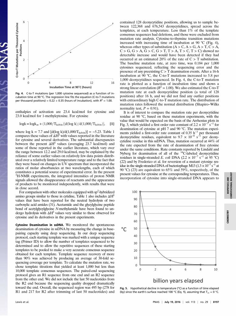
contained 128 deoxycytidine positions, allowing us to sample be-tween 122,368 and 676,543 deoxycytidines, spread across thetemplates, at each temperature. Less than 1% of the templateconsensus sequences had deletions, and those were excluded frommutation rate analysis. Cytosine-to-thymine transition mutationsincreased with increasing time of incubation at 90 °C (Fig. 4),whereas other types of substitution (A > C, A > G, A > T, C > A,C > G, G > A, G > C, G > T, T > A, T > C, T > C) showed nodetectable increase and would have been detected if they hadoccurred at an estimated 20% of the rate of C > T substitution.The baseline mutation rate, at zero time, was 0.184 per 1,000cytosines sequenced, reflecting the sequencing error and thepresence of any preexisting C > T deamination events. After a 16-hincubation at 90 °C, the C-to-T mutations increased to 5.8 per1,000 deoxycytidines sequenced. In Fig. 4, the C-to-T mutationrate is plotted as a function of incubation time and shows astrong linear correlation (R2 = 1.00). We also estimated the C-to-Tmutation rate at each deoxycytidine position (a total of 128positions) after 16 h, and we did not find any outlier positionswith extraordinary high C-to-T mutation rate. The distribution ofmutation rates followed the normal distribution (Shapiro–Wilksnormality test, P = 0.91).It is of interest to compare the mutation rate per deoxycytidine
residue at 90 °C, based on these mutation experiments, with thevalue that would be expected on the basis of the Arrhenius plots inFig. 3, which yielded a first order rate constant of 2.2 × 10⁻⁷ s−1 fordeamination of cytosine at pH 7 and 90 °C. The mutation experi-ments yielded a first-order rate constant of 0.35 h−1 per thousanddeoxycytidine residues, equivalent to 9.7 × 10⁻⁸ s−1 per deoxy-cytidine residue in this ssDNA. Thus, mutation occurred at 44% ofthe rate expected from the rate of deamination of free cytosineunder the same conditions. Rate constants reported by Lindahl andNyberg for deamination of all of the 14C-labeled deoxycytidineresidues in single-stranded E. coli DNA (2.2 × 10⁻⁷ s−1 at 95 °C)(22) and by Frederico et al. for reversion of a mutant cytosine res-idue in the single-strandedDNAof bacteriophageM13 (1.3×10⁻⁷ s−1 at90 °C) (23) are equivalent to 65% and 59%, respectively, of thepresent values for cytosine at the corresponding temperatures. Thus,incorporation of cytosine into single-stranded DNA appears to
Fig. 4. C-to-T mutations (per 1,000 cytosine sequenced) as a function of in-cubation time at 90 °C. The regression line fits the equation (C-to-T mutationsper thousand positions) = 0.22 + 0.35 (hours of incubation), with R2 = 1.00.
Fig. 5. Hypothetical decline in temperature (°C) as a function of time elapsed(by) since the earth’s surface reached 100 °C (Eq. 1), assuming that T∞ = 0 °C.
Lewis et al. PNAS | July 19, 2016 | vol. 113 | no. 29 | 8197
BIOPH
YSICSAND
COMPU
TATIONALBIOLO
GY
CHEM
ISTR
Y
reduce its rate of deamination by a factor of ∼2. It should beadded that the rates of deamination of individual cytosine residuesin bacteriophage T4 DNA are not uniform but appear to be sitespecific in bacteriophage T4 (32).
DiscussionIt is of interest to consider the probable time course of deamination-based mutation during Earth’s history, based on the temperature-dependence of cytosine deamination. In the absence of detailedinformation about the thermal history of the Earth, it seemsreasonable to expect a roughly exponential decline of the surfacetemperature, following Newton’s law of cooling, toward a steadystate maintained by the balance between the loss of heat to spaceand the gain of heat from the core and from solar radiation.For the sake of argument, let us suppose that the surface tem-
perature declines from T0 = 373° K (100 °C) as a starting point—below which water becomes a liquid at Earth’s surface and lifebecomes possible—toward an ultimate steady state value (T∞) of273 °K (0 °C). That choice of T∞ is arbitrary, but its actual value isnot expected to affect our conclusions in a qualitative sense.If, after the elapse of 4 billion years, the surface temperature is
now 298 °C (25 °C), then the half-life for cooling is 2 billion years,and the surface temperature decreases with time according to thefirst-order equation
ln ðT−T∞Þ= ln ðTo− −T∞Þ+ ðln 0.5ÞðtÞ, [2]
where To = 100 °C and t = billions of years that have elapsed since4 billion years ago. Fig. 5 illustrates the expected decline in tem-perature over the first 10 billion years.Suppose further that the rate constant for mutation (kmut), like
the rate constant for cytosine deamination, varies as a logarithmicfunction of the reciprocal of absolute temperature according tothe Arrhenius relationship (Eq. 1 and Fig. 3), with a heat ofactivation (ΔH‡) of 24 kcal/mol.Combining the expected rate of mutation as a function of
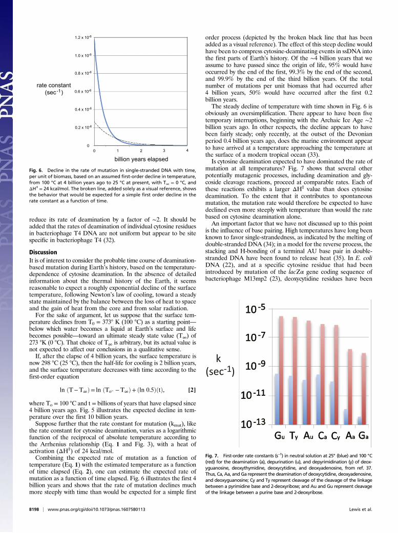
temperature (Eq. 1) with the estimated temperature as a functionof time elapsed (Eq. 2), one can estimate the expected rate ofmutation as a function of time elapsed. Fig. 6 illustrates the first 4billion years and shows that the rate of mutation declines muchmore steeply with time than would be expected for a simple first
order process (depicted by the broken black line that has beenadded as a visual reference). The effect of this steep decline wouldhave been to compress cytosine-deaminating events in ssDNA intothe first parts of Earth’s history. Of the ∼4 billion years that weassume to have passed since the origin of life, 95% would haveoccurred by the end of the first, 99.3% by the end of the second,and 99.9% by the end of the third billion years. Of the totalnumber of mutations per unit biomass that had occurred after4 billion years, 50% would have occurred after the first 0.2billion years.The steady decline of temperature with time shown in Fig. 6 is
obviously an oversimplification. There appear to have been fivetemporary interruptions, beginning with the Archaic Ice Age ∼2billion years ago. In other respects, the decline appears to havebeen fairly steady; only recently, at the outset of the Devonianperiod 0.4 billion years ago, does the marine environment appearto have arrived at a temperature approaching the temperature atthe surface of a modern tropical ocean (33).Is cytosine deamination expected to have dominated the rate of
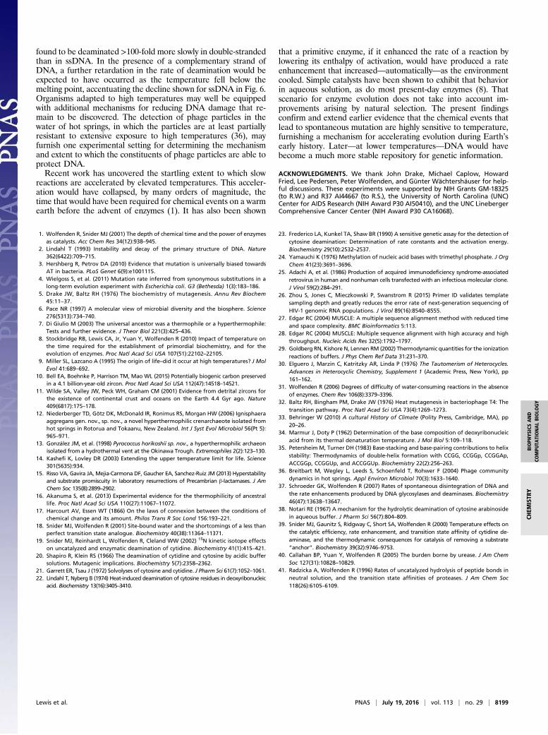
mutation at all temperatures? Fig. 7 shows that several otherpotentially mutagenic processes, including deamination and gly-coside cleavage reactions, proceed at comparable rates. Each ofthese reactions exhibits a larger ΔH‡ value than does cytosinedeamination. To the extent that it contributes to spontaneousmutation, the mutation rate would therefore be expected to havedeclined even more steeply with temperature than would the ratebased on cytosine deamination alone.An important factor that we have not discussed up to this point
is the influence of base pairing. High temperatures have long beenknown to favor single-strandedness, as indicated by the melting ofdouble-stranded DNA (34); in a model for the reverse process, thestacking and H-bonding of a terminal AU base pair in double-stranded DNA have been found to release heat (35). In E. coliDNA (22), and at a specific cytosine residue that had beenintroduced by mutation of the lacZα gene coding sequence ofbacteriophage M13mp2 (23), deoxycytidine residues have been
Fig. 6. Decline in the rate of mutation in single-stranded DNA with time,per unit of biomass, based on an assumed first-order decline in temperature,from 100 °C at 4 billion years ago to 25 °C at present, with T∞ = 0 °C, andΔH‡ = 24 kcal/mol. The broken line, added solely as a visual reference, showsthe behavior that would be expected for a simple first order decline in therate constant as a function of time.
Fig. 7. First-order rate constants (s−1) in neutral solution at 25° (blue) and 100 °C(red) for the deamination (a), depurination (u), and depyrimidination (y) of deox-yguanosine, deoxythymidine, deoxycytidine, and deoxyadenosine, from ref. 37.Thus, Ca, Aa, and Ga represent the deamination of deoxycytidine, deoxyadenosine,and deoxyguanosine; Cy and Ty represent cleavage of the cleavage of the linkagebetween a pyrimidine base and 2-deoxyribose; and Au and Gu represent cleavageof the linkage between a purine base and 2-deoxyribose.
8198 | www.pnas.org/cgi/doi/10.1073/pnas.1607580113 Lewis et al.
found to be deaminated >100-fold more slowly in double-strandedthan in ssDNA. In the presence of a complementary strand ofDNA, a further retardation in the rate of deamination would beexpected to have occurred as the temperature fell below themelting point, accentuating the decline shown for ssDNA in Fig. 6.Organisms adapted to high temperatures may well be equippedwith additional mechanisms for reducing DNA damage that re-main to be discovered. The detection of phage particles in thewater of hot springs, in which the particles are at least partiallyresistant to extensive exposure to high temperatures (36), mayfurnish one experimental setting for determining the mechanismand extent to which the constituents of phage particles are able toprotect DNA.Recent work has uncovered the startling extent to which slow
reactions are accelerated by elevated temperatures. This acceler-ation would have collapsed, by many orders of magnitude, thetime that would have been required for chemical events on a warmearth before the advent of enzymes (1). It has also been shown
that a primitive enzyme, if it enhanced the rate of a reaction bylowering its enthalpy of activation, would have produced a rateenhancement that increased—automatically—as the environmentcooled. Simple catalysts have been shown to exhibit that behaviorin aqueous solution, as do most present-day enzymes (8). Thatscenario for enzyme evolution does not take into account im-provements arising by natural selection. The present findingsconfirm and extend earlier evidence that the chemical events thatlead to spontaneous mutation are highly sensitive to temperature,furnishing a mechanism for accelerating evolution during Earth’searly history. Later—at lower temperatures—DNA would havebecome a much more stable repository for genetic information.
ACKNOWLEDGMENTS. We thank John Drake, Michael Caplow, HowardFried, Lee Pedersen, Peter Wolfenden, and Günter Wächtershäuser for help-ful discussions. These experiments were supported by NIH Grants GM-18325(to R.W.) and R37 AI44667 (to R.S.), the University of North Carolina (UNC)Center for AIDS Research (NIH Award P30 AI50410), and the UNC LinebergerComprehensive Cancer Center (NIH Award P30 CA16068).
1. Wolfenden R, Snider MJ (2001) The depth of chemical time and the power of enzymesas catalysts. Acc Chem Res 34(12):938–945.
2. Lindahl T (1993) Instability and decay of the primary structure of DNA. Nature362(6422):709–715.
3. Hershberg R, Petrov DA (2010) Evidence that mutation is universally biased towardsAT in bacteria. PLoS Genet 6(9):e1001115.
4. Wielgoss S, et al. (2011) Mutation rate inferred from synonymous substitutions in along-term evolution experiment with Escherichia coli. G3 (Bethesda) 1(3):183–186.
5. Drake JW, Baltz RH (1976) The biochemistry of mutagenesis. Annu Rev Biochem45:11–37.
6. Pace NR (1997) A molecular view of microbial diversity and the biosphere. Science276(5313):734–740.
7. Di Giulio M (2003) The universal ancestor was a thermophile or a hyperthermophile:Tests and further evidence. J Theor Biol 221(3):425–436.
8. Stockbridge RB, Lewis CA, Jr, Yuan Y, Wolfenden R (2010) Impact of temperature onthe time required for the establishment of primordial biochemistry, and for theevolution of enzymes. Proc Natl Acad Sci USA 107(51):22102–22105.
9. Miller SL, Lazcano A (1995) The origin of life–did it occur at high temperatures? J MolEvol 41:689–692.
10. Bell EA, Boehnke P, Harrison TM, Mao WL (2015) Potentially biogenic carbon preservedin a 4.1 billion-year-old zircon. Proc Natl Acad Sci USA 112(47):14518–14521.
11. Wilde SA, Valley JW, Peck WH, Graham CM (2001) Evidence from detrital zircons forthe existence of continental crust and oceans on the Earth 4.4 Gyr ago. Nature409(6817):175–178.
12. Niederberger TD, Götz DK, McDonald IR, Ronimus RS, Morgan HW (2006) Ignisphaeraaggregans gen. nov., sp. nov., a novel hyperthermophilic crenarchaeote isolated fromhot springs in Rotorua and Tokaanu, New Zealand. Int J Syst Evol Microbiol 56(Pt 5):965–971.
13. González JM, et al. (1998) Pyrococcus horikoshii sp. nov., a hyperthermophilic archaeonisolated from a hydrothermal vent at the Okinawa Trough. Extremophiles 2(2):123–130.
14. Kashefi K, Lovley DR (2003) Extending the upper temperature limit for life. Science301(5635):934.
15. Risso VA, Gavira JA, Mejia-Carmona DF, Gaucher EA, Sanchez-Ruiz JM (2013) Hyperstabilityand substrate promiscuity in laboratory resurrections of Precambrian β-lactamases. J AmChem Soc 135(8):2899–2902.
16. Akanuma S, et al. (2013) Experimental evidence for the thermophilicity of ancestrallife. Proc Natl Acad Sci USA 110(27):11067–11072.
17. Harcourt AV, Essen WT (1866) On the laws of connexion between the conditions ofchemical change and its amount. Philos Trans R Soc Lond 156:193–221.
18. Snider MJ, Wolfenden R (2001) Site-bound water and the shortcomings of a less thanperfect transition state analogue. Biochemistry 40(38):11364–11371.
19. Snider MJ, Reinhardt L, Wolfenden R, Cleland WW (2002) 15N kinetic isotope effectson uncatalyzed and enzymatic deamination of cytidine. Biochemistry 41(1):415–421.
20. Shapiro R, Klein RS (1966) The deamination of cytidine and cytosine by acidic buffersolutions. Mutagenic implications. Biochemistry 5(7):2358–2362.
21. Garrett ER, Tsau J (1972) Solvolyses of cytosine and cytidine. J Pharm Sci 61(7):1052–1061.22. Lindahl T, Nyberg B (1974) Heat-induced deamination of cytosine residues in deoxyribonucleic
acid. Biochemistry 13(16):3405–3410.
23. Frederico LA, Kunkel TA, Shaw BR (1990) A sensitive genetic assay for the detection ofcytosine deamination: Determination of rate constants and the activation energy.Biochemistry 29(10):2532–2537.
24. Yamauchi K (1976) Methylation of nucleic acid bases with trimethyl phosphate. J OrgChem 41(23):3691–3696.
25. Adachi A, et al. (1986) Production of acquired immunodeficiency syndrome-associatedretrovirus in human and nonhuman cells transfected with an infectious molecular clone.J Virol 59(2):284–291.
26. Zhou S, Jones C, Mieczkowski P, Swanstrom R (2015) Primer ID validates templatesampling depth and greatly reduces the error rate of next-generation sequencing ofHIV-1 genomic RNA populations. J Virol 89(16):8540–8555.
27. Edgar RC (2004) MUSCLE: A multiple sequence alignment method with reduced timeand space complexity. BMC Bioinformatics 5:113.
28. Edgar RC (2004) MUSCLE: Multiple sequence alignment with high accuracy and highthroughput. Nucleic Acids Res 32(5):1792–1797.
29. Goldberg RN, Kishore N, Lennen RM (2002) Thermodynamic quantities for the ionizationreactions of buffers. J Phys Chem Ref Data 31:231–370.
30. Elguero J, Marzin C, Katritzky AR, Linda P (1976) The Tautomerism of Heterocycles.Advances in Heterocyclic Chemistry, Supplement 1 (Academic Press, New York), pp161–162.
31. Wolfenden R (2006) Degrees of difficulty of water-consuming reactions in the absenceof enzymes. Chem Rev 106(8):3379–3396.
32. Baltz RH, Bingham PM, Drake JW (1976) Heat mutagenesis in bacteriophage T4: Thetransition pathway. Proc Natl Acad Sci USA 73(4):1269–1273.
33. Behringer W (2010) A cultural History of Climate (Polity Press, Cambridge, MA), pp20–26.
34. Marmur J, Doty P (1962) Determination of the base composition of deoxyribonucleicacid from its thermal denaturation temperature. J Mol Biol 5:109–118.
35. Petersheim M, Turner DH (1983) Base-stacking and base-pairing contributions to helixstability: Thermodynamics of double-helix formation with CCGG, CCGGp, CCGGAp,ACCGGp, CCGGUp, and ACCGGUp. Biochemistry 22(2):256–263.
36. Breitbart M, Wegley L, Leeds S, Schoenfeld T, Rohwer F (2004) Phage communitydynamics in hot springs. Appl Environ Microbiol 70(3):1633–1640.
37. Schroeder GK, Wolfenden R (2007) Rates of spontaneous disintegration of DNA andthe rate enhancements produced by DNA glycosylases and deaminases. Biochemistry46(47):13638–13647.
38. Notari RE (1967) A mechanism for the hydrolytic deamination of cytosine arabinosidein aqueous buffer. J Pharm Sci 56(7):804–809.
39. Snider MJ, Gaunitz S, Ridgway C, Short SA, Wolfenden R (2000) Temperature effects onthe catalytic efficiency, rate enhancement, and transition state affinity of cytidine de-aminase, and the thermodynamic consequences for catalysis of removing a substrate“anchor”. Biochemistry 39(32):9746–9753.
40. Callahan BP, Yuan Y, Wolfenden R (2005) The burden borne by urease. J Am ChemSoc 127(31):10828–10829.
41. Radzicka A, Wolfenden R (1996) Rates of uncatalyzed hydrolysis of peptide bonds inneutral solution, and the transition state affinities of proteases. J Am Chem Soc118(26):6105–6109.
Lewis et al. PNAS | July 19, 2016 | vol. 113 | no. 29 | 8199
BIOPH
YSICSAND
COMPU
TATIONALBIOLO
GY
CHEM
ISTR
Y