Embed Size (px)
Citation preview

Cytoplasmic localization of PXR and ligand-dependent nuclear translocation in mouse
liver
E. James Squires*, Tatsuya Sueyoshi and Masahiko Negishi
Pharmacogenetics Section, Laboratory of Reproductive and Developmental Toxicology,
National Institute of Environmental Health Sciences, National Institutes of Health,
Research Triangle Park, North Carolina, 27709
Running title: Nuclear translocation of mouse PXR
All correspondence to:
Masahiko Negishi at the above address
Telephone 919-541-2404; Fax 919-541-0696
E-mail address, [email protected]
*Permanent address: Dept. of Animal and Poultry Science, University of Guelph, Guelph,
Ontario, Canada N1G 2W1
1
JBC Papers in Press. Published on September 2, 2004 as Manuscript M407281200 by guest on July 8, 2018
http://ww
w.jbc.org/
Dow
nloaded from

SUMMARY
The pregnane X receptor (PXR) plays an important role in the response to xenobiotics
and endogenous toxins. We have used a specific anti-PXR antibody in Western blotting
of mouse liver nuclear extracts to show that PXR is accumulated in the nucleus after
treatment with 5-pregnen-3²-ol-20-one-16±-carbonitrile (PCN), followed by an
increase in Cyp3a11 mRNA. Expression of wild type PXR and various mutants as GFP
fusion proteins in mouse livers showed that PXR was retained in the cytoplasm from
where PCN treatment translocated PXR into the nucleus. Furthermore, the xenochemical
response signal (XRS), the nuclear translocation signal (NLS) and the AF2 domain were
all required for the nuclear translocation to occur. Immunoprecipitation experiments
using hsp90 antibody demonstrated the presence of PXR in a complex with the
endogenous cytoplasmic CAR retention protein (CCRP) in HepG2 cells. FRET analysis
of mouse liver sections after co-expression of CFP-CCRP and YFP-PXR also indicated
that CCRP and PXR were closely associated in vivo. Over expression of exogenous
CCRP increased the cytoplasmic level of the PXR:CCRP:hsp90 complex, whereas a
decrease in endogenous CCRP by treatment with siRNA for CCRP repressed the PXR-
mediated reporter activity in HepG2 cells. We conclude that the CCRP mediates the
retention of PXR in the cytosol and modulates the activation of PXR in response to PCN
treatment.
2
by guest on July 8, 2018http://w
ww
.jbc.org/D
ownloaded from

Abbreviations used: AF2, activation function 2; AhR, aryl hydrocarbon receptor; CAR,
constitutive active/androstane receptor; CCRP, cytoplasmic CAR retention protein;
DMSO, dimethyl sulfoxide; FRET, fluorescence resonance energy transfer; GFP, green
fluorescent protein; GR, glucocorticoid receptor; hsp90, heat shock protein 90; LBD,
ligand binding domain; NES, nuclear export sequence; NLS, nuclear localization
sequence; PB, phenobarbital; PCN, 5-pregnen-3²-ol-20-one-16±-carbonitrile; PXR,
pregnane X receptor; hPXR, human PXR; mPXR, mouse PXR; RIF, rifampicin; TPR,
tetratrico peptide repeat; VDR, vitamin D receptor; XRS, xenochemical response
sequence.
3
by guest on July 8, 2018http://w
ww
.jbc.org/D
ownloaded from
INTRODUCTION
The pregnane X receptor (PXR, NR1I2) and constitutive active/androstane receptor
(CAR, NR1I3) are involved in the primary response to xenobiotics and endogenous
toxins (1, 2). These receptors respond to ligands by activating the expression of genes
encoding enzymes involved in phase I (oxidation) and phase II (conjugation) metabolism
as well as proteins involved in the efflux of toxins from the cell (3-5). CAR is normally
sequestered in the cytoplasm of untreated liver cells and translocates to the nucleus after
exposure to PB and PB-like chemicals (6). The cytoplasmic CAR retention protein
(CCRP and designated Dnajc7 in the official gene symbol in the NCBI database) has
been shown to maintain the cytoplasmic localization of CAR by forming a complex with
CAR and hsp90 (7, 8). Other nuclear receptors, such as the glucocorticoid receptor (GR),
vitamin D receptor (VDR) and aryl hydrocarbon receptor also exist as complexes with
receptor-specific co-chaperones and hsp90 that play an important role in locating these
receptors in the cytoplasm of unstimulated cells (9). These co-chaperones, including
immunophilin-FK-binding proteins and hepatitis B virus protein X-associated protein 2
contain multiple tetratricopeptide repeat (TPR) motifs, which are 34 amino acid
sequences that form a pair of anti-parallel ± helices that mediate protein-protein
interactions and the assembly of multi-protein complexes (10).
4
by guest on July 8, 2018http://w
ww
.jbc.org/D
ownloaded from
The movement of proteins between the nucleus and the cytoplasm is an energy dependent
process, determined by both nuclear localization sequence (NLS) and nuclear export
sequence (NES) on the protein (11). The typical NLS consists of a single or repeat cluster
of basic amino acids which associate with factors such as importins that carry the proteins
into the nucleus through a nuclear pore complex. The typical NES is a leucine rich
region, and has been identified on a number of proteins, including the AhR (12, 13). After
binding to ligand, the nuclear receptor GR moves along cytoskeletal tracks towards the
nucleus by connecting with dynein motors (reviewed in 13). The C-terminal AF2 domain
is required for ligand dependent transcriptional activation by steroid hormone and many
other nuclear receptors (14), and it is displaced upon binding of ligand allowing it to
interact with co-activators. The removal of the AF2 domain prevents the nuclear
translocation of the VDR and GR (15). However, the AF2 domain is not required for the
nuclear translocation of CAR following PB exposure (17). The nuclear translocation of
CAR following drug exposure is dependent on the xenochemical response sequence
(XRS), a leucine rich sequence near the C-terminus conserved as L/(M)XXLXXL (16) in
mouse and human CAR and PXR. However, mutations in the key leucine residues in the
XRS did not affect the formation of heterodimers between CAR and RXR or the co-
activation of CAR by SRC-1. This suggests that the XRS regulates the translocation but
not the activation of CAR.
5
by guest on July 8, 2018http://w
ww
.jbc.org/D
ownloaded from

In contrast to CAR, PXR is generally thought to be primarily retained in the nucleus,
where it activates the expression of genes such as Cyp3a11 after binding ligands such as
PCN. Thus, the possibility that co-chaperones could regulate the cellular localization of
PXR has not been investigated. However, recent results of immunostaining of mouse
liver sections using a commercially available antibody suggest that mPXR may be
located in the cytosol of untreated liver cells (18); however, additional supporting
experiments, such as Western blot analysis, were not presented. Moreover, in transformed
cells, human hPXR (SXR) is located primarily in the nucleus and mutation of specific
amino acids in the so-called NLS region of the DNA binding domain of hPXR resulted
in cytoplasmic localization of the receptor in transformed cells (18). However, the role of
the NLS as well as other specific motifs (AF2 and XRS) in the nuclear translocation of
mPXR in the liver following drug treatment remains unexplored.
Our goal was to use more definitive methods to determine if mPXR was located in the
cytoplasm of untreated liver and translocated to the nucleus only after exposure to
xenochemicals. To achieve this, we produced specific antibodies against the hinge region
of mPXR and used these antibodies in Western blotting of liver nuclear extracts prepared
from control mice and those treated with PCN. We also investigated the interaction of
mPXR with CCRP in the cytosol to determine if CCRP played a role in maintaining
mPXR in the cytosol. We then looked at the NLS, XRS and the AF2 domain which may
6
by guest on July 8, 2018http://w
ww
.jbc.org/D
ownloaded from

be involved in nuclear translocation of mPXR in response to drug treatment, and asked if
these components also affected the association of mPXR with CCRP. Finally, we
investigated the role of endogenous CCRP in the activation of mPXR in response to drug
treatment.
7
by guest on July 8, 2018http://w
ww
.jbc.org/D
ownloaded from

EXPERIMENTAL PROCEDURES
Antibodies— Anti-mPXR antibody was raised in rabbits using the hinge region peptide
CSNAAVEQRRALIKRKKRE conjugated to keyhole limpet hemocyanin as the antigen.
The antibody was purified from serum by affinity chromatography using the peptide
coupled to SulfoLink gel (Pierce, Rockford. IL). Rabbit anti-CAR antibody and rabbit
anti-CCRP antibody were produced as previously described (7, 19). Anti-hsp90
monoclonal antibody was purchased from Affinity Bioreagents (catalog # MA3-011,
Golden, Co), control IgM from BD Biosciences, anti-V5-HRP antibody was purchased
from Invitrogen (Carlsbad, CA). Anti-Lamin B was from Santa Cruz Biotechonology
(sc-20682, Santa Cruz, CA).
Plasmids—cDNAs encoding full length mPXR, mPXR”AF2 (residues 1-416), hPXR,
mCAR and CCRP were PCR amplified using appropriate primers and cloned into
pcDNA3.1/V5-His-TOPO (Invitrogen) to produce pcDNA3.1/mPXR-V5-His,
pcDNA3.1/mPXRAF2-V5-His, pcDNA3.1/hPXR-V5-His, pcDNA3.1/mCAR-V5-His
and pcDNA3.1/CCRP-V5-His. mPXR and CCRP were also amplified using 5’ primers
containing an in-frame XhoI site and 3’ primers containing an EcoRI site and cloned into
pEYFP-C1 or pECFP-C1 expression vectors (BD Biosciences Clontech, Palo Alto, CA)
to produce an N terminal fusion with green fluorescent protein. The XREM-3A4-Luc
8
by guest on July 8, 2018http://w
ww
.jbc.org/D
ownloaded from
(p3A4-362(7836/7208ins)) reporter plasmid was kindly provided by Bryan Goodwin
(20). The following mutants were constructed using the QuikChange site-directed
mutagenesis kit (Stratagene, Cedar Creek, TX) and appropriate primers:
pcDNA3.1/mPXR-V5-His or pEYFP-mPXR containing either the mutations M391A,
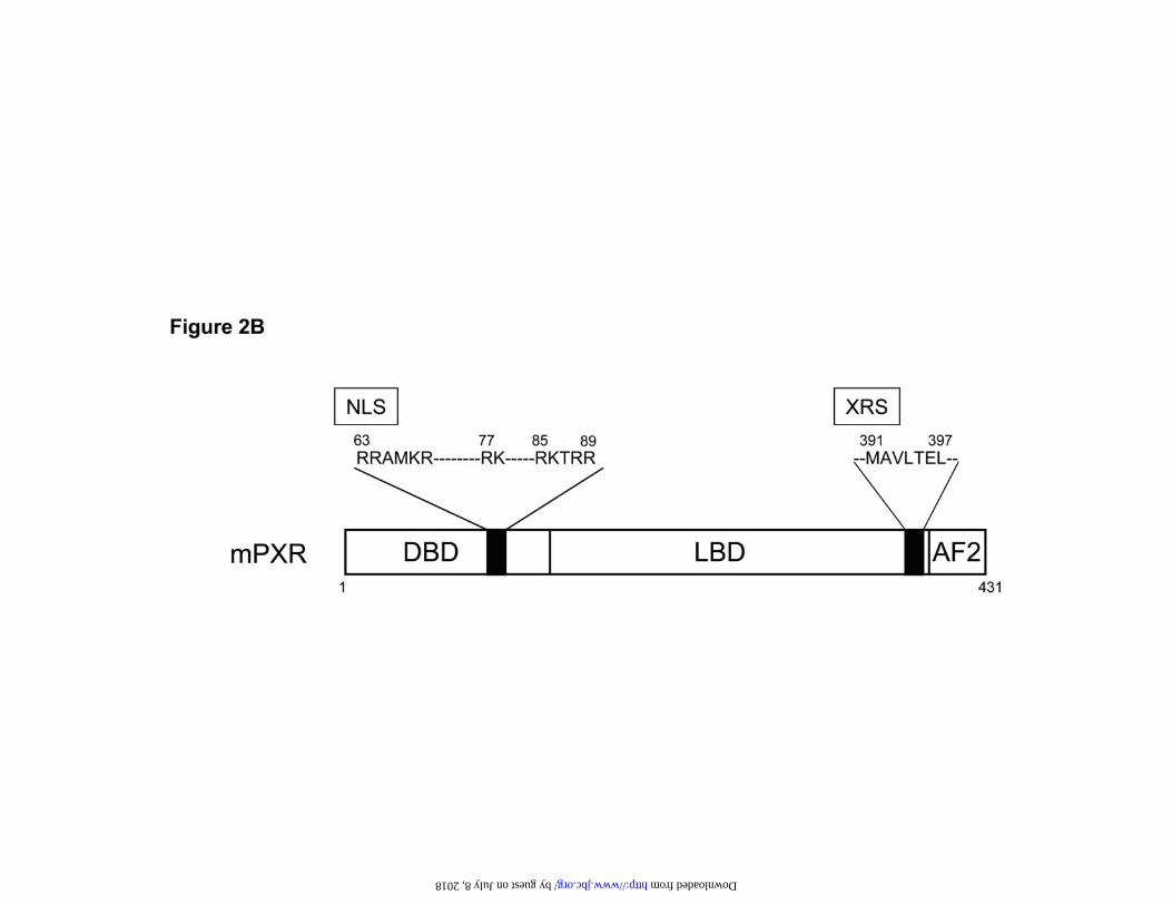
L394A or L397A in the XRS region, R63/R64A/R85/K86A or R63/R64A/R88A/R89A
in the NLS region. All plasmids were verified by nucleotide sequencing and prepared
using Qiagen Plasmid Maxi kit (Qiagen, Valencia, CA).
Expression of fluorescent protein-tagged PXR and CCRP in mouse liver in vivo—
Expression of pEYFP-mPXR, its various mutants and pECFP-CCRP in mouse liver in
vivo and their detection by microscopy was performed as described previously (7, 21).
Plasmids were injected via the tail vein of male CD-1 mice using the TransIT In Vivo
gene Delivery System (Mirus, Maddison, WI) according to the manufacturer’s
instructions. Two hours later, the mice were injected intraperitoneally with either 15µg/kg
PCN, 100µg/kg PB or DMSO vehicle and killed 6 hrs later. Liver sections were analyzed
by confocal laser scanning microscopy using a Zeiss LSM510 microscopy system
equipped with a META detector. For FRET analysis, 458nm excited signals were
captured twice before and twice after YFP photobleaching using twenty-five pulses of
514 nm laser. Wavelength scanning images between 450 nm and 540 nm were collected
to extract simultaneously each of the YFP and CFP emissions using software provided by
9
by guest on July 8, 2018http://w
ww
.jbc.org/D
ownloaded from

Zeiss. Dequenching of CFP was quantified for multiple liver cells and FRET efficiency E
was calculated with the equation E=1-(Ii/Ii0), where Ii is fluorescence intensity before
the bleaching and Ii0 is that of after the bleaching. The distance between the CFP donor
and YFP acceptor was calculated using a R0 value, the distance at which energy transfer
efficiency is 50% in ideal conditions, of 4.9 nm (22).
Cells and transfection assay—HepG2 cells were cultured in Minimal Essential Medium
supplemented with 10% fetal bovine serum and antibiotics (100 units/ml of penicillin and
100 ¼g/ml streptomycin). At approximately 50% confluence, the cells were transfected
with plasmids using Lipofectamine 2000 (Invitrogen) according to manufacturers
instructions. Twenty four hours later, the cells were given fresh media and a further 24
hrs later the cells were harvested, washed twice with PBS and homogenized with a
Dounce homogenizer in buffer A (10 mM HEPES buffer, pH 7.6 containing 10 mM KCl,
1.5 mM MgCl2, 20 mM Na2MoO4, 0.3% Nonidet P-40 (Calbiochem, La Jolla, CA),
1mM dithiothreitol and Complete protease inhibitor (Roche Diagnostics, Mannheim,
Germany) as described (7, 23). The homogenate was centrifuged at 4,000 x g for 10 min.
to obtain a nuclear pellet and the supernatant was centrifuged at 17,800 x g for 10 min to
obtain a clear cytosolic fraction for use in immunoprecipitation. The pellet was washed
once in buffer A, once in buffer A without Nonidet P-40 and then suspended in lysis
10
by guest on July 8, 2018http://w
ww
.jbc.org/D
ownloaded from
buffer (10 mM HEPES buffer, pH 7.6 containing 10% glycerol, 100 mM KCl, 3 mM
MgCl2, 1 mM Na3VO4, 1 mM dithiothreitol and Complete protease inhibitor). The
resulting suspension was mixed at 4 ºC for 12 hours in the presence of 0.4 M NaCl and
centrifuged for 10 min at 17,800 x g to obtain a clear nuclear extract.
Preparation of mouse liver nuclear extracts— Nuclear extracts were prepared based on
published methods (24, 25). Liver was homogenized in 20 volumes of 10 mM HEPES
buffer, pH 7.6 containing 2 M sucrose, 10% glycerol, 25 mM KCl, 0.15 mM spermidine,
0.5 mM spermine and 1mM EDTA using a motor driven glass Teflon homogenizer kept
on ice. The homogenate was then layered over a cushion of the same buffer and
centrifuged at 85,000 x g for 60 min at 4 °C. The pellet was suspended in 1 ml of lysis
buffer (as described above), mixed at 4 °C for 30 min in the presence of 0.4 M NaCl and
centrifuged at 100,000 x g for 30 min. The supernatant was dialysed overnight against 20
mM HEPES buffer, pH 7.6 containing 20% glycerol, 0.2 mM EDTA, 1mM Na2MoO4,
1mM dithiothreitol and Complete protease inhibitor.
Immunoprecipitation—Anti-hsp90 (5 ¼l) or IgM (1 ¼g) as control was added to 2 mg of
cytosolic protein from HepG2 cells and incubated overnight at 4 °C. Twenty ¼l of a 50%
slurry of Protein L Sepharose (ImmunoPure immobilized Protein L, Pierce), previously
11
by guest on July 8, 2018http://w
ww
.jbc.org/D
ownloaded from

washed in buffer A, was added and the mixture incubated for 1 hour at 4 °C. The resin
was recovered by centrifugation and washed 5 times with 1 ml of 50mM Tris buffer, pH
7.5 containing 0.15 M NaCl, 20 mM Na2MoO4 and 0.2% Nonidet P-40. The
immunoprecipitated proteins were extracted from the resin with NuPAGE LDS buffer
(Invitrogen) and subjected to Western blotting.
Western blotting—Proteins were separated either on a NuPage 4-10% Bis-Tris gel in
NuPAGE MOPS SDS running buffer (Invitrogen) or a 10% SDS gel in Tris-Glycine
buffer and transferred to nitrocellulose membrane using the SemiPhor semi-dry transfer
unit (Hoefer Scientific Instruments, San Francisco, CA). The membrane was then
incubated for 1 hr in TBS-0.1%Tween 20 containing 5% Blotto milk powder, followed
by 1 hr incubation with primary antibody (rabbit anti-mPXR, rabbit anti-mCAR) in the
same medium and 1 hr with secondary antibody (donkey anti rabbit IgG-HRP conjugate,
Santa Cruz Biotechnology). For the detection of V5-tagged proteins, the nitrocellulose
membranes were incubated with anti-V5-HRP conjugate in TBS-Tween with 5%
Blotto. Protein bands were visualized on the membranes using Lumigen PS-3 ECL
detection reagent (Amersham Biosciences). In some cases, the immunoblots were
stripped with Restore Western Blot stripping buffer (Pierce) and restained with
antibodies.
12
by guest on July 8, 2018http://w
ww
.jbc.org/D
ownloaded from
GST pull down assay—The GST-CCRP fusion protein was expressed and purified using
glutathione–Sepharose 4B (Amersham Biosciences) (7). 35S-labelled mPXR and mCAR
were produced from pcDNA3.1/mPXR-V5-His and pcDNA3.1/mCAR-V5-His using
the TnT T7 quick coupled transcription/translation system (Promega) along with 35S-
labled methionine. GST-CCRP or GST coupled to the glutathione Sepharose 4B was
incubated with the 35S-labelled mPXR or mCAR in 50 mM HEPES buffer, pH 7.5
containing 0.1 M NaCl and 0.1% Triton X100 for 20 min at room temperature. The resin
was then recovered by centrifugation and washed 3 times in the same buffer. Proteins
were extracted from the resin by heating for 10 min at 70 °C in NuPAGE LDS sample
buffer (Invitrogen) and separated on a NuPage 4-10% Bis-Tris gel in NuPAGE MOPS
SDS running buffer for 1 hour at 150 volts. The gel was then stained with Coomassie
Blue, destained and dried under vacuum and the proteins detected by autoradiography.
siRNA Treatment and Reporter assay— HepG2 cells were cultured in Minimal Essential
Medium supplemented with 10% fetal bovine serum and antibiotics (100units/ml of
penicillin and 100 ¼g/ml streptomycin) to about 50% confluence. The cells were
transfected with pcDNA3.1/mPXR-V5-His or pcDNA3.1/hPXR-V5-His, the XREM-
3A4-Luc reporter and phRL-tk as a transfection control (Promega) using Lipofectamine
2000 (Invitrogen) according to manufacturer’s instructions. For RNAi experiments,
13
by guest on July 8, 2018http://w
ww
.jbc.org/D
ownloaded from

HepG2 cells were transfected with siRNA against CCRP (SMART pool siRNA M-
019566-00 from Dharmacon, Inc, Boulder, CO) or an unrelated scramble (5’-
ACUCUAUCGCCAGCGUGACUUdTdT-3’) using lipofectamine 2000 reagent
according to the manufacturer’s directions. After 48 hr, half of the wells were treated
with PCN or RIF and after a further 24 hr the luciferase activity of the XREM-3A4-Luc
reporter and the control phRL-tk was measured using the Dual luciferase Reporter Assay
System (Promega). The ratio of XREM-3A4-Luc reporter activity to Renilla luciferase
control was calculated for triplicate transfections.
Real time PCR—To measure CYP3A11 mRNA, liver samples were homogenized in
TRIZOL reagent (Invitrogen) and total RNA was prepared as per the manufacturer’s
instructions. Five ¼g of RNA was used as a template for cDNA synthesis using the
SuperScript first-strand synthesis system (Invitrogen) with random hexamers as primers.
One-twentieth of the cDNA was used for real time PCR with an ABI Prism 7700
Sequence Detector using TaqMan Universal PCR reaction mix and primers for mouse
CYP3A11 (Applied Biosystems, Foster City, CA) or GAPDH as a control.
14
by guest on July 8, 2018http://w
ww
.jbc.org/D
ownloaded from

RESULTS
mPXR accumulates in the nucleus of liver cells after drug treatment— In order to
determine the cellular localization of endogenous mPXR in mouse liver in vivo, Western
blotting of liver nuclear extracts was performed using an antibody raised against the
hinge region of mPXR (Fig.1A). Levels of mPXR were lowest in nuclear extracts from
untreated mice or mice treated with DMSO vehicle and increased following PCN
treatment, but not after PB treatment. When the same blot was stripped and then
immunostained using antibody against mouse CAR, levels of CAR were highest in
nuclear extracts from PB treated mice, with no effect of PCN treatment. The results
obtained from the present Western blot analysis have now established that mPXR
accumulates in the nucleus of liver cells following PCN treatment.
This nuclear accumulation of mPXR occurred in time-dependent manner. Livers were
removed from untreated mice and at various times up to 40 hr after treatment with PCN
and nuclear extracts were prepared and used for Western blotting using anti-mPXR
antibody. The results (Fig. 1B) show that levels of mPXR in the nucleus increased rapidly
after treatment with PCN and were already maximal at the first time point of 3 hr after
treatment. Levels of mPXR remained high in the nuclear extracts at 8 hr after PCN
treatment and decreased by the 17 hr time point. To compare the time course for gene
15
by guest on July 8, 2018http://w
ww
.jbc.org/D
ownloaded from

activation with the accumulation of mPXR in the nucleus after treatment with PCN,
levels of CYP3A11 mRNA were measured in the livers by real time PCR. Levels of
CYP3A11 mRNA increased to a maximum at the 17 hr time point after PCN treatment
and remained elevated over untreated control values at 40 hrs after treatment. This
indicates that activation of the CYP3A11 gene occurred after mPXR accumulates in the
nucleus in response to PCN treatment. Thus, mPXR moves into the nucleus in response
to PCN treatment, activates transcription of responsive genes, and then is cleared from
the nucleus.
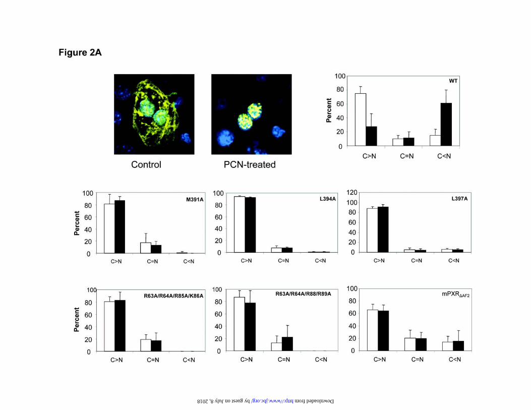
Structural components of mPXR that affect nuclear translocation—We investigated the
cellular localization of mPXR by in vivo transfection of YFP tagged mPXR in mouse
liver. In untreated liver, YFP-mPXR was localized throughout the cytoplasm of the liver
cell (Fig. 2A). However, in liver sections from mice that had been treated with PCN,
YFP-mPXR was present predominantly in the nucleus. This cytoplasmic retention and
nuclear translocation of YFP-mPXR are reminiscent of what occurs with the endogenous
receptor. In order to identify the structural features of mPXR that affect nuclear
translocation in vivo, we expressed mPXR and its various mutants as YFP fusion proteins
in mouse livers. These mPXR mutants contained substitutions in the XRS region or the
NLS region in the DNA binding domain or had the C terminal AF2 region deleted (Fig.
2B). We then measured the percentage of cells with mPXR expressed predominantly in
16
by guest on July 8, 2018http://w
ww
.jbc.org/D
ownloaded from
the cytoplasm, the percentage with equal nuclear and cytoplasmic localization and the
percentage with primarily nuclear localization of mPXR (Fig. 2A). Wild type YFP-
mPXR was located primarily in the cytoplasm in untreated mouse liver and translocated
to the nucleus after treatment with PCN. However, mutations in key amino acid residues
in the XRS or NLS region or deletion of the AF2 domain prevented nuclear translocation
in vivo in response to treatment with PCN. The distribution of the NLS and XRS mutants
remained almost entirely cytoplasmic, while the AF2-deletion mutant had slightly more
nuclear distribution, which was not affected by PCN treatment. These results suggest that
the XRS, NLS and AF2 regions are all required for the nuclear translocation of mPXR
after PCN treatment in vivo.
Role of CCRP in cytoplasmic localization of mPXR—CCRP is involved in maintaining
the cytoplasmic localization of CAR (7). We hypothesized that CCRP could also bind to
mPXR and was involved in the retention of mPXR in the cytoplasm. As a first step, we
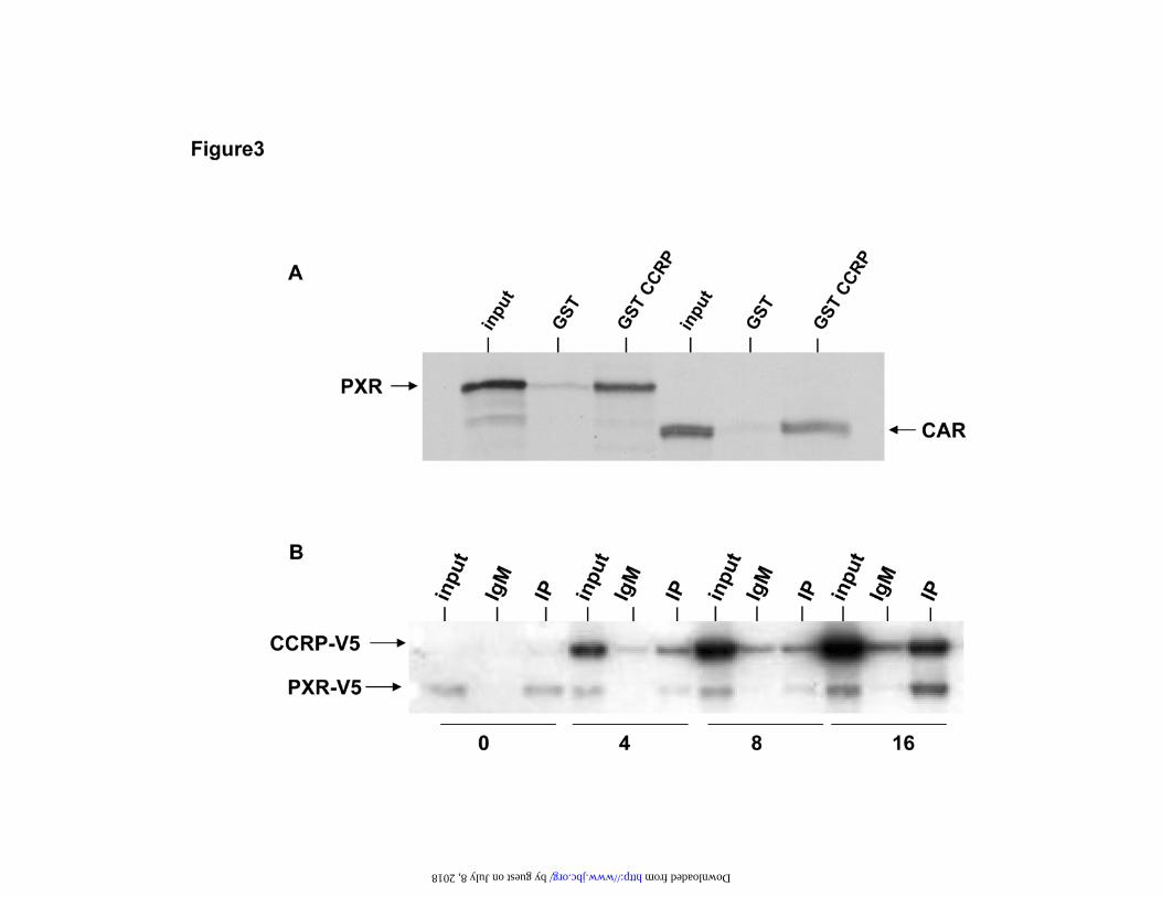
used a GST pull down assay to demonstrate that GST-CCRP can bind to mPXR as well
as binding to CAR (Fig. 3A). Next we expressed mPXR-V5 and CCRP-V5 in HepG2
cells and found that they formed a complex in the cytosol with hsp90 that can be
immunoprecipitated by anti-hsp90 antibody (Fig.3B). Furthermore, when the ratio of
CCRP to mPXR used in transfecting the HepG2 cells is increased, the retention of mPXR
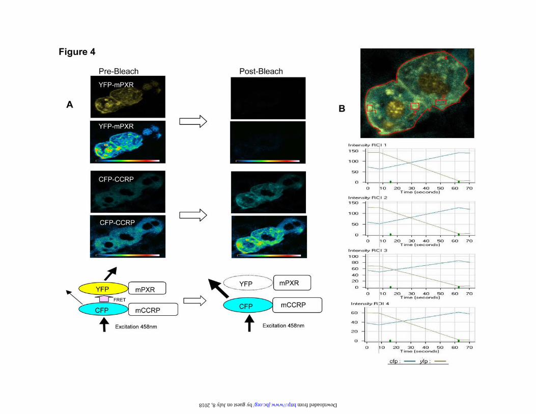
in the cytosol is also increased. We then did FRET analysis of mouse liver sections after
17
by guest on July 8, 2018http://w
ww
.jbc.org/D
ownloaded from

co-expression of CFP-CCRP and YFP-mPXR. The results indicated that CCRP and
mPXR are closely associated in vivo to allow FRET to occur. Before photo bleaching of
YFP, excitation at 458 nm produced a clear FRET signal from CFP-CCRP to increase
the signal observed from YFP-mPXR (Fig 4A). After photo bleaching of YFP, this
FRET did not occur and the CFP signal intensity increased. The CFP and YFP signal
intensity was measured before and after photo bleaching in four different regions of the
cell (Fig. 4B). After photo bleaching, the YFP signal intensity decreased rapidly with a
concomitant increase in the signal intensity of CFP. Measurements of FRET efficiency in
7 different cells ranged from 49% - 66%, which suggested that CFP-CCRP and YFP-
mPXR were within an estimated distance of 4.69 ± 0.23 nm of each other. Taken
together, these results demonstrate that mPXR forms a complex with CCRP that may
promote the retention of mPXR in the cytoplasm of liver cells.
Given that mPXR and CCRP could form a complex with hsp90, we expressed mPXR-V5
and its various mutants in HepG2 cells along with CCRP-V5 in order to investigate
whether the signature motifs (NLS, XRS and AF2) were involved in the formation of the
mPXR:CCRP:hsp90 complex. Immunoprecipitation of cytosols with hsp90 antibody
demonstrated that the various mPXR mutants can all form a complex with CCRP and
hsp90 in the cytosol with approximately equal efficiency (Fig. 5). Thus, the extent of
binding of mPXR mutants to CCRP in the cytosolic complex is not correlated with the
18
by guest on July 8, 2018http://w
ww
.jbc.org/D
ownloaded from
amount of the mPXR mutant that translocates to the nucleus after PCN treatment as
shown in Fig 2A. Thus, the CCRP-mediated retention of mPXR in the cytosol of HepG2
cells does not fully explain the mechanism of nuclear translocation of mPXR in liver in
vivo.
Role of CCRP in regulating the activity of PXR— We next transfected HepG2 cells with
pcDNA3.1/mPXR-V5-His and pcDNA3.1/CCRP-V5-His and immunoprecipitated the
CCRP:mPXR complex with anti-hsp90 antibody. When the Western blot was stained
with anti-CCRP antibody, the endogenous CCRP that was immunoprecipitated along
with the CCRP-V5 could be clearly seen, indicating that the endogenous CCRP also
forms a complex with endogenous hsp90 and the transfected mPXR in HepG2 cells
(Fig.6A). Immunoprecipitation experiments using anti-V5 antibody also confirmed that
hsp90 was precipitated along with mPXR-V5 and CCRP-V5 (data not shown). We then
wanted to determine if CCRP affects the activation of PXR in response to drug treatment.
When siRNA for CCRP was transfected in HepG2 cells, a clear decrease in endogenous
CCRP protein was observed by Western blotting (Fig 6B), while there was no effect on
levels of mPXR in the nucleus (data not shown). The decrease in CCRP expression by
siRNA treatment resulted in an attenuated response in a reporter assay for mouse PXR
activity, with a more dramatic response obtained with 50 ¼M PCN than 10 ¼M PCN. In
addition, the response of human PXR to activation by RIF was also attenuated by
19
by guest on July 8, 2018http://w
ww
.jbc.org/D
ownloaded from
treatment with siRNA for CCRP. This clearly demonstrates that CCRP plays a role in the
activation of both mouse and human PXRs following treatment with their activators.
20
by guest on July 8, 2018http://w
ww
.jbc.org/D
ownloaded from
DISCUSSION
We produced an antibody directed against the hinge region of mPXR, and used this
antibody in Western blots to show that the levels of mPXR initially increase in the
nucleus of mouse liver cells after PCN treatment, followed by activation of a mPXR
responsive gene, and then levels of mPXR decrease in the nucleus. Expression of YFP-
mPXR in mouse liver in vivo revealed that mPXR is expressed in the cytoplasm of
untreated liver cells and is translocated in the nucleus after PCN treatment. These data
definitively demonstrate that nuclear translocation of mPXR occurs, as with other nuclear
receptors such as CAR, GR and VDR, as a primary step in the activation of mPXR
responsive genes.
PXR transfected into transformed cells normally accumulates spontaneously in the
nucleus without drug treatment. Using this model, Kawana and his associates identified a
NLS region in hPXR (SXR) that when mutated resulted in accumulation of the receptor
in the cytosol of transformed cells (18). They concluded that the NLS is essential in the
ligand-independent translocation of human PXR in HeLa cells. They also found that
hPXR with the AF2 domain deleted was accumulated in the nucleus, suggesting that the
AF2 domain was not necessary for nuclear translocation. The results of our experiments,
in which various mutants of mPXR were expressed in mouse liver in vivo, demonstrate
21
by guest on July 8, 2018http://w
ww
.jbc.org/D
ownloaded from

that the PCN-dependent nuclear translocation requires not only the NLS but also the AF2
domain. In addition to the NLS and the AF2 domain, mPXR contains the XRS motif that
is known to regulate the nuclear translocation of both mouse and human CAR in mouse
liver in vivo (17). Our experiments indicate that the XRS motif is also required for the
nuclear translocation of mPXR to occur in response to PCN treatment. The differences
between the findings of Kawana and associates (18) and the present work may be due to
differences between hPXR and mPXR, cell types used and/or differences between the
model of spontaneous accumulation of PXR in the nucleus and nuclear translocation of
PXR in response to drug treatment.
Since all of the NLS, XRS and AF2 domains are required for nuclear translocation of
mPXR in vivo, they may all function in the mechanism of nuclear translocation of
mPXR.
In contrast to mPXR, hCAR does not need either the NLS or the AF2 domain for nuclear
translocation, requiring only the ligand binding domain containing the XRS region. CAR
lacking its AF2 domain or everything except the C-terminal half of the LB domain
(residues 181 to 348) translocated to the nucleus in mouse liver in vivo, following
treatment with CAR activators (17, 21). The NLS region of CAR may not be effective for
nuclear translocation due to the substitution of a highly conserved lysine by serine in
human, rat and mouse CAR compared to PXR and VDR (18). In fact, a chimeric hPXR
22
by guest on July 8, 2018http://w
ww
.jbc.org/D
ownloaded from

containing the NLS of hCAR underwent nuclear translocation as observed with hCAR
(unpublished observation). The AF2 domain regulates receptor function and is activated
by the direct binding of ligand. The direct binding of agonistic ligand initiates GR and
VDR to translocate into the nucleus (16). In contrast, the mechanism of nuclear
translocation of CAR may be distinct, since the CAR activator PB does not bind to CAR
and the AF2 domain is not required for translocation of CAR. Since PCN binds directly
to mPXR and the translocation requires the AF2 domain, the regulatory mechanism for
nuclear translocation may be similar to that of GR and VDR.
CCRP (a mouse ortholog of human TPR2 (26)), a co-chaperone of the class III tetratrico
peptide repeat (TPR) family that includes proteins that are involved in antiviral interferon
response, the stress response and protein import, was identified as a CAR binding protein
by yeast two-hybrid screening of a mouse liver cDNA library using CAR as bait. CCRP,
acting as a co-chaperone, mediates the formation of the CAR:CCRP-hsp90 complex in
the cytosol of HepG2 cells (7). We have shown here that mPXR can also bind to the co-
chaperone CCRP to form a complex within the cytosol along with hsp90, increasing the
retention of mPXR in the cytosol of HepG2 cells. In addition, our FRET analysis
indicates that CCRP and mPXR are closely associated in mouse liver in vivo. CCRP has
also been reported to bind to the GR and is required at a narrowly defined expression
limit for maximal activation of the GR (27). Similar to the GR activity, we have found
23
by guest on July 8, 2018http://w
ww
.jbc.org/D
ownloaded from

that the mPXR-mediated trans-activation of genes is modulated by CCRP in HepG2
cells. These observations suggest that CCRP may play a role not only in the cytoplasmic
retention of the receptors, but also in their activation in the nucleus. The role of hsp90
and TPR proteins in the formation of complexes with signaling proteins such as steroid
receptors and their movement to the nucleus has recently been reviewed (14). The GR
forms a complex with hsp90 and immunophilins, which are TPR proteins that link the
complex to dynein motor proteins for retrograde movement along microtubules to the
nucleus. However, the mechanism by which CCRP regulates the retention and activation
of these receptors remains an interesting target for further investigations.
In summary, we have shown that mPXR is located in the cytoplasm of untreated liver
cells and is concentrated in the nucleus following drug treatment. mPXR forms a
complex with CCRP and hsp90 that maintains the receptor in the cytosol. The formation
of the mPXR:CCRP:hsp90 complex is not dependent on the XRS, NLS or AF2 domains
in mPXR. However, these regions are all required for the nuclear translocation of mPXR.
Thus, although CCRP is involved in maintaining mPXR in the cytosol, the binding of
mPXR to CCRP does not regulate the nuclear translocation of mPXR in response to PCN
treatment. However, CCRP does modulate the activation of mPXR in response to drug
treatment. This demonstration of the cytoplasmic localization of mPXR in untreated liver
and the role of CCRP should stimulate future research on the mechanism of nuclear
24
by guest on July 8, 2018http://w
ww
.jbc.org/D
ownloaded from

localization and activation of PXR following drug treatment.
25
by guest on July 8, 2018http://w
ww
.jbc.org/D
ownloaded from

REFERENCES
1. Goodwin, B., Redinbo, M. R., and Kliewer, S. A. (2002) Annu. Rev. Pharmacol.
Toxicol. 42, 1-23
2. Sueyoshi, T., and Negishi, M. (2001) Annu Rev Pharmacol Toxicol 41, 123-143
3. Honkakoski, P., Sueyoshi, T., and Negishi, M. (2003) Ann. Med. 35, 172-182
4. Sonoda, J., Rosenfeld, J. M., Xu, L., Evans, R. M., and Xie, W. (2003) Curr. Drug
Metab. 4, 59-72
5. Swales, K., and Negishi, M. (2004) Mol. Endcrinol. 18, 1589-1598
6. Kawamoto, T., Sueyoshi, T., Zelko, I., Moore, R., Washburn, K. and Negishi, M.
(1999) Mol. Cell. Biol. 19, 6318-6322
7. Kobayashi, K., Sueyoshi, T., Inoue, K, Moore, R and Negishi, M. (2003) Mol.
Pharmacol. 64, 1-7
8. Yoshinari, K., Kobayashi, K., Moore, R., Kawamoto, T., and Negishi, M. (2003) FEBS
lett. 548, 17-20
9. Pratt, W. B., and Toft, D. O. (1997) Endocr. Rev. 18, 306-360
10. D’Andrea, L.D. and Regan, L. (2003) Trends Biochem Sci. 28, 655-662
11. Hood, J.K. and Silver, P.A. (1999) Curr. Opin. Cell Biol. 11, 241-247.
12. Ikuta, T., Eguchi, H., Tachibana, T., Yoneda, Y. and Kawajiri, K. (1998) J. Biol.
Chem. 273, 2895-2904.
26
by guest on July 8, 2018http://w
ww
.jbc.org/D
ownloaded from

13. Berg, P., and Pongratz, I. (2001) J. Biol. Chem. 276, 43231-43238.
14. Pratt, W.B., Galigniana, M.D., Harrell, J.M. and DeFranco, D.B. (2004) Cell. Signal.
16, 857-872.
15. Glass, C.K., Rose, D.W., and Rosenfeld, M.G. (1997) Curr. Opin. Cell Biol. 9, 222-
232.
16. Racz, A. and Barsony, J. (1999) J. Biol. Chem. 274, 19352-60.
17. Zelko, I., Sueyoshi, T., Kawamoto, T., Moore, R. and Negishi, M. (2001) Mol. Cell.
Biol. 21, 2838-2846
18. Kawana, K., Ikuta, T., Kobayashi, Y., Gotoh, O., Takeda, K., and Kawajiri, K. (2003)
Mol. Pharmacol. 63, 524-531
19. Honkakoshi, P., Zelko, I., Sueyoshi, T. and Negishi, M. (1998). Mol Cell Biol. 18,
5652-5658
20. Goodwin, B., Hodgson, E. and Liddle, C. (1999) Mol. Pharmacol. 56, 1329-1339
21. Sueyoshi, T., Moore, R., Pascussi, J-M. and Negishi, M. (2002). Methods Enzymol.
357, 205-213
22. Harpur AG, Bastiaens P I H (2001) In Sambrook J and Russell DW. Molecular
Cloning: A Laboratory Manual, 3rd Ed. Cold Spring Harbor Laboratory, Cold Spring
Harbor, New York, pp18.69-18.95
23. Dignam, J.D., Lebovitz, R.M. and Roeder, R.G. (1983) Nucleic Acid Res. 11, 1475-
1489.
27
by guest on July 8, 2018http://w
ww
.jbc.org/D
ownloaded from

24. Gorski, K., Carneiro, M. and Schibler, U. (1986). Cell 47, 767-776
25. Sueyoshi, T., Kobayashi, R., Nishio, K., Aida, K., Moore, R., Wada, T., Handa, H
and Negishi, M. (1995) Mol. Cell. Biol. 15, 4158-4166
26. Murthy, A.E., Bernards, A., Church, D., Wasmuth, J. and Gusella, J.F. (1996) DNA
Cell Biol 15, 727-735.
27. Brychzy, A., Rein, T., Winklhofer, K.F., Hartl, F.U., Young, J.C. and Obermann,
W.M.J. (2003) EMBO Journal 22, 3613-3623.
28
by guest on July 8, 2018http://w
ww
.jbc.org/D
ownloaded from

FIGURE LEGENDS
Figure1. Effect of drug treatment on PXR and CAR content in mouse liver nuclear
extracts. A. Mice were either untreated or injected intraperitoneally with either 15 µg/kg
PCN, 100 µg/kg PB or DMSO vehicle. The livers were removed 6 hours later and nuclear
extracts were prepared and subjected to Western blotting using anti-PXR, anti-CAR
antibody or anti-lamin B (Lmn B). B Mice were injected intraperitoneally with 15 µg/kg
PCN and the livers were removed at various times afterwards and used to prepare nuclear
extracts for Western blotting using anti-PXR antibody and for isolation of RNA for real
time PCR analysis of CYP3A11 mRNA.
Figure 2. Cellular localization of wild type mPXR, XRS mutants, NLS mutants and
mPXR-AF2 in mouse liver before and after drug treatment. Plasmids encoding mPXR
and its various mutants as YFP fusion proteins were injected via the tail vein using the
TransIT In Vivo gene Delivery System. A. Mice were either untreated or injected
intraperitoneally with 15 ¼g/kg PCN and killed 6 hours later. Liver sections were
prepared and examined by microscopy for YFP expression (in yellow) and Hoechst
S33258 (0.5µg/ml) staining for nuclei (in blue). Liver sections from untreated mice
showing cytoplasmic expression of YFP mice injected intraperitoneally with PCN
showing nuclear localization of YFP. The percentage of cells with PXR expressed
29
by guest on July 8, 2018http://w
ww
.jbc.org/D
ownloaded from
predominantly in the cytoplasm (C>N), the percentage with equal nuclear and
cytoplasmic localization (C=N) and the percentage with primarily nuclear localization of
PXR (C<N) were determined. At least 50 cells from 4 different sections were examined
for each treatment and the results are expressed as mean ± standard deviation, using open
and closed bars for control and PCN treatments, respectively. WT, pEYFP-mPXR;
M391A, pEYFP-mPXR M391A; L394A, pEYFP-mPXR L394A; L397A, pEYFP-
mPXR L397A; R63A/R64A/R85A/K86A, pEYFP-mPXR R63A/R64A/R85A/K86A;
R63A/R64A/R88A/R89A, pEYFP-mPXR R63A/R64A/R88A/R89A; mPXR”AF2,
pEYFP-mPXR”AF2 with the C terminal AF2 region deleted to include amino acid
residues 1-416. B. Schematic representation of NLS, XRS and AF2 in mPXR molecule.
Figure 3. Association of mPXR and CCRP. A. GST pull down assay. 35S-labelled
mPXR and mCAR were produced by in vitro translation, incubated with equal amounts
of either GST-CCRP or GST coupled to glutathione Sepharose 4B and the proteins
bound to the resin were examined by SDS-PAGE and autoradiography. The input
represents 5% of the amount of 35S-labelled mPXR and mCAR used in the pull down
assay. B. CCRP forms cytoplasmic complex with mPXR in HepG2 cells. HepG2 cells
were transfected with 8 ¼g pcDNA3.1/mPXR-V5-His and from 0 to 16 ¼g of
pcDNA3.1/CCRP-V5-His. Cytosols were prepared and used for immunoprecipitation
30
by guest on July 8, 2018http://w
ww
.jbc.org/D
ownloaded from

using anti-hsp90 antibody or normal IgM and Western blotting stained with anti-V5
antibody. The input represents 0.5% of the amount of cytosol used for the
immunoprecipitation.
Figure 4. Association of mPXR and CCRP: FRET assay. pEYFP-mPXR and pECFP-
CCRP were injected via the tail vein using the TransIT In Vivo gene Delivery System
and liver sections were prepared for confocal laser scanning microscopy. A. CFP and
YFP protein images before and after bleaching of YFP-mPXR. Before bleaching of YFP,
FRET from CFP increases the signal intensity from YFP and decreases the signal from
CFP. After bleaching of YFP, FRET does not occur, so that signal from CFP is increased.
B. Quantification of CFP and YFP intensities from three different regions of a cell
(boxes 1, 2 and 3) and the whole cell overall (box 4) over time. Photo bleaching began
after recording the baseline CFP and YFP intensities twice.
Figure 5. Immunoprecipitation of CCRP and mPXR mutants from HepG2 cells with
anti-hsp90 antibody. HepG2 cells were transfected with pcDNA3.1/CCRP-V5-His and
various mutants of pcDNA3.1/mPXR-V5-His. Cytosols were prepared and used for
immunoprecipitation using anti-hsp90 antibody or normal IgM and Western blotting
using anti-V5 antibody. The input represents 0.5% of the amount of cytosol used for the
immunoprecipitation. M391A, pcDNA3.1/mPXR M391A V5-His; L394A,
31
by guest on July 8, 2018http://w
ww
.jbc.org/D
ownloaded from

pcDNA3.1/mPXR L394A V5-His; L397A, pcDNA3.1/mPXR L397A V5-His;
R63A/R64A/R85A/K86A, pcDNA3.1/mPXR R63A/R64A/R85A/K86A V5-His;
R63A/R64A/R88A/R89A, pcDNA3.1/mPXR R63A/R64A/R88A/R89A V5-His;
mPXR”AF2, pcDNA3.1/mPXRAF2 V5-His with the C terminal AF2 region deleted to
include amino acid residues 1-416.
Figure 6. Effects of endogenous CCRP on PXR in HepG2 cells. A. Immunoprecipitation
of endogenous CCRP. HepG2 cells were transfected with 8 ¼g pcDNA3.1/mPXR-V5-
His and 0, 4 or 8 ¼g of pcDNA3.1/CCRP-V5-His. Cytosols were prepared and used for
immunoprecipitation using anti-hsp90 antibody or normal IgM and Western blotting and
stained with anti-CCRP antibody. The input represents 0.5% of the amount of cytosol
used for the immunoprecipitation. B. Effect of treatment with siRNA on expression of
CCRP. HepG2 cells were transfected with pcDNA3.1/mPXR-V5-His or
pcDNA3.1/hPXR-V5-His along with siRNA for CCRP or siRNA scramble control.
Total cell extract was prepared for Western blotting and stained with anti-CCRP
antibody or anti-PXR antibody. C. Reporter assay showing the fold induction of PXR
activity due to drug treatment (solid bars) compared to untreated controls (open bars).
Values are expressed as mean ± standard deviation.
32
by guest on July 8, 2018http://w
ww
.jbc.org/D
ownloaded from

E. James Squires, Tatsuya Sueyoshi and Masahiko Negishiliver
Cytoplasmic localization of PXR and ligand-dependent nuclear translocation in mouse
published online September 2, 2004J. Biol. Chem.
10.1074/jbc.M407281200Access the most updated version of this article at doi:
Alerts:
When a correction for this article is posted•
When this article is cited•
to choose from all of JBC's e-mail alertsClick here
by guest on July 8, 2018http://w
ww
.jbc.org/D
ownloaded from


![PXR-D RevBUserManual English[1]](https://img.dokumen.tips/doc/110x75/55cf882a55034664618e0bd6/pxr-d-revbusermanual-english1.jpg)