Embed Size (px)
Citation preview

PANCREATIC CYSTICNEOPLASMSKings County Hospital February 2009Joelle PierreJoelle Pierre
www.downstatesurgery.org
CASE PRESENTATION
33 y/o female presented with an incidentally y p yfound pancreatic mass on CT scanHad vague abdominal pain x several monthsPalpable mass on examPMHx: noneP H PsurgHx: noneMeds: OCP
www.downstatesurgery.org
CT SCAN
Large mass with necrotic center arising from the g ghead of the pancreas measuring 10.0 cm x 7.7 cm x 11 cm
l h d thno lymphadenopathyNo liver masses Clear fascial plane between the mass and the Clear fascial plane between the mass and the superior mesenteric artery
www.downstatesurgery.org

FNA OF THE MASS
EGD Endoscopic transmural pancreatic biopsy
PathologySolid Pseudopapillary tumor
www.downstatesurgery.org
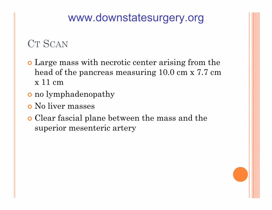
OPERATIVE INTERVENTION
Pylorus sparing y p gpancreatico-duodenectomyO ti th lOperative pathology
8x5x5 cm tumorPseudopapillary tumor p p yof the pancreas
www.downstatesurgery.org
CYSTIC NEOPLASMS OF THEPANCREAS
www.downstatesurgery.org
CYSTIC NEOPLASMS OF THE PANCREAS
WILLIAM R. BRUGGE, M.D., GREGORY Y. LAUWERS, M.D., DUSHYANTSAHANI, M.D.,CARLOS FERNANDEZ DEL CASTILLO M D AND ANDREW L WARSHAW M D NCARLOS FERNANDEZ-DEL CASTILLO, M.D., AND ANDREW L. WARSHAW, M.D.NENGL J MED 351;12 WWW.NEJM.ORG SEPTEMBER 16, 2004
Cystic neoplasms - <10% of pancreatic neoplasmsCyst c eop as s 0% o pa c eat c eop as sbenign, malignant, and borderlineneoplasms that either are primarily cystic or result from the cystic degeneration of solid tumors
www.downstatesurgery.org

CYSTIC NEOPLASMS
Types of cystic Solid pseudopapillary serous yp y
neoplasms includeserous cystadenomas (32 to 39 %)
pseudopapillary
to 39 %),mucinous cystic neoplasms (10 to 45 %)
papillary mucinous neoplasms (21 to 33 %)Solid pseudopapillary mucinous
tumors (<3-5%)pseudocyst
www.downstatesurgery.org
PRESENTATION
Can be asymptomaticy pRecurrent pain, jaundice or pancreatitis
Involving or connected to the pancreatic ductAdvanced
Pain, weight loss, jaundiceCan present with pseudocystsCan present with pseudocysts
Pain, and even early satiety if compressing the stomach or small bowel; jaundice secondary to compression of the common bile duct
www.downstatesurgery.org
DIAGNOSISCT
i iti l d t ti f l iinitial detection of a lesionvisualization of calcifications, septa, mural nodules, pancreatitis
MRI/MRCPbetter characterization of the morphologic features of a cyst showing a communication between the cyst and the showing a communication between the cyst and the pancreatic duct.
Transabdominal ultrasonographyThe use of PET is not firmly established
www.downstatesurgery.org
CYTOLOGICAL INVESTIGATION
Cytologic examination of cyst fluid y g yanalysis the aspirated fluid for a variety of biochemical markers and tumor cellst l i l i f t fl id h id tifi d ll cytologic analysis of cyst fluid has identified cells
to confirm of malignant disease mucinous cystic lesion in perhaps only half the aspirates obtained
www.downstatesurgery.org

CHARACTERISTICS TO BE EXAMINED
Signs and symptomsg y pHistologyLocation in the pancreasDiagnostic featuresSurgical treatmentMalignant potentialPrognosis
www.downstatesurgery.org
DIAGNOSIS AND MANAGEMENT OF CYSTICDIAGNOSIS AND MANAGEMENT OF CYSTICNEOPLASMS OF THE PANCREAS: AN
EVIDENCE-BASED APPROACHMATTHEW H G KATZ, MD, MELINDA M MORTENSON, MD,
HUAMINWANG, MD, PHD, ROSA HWANG, MD,ERIC P TAMM, MD GREGG STAERKEL MD JEFFREY H LEE MD DOUGLAS B MD, GREGG STAERKEL, MD, JEFFREY H LEE, MD, DOUGLAS B
EVANS, MD, FACS, JASON B FLEMING, MD, FACSJOURNAL OF AMERICAN COLLEGE OF SURGEONS
VOL. 207, NO. 1, JULY 2008VOL. 207, NO. 1, JULY 2008
www.downstatesurgery.org
SEROUS CYSTADENOMAS
> 30 % of cystic pancreatic neoplasmsy p pWomen in their 7th decadeMainly in body and tail but can be anywhere in the pancreasUsually asymptomatic and found incidentally Wh t ti i t i i bd i l When symptomatic - epigastric pain, abdominal fullness and weight loss; rarely jaundice – even in the pancreatic head.
www.downstatesurgery.org
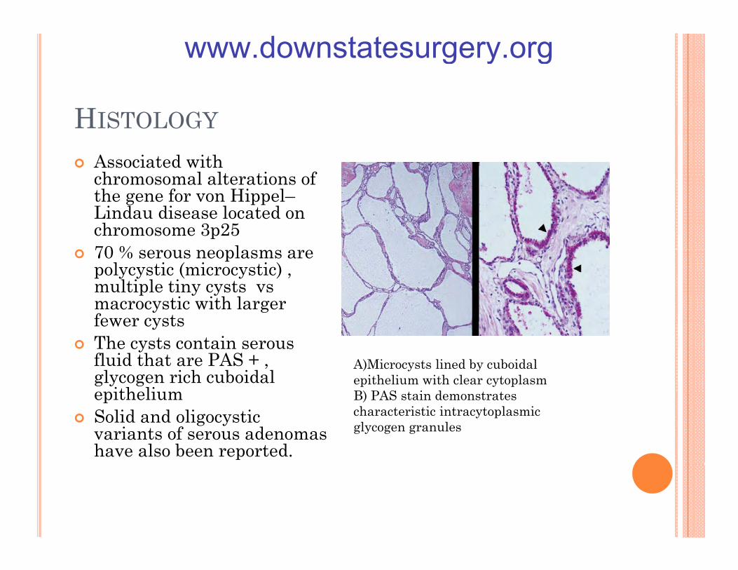
HISTOLOGYAssociated with chromosomal alterations of chromosomal alterations of the gene for von Hippel–Lindau disease located on chromosome 3p2570 % l 70 % serous neoplasms are polycystic (microcystic) , multiple tiny cysts vsmacrocystic with larger fewer cysts fewer cysts The cysts contain serous fluid that are PAS + , glycogen rich cuboidalepithelium
A)Microcysts lined by cuboidal epithelium with clear cytoplasm B) PAS t i d t t epithelium
Solid and oligocysticvariants of serous adenomas have also been reported.
B) PAS stain demonstrates characteristic intracytoplasmic glycogen granules
www.downstatesurgery.org
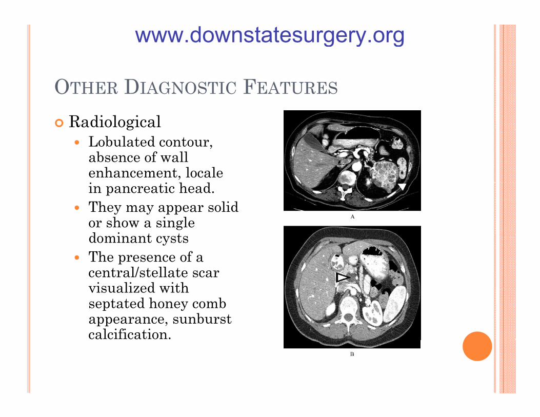
OTHER DIAGNOSTIC FEATURES
RadiologicalgLobulated contour, absence of wall enhancement, locale
in pancreatic head. They may appear solid or show a single d i t tdominant cystsThe presence of a central/stellate scar visualized with visualized with septated honey comb appearance, sunburst calcification.
www.downstatesurgery.org
SEROUS CYSTADENOMAS
Cyst fluid - 20 percent of serous cystadenomasLow CEALow CA19-9Low amylase
low potential for malignant diseaselow potential for malignant disease.Observed no significant increase in diameter of the tumor after 69 months G h f 0 2 f Growth of 0.12 cm per year for tumors < 4 cm Larger tumors >1.98 cm per year
www.downstatesurgery.org
SEROUS CYSTADENOMAS
Treatment –“our bias is that that operations are applied too early and too often for patients with this disease.”
There is some data that tumors <4 cm can be observed
Admittedly the number four is arbitrary. Does not apply for tumors of undetermined pathologyOperative procedure depends of the locale in the pancreas –
Pancreaticoduodectomy , central or partial pancreatectomy, distal pancreatectomy
www.downstatesurgery.org
SEROUS CYSTADENOMA PROGNOSIS
In a series by Bassi and colleagues’ y g50 patients with SCA were treated with definitive surgical resection. At a median followup of 43 months all patients were At a median followup of 43 months, all patients were alive and free of disease except one, who died of other causes.
I li i P k d ll In an earlier series Pyke and colleagues reported a 5-year survival of approximately 81% in 36 patients who underwent resection.
www.downstatesurgery.org
SEROUS CYSTADENOCARCINOMA
Aggressive behaviorggLymphovascular invasion Microscopic infiltrationS h t h t ti Synchronous or metachronous extrapancreatic metastasis
www.downstatesurgery.org

MUCINOUS CYSTIC NEOPLASM (MCN)
Mucin producing cystic neoplasm Includes intraductal mucinous neoplasms
IPMNMCN
Pre-malignant Can progress to invasive cancer
Can be determined from cyst aspirate
www.downstatesurgery.org
MUCINOUS CYSTIC NEOPLASM (MCN)45% of all resected pancreatic neoplasmsp pAlmost all are female Middle aged>90% located in the body and tailSymptoms can include discomfort, nausea, d idyspepsiaJaundice is uncommon
www.downstatesurgery.org
MUCINOUS CYSTIC NEOPLASM (MCN)Typically round, thick walled and septated.yp y , pNo communication with the pancreatic ductal systemHistopathological features of mucinous cystic neoplasms
include a dense mesenchymal ovarian-like stroma include a dense mesenchymal ovarian like stroma, requisite feature of mucinous cystic neoplasms.
www.downstatesurgery.org
MUCINOUS CYSTIC NEOPLASM (MCN)CT – thick cyst wall. Macrocystic lesion that can y ybe multiloculated. Peripheral eggshell calcification on CT
uncommon specific to a mucinous cystic neoplasmhighly predictive of cancerg y p e c ve o ca ce
The use of PET is not firmly established
www.downstatesurgery.org
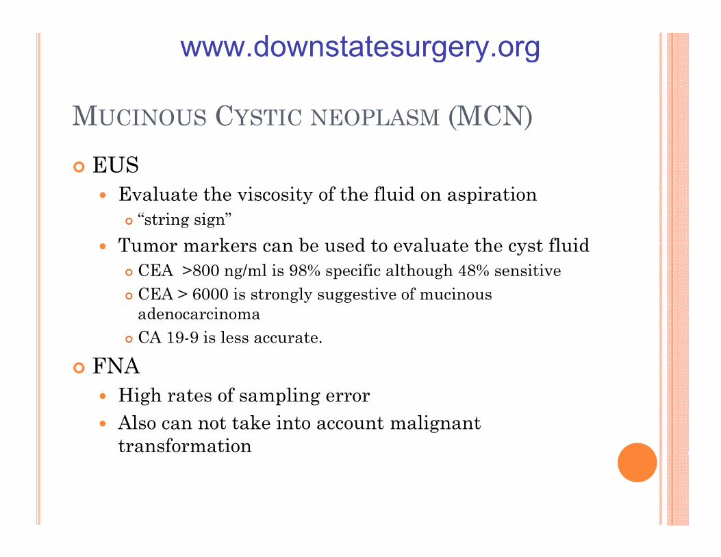
MUCINOUS CYSTIC NEOPLASM (MCN)EUS
Evaluate the viscosity of the fluid on aspiration “string sign”
Tumor markers can be used to evaluate the cyst fluidTumor markers can be used to evaluate the cyst fluidCEA >800 ng/ml is 98% specific although 48% sensitiveCEA > 6000 is strongly suggestive of mucinous adenocarcinomaadenocarcinomaCA 19-9 is less accurate.
FNAHigh rates of sampling errorAlso can not take into account malignant transformation
www.downstatesurgery.org
MUCINOUS CYSTIC NEOPLASM (MCN) PROGNOSIS
Survival after surgical resection correlates with gthe presence or absence of invasive disease. Surgical resection is curative for non invasive di h th h b f disease where as there have been cases of recurrence with invasive disease.
www.downstatesurgery.org
MUCINOUS CYSTIC NEOPLASM (MCN)MUCINOUS CYSTIC NEOPLASM (MCN)
In a series of 56 patients who underwent surgical resection f MCN i h l d d h of MCN, neither tumor recurrence or tumor-related death
was observed in 34 patients with adenomas or borderline MCN during a median follow-up period of 42 months and 69 months, respectively.69 months, respectively.Six patients with noninvasive carcinoma (carcinoma in situ) were also all alive without recurrence at a median followup of 76 months. In contrast, 8 (50%) of16 patients with invasive MCN died within 45 months.Reddy and colleagues found that none of the 52 patients with noninvasive MCN had recurrence of disease after resection, Goh and colleagues identified only 4 (2%) recurrences
189 t d ti t ith i i MCN i among 189 resected patients with noninvasive MCN in a pooled analysis of 344 previously reported patients
www.downstatesurgery.org
INTRADUCTAL PAPILLARY MUCINOUS NEOPLASM (IPMN)
25% of pancreatic cystic neoplasmsp y p20% of pancreatic resections for malignancyDisease of the elderly Higher prevalence of invasive adenocarcinoma with advanced ageE l di t ib ti t th dEqual distribution amongst the gendersSalient feature – connection to pancreatic ductClinical symptoms are generally non specificClinical symptoms are generally non specific
Can also include : pancreatitisDiabetes, weight loss or jaundice is generally
l d i h i i dicorrelated with invasive disease
www.downstatesurgery.org
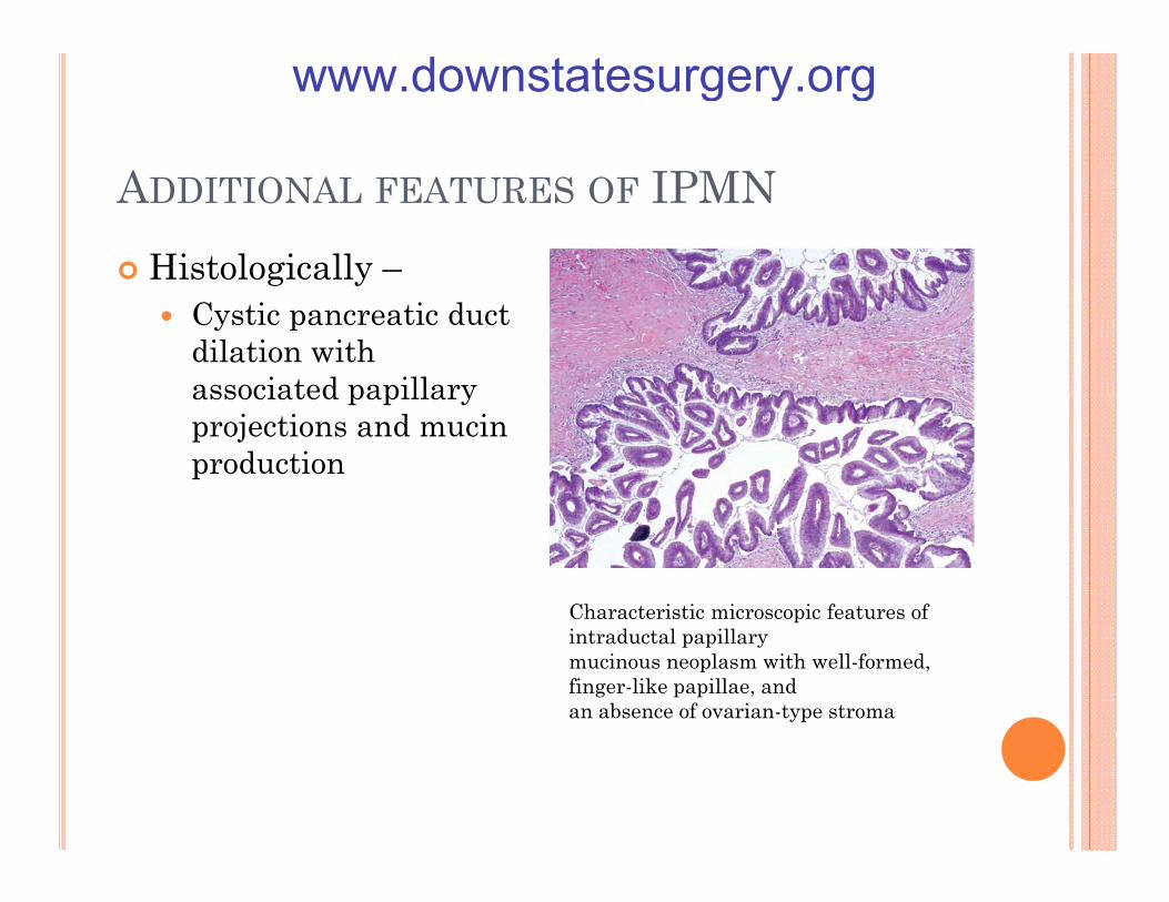
ADDITIONAL FEATURES OF IPMNHistologically –g y
Cystic pancreatic duct dilation with associated papillary associated papillary projections and mucin production
Characteristic microscopic features of pintraductal papillarymucinous neoplasm with well-formed, finger-like papillae, andan absence of ovarian-type stroma
www.downstatesurgery.org
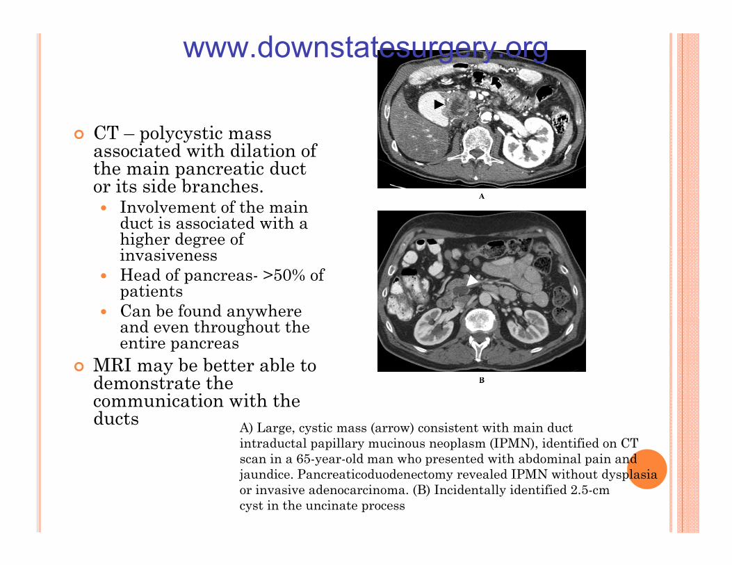
CT – polycystic mass associated with dilation of the main pancreatic duct por its side branches.
Involvement of the main duct is associated with a higher degree of g ginvasivenessHead of pancreas- >50% of patientsCan be found anywhere yand even throughout the entire pancreas
MRI may be better able to demonstrate the communication with the ducts A) Large, cystic mass (arrow) consistent with main duct
intraductal papillary mucinous neoplasm (IPMN), identified on CTscan in a 65-year-old man who presented with abdominal pain andscan in a 65 year old man who presented with abdominal pain andjaundice. Pancreaticoduodenectomy revealed IPMN without dysplasiaor invasive adenocarcinoma. (B) Incidentally identified 2.5-cmcyst in the uncinate process
www.downstatesurgery.org
IPMNERCP may show mucin production from an y penlarged papillaIPMN can be associated with a focus of ductal
i l h i th carcinoma elsewhere in the pancreasCurrent recommendations
Resection should be offered to all pts with main duct Resection should be offered to all pts with main duct IPMNsTumors with side branch involvement that are symptomatic or greater than 3 cm in size should be symptomatic or greater than 3 cm in size should be be resected < 3cm can be observed
www.downstatesurgery.org

IPMN PROGNOSIS
Postoperative survival of patients is related p pprimarily to the presence of invasive adenocarcinoma. Fi t ti i l f ti t ith Five-year postoperative survival of patients with noninvasive disease is between 77% and 100%In contrast, the prognosis for patients with In contrast, the prognosis for patients with invasive IPMN is similar to that of patients with invasive ductal adenocarcinoma, with the most optimistic 5 year survival rates no better than optimistic 5-year survival rates no better than approximately 36%.Noninvasive IPMN carries a recurrence rate after resection of 10%.
www.downstatesurgery.org
SOLID PSEUDOPAPILLARY TUMOR
RarePredominates in womenMedian age around 30 yearsCan occur anywhere in the pancreasSymptoms are usually vague and associated with i f th tsize of the tumor
www.downstatesurgery.org

Typically large Typically large encapsulated lesions with solid and cystic components.co po e s.Have pseudopapillarypatterns on histology
CT CT Well – encapuslatedsolid masses with thickened capsules and thickened capsules and variable amount of internal hemorrhage, cystic degeneration and cystic degeneration and calcification A) specimen demonstrating focal hemorrhage and cystic
degeneration consistent (B) A 4.5-cm solid andcystic lesion with coarse internal calcification, (arrow), found incidentally on a CT scan performed for workup ( ), y p pof nephrolithiasis.
www.downstatesurgery.org

SOLID PSEUDOPAPILLARY TUMOR
Operation is offered for this low grade tumor p gPrevent local tumor growth and metsPalliate symptomsC l d t f bl i l ith l l t Can lead to favorable survival even with local tumor extension or metsUsually requires distal pancreatectomy or a pancreaticoduodenectomy because of its large size
www.downstatesurgery.org
SOLID PSEUDOPAPILLARY TUMORPROGNOSIS
A single center report by Tipton and colleaguesdescribed 14 patients with a median tumor diameter of 7 cm.13 of the patients in whom curative resection was performed p12 were alive after longterm followup.
In a similar series by Martin and colleagues 18 patients who underwent resection for localized SPPT 100% recurrence-free survivalOf these, four patients presented with synchronous liver metastasis underwent combined pancreatectomy and metastasectomyyled to survival of 6 years and 11 years in 2 of the 4 patients. Overall, aggressive surgical resection is associated with a 5-year survival of 95%5 year survival of 95%.
www.downstatesurgery.org
LYMPHOEPITHELIAL CYSTS
The rarest : <70 pts in the literaturepMore common in men5th to 7th decadeUsually asymptomatic and discovered incidentallyU di t ib t d th h t th Usu distributed throughout the pancreasHistology
lined by a layer of stratified squamous epithelium lined by a layer of stratified squamous epithelium surrounded by a characteristic layer of lymphoid tissue.Cysts are filled with a dense material composed Cysts are filled with a dense material, composed mainly of debris, keratin, and cholesterol crystals.
www.downstatesurgery.org
LYMPHOEPITHELIAL CYSTS
There are no pathognomonic features on cross-p gsectional imaging, but several radiographic features are highly suggestive of this diagnosis. CT can demonstrate either a multi- or a CT can demonstrate either a multi or a unilocular cyst, which is well-encapsulated by an enhancing thin wall protruding from the body of the gland the gland. A cystic component of low attenuation is typical, but a solid component of variable magnitude can also existalso exist.MRI can be useful;
the high-keratin content of cyst fluid often produces a hyperintense signal on T1- and a hypointense signal on T2-weighted images
www.downstatesurgery.org
LYMPHOEPITHELIAL CYSTS
Few reports exist of FNA biopsy and analysis of p p y ycyst fluid for cytology and biochemical analysis.Cytologic evidence of squamous cells, keratin d b i l h t d h l t l t l debris,lymphocytes, and cholesterol crystals can help confirm the diagnosis.lymphoepithelial cysts are benignlymphoepithelial cysts are benign
observation is appropriate for the asymptomatic patient if you have definitive diagnosis has been secured secured. Surgical therapy should be reserved for symptomatic patients or for those in whom the diagnosis is equivocal equivocal.
www.downstatesurgery.org

ALGORITHM FOR PANCREATIC CYSTIC TUMORS/NEOPLASMS
www.downstatesurgery.org
QUESTIONS
1. Which of the following is a important g pdistinguising characteristic of an IPMNa) Elevated CEAb) L l t d tb) Loculated cystsc) Connection with the pancreatic ductsd) Ring enhancment on MRI
www.downstatesurgery.org

1. Which of the following is a important g pdistinguising characteristic of an IPMNa) Elevated CEAb) L l t d tb) Loculated cystsc) Connection with the pancreatic ductsd) Ring enhancment on MRI
www.downstatesurgery.org
2. Which of the following are considered mucin gproducing tumors a) IPMNb) SPPTb) SPPTc) Lymphoepitheliald) Serous cystoadenoma
www.downstatesurgery.org

2. Which of the following are considered mucin gproducing tumors a) IPMNb) SPPTb) SPPTc) Lymphoepitheliald) Serous cystoadenoma
www.downstatesurgery.org
3.What are the histological characteristics of gserous adenomas a) lined by a layer of stratified squamous epithelium
surrounded by a characteristic layer of lymphoid surrounded by a characteristic layer of lymphoid tissue.
b) Characteristic microscopic features of intraductal papillary mucinous neoplasm with well-formed finger-papillary mucinous neoplasm with well-formed, finger-like papillae, and an absence of ovarian-type stroma
c) pseudopapillary patternsd) Microcysts lined by cuboidal epithelium with clear d) Microcysts lined by cuboidal epithelium with clear
cytoplasm and PAS stain demonstrates characteristic intracytoplasmic glycogen granules
www.downstatesurgery.org
3.What are the histological characteristics of gserous adenomas a) lined by a layer of stratified squamous epithelium
surrounded by a characteristic layer of lymphoid surrounded by a characteristic layer of lymphoid tissue.
b) Characteristic microscopic features of intraductal papillary mucinous neoplasm with well-formed finger-papillary mucinous neoplasm with well-formed, finger-like papillae, and an absence of ovarian-type stroma
c) pseudopapillary patternsd) Microcysts lined by cuboidal epithelium with d) Microcysts lined by cuboidal epithelium with
clear cytoplasm and PAS stain demonstrates characteristic intracytoplasmic glycogen granulesgranules
www.downstatesurgery.org
4. Serous cystadenomas are associated with ywhich of the following genetic disorders a) Von Hippel Landau’s diseaseb) BRC 1b) BRCa-1c) Lynch Syndromed) MEN 2
www.downstatesurgery.org
Serous cystadenomas are associated with which yof the following genetic disorders a) Von Hippel Landau’s diseaseb) BRC 1b) BRCa-1c) Lynch Syndromed) MEN 2
www.downstatesurgery.org
5. With which of the following can the GI guys g g ysee a “string sign”a) IPMNb) S t db) Serous cystadenomac) SPPTd) MCN
www.downstatesurgery.org
5. With which of the following can the GI guys g g ysee a “string sign”a) IPMNb) S t db) Serous cystadenomac) SPPTd) MCN
www.downstatesurgery.org