Embed Size (px)
Citation preview

Pediatric Pulmonology 15:187-198 (1993)
Conference Report -
Cystic Fibrosis Foundation Consensus Conference Report on Pulmonary Complications of Cystic Fibrosis
Daniel V. Schidlow, MD,‘ Lynn M. Taussig, M D , ~ and Michael R. Knowles, MD3
INTRODUCTION
Management of complications of cystic fibrosis (CF) is the subject of perennial controversy and ongoing change. The literature only partially reflects the wealth of experi- ence accumulated by caregivers and is becoming rapidly outdated.
A group of physicians representing a vast array of disciplines joined in discussion to arrive at a set of recom- mendations that reflect the current trend in management of complications of the pulmonary disease in CF.
Consensus documents represent the combined wisdom and clinical experience of a group of experts in the field, rather than a mere distillation of the literature. We expect the opinions herein to evolve with time, as new therapeu- tic approaches are developed, in the same manner as this document has emerged from a critical review of what is known and published.
PNEUMOTHORAX Introduction
Pneumothorax, or “air in the pleural space,” can result in dangerous pressure on the lung. In patients with CF, this is most often caused by rupture of subpleural blebs through the visceral pleura. Another, much less common cause is “traumatic pneumothorax” secondary to lung puncture during procedures such as intravascular line in- sertion, or barotrauma (e.g., during mechanical ventila- tion).
Incidence The approximate incidence of pneumothorax is one
percent per year (Cystic Fibrosis Foundation Patient Reg- istry data). Five to eight percent of all patients with CF will eventually experience a pneumothorax. The inci- dence increases with age and severity of disease. Approx- imately 16 to 20% of adults (over 18) with CF will expe- rience a pneumothorax at some time in their lives.
0 1993 Wiley-Liss, Inc.
Diagnosis Pneumothorax should be suspected in patients with CF
who experience the sudden onset of chest pain and respi- ratory distress. Other patients can be completely asymp- tomatic and the pneumothorax is detected on a chest roentgenograph obtained for other reasons.
Common signs and symptoms of pneumothorax in- clude tachypnea, tachycardia, dyspnea, pallor, and cy- anosis. On physical examination the following are found: decreased breath sounds and vocal fremitus, and de- creased excursion of the affected hemithorax. In addi- tion, tension pneumothorax may cause signs of mediasti- nal shift, including deviation of the point of maximal impulse of the heart and deviation of the trachea from the midline. Subcutaneous emphysema and signs of circula- tory compromise also are likely during tension pneu- mothorax.
The most important laboratory examination is the chest radiograph, which should include frontal and lateral views. In addition, inspiratory, expiratory, and lateral decubitus views may help detect free air in the pleural space, when pneumothorax is difficult to detect in a plain
From the Department of Pediatrics, Temple University School of Med- icine, and Section of Pediatric Pulmonology, St. Christopher’s Hospi- tal for Children, Philadelphia, Pennsylvania;’ Department of Pediat- rics, Arizona Health Sciences Center, Tucson, Arizona;’ Department of Internal Medicine, University of North Carolina School of Medi- cine, Chapel Hill, North Carolina.3 Contributing members of the Con- sensus Panel: A. M. Cohen, MD, Carolyn R . Denning, MD, Thomas M. Egan, MD, Howard Eigen, MD, Richard J . Lemen, MD, Robert B . Mel- lins, MD, David M. Orenstein, MD, Lewis J . Rubin, MD, Michael L. Spector, MD, Robert C. Stem, MD, Robert E. Wood, MD, phi), and James R . Yankaskas, MD.
Received September 5 . 1992; accepted September 8 , 1992.
Address correspondence and reprint requests to D. V. Schidlow, De- partment of Pediatrics, Temple University School of Medicine, Sec- tion of Pediatric Pulmonology, St. Christopher’s Hospital for Chil- dren, Erie Avenue at Front Street, Philadelphia, PA 19134-1095.

188 Schidlow et al.
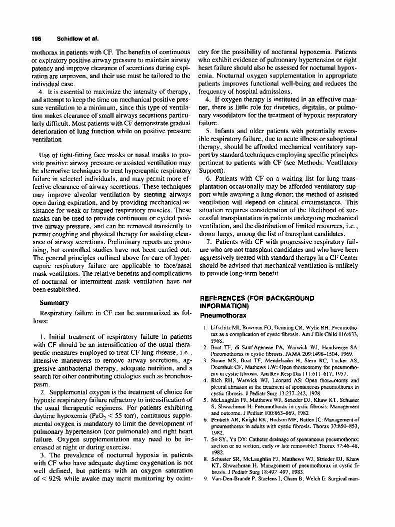
URGE (B) MI SYMPTOMATlc M X
Fig. 1. Management of small pneumothorax.
film. Air will become evident as the lung deflates on expiration or when the affected side is up (i.e., the air will rise and a rim of air will become evident on the left lateral decubitus film on a right sided pneumothorax).
Treatment Every patient with a newly diagnosed pneumothorax,
even if asymptomatic, should be hospitalized and ob- served for a minimum of 24 hr. For an asymptomatic patient in whom pneumothorax is an incidental finding, a chest radiograph should be obtained 24 hr after admis- sion. If such patient remains stable, asymptomatic, and the chest radiograph shows no increase in the size of the pneumothorax, the patient can be discharged and followed as an outpatient (Fig. 1). However, if the pneumothorax has increased, full treatment should begin (Fig. 2).
If the patient is symptomatic, or the pneumothorax appears to be 20% or more of the total affected hemitho- rax volume, a chest tube should be inserted.For suction a negutive pressure of no more than - 20 cm H,O should be applied. Because the lung reexpands by itself once free air is evacuated, greater negative pressure does not necessarily cause faster pneumothorax resolution. Fur- thermore, many physicians feel that application of greater negative pressure causes laceration of the lung secondary to apposition of the source of this negative pressure (tube) to the visceral pleural surface.
Large pneumothoraces are best managed by applying no suction to the chest tube initially, letting the lung slowly reexpand. This cautionary approach should avoid both patient discomfort and edema of the affected lung when suddenly reinflated. Some practitioners administer 100% inspired oxygen to facilitate reabsorption. This method is not universally accepted and is probably inef-
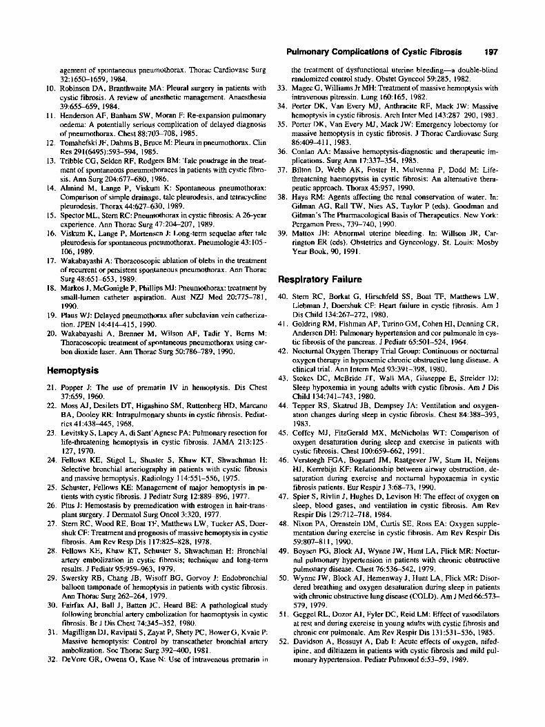
,-, +, PTX PERSISTS
REMOVE CHEST N E E
Fig. 2. Management of large pneumothorax.
fective beyond the neonatal period. No controlled studies have clarified this controversy.
After chest tube insertion, a new chest radiograph should be obtained. If a substantial collection of air per- sists, insertion of additional chest tubes should be consid- ered. Such procedures can be aided by the guidance of diagnostic imaging such as fluoroscopy or ultrasound. Following reduction of the size of pneumothorax, a wait- ing period of 24-48 hr is prudent. However, if after this waiting period pneumothorax is still present, surgery must be considered.
Once the pneumothorax is resolved and the air leak stops (i.e., apposition of the visceral pleura to the tho- racic wall is achieved) the chest tube can be removed.
If the patient remains asymptomatic and the lung stays expanded, the pneumothorax is considered resolved. If in spite of this an air leak persists, observation of the patient should continue. If after approximately 5 days there is no cessation of the air leak, suction should be discontinued and the tube left in place with underwater seal. A new radiographic and clinical evaluation should be camed out; if air reaccumulated in the pleural space, surgery should be considered.
No full agreement has been reached as to whether tubes should be clamped before removal, and whether tubes should be removed as soon as the pneumothorax is con- sidered resolved. Most practitioners will clamp the tube for approximately 12 hr prior to withdrawing it. Reoccur- rence of pneumothorax indicates the need for surgery.
Sclerosing agents have been used as a treatment for pneumothorax in patients with CF. The most popular

Pulmonary Complications of Cystic Fibrosis 189
3. Needle aspiration is an emergency procedure which can precede but never replace chest-tube insertion. Nee- dle aspiration is indicated only when patients are severely compromised upon presentation and require immediate intervention. Otherwise, needle aspiration is not appro- priate in the management of pneumothorax in CF.
4. Heimlich valves have a limited role in the tempo- rary control of pneumothorax. They could be used during patient transport and for patients awaiting an imminent lung transplantation. These valves are not always effec- tive in completely evacuating air but provide a temporary prevention of tension pneumothorax.
5. Chemical pleurodesis should be reserved for pa- tients whose clinical condition is such that the risk of surgery outweighs its benefits (e.g., extremely poor anes- thetic risks, patients in heart failure or respiratory fail- ure), or for patients who absolutely refuse surgery. Avail- ability of chemical agents may limit the performance of these procedures.
TABLE 1-Unpublished Data From a Retrospective Review of Treatment of Pneumothorax in 192 Patients (Denning et al., 1991) Treatment Success
Open thoracotomy Quinacrine sclerosis Tetracycline sclerosis
50/52 (96%) 31/35 (89%) 21/36 (58%)
sclerosing agents are quinacrine and tetracycline. In a large unpublished study (Denning et al., 1991), instilla- tion of quinacrine into the pleural space resolved pneu- mothoraces unresponsive to chest tube insertion and air drainage alone, in about 90% of all cases. In that same series, tetracycline was successful in 58% and open tho- racotomy in 96% of pneumothoraces (Table 1).
Instillation of sclerosing agents has two advantages. First, no surgical intervention is required. Second, it can be accomplished in gravely ill patients without general anesthesia. The disadvantages include lack of direct visu- alization of the distribution and effect of the chemical, unpredictable results, with variable success rate. Finally, quinacrine and tetracycline are no longer manufactured by U.S. pharmaceutical companies and access to these products has become extremely difficult.
Talcum powder is a very effective and readily avail- able sclerosing agent. A major drawback of using talcum is the high probability of causing lung adhesions to the parietal pleura. This complication is particularly serious if it involves the diaphragmatic pleura because it may render the diaphragm unable to contract properly.
Thoracoscopy and COz laser abrasion, with or without stapling of blebs or instillation of chemical agents, has recently emerged as another treatment modality for pneu- mothorax.
Stapling and abrasion are facilitated by collapse of the lung. The lungs of patients with CF tend not to fully collapse following pneumothorax because of pleural ad- hesions and poor elastic recoil. Thus, adequate visualiza- tion may be less than optimal for thoracoscopic manage- ment in some of these patients.
Data concerning the use of thoracoscopy in CF are scant. Successful management of pneumothorax in five patients using talcum insufflation through thoracoscopy was recently reported. No data are available on the long- term outcome of this procedure.
Recommendations
1 . Treatment options for pneumothorax unresponsive to chest tube drainage are thoracotomy, limited surgical pleurodesis, oversewing or stapling subpleural blebs, and chemical pleurodesis.
2. The benefits of surgery in managing persistent pneumothorax far outweigh the risk of this procedure for most patients.
Technical Considerations Chest Tube Placement The chest tube should be of adequate size to evacuate
the air and allow for sufficient flow to accommodate the air leak from the ruptured bleb(s). In general, a 24 French size tube should suffice for an adult-sized patient. Smaller tubes may be adequate for smaller patients.
The site of the chest tube insertion will depend on the location of the pneumothorax. Consideration must be given to the fact that surgery may ultimately be required. Since the preferred surgical approaches are transaxillary (third intercostal space) or anterior (fourth or fifth inter- costal space), the chest tubes should nor be placed in these locations.
The optimal insertion site is in the mid-axillary line in the fifth or sixth intercostal space below the axillary tail of the breast in females. It is important to direct the tube to the apex of the chest and to insert a sufficient length so that the end of the tube sits in the apex.
High anterior chest tubes (second intercostal space) are sometimes useful. However, they are difficult to position and maintain properly, and their insertion is more pain- ful, also risking injury to the internal mammary artery if placed too far medially. Diagnostic imaging may help position the tube, particularly when placement of a sec- ond tube becomes necessary.
Several commercially available pleural drainage units offer closed suction systems. Connections should be air- tight with appropriate pleural pressures. Occasionally, these units can malfunction if they are overturned or if the negative inspiratory force exerted by the patient exceeds - 30 cm H,O. In these situations, fluid can be expelled via a valve designed to ensure egress of air. For patients
190 Schidlow et al.
with restrictive lung disease this can be a frequent occur- rence and therefore mandates diligent surveillance.
Chest tube removal may cause recurrence of pneu- mothorax when the tract formed by the tube remains in communication with the pleural space. A purse string or mattress suture can be used to assure a proper skin seal.
Technique of Pleurodesis (Production of Adhesions) The area of air leak is identified through a small thora-
cotomy. Identifiable blebs are usually excised after their base is stapled. The parietal pleural surface is then abraded with a gauze sponge. By rubbing the sponge over the pleural surface, the surgeon can see the inflammation that this produces as the pleura becomes erythematous visible to the naked eye. By manipulating the lung the surgeon has the ability to see and control the extent and location of the area of abrasion.
Pleurodesis and Lung Transplantation Pleurodesis is not an absolute contraindication for lung
transplantation. The transplant coordinator at the specific center should be contacted with questions concerning management of pneumothorax if referral is planned.
Patients who experience pneumothorax while awaiting transplantation (on a transplant list) should be managed as others with CF. It is prudent, however, to contact the transplant team at the referral institution before beginning pleurodesis.
Complications Complications of pneumothorax include respiratory
compromise, shock, hemorrhage (due to shearing of ves- sels), and empyema. Efforts should be made to continue physical therapy. The performance of this treatment will not interfere with pneumothorax resolution. Efforts should be made to relieve discomfort resulting from the presence of a chest tube or a thoracotomy with adequate analgesic therapy.
Prognosis Spontaneous penumothorax in CF is, generally, a bad
prognostic sign. Available data indicate that survival af- ter the onset of a pneumothorax is approximately 30 months.
Prevention To prevent pneumothorax, patients with CF should
avoid maneuvers or situations which will create marked fluctuations in intrapleural pressure. These include power weight lifting, intense isometric exercises. During scuba diving, individuals are subjected to increased pressure followed by decompression, which may create changes in lung parenchyma that could cause pneumothorax. Under-
water swimming in pools and shallow diving probably do not increase the risks of pneumothorax.
No air travel or pulmonary function testing should be undertaken for at least 2 weeks following resolution of pneumothorax.
HEMOPTYSIS Minor Hemoptysis (“Blood Streaking”)
Blood streaking of the sputum is common in patients with CF and requires no specific treatment. Persistent streaking, however, may indicate a pulmonary exacerba- tion requiring appropriate therapy. The physician should determine that there are no other potential contributing factors to this condition such as the chronic use of aspirin.
Major Hemoptysis (“Major Bleeding”) Major hemoptysis is defined as acute bleeding of a
large amount of blood (often defined as 240 mL in a 24-hr period) which could be life-threatening due to asphyxia- tion from airway obstruction and/or could lead to acute hypotension.
Recurrent bleeding of substantial volume (e.g., more than 100 mWday) over a short period of time (e.g.. 3-7 days) is also termed “major hemoptysis.” These recurrent episodes may have the following characteristics:
threaten life due to asphyxiation; produce airway obstruction; produce hypotension; produce anemia; produce chemical pneumonitis; and trigger a pulmonary exacerbation
Incidence About 1% of patients with CF have one episode of
major bleeding each year, according to the Cystic Fibro- sis Foundation Patient Registry data, which defines he- moptysis as a bleed of more than 240 mL/24 hr or requir- ing transfusion. Obviously, the number of patients with minor hemoptysis, as defined above, will be somewhat larger. The vast majority of patients with major hemopty- sis are 16 years or older (Registry data).
Pathogenesis Major hemoptysis in patients with CF is nearly always
of systemic arterial origin. The bleeding usually arises from markedly enlarged and tortuous bronchial arteries which vary greatly in number and origin. Two-thirds of bronchial arteries arise from the ventral surface of the aorta, the remaining one-third arise from other arteries including the internal mammary and intercostal arteries. Major hemoptysis also can be produced by nonbronchial arteries which collateralize with the bronchial circulation or enter the lung through granulation tissue. Such arteries
Pulmonary Complications of Cystic Fibrosis 191
unknown, all large and tortuous bronchial arteries may be embolized since collaterals often have developed from mediastinal and contralateral vessels.
7. When the patient is admitted, a surgeon and a radi- ologist experienced in interventional angiography should be notified. If such personnel are not readily available, referral to another center should be considered.
include the phrenic, intercostal, internal mammary, thy- rocervical, costocervical, and branches of the subclavian and axillary arteries.
Evaluation/Diagnosis
1. It is essential to differentiate major hemoptysis from bleeding from the upper airway or from the gas- trointestinal tract. In addition, the examination should rule out the presence of a foreign body. A careful history may help to localize the site of bleeding by the patient noticing localized gurgling, warmth, discomfort, full- ness, etc. Physical examination may reveal new, focal pulmonary findings or bleeding from an upper airway site. Differentiating a pulmonary bleeding from other sites may require assessing associated symptoms, mea- suring pH of the expectorated blood, placing a nasogas- tric tube, endoscopy, etc.
2. Use of other drugs which could contribute to the bleeding, such as aspirin, nonsteroidal antiinflammatory drugs (NSAID), penicillin, etc. should be ascertained.
3. A chest radiograph should be obtained for several reasons: to determine if there have been any acute changes in the lungs, to try to localize the site of the bleeding, and to determine if there are any other unusual findings which could explain the bleeding such as a new thick-walled cyst (suggesting infection with an unusual organism such as atypical mycobacteria), and localized or unilateral hyperinflation (suggesting a foreign body). 4. The following laboratory tests should be obtained:
complete blood count with platelet count, prothrombin time (PT) and partial thromboplastin time (PTT), liver function tests, and typing and crossing the patient’s blood for possible transfusion.
5. A sputum for culture and sensitivity should be ob- tained. If the clinical situation and radiographic findings suggest other potential infections, the sputum should also be stained and cultured for organisms such as mycobacte- ria and fungi.
6. Bronchoscopy may help localize the site of bleed- ing; however, the procedure is not always successful. This occurs when the patient has stopped bleeding or when massive bleeding prevents adequate visualization of the airways. The vast majority of major pulmonary bleeds in CF patients are acute, self-limited, and may not require diagnostic tests such as bronchoscopy . Bronchos- copy is indicated when the acute bleeding continues, when it appears to be progressive or frequently recurs, and if surgery or local airway therapy will be used. It is strongly recommended that both rigid and flexible bron- choscopes be available since either or both may be re- quired to assess and manage the bleeding. If the site of bleeding is known, it is acceptable to embolize only the artery(ies) to that area. If embolization of bronchial arter- ies is to be the therapy of choice and the site of bleeding is
Treatment Many patients with CF having major hemoptysis, will
stop bleeding spontaneously in less than 4 days. This may occur with no change in their usual therapy and/or with- out specific treatment for the bleeding.
There are almost no scientific data to support most of the therapeutic options noted below for treatment of he- moptysis in CF patients. The recommendations are based on the clinical observations and experiences of the physi- cians at the Consensus Conference, on case reports in the literature, published studies, and theoretical consider- ations derived from the literature.
1. First, the patient should be reassured and calmed. Psychological support may be necessary. Occasionally, sedation may be required.
2. Drugs which could interfere with coagulation should be discontinued. These include aspirin, penicillin, and NSAID. Inhaled drugs which may be pulmonary irritants such as N-acetylcysteine and aerolized antibiot- ics should also be discontinued. In vitro and animal stud- ies have demonstrated that inhaled P-adrenergic drugs may dilate the bronchial vasculature whereas anticholin- ergic drugs may constrict it. There are no data in humans. Therefore, the decision to continue or discontinue any of the inhaled bronchodilators is based on theoretical con- siderations. No specific recommendations are made with respect to their use.
3. Coagulation defects should be corrected with vita- min K, fresh frozen plasma, or specific factors as indi- cated. If intravenous vitamin K is used, the drug should be given slowly to avoid hypotension and anaphylaxis. Since the PT and PTT may not always reflect low levels of clotting factors, intramuscular vitamin K is often given during a major bleed, in spite of a normal PT and PTT. 4. Acute blood loss should be corrected with transfu-
sions as clinically indicated. 5. Most major episodes of hemoptysis appear to be
associated with pulmonary exacerbations and, therefore, should be treated with appropriate intravenous antibiotics and other usual therapies (as discussed earlier, penicillins probably should not be used).
6. Placing the lung which appears to be bleeding in the dependent position may help to prevent contamination of the nonbleeding lung. However, individual patients may respond to various positions with increased bleeding or

192 Schidlow et al.
breathing difficulty. Thus, the positioning of patients with major hemoptysis needs to be individualized. 7. The patient should be encouraged to cough to max-
imize blood expectoration, thereby keeping the airways as patent as possible. There is no evidence that discon- tinuing chest physiotherapy is either beneficial or harm- ful; the decision of whether or not to continue it is left to the physician and patient.
8. Placing ice packs on the chest has no demonstrable benefit and may be uncomfortable for the patient.
9. Intravenous premarin (water-soluble, conjugated estrogen) may rapidly stop pulmonary bleeding by en- hancing platelet aggregation, decreasing capillary perme- ability, interfering with tissue reactions to bradykinin, and by other poorly understood mechanisms. This medi- cal treatment may warrant consideration before more in- vasive therapy. Recommended dose is 10-25 mg over 1&15 min every 4-6 hr for 3 4 doses.
10. Intravenous pitressin (vasopressin and desmo- pressin) may rapidly stop bleeding by enhancing arterio- lar smooth muscle contraction and the release of coagula- tion factors from the vascular endothelium. This medical treatment may be considered before more invasive ther- apy. With the use of pitressin, there may be considerable water retention for which diuretics may need to be given in the following doses: (1) vasopressin, 20 units over 15 min followed by continuous infusion of 0.2 unitdmin; (2) desmopressin, 4 pg intravenously followed by infusion of 0.3 p/kg over 12 hr.
1 1. Local/topical treatment may be indicated when the bleeding appears to be immediately life-threatening . Such therapy may include some or all of the following:
Endobronchial tamponade with a catheter/balloon system Direct airway tamponade with gel foam pledgets Iced saline lavage Topical therapy with a-agonists or thrombin Selective or double-lumen intubation Removal of clots (while removing clots may improve ventilation, it can also lead to a marked increase in bleeding).
Topical or local treatment is usually not indicated in nonemergency situations.
12. Arterial embolization may be indicated for major hemoptysis (as defined above) as well as for persistent hemoptysis of lesser volume when it interferes with a patient’s life-style and/or medical management. While embolization often produces acute cessation of bleeding, the procedure has potentially major complications includ- ing paralysis, organ infarction, and death. Therefore, this procedure should be performed only by an experienced angiographer and interventional radiologist who have ex- perience with this specific procedure. If such expert is not
available, the patient should be referred to an institution where the embolization can be performed. Recurrence of bleeding is relatively common following embolization and should be managed as a de novo event. If bleeding does not stop acutely after embolization, reembolization should be considered. It is important in this conjunction to remember the anatomic variability of the bronchial vessels and the role other arteries play in producing major hemoptysis. A theoretical complication of embolization is airway ischemia, which could affect subsequent lung transplantation.
1 3. In rare cases where bleeding cannot be stopped by the methods described above, including embolization, local pulmonary resection may be indicated. In these situations, it is mandatory to localize the site of bleeding prior to surgery. If surgery is seriously contemplated, other factors such as the patient’s baseline lung function and overall clinical status must be considered.
Prognosis Recent data indicate that an episode of major hemopty-
sis does not necessarily change the long-term prognosis for any individual patient.
RESPIRATORY FAILURE Introduction
Respiratory failure in patients with CF is a sign of severe lung disease and defined by hypoxemia and/or hypercapnia associated with obstructive airway disease. The topic merits review for several reasons. Over the past 20 years, we have developed a better understanding of the treatment of hypoxic respiratory failure with supplemen- tal oxygen, and we have observed technical advances in modes of ventilatory support. Further, the availability of lung transplantation makes the short-term treatment of respiratory failure of patients with CF pertinent, because their long-term quality of life may be improved with transplantation. This section will describe our current understanding of the pathophysiology of respiratory fail- ure in CF and the state of the art for therapeutic interven- tions. These concepts rely, in part, on information gleaned from treatment of non-CF patients with obstruc- tive airways disease and respiratory failure.
Pathophysiology of Respiratory Failure Retained airway secretions and bronchiectasis are ma-
jor features of the obstructive airway pathobiology in CF. Pathogenic factors in the development of respiratory fail- ure include (1) thickened airway secretions with bacterial infection and mucus hypersecretion, (2) bronchoconstric- tion, (3) airway mucosal edema and inflammation, and (4) respiratory muscle weakness and fatigue. These fea- tures contribute to the development of respiratory failure, and hypoxemia usually precedes hypercapnia. Figure 3

Pulmonary Complications of Cystic Fibrosis 193
airway secretions. Antiinflammatory agents, particularly systemic corticosteroids, may play a useful short-term role in reducing mucosal inflammation, edema, and bron- chospasm, and improving clearance of airway secretions. The long-term benefit of antiinflammatory agents how- ever is undefined. Adequate nutrition is mandatory for providing appropriate muscle strength and endurance. Nutritional supplementation may be required. Exercise rehabilitation may be useful, not only to improve cardio- vascular conditioning, enhance oxygen delivery, and im- prove respiratory muscle strength and endurance, but also to assist in mechanical clearance of airway secretions. The initial therapeutic approach also should include a search for unusual pathogens such as nontuberculous my- cobacteria, and for other etiologies such as pneumotho- rax, allergic bronchopulmonary aspergillosis, or reactive airways disease.
Treatment: Oxygen Supplementation Chronic (Daily) Hypoxemia If intensification of the standard therapeutic regimen
does not correct hypoxemia (PaO, < 55 torr in room air at rest), continuous supplemental oxygen is mandatory to limit the development of pulmonary hypertension and cor pulmonale. Although supplemental oxygen for I2 hr/day is better than no oxygen supplementation, continuous oxygen therapy (18-24 hr/day) reduces mortality in pa- tients with chronic obstructive pulmonary disease (COPD) by 50% when compared to nocturnal oxygen alone. Oxygen supplementation should be aimed at achieving a PaO, > 60 torr without significant adverse effect on arterial PaC02 and pH. In patients who chroni- cally retain C02, oxygen supplementation should be ini- tiated cautiously and PaCO, rechecked. The usual mode of oxygen supplementation is by a nasal cannula. Al- though transtracheal oxygen delivery is being used in patients with COPD, there is little experience in patients with CF, in whom transtracheal oxygen may dry airway secretions and impair the clearance of airway secretions. Many patients will require increased amounts of oxygen at night to overcome the effects of hypoventilation with REM sleep and worsening ventilation to perfusion (V/Q) mismatch secondary to airway closure. Patients on exer- cise rehabilitation programs also are likely to require increased levels of oxygen supplementation during exer- cise. The dosing requirement for additional oxygen sup- plementation during sleep or exercise must be determined empirically (Table 2).
Nocturnal Hypoxemia Some patients with CF may have adequate daytime
oxygenation, but experience hemoglobin desaturation during sleep that is associated with REM sleep, shorter total sleep time, and increased pulmonary artery pres- sures. These patients should receive supplemental oxy-
AIRWAYS OBSTRUCTION - + +
ALVEOLAR HYPOVENTllATlON I
RESPIRATORY FAILURE
RESPIRATORY FAILURE
Fig. 3. Major pathophysiologic derangements associated with hypoxemic and hypercapnic respiratory failure in cystic fibro- sis.
displays the pathophysiologic consequences of airway obstruction that contribute to respiratory failure. Obstruc- tion of airflow in small and large conducting airways results in hypoxemia due to altered matching of ventila- tion and perfusion, and this results in hypercapnia due to alveolar hypoventilation.
Treatment of Respiratory Failure: Initial Approach Goal The goal of treatment is to correct hypoxemia and to
achieve adequate pulmonary carbon dioxide elimination and a stable acid-base balance, thereby retarding the development of pulmonary hypertension and cor pulmo- nale, and prolonging functional life. The treatment of respiratory failure must target the pathogenic factors, i .e., abnormally thickened airway secretions, bacterial infection, bronchoconstriction, mucosal edema and in- flammation, and respiratory muscle weakness and fa- tigue.
General Principles The initial approach to the treatment of respiratory
failure is to intensify the usual regimen used to treat the pulmonary disease, i.e., clearance of retained airway se- cretions and control of bacterial infection with parented antibiotics. This approach should involve more intensive and more frequent sessions of chest physical therapy with postural drainage, including intensive care resources if necessary. It may also involve higher doses and/or longer durations of multiple antipseudomonal drug treatment and, sometimes, antistaphyloccocal therapy, even if sta- phylococci are not recovered from sputum cultures. In- haled or systemic bronchodilators may assist clearance of

194 Schidlow et al.
TABLE 2-Hypoxemic Respiratory Failure: Guidelines for Oxygen Therapy.
Daytime room air: PaO, < 55 tom or PaO, < 59 tom, plus one of the following: edema, hematocrit >55%, or p-pulmonale wave pattern on EKG
oxygen saturation < 88-90% for >lo% of total sleep time
Nocturnal:
Exercise: oxy-hemoglobin saturation < 88-90%
“Uscd by third-party payers for reimbursement.
TABLE 3-Hypercapnic Respiratory Failure: Guidelines for Assisted Ventilation
Indications Infants (most cases) Reversible or potentially reversible
complications Pneumo thorax Bronchospastic obstruction Central mucous plugging Suboptimal therapeutic regimen
Possible indication Not indicatcd
Patient accepted as lung transplant candidate Progressive respiratory failure, unresponsive
to intensive standard therapy
gen at night because it improves functional well-being and reduces the frequency of hospital admissions; fur- ther, nocturnal hypoxemia may contribute to pulmonary hypertension and vascular remodeling. The prevalence of nocturnal hypoxia in patients who have adequate daytime oxygenation is not well defined, but secondary poly- cythemia is not a reliable index of nocturnal desaturation. Assessment of oximetry is required, and screening for nocturnal hypoxemia should be considered in patients with saturation less than 92% when awake. Patients who exhibit evidence of pulmonary hypertension or right heart failure should be evaluated for the possibility of nocturnal hypoxemia, even if they have adequate PaO, during the daytime (Table 2).
Treatment: Other Medications If oxygen therapy is satisfactorily instituted, there is
little role for diuretics, digitalis, or vasodilators for the treatment of hypoxic respiratory failure in patients with CF. Diuretics may have an adverse effect by decreasing systemic vascular volume without reducing pulmonary vascular pressures; this leads to a decrease in right heart filling and cardiac output may be adversely affected since the right ventricle is a “volume” pump. Cardiac glyco- sides provide little benefit for right-sided cardiac failure, and their use may be associated with arrhythmias, partic- ularly when pulmonary hypertension is present. The use of vasodilators (other than supplemental oxygen) is dis- couraged because they may induce larger reductions in systemic vascular resistance than in pulmonary vascular resistance, leading to decreased cardiac output, hypoten- sion, and even death. The use of theophylline may con- tribute to improved diaphragmatic muscle function, but it is not routinely beneficial and may be associated with increased risk of gastroesophageal reflux, which is com- mon in CF patients.
Treatment: Mechanical (Assisted) Ventilation Introduction The general goal of medical therapy in patients with
CF is to improve longevity and quality of life. Initial experience with mechanical ventilation in the 1960s and early 1970s yielded poor results; few patients were suc-
cessfully weaned from ventilators, and those weaned had short survival. However, assisted ventilation may be ap- propriate when there are reversible causes of respir- atory failure, or when lung transplantation is imminent (Table 3).
Infants Early experience with mechanical ventilation in infants
with CF (less than 12 months old) in respiratory failure was associated with an 80% mortality rate, but recent studies indicate that infants and young children with CF may have greatly improved acute survival (-80%) with current methods of ventilatory support, and significant long-term survival (5040% for several years). Thus, such patients should be afforded mechanical ventilatory support as indicated; many will survive and achieve long- term functional status.
Acute/Reversible Respiratory Failure Patients with CF with potentially reversible respiratory
failure due to an acute illness, e.g., bronchospasm, viral pulmonary infection, pneumothorax, hemoptysis, mas- sive aspiration, or with possibly reversible respiratory failure due to suboptimal medical therapy, should be treated with mechanical ventilatory support as indicated. The majority of these patients will survive short-term ventilatory support, and their overall prognosis is more closely linked to the underlying severity of the lung dis- ease than to the episode of respiratory failure. Several general principles should be carefully observed. First, the period of intubation should be as short as possible. Sec- ond, aggressive attention should be paid to mechanical removal of airway secretions to prevent impaction of secretions. Third, respiratory and skeletal muscles should be exercised (including patient ambulation with Am- bubag) to prevent muscle deconditioning and atrophy.
Chronic Respiratory Failure in Patients Accepted for Lung Transplantation In some patients with CF, the possibility of lung trans-
plantation increases the likelihood of long-term improved quality of life, and adds another potential indication for

Pulmonary Complications of Cystic Fibrosis 195
ticular attention should be paid to correcting any acute or reversible components of respiratory failure, and the usual regimen of care for CF lung disease must be inten- sified. This includes use of frequent (every 1 4 hr) and aggressive chest physical therapy and other techniques to clear airway secretions. Bronchoscopy is occasionally useful to diagnose and treat central mucous plugging, but it is not usually beneficial for clearing secretions from small airways. Parenteral antibiotics should be used in- tensively, with multiple drugs and high doses. Parenteral bronchodilators and/or corticosteroids should be consid- ered for bronchospasdmucosal edema unless contraindi- cated, e.g., corticosteroids may be contraindicated in lung transplant candidates. Maintenance of adequate nu- trition is essential, and must be balanced with the associ- ated increase in CO, production.
In contrast to some patients with severe COPD or sta- tus asthmaticus, sedation and paralysis to reduce C02 production are not generally appropriate; sedation should be minimized. Patients with CF should exercise respira- tory and skeletal muscles, even walking (while intubated) with Ambu-bag assisted ventilation. Aggressive therapy to reduce production of and to remove airway secretions should be quickly instituted. Rapid treatment of revers- ible etiologic factors and early extubation is essential, since prolonged positive pressure ventilation tends to worsen small airway obstruction and reduces the likeli- hood of successful extubation.
Techniques Positive pressure ventilation through cuffed endotra-
cheal tubes is the current standard of care for hypercapnic respiratory failure. Negative pressure ventilatory devices have not been effective in CF, in part because of their interference with chest physiotherapy. The endotracheal tube should be as large as possible to permit adequate suctioning and, occasionally, bronchoscopy . Tracheos- tomy generally is not necessary, and may compromise candidacy for lung transplantation in some patients. Many volume- and pressure-limited ventilator modes and waveforms are available on positive pressure mechanical ventilators. The modes selected must be adapted to the individual patient and situation. However, some general principles apply in virtually all situations:
assisted ventilation. However, the evolving criteria for candidacy for lung transplantation, and the limited avail- ability of donor organs, make it difficult to define specific recommendations at this time. In general, patients receiv- ing assisted ventilation will not be accepted as transplant candidates. The criteria for the use of assisted ventilation in patients who had been previously accepted as lung transplantation candidates, will have to be defined by the experience of individual lung transplant centers. This situation requires consideration of the possible duration and complications of mechanical ventilation, the avail- ability and assignment of donor organs, and the likeli- hood of successful transplantation in mechanically venti- lated patients.
Chronic Respiratory Failure in Patients Not Accepted for Lung Transplantation Patients who develop respiratory failure despite ag-
gressive treatment, consistent with standard measures in a CF Center, should be advised that mechanical ventila- tion is unlikely to provide long-term benefit. Unless there is an acute and potentially reversible component to the respiratory failure, the great majority of these patients will not be successfully weaned from mechanical ventila- tion, and the survival time and functional capability of any patient successfully weaned are likely to be re- stricted. A time-limited trial of intubation and mechanical ventilation is occasionally appropriate to accomplish last wishes, but this approach should be discouraged by edu- cating the patient about terminal respiratory complica- tions of the disease. Recent experience suggests that non- invasive ventilatory support (i.e., by face or nasal mask) may be useful in selected patients, but this does little to improve functional status. It improves or stabilizes the blood gases so that patients do not have continuing in- crease in PaCO, or acidosis but does not otherwise change pulmonary functions.
Methods: Ventilatory Support Goal Assisted ventilation in patients with CF is usually di-
rected at the treatment of hypercapnic respiratory failure; hypoxemic respiratory failure can usually be treated suc- cessfully with supplemental oxygen. The goal of me- chanical ventilatory support is to increase alveolar venti- lation sufficiently to sustain life and permit treatment of the underlying cause of hypercapnic respiratory failure.
General Principles Because clearance of secretions from small airways by
endotracheal suctioning is less effective than coughing, all possible measures should be instituted to limit the duration of intubation and thereby prevent irreversible “impaction” of secretions and loss of lung function. Par-
1. Ventilatory modes that permit and encourage pa- tient to use their respiratory muscles (e.g., pressure sup- port ventilation) are more useful for adults and older children.
2. Waveform and inspiratory flow rate must be ad- justed to allow adequate expiratory time and to minimize additional air-trapping that may occur in patients with obstructive lung disease.
3. Maximal and mean airway pressures should be as low as possible because of the high incidence of pneu-
196 Schidlow et al.
mothorax in patients with CF. The benefits of continuous or expiratory positive airway pressure to maintain airway patency and improve clearance of secretions during expi- ration are unproven, and their use must be tailored to the individual case.
4. It is essential to maximize the intensity of therapy, and attempt to keep the time on mechanical positive pres- sure ventilation to a minimum, since this type of ventila- tion makes clearance of small airways secretions particu- larly difficult. Most patients with CF demonstrate gradual deterioration of lung function while on positive pressure ventilation
Use of tight-fitting face masks or nasal masks to pro- vide positive airway pressure or assisted ventilation may be alternative techniques to treat hypercapnic respiratory failure in selected individuals, and may permit more ef- fective clearance of airway secretions. These techniques may improve alveolar ventilation by stenting airways open during expiration, and by providing mechanical as- sistance for weak or fatigued respiratory muscles. These masks can be used to provide continuous or cycled posi- tive airway pressure, and can be removed transiently to permit coughing and physical therapy for assisting clear- ance of airway secretions. Preliminary reports are prom- ising, but controlled studies have not been carried out. The general principles outlined above for care of hyper- capnic respiratory failure are applicable to facehasal mask ventilators. The relative benefits and complications of nocturnal or intermittent mask ventilation have not been established.
Summary
Respiratory failure in CF can be summarized as fol- lows:
1. Initial treatment of respiratory failure in patients with CF should be an intensification of the usual thera- peutic measures employed to treat CF lung disease, i.e., intensive maneuvers to remove airway secretions, ag- gressive antibacterial therapy, adequate nutrition, and a search for other contributing etiologies such as bronchos- pasm.
2. Supplemental oxygen is the treatment of choice for hypoxic respiratory failure refractory to intensification of the usual therapeutic regimens. For patients exhibiting daytime hypoxemia (PaO, < 55 ton-), continuous supple- mental oxygen is mandatory to limit the development of pulmonary hypertension (cor pulmonale) and right heart failure. Oxygen supplementation may need to be in- creased at night or during exercise.
3. The prevalence of nocturnal hypoxia in patients with CF who have adequate daytime oxygenation is not well defined, but patients with an oxygen saturation of < 92% while awake may merit monitoring by oxim-
etry for the possibility of nocturnal hypoxemia. Patients who exhibit evidence of pulmonary hypertension or right heart failure should also be assessed for nocturnal hypox- emia. Nocturnal oxygen supplementation in appropriate patients improves functional well-being and reduces the frequency of hospital admissions.
4. If oxygen therapy is instituted in an effective man- ner, there is little role for diuretics, digitalis, or pulmo- nary vasodilators for the treatment of hypoxic respiratory failure.
5 . Infants and older patients with potentially revers- ible respiratory failure, due to acute illness or suboptimal therapy, should be afforded mechanical ventilatory sup- port by standard techniques employing specific principles pertinent to patients with CF (see Methods: Ventilatory
6. Patients with CF on a waiting list for lung trans- plantation occasionally may be afforded ventilatory sup- port while awaiting a lung donor; the method of assisted ventilation will depend on clinical circumstances. This situation requires consideration of the likelihood of suc- cessful transplantation in patients undergoing mechanical ventilation, and the distribution of limited resources, i.e., donor lungs, among the list of transplant candidates.
7. Patients with CF with progressive respiratory fail- ure who are not transplant candidates and who have been aggressively treated with standard therapy in a CF Center should be advised that mechanical ventilation is unlikely to provide long-term benefit.
Support).
REFERENCES (FOR BACKGROUND INFORMATION) Pneumothorax
I . Lifschitz MI, Bowman FO, Denning CR, Wylie RH: Pneumotho- rax as a complication of cystic fibrosis. Am J Dis Child I 16:633, 1968.
2. Boat TF, di Sant'Agenese PA, Warwick WJ, Handwerge SA: Pneumothorax in cystic fibrosis. JAMA 209: 1498-1504, 1969.
3. Stowe MS, Boat TF, Mendelsohn H, Stem RC, Tucker AS, Doershuk CF, Mathews LW: Open thoracotomy for pneumotho- rax in cystic fibrosis. Am Rev Resp Dis 1 1 1:611417. 1957.
4. Rich RH. Warwick WJ, Leonard AS: Open thoracotomy and pleural abrasion in the treatment of spontaneous pneumothorax in cystic fibrosis. J Pediatr Surg 13:237-242, 1978.
5 . McLaughlin FJ, Matthews WJ, Strieder DJ, Khaw KT, Schuster S, Shwachman H: Pneumothorax in cystic fibrosis: Management and outcome. J Pediatr 100:863-869, 1982
6. Penketh AR, Knight RK, Hodson ME, Batten JC: Management of pneumothorax in adults with cystic fibrosis. Thorax 37:85&853, 1982.
7 . So SY, Yu DY: Catheter drainage of spontaneous pneumothorax: suction or no suction, early or late removable'? Thorax 37:4648, 1982.
8. Schuster SR, McLaughlin FJ, Matthews WJ, Strieder DJ. Khaw KT, Shwachman H. Management of pneumothorax in cystic li- brosis. J Pediatr Surg 18:492-497, 1983.
9. Van-Den-Brande P. Staelens I , Cham B , Welch E: Surgical man-
Pulmonary Complications of Cystic Fibrosis 197
the treatment of dysfunctional uterine bleeding-a douhle-blind randomized control study. Obstet Gynecol 59:285, 1982.
33. Magee G, Williams Jr MH: Treatment of massive hemoptysis with intravenous pitressin. Lung 160: 165, 1982.
34. Porter DK, Van Every MJ, Anthracite RF, Mack JW: Massive hemoptysis in cystic fibrosis. Arch Inter Med 143:287-290, 1983.
35. Porter DK, Van Every MJ, Mack JW: Emergency lobectomy for massive hemoptysis in cystic fibrosis. J Thorac Cardiovasc Surg 86:409411, 1983.
36. Conlan AA: Massive hemoptysis-diagnostic and therapeutic im- plications. Surg Ann I7:337-354, 1985.
37. Bilton D, Webb AK, Foster H, Mulvenna P, Dodd M: Life- threatening haemopytsis in cystic fibrosis: An alternative thera- peutic approach. Thorax 45:957, 1990.
38. Hays RM: Agents affecting the renal conservation of water. In: Gilman AG, Rall TW, Nies AS, Taylor P (eds). Goodman and Gilman’s The Pharmacological Basis of Therapeutics. New York: Pergarnon Press, 739-740, 1990.
39. Mattox JH: Abnormal uterine bleeding. In: Willson JR, Car- rington ER (eds). Obstetrics and Gynecology. St. Louis: Mosby Year Book, 90, 1991.
agement of spontaneous pneurnothorax. Thorac Cardiovasc Surg 32:165@-1659, 1984.
10. Robinson DA, Branthwaite MA: Pleural surgery in patients with cystic fibrosis. A review of anesthetic management. Anaesthesia 39:655-659, 1984.
11. Henderson AF, Banham SW, Moran F: Re-expansion pulmonary oedema: A potentially serious complication of delayed diagnosis of pneumothorax. Chest 88:703-708, 1985.
12. Tomahefski JF, Dahms B. Bruce M: Pleura in pneumothorax. Clin Res 291(6495):593-594, 1985.
13. Tribble CG, Selden RF, Rodgers BM: Talc poudrage in the treat- ment of spontaneous pneumothoraces in patients with cystic fibro- sis. Ann Surg 204:677-680, 1986.
14. Almind M, Lange P, Viskum K: Spontaneous pneumothorax: Comparison of simple drainage, talc pleurodesis, and tetracycline pleurodesis. Thorax 44:627-630, 1989.
15. Spector ML, Stem RC: Pneurnothorax in cystic fibrosis: A 26-year experience. Ann Thorac Surg 47:204-207, 1989.
16. Viskum K, Lange P, Mortensn J: Long-term sequelae after talc pleurodesis for spontancous pneumothorax. Pneumologie 43: 105- 106, 1989.
17. Wakdbayashi A: Thoracoscopic ablation of blebs in the treatment of recurrent or persistent spontaneous pneumothorax. Ann Thorac Surg 48:651-653, 1989.
18. Markos J, McGonigle P, Phillips MJ: Pneumothorax: treatment by small-lumen catheter aspiration. Aust NZI Med 20:775-781, 1990.
19. Plaus WJ: Delayed pneumothordx after subclavian vein catheriza- tion. JPEN 14:414-415, 1990.
20. Wakabayashi A, Brenner M, Wilson AF, Tadir Y, Bems M: Thoracoscopic treatment of spontaneous pneumothorax using car- bon dioxide laser. Ann Thorac Surg 50786-789, 1990.
Hemoptysis
21. Popper J: The use of premarin IV in hemoptysis. Dis Chest 37:659, 1960.
22. Moss AJ, Desilets DT, Higashino SM, Ruttenberg HD, Marcano BA, Dooley RR: Intrapulmonary shunts in cystic fibrosis. Pediat- rics 41:43845, 1968.
23. Levitsky S, Lapey A, di Sant’Agnese PA: Pulmonary resection for life-threatening hemoptysis in cystic fibrosis. JAMA 2 13: 125- 127, 1970.
24. Fellows KE, Stigol L, Shuster S, Khaw KT, Shwachman H: Selective bronchial arteriography in patients with cystic fibrosis and massive hemoptysis. Radiology 114551-556, 1975.
25. Schuster, Fellows KE: Management of major hemoptysis in pa- tients with cystic fibrosis. J Pediatr Surg 12:88%896, 1977.
26. Plus J: Hemostasis by premedication with estrogen in hair-trans- plant surgery. J Dermatol Surg Oncol3:320, 1977.
27. Stem RC, Wood RE, Boat TF, Matthews LW, Tucker AS, Doer- shuk CF: Treatment and prognosis of massive hemoptysis in cystic fibrosis. Am Rev Resp Dis 117:825-828, 1978.
28. Fellows KE, Khaw KT, Schuster S, Shwachman H: Bronchial artery embolization in cystic fibrosis; technique and long-term results. J Pediatr 95:959-963, 1979.
29. Swersky RB, Chang JB, Wisoff BG, Gorvoy J: Endobronchial balloon tamponade of hemoptysis in patients with cystic fibrosis. Ann Thorac Surg 262-264, 1979.
30. Fairfax AJ, Ball J , Batten JC, Heard BE: A pathological study following bronchial artery emholization for haemoptysis in cystic fibrosis. Br J Dis Chest 74:345-352, 1980.
31. Magilligan DJ, Ravipati S, Zayat P, Shety PC, Bower G, Kvale P: Massive hemoptysis: Control by transcatheter bronchial artery ambolization. Soc Thorac Surg 3 9 2 4 0 , 1981.
32. DeVore GR, Owens 0, Kase N: Use of intmvenous premarin in
Respiratory Failure
40. Stem RC, Borkat G, Hirschfeld SS, Boat TF, Matthews LW, Liebman J, Doershuk C F Heart failure in cystic fibrosis. Am J Dis Child 134:267-272. 1980.
41. Goldring RM, Fishman AP, Turino GM, Cohen HI, Denning CR, Andersen DH: Pulmonary hypertension and cor pulmonale in cys- tic fibrosis of the pancreas. J Pediatr 65501-524. 1964.
42. Nocturnal Oxygen Therapy Trial Group: Continuous or nocturnal oxygen therapy in hypoxemic chronic obstructive lung disease. A clinical trial. Ann Intern Med 93:391-398, 1980.
43. Stokes DC, McBride JT, Wall MA, Giuseppe E. Streider DJ: Sleep hypoxemia in young adults with cystic fibrosis. Am J Dis Child 134:741-743, 1980.
44. Tepper RS, Skatrud JB, Dempsey JA: Ventilation and oxygen- ation changes during sleep in cystic fibrosis. Chest 84:388-393, 1983.
45. Coffey MJ, FitzGerald MX, McNicholas WT: Comparison of oxygen desaturation during sleep and exercise in patients with cystic fibrosis. Chest 1W659-662, 1991.
46. Versteegh FGA, Bogaard JM, Raatgever JW, Stam H, Neijens HJ, Kerrebijn KF: Relationship between airway obstruction, de- saturation during exercise and nocturnal hypoxaemia in cystic fibrosis patients. Eur Respir J 3:68-73, 1990.
47. Spier S, Rivlin J, Hughes D, Levison H: The effect of oxygen on sleep, blood gases, and ventilation in cystic fibrosis. Am Rev Respir Dis 129:712-718, 1984.
48. Nixon PA, Orenstein DM, Curtis SE. Ross EA: Oxygen supple- mentation during exercise in cystic fibrosis. Am Rev Respir Dis
49. Boysen PG, Block AJ, Wynne JW, Hunt LA, Flick MR: Noctur- nal pulmonary hypertension in patients with chronic obstructive pulmonary disease. Chest 76536-542, 1979.
50. Wynne JW, Block AJ. Hemenway J , Hunt LA, Flick MR: Disor- dered breathing and oxygen desaturation during sleep in patients with chronic obstructive lung disease (COLD). Am J Med 66573- 579, 1979.
5 I . Geggel RL, Dozor AJ, Fyler DC, Reid LM: Effect of vasodilators at rest and during exercise in young adults with cystic fibrosis and chronic cor pulmonale. Am Rev Respir Dis 131531-536, 1985.
52. Davidson A, Bossuyt A, Dab I: Acute effects of oxygen, nifed- ipine, and diltiazem in patients with cystic fibrosis and mild pul- monary hypertension. Pediatr Pulmonol6:53-59, 1989.
59:807-81 I, 1990.

198 Schidlow et el.
53. Matthay RA, Berger HJ: Cardiovascular function in cor pulmon- ale. Clin Chest Med 4:269-295, 1983.
54. Mathur PN, Powles P, Pugsley SO, McEwan MP, Campbell EJM: Effect of digoxin on right ventricular function in severe chronic airflow obstruction. A controlled clinical trial. Ann Intern Med 95:283-288, 1981.
55 . Packer M: Vasodilator therapy for primary pulmonary hyperten- sion. Limitations and hazards. Ann Intern Med 103:258-270, 1985.
56. Aubier M, de Troyer A, Sampson M, Macklem PT, Roussos C: Aminophylline improves diaphragmatic contractility. N Engl J Med 305:249-252, 1981.
57. Davis PB, di Sant’Agnese PA: Assisted ventilation for patients with cystic fibrosis. JAMA 239:1851-1854, 1978.
58. Garland JS, Chan YM, Kelly KJ, Rice TB: Outcome of infants with cystic fibrosis requiring mechanical ventilation for respira- tory failure. Chest 96:13&138, 1989.
59. Ackerman V, Eigen H, Newth C: A positive outcome of mechm- ical ventilation in young cystic fibrosis patients with acute respira- tory compromise. Pediatr Pulmonol Suppl 1: 132 (Abstr), 1987.
60. Waldhom RE: Nocturnal nasal intermittent positive pressure ven- tilation with bi-level positive airway pressure (BiPAP) in respira- tory failure. Chest 101:516-521, 1992.
61. Hodson ME, Madden BP. Steven MH, Tsang VT, Yacoub MH: Non-invasive mechanical ventilation for cystic fibrosis pa- tients-a potential bridge to transplantation. Eur Respir J 4524- 527, 1991.
62. Regnis JA, Piper AJ, Henke KG, Parker S, Bye PTP, Sullivan CE: Effect of nasal CPAP (ncPAP) on oxygen saturation and respiratory disturbance index (RDI) during sleep in patients with cystic fibrosis (CF). Am Rev Respir Dis 145:AI 17 (Abstr), 1992.