Embed Size (px)
Citation preview

*For correspondence:
†These authors contributed
equally to this work
‡Deceased
Competing interests: The
authors declare that no
competing interests exist.
Funding: See page 22
Received: 21 May 2016
Accepted: 07 October 2016
Published: 14 October 2016
Reviewing editor: Michael R
Botchan, University of California,
Berkeley, United States
Copyright Mansilla et al. This
article is distributed under the
terms of the Creative Commons
Attribution License, which
permits unrestricted use and
redistribution provided that the
original author and source are
credited.
Cyclin Kinase-independent role ofp21CDKN1A in the promotion of nascentDNA elongation in unstressed cellsSabrina F Mansilla1, Agustina P Bertolin1, Valerie Bergoglio2,3,4,5†,Marie-Jeanne Pillaire2,3,4,5†, Marina A Gonzalez Besteiro1, Carlos Luzzani6,Santiago G Miriuka6, Christophe Cazaux2,3,4,5‡, Jean-Sebastien Hoffmann2,3,4,5,Vanesa Gottifredi1*
1Fundacion Instituto Leloir-Instituto de Investigaciones Bioquımicas de BuenosAires, Consejo de Investigaciones Cientıficas y Tecnicas, Buenos Aires, Argentina;2Centre de Recherches en Cancerologie de Toulouse, Toulouse, France; 3INSERM,Universite Paul Sabatier-CNRS, Universite de Toulouse, Toulouse, France;4Laboratoire d’Excellence TOUCAN, Toulouse, France; 5Equipe labellisee La Liguecontre le Cancer, Toulouse, France; 6Laboratorio de Investigaciones Aplicadas enNeurociencias, Fundacion para la Lucha contra las Enfermedades Neurologicas dela Infancia, Belen de Escobar, Argentina
Abstract The levels of the cyclin-dependent kinase (CDK) inhibitor p21 are low in S phase and
insufficient to inhibit CDKs. We show here that endogenous p21, instead of being residual, it is
functional and necessary to preserve the genomic stability of unstressed cells. p21depletion slows
down nascent DNA elongation, triggers permanent replication defects and promotes the instability
of hard-to-replicate genomic regions, namely common fragile sites (CFS). The p21’s PCNA
interacting region (PIR), and not its CDK binding domain, is needed to prevent the replication
defects and the genomic instability caused by p21 depletion. The alternative polymerase kappa is
accountable for such defects as they were not observed after simultaneous depletion of both p21
and polymerase kappa. Hence, in CDK-independent manner, endogenous p21 prevents a type of
genomic instability which is not triggered by endogenous DNA lesions but by a dysregulation in
the DNA polymerase choice during genomic DNA synthesis.
DOI: 10.7554/eLife.18020.001
IntroductionThe p21 protein (also known as p21CDKN1A and p21Cip1/Waf1), is a member of the family of cyclin-
dependent kinase (CDK) inhibitors (CKIs) which has long been known for its ability to consolidate the
G1 and G2 arrest after DNA damage caused by genotoxic agents, such as g irradiation (g
IR) (Brugarolas et al., 1995; Bunz et al., 1998; Deng et al., 1995; Dulic et al., 1998). As p21 has
no enzymatic domain, it was not surprising to discover that a robust increase in p21 protein levels is
required to achieve efficient CDK inhibition in response to DNA damage (Boulaireet al., 2000;
Cai and Dynlacht, 1998). Such observations have led to the widely-held assumption that the lower
amounts of p21 in cycling cells are residual and insufficient to achieve any biological relevant func-
tion (Bertolin et al., 2015; Soria and Gottifredi, 2010).
However, p21 levels in cycling cells are not null. Albeit p21 does not efficiently inhibit CDK activity
in cycling cells (Cai and Dynlacht, 1998) it could still regulate CDK-independent processes. CDK-
independent functions of p21 could rely on its proliferating cell nuclear antigen (PCNA)-interacting
Mansilla et al. eLife 2016;5:e18020. DOI: 10.7554/eLife.18020 1 of 26
RESEARCH ARTICLE
region (PIR) located on the C-terminus of p21, which binds the interdomain connecting loop (IDCL)
of PCNA with high affinity ([Prives and Gottifredi, 2008] and references there in).However, no role
for the p21/PCNA complex formation has been yet described. On the contrary, research has focused
only on the biological relevance of disrupting the p21/PCNA interaction.
As DNA polymerases (pols) also bind the IDCL of PCNA through PIR or PIP (PCNA interacting
protein) boxes, p21 should be capable of negatively regulating all PCNA-dependent DNA synthesis
processes in cells (Moldovan et al., 2007; Tsanov et al., 2014). In fact, in vitro experiments demon-
strated that the large excess of p21´s PIR inhibits the interaction of PCNA with DNA replication and
nucleotide excision repair (NER) factors (for original papers refer to [Prives and Gottifredi, 2008]),
impairing replication- and repair-associated DNA synthesis respectively (see examples in
[Cooper et al., 1999; Gottifredi et al., 2004; Prives and Gottifredi, 2008]). However, the amount
of p21 used in such experiments were much higher than the maximal p21 levels that can be accumu-
lated in cells, even after genotoxic treatments (discussed in [Prives and Gottifredi, 2008]).The over-
expression of p21 to levels in the range of those induced by genotoxins, inhibits neither replication-
nor repair- associated DNA synthesis events (Soria et al., 2008) which are mostly dependent on rep-
licative DNA pols (Burgers, 1998; Soria and Gottifredi, 2010). These data all together indicate that
in cycling cells, physiological levels of p21 are not capable of inhibiting PCNA-dependent DNA syn-
thesis by replicative DNA pols, even when p21 is induced by external stress.
However, PCNA-dependent synthesis by other DNA pols could be inhibited by endogenous p21.
In fact, endogenous p21 levels drop dramatically after ultraviolet irradiation (UV) and Methyl meth-
ane sulfonate (MMS) treatments (Soria et al., 2006). We have previously shown that p21 downregu-
lation after UV facilitates nascent DNA elongation across UV-damaged DNA templates by enabling
the recruitment of alternative (alt) DNA pols to replication factories (Mansilla et al., 2013). Strik-
ingly, UV irradiation couples translesion DNA synthesis (TLS) by alt DNA pols with the activation of
the CRL4Cdt2 E3 ligase at replisomes (Havens and Walter, 2011; Nishitani et al., 2008; Soria and
Gottifredi, 2010). The CRL4Cdt2 E3 ligase binds and degrades p21 only when it is complexed with
chromatin-associated PCNA (Abbas et al., 2008; Havens and Walter, 2011). The list of genotoxic
treatments that triggers p21 proteolysis has expanded lately and includes UV, MMS, cisplatin, hyp-
oxia, hypoxia mimicking factors, hydroxyurea (HU), aphidicolin (APH), hydrogen peroxide, and potas-
sium bromide (Savio et al., 2009). In conclusion, the degradation of endogenous p21 at replication
sites in S phase allows full TLS activation or fork-restart when required.
eLife digest Cancer develops when cells in the body mutate in ways that allow them to rapidly
grow and divide. To protect cells from becoming cancerous, various molecules act like guardians to
prevent cells from dividing when their DNA is damaged, or if they are short of energy. Other
guardian molecules monitor the DNA copying process to ensure that the newly-made DNA is as
identical as possible to the original DNA template.
A protein called p21 belongs to the first group of guardian molecules: DNA damage triggers the
production of p21, which prevents the cell from copying its DNA. This role relies on a section of the
protein called the CDK binding domain. Cells that have already started to copy their genetic
material also have low levels of p21.
Mansilla et al. used human cells to investigate whether p21 is also involved in the process of
copying DNA. The experiments show that the low levels of p21 act to increase the speed at which
the DNA is copied. This activity helps to ensure that all of the cell’s DNA is copied within the time
available, including sections of DNA that are harder to copy because they are more fragile and
prone to damage. This newly identified role does not involve the CDK binding domain, but instead
requires a different section of the p21 protein known as the PCNA interacting region.
Mansilla et al. propose that p21 plays a dual role in protecting us from developing cancer. The
PCNA interacting region is also found in other proteins that are involved in copying DNA. Therefore,
a future challenge is to find out how these proteins interact with each other to ensure that cells
accurately copy their DNA in a timely fashion.
DOI: 10.7554/eLife.18020.002
Mansilla et al. eLife 2016;5:e18020. DOI: 10.7554/eLife.18020 2 of 26
Research article Cancer Biology Cell Biology

While the above mentioned reports demonstrate the relevance of disrupting p21-PCNA interac-
tion in cells, no report has ever addressed the relevance of the PCNA-p21 complex in cells. Here we
report that endogenous p21 localizes at replication factories through PCNA binding, thereby avoid-
ing DNA polymerase k (Pol k) to be recruited at replication factories. Surprisingly, in contradiction
with its function as a negative regulator of CDKs, p21 facilitates S phase progression; that is p21 pro-
motes nascent DNA elongation.The DNA replication defects caused by p21-depletion caused accu-
mulation of replication stress markers, such as gH2AX and 53BP1, instability of common fragile sites
and micronuclei (MN) accumulation. Interestingly, all the replication defects observed in p21-
depleted cells were eliminated when Pol k was depleted, and were also complemented by a p21
mutant with an intact PCNA binding domain and a disrupted CDK binding site. Collectively, our
data demonstrate that, although expressed at low levels in S phase, p21 fine-tunes the dynamics of
DNA replication by regulating Pol k loading to replisomes. Therefore, while the CDKs/p21 interac-
tion is crucial to the cellular response to DNA damage, the PCNA/p21 interaction prevents the accu-
mulation of DNA-damage independent genomic instability in unstressed cycling cells.
Results
p21 localizes to replication factories in cycling cellsThe limited amounts of p21 in cycling cells allow CDK-dependent cell cycle progression (Kreis et al.,
2014). Indeed, p21 levels in cycling cells are not null and can be detected on EdU positive cells with
p21 specific antibodies (Figure 1A and B) as reported recently (Coleman et al., 2015). Remarkably,
while p21 levels are at the lowest in S phase (Figure 1—figure supplement 1A,B), they are sufficient
to impair TLS-dependent DNA synthesis (Mansilla et al., 2013; Soria and Gottifredi, 2010) if not
degraded after UV irradiation (Figure 1—figure supplement 1A, B). Notably, the function of p21
during unperturbed cell cycle progression remained unknown. A hint of such function was revealed
by a Proximity ligation assay (PLA) which revealed a chromatin bound PCNA/p21 interaction in
cycling cells. Such complexes resisted a mild extraction with CSK buffer which removes proteins
unbound to chromatin (Figure 1C–D). Consistent with our previous findings, the percentage of cells
with PLA spots was reduced by UV irradiation and PLA spots were not detected upon p21 depletion
(Figure 1C–D). In agreement, endogenous p21 colocalized with PCNA (Figure 1E and F) and EdU-
labelled replication factories (Figure 1—figure supplement 1C). The colocalization of p21 and GFP-
PCNA became more evident following removal of proteins unbound to chromatin (Figure 1—figure
supplement 2A). We next evaluated the requirement of the p21 PIR region for p21-PCNA colocali-
zation. To this end, we transfected cells with either p21 or p21PIPMut*, bearing an intact or a dis-
rupted PIR, respectively (Mansilla et al., 2013; Soria et al., 2008). The disruption of the CDK-
binding site by point mutations in both constructs (Mansilla et al., 2013; Soria et al., 2006, 2008),
prevented the arrest outside S phase expected after p21 overexpression (Figure 1—figure supple-
ment 2B,C). Similar to endogenous p21, overexpressed p21 localized to replication factories
(Figure 1G and Figure 1—figure supplement 3A,B). However, p21PIPMut* lost its ability to form foci
at replication factories (Figure 1H and Figure 1—figure supplement 3C), did not colocalize with
PCNA (Figure 1—figure supplement 3C) and showed reduced chromatin retention (Figure 1—fig-
ure supplement 3D and E). Hence, the PIR of p21 is required for the localization of p21 to replica-
tion factories in cycling cells.
Endogenous p21 preserves DNA replication homeostasis in cycling cellsOn the basis of the localization of p21 to replication factories, we speculated that p21 could regulate
the DNA replication dynamics during S phase. To test this hypothesis, we used p21-depleted U2OS
osteosarcoma cells (Figure 2A). The number of cells positive for EdU and with PCNA bound to chro-
matin increased after p21 depletion (Figure 2B and C). An enrichment in PCNA focal distribution
corresponding to mid-to-late S phase (Essers et al., 2005; Rey et al., 2009), was found in p21-
depleted samples (Figure 2D) which suggested a defect intrinsic to S phase and independent from
the G1/S transition.
Therefore, we evaluated DNA replication parameters specific to S phase in p21-depleted U2OS
cells by means of the DNA spreading technology. Nascent DNA was labelled with a 10-min pulse of
CldU (Chlorodeoxyuridine), followed by a 30-min pulse of IdU (Iododeoxyuridine). After
Mansilla et al. eLife 2016;5:e18020. DOI: 10.7554/eLife.18020 3 of 26
Research article Cancer Biology Cell Biology

Figure 1. The PCNA interacting region of p21 facilitates the recruitment of p21 to replication factories in cycling cells. (A) Representative images of p21
in EdU positive and negative cells (left panel) and from U2OS cells transfected with control siRNA (siLuc) or sip21 (right panel). (B) p21 intensity in the
indicated samples. Nuclei were counterstained with DAPI. 70 nuclei/sample; two independent experiments were performed. (C) Representative images
from Proximity ligation assay (PLA) performed after mild extraction on the indicated samples. (D) Quantification of PLA experiments described in C. 100
Figure 1 continued on next page
Mansilla et al. eLife 2016;5:e18020. DOI: 10.7554/eLife.18020 4 of 26
Research article Cancer Biology Cell Biology
denaturalization, stretching and labelling with specific antibodies, DNA track lengths were quanti-
fied. Surprisingly, p21 loss resulted in a reduced track length suggesting that p21 is required for a
proper elongation of the replication forks (representative tracks and fields are shown in Figure 2E–
F). These findings were confirmed in an additional cellular model, the HCT116 p21 null cells which
were compared with the HCT116 p21+/+ counterparts (Figure 2—figure supplement 1A–C). Such
findings were unexpected from the perspective of the prevalent notion of p21 as a negative regula-
tor of the cell cycle. Since defective fork elongation is generally coupled with increased origin firing
(Blow et al., 2011; Pillaire et al., 2007; Techer et al., 2016), we measured the origin frequency as
we did in the past (Vallerga et al., 2015) [number of red-green-red tracks + red tracks only/total
fibers] (Figure 2G). Results indicated that p21 depletion upregulated origin firing in the absence of
stress (Figure 2H). Thus endogenous p21 is required for the optimal execution of the DNA replica-
tion program during unperturbed S phase.
p21 low levels in cycling cells prevent replication stress in the absenceof DNA damageOn the basis of the contribution of p21 to unperturbed DNA replication, we tested the effect of p21
depletion on the accumulation of markers of DNA replication stress. The intensity of gH2AX
(Mansilla et al., 2013; Ward and Chen, 2001) increased both in U2OS transfected with p21 siRNA
(Figure 3A) and in p21 �/� HCT116 cells (Figure 3—figure supplement 1A–B). Also, the number
of cells with more than five 53BP1 foci (Mansilla et al., 2013; Noon and Goodarzi, 2011)
(Figure 3B and Figure 3—figure supplement 1C) and the number of cells with RPA foci which
reveals long stretches of single stranded DNA (Bergoglio et al., 2013; Oakley and Patrick, 2010)
increased when p21 was depleted (Figure 3C).Thus, fork stalling and/or uncoupling events are more
frequent in cells attempting to replicate DNA in the absence of p21 than in control samples.
Replication-associated defects could trigger the accumulation of perinuclear DNA or micronuclei
(MN) in binucleated cells that have finished karyocinesis (Fenech, 2000). Notably, MN acumulated in
cells transiently or permanently depleted from p21 (Figure 3D and Figure 3—figure supplement
1D, respectively). Another marker of genomic instability that is highly sensitive to replication defects
is the rearrangements of common fragile sites (CFS) (Le Tallec et al., 2014; Letessier et al., 2011).
Because of low origin density, the replication of CFSs is easily compromised by alterations in the rep-
lication program (Letessier et al., 2011; Ozeri-Galai et al., 2011, 2012). Noteworthy, data in
Figure 3E and F revealed that p21 depletion caused the accumulation of the FRA7H CFS instability
to an extent similar to that caused by low doses of APH, a known inducer of CFS instability
(Bergoglio et al., 2013; Sutherland et al., 1985). It follows that the depletion of p21 jeopardizes
the duplication of hard-to-replicate genomic regions triggering replication-associated genomic
instability.
Figure 1 continued
nuclei/sample; two independent experiments were performed. (E) Colocalization of p21 foci with GFP-PCNA which reveal replication factories (mild
extraction was applied). (F) Profiles of signal intensity along an arbitrary line (showed in E) drawn across the nuclei. (G and H) Samples were transfected
with the indicated p21 mutants. Representative images are shown. Samples treated or not with CSK buffer were classified into the indicated
categories.100 nuclei/ sample; two independent experiments were analysed. For all figures in this manuscript: significance of the differences are:
*p<0.1; **p<0.01; ***p<0.001. When the difference is not statistically significant, the p value is not shown. Error bars represent SEM (standard error of
the mean).
DOI: 10.7554/eLife.18020.003
The following figure supplements are available for figure 1:
Figure supplement 1. Endogenous p21 localizes to replication factories.
DOI: 10.7554/eLife.18020.004
Figure supplement 2. Chromatin bound p21 is localized at replication factories.
DOI: 10.7554/eLife.18020.005
Figure supplement 3. The PIR domain is required for p21 recruitment to replication factoriesU2OS cells were transfected with GFP-PCNA and p21 or
p21PIPMut* respectively.
DOI: 10.7554/eLife.18020.006
Mansilla et al. eLife 2016;5:e18020. DOI: 10.7554/eLife.18020 5 of 26
Research article Cancer Biology Cell Biology

p21 prevents the aberrant use of the alternative DNA polymerase k
during the replication of undamaged DNABesides p21, novel negative regulators of alt DNA pols have been recently identified
(Bertolin et al., 2015). One of them, USP1, has the ability to remove the ubiquitin moiety from
PCNA (Huang et al., 2006; Niimi et al., 2008). Mono-ubiquitinated PCNA interacts with the UBM
and UBZ domains of alternative DNA pols favouring their loading to the replisomes (Bienko et al.,
2005; Kannouche and Lehmann, 2004; Plosky et al., 2006). During unperturbed replication, PCNA
ubiquitination is limited by USP1 (Huang et al., 2006; Niimi et al., 2008), as evidenced by increased
PCNA ubiquitination upon USP1 depletion (Figure 4A and B). In constrast, the level of PCNA ubiq-
uitination was not modified when p21 was depleted (Figure 4A). Hence, p21 and USP1 regulate the
Figure 2. The depletion of endogenous p21 impairs the choreography of unperturbed DNA replication. (A) Western blot (WB) analysis showing p21
levels in U2OS cells transfected with the indicated siRNAs. (B) EdU positive cells. 200 nuclei/sample were analysed in three independent experiments.
(C) The percentage of cells with CSK-resistant PCNA nuclear retention. 300 nuclei/sample were analysed in three independent experiments. (D) Relative
amount of cells with early or mid/late PCNA distribution. 100 nuclei/sample were examined in three independent experiments. (E) Representative fibers
from control (siLuc) or sip21 transfected cells. (F) IdU track length. 100 fibers/samples were analysed in three independent experiments. (G) Schematic
representation of the different structures that can be measured in the fiber analysis. (H) Samples in F were used to analyse the frequency of origin firing
as the relative number of origins [(red-green-red + red only fibers)/total fibers]. 200 fibers/samples were analysed in three independent experiments.
DOI: 10.7554/eLife.18020.007
The following figure supplement is available for figure 2:
Figure supplement 1. Stable p21 depletion cause alterations in the DNA replication choreography of HCT116 cells.
DOI: 10.7554/eLife.18020.008
Mansilla et al. eLife 2016;5:e18020. DOI: 10.7554/eLife.18020 6 of 26
Research article Cancer Biology Cell Biology

Figure 3. In the absence of DNA damage, replication stress markers and genomic instability increase when p21 is depleted. (A) Quantification of g
H2AX intensity in the nucleus of U2OS transfected with the indicated siRNA. 200 nuclei/sample were examined in three independent experiments.
Representative images are shown in the lower part of the panel. (B) U2OS cells with more than five 53BP1 foci were quantified. 200 cells/sample were
analysed in three independent experiments. Representative images are shown in the lower part of the panel. (C) Quantification of cells with RPA foci.
Figure 3 continued on next page
Mansilla et al. eLife 2016;5:e18020. DOI: 10.7554/eLife.18020 7 of 26
Research article Cancer Biology Cell Biology
recruitment of alt pols by distinct mechanisms (Figure 4C). Intriguingly, despite such mechanistic dif-
ferences, both p21 and USP1 depletion caused a similar effect (both in quality and extent) on
nascent DNA elongation (Figure 4D), origin frequency (Figure 4E), the accumulation of cells with
53BP1 foci (Figure 4F) and the number of binucleated cells with MN (Figure 4G). These results rein-
force the function of p21 as a negative regulator of alt DNA pols. Moreover, mechanistically distinct
regulators of alt DNA pols are equally required to preserve DNA replication and genomic stability
during unperturbed replication.
T. Huang and colleagues have previously reported that MN accumulation induced by USP1 deple-
tion depends on Pol k (Jones et al., 2012). Therefore, we set to explore the effect of p21 on Pol k
recruitment to DNA replication factories. First, we observed that Pol k foci were formed only in a
modest percentage of control cycling cells (siLuc in Figure 5A–B). However, when p21 was
depleted, the percentage of cells with Pol k foci raised significantly (Figure 5A–B). Second, the
interaction of PCNA and GFP-Pol k in the chromatin fraction increased when p21 was depleted
(Figure 5C). Third, using PLA an increase in the number of endogenous PCNA/Pol k interacting foci
was revealed in p21-depleted samples (Figure 5D–E). We hypothesized that an increased recruit-
ment of Pol k to the replication forks in p21-depleted cell may slow down DNA elongation, trigger-
ing fork collapse and/or the generation of under-replicated DNA. To test this hypothesis, Pol k was
down-regulated in p21-depleted cells (Figure 6A) and different DNA replication parameters were
tested. Forty eight hours after siRNA transfection, Pol k depletion alone had no effect on most
parameters, except from a modest increase in RPA foci formation (Figure 6—figure supplement 1).
Such result may be in agreement with the role of Pol k in the replication of non-B DNA regions such
as G4 cuadruplex (Betous et al., 2009). Notably however, the simultaneous elimination of Pol k and
p21 prevented all the phenotypes associated with p21 depletion. Specifically, Pol k depletion res-
cued the defective nascent DNA elongation (Figure 6B), the origin frequency (Figure 6C), the per-
centage of EdU positive cells (Figure 6—figure supplement 1A) and the increased number of cells
with chromatin bound-PCNA (Figure 6—figure supplement 1B). Similarly, markers of replication
stress such as RPA foci (Figure 6—figure supplement 1C), gH2AX (Figure 6—figure supplement
1D) and 53BP1 foci (Figure 6D) were downregulated after simultaneous depletion of p21 and Pol k.
Similar results were obtained when using a second siRNA specific for Pol k in U2OS cells (Figure 6—
figure supplement 2A–D) and when employing a different cell line, HCT116 p21 �/� cells (Fig-
ure 6—figure supplement 2E–F).
While MN accumulation was evident when p21 was knocked down, this was not observed after
simultaneous depletion of Pol k and p21 (Figure 6E). CFS instability modestly increased in Pol k-
depleted cells and more pronouncedly in p21-samples. Remarkably, in the context of p21 elimina-
tion, instead of increasing CFS instability Pol k depletion reverted the instability caused by p21
depletion (Figure 6F–G) Collectively, these findings indicate that the misuse of Pol k causes multiple
alterations in the DNA replication of p21-deficient cells.
The recruitment of the alternative DNA polymerase pol eta (Pol h) to replication factories was
also stimulated in the absence of p21 (Figure 6—figure supplement 3A–C). However, in contrast to
Pol k, Pol h depletion did not rescue the replication defects triggered by p21 depletion (see Fig-
ure 6—figure supplement 3D–H). Hence, the parameters of genomic instability explored in this
study are predominantly associated with the misuse of Pol k in p21-depleted samples.
Figure 3 continued
150 nuclei positive for PCNA-resistance to CSK extraction/sample were examined in three independent experiments. (D) Quantification of cells with
perinuclear DNA (MN) accumulation. 300 binucleated U2OS cells/samples were inspected in three independent experiments. Representative images
are shown on the right. (E) Quantification of CFS expression for the indicated conditions. APH treatment corresponds to 0.2 mm for 24 hr. 50
metaphases from HCT116 cells/sample were examined in three independent experiments.
DOI: 10.7554/eLife.18020.009
The following figure supplement is available for figure 3:
Figure supplement 1. Stable p21 depletion cause alterations in the genomic stability of HCT116 cells.
DOI: 10.7554/eLife.18020.010
Mansilla et al. eLife 2016;5:e18020. DOI: 10.7554/eLife.18020 8 of 26
Research article Cancer Biology Cell Biology
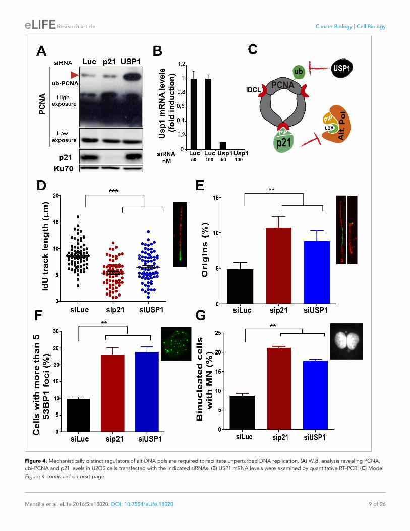
Figure 4. Mechanistically distinct regulators of alt DNA pols are required to facilitate unperturbed DNA replication. (A) W.B. analysis revealing PCNA,
ubi-PCNA and p21 levels in U2OS cells transfected with the indicated siRNAs. (B) USP1 mRNA levels were examined by quantitative RT-PCR. (C) Model
Figure 4 continued on next page
Mansilla et al. eLife 2016;5:e18020. DOI: 10.7554/eLife.18020 9 of 26
Research article Cancer Biology Cell Biology

p21 prevents mitotic transmission of DNA damage induced by Pol k–dependent replicative stressIn addition to the well characterized and direct consequences of DNA replication stress, namely
chromosomal breakage and aberrations, it has been shown that a fraction of under-replicated/unre-
solved genomic loci enter into mitosis. When not accurately processed in M phase, such DNA
regions are converted into complex broken-DNA structures that are transmitted to daughter cells
(Minocherhomji et al., 2015). In G1 phase such DNA structures are sequestered and shielded in
nuclear compartments described as nuclear 53BP1 bodies (Bergoglio et al., 2013; Harrigan et al.,
2011; Lukas et al., 2011). We therefore explored whether the altered DNA replication dynamics of
p21-depleted cells could lead to the mitotic transmission of DNA damage. We first noticed an
increase in the percentage of cells positive for the phosphorylated histone H3, a bona-fide marker of
G2/M transition (Minocherhomji et al., 2015), upon p21 depletion which was again totally reversed
by Pol k depletion (Figure 7A). To confirm the persistence of under-replicated DNA in mitosis, we
used previously reported protocols (Federico et al., 2015; Minocherhomji et al., 2015) to quantify
53BP1 body formation in G1 (EdU-negative). We found a significant increase in the number of spon-
taneous 53BP1 nuclear bodies in G1 in the absence of p21, as hallmark of incomplete DNA replica-
tion during the previous cell cycle, which was again dependent on Pol k (Figure 7B–D). We
conclude from these experiments that the choice of Pol k in the absence of p21 is sufficient to
increase the vulnerability of fragile genomic regions by delaying replication completion at these
sequences. Such alteration of the DNA replication dynamics appears to be specific to Pol k since
they were not rescued when Pol h was depleted (Figure 7E–F).
The PCNA-binding domain of p21 is necessary and sufficient to preventthe replication defects introduced by Pol kHaving established that Pol k triggers replication defects of p21-depleted cells, it was important to
determine whether p21 prevents the loading of Pol k to replication factories and if its ability to inter-
act with PCNA is relevant to such function. We used the p21 mutants described in Figure 1, which
are refractory to an siRNA directed to the 3´UTR of p21 (Figure 8A). Lentiviral transduction allowed
the expression of p21 and p21PIPMut* in almost all cells (Figure 8B). A fully functional PCNA binding
domain in p21 was required to down-modulate Pol k foci formation to control levels (Figure 8C). In
agreement, the fork elongation defects and the excessive origin firing observed in p21- depleted
samples were complemented by p21, but not by p21PIPMut* (Figure 8D–E). Additionally, the key role
of the PIP domain of p21 was supported by experiments performed in HCT116 p21�/� cells. Such
experiments revealed that p21 but not the p21PIPMut* mutant complement the replication defects of
cells with a null mutation of the endogenous p21 alleles (Figure 8 F–H). Notably, the accumulation
of markers of replication stress and genomic instability in p21-depleted cells was abolished when
overexpressing p21, but not p21PIPMut* (Figure 8I–J). Hence, we postulate a key role of the PIP
domain of p21 in the preservation of DNA replication homeostasis.
p21 preserves the genomic stability of primary cellsGiven that our results indicate a novel antioncogenic role of p21 in the promotion of DNA replication
it was important to determine whether this phenotype was not limited to cancer cells. To address
such question we used primary cells from two independent sources: (a) human foreskin fibroblast
(HFF) and (b) mesenchyimal stem cells isolated from umbilical cord (MSC). As shown in Figure 9A
and B, transfection of p21 siRNA efficiently depleted p21 from both cell types. p21 elimination
caused a reduction in the elongation of nascent DNA (Figure 9A–C), which was accompanied with
Figure 4 continued
depicting the different mechanisms of PCNA regulation by p21 and USP1. (D) IdU track length was measured in 85 fibers/sample in three independent
experiments. (E) Origin firing frequency. 150 fibers/sample were analysed in three independent experiments. (F) Quantification of cells with more than
five 53BP1 foci. 200 U2OS cells/sample were analysed in three independent experiments. (G) MN accumulation. 300 binucleated cells/sample were
analyzed in three independent experiments. Data on the effect of USP1 downregulation on the modulation of nascent DNA elongation (Figure 4D) and
accumulation of cells with 53BP1 foci (Figure 4F) and micronuclei (Figure 4G) was reproduced from Jones et al, EMBO.
DOI: 10.7554/eLife.18020.011
Mansilla et al. eLife 2016;5:e18020. DOI: 10.7554/eLife.18020 10 of 26
Research article Cancer Biology Cell Biology

an increase in origin firing (Figure 9D). In turn, such alteration in DNA replication parameters corre-
lated with the accumulation of cells with 53BP1 foci (Figure 9E–F) and micronuclei (Figure 9 G–I).
Hence, we postulate that p21 regulates protein-complex formation at the replisomes, promoting the
choice of the most adequate polymerase and therefore protecting DNA replication homeostasis.
Figure 5. The recruitment of pol k to replication-associated structures increases in the absence of p21. (A) U2OS
cells were transfected with GFP-polk. Representative images of cells with and without with GFP-pol k foci. (B)
Percentages of cells with GFP-pol k focal organization. 150 nuclei/sample were analyzed in two independent
experiments. (C) siLuc and sip21 depleted samples were subjected to chromatin immunoprecipitation using a
monoclonal PCNA antibody. GFP-pol k recruitment to chromatin was revealed by using GFP antibodies. The
result was reproduced in three independent experiments. (D) Proximity Ligation Assay (PLA) between PCNA and
endogenous pol k was performed in U2OS cells. Two representative images of PLA in siLuc and sip21 cells are
shown. (E) Quantification of PLA foci per nuclei. More than 1000 nuclei were analysed in three independent
experiments.
DOI: 10.7554/eLife.18020.012
Mansilla et al. eLife 2016;5:e18020. DOI: 10.7554/eLife.18020 11 of 26
Research article Cancer Biology Cell Biology
Figure 6. Pol k depletion prevents the DNA replication defects and the genomic instability caused by p21 downmodulation. (A) Western blots showing
GFP-pol k and p21 levels in U2OS cells transfected with the indicated siRNAs. (B) Total IdU track length was evaluated in 100 fibers/sample in three
independent experiments. (C) Frequency of origin firing. 200 fibers were analysed in three experiments. (D) Quantification of cells with 53BP1 foci. 200
cells were analysed in three independent experiments. (E) Quantification of MN accumulation. 300 binucleated cells/sample were analysed in three
Figure 6 continued on next page
Mansilla et al. eLife 2016;5:e18020. DOI: 10.7554/eLife.18020 12 of 26
Research article Cancer Biology Cell Biology

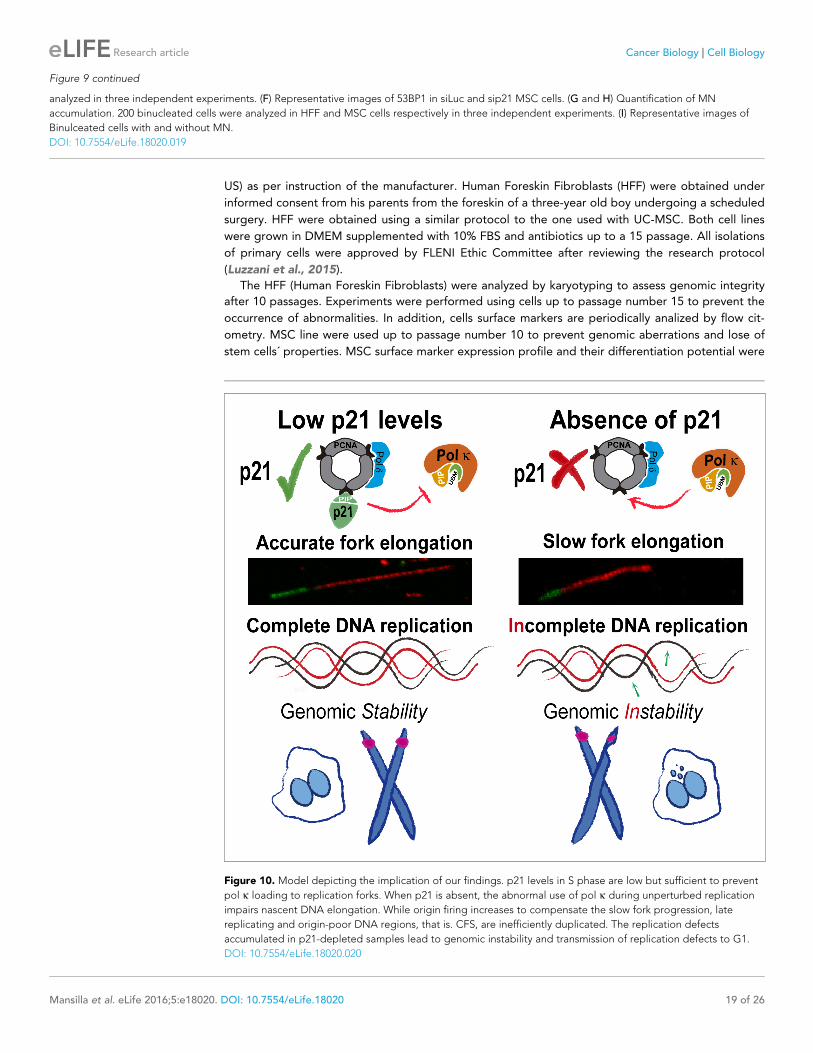
Such novel function of p21 depends exclusively on its ability to interact with PCNA and is needed at
every S phase to ensure the accurate finalization of DNA replication (see model in Figure 10).
DiscussionWe describe a biologically relevant contribution of the interaction of p21 and PCNA in cells. We
show that such interaction improves DNA replication dynamics since it is required to ensure the best
rate of nascent DNA elongation. By promoting accurate DNA polymerase choice at the replisome,
p21 acts as a tumor suppressor chronically at each duplication cycle, in the absence of exogenous
sources of DNA damage.
A novel function of p21 in the fine-tuning of DNA replication dynamicsAn important implication of our findings is that the idea that p21 acts exclusively as a negative regu-
lator of the cell cycle through the inhibition of cycling kinases (Warfel and El-Deiry, 2013) is simpli-
fied, incomplete and limited to specific DNA-damaging scenarios. The results presented herein
conclusively show that p21 is more often a positive rather than a negative regulator of the cell cycle,
as its contribution is required at every S phase. Remarkably, such contribution relies exclusively on
the p21/PCNA interaction.
Previous work indicates that p21 can displace alt DNA pols from replicating DNA. First, Z. Livneh
and colleagues showed that p21 impairs TLS events on transfected plasmids (Avkin et al., 2006).
Second, lower levels of p21 in cycling cells must be eliminated to promote nascent DNA elongation
across UV damage templates by alt DNA pols (Mansilla et al., 2013). Third, endogenous p21 delays
the recruitment of alt DNA pols to UV-damaged replication factories in a manner that correlates
with the extent of p21 degradation (Mansilla et al., 2013; Soria et al., 2008). Fourth, the PCNA-
binding domain of p21 is required to inhibit TLS activation (Avkin et al., 2006; Mansilla et al.,
2013; Soria et al., 2008). It follows that alt DNA pols might be selectively sensitive to p21 levels
which are insufficient to inhibit CDKs (Soria and Gottifredi, 2010). Such a difference in the levels of
p21 required to inhibit different cellular processes indicates that p21 is locally concentrated at, and/
or has very high affinity for PCNA (Soria and Gottifredi, 2010). Both replicative and alt DNA pols
bind the IDCL of PCNA through PIR or PIP (PCNA interacting protein) regions (Bertolin et al., 2015;
Moldovan et al., 2007). The PIR of p21 binds PCNA strongly, so that p21 is capable of disrupting
the weaker interactions between PCNA and alt DNA pols (Hishiki et al., 2009; Mansilla et al.,
2013; Tsanov et al., 2014) without affecting replicative DNA pols which have multiple PIP domains
(Soria and Gottifredi, 2010; Soria et al., 2008). Such difference may relate to the fact that replica-
tive DNA pols utilize multiple domains and different sites to interact with PCNA (Johansson et al.,
2004; Moldovan et al., 2007).
There is a tight cross-regulation of PIP box-containing proteins at replication forks which is not
yet completely understood. On the one hand, it has been demonstrated that proteins with strong
PIP boxes such as p21 can remove alt pols such as Pol k from replication factories (this report and
[Tsanov et al., 2014]). On the other hand, this and previous reports (Jones et al., 2012;
Pillaire et al., 2007) suggest that the alt Pol k can displace replicative pols from replisomes. Con-
versely however, p21-PCNA interaction in S phase does not impair genomic DNA synthesis
Figure 6 continued
independent experiments. (F) Quantification of CFS instability in HCT116 cells transfected with the indicated siRNAs. 50 metaphases/sample were
analysed in three independent experiments. (G) Representative images of the CFS analysed in F.
DOI: 10.7554/eLife.18020.013
The following figure supplements are available for figure 6:
Figure supplement 1. Pol k depletion prevents the accumulation of DNA replication stress markers caused by p21 downmodulation.
DOI: 10.7554/eLife.18020.014
Figure supplement 2. Pol k prevents the accumulation of DNA replication stress in p21 depleted cells independently of the siRNA used and in cells
stably lacking p21.
DOI: 10.7554/eLife.18020.015
Figure supplement 3. Pol k but not pol h depletion prevents the DNA replication defects and the genomic instability caused by p21 downmodulation.
DOI: 10.7554/eLife.18020.016
Mansilla et al. eLife 2016;5:e18020. DOI: 10.7554/eLife.18020 13 of 26
Research article Cancer Biology Cell Biology

Figure 7. Pol k-mediated replication defects of p21-depleted cells are transmitted to the M and G1 phases of the cell cycle. (A) Quantification of
phospho-H3 positive U2OS. 200 cells/sample were analysed in three independent experiments. (B) Representative images of 53BP1 bodies outside S
phase. Yellow arrows indicate EdU negative cells with 53BP1 foci (C) Percentages of EdU negative cells which are positive for 53BP1 bodies. 200 nuclei/
sample were analysed in three independent experiments. (D) Distribution of EdU negative cells with increasing number of 53BP1 bodies per cell in the
Figure 7 continued on next page
Mansilla et al. eLife 2016;5:e18020. DOI: 10.7554/eLife.18020 14 of 26
Research article Cancer Biology Cell Biology

(Soria et al., 2008) suggesting that p21 cannot displace replicative DNA pols from the replisome.
Hence, some yet unknown factors/events need to be identified to understand the hierarchy of PCNA
partners at forks. Moreover, variables such as the sequence of the DNA which is being replicated
and the average retention time of a PCNA partner at different regions of the chromatin might have
a role in such a puzzling cross-regulation among PCNA partners. We have shown that Pol k is
required for checkpoint activation at stalled forks (Betous et al., 2013) and during the replication of
repetitive sequences (Hile et al., 2012) or naturally occurring non-B DNA structures (Betous et al.,
2009). In agreement, Pol k depletion affected the CFS expression and RPA accumulation in our
experimental settings. It is therefore possible that certain DNA regions may benefit from Pol k-
dependent DNA synthesis (Betous et al., 2009) while others it might require active displacement of
Pol k by p21. If that is the case, p21 may prevent Pol k recruitment to specific regions of the
genome while prompt p21 degradation by CLR4Cdt2 might rapidly allow Pol k binding to others.
Such p21 function could be executed either by promoting the dissociation of Pol k from replisomes
or by preventing its recruitment to DNA regions that must be necessarily copied by replicative DNA
pols (such as for example B-regions in the exonic DNA).
The complex regulation of Pol k by p21The PIR domain of p21 prevents the recruitment of Pol h, Pol i, Pol k and Rev1 to UV-damaged rep-
lication factories (Bertolin et al., 2015; Mansilla et al., 2013). Hence, it is unlikely that during unper-
turbed replication p21 would act as a specific inhibitor of Pol k. The same is valid for USP1 because
the modulation of PCNA ubiquitination should regulate all alt DNA pols (Huang and D’Andrea,
2006; Jones et al., 2012). Hence, p21 and USP1 depletion may result in increased loading of all alt
DNA pols to undamaged DNA. In fact, others have reported increased spontaneous mutation fre-
quency in the hypoxanthine phosphoribosyl transferase (hprt) locus of p21�/� cells,
(McDonald et al., 1996). Such defects might depend on the unleashed activity of alt DNA pols other
than Pol k (Yang and Woodgate, 2007). Notwithstanding this, the phenotypes described herein are
intimately associated with processive DNA synthesis events, and Pol k is a very processive alt DNA
pol (the most processive in the Y family) (Ohashi et al., 2000; Zhang et al., 2000). Because Polk is
less processive than replicative DNA pols (McCulloch and Kunkel, 2008; Ohashi et al., 2000), the
coupling of a replicative DNA pol and Pol k at a single replisome may most likely generate asyn-
chronic speed in both DNA strands. In turn, this may cause the transient accumulation of ssDNA in
one strand (see Figure 3C). In fact, low doses of APH, which are known to disrupt the DNA pol
homeostasis during DNA replication, cause similar levels of CFS expression as p21 depletion (see
Figure 3E). Hence, the local degradation of p21 by CLR4CDT2 may maintain the most efficient DNA
elongation speed by allowing the rapid and dynamic exchange of replicative DNA pols for Pol k. In
fact, a minor shift in the timing of CLR4CDT2-dependent p21 degradation is sufficient to accumulate
cells in S phase (Coleman et al., 2015). Notably, the elimination of not only p21 but also USP1
(Jones et al., 2012) increases the use of Pol k during undamaged DNA replication, suggesting that
the slightest modulation of Pol k activity perturbs the DNA replication dynamics. In agreement, the
expression of a Pol k mutant with increased affinity for PCNA and the overexpression of Pol k gener-
ate genomic instability and tumor formation (Bavoux et al., 2005a; 2005b; Bergoglio et al., 2002;
Hoffmann and Cazaux, 2010; O-Wang et al., 2001; Jones et al., 2012).
The biological consequences of excessive genomic DNA synthesis byPol kIt is relevant to mention that the p21 knockout mice develop spontaneous tumors at 16 months
(Martin-Caballero et al., 2001), suggesting that modest but accumulative defects eventually trigger
oncogenic transformation. Such observation highlights a role of p21 in the face of chronic, rather
Figure 7 continued
experiments showed in C. 100 nuclei/sample were analysed in three independent experiments. (E) Phospho-H3 accumulation was quantified in 200
nuclei/sample of three independent experiments. Samples depleted from pol k and pol h were compared. (F) Percentages of cells with more than five
53BP1 foci were determined after analysing 200 nuclei/sample in three independent experiments. Samples depleted from pol k and pol h were
compared.
DOI: 10.7554/eLife.18020.017
Mansilla et al. eLife 2016;5:e18020. DOI: 10.7554/eLife.18020 15 of 26
Research article Cancer Biology Cell Biology

Figure 8. The PIR domain of p21 and not the CDK-binding domain is required to prevent DNA replication defects in p21 depleted cells during
the unperturbed S phase. (A) W.B. analysis was performed to evaluate p21 expression in U2OS cells transfected with control or p21 siRNA and infected
with p21 or p21PIPMut* lentiviruses 6 hr later. (B) Representative panel showing the efficiency of lentiviral infection (>90%). (C) GFP-pol k focal
organization in samples depleted from p21 and infected with p21 or p21PIPMut*. (D) IdU track length was measured in 100 fibers/sample in three
Figure 8 continued on next page
Mansilla et al. eLife 2016;5:e18020. DOI: 10.7554/eLife.18020 16 of 26
Research article Cancer Biology Cell Biology

than acute stress and may correlate with the multiple defects in DNA replication revealed when
depleting p21 from primary cells (Figure 9). While p21 is a negative regulator of TLS, its role in
undamaged DNA replication (revealed in this manuscript) is most possibly unrelated to TLS inhibi-
tion. In fact, if p21 depletion caused the accumulation of DNA lesions, the elimination of an alt poly-
merase should: (a) aggravate the defect (if the alt pol is essential to promote DNA replication across
the replication barrier) or (b) not alter such defect (if the replication across such barrier is also
achieved by another alt polymerase) (Bertolin et al., 2015). As our data show that the elimination of
Pol k promotes DNA replication, it does not support a model in which p21 depletion generates sub-
strates (DNA lesions) for alt pols. On the contrary, it suggests that undamaged DNA is being mis-
used by the alternative polymerase pol k as a replication template. Hence, the contribution of p21
to unperturbed-DNA replication is most probably related to the control of DNA synthesis events on
undamaged templates. In such scenario, the colocalization of the p21-CLR4cdt2 complex at replica-
tion forks may be crucial to control the composition of the active replisome, allowing the selection
of the most adequate polymerase ’on the go’. Such dynamic regulation would favour the most effec-
tive DNA replication speed allowing the timely activation of late and hard-to-replicate genomic
regions. The coupling of p21 with ongoing replisomes may also favor other p21 functions as homolo-
gous recombination (HR) events. While such p21 function has been linked to CDK inhibition
(Mauro et al., 2012), it is still possible that the loading of p21 to PCNA serves to orchestrate multi-
ple transactions at the elongating DNA. While more work is required to reveal yet hidden cross-reg-
ulations among members of the replisome, our work unveils a novel tumor suppressor function of
p21 dependent on its ability to interact with PCNA. Such a role of p21 is central for genome mainte-
nance in the absence of DNA damage at every S phase and which might be key to prevent sponta-
neous oncogenic events.
Materials and methods
Cell culture and reagentsAll cell lines were received from reliable sources, either public repositories or directly, from the labo-
ratory that generated them. They were never frozen at passage higher than eight. U2OS (source:
ATCC), and HCT116 p21+/+ and HCT116 p21�/� (gifts from B. Vogelstein-Johns Hopkins Univer-
sity, Baltimore) were received directly from ATCC and Johns Hopkins University respectively. Sam-
ples were minimally amplified and frozen after the reception and were used for limited passages
after thawing of those primary stocks. We have verified the identity of the cell lines in terms of the
pathways which are key for this study. We have verified the p53 and p21 status by RT-PCR. The
three cell lines are positive for p53 and only U2OS and HCT 116 p21+/+ were positive for p21, while
HCT 116 p21�/� was not. None of the used cell lines were in the list of commonly misidentified cell
lines maintained by the International Cell Line Authentication Committee (Capes-Davis et al., 2010)
updated in http://iclac.org/databases/cross-contaminations/). Samples were grown in Dulbecco’s
modified Eagle’s medium (Invitrogen, Cambridge, Massachusetts) with 10% fetal calf serum in a 5%
CO2 atmosphere. Samples are routinely tested for mycoplasma by PCR every two weeks.
Umbilical cord mesenchymal stem cells (UC-MSC) were isolated from Wharton jelly tissues. Small
pieces of the umbilical cord, excluding the major vessels, were layered onto plastic culture plates
and cultured in D-MEM supplemented with 10% SFB and penicillin-streptomycin. After 14 days, cells
started to emerge from the tissue pieces. When plates were almost confluent, umbilical cord pieces
were removed and cells trypsinized and frozen. Multilineage differentiation potential of UC-MSC
was assessed using StemPro Adipogenesis, Osteogenesis or Chondrogenesis Kit (Life Technologies,
Figure 8 continued
independent experiments. (E) Frequency of origin firing. 100 fibers/sample were analysed in three experiments. (F) Infection were performed in HCT116
p21+/+ and p21�/� cells. Representative panel showing the efficiency of lentiviral infection in HCT116 p21�/� s. (G) IdU track length was measured in
100 fibers/sample in three independent experiments. (H) Frequency of origin firing was measured in the experiments shown in G. 200 fibers/sample
were analysed. (I) Quantification of the percentage of cells with more than 5 53BP1 foci/cell. 200 cells/sample were evaluated in three independent
experiments. (J) Quantification of MN accumulation. 200 binucleated cells were analysed in three independent experiments.
DOI: 10.7554/eLife.18020.018
Mansilla et al. eLife 2016;5:e18020. DOI: 10.7554/eLife.18020 17 of 26
Research article Cancer Biology Cell Biology

Figure 9. p21 depletion promotes replication stress and genomic instability in primary human cells. (A and B) IdU track length in Human Foreskin
fibroblasts (HFF) and Umbilical Cord Mesenchymal Stem cells (MSC). 100 fiber/sample were analysed in three independent experiments. Western blot
showing p21 depletion in the indicated cell line. (C) Representative fibers in siLuc and sip21 HFF cells. (D) Origin frequency in HFF cells. 200 fiber/
sample were analyzed in three independent experiments. (E) Cells with more than 5 53BP1 foci were analyzed in MSC cells. 200 nuclei/sample were
Figure 9 continued on next page
Mansilla et al. eLife 2016;5:e18020. DOI: 10.7554/eLife.18020 18 of 26
Research article Cancer Biology Cell Biology
US) as per instruction of the manufacturer. Human Foreskin Fibroblasts (HFF) were obtained under
informed consent from his parents from the foreskin of a three-year old boy undergoing a scheduled
surgery. HFF were obtained using a similar protocol to the one used with UC-MSC. Both cell lines
were grown in DMEM supplemented with 10% FBS and antibiotics up to a 15 passage. All isolations
of primary cells were approved by FLENI Ethic Committee after reviewing the research protocol
(Luzzani et al., 2015).
The HFF (Human Foreskin Fibroblasts) were analyzed by karyotyping to assess genomic integrity
after 10 passages. Experiments were performed using cells up to passage number 15 to prevent the
occurrence of abnormalities. In addition, cells surface markers are periodically analized by flow cit-
ometry. MSC line were used up to passage number 10 to prevent genomic aberrations and lose of
stem cells´ properties. MSC surface marker expression profile and their differentiation potential were
Figure 9 continued
analyzed in three independent experiments. (F) Representative images of 53BP1 in siLuc and sip21 MSC cells. (G and H) Quantification of MN
accumulation. 200 binucleated cells were analyzed in HFF and MSC cells respectively in three independent experiments. (I) Representative images of
Binulceated cells with and without MN.
DOI: 10.7554/eLife.18020.019
Figure 10. Model depicting the implication of our findings. p21 levels in S phase are low but sufficient to prevent
pol k loading to replication forks. When p21 is absent, the abnormal use of pol k during unperturbed replication
impairs nascent DNA elongation. While origin firing increases to compensate the slow fork progression, late
replicating and origin-poor DNA regions, that is. CFS, are inefficiently duplicated. The replication defects
accumulated in p21-depleted samples lead to genomic instability and transmission of replication defects to G1.
DOI: 10.7554/eLife.18020.020
Mansilla et al. eLife 2016;5:e18020. DOI: 10.7554/eLife.18020 19 of 26
Research article Cancer Biology Cell Biology

periodically analized by flow citometry and by chondregic, adipogenic and osteogenic differentia-
tion, respectively. MSC immunomodulatory properties were also assessed by CFSE lymphocyte pro-
liferation assay. All cell lines were weekly analyzed for mycoplasm contamination by PCR.
Transfections of siRNAs were performed using Lipofectamine RNAiMAX (Thermofisher) and Jet
Prime (Polyplus, France) was used when delivering both siRNAs and plasmid expression vectors.
siRNAs, vector expression plasmids and lentiviral infectionGFP-Pol h and GFP-Pol k were gifts from Dr. A Lehmann (Kannouche et al., 2001). GFP-PCNA was
kindly provided by Dr. M C Cardoso (Max Delbruck Center for Molecular Medicine, Berlin, Germany)
(Leonhardt et al., 2000) siRNAs were purchased from Dharmacon (Lafayette , Colorado):
siRNA p21 3’UTR:GGAACAAGGAGUCAGACAU;
siRNAUSP1:UCGGCAAUACUUGCUAUCUUA (Jones et al., 2012);
siRNA Pol k1: AAGAUUAUGAAGCCCAUCCAA (Vallerga et al., 2015);
siRNA Pol k2: AACCUCUAGAAAUGUCUCAUA (Jones et al., 2012)
siRNA Pol h: CUGGUUGUGAGCAUUCGUGUA (Vallerga et al., 2015).
In this work, we used a previously described p21 expression vectors (CS2-p21) and generated the
p21PIPMut* (CS2-p21 with the M147 to A, D149 to A and F150 to A mutations which were shown to
disrupt the PCNA/p21 interaction [Gulbis et al., 1996; Mansilla et al., 2013]) Both the p21 and the
p21PIPMut* vectors bear three point mutations in its CDK binding domain (W49 to R,F51 to S and
D52 to A) which disrupt the CDK2/p21 interaction (Gulbis et al., 1996; Mansilla et al., 2013). Pri-
mers used to generate p21 mutations were described and validated in our previous work
(Mansilla et al., 2013) and both mutations were characterized previously (Gulbis et al., 1996).
p21 and p21PIPMut* were cloned into the lentiviral transfer plasmid pLenti CMV/TO Puro (Plasmid
#17482, Addgene, 3rd generation) using BamHI and XbaI. Packaging and envelope 2nd generation
vectors used were psPAX2 and PMD2.G respectively. Lentiviral particles were obtained by transfect-
ing with Jet prime Hek293T cells in 60 mm dishes in a 5:5:1 ratio (pLenti: psPAX2:PMD2). 48 hr after
transfection lentiviral particles were collected from the supernatant of Hek293T cells, centrifuged, fil-
tered with a 0.45 mm filter and stored at �80˚C. Lentiviral preparations were slowly thawed and used
to infect U2OS cells in 24/12 well dishes in the presence of 8 mg/ml of polybrene. Infection efficiency
was approximately 90% (Figure 7B).
Immunostaining and fluorescence detectionFor the quantification and inmunodetection of specialized GFP tagged Pol k and Pol h, 53BP1 and
gH2AX respectively, cells were fixed in 2% paraformaldehyde (PFA)/2% sucrose and permeablized
with 0.1% Triton X-100 in phosphate buffered saline (PBS) as described previously (Mansilla et al.,
2013). For the detection of chromatin-bound protein ice cold, 0.5% Triton CSK buffer was used
(10 mM Pipes, pH 7.5, 100 mM NaCl, 300 mM sucrose, 3 mM MgCl2). Proteins were preextracted 1
min or 5 min when detecting p21 or RPA/PCNA respectively. EdU was detected following manufac-
turer’s instructions (Click-iT EdU kit– C10338 Invitrogen). Blocking was performed overnight in PBS
2% donkey serum (Sigma, St. Louis, Missouri). Coverslips were incubated for 1 hr in primary antibod-
ies: 53BP1(Santa Cruz, Dallas, Texas), gH2AX (EMD Millipore, Massachusetts), p21 (c-19 Santa Cruz),
RPA (NA18 EMD Millipore), PCNA (Abcam), pH3 (ser10 Millipore). Secondary anti-mouse/rabbit-
conjugated Cy2/Cy3 antibodies were from Jackson Immuno Research and anti-rabbit alexa 488 from
Invitrogen. GFP-tagged specialized Y polymerases and GFP-PCNA were detected by GFP autofluor-
escence. Nuclei were stained with DAPI (SIGMA). Images were obtained with a Zeiss Axioplan confo-
cal microscope or a Zeiss Axio Imager A2.
Protein analysisFor Western blot analysis, samples were lysed in Laemmli buffer. Antibodies used were anti-p21 (c-
19; Santa Cruz Biotechnology) anti-polh (H-300; Santa Cruz Biotechnology), anti-KU70 (A9; Santa
Cruz Biotechnology), and anti-GFP (Santa Cruz Biotechnology). Secondary antibodies (Sigma) and
ECL detection (Amersham GE Healthcare, Milwaukee, Wisconsin) were used according to the manu-
facturers’ instructions. Western blot images were taken with Image QuantLAS4000 (GE Healthcare),
which allows capture and quantification of images within a linear range. These images were then
quantified with the ImageJ software when indicated.
Mansilla et al. eLife 2016;5:e18020. DOI: 10.7554/eLife.18020 20 of 26
Research article Cancer Biology Cell Biology
DNA fiber spreadingDNA fibers were analysed using a protocol previously used by us (Mansilla et al., 2013) with a minor
change in the time of labelling. Exponentially growing cells were pulse labeled with CldU (20 mM) for
10 min, washed twice,and incubated with IdU(200 mM) for additional 30 min. Cells were trypsinized
and lysed with 6 ml of 0.5% SDS, 200 mM Tris–HCL (pH 7.4) and 50 mM EDTA buffer onto clean
glass slides, which were tilted, allowing DNA to unwind. Samples were fixed in 3:1 methanol/acetic
acid and denatured with HCL (2.5 N) for 1 h, blocked in PBS 5%Bovine serum albumin (BSA) for
15 min and incubated with the mouse anti-BrdU (Becton Dickinson, USA) to detect IdU, donkey anti-
mouse Cy3-conjugated secondary antibody (Jackson Immuno Research, West Grove, Pennsylvania),
rat anti-BrdU (Accurate Chemicals, Westbury, New York) to detect CldU and donkey antirat Alexa
488 secondary antibody (Invitrogen). Slides were mounted with Mowiol 488 (Calbiochem), and DNA
fibers were visualized using a Zeiss Axioplanconfocal microscope. Images were analysed using Zeiss
LSM Image Browser software and Image J software. Each data set is derived from measurement of
85–100 fibers.
Chromatin immunoprecipitation of PCNAChromatin immunoprecipitations were performed as described in (Soria et al., 2008). U2OS cells
were plated in 100 mm culture dishes, transfected with either Luc or p21 siRNA and 24 hr later trans-
fected with GFP-Pol Kappa. Cells were rinsed twice with cold PBS and then extracted with 5 mL of
CSK buffer (250 mM sucrose, 25 mM KCl, 10 mM HEPES at pH 8.0, 1 mM EGTA, and 1 mM MgCl2)
for 10 min in ice. The CSK-extracted cells were fixed with 1% formaldehyde in PBS (4.5 mL) for
12 min. Then, 0.5 mL of 1 M glycine was added for 5 min to quench the cross-linking reaction. The
cross-linked nuclei were rinsed with PBS and then lysed in 750 mL of IP lysis buffer (10 mM Tris�HCl
at pH 7.5, 25 mM FNa, 20 mM NaCl, 1% Nonidet P-40, 1% Na-Deoxicholate, and 0.1% SDS) freshly
supplemented with protease and phosphatase inhibitors. Lysates were scraped from the plates and
transferred into 1.5-mL Microfuge tubes. Samples were sonicated (Bioruptor Sonication System, Dia-
genode) and clarified by centrifugation at 12,000 � g for 30 min at 4˚C. PCNA was immunoprecipi-
tated overnight at 4˚C with 5 mL of monoclonal PCNA antibody (PC-10 AC; Santa Cruz
Biotechnology). Samples were washed, lysed in Laemmli buffer and heated at 99˚C for 30 min to
revert the crosslink, and resolved in SDS/PAGE for Western blot analysis.
Proximity ligation assay (PLA)U2OS cells were seeded onto 22 mm x 22 mm coverslips in 6 well plates. 24 hr later cells were pre-
extracted with ice cold CSK buffer (PIPES 10 mM, NaCl 100 mM, sucrose 300 mM, MgCl2 3 mM,tri-
ton 0.5%) for 2 min and fixed with PFA 4% for 10 min. After pre-extraction and fixation, interaction
between endogenous p21 and PCNA was detected following manufacturer’s instructions Duolink In
Situ – Fluorescence (SIGMA). Briefly PLA’s principle is based on detecting proteins in close proximity
(30–40 nm) by using different species of primary antibodies. A pair of oligonucleotide (PLA probes)
detect the primary antibodies bound the proteins of interest and generates a signal only when the
two PLA probes have bound in close proximity, meaning that the samples are localized in close
proximity. The signal from each detected pair of PLA probes is visualized as an individual fluorescent
spot. These PLA signals can be quantified (counted) and assigned to a specific subcellular location
based on microscopic images. Images were obtained with a Zeiss Axioplan confocal microscope and
PLA spots were counted using Image J software, cells were counted as positive when more than 3
PLA spots were detected. When analyzing pol k/PCNA interaction a pol k ’home made’ monoclonal
antibody from mice purified by Biotem (clone n˚16A7-3A11) and PCNA rabbit polyclonal (Abcam ref
ab18197) were used. For the quantification of number of foci per nuclei the Cell Profiler program
was used to analyze more than 1000 nuclei.
Quantitative real-time PCRAfter transfection with the indicated siRNAs, cells were lysed and total RNA was extracted using TRI-
zol Reagent (Invitrogen). Total RNA (1 mg) was used as a template for cDNA synthesis utilizing an
ImProm-II Reverse Transcription System (Promega) and oligo-dT as a primer. Quantitative real-time
PCR was performed using the MX3005P quantitative PCR instrument (Stratagene) with Taq DNA
poly-merase (Invitrogen) and SyberGreen and ROX as reference dyes (Invitrogen). Samples were
Mansilla et al. eLife 2016;5:e18020. DOI: 10.7554/eLife.18020 21 of 26
Research article Cancer Biology Cell Biology

normalized using GAPDH primers. Both target gene and GAPDH amplification reactions approached
100% efficiency as determined by standard curves. Three independent biological samples were ana-
lyzed, and one representative set of results is shown. Primer sequences were as follows: USP1 (For-
ward): 5’- GGACGCGTTGCTTGGAATGT-3’ (Reverse) 5’-TGCCCATCTCAGGGTCTTCA-3’. GADPH:
(Forward) 5’-AGCCTCCCGCTTCGCTCTCT-3’ (Reverse) 5’-GAGCGATGTGGCTCGGCTGG-3’
(Vallerga et al., 2015).
MN assayTransfected cells were replated at low density. 24 hr after replating cytochalasine B (4.5 mg/ml-
Sigma) was added to the media and 40 hr later cells were washed 1 min with hypotonic buffer (KCl
0.0075 M) diluted 1/10 from stock solution in PBS 1X, twice with PBS 1X and fixed with PFA/sucrose
2% for 20 min. DAPI staining served to visualize cell nuclei. 300 binucleated cells were analyzed.
FISH analysisCells were treated with 0.1 mg/ml colcemid (Gibco) for 3 hr. Cells were trypsinized for 1 min trying
to take off only the mitotic cells. After centrifugation cell pellets were resuspended in hypotonic
solution (0.075 M KCl) and incubated for 15 min at 37˚C. Samples were then fixed in a methanol/
acetic acid solution (3:1), washed three times in the fixative solution and dropped on slides to obtain
spread chromosomes. Metaphase slides were incubated with Rnase (10 mg/ml in 2� SSC) for 1 hr at
37˚C followed by dehydration in successive ethanol baths (70, 85, and 100%). The RP 11 36B6 BAC
(corresponding to FRA7H locus) was labeled by nick translation (Abbott) with green dUTP (Vysis
Spectrum) and ethanol precipitated with human Cot-1 DNA (Roche, California) overnight at �20˚C.Precipitated DNA was incubated for 1 hr at 37˚C in hybridization mix. The probe was denatured for
8 min at 70˚C and applied to denatured metaphases. Samples were incubated over night at 37˚Cand, after washing (1X SSC, 5 min at 72˚C and SCC 1X 5 min at RT) and mounted in VECTASHIELD
Antifade Mounting Medium with DAPI. The images were acquired by using an inverted wide-field
Zeiss Axio Observer Z1 microscope fitted with a x63 oil NA 1.4 objective and an Axiocam MRm
CDD camera.
Statistical analysisFrequency distributions of DNA track length were determined with GraphPad Prism 5 software. In
non-Gaussian distributions, Mann–Whitney and Kruskal–Wallis tests were used for statistical analyses
when comparing two and more than three variables, respectively. Other statistical analyses were per-
formed with GraphPad in Stat software using the Student’s t test and the one-way ANOVA test
when applicable.
AcknowledgementsWe would like to thank Professors AR Lehmann (University of Sussex) and Maria Cristina Cardoso
(Max Delbruck Center for Molecular Medicine, Berlin, Germany) for the gift of reagents.This work
was supported by grants from National Institute of Health (R03 TW008924) and the Agencia Nacio-
nal de Promocion Cientıfica y Tecnologica (ANPCyT) to VG and from La Ligue Nationale contre le
Cancer (Equipe Labellisee) and the Laboratoire d’Excellence Toulouse Cancer LABEX TOUCAN
grant (Integrative analysis of resistance in hematological cancers) to JSH. VG and MGB are research-
ers from the National Council of Scientific and Technological Research (CONICET). SFM and APB
are supported by fellowships from CONICET.SFM was also supported by a short-term fellowship
from The Journal of Cell Science to visit the JSH laboratory. Christophe Casaux is listed as an author
as he contributed actively to this project and sadly passed away before submitting the manuscript.
Additional information
Funding
Funder Grant reference number Author
National Institutes of Health R03 TW008924 Vanesa Gottifredi
Mansilla et al. eLife 2016;5:e18020. DOI: 10.7554/eLife.18020 22 of 26
Research article Cancer Biology Cell Biology
Agencia Nacional de Promo-cion Cientıfica y Tecnologica
PICT-2012-1371 Vanesa Gottifredi
Agencia Nacional de Promo-cion Cientıfica y Tecnologica
PICT-2013-1049 Vanesa Gottifredi
Company of Biologists Travel Fellowship Sabrina F Mansilla
Laboratoire d’Excellence Tou-louse Cancer LABEX TOUCAN
Jean-Sebastien Hoffmann
La Ligue Nationale contra leCancer
Jean-Sebastien Hoffmann
The funders had no role in study design, data collection and interpretation, or the decision tosubmit the work for publication.
Author contributions
SFM, APB, Approval of the final version to be published, Conception and design, Acquisition of
data, Analysis and interpretation of data, Drafting or revising the article; VB, M-JP, Approval of the
final version to be published, Acquisition of data, Analysis and interpretation of data, Drafting or
revising the article; MAGB, Approval of the final version to be published, Drafting or revising the
article, Contributed unpublished essential data or reagents; CL, SGM, Critical revision of the final
draft for important intellectual content, Approval of the final version to be published, Drafting or
revising the article, Contributed unpublished essential data or reagents; CC, Conception and design,
Contributed unpublished essential data or reagents; J-SH, Approval of the final version to be pub-
lished, Conception and design, Drafting or revising the article; VG, Approval of the final version to
be published, Conception and design, Analysis and interpretation of data, Drafting or revising the
article
Author ORCIDs
Santiago G Miriuka, http://orcid.org/0000-0003-2402-3920
Vanesa Gottifredi, http://orcid.org/0000-0001-9656-5951
ReferencesAbbas T, Sivaprasad U, Terai K, Amador V, Pagano M, Dutta A. 2008. PCNA-dependent regulation of p21ubiquitylation and degradation via the CRL4Cdt2 ubiquitin ligase complex. Genes & Development 22:2496–2506. doi: 10.1101/gad.1676108, PMID: 18794347
Avkin S, Sevilya Z, Toube L, Geacintov N, Chaney SG, Oren M, Livneh Z. 2006. p53 and p21 regulate error-proneDNA repair to yield a lower mutation load. Molecular Cell 22:407–413. doi: 10.1016/j.molcel.2006.03.022,PMID: 16678112
Bavoux C, Hoffmann JS, Cazaux C. 2005a. Adaptation to DNA damage and stimulation of genetic instability: thedouble-edged sword mammalian DNA polymerase kappa. Biochimie 87:637–646. doi: 10.1016/j.biochi.2005.02.007, PMID: 15989980
Bavoux C, Leopoldino AM, Bergoglio V, O-Wang J, Ogi T, Bieth A, Judde JG, Pena SD, Poupon MF, Helleday T,Tagawa M, Machado C, Hoffmann JS, Cazaux C. 2005b. Up-regulation of the error-prone DNA polymerase{kappa} promotes pleiotropic genetic alterations and tumorigenesis. Cancer Research 65:325–330.PMID: 15665310
Bergoglio V, Bavoux C, Verbiest V, Hoffmann JS, Cazaux C. 2002. Localisation of human DNA polymerase kappato replication foci. Journal of Cell Science 115:4413–4418. doi: 10.1242/jcs.00162, PMID: 12414988
Bergoglio V, Boyer AS, Walsh E, Naim V, Legube G, Lee MY, Rey L, Rosselli F, Cazaux C, Eckert KA, HoffmannJS. 2013. DNA synthesis by Pol h promotes fragile site stability by preventing under-replicated DNA in mitosis.The Journal of Cell Biology 201:395–408. doi: 10.1083/jcb.201207066, PMID: 23609533
Bertolin AP, Mansilla SF, Gottifredi V. 2015. The identification of translesion DNA synthesis regulators: Inhibitorsin the spotlight. DNA Repair 32:158–164. doi: 10.1016/j.dnarep.2015.04.027, PMID: 26002196
Bienko M, Green CM, Crosetto N, Rudolf F, Zapart G, Coull B, Kannouche P, Wider G, Peter M, Lehmann AR,Hofmann K, Dikic I. 2005. Ubiquitin-binding domains in Y-family polymerases regulate translesion synthesis.Science 310:1821–1824. doi: 10.1126/science.1120615, PMID: 16357261
Blow JJ, Ge XQ, Jackson DA. 2011. How dormant origins promote complete genome replication. Trends inBiochemical Sciences 36:405–414. doi: 10.1016/j.tibs.2011.05.002, PMID: 21641805
Boulaire J, Fotedar A, Fotedar R. 2000. The functions of the cdk-cyclin kinase inhibitor p21WAF1. Pathologie-Biologie 48:190–202. PMID: 10858953
Brugarolas J, Chandrasekaran C, Gordon JI, Beach D, Jacks T, Hannon GJ. 1995. Radiation-induced cell cyclearrest compromised by p21 deficiency. Nature 377:552–557. doi: 10.1038/377552a0, PMID: 7566157
Mansilla et al. eLife 2016;5:e18020. DOI: 10.7554/eLife.18020 23 of 26
Research article Cancer Biology Cell Biology

Bunz F, Dutriaux A, Lengauer C, Waldman T, Zhou S, Brown JP, Sedivy JM, Kinzler KW, Vogelstein B. 1998.Requirement for p53 and p21 to sustain G2 arrest after DNA damage. Science 282:1497–1501. doi: 10.1126/science.282.5393.1497, PMID: 9822382
Burgers PM. 1998. Eukaryotic DNA polymerases in DNA replication and DNA repair. Chromosoma 107:218–227.doi: 10.1007/s004120050300, PMID: 9745046
Betous R, Pillaire MJ, Pierini L, van der Laan S, Recolin B, Ohl-Seguy E, Guo C, Niimi N, Gruz P, Nohmi T,Friedberg E, Cazaux C, Maiorano D, Hoffmann JS. 2013. DNA polymerase k-dependent DNA synthesis atstalled replication forks is important for CHK1 activation. The EMBO Journal 32:2172–2185. doi: 10.1038/emboj.2013.148, PMID: 23799366
Betous R, Rey L, Wang G, Pillaire MJ, Puget N, Selves J, Biard DS, Shin-ya K, Vasquez KM, Cazaux C, HoffmannJS. 2009. Role of TLS DNA polymerases eta and kappa in processing naturally occurring structured DNA inhuman cells. Molecular Carcinogenesis 48:369–378. doi: 10.1002/mc.20509, PMID: 19117014
Cai K, Dynlacht BD. 1998. Activity and nature of p21WAF1 complexes during the cell cycle. PNAS 95:12254–12259. doi: 10.1073/pnas.95.21.12254
Capes-Davis A, Theodosopoulos G, Atkin I, Drexler HG, Kohara A, MacLeod RA, Masters JR, Nakamura Y, ReidYA, Reddel RR, Freshney RI. 2010. Check your cultures! A list of cross-contaminated or misidentified cell lines.International Journal of Cancer 127:1–8. doi: 10.1002/ijc.25242, PMID: 20143388
Coleman KE, Grant GD, Haggerty RA, Brantley K, Shibata E, Workman BD, Dutta A, Varma D, Purvis JE, CookJG. 2015. Sequential replication-coupled destruction at G1/S ensures genome stability. Genes & Development29:1734–1746. doi: 10.1101/gad.263731.115, PMID: 26272819
Cooper MP, Balajee AS, Bohr VA. 1999. The C-terminal domain of p21 inhibits nucleotide excision repair In vitroand In vivo. Molecular Biology of the Cell 10:2119–2129. doi: 10.1091/mbc.10.7.2119, PMID: 10397753
Deng C, Zhang P, Harper JW, Elledge SJ, Leder P. 1995. Mice lacking p21CIP1/WAF1 undergo normaldevelopment, but are defective in G1 checkpoint control. Cell 82:675–684. doi: 10.1016/0092-8674(95)90039-X, PMID: 7664346
Dulic V, Stein GH, Far DF, Reed SI. 1998. Nuclear accumulation of p21Cip1 at the onset of mitosis: a role at theG2/M-phase transition. Molecular and Cellular Biology 18:546–557. doi: 10.1128/mcb.18.1.546, PMID: 9418901
Essers J, Theil AF, Baldeyron C, van Cappellen WA, Houtsmuller AB, Kanaar R, Vermeulen W. 2005. Nucleardynamics of PCNA in DNA replication and repair. Molecular and Cellular Biology 25:9350–9359. doi: 10.1128/MCB.25.21.9350-9359.2005, PMID: 16227586
Federico MB, Vallerga MB, Radl A, Paviolo N, Bocco JL, Di Giorgio M, Gottifredi V. 2015. Chromosomal integrityafter UV irradiation relays on FANCD2-mediated repair of double strand breaks. PLoS Genetics 12:e1005792.doi: 10.1371/journal.pgen.1005792
Fenech M. 2000. The in vitro micronucleus technique. Mutation Research/Fundamental and MolecularMechanisms of Mutagenesis 455:81–95. doi: 10.1016/S0027-5107(00)00065-8
Gottifredi V, McKinney K, Poyurovsky MV, Prives C. 2004. Decreased p21 levels are required for efficient restartof DNA synthesis after S phase block. Journal of Biological Chemistry 279:5802–5810. doi: 10.1074/jbc.M310373200, PMID: 14597617
Gulbis JM, Kelman Z, Hurwitz J, O’Donnell M, Kuriyan J. 1996. Structure of the C-terminal region of p21(WAF1/CIP1) complexed with human PCNA. Cell 87:297–306. doi: 10.1016/S0092-8674(00)81347-1, PMID: 8861913
Harrigan JA, Belotserkovskaya R, Coates J, Dimitrova DS, Polo SE, Bradshaw CR, Fraser P, Jackson SP. 2011.Replication stress induces 53BP1-containing OPT domains in G1 cells. The Journal of Cell Biology 193:97–108.doi: 10.1083/jcb.201011083, PMID: 21444690
Havens CG, Walter JC. 2011. Mechanism of CRL4(Cdt2), a PCNA-dependent E3 ubiquitin ligase. Genes &Development 25:1568–1582. doi: 10.1101/gad.2068611, PMID: 21828267
Hile SE, Wang X, Lee MY, Eckert KA. 2012. Beyond translesion synthesis: polymerase k fidelity as a potentialdeterminant of microsatellite stability. Nucleic Acids Research 40:1636–1647. doi: 10.1093/nar/gkr889,PMID: 22021378
Hishiki A, Hashimoto H, Hanafusa T, Kamei K, Ohashi E, Shimizu T, Ohmori H, Sato M. 2009. Structural basis fornovel interactions between human translesion synthesis polymerases and proliferating cell nuclear antigen.Journal of Biological Chemistry 284:10552–10560. doi: 10.1074/jbc.M809745200, PMID: 19208623
Hoffmann JS, Cazaux C. 2010. Aberrant expression of alternative DNA polymerases: a source of mutatorphenotype as well as replicative stress in cancer. Seminars in Cancer Biology 20:312–319. doi: 10.1016/j.semcancer.2010.10.001, PMID: 20934518
Huang TT, D’Andrea AD. 2006. Regulation of DNA repair by ubiquitylation. Nature Reviews Molecular CellBiology 7:323–334. doi: 10.1038/nrm1908, PMID: 16633336
Huang TT, Nijman SM, Mirchandani KD, Galardy PJ, Cohn MA, Haas W, Gygi SP, Ploegh HL, Bernards R,D’Andrea AD. 2006. Regulation of monoubiquitinated PCNA by DUB autocleavage. Nature Cell Biology 8:339–347. doi: 10.1038/ncb1378, PMID: 16531995
Johansson E, Garg P, Burgers PM. 2004. The Pol32 subunit of DNA polymerase delta contains separabledomains for processive replication and proliferating cell nuclear antigen (PCNA) binding. Journal of BiologicalChemistry 279:1907–1915. doi: 10.1074/jbc.M310362200, PMID: 14594808
Jones MJ, Colnaghi L, Huang TT. 2012. Dysregulation of DNA polymerase k recruitment to replication forksresults in genomic instability. The EMBO Journal 31:908–918. doi: 10.1038/emboj.2011.457, PMID: 22157819
Kannouche P, Broughton BC, Volker M, Hanaoka F, Mullenders LH, Lehmann AR. 2001. Domain structure,localization, and function of DNA polymerase eta, defective in xeroderma pigmentosum variant cells. Genes &Development 15:158–172. doi: 10.1101/gad.187501, PMID: 11157773
Mansilla et al. eLife 2016;5:e18020. DOI: 10.7554/eLife.18020 24 of 26
Research article Cancer Biology Cell Biology

Kannouche PL, Lehmann AR. 2004. Ubiquitination of PCNA and the polymerase switch in human cells. Cell Cycle3:1011–1013. doi: 10.4161/cc.3.8.1074, PMID: 15280666
Kreis NN, Sanhaji M, Rieger MA, Louwen F, Yuan J. 2014. p21Waf1/Cip1 deficiency causes multiple mitoticdefects in tumor cells. Oncogene 33:5716–5728. doi: 10.1038/onc.2013.518, PMID: 24317508
Le Tallec B, Koundrioukoff S, Wilhelm T, Letessier A, Brison O, Debatisse M. 2014. Updating the mechanisms ofcommon fragile site instability: how to reconcile the different views? Cellular and Molecular Life Sciences 71:4489–4494. doi: 10.1007/s00018-014-1720-2, PMID: 25248392
Leonhardt H, Rahn HP, Weinzierl P, Sporbert A, Cremer T, Zink D, Cardoso MC. 2000. Dynamics of DNAreplication factories in living cells. The Journal of Cell Biology 149:271–280. doi: 10.1083/jcb.149.2.271,PMID: 10769021
Letessier A, Millot GA, Koundrioukoff S, Lachages AM, Vogt N, Hansen RS, Malfoy B, Brison O, Debatisse M.2011. Cell-type-specific replication initiation programs set fragility of the FRA3B fragile site. Nature 470:120–123. doi: 10.1038/nature09745, PMID: 21258320
Lukas C, Savic V, Bekker-Jensen S, Doil C, Neumann B, Pedersen RS, Grøfte M, Chan KL, Hickson ID, Bartek J,Lukas J. 2011. 53BP1 nuclear bodies form around DNA lesions generated by mitotic transmission ofchromosomes under replication stress. Nature Cell Biology 13:243–253. doi: 10.1038/ncb2201, PMID: 21317883
Luzzani C, Neiman G, Garate X, Questa M, Solari C, Fernandez Espinosa D, Garcıa M, Errecalde AL, GubermanA, Scassa ME, Sevlever GE, Romorini L, Miriuka SG. 2015. A therapy-grade protocol for differentiation ofpluripotent stem cells into mesenchymal stem cells using platelet lysate as supplement. Stem Cell Research &Therapy 6:6. doi: 10.1186/scrt540, PMID: 25582222
Mansilla SF, Soria G, Vallerga MB, Habif M, Martınez-Lopez W, Prives C, Gottifredi V. 2013. UV-triggered p21degradation facilitates damaged-DNA replication and preserves genomic stability. Nucleic Acids Research 41:6942–6951. doi: 10.1093/nar/gkt475, PMID: 23723248
Martın-Caballero J, Flores JM, Garcıa-Palencia P, Serrano M. 2001. Tumor susceptibility of p21(Waf1/Cip1)-deficient mice. Cancer Research 61:6234–6238. PMID: 11507077
Mauro M, Rego MA, Boisvert RA, Esashi F, Cavallo F, Jasin M, Howlett NG. 2012. p21 promotes error-freereplication-coupled DNA double-strand break repair. Nucleic Acids Research 40:8348–8360. doi: 10.1093/nar/gks612, PMID: 22735704
McCulloch SD, Kunkel TA. 2008. The fidelity of DNA synthesis by eukaryotic replicative and translesion synthesispolymerases. Cell Research 18:148–161. doi: 10.1038/cr.2008.4, PMID: 18166979
McDonald ER, Wu GS, Waldman T, El-Deiry WS. 1996. Repair Defect in p21 WAF1/CIP1 -/- human cancer cells.Cancer Research 56:2250–2255. PMID: 8625293
Minocherhomji S, Ying S, Bjerregaard VA, Bursomanno S, Aleliunaite A, Wu W, Mankouri HW, Shen H, Liu Y,Hickson ID. 2015. Replication stress activates DNA repair synthesis in mitosis. Nature 528:286–290. doi: 10.1038/nature16139, PMID: 26633632
Moldovan GL, Pfander B, Jentsch S. 2007. PCNA, the maestro of the replication fork. Cell 129:665–679. doi: 10.1016/j.cell.2007.05.003, PMID: 17512402
Niimi A, Brown S, Sabbioneda S, Kannouche PL, Scott A, Yasui A, Green CM, Lehmann AR. 2008. Regulation ofproliferating cell nuclear antigen ubiquitination in mammalian cells. PNAS 105:16125–16130. doi: 10.1073/pnas.0802727105
Nishitani H, Shiomi Y, Iida H, Michishita M, Takami T, Tsurimoto T. 2008. CDK inhibitor p21 is degraded by aproliferating cell nuclear antigen-coupled Cul4-DDB1Cdt2 pathway during S phase and after UV irradiation.Journal of Biological Chemistry 283:29045–29052. doi: 10.1074/jbc.M806045200, PMID: 18703516
Noon AT, Goodarzi AA. 2011. 53BP1-mediated DNA double strand break repair: insert bad pun here. DNARepair 10:1071–1076. doi: 10.1016/j.dnarep.2011.07.012, PMID: 21868291
O-Wang J, Kawamura K, Tada Y, Ohmori H, Kimura H, Sakiyama S, Tagawa M. 2001. DNA polymerase kappa,implicated in spontaneous and DNA damage-induced mutagenesis, is overexpressed in lung cancer. CancerResearch 61:5366–5369. PMID: 11454676
Oakley GG, Patrick SM. 2010. Replication protein A: directing traffic at the intersection of replication and repair.Frontiers in Bioscience 15:883–900. doi: 10.2741/3652
Ohashi E, Bebenek K, Matsuda T, Feaver WJ, Gerlach VL, Friedberg EC, Ohmori H, Kunkel TA. 2000. Fidelity andprocessivity of DNA synthesis by DNA polymerase kappa, the product of the human DINB1 gene. Journal ofBiological Chemistry 275:39678–39684. doi: 10.1074/jbc.M005309200, PMID: 11006276
Ozeri-Galai E, Bester AC, Kerem B. 2012. The complex basis underlying common fragile site instability in cancer.Trends in Genetics 28:295–302. doi: 10.1016/j.tig.2012.02.006, PMID: 22465609
Ozeri-Galai E, Lebofsky R, Rahat A, Bester AC, Bensimon A, Kerem B. 2011. Failure of origin activation inresponse to fork stalling leads to chromosomal instability at fragile sites. Molecular Cell 43:122–131. doi: 10.1016/j.molcel.2011.05.019, PMID: 21726815
Pillaire MJ, Betous R, Conti C, Czaplicki J, Pasero P, Bensimon A, Cazaux C, Hoffmann JS. 2007. Upregulation oferror-prone DNA polymerases beta and kappa slows down fork progression without activating the replicationcheckpoint. Cell Cycle 6:471–477. doi: 10.4161/cc.6.4.3857, PMID: 17329970
Plosky BS, Vidal AE, Fernandez de Henestrosa AR, McLenigan MP, McDonald JP, Mead S, Woodgate R. 2006.Controlling the subcellular localization of DNA polymerases iota and eta via interactions with ubiquitin. TheEMBO Journal 25:2847–2855. doi: 10.1038/sj.emboj.7601178, PMID: 16763556
Prives C, Gottifredi V. 2008. The p21 and PCNA partnership: a new twist for an old plot. Cell Cycle 7:3840–3846.doi: 10.4161/cc.7.24.7243, PMID: 19066467
Mansilla et al. eLife 2016;5:e18020. DOI: 10.7554/eLife.18020 25 of 26
Research article Cancer Biology Cell Biology

Rey L, Sidorova JM, Puget N, Boudsocq F, Biard DS, Monnat RJ, Cazaux C, Hoffmann JS. 2009. Human DNApolymerase eta is required for common fragile site stability during unperturbed DNA replication. Molecular andCellular Biology 29:3344–3354. doi: 10.1128/MCB.00115-09, PMID: 19380493
Savio M, Coppa T, Cazzalini O, Perucca P, Necchi D, Nardo T, Stivala LA, Prosperi E. 2009. Degradation ofp21CDKN1A after DNA damage is independent of type of lesion, and is not required for DNA repair. DNARepair 8:778–785. doi: 10.1016/j.dnarep.2009.02.005, PMID: 19321391
Soria G, Gottifredi V. 2010. PCNA-coupled p21 degradation after DNA damage: The exception that confirms therule? DNA Repair 9:358–364. doi: 10.1016/j.dnarep.2009.12.003, PMID: 20060369
Soria G, Podhajcer O, Prives C, Gottifredi V. 2006. P21Cip1/WAF1 downregulation is required for efficient PCNAubiquitination after UV irradiation. Oncogene 25:2829–2838. doi: 10.1038/sj.onc.1209315, PMID: 16407842
Soria G, Speroni J, Podhajcer OL, Prives C, Gottifredi V. 2008. p21 differentially regulates DNA replication andDNA-repair-associated processes after UV irradiation. Journal of Cell Science 121:3271–3282. doi: 10.1242/jcs.027730, PMID: 18782865
Sutherland GR, Parslow MI, Baker E. 1985. New classes of common fragile sites induced by 5-azacytidine andbromodeoxyuridine. Human Genetics 69:233–237. doi: 10.1007/BF00293031, PMID: 2579891
Tsanov N, Kermi C, Coulombe P, Van der Laan S, Hodroj D, Maiorano D. 2014. PIP degron proteins, substratesof CRL4Cdt2, and not PIP boxes, interfere with DNA polymerase h and k focus formation on UV damage.Nucleic Acids Research 42:3692–3706. doi: 10.1093/nar/gkt1400, PMID: 24423875
Techer H, Koundrioukoff S, Carignon S, Wilhelm T, Millot GA, Lopez BS, Brison O, Debatisse M. 2016. Signalingfrom Mus81-Eme2-Dependent DNA damage elicited by Chk1 deficiency modulates replication fork speed andorigin usage. Cell Reports 14:1114–1127. doi: 10.1016/j.celrep.2015.12.093, PMID: 26804904
Vallerga MB, Mansilla SF, Federico MB, Bertolin AP, Gottifredi V. 2015. Rad51 recombinase prevents Mre11nuclease-dependent degradation and excessive PrimPol-mediated elongation of nascent DNA after UVirradiation. PNAS 112:E6624–E6633. doi: 10.1073/pnas.1508543112
Ward IM, Chen J. 2001. Histone H2AX is phosphorylated in an ATR-dependent manner in response toreplicational stress. The Journal of Biological Chemistry 276:47759–47762. doi: 10.1074/jbc.C100569200,PMID: 11673449
Warfel NA, El-Deiry WS. 2013. p21WAF1 and tumourigenesis: 20 years after. Current Opinion in Oncology 25:52–58. doi: 10.1097/CCO.0b013e32835b639e, PMID: 23159848
Yang W, Woodgate R. 2007. What a difference a decade makes: Insights into translesion DNA synthesis. PNAS104:15591–15598. doi: 10.1073/pnas.0704219104
Zhang Y, Yuan F, Xin H, Wu X, Rajpal DK, Yang D, Wang Z. 2000. Human DNA polymerase kappa synthesizesDNA with extraordinarily low fidelity. Nucleic Acids Research 28:4147–4156. doi: 10.1093/nar/28.21.4147,PMID: 11058111
Mansilla et al. eLife 2016;5:e18020. DOI: 10.7554/eLife.18020 26 of 26
Research article Cancer Biology Cell Biology


![Elevated Cyclins and Cyclin-dependent Kinase Activity in ...[CANCER RESEARCH 58, 2042-2049, May I, 1998] Elevated Cyclins and Cyclin-dependent Kinase Activity in the Rhabdomyosarcoma](https://img.dokumen.tips/doc/110x75/5e4e63ca3358114ff2317f00/elevated-cyclins-and-cyclin-dependent-kinase-activity-in-cancer-research-58.jpg)