Embed Size (px)
Citation preview

Washington University School of MedicineDigital Commons@Becker
Open Access Publications
2013
Cyclin A degradation by primate cytomegalovirusprotein pUL21a counters its innate restriction ofvirus replicationNicolas CaffarelliWashington University School of Medicine in St. Louis
Anthony R. FehrWashington University School of Medicine in St. Louis
Dong YuWashington University School of Medicine in St. Louis
Follow this and additional works at: https://digitalcommons.wustl.edu/open_access_pubs
This Open Access Publication is brought to you for free and open access by Digital Commons@Becker. It has been accepted for inclusion in OpenAccess Publications by an authorized administrator of Digital Commons@Becker. For more information, please contact [email protected].
Recommended CitationCaffarelli, Nicolas; Fehr, Anthony R.; and Yu, Dong, ,"Cyclin A degradation by primate cytomegalovirus protein pUL21a counters itsinnate restriction of virus replication." PLoS Pathogens.9,12. e1003825. (2013).https://digitalcommons.wustl.edu/open_access_pubs/2179
Cyclin A Degradation by Primate CytomegalovirusProtein pUL21a Counters Its Innate Restriction of VirusReplicationNicolas Caffarelli., Anthony R. Fehr.¤, Dong Yu*
Department of Molecular Microbiology, Washington University School of Medicine, Saint Louis, Missouri, United States of America
Abstract
Cyclin A is critical for cellular DNA synthesis and S phase progression of the cell cycle. Human cytomegalovirus (HCMV) canreduce cyclin A levels and block cellular DNA synthesis, and cyclin A overexpression can repress HCMV replication. Thisinteraction has only been previously observed in HCMV as murine CMV does not downregulate cyclin A, and the responsibleviral factor has not been identified. We previously reported that the HCMV protein pUL21a disrupted the anaphase-promoting complex (APC), but a point mutant abrogating this activity did not phenocopy a UL21a-deficient virus,suggesting that pUL21a has an additional function. Here we identified a conserved arginine-x-leucine (RxL) cyclin-bindingdomain within pUL21a, which allowed pUL21a to interact with cyclin A and target it for proteasome degradation.Homologous pUL21a proteins from both chimpanzee and rhesus CMVs also contained the RxL domain and similarlydegraded cyclin A, indicating that this function is conserved in primate CMVs. The RxL point mutation disabled the virus’ability to block cellular DNA synthesis and resulted in a growth defect similar to pUL21a-deficient virus. Importantly,knockdown of cyclin A rescued growth of UL21a-deficient virus. Together, these data show that during evolution, thepUL21a family proteins of primate CMVs have acquired a cyclin-binding domain that targets cyclin A for degradation, thusneutralizing its restriction on virus replication. Finally, the combined proteasome-dependent degradation of pUL21a and itscellular targets suggests that pUL21a may act as a novel suicide protein, targeting its protein cargos for destruction.
Citation: Caffarelli N, Fehr AR, Yu D (2013) Cyclin A Degradation by Primate Cytomegalovirus Protein pUL21a Counters Its Innate Restriction of VirusReplication. PLoS Pathog 9(12): e1003825. doi:10.1371/journal.ppat.1003825
Editor: William J. Britt, University of Alabama at Birmingham, United States of America
Received June 15, 2013; Accepted October 28, 2013; Published December 26, 2013
Copyright: � 2013 Caffarelli et al. This is an open-access article distributed under the terms of the Creative Commons Attribution License, which permitsunrestricted use, distribution, and reproduction in any medium, provided the original author and source are credited.
Funding: This study was supported by Public Health Service grant RO1CA120768. DY holds an Investigators in the Pathogenesis of Infectious Disease award fromthe Burroughs Wellcome Fund. The funders had no role in study design, data collection and analysis, decision to publish, or preparation of the manuscript.
Competing Interests: The authors have declared that no competing interests exist.
* E-mail: [email protected], [email protected]
. These authors contributed equally to this work.
¤ Current address: Department of Microbiology, University of Iowa Carver College of Medicine, Iowa City, Iowa, United States of America.
Introduction
Human cytomegalovirus (HCMV), a widespread b-herpesvirus
that establishes a lifelong infection, is capable of causing severe
complications in immuno-naı̈ve populations and immuno-com-
promised patients [1]. HCMV is known to use a number of
mechanisms to manipulate the host cell cycle so that infected cells
are arrested at the G1/S boundary. Some of these mechanisms are
inhibition of Rb and activation of the E2F family of transcription
factors [2–6], modulation of the anaphase-promoting complex
(APC) [7,8], suppression of the mini-chromosome maintenance
complex (MCM) [9], and alteration of cyclin/cyclin-dependent
kinase (CDK) activity [10–15]. It has been postulated that these
regulations provide essential nutrients and cellular enzymes
needed for viral DNA synthesis while preventing cellular DNA
synthesis from competing for these important resources.
Cyclins are regulatory proteins that interact with CDKs to
phosphorylate numerous substrates involved in cell cycle progres-
sion. It has been established that HCMV dramatically affects the
levels and activity of several cyclin-CDK complexes [5,12,15–18],
as well as CDK inhibitors such as p21 [13,19,20]. During
infection, cyclins B and E are upregulated, cyclin D is largely
unchanged, and cyclin A is inhibited [12,15–17], whereas CDK
levels are largely unaffected [15]. While the roles of cyclins B, E,
and D on virus replication and virus-induced cell cycle manipu-
lation remain to be determined, overexpression of cyclin A
represses HCMV replication [14]. Cells overexpressing cyclin A
had delayed IE viral gene expression, and a more noticeable block
on splicing of IE transcripts as well as early and late gene
expression, resulting in a multi-log reduction in viral titers. While it
is likely that HCMV actively represses the expression of cyclin A to
avoid its detrimental effects, the viral factor responsible and its
mechanism of action remain unknown. Importantly, murine CMV
(MCMV) does not actively block cyclin A expression or activity,
and cyclin A overexpression does not affect MCMV gene
expression [14], suggesting that the antiviral activity of cyclin A
is specific to human or primate CMVs.
We have previously identified an HCMV protein, pUL21a,
which is required for efficient virus growth by promoting viral
DNA synthesis and late accumulation of spliced IE transcripts
[21,22]. Recently we found that this protein disrupted the APC by
proteasome-dependent degradation of subunits APC4 and APC5
[7]. However, a point mutation that rendered pUL21a unable to
regulate the APC (PR109-110AA) was not sufficient to alter virus
PLOS Pathogens | www.plospathogens.org 1 December 2013 | Volume 9 | Issue 12 | e1003825

growth by itself. This pUL21 point-mutant virus was attenuated
only when pUL97, the second viral APC regulator, was
simultaneously deleted, indicating that APC regulation is required
for efficient HCMV replication. The different phenotypes between
UL21a deletion virus and APC-binding point mutant virus also
suggest that pUL21a must have an additional function. Here we
show that pUL21a interacts with and targets cyclin A for
proteasome-dependent degradation through its primate CMV
conserved RxL cyclin-binding domain. This activity is completely
independent of its ability to inhibit the APC. Loss of this domain
results in a marked increase in cellular DNA replication and
significantly attenuates virus replication. Furthermore, knockdown
of cyclin A can rescue pUL21a mutant virus replication, indicating
that pUL21a targets cyclin A for degradation to counter its
restriction activity during HCMV replication.
Results
pUL21a Interacts with Cyclin A through Its Conserved RxLDomain
Using the Eukaryotic Linear Motif Resource (ELM) [23], we
identified an arginine-x-leucine (RxL) cyclin-binding domain
[24,25] in pUL21a proteins from human CMV (HCMV),
chimpanzee CMV (ChCMV), and rhesus CMV (RhCMV)
(Fig. 1A). To determine whether pUL21a was able to bind to
cyclins, we transfected 293T cells with constructs expressing GFP-
tagged pUL21a isoforms and immunoprecipitated cell lysates with
GFP antibody (Fig. 1B). Cyclin A is present in two forms: cyclin
A1 that is expressed during embryogenesis and cyclin A2 that is
expressed in all other dividing cells. We found that GFP-tagged
pUL21a co-immunoprecipitated with cyclin A2 (termed cyclin A
herein) but not cyclins B or E (lane 5). This interaction was
independent of GFP as the tagged UL21a stop mutant (gfpUL21astop)
[7] failed to co-immunoprecipitate with any cyclins (lane 6).
Moreover, this interaction was also independent of the binding of
pUL21a to the APC, as the APC-binding mutant (gfpUL21aPR-AA)
[7] co-immunoprecipitated with cyclin A even though it failed to pull
down APC3 (lane 7). Importantly, mutation of the RxL domain
(gfpUL21aRxL-AxA) abrogated pUL21a’s ability to co-immunopre-
cipitate with cyclin A but not APC3 (lane 8), indicating that the RxL
domain is necessary and specific for pUL21a to bind to cyclin A but
not the APC. These interactions were also confirmed by reciprocal
immunoprecipitation using cyclin A antibody. As expected, cyclin A
co-immunoprecipitated with wildtype and PR-AA mutant pUL21a
(lanes 9 and 11), but not the stop mutant or RxL mutant pUL21a
(lanes 10 and 12).
To test whether pUL21a interacts with cyclin A during HCMV
infection, we performed an immunoprecipitation assay on cell
lysates infected with control wildtype virus expressing free GFP
(ADgfp) or recombinant HCMV expressing GFP-tagged UL21a
(ADgfpUL21a) (Fig. 1C). GFP tagged pUL21a co-immunoprecip-
itated with cyclin A and APC3 but not cyclins B and E (lane 4),
and cyclin A co-immunoprecipitated with both native and GFP
tagged pUL21a (lanes 5 and 6). Together, these results indicate
that pUL21a uses its conserved RxL cyclin-binding domain to
interact with cyclin A, and that the ability of pUL21a to bind to
cyclin A or the APC is independent of each other.
pUL21a Reduces Cyclin A Protein LevelsTo test the consequence of pUL21a binding on cyclin A protein
levels, we analyzed cyclin A accumulation during infection of
recombinant HCMV viruses. Fibroblasts were synchronized by
contact inhibition and released 24 hours before infection. This
results in a sharp rise and then decrease of cyclin A levels over
72 hours. Consistent with previous studies [15,16], wildtype virus
(ADgfp) markedly reduced cyclin A protein levels during infection
(Fig. 2A). However, cells infected with UL21a-deficient virus
(ADsubUL21a) or cyclin binding-deficient virus (ADpmUL21aRxL-AxA)
accumulated much higher levels of cyclin A. The inability of
ADpmUL21aRxL-AxA virus to reduce cyclin A accumulation was not
due to an inability to induce APC subunit degradation as it degraded
APC4 as efficiently as wildtype virus (Figs. 2A and 2B). This inability
was also not the result of the somewhat reduced stability of mutant
pUL21a at 72 hours post infection (hpi), as the mutant virus could
degrade the APC efficiently at this time point. Furthermore, mutant
virus ADpmUL21aPR-AA, which was unable to inhibit the APC, was
still able to reduce cyclin A protein levels (Fig. 2B). Thus, not only the
binding (Fig. 1), but also the ability to reduce cyclin A and APC
subunit protein levels by pUL21a are genetically separable functions
that do not depend on each other. Differences in cyclin A levels
between wildtype and UL21a-deficient virus were apparent as early as
6–9 hours post infection (hpi) and coincides with expression kinetics of
pUL21a (Fig. 2C). This suggests that the phenotype is not due to
reduced levels of late gene expression seen in UL21a-deficient virus
infection [22]. Together, these results indicate that pUL21a is
necessary for virus-induced cyclin A reduction during HCMV
infection.
To test if pUL21a was sufficient to reduce cyclin A protein
levels, we used a tetracycline-inducible system to over-express
pUL21a in MRC-5 cells [7]. Tetracycline induced pUL21a
expression, leading to the loss of cyclin A expression when
compared to the non-induced control (Fig. 2D). While both
wildtype and RxL mutant pUL21a reduced APC4 levels, mutant
pUL21a failed to reduce cyclin A levels, further supporting the
conclusion that pUL21a regulation of cyclin A but not APC4 is
dependent on the RxL cyclin-binding domain. As the RxL domain
is present in UL21a proteins of primate CMVs (Fig. 1A), we
hypothesized that cyclin A regulation is a conserved function of
pUL21a. To test this, we used the same inducible system to express
UL21a proteins from ChCMV and RhCMV. Both viral proteins
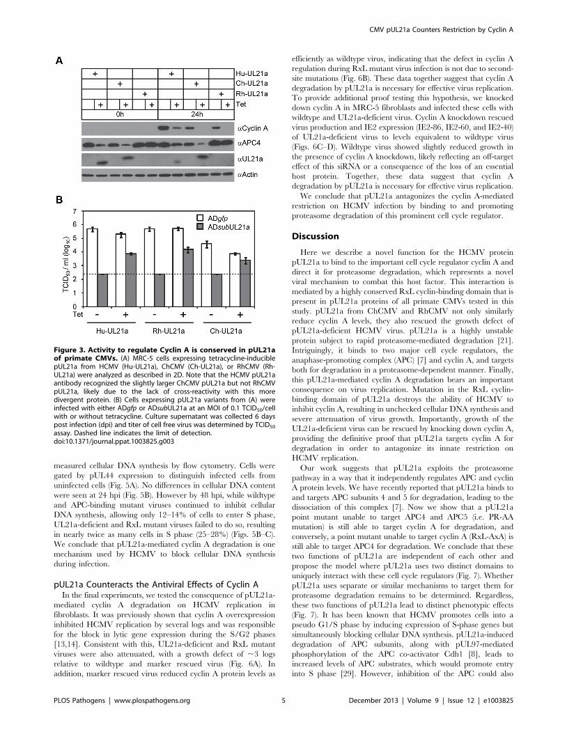
were able to reduce cyclin A levels (Fig. 3A). Furthermore, both
Author Summary
Cyclins are evolutionarily conserved proteins that associatewith cyclin-dependent kinases (CDKs) to regulate phos-phorylation of multiple substrates to promote cell-cycleprogression. Many viruses manipulate the cell cycle inorder to create an environment suitable for replication;however, only few examples exist where viruses modulatecyclin activity. Here, we identified a cyclin-binding domainwithin the human cytomegalovirus (HCMV) protein pU-L21a that confers its ability to interact with cyclin A andtarget it for proteasome degradation. Cyclin A promotescellular DNA replication, which consumes importantenzymes and metabolites needed for viral replication,making it important for large viruses like HCMV to blockthis protein’s activity. In accord, the ability of pUL21a todegrade cyclin A was necessary for the virus to blockcellular DNA replication and promote viral replication.Importantly, ablating cyclin A expression restored replica-tion to a virus lacking pUL21a, demonstrating that cyclin Ahas the intrinsic ability to restrict viral replication, but isspecifically countered by pUL21a. Together with ourprevious work showing that pUL21a also regulates theanaphase-promoting complex, another master cell cycleregulator, our studies have now revealed that HCMV haselegantly evolved dual functions within one proteintargeting the cell cycle machinery for viral replication.
CMV pUL21a Counters Restriction by Cyclin A
PLOS Pathogens | www.plospathogens.org 2 December 2013 | Volume 9 | Issue 12 | e1003825

proteins were able to rescue UL21a-deficient HCMV virus to
levels similar to that by HCMV pUL21a (Fig. 3B). Importantly,
the RhCMV UL21a protein did not degrade APC4 (Fig. 3A) but
still rescued UL21a-deficient HCMV virus (Fig. 3B), providing
evidence that the activity of pUL21a to inhibit cyclin A is critical
for efficient HCMV replication. The ChCMV UL21a-expressing
cells overall did not support HCMV infection as well as other cells
did, likely due to differences in the nature of the protein and where
in the cellular genome lentivirus was transduced. Together, these
data strongly suggest that pUL21a-mediated reduction of cyclin A
is a conserved and important function of primate CMVs.
pUL21a Induces Proteasome-Dependent Degradation ofCyclin A
We next investigated the potential mechanism of how pUL21a
reduced cyclin A levels. pUL21a became detectable at 10 hpi and
peaked at 24–48 hpi (Fig. 2A) [21]. Its protein levels markedly
increased in the presence of proteasome inhibitors, indicating that
this viral protein was inherently unstable and was targeted for
proteasome degradation during HCMV infection [21]. Intrigu-
ingly, pUL21a also induced proteasome-mediated degradation of
APC4 and APC5 [7]. We therefore hypothesized that pUL21a
regulated cyclin A levels by inducing its proteasome-dependent
degradation. To test this, we measured cyclin A protein levels in
the presence of the proteasome inhibitors MG132 and epoxomicin
during infection. Both MG132 and epoxomicin restored cyclin A
protein levels in wildtype infection to those seen in mock and
UL21a-deficient virus infected cells (Fig. 4A). While cyclin A
transcript levels were reduced during infection as previously
reported [12,16], wildtype and UL21a-deficient virus had similar
levels of cyclin A transcripts (Fig. 4B). Moreover, cyclin A protein
levels remained high despite drastic reduction in its transcript
levels following MG132 treatment (Figs. 4A and 4B). These data
suggest that regulation of cyclin A by pUL21a occurs at the
protein levels. Cyclin A contains a D-box motif that is necessary
for recognition by the APC, the cellular E3 ubiquitin ligase that
ubiquitinates and targets cyclin A for degradation during the cell
cycle [14,26,27]. To test if the D-box motif was required for
pUL21a-mediated degradation of cyclin A, we overexpressed
tetracycline-inducible FLAG-tagged wildtype and D-box mutant
Figure 1. HCMV pUL21a interacts with cyclin A through its cyclin-binding domain. (A) Alignment of the N-terminus of the UL21a codingsequence from HCMV (top), ChCMV (middle) and RhCMV (bottom). Asterisks denote the residues of the RxL motif that were subject to mutagenesis.(B) 293T cells were transfected with construct expressing a GFP tagged version of wildtype (gfpUL21a), stop mutant (gfpUL21astop), APC-bindingdomain mutant (gfpUL21aPR-AA), or cyclin-binding domain mutant (gfpUL21aRxL-AxA) pUL21a. Lysates were collected at 72 hours post infection (hpi),and immunoprecipitated with GFP or cyclin A antibody. Cell lysates and eluted proteins were analyzed by immunoblotting with indicated antibodies.Arrow indicates free GFP (bottom) or GFP-tagged pUL21a (top). Partial proteolysis was often seen with the GFP-tagged UL21a protein. (C) MRC-5 cellswere infected with wildtype HCMV expressing free GFP (ADgfp) or GFP-tagged UL21a (ADgfpUL21a). Lysates were collected at 48 hpi andimmunoprecipitated with GFP or cyclin A antibody. Cell lysates and eluted proteins were analyzed by immunoblotting. Arrow indicates free GFP,native pUL21a, or GFP-tagged pUL21a.doi:10.1371/journal.ppat.1003825.g001
CMV pUL21a Counters Restriction by Cyclin A
PLOS Pathogens | www.plospathogens.org 3 December 2013 | Volume 9 | Issue 12 | e1003825

cyclin A by lentiviral transduction in HCMV-infected cells. In the
absence of tetracycline, only endogenous cyclin A was expressed,
and its protein levels were reduced by almost 8-fold during
wildtype virus infection as compared to those during pUL21a-
deficient virus infection (Fig. 4C). In the presence of tetracycline,
expressions of FLAG-tagged cyclin A was induced, and their
expression was driven by the CMV promoter that was further
potentiated during CMV infection. Therefore, unlike endogenous
cyclin A, the levels of FLAG-tagged cyclin As were higher in
HCMV-infected cells relative to those in mock-infected cells
(Fig. 4C). Nonetheless, in the presence of tetracycline, pUL21a was
able to similarly reduce protein levels of endogenous cyclin A,
overexpressed FLAG-tagged wildtype, and D-box mutant cyclin A
during HCMV infection (Fig. 4C). We noted that cyclin A
reduction by pUL21a in the presence of tetracycline was less
pronounced than that in the absence of tetracycline (i.e. ,8-fold
vs. ,3-fold) (Fig. 4C). The lesser reduction of cyclin A in the
presence of tetracycline was likely due to overexpression of cyclin
A which overwhelmed the capacity of pUL21a to target it for
degradation. Overall, the ability of pUL21a to degrade D-box
cyclin A mutant, along with independent binding and degradation
of the APC and cyclin A by pUL21a (Figs. 1 and 2), suggest that
pUL21a-mediated degradation of cyclin A is independent of the
APC.
Together, our data show that pUL21a regulates cyclin A by
inducing its proteasome-dependent degradation, and that this
regulation is likely independent of the APC, the cellular E3 ligase
known to regulate cyclin A ubiquitination and degradation.
The pUL21a Cyclin-Binding Domain Is Necessary toPrevent Cellular DNA Synthesis during HCMV Infection
HCMV inhibits cellular DNA synthesis [28], and cyclin A is a
critical factor for promoting cellular DNA synthesis. We hypoth-
esized that pUL21a regulation of cyclin A was important to
prevent cellular DNA synthesis during HCMV infection. To test
this, we infected MRC-5 fibroblasts with recombinant virus and
Figure 2. pUL21a reduces cyclin A protein levels. (A) MRC-5 cells were mock infected or infected with indicated recombinant HCMV. Cell lysateswere collected at 24, 48, or 72 hpi, and analyzed by immunoblotting. IE1 and actin were used as infection and loading controls, respectively. (B) MRC-5 cells were infected as in A with indicated recombinant HCMV. Cell lysates were collected at 48 hpi and analyzed by immunoblotting. (C) MRC-5 cellswere infected as in A with indicated HCMV. Cell lysates were collected at indicated times and analyzed by immunoblotting. * indicates nonspecificcross-reacting bands. (D) MRC-5 cells expressing wildtype pUL21a (UL21a) or RxL mutant (UL21aRxL-AxA) under a tetracycline-inducible promoter werecreated by lentiviral transduction. Transduced cells were serum-starved for 24 hours, then treated with or without tetracycline (1 mg/ml) for 24 hoursand re-stimulated with serum to induce cyclin A expression. Cell lysates were collected at 0 or 24 hours post serum stimulation and analyzed byimmunoblotting.doi:10.1371/journal.ppat.1003825.g002
CMV pUL21a Counters Restriction by Cyclin A
PLOS Pathogens | www.plospathogens.org 4 December 2013 | Volume 9 | Issue 12 | e1003825
measured cellular DNA synthesis by flow cytometry. Cells were
gated by pUL44 expression to distinguish infected cells from
uninfected cells (Fig. 5A). No differences in cellular DNA content
were seen at 24 hpi (Fig. 5B). However by 48 hpi, while wildtype
and APC-binding mutant viruses continued to inhibit cellular
DNA synthesis, allowing only 12–14% of cells to enter S phase,
UL21a-deficient and RxL mutant viruses failed to do so, resulting
in nearly twice as many cells in S phase (25–28%) (Figs. 5B–C).
We conclude that pUL21a-mediated cyclin A degradation is one
mechanism used by HCMV to block cellular DNA synthesis
during infection.
pUL21a Counteracts the Antiviral Effects of Cyclin AIn the final experiments, we tested the consequence of pUL21a-
mediated cyclin A degradation on HCMV replication in
fibroblasts. It was previously shown that cyclin A overexpression
inhibited HCMV replication by several logs and was responsible
for the block in lytic gene expression during the S/G2 phases
[13,14]. Consistent with this, UL21a-deficient and RxL mutant
viruses were also attenuated, with a growth defect of ,3 logs
relative to wildtype and marker rescued virus (Fig. 6A). In
addition, marker rescued virus reduced cyclin A protein levels as
efficiently as wildtype virus, indicating that the defect in cyclin A
regulation during RxL mutant virus infection is not due to second-
site mutations (Fig. 6B). These data together suggest that cyclin A
degradation by pUL21a is necessary for effective virus replication.
To provide additional proof testing this hypothesis, we knocked
down cyclin A in MRC-5 fibroblasts and infected these cells with
wildtype and UL21a-deficient virus. Cyclin A knockdown rescued
virus production and IE2 expression (IE2-86, IE2-60, and IE2-40)
of UL21a-deficient virus to levels equivalent to wildtype virus
(Figs. 6C–D). Wildtype virus showed slightly reduced growth in
the presence of cyclin A knockdown, likely reflecting an off-target
effect of this siRNA or a consequence of the loss of an essential
host protein. Together, these data suggest that cyclin A
degradation by pUL21a is necessary for effective virus replication.
We conclude that pUL21a antagonizes the cyclin A-mediated
restriction on HCMV infection by binding to and promoting
proteasome degradation of this prominent cell cycle regulator.
Discussion
Here we describe a novel function for the HCMV protein
pUL21a to bind to the important cell cycle regulator cyclin A and
direct it for proteasome degradation, which represents a novel
viral mechanism to combat this host factor. This interaction is
mediated by a highly conserved RxL cyclin-binding domain that is
present in pUL21a proteins of all primate CMVs tested in this
study. pUL21a from ChCMV and RhCMV not only similarly
reduce cyclin A levels, they also rescued the growth defect of
pUL21a-deficient HCMV virus. pUL21a is a highly unstable
protein subject to rapid proteasome-mediated degradation [21].
Intriguingly, it binds to two major cell cycle regulators, the
anaphase-promoting complex (APC) [7] and cyclin A, and targets
both for degradation in a proteasome-dependent manner. Finally,
this pUL21a-mediated cyclin A degradation bears an important
consequence on virus replication. Mutation in the RxL cyclin-
binding domain of pUL21a destroys the ability of HCMV to
inhibit cyclin A, resulting in unchecked cellular DNA synthesis and
severe attenuation of virus growth. Importantly, growth of the
UL21a-deficient virus can be rescued by knocking down cyclin A,
providing the definitive proof that pUL21a targets cyclin A for
degradation in order to antagonize its innate restriction on
HCMV replication.
Our work suggests that pUL21a exploits the proteasome
pathway in a way that it independently regulates APC and cyclin
A protein levels. We have recently reported that pUL21a binds to
and targets APC subunits 4 and 5 for degradation, leading to the
dissociation of this complex [7]. Now we show that a pUL21a
point mutant unable to target APC4 and APC5 (i.e. PR-AA
mutation) is still able to target cyclin A for degradation, and
conversely, a point mutant unable to target cyclin A (RxL-AxA) is
still able to target APC4 for degradation. We conclude that these
two functions of pUL21a are independent of each other and
propose the model where pUL21a uses two distinct domains to
uniquely interact with these cell cycle regulators (Fig. 7). Whether
pUL21a uses separate or similar mechanisms to target them for
proteasome degradation remains to be determined. Regardless,
these two functions of pUL21a lead to distinct phenotypic effects
(Fig. 7). It has been known that HCMV promotes cells into a
pseudo G1/S phase by inducing expression of S-phase genes but
simultaneously blocking cellular DNA synthesis. pUL21a-induced
degradation of APC subunits, along with pUL97-mediated
phosphorylation of the APC co-activator Cdh1 [8], leads to
increased levels of APC substrates, which would promote entry
into S phase [29]. However, inhibition of the APC could also
Figure 3. Activity to regulate Cyclin A is conserved in pUL21aof primate CMVs. (A) MRC-5 cells expressing tetracycline-induciblepUL21a from HCMV (Hu-UL21a), ChCMV (Ch-UL21a), or RhCMV (Rh-UL21a) were analyzed as described in 2D. Note that the HCMV pUL21aantibody recognized the slightly larger ChCMV pUL21a but not RhCMVpUL21a, likely due to the lack of cross-reactivity with this moredivergent protein. (B) Cells expressing pUL21a variants from (A) wereinfected with either ADgfp or ADsubUL21a at an MOI of 0.1 TCID50/cellwith or without tetracycline. Culture supernatant was collected 6 dayspost infection (dpi) and titer of cell free virus was determined by TCID50
assay. Dashed line indicates the limit of detection.doi:10.1371/journal.ppat.1003825.g003
CMV pUL21a Counters Restriction by Cyclin A
PLOS Pathogens | www.plospathogens.org 5 December 2013 | Volume 9 | Issue 12 | e1003825

increase cyclin A levels, which is detrimental to HCMV
replication. Therefore, HCMV has developed a second function
within pUL21a, which is to promote cyclin A degradation. This
will antagonize the antiviral activity of cyclin A, inhibit cellular
DNA synthesis, and phenotypically prevent infected cells from
entering S phase. Thus the virus has elegantly evolved two
mechanisms within one protein to harness the benefits of
inhibiting the APC as well as overcoming any detrimental
consequences of such regulation.
How does pUL21a target cyclin A for proteasome degradation?
Cyclin A is normally targeted for ubiquitination and proteasome
degradation during M phase of the cell cycle. Preliminary work
suggests a modest increase in cyclin A ubiquitination in the
presence of pUL21a (data not shown), so it is possible that pUL21a
may have a role in cyclin A ubiquitination. If so, as pUL21a
contains no domain indicating it as an E3 ligase, it could recruit an
E3 ligase to ubiquitinate cyclin A or block the activity of a
deubiquitinase (DUB). Alternatively, it is also entirely possible that
pUL21a may directly target cyclin A to the proteasome in a
ubiquitin-independent manner. Cyclin A is normally targeted by
the APC for ubiquitination and proteasome degradation. Howev-
er, inhibition of the APC by pUL21a and pUL97 [7,8], along with
our present data showing that pUL21a can mediate cyclin A
degradation in the absence of its APC recognition motif (D-box),
suggests that cyclin A degradation during HCMV infection is
APC-independent. Interestingly, pUL21a itself is rapidly degraded
by the proteasome in a ubiquitin-independent manner, and is
highly sensitive to proteasome inhibition [21]. It is tempting to
speculate that pUL21a may act as a novel ‘‘suicide’’ protein that
delivers substrates to the proteasome and is degraded with them.
Mechanistically, pUL21a could directly bind to the proteasome, or
could be recognized as an unfolded protein due to its proline-rich,
predicted unstructured C-terminus tail [7], and transported to the
proteasome by chaperones. Additional biochemical, genetic, and
structural analysis will be critical to testing these hypotheses.
What is the mechanism for cyclin A to restrict virus replication?
Cyclin A and its concomitant CDK’s phosphorylate a number of
substrates that promote cellular DNA synthesis. It is possible that
active cellular DNA synthesis would limit the resources available
for viral genome amplification and interfere with the process of
viral DNA replication, ultimately attenuating virus growth. Thus,
pUL21a mutants may provide us with a tool to test the long held
belief that cellular DNA replication is detrimental to herpesvirus
replication. Alternatively, a substrate or set of substrates of cyclin A
may also have more direct and specific antiviral activity that blocks
HCMV replication. This pUL21a-mediated cyclin A regulation
appears to be unique to CMVs of high mammals, such as
primates, as murine CMV does not encode a UL21a homologous
protein. It is possible that murine CMV has evolved alternative
mechanisms to arrest the cell cycle, or that its shorter life cycle
Figure 4. pUL21a induces proteasome-dependent degradation of cyclin A. (A) MRC-5 cells were mock infected or infected with ADgfp orADsubUL21a. Infected cells were treated with or without MG132 or epoxomicin for 9 hours prior to collection. Cell lysates were collected at 24 hpiand analyzed by immunoblotting. (B) MRC-5 cells were infected as described in A. Cells were treated with or without MG132 at 9 hpi, and RNA wasextracted at 18 hpi. Cyclin A transcripts were quantified by real-time quantitative PCR (RT-qPCR) and normalized to GAPDH. The normalized levels ofcyclin A transcripts in mock infected samples were arbitrarily set to 1. p values were determined using the student’s t test. *, p value ,0.05; n.s. (notsignificant), p value .0.05. (C) MRC-5 cells expressing tetracycline-inducible wildtype cyclin A (flagCyclin A) or D-box mutant cyclin A (flagCyclin A DD)were created by lentiviral transduction. Cells were then infected with indicated viruses, treated with or without tetracycline, and analyzed byimmunoblotting at 24 hpi. Arrow indicates FLAG-tagged cyclin A variants detected by cyclin A antibody. Protein bands of FLAG-tagged cyclin A (inanti-FLAG blot) and endogenous cyclin A (in anti-cyclin A blot) were quantitated using Image J software and normalized to ADsubUL21a under eachcondition.doi:10.1371/journal.ppat.1003825.g004
CMV pUL21a Counters Restriction by Cyclin A
PLOS Pathogens | www.plospathogens.org 6 December 2013 | Volume 9 | Issue 12 | e1003825

(,32 hours) circumvents the need to inhibit cyclin A activity of
host cells. Consistent with this, murine CMV upregulates cyclin A
levels and initial viral gene expression is unaffected by cyclin A
overexpression [14], highlighting an interesting and striking
difference between closely related virus families.
Finally, the discovery of pUL21a as a cyclin A modulator can
also provide a useful tool to delineate the activity of this important
molecule in cell biology. Only recently have large-scale screenings
been used to systematically identify cyclin A substrates in
mammalian cells [30]. pUL21a may be used to confirm these
Figure 5. The pUL21a cyclin-binding domain is essential for HCMV to prevent cellular DNA synthesis. MRC-5 cells were infected withindicated viruses in the presence of viral DNA replication inhibitor phosphonoacetic acid (PAA). Cells were collected at 24 and 48 hpi, fixed, and co-stained with propidium iodide (PI) and antibody to viral protein pUL44. Cell cycle profiles were analyzed by flow cytometry. (A) The PI and pUL44staining profiles of representative mock- and wildtype virus- infected cells at 48 hpi. Also shown is gating used to separate pUL44-positive (infected)and pUL44-negative (uninfected) cells. (B) Both 24 and 48 hpi DNA content profiles of mock-infected cells or pUL44-positive, virus-infected cells. (C)Percentage of pUL44-positive, virus-infected cells in G1, S, or G2/M phase at 48 hpi based on DNA content. p values were based on the numbers of Sphase cells and determined using the student’s t test. *, p value ,0.05; n.s. (not significant), p value .0.05. Shown are data from two independentexperiments.doi:10.1371/journal.ppat.1003825.g005
CMV pUL21a Counters Restriction by Cyclin A
PLOS Pathogens | www.plospathogens.org 7 December 2013 | Volume 9 | Issue 12 | e1003825

recently identified cyclin A substrates, explore the role of these
substrates in cellular DNA synthesis, or discover additional
substrates that play roles in cellular DNA synthesis and
proliferation. Viral systems have been instrumental in many
seminal discoveries in the history of cell biology. Novel virus-
encoded regulators such as pUL21a can be powerful tools to probe
the biology of host cells that are otherwise difficult to study.
Materials and Methods
Cell CultureHuman foreskin fibroblasts (HFFs), primary embryonic lung
fibroblasts (MRC-5), and 293T cells were propagated in
Dulbecco’s modified Eagle medium (DMEM) containing 10%
fetal bovine serum and penicillin-streptomycin. Expression con-
structs were transfected into 293T cells with 1 mg/ml polyethy-
leneimine (PEI, Polysciences) in Opti-MEM media.
Plasmids and ReagentsPrimers used in this study are listed in Table 1. Point mutant
UL21a sequences were constructed using PCR with the desired
mutations incorporated into complementary primers. To create
expression constructs, the RxL mutant UL21a PCR fragment was
digested with restriction enzymes Bgl II and EcoR I, and ligated
into a pLPCX (Clontech)-derived over-expression vector with an
N-terminal GFP tag to create retroviral expression construct pYD-
Figure 6. Depletion of cyclin A alleviates the requirement of pUL21a for HCMV replication. (A) MRC-5 cells were infected with indicatedviruses at an MOI of 0.01 TCID50/cell and production of cell free virus was determined by TCID50 assay at indicated time points. (B) MRC-5 cells wereinfected with indicated viruses at an MOI of 3, and cell lysates were collected at 24 and 48 hpi and analyzed by immunoblotting. * indicatesnonspecific cross-reacting bands. (C) MRC-5 cells were treated with siRNA against cyclin A (siCyclin A) or luciferase control (siCont), and then infectedwith indicated virus at an MOI of 3. At 96 hpi, production of cell free virus was determined by TCID50 assay, and (D) cell lysates were analyzed byimmunoblotting.doi:10.1371/journal.ppat.1003825.g006
CMV pUL21a Counters Restriction by Cyclin A
PLOS Pathogens | www.plospathogens.org 8 December 2013 | Volume 9 | Issue 12 | e1003825

C760; or digested with Sal I and EcoR I and ligated into pYD-
C639, a pLKO-based lentiviral vector under a tetracycline-
inducible CMV-TetO2 promoter [31] (generous gift from Roger
Everett, University of Glasgow), to create lentiviral expression
construct pYD-C762. RhCMV UL21a was amplified from
RhCMV BAC [32], ChCMV UL21a was constructed from long
overlapping primers, and FLAG tagged wildtype cyclin A as well
as D-box mutant cyclin A (cyclin A DD) were amplified from
plasmids provided by Anindya Dutta (University of Virginia
School of Medicine) [26]. They were all cloned into pYD-C639 to
create lentiviral expression constructs. All other constructs used in
this study have been previously described [7].
Lentivirus was produced by PEI transfection of corresponding
expression constructs as described above, along with appropriate
packaging plasmids, into 293T cells. To create stable expression
cells, MRC-5 cells expressing GFP-TetR [7] were transduced with
lentivirus, and selected with 2 mg/mL puromycin (Sigma-Aldrich)
to produce stable cells expressing various forms of UL21a, FLAG-
cyclin, or HA-ubiquitin under the CMV-TetO2 promoter.
To knock down cyclin A by RNAi, MRC-5 cells were
transfected with siGENOME siRNA against cyclin A or luciferase
control (Thermo Scientific), using the procedure of siLENTFECT
(Bio-Rad) according to manufacturer’s instructions. Cells were left
in serum-free medium and infected 48 hours later in serum-
containing medium.
Primary antibodies used included anti-b actin (AC-15, Abcam);
anti-GFP (3E6 and A6455, Invitrogen); anti-APC3 (35/CDC27,
BD Biosciences); anti-APC4 (A301-176A, Bethyl laboratories); anti-
UL21a [21]; anti-IE2 (mAB8140, Chemicon); anti-UL44 (10D8,
virusys); anti-Cyclin A (B-8 and H-432, Santa Cruz); anti-cyclin B
(GNS1, Thermo Scientific); anti-cyclin E (HE12, BD Biosciences);
anti-FLAG (F1804, Sigma); anti-HA (16B12, Covance); and anti-
IE1 (generous gift from Thomas Shenk, Princeton University).
Recombinant HCMV VirusBAC-HCMV clones used in the present study were constructed
using PCR-based two-step linear recombination as previously
reported [33]. pADgfp, which carried the genome of
HCMVAD169 strain and a simian virus 40 early promoter-driven
GFP gene in place of the viral US4–US6 region, was used to
produce wildtype virus ADgfp [34]. pADpmUL21aRxL-AxA, which
carried the point mutation RxL42-44AxA in the UL21a coding
sequence, and pADrevUL21aRxL-AxA, in which RxL42-44AxA
mutation was subsequently repaired, were used to produce UL21a
cyclin-binding domain point mutant virus ADpmUL21aRxL-AxA
and its marker rescued virus ADrevUL21aRxL-AxA, respectively.
Figure 7. Model for dual roles of pUL21a in HCMV infection. pUL21a independently binds to cyclin A or the anaphase-promoting complex(APC) through the RxL or PR domain, respectively, and targets each for proteasome-dependent degradation. pUL21a-induced degradation of the APCbridge, in concordance with pUL97-induced phosphorylation of Cdh1, leads to an increase in APC substrates, which helps to create a favorable, S-phase like cellular environment for DNA synthesis. However, pUL21a-induced degradation of cyclin A allows HCMV to specifically prevent host DNAsynthesis. Together, these two independent activities of pUL21a help to subvert host cells for efficient HCMV growth.doi:10.1371/journal.ppat.1003825.g007
CMV pUL21a Counters Restriction by Cyclin A
PLOS Pathogens | www.plospathogens.org 9 December 2013 | Volume 9 | Issue 12 | e1003825

These recombinant BAC clones were confirmed by PCR,
restriction digest, and sequencing. All other recombinant BAC-
HCMV clones used were described previously [7]. Recombinant
HCMV AD169 viruses were reconstituted from transfection of
corresponding BAC-HCMV clones as previously described [33].
Viral stocks were harvested from infected cell culture supernatant
and concentrated by ultracentrifugation through 20% D-sorbitol.
Virus titers were determined in duplicate by tissue culture
infectious dose 50 (TCID50) assay on HFFs. Relative viral genome
numbers were determined by extracting virion DNA and
performing real-time quantitative PCR (qPCR) with either a
taqman probe and primers specific to viral gene UL54, or with
SYBR green and primers to UL32 [21].
HCMV InfectionConfluent MRC-5 cells were split, and 24 hours later infected
with HCMV at an input genome number equivalent to that of 3
infectious units of wildtype virus/cell, unless otherwise indicated.
Cells were inoculated for 1 hour with virus and then replenished
with fresh media. For cell cycle profiling, cells were treated with
phosphonoacetic acid (PAA, 100 mg/ml, Sigma-Aldrich) immedi-
ately after infection. For proteosome inhibition experiments, cells
were treated with MG132 (20 mM, Santa Cruz) or epoxomicin
(40 mM, Santa Cruz). Virus titers in the supernatant of infected
cultures were determined by TCID50 assay.
Protein AnalysisFor immunoprecipitation, 293T and MRC-5 cells were lysed
in NP40 buffer (0.5% NP40, 50 mM Tris-Cl pH 8.0, 125 mM
NaCl) and EBC2 buffer (0.5% NP40, 50 mM Tris-Cl pH 8.0,
300 mM NaCl), respectively. Lysis buffers were supplemented
with protease and phosphatase inhibitors (Roche and Sigma-
Aldrich, respectively). Mouse anti-GFP antibody (3E6, Invitro-
gen) or mouse anti-Cyclin A antibody (B-8, Santa Cruz) was
conjugated to protein A dynabeads (Invitrogen) with BS3
(Thermo Scientific) according to manufacturer’s instructions.
Cleared cell lysates were incubated with conjugated dynabeads by
gentle rotation at 4uC. Beads were washed once with lysis buffer
and twice with PBS, and bound proteins were eluted in reducing
sample buffer (200 mM Tris pH 6.8, 6% SDS, 12% b-
mercaptoethanol, 18% glycerol) by incubating at 55uC. For
whole cell lysate control (WCL), cell lysates were similarly mixed
with reducing sample buffer and incubated at 90uC for
5 minutes.
For immunoblotting, cells were lysed in reducing sample buffer
containing protease and phosphatase inhibitors. Proteins from
equivalent cell numbers were resolved on a SDS poly-acrylamide
gel, transferred to a polyvinylidene difluoride membrane, hybrid-
ized with primary antibody, reacted with horseradish peroxidase-
conjugated secondary antibody, and visualized using chemilumi-
nescent substrate (Thermo Scientific).
Analysis of Cellular DNA ContentCells were trypsinized, washed with PBS, and fixed in 70%
ethanol. Fixed cells were double stained with propidium iodide (PI)
for DNA and with anti-pUL44 antibody to identify infected cells,
and analyzed by flow cytometry. Cells were gated for pUL44 and
PI staining, and percentages of cells in each cell cycle compart-
ment were calculated using FlowJo software.
Reverse Transcription Coupled-Quantitative PCR Analysis(RT-qPCR)
Total RNA was extracted with TRIzol (Invitrogen), treated with
TURBO DNA-free (Ambion) to remove DNA contaminants, and
reverse transcribed with random hexamer primers using the High
Capacity cDNA RT Kit (Applied Biosystems). The cDNA was
Table 1. Primers used in this study.
Purpose Primer sequencea Construct name
Overlapping PCR primers tocreate UL21a RxL-AxA mutation
59cgagctcgtgcgagggctttccaaaatc39
59gattttggaaagccctcgcacgagctcg39
Non applicable
Overlapping primers to createtemplate for amplifying ChUL21a
59atgggaggcagtcccgaccccgatctctcggtggccaccgagggtgtgaggcccgtggtccgcgcggacctgttccgagcccg-ccggcccctgc39
59cctgaaggtgctggtgatgctgatggtagttctcaaaaagccggcgccgggcccgcggagcataga-aagccagacggcgcaggggccggcgggct39
59tcaccagcaccttcaggtcccgccgccgattcatcggatcgtcgctgtgcc-cggaggggacgaggaagcgataccgatggacctgccgagggaga39
59tgggcggaacgtcgtccaacagcagcaccagc-gggttgggcagcgggcggtcgggcggtatatcggcggcgacctggatctccctcggcaggtcc39
59ttaaaaatgttcccagttctcttcgcgtatcacggggtactcgcggggcactcggaaaggagcgaagccgggcggca-tgggcggaacgtcgtcc39
Non applicable
PCR primers to cloneUL21a into pLKO vector
59gtcgaccgagatgggaggtagccctgttcc39
59ggaattcttaaaactggtcccaatgttctt39
pLKO-UL21a pLKO-UL21a RxL-AxA
PCR primers to cloneRhUL21a into pLKO vector
59gtcgaccgagatgggaggcagcaccga39
59ggaattcttaagcgtgtgcttcttcat39
pLKO-RhUL21a
PCR primers to cloneChUL21a into pLKO vector
59gggtcgacatgggaggcagtcccgac39
59gggaattcttaaaaatgttcccagttctc39
pLKO-ChUL21a
PCR primers to clone3xFlag-Cyclin A into pLKO vector
59gcgtcgacatggactacaaagaccatgacggtgatttaaagatcatgatatcgattacaaggatga-cgatgacaagttgggcaactctgcgcc39
59gcgaattcttacagatttagtgtctctggtggg39
pLKO-3xFlag-Cyclin ApLKO-3xFlag-Cyclin ADD-box
PCR primers to clone HA-Ubiquitininto pLKO vector
59gcgctagcatgtacccatacgacgtccca39
59gcgaattctcacccacctcttagtctt39
pLKO-HA-Ubiquitin
Primer pairs to quantify cyclinA transcript levels by RT-qPCR
59gcatgtcaccgttcctcctt39
59cagggcatcttcacgctctat39
Non applicable
Primer pairs to quantify GAPDHtranscript levels by RT-qPCR
59ctgttgctgtagccaaattcgt39
59acccactcctccacctttgac39
Non applicable
CMV pUL21a Counters Restriction by Cyclin A
PLOS Pathogens | www.plospathogens.org 10 December 2013 | Volume 9 | Issue 12 | e1003825

quantified by qPCR using SYBR green SYBR Advantage qPCR
Premix (Clontech) with primers for the cellular genes Cyclin A or
GAPDH (glyceraldehyde-3-phosphate dehydrogenase) (Table 1).
cDNA from six arbitrary samples were mixed together and serially
diluted to generate a standard curve used to calculate the relative
amount of specific RNA.
Acknowledgments
We thank Roger Everett (University of Glasgow) for the pLKO-based
tetracycline-inducible lentiviral expression system, Anindya Dutta (Uni-
versity of Virginia) for FLAG tagged wildtype and D-box mutant cyclin A
constructs, Ted Hansen (Washington University) for HA-ubiquitin
construct, Thomas Shenk (Princeton University) for antibodies, the High
Speed Cell Sorter Core at the Siteman Cancer Center for excellent
technical assistance with flow cytometry, and members of the Yu lab for
critical reading of the manuscript.
Author Contributions
Conceived and designed the experiments: ARF DY. Performed the
experiments: NC ARF. Analyzed the data: NC ARF DY. Contributed
reagents/materials/analysis tools: NC ARF DY. Wrote the paper: NC
ARF DY.
References
1. Revello MG, Gerna G (2002) Diagnosis and management of human
cytomegalovirus infection in the mother, fetus, and newborn infant. Clin
Microbiol Rev 15: 680–715.
2. Poma EE, Kowalik TF, Zhu L, Sinclair JH, Huang ES (1996) The human
cytomegalovirus IE1-72 protein interacts with the cellular p107 protein and
relieves p107-mediated transcriptional repression of an E2F-responsive promot-
er. J Virol 70: 7867–7877.
3. Kamil JP, Hume AJ, Jurak I, Munger K, Kalejta RF, et al. (2009) Human
papillomavirus 16 E7 inactivator of retinoblastoma family proteins complements
human cytomegalovirus lacking UL97 protein kinase. Proc Natl Acad Sci U S A
106: 16823–16828.
4. Hagemeier C, Caswell R, Hayhurst G, Sinclair J, Kouzarides T (1994)
Functional interaction between the HCMV IE2 transactivator and the
retinoblastoma protein. Embo J 13: 2897–2903.
5. Song YJ, Stinski MF (2002) Effect of the human cytomegalovirus IE86 protein
on expression of E2F-responsive genes: a DNA microarray analysis. Proc Natl
Acad Sci U S A 99: 2836–2841.
6. Hume AJ, Finkel JS, Kamil JP, Coen DM, Culbertson MR, et al. (2008)
Phosphorylation of retinoblastoma protein by viral protein with cyclin-
dependent kinase function. Science 320: 797–799.
7. Fehr AR, Gualberto NC, Savaryn JP, Terhune SS, Yu D (2012) Proteasome-
dependent disruption of the E3 ubiquitin ligase anaphase-promoting complex by
HCMV protein pUL21a. PLoS Pathog 8: e1002789.
8. Tran K, Kamil JP, Coen DM, Spector DH (2010) Inactivation and disassembly
of the anaphase-promoting complex during human cytomegalovirus infection is
associated with degradation of the APC5 and APC4 subunits and does not
require UL97-mediated phosphorylation of Cdh1. J Virol 84: 10832–10843.
9. Qian Z, Leung-Pineda V, Xuan B, Piwnica-Worms H, Yu D (2010) Human
cytomegalovirus protein pUL117 targets the mini-chromosome maintenance
complex and suppresses cellular DNA synthesis. PLoS Pathog 6: e1000814.
10. Sanchez V, McElroy AK, Yen J, Tamrakar S, Clark CL, et al. (2004) Cyclin-
Dependent Kinase Activity Is Required at Early Times for Accurate Processing
and Accumulation of the Human Cytomegalovirus UL122-123 and UL37
Immediate-Early Transcripts and at Later Times for Virus Production. J Virol
78: 11219–11232.
11. Sanchez V, Spector DH (2006) Cyclin-dependent kinase activity is required for
efficient expression and posttranslational modification of human cytomegalovi-
rus proteins and for production of extracellular particles. J Virol 80: 5886–5896.
12. Shlapobersky M, Sanders R, Clark C, Spector DH (2006) Repression of
HMGA2 gene expression by human cytomegalovirus involves the IE2 86-
kilodalton protein and is necessary for efficient viral replication and inhibition of
cyclin A transcription. J Virol 80: 9951–9961.
13. Zydek M, Hagemeier C, Wiebusch L (2010) Cyclin-dependent kinase activity
controls the onset of the HCMV lytic cycle. PLoS Pathog 6: e1001096.
14. Oduro JD, Uecker R, Hagemeier C, Wiebusch L (2012) Inhibition of human
cytomegalovirus immediate-early gene expression by cyclin A2-dependent kinase
activity. J Virol 86: 9369–9383.
15. Jault FM, Jault JM, Ruchti F, Fortunato EA, Clark C, et al. (1995)
Cytomegalovirus infection induces high levels of cyclins, phosphorylated Rb,
and p53, leading to cell cycle arrest. J Virol 69: 6697–6704.
16. Salvant BS, Fortunato EA, Spector DH (1998) Cell cycle dysregulation byhuman cytomegalovirus: influence of the cell cycle phase at the time of infection
and effects on cyclin transcription. J Virol 72: 3729–3741.17. Bresnahan WA, Albrecht T, Thompson EA (1998) The cyclin E promoter is
activated by human cytomegalovirus 86-kDa immediate early protein. J Biol
Chem 273: 22075–22082.18. Du G, Dutta N, Lashmit P, Stinski MF (2011) Alternative splicing of the human
cytomegalovirus major immediate-early genes affects infectious-virus replicationand control of cellular cyclin-dependent kinase. J Virol 85: 804–817.
19. Chen Z, Knutson E, Kurosky A, Albrecht T (2001) Degradation of p21cip1 incells productively infected with human cytomegalovirus. J Virol 75: 3613–3625.
20. Noris E, Zannetti C, Demurtas A, Sinclair J, De Andrea M, et al. (2002) Cell
cycle arrest by human cytomegalovirus 86-kDa IE2 protein resembles prematuresenescence. J Virol 76: 12135–12148.
21. Fehr AR, Yu D (2010) Human cytomegalovirus gene UL21a encodes a short-lived cytoplasmic protein and facilitates virus replication in fibroblasts. J Virol
84: 291–302.
22. Fehr AR, Yu D (2011) Human cytomegalovirus early protein pUL21a promotesefficient viral DNA synthesis and the late accumulation of immediate-early
transcripts. J Virol 85: 663–674.23. Dinkel H, Michael S, Weatheritt RJ, Davey NE, Van Roey K, et al. (2012)
ELM–the database of eukaryotic linear motifs. Nucleic Acids Res 40: D242–251.
24. Adams PD, Sellers WR, Sharma SK, Wu AD, Nalin CM, et al. (1996)Identification of a cyclin-cdk2 recognition motif present in substrates and p21-
like cyclin-dependent kinase inhibitors. Mol Cell Biol 16: 6623–6633.25. Schulman BA, Lindstrom DL, Harlow E (1998) Substrate recruitment to cyclin-
dependent kinase 2 by a multipurpose docking site on cyclin A. Proc Natl AcadSci U S A 95: 10453–10458.
26. Machida YJ, Dutta A (2007) The APC/C inhibitor, Emi1, is essential for
prevention of rereplication. Genes Dev 21: 184–194.27. Geley S, Kramer E, Gieffers C, Gannon J, Peters JM, et al. (2001) Anaphase-
promoting complex/cyclosome-dependent proteolysis of human cyclin A startsat the beginning of mitosis and is not subject to the spindle assembly checkpoint.
J Cell Biol 153: 137–148.
28. Sanchez V, Spector DH (2008) Subversion of cell cycle regulatory pathways.Curr Top Microbiol Immunol 325: 243–262.
29. Peters JM (2006) The anaphase promoting complex/cyclosome: a machinedesigned to destroy. Nat Rev Mol Cell Biol 7: 644–656.
30. Chi Y, Welcker M, Hizli AA, Posakony JJ, Aebersold R, et al. (2008)Identification of CDK2 substrates in human cell lysates. Genome Biol 9: R149.
31. Everett RD, Boutell C, McNair C, Grant L, Orr A (2010) Comparison of the
biological and biochemical activities of several members of the alphaherpesvirusICP0 family of proteins. J Virol 84: 3476–3487.
32. Lilja AE, Shenk T (2008) Efficient replication of rhesus cytomegalovirus variantsin multiple rhesus and human cell types. Proc Natl Acad Sci U S A 105: 19950–
19955.
33. Paredes AM, Yu D (2012) Human cytomegalovirus: bacterial artificialchromosome (BAC) cloning and genetic manipulation. Curr Protoc Microbiol
Chapter 14: Unit14E 14.34. Qian Z, Xuan B, Hong TT, Yu D (2008) The full-length protein encoded by
human cytomegalovirus gene UL117 is required for the proper maturation ofviral replication compartments. J Virol 82: 3452–3465.
CMV pUL21a Counters Restriction by Cyclin A
PLOS Pathogens | www.plospathogens.org 11 December 2013 | Volume 9 | Issue 12 | e1003825