Embed Size (px)
Citation preview
CutaneousCutaneousSquamousSquamous Cell Carcinoma…Cell Carcinoma…
Good Grades Are Not Enough!Good Grades Are Not Enough!
Paul K. Shitabata, M.D.Paul K. Shitabata, M.D.DermatopathologistDermatopathologist
APMGAPMG
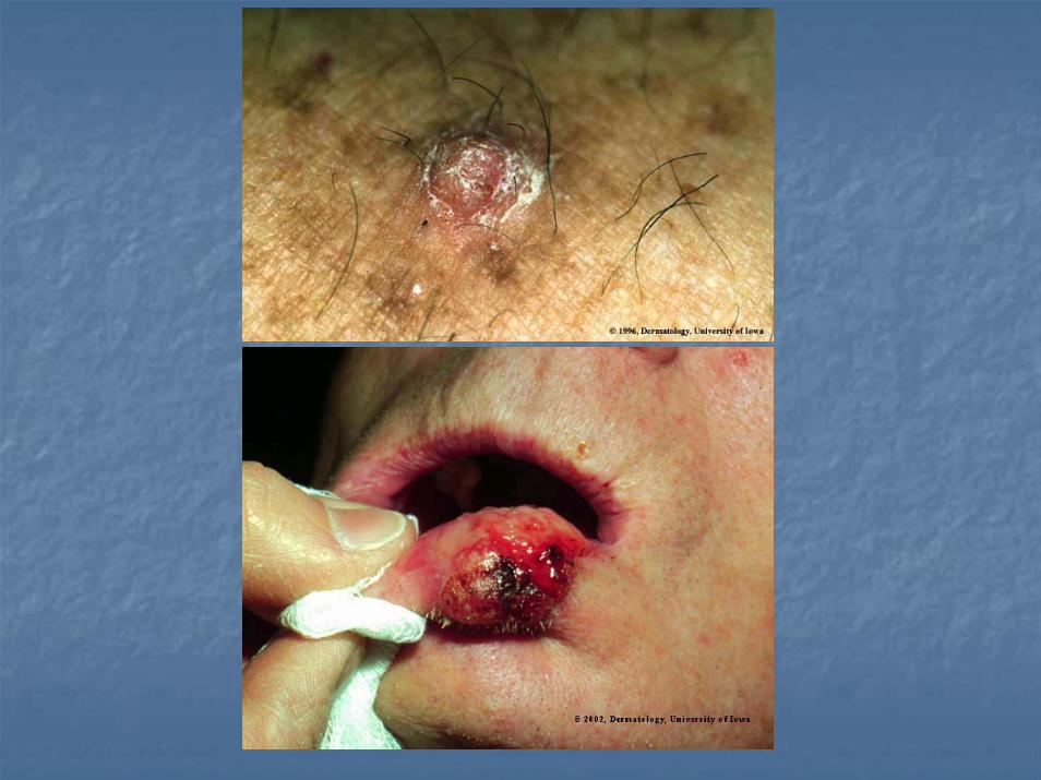
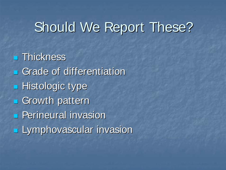
Should We Report These?Should We Report These?
ThicknessThicknessGrade of differentiationGrade of differentiationHistologicHistologic typetypeGrowth patternGrowth patternPerineuralPerineural invasioninvasionLymphovascularLymphovascular invasioninvasion
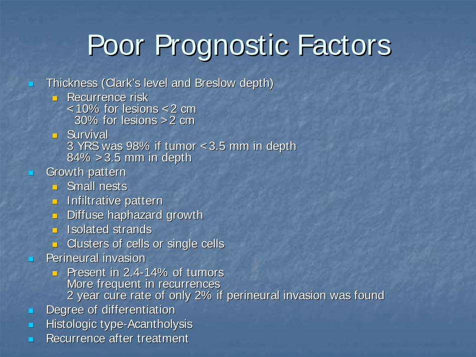
Poor Prognostic FactorsPoor Prognostic FactorsThickness (Clark’s level and Thickness (Clark’s level and BreslowBreslow depth)depth)
Recurrence riskRecurrence risk<10% for lesions <2 cm<10% for lesions <2 cm
30% for lesions >2 cm30% for lesions >2 cmSurvivalSurvival3 YRS was 98% if tumor <3.5 mm in depth3 YRS was 98% if tumor <3.5 mm in depth84% >3.5 mm in depth84% >3.5 mm in depth
Growth patternGrowth patternSmall nestsSmall nestsInfiltrative patternInfiltrative patternDiffuse haphazard growthDiffuse haphazard growthIsolated strandsIsolated strandsClusters of cells or single cells Clusters of cells or single cells
PerineuralPerineural invasioninvasionPresent in 2.4Present in 2.4--14% of tumors14% of tumorsMore frequent in recurrencesMore frequent in recurrences2 year cure rate of only 2% if 2 year cure rate of only 2% if perineuralperineural invasion was found invasion was found
Degree of differentiationDegree of differentiationHistologicHistologic typetype--AcantholysisAcantholysisRecurrence after treatmentRecurrence after treatment
Questionable SignificanceQuestionable Significance
Location (nonLocation (non--mucosal surfaces excluded)mucosal surfaces excluded)UlcerationUlcerationInflammationInflammation
DermatolDermatol SurgSurg 2002 Mar;28(3):2682002 Mar;28(3):268--73 73
Proposed Classification SchemesProposed Classification Schemes
Low Risk of Aggressive BehaviorLow Risk of Aggressive BehaviorBowen’s Disease/Carcinoma in situBowen’s Disease/Carcinoma in situ
ErythroplasiaErythroplasia of of QueyratQueyratActinic Actinic keratosiskeratosis/KIN I/KIN I--IIIIIIKeratoacanthomaKeratoacanthomaVerrucousVerrucousPapillary Papillary
High Risk of Aggressive BehaviorHigh Risk of Aggressive BehaviorMarjolin’sMarjolin’s UlcerUlcerAcantholyticAcantholyticDesmoplastic/SarcomatoidDesmoplastic/SarcomatoidInvasive Invasive BowenoidBowenoidAdenosquamousAdenosquamousLymphoepitheliomaLymphoepithelioma--like carcinoma like carcinoma Transplant relatedTransplant related
KeratoacanthomaKeratoacanthoma
Resemble Resemble squamoussquamous cell carcinomas both cell carcinomas both clinically and clinically and histologicallyhistologicallyUsually nodules with a scaly central plug. Usually nodules with a scaly central plug. History of rapid onset with growth to 1History of rapid onset with growth to 1--2 2 cm over a period of 1cm over a period of 1--2 months2 monthsMany spontaneously Many spontaneously involuteinvolute or regress or regress after 3after 3--6 months6 monthsMales in their 6Males in their 6--7th decades. 7th decades.

KeratoacanthomaKeratoacanthoma Clinical VariantsClinical Variants
GiantGiantMultipleMultiple--Ferguson Smith Type Ferguson Smith Type MultipleMultiple--GrzybowskiGrzybowski (Eruptive) Type (Eruptive) Type SubungualSubungual
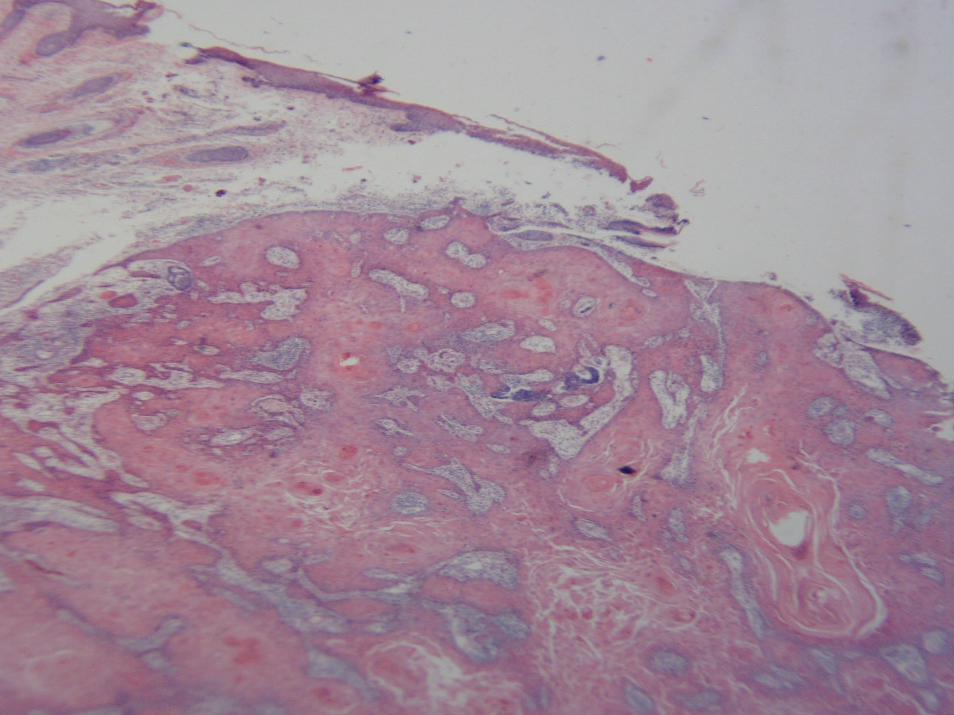
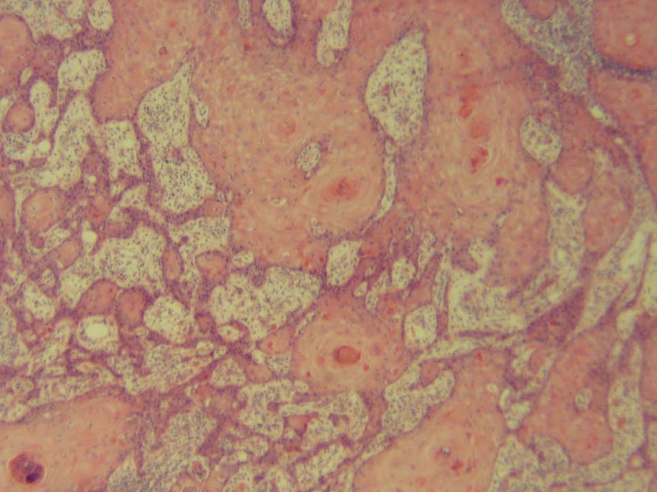
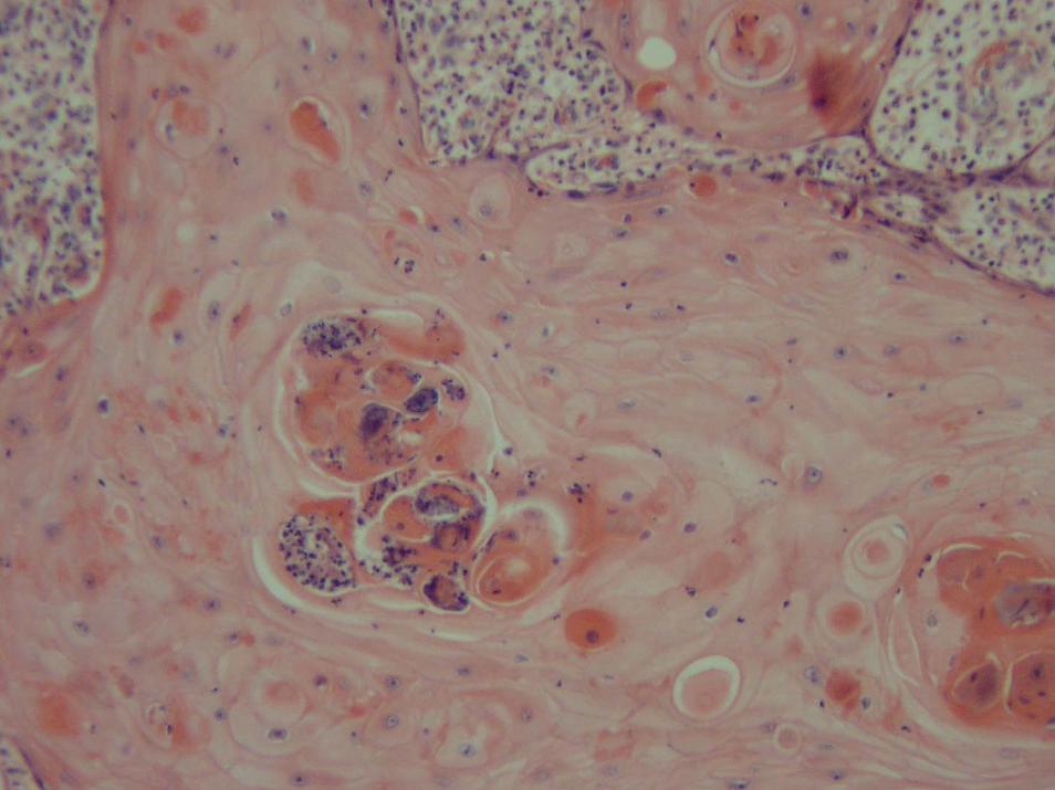
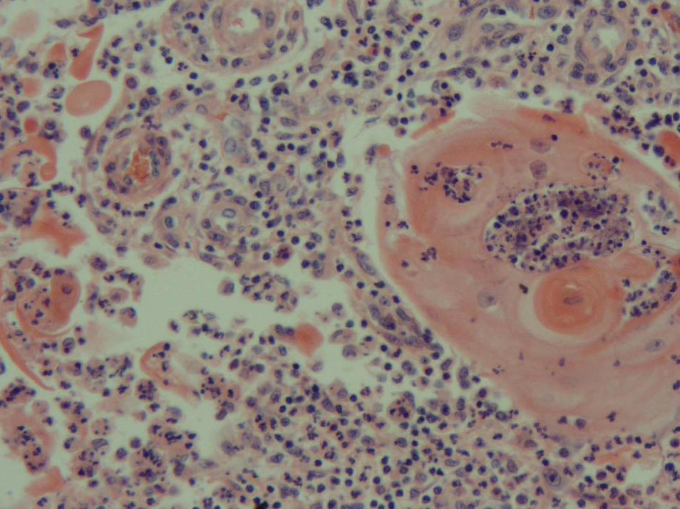
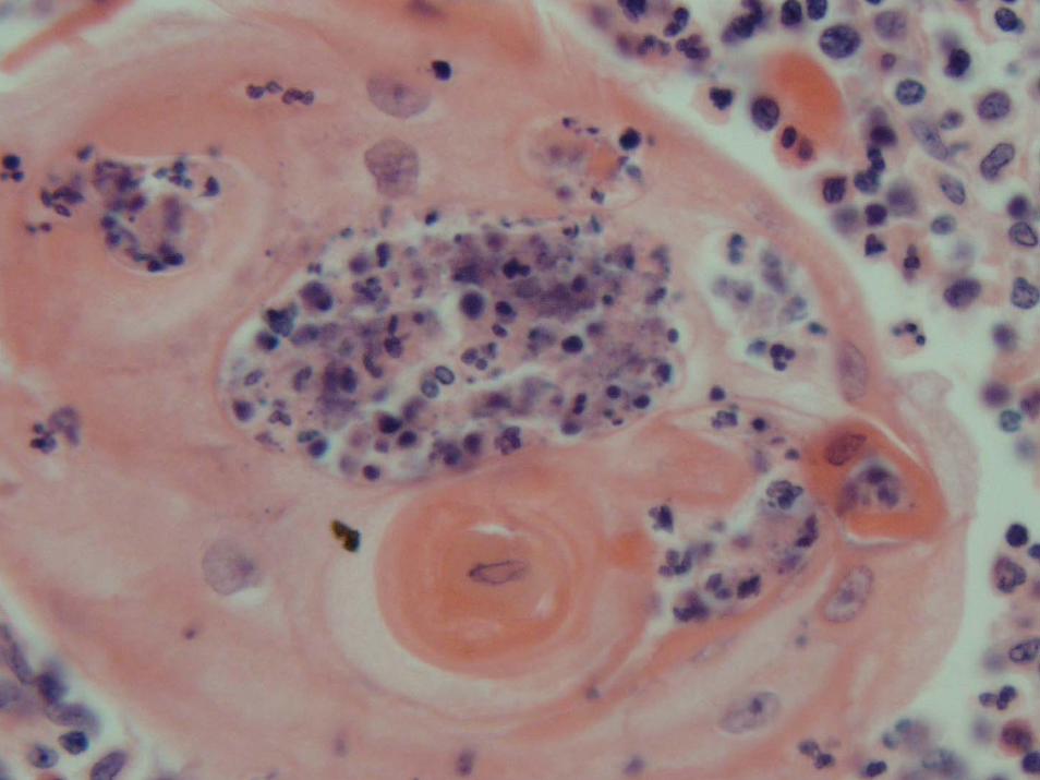
KAKA--HistologyHistology
ExoendophyticExoendophytic proliferation of wellproliferation of well--differentiated differentiated keratinocyteskeratinocytes with a large keratin filled crater. with a large keratin filled crater. Surrounding this keratin crater are buttressing Surrounding this keratin crater are buttressing or or lippinglipping of the epidermal edgesof the epidermal edgesKeratinocytesKeratinocytes have a distinct have a distinct eosinophiliceosinophiliccytoplasmiccytoplasmic appearance with a bland cytology appearance with a bland cytology and rare mitotic figuresand rare mitotic figuresFrequent Frequent eosinophilseosinophils and and neutrophilsneutrophils
KAKA--Differentiation from SCCDifferentiation from SCCICAM (CDICAM (CD--54) 54) ligandligand for the cell adhesion receptor LFAfor the cell adhesion receptor LFA--1, important in 1, important in immune stimulation that is upgraded in inflammatory immune stimulation that is upgraded in inflammatory cutaneouscutaneous disordersdisorders
Increase in expression in the fully developed Increase in expression in the fully developed keratoacanthomakeratoacanthoma and and absent in the evolving and resolved absent in the evolving and resolved keratoacanthomakeratoacanthomaSquamousSquamous cell carcinomas expression focally observed in the wellcell carcinomas expression focally observed in the well--differentiated tumors, dramatic increase in poorly differentiatedifferentiated tumors, dramatic increase in poorly differentiated tumorsd tumors
VCAM (CDVCAM (CD--106) adhesion molecule normally found in stimulated 106) adhesion molecule normally found in stimulated endothelium, plays a critical role in the migration of leukocyteendothelium, plays a critical role in the migration of leukocytess
Expressed in the fully developed Expressed in the fully developed keratoacanthomakeratoacanthoma and was absent in and was absent in the evolving and resolved the evolving and resolved keratoacanthomakeratoacanthomaModerate expression in the wellModerate expression in the well--differentiated differentiated squamoussquamous cell carcinoma, cell carcinoma, and intense expression was seen in the fully developed and intense expression was seen in the fully developed keratoacanthomakeratoacanthoma and poorly differentiated and poorly differentiated squamoussquamous cell carcinoma. cell carcinoma. Temporal related increase in expression of VCAM (CDTemporal related increase in expression of VCAM (CD--106) in 106) in conjunction with the evolution of conjunction with the evolution of keratoacanthomakeratoacanthoma
Increased expression of both markers is seen with Increased expression of both markers is seen with squamoussquamous cell cell carcinoma dedifferentiation. carcinoma dedifferentiation. Mod Mod PatholPathol 2003 Jan;16(1):82003 Jan;16(1):8--1313
KAKA--Differentiation from SCCADifferentiation from SCCA
SyndecanSyndecan--1, 1, heparanheparan sulfate sulfate proteoglycansproteoglycans that mediates intercellular and that mediates intercellular and cell to matrix adhesioncell to matrix adhesion
Expression appears to be inversely correlated with tumor aggressExpression appears to be inversely correlated with tumor aggressiveness and iveness and invasiveness. invasiveness. Previous studies have shown decreased levels of syndecanPrevious studies have shown decreased levels of syndecan--1 expression in 1 expression in invasive invasive cutaneouscutaneous SCC, correlating with tumor deSCC, correlating with tumor de--differentiation. However, a differentiation. However, a similar study has never been done on KA. similar study has never been done on KA.
All 24 All 24 KAsKAs were positive for syndecanwere positive for syndecan--1 expression. Staining intensity of 18 1 expression. Staining intensity of 18 cases was comparable with that of SCC in situ or adjacent normalcases was comparable with that of SCC in situ or adjacent normal epidermisepidermisBy comparison, invasive SCC showed significantly diminished staiBy comparison, invasive SCC showed significantly diminished staining. ning. Reduced staining in focal areas of Reduced staining in focal areas of cytologiccytologic atypiaatypia at the base was present at the base was present in three in three KAsKAs. . SyndecanSyndecan--1 expression in KA mirrors that of SCC in situ and normal 1 expression in KA mirrors that of SCC in situ and normal epidermis, providing a molecular basis that biologically KA may epidermis, providing a molecular basis that biologically KA may be closely be closely related to SCC in situ but distinctively different from invasiverelated to SCC in situ but distinctively different from invasive SCC. SCC. Mod Mod PatholPathol 2002;15:452002;15:45--49 49
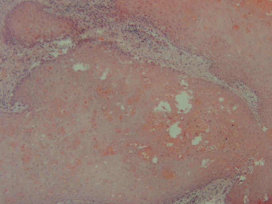
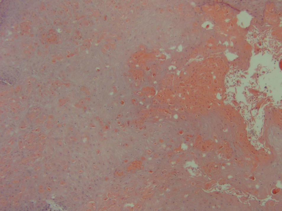
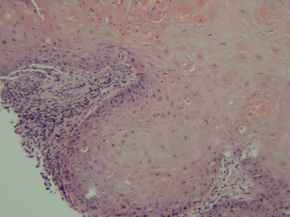
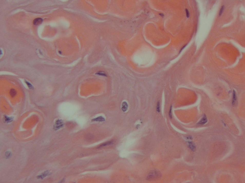
SubungualSubungual KeratoacanthomaKeratoacanthoma
Differs from the common solitary Differs from the common solitary keratoacanthomakeratoacanthoma::Clinical similarity to Clinical similarity to verrucaverruca vulgarisvulgarisMore More dyskeratoticdyskeratotic cells and fewer cells and fewer neutrophilsneutrophils and and eosinophilseosinophilsMore vertical in orientation (longer than it is broad)More vertical in orientation (longer than it is broad)Failure to regress spontaneouslyFailure to regress spontaneouslyLonger courseLonger courseTendency to destroy boneTendency to destroy boneKeratoacanthomaKeratoacanthoma situated in the situated in the subungualsubungual region is region is a more destructive neoplasm than a a more destructive neoplasm than a squamoussquamous cell cell carcinoma there carcinoma there
VerrucousVerrucous CarcinomaCarcinoma
ExoExo or or endophyticendophytic tumors often growing tumors often growing at sites of chronic irritationat sites of chronic irritationClassified based upon locationClassified based upon location
OralOralPlantarPlantarBuschkeBuschke--Lowenstein tumors Lowenstein tumors
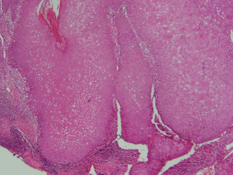
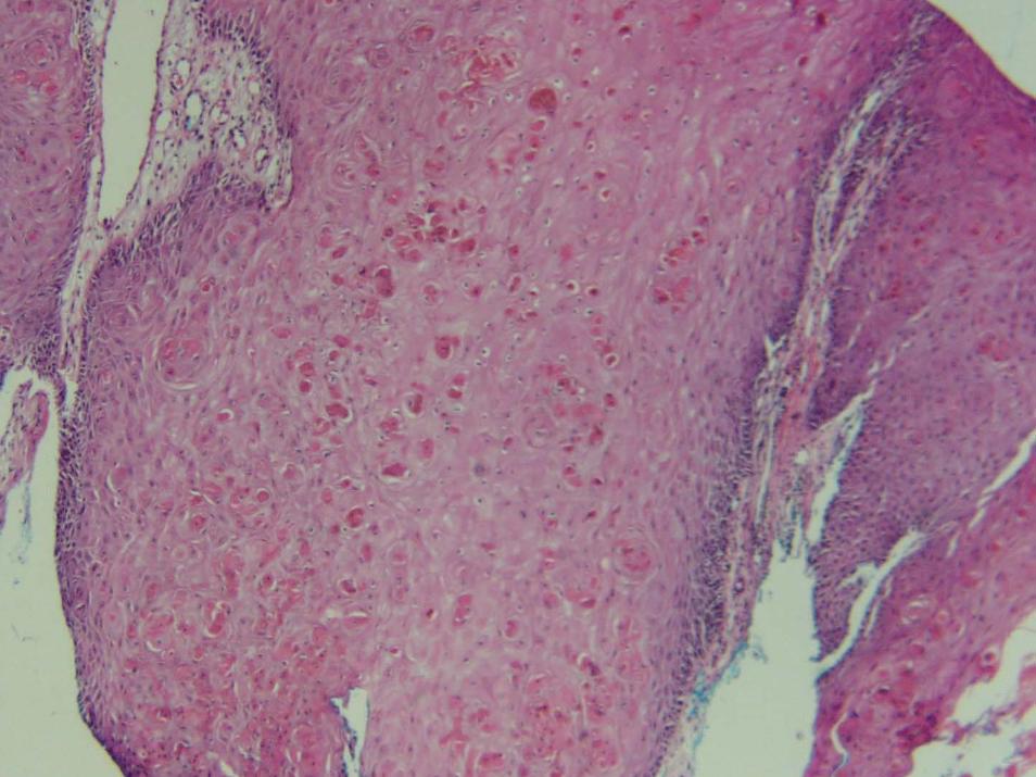
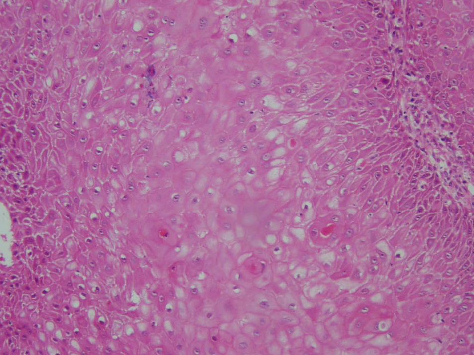
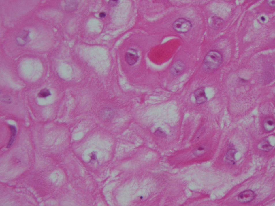
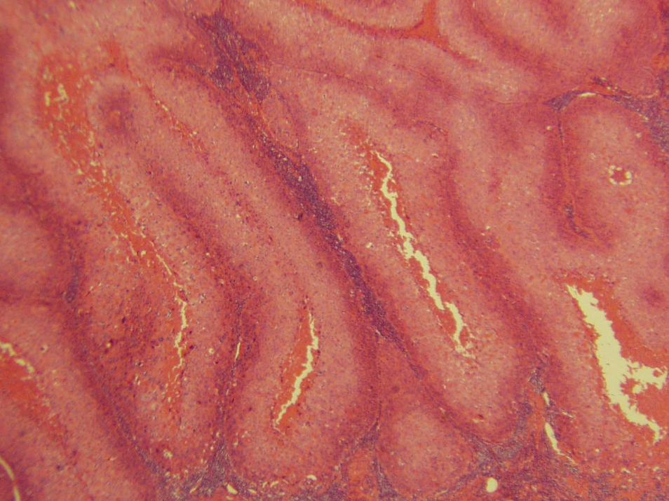
Papillary SCCAPapillary SCCA
ExophyticExophytic verrucousverrucous growthgrowthHigh grade nuclear changesHigh grade nuclear changesProminent papillary growth pattern with several layers of notablProminent papillary growth pattern with several layers of notably y atypical atypical squamoussquamous epithelium overlying a fibroepithelium overlying a fibro--vascular core in both vascular core in both casescases
Mitoses were frequentMitoses were frequentLack deep invasion although focal invasion of the stalk may occuLack deep invasion although focal invasion of the stalk may occurrThese tumors were These tumors were histologicallyhistologically distinct from distinct from verrucousverrucous carcinoma, carcinoma, verrucousverrucous Bowen's disease, and previously described Bowen's disease, and previously described adnexaladnexalcarcinomas. The lack of deep invasion and the absence of local carcinomas. The lack of deep invasion and the absence of local recurrence or recurrence or metastaticmetastatic disease after 18 months followdisease after 18 months follow--up suggest that up suggest that this this histologichistologic variant is a lowvariant is a low--grade malignancy, although study of grade malignancy, although study of more cases and longer followmore cases and longer follow--up will be necessary to accurately assess up will be necessary to accurately assess the biology of this papillary variant of SCC. the biology of this papillary variant of SCC.

Aggressive VariantsAggressive Variants
Marjolin’sMarjolin’s UlcerUlcerAcantholyticAcantholyticSarcomatoidSarcomatoid (Adenoid, (Adenoid, PseudovascularPseudovascular))Invasive Invasive BowenoidBowenoidAdenosquamousAdenosquamous
Marjolin’sMarjolin’s UlcerUlcer
Aggressive form of Aggressive form of squamoussquamous cell cell carcinoma that arises from sites of chronic carcinoma that arises from sites of chronic injury, scars, burns, or irradiation sitesinjury, scars, burns, or irradiation sites
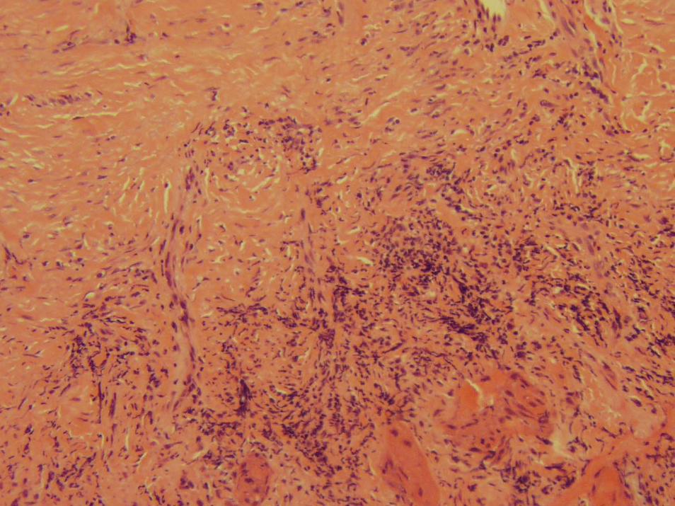
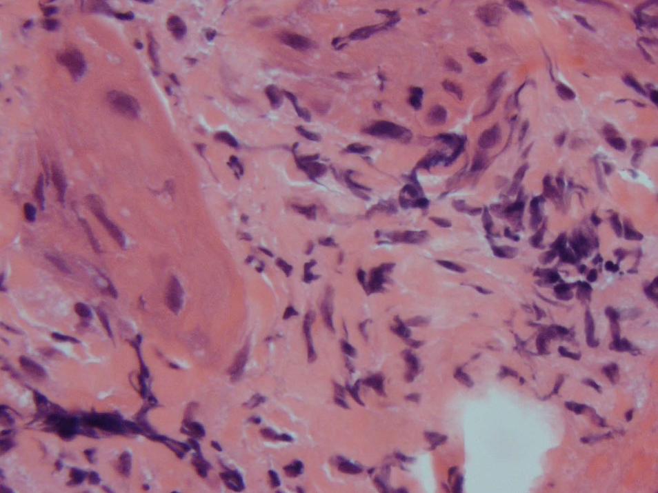
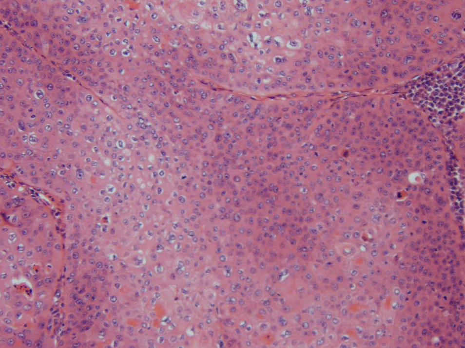
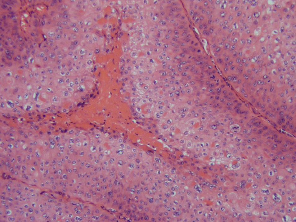
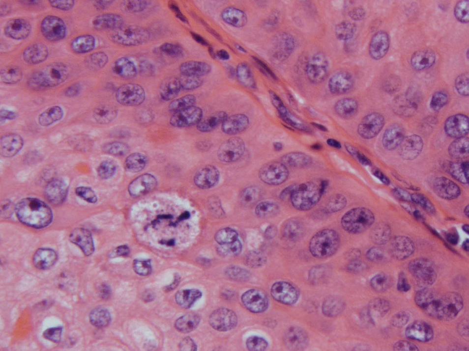
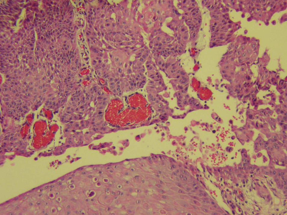
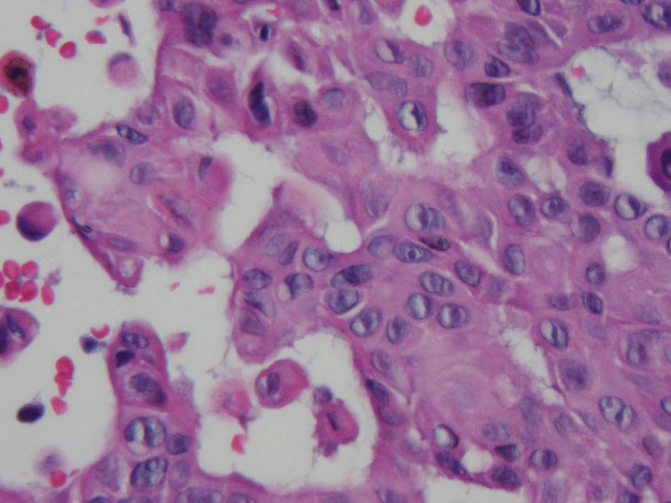
AcantholyticAcantholytic SCCA SCCA ((PseudovascularPseudovascular))
Usually present as ulcer on the head and neck of men in 5Usually present as ulcer on the head and neck of men in 5--6th decade6th decadeHas been associated with recurrences following radiation therapyHas been associated with recurrences following radiation therapyPseudoglandularPseudoglandular acantholyticacantholytic changeschanges
InteranastomosingInteranastomosing cordlike arrays of polygonal or flattened tumor cells, cordlike arrays of polygonal or flattened tumor cells, with internal with internal pseudoluminapseudolumina that contained detached tumor cellsthat contained detached tumor cellsConnection between the dermal neoplasm and the epidermis was Connection between the dermal neoplasm and the epidermis was apparent in three cases, but it was focalapparent in three cases, but it was focalErythrocytes were seen in Erythrocytes were seen in pseudovascularpseudovascular spaces spaces
IPOXIPOXPositive for CK and EMAPositive for CK and EMANegative for FVIII and CD34Negative for FVIII and CD34
Am J Am J SurgSurg PatholPathol 1992 May;16(5):4291992 May;16(5):429--38 38
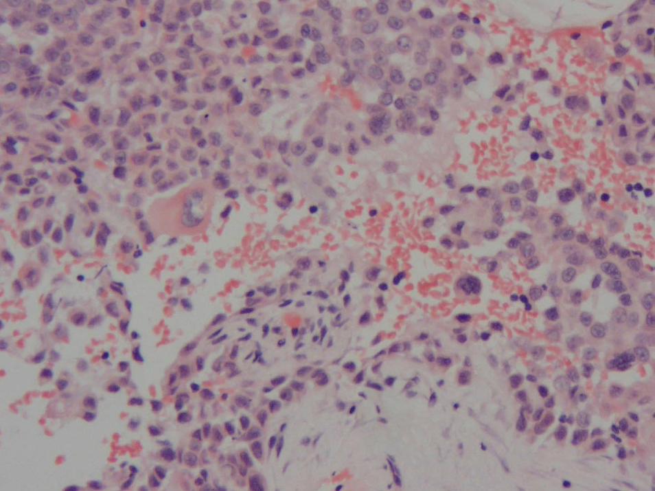
Poorly Differentiated/Poorly Differentiated/SarcomatoidSarcomatoidSCCASCCA
Spindle cellSpindle cellDesmoplasticDesmoplasticCarcinosarcomaCarcinosarcoma
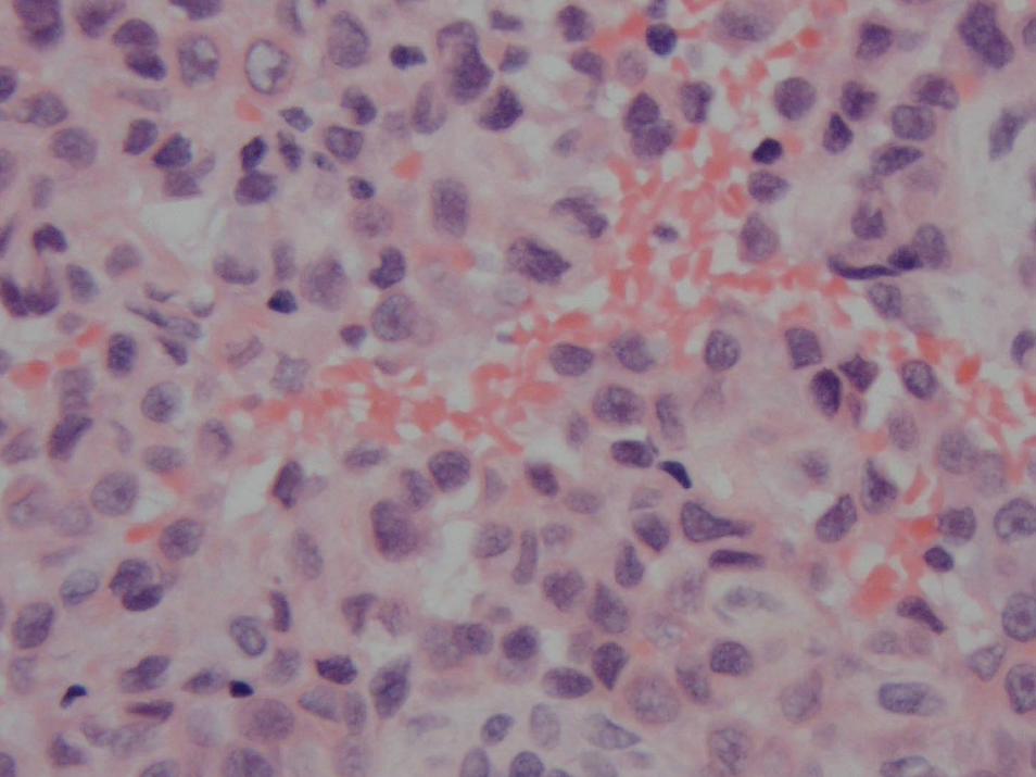
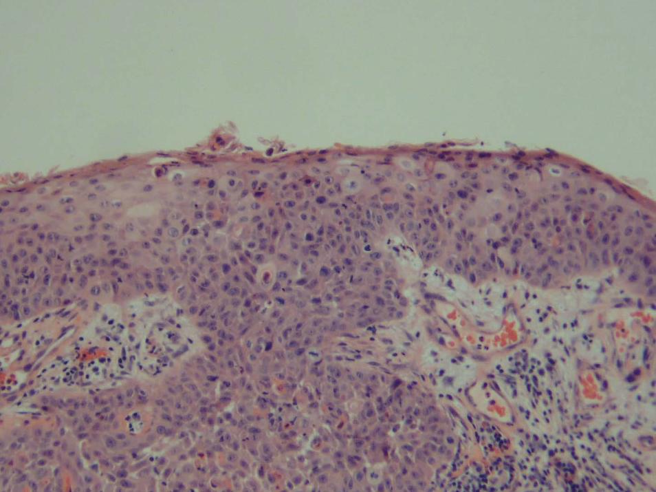
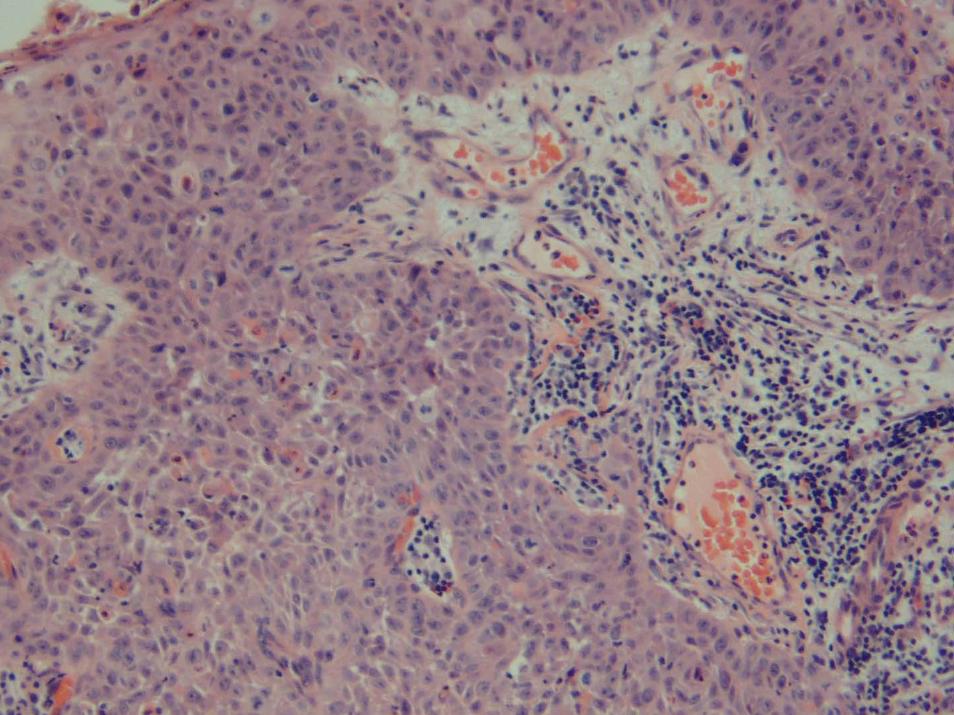
BowenoidBowenoid SCCASCCA
In situ carcinoma with In situ carcinoma with neoplasticneoplastickeratinocyteskeratinocytes invade the dermisinvade the dermisHPV 2 associated in HPV 2 associated in extragenitalextragenital lesionslesionsHPV 16 most common in genital lesionsHPV 16 most common in genital lesions
ClinClin DermatolDermatol 1993;11:431993;11:43--4646
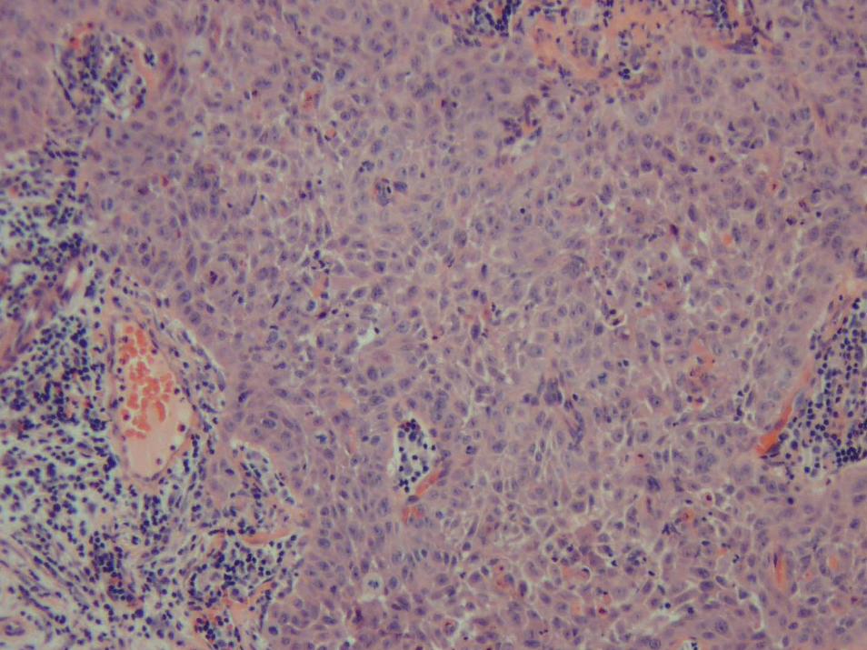
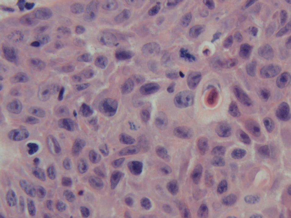
AdenosquamousAdenosquamous CACA
RareRareLess than 15 wellLess than 15 well--documenteddocumentedTerm such as Term such as mucoepidermoidmucoepidermoid carcinomas and carcinomas and acantolyticacantolyticsquamoussquamous cell carcinomas have been usedcell carcinomas have been used
Two componentsTwo componentsConventional Conventional squamoussquamous cell carcinoma merging with cell carcinoma merging with adenocarcinomaadenocarcinoma
PrognosisPrognosisLocal recurrence with later lymph node metastasesLocal recurrence with later lymph node metastasesNo evidence of disease 8 months laterNo evidence of disease 8 months later
Always exclude metastases to skinAlways exclude metastases to skin
Journal of Journal of CutaneousCutaneous Pathology 2001;28 (10), 542Pathology 2001;28 (10), 542--545545

Unusual VariantsUnusual Variants
MucinousMucinousPigmentedPigmented
MucinousMucinous SCCASCCA
MucinousMucinous changechangeDifferentiate from Differentiate from basosquamousbasosquamous CACARule out Rule out metastaticmetastatic adenocarcinomaadenocarcinoma
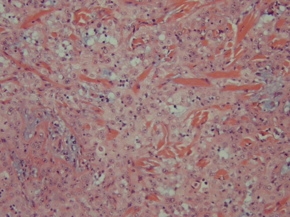
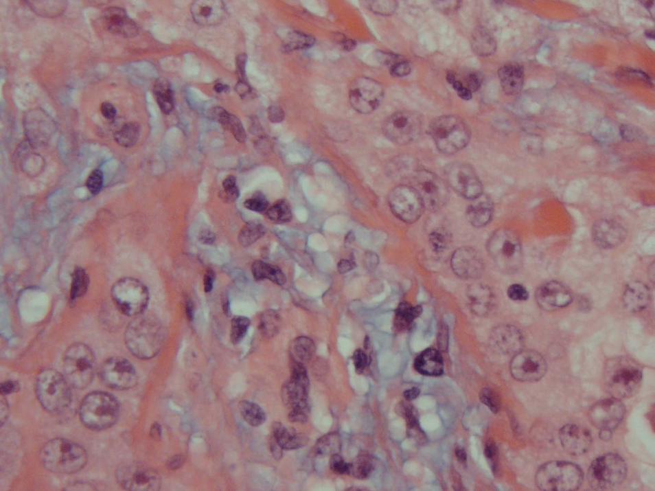
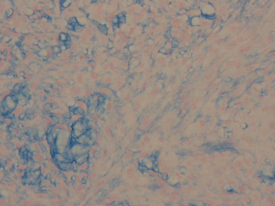
Pigmented SCCAPigmented SCCA
RareRare5/46,791 archived cases5/46,791 archived casesRelative frequency of approximately 0.01%Relative frequency of approximately 0.01%
Rapidly growing crusted papule on actinic damaged skin of the faRapidly growing crusted papule on actinic damaged skin of the faceceMixture of Mixture of keratininizedkeratininized squamoussquamous cells and melanincells and melanin--producing producing dendriticdendriticmelanocytesmelanocytes. . IPOXIPOX
SquamousSquamous cells stained for epithelial membrane antigen, low and high cells stained for epithelial membrane antigen, low and high molecular keratinsmolecular keratinsMelanocytesMelanocytes stained for Sstained for S--100 and HMB100 and HMB--4545Matched series of 31 Matched series of 31 SCCsSCCs failed to show failed to show intratumoralintratumoral melanocytesmelanocytes. .
J J CutanCutan PatholPathol 2000 Sep;27(8):3812000 Sep;27(8):381--6 6
TreatmentTreatment
SurgerySurgeryMOHS MOHS RadiationRadiationSentinel lymph node excisionSentinel lymph node excision
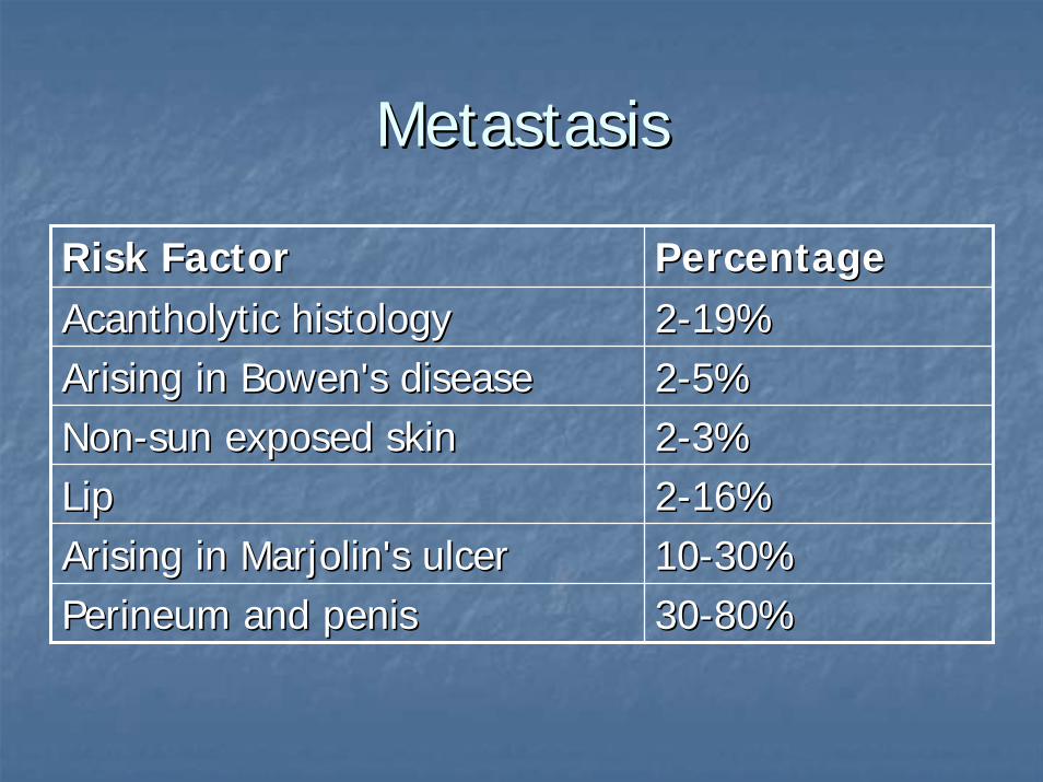
MetastasisMetastasis
Risk FactorRisk Factor PercentagePercentageAcantholyticAcantholytic histologyhistology 22--19%19%Arising in Bowen's diseaseArising in Bowen's disease 22--5%5%NonNon--sun exposed skinsun exposed skin 22--3%3%LipLip 22--16%16%Arising in Arising in Marjolin'sMarjolin's ulcerulcer 1010--30%30%Perineum and penisPerineum and penis 3030--80%80%
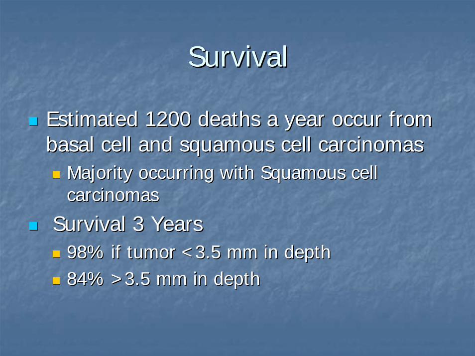
SurvivalSurvival
Estimated 1200 deaths a year occur from Estimated 1200 deaths a year occur from basal cell and basal cell and squamoussquamous cell carcinomascell carcinomas
Majority occurring with Majority occurring with SquamousSquamous cell cell carcinomascarcinomas
Survival 3 YearsSurvival 3 Years98% if tumor <3.5 mm in depth98% if tumor <3.5 mm in depth84% >3.5 mm in depth84% >3.5 mm in depth
ReferencesReferences
J J EurEur AcadAcad DermatolDermatol VenereolVenereol 1998;11:371998;11:37--4444J Am J Am AcadAcad DermatolDermatol 1992;26:9761992;26:976--990990J J DermatolDermatol SurgSurg OncolOncol 1982;8:5891982;8:589--600600J Am J Am AcadAcad DermatolDermatol 1992;26:9761992;26:976--990 990


![ARID1A prevents squamous cell carcinoma initiation and ...SCCs include the skin, head and neck, esophagus, lung, and cervix [2]. Cutaneous squamous cell carcinoma (cSCC) is a nonmelanoma](https://img.dokumen.tips/doc/110x75/6012df67f7a82c062d6f1b92/arid1a-prevents-squamous-cell-carcinoma-initiation-and-sccs-include-the-skin.jpg)