Embed Size (px)
Citation preview
Cut and move: protein machinery for DNA processing inbacterial conjugationF Xavier Gomis-Ruth1 and Miquel Coll1,2
Conjugation is a paradigmatic example of horizontal or lateral
gene transfer, whereby DNA is translocated between bacterial
cells. It provides a route for the rapid acquisition of new genetic
information. Increased antibiotic resistance among pathogens
is a troubling consequence of this microbial capacity. DNA
transfer across cell membranes requires a sophisticated
molecular machinery that involves the participation of several
proteins in DNA processing and replication, cell recruitment,
and the transport of DNA and proteins from donor to recipient
cells. Although bacterial conjugation was first reported in the
1940s, only now are we beginning to unravel the molecular
mechanisms behind this process. In particular, structural
biology is revealing the detailed molecular architecture of
several of the pieces involved.
Addresses1 Institut de Biologia Molecular de Barcelona (CSIC), Parc Cientıfic de
Barcelona, Josep Samitier 1-5, 08028 Barcelona, Spain2 Institut de Recerca Biomedica, Parc Cientıfic de Barcelona, Josep
Samitier 1-5, 08028 Barcelona, Spain
Corresponding author: Coll, Miquel ([email protected])
Current Opinion in Structural Biology 2006, 16:744–752
This review comes from a themed issue on
Proteins
Edited by Martino Bolognesi and Janet L Smith
Available online 31st October 2006
0959-440X/$ – see front matter
# 2006 Elsevier Ltd. All rights reserved.
DOI 10.1016/j.sbi.2006.10.004
IntroductionMechanisms leading to lateral gene transfer in bacteria
are classically categorized as transduction, transformation
or conjugation [1–3]. Transduction occurs via bacterio-
phages, which can incorporate portions of the host bac-
terial DNA and introduce them into newly infected hosts.
Transformation consists of the uptake of naked DNA
from the environment. Finally, conjugation is the uni-
directional transfer of single-stranded (ss) DNA (known
as the T-strand) of conjugative plasmids (or chromosome-
integrated conjugative elements) from a donor to a reci-
pient cell by intimate cell-to-cell contact [3–6]. After
transfer, the recipient becomes a transconjugant, posses-
sing the capacity to start new rounds of conjugation.
Through this highly efficient mechanism, a few conju-
gative-plasmid-harbouring cells within a strain can spread
this information among the whole population within short
timescales, thus enabling rapid dissemination of adaptive
Current Opinion in Structural Biology 2006, 16:744–752
genes and infectious or antibiotic resistance factors.
Studies of Escherichia coli strain K12 plasmid F led to
the discovery of bacterial conjugation in the 1940s; this
plasmid has since become a model for plasmid-encoded
conjugation systems in Gram-negative bacteria [7,8].
Another example is the enterobacterial plasmid R388,
which confers resistance to the antibiotics sulphonamide
and trimethoprim [9]. Conjugative plasmids have also
been found in several Gram-positive bacterial genera,
such as Streptococcus, Enterococcus and Staphylococcus[10–12]. Conjugative-like DNA delivery further occurs
between bacteria and eukaryotic plant and fungi cells. A
well-known example is Agrobacterium tumefaciens, the
etiological agent of crown gall disease, which transfers
the tumour-causing plasmid pTi to plants [13].
Most of the proteins engaged in conjugation are encoded
by plasmid genes located in the tra (transfer) region, which
includes the mpf (mating-pair formation) and dtr (DNA
processing and transport) genes [14,15,16�]. Dtr encodes
proteins responsible for the process in which the T-strand
is prepared for transfer. It includes the formation of the
relaxosome [17], a multicomponent nucleoprotein com-
plex comprising an ATP-dependent relaxase/helicase, the
T-strand, a transcriptional regulator and the host-encoded
integration host factor (IHF), and its recruitment to the
membrane transport pore (see Figure 1). Mpf encodes
proteins that participate in pilus formation and assembly
of a type IV secretion system (T4SS), a multiprotein
organelle required for horizontal transfer through mem-
branes in Gram-negative bacteria (for recent reviews, see
[15,18–22]). Conjugation initiates when the pilus,
anchored on the donor cell surface, binds to the surface
of the recipient cell through its distal end and subsequently
retracts to enable stable intercellular wall-to-wall contact.
An unknown mating signal then triggers mobilisation
of donor DNA, which leads to a site-specific nick in
the plasmid T-strand. The relaxosome is subsequently
coupled to the T4SS by T4CP, a dtr-encoded receptor
or coupling protein ([22,23]; see Figure 1).
In recent years, structural biology has revealed the
detailed molecular architecture of several of the pieces
involved in the intricate scenario of conjugation. Here, we
review the structures of proteins that participate in the
first two stages of DNA transfer, namely processing and
recruitment to the cell membrane.
Relaxase/helicaseA key player in the generation of the transferable
T-strand is the relaxase/helicase, TrwC in the R388
www.sciencedirect.com
Bacterial conjugation DNA processing machinery Gomis-Ruth and Coll 745
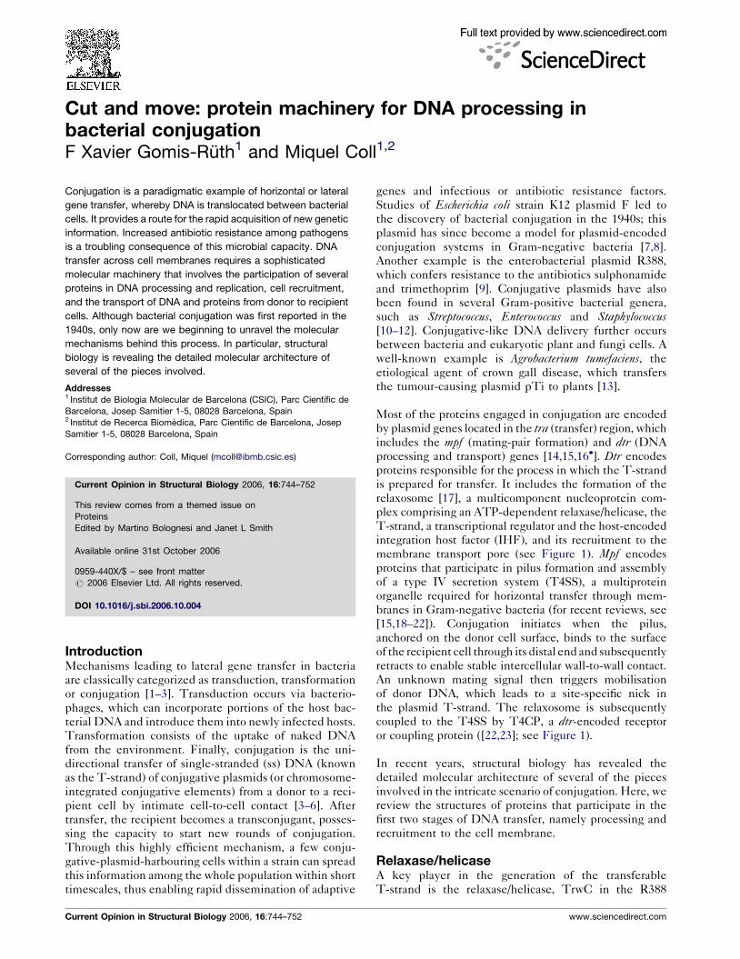
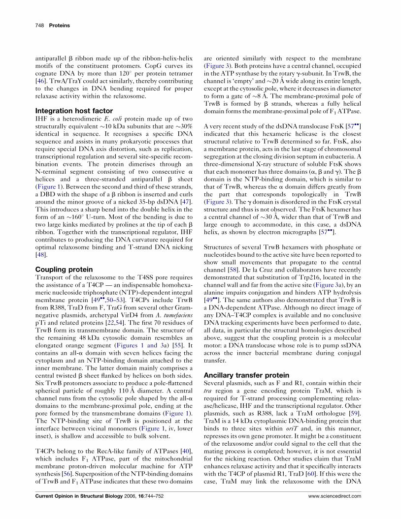
Figure 1
DNA processing and recruitment of the relaxosome to the T4SS in the E. coli plasmid R388 system [59]. Initially, the relaxosome is assembled
from the transcriptional regulator TrwA, host-encoded IHF, relaxase/helicase TrwC and the plasmid dsDNA around oriT (upper centre). After
nicking the T-strand DNA, the 50 end remains attached to TrwC and the nucleoprotein complex is transported to the T4SS pore (proteins TrwD
to TrwN in R388; see [22,54]) to be driven into the recipient cell. This operation is mediated by the T4CP (TrwB in this system), which is inserted
into the cytoplasmic membrane. Richardson plots of the following structures are presented. (i) CopG dimer of dimers (blue/cyan and
orange/yellow; PDB code 1b01) in complex with its cognate 19-bp dsDNA, as a model for TrwA. The dimerisation domain of TrwA bears
distant sequence similarity to a region of Bacillus licheniformis b-lactamase (PDB code 4blm), which was used to tentatively model this domain
(faded part). (ii) E. coli IHF heterodimer (yellow and orange ribbon) in complex with its cognate 35-bp dsDNA (magenta and violet), which
forms a U-turn (PDB code 1ihf). (iii) TrwC relaxase domain (cyan ribbon) in complex with a 25-base DNA hairpin featuring the recognition
sequence around oriT (PDB code 1qx0). (iv) T4CP TrwB hexamer in complex with an ATP analogue, showing each chain in one colour
and the tentatively modelled transmembrane helices (faded part) (PDB code 1gl7). The insets depict the NTP-binding site of TrwB
(lower inset; ATP analogue as white stick model) and the N-terminal domain of plasmid R1 TraM (upper inset; PDB code 1dp3), which is
structurally similar to a region of the all-a domain of TrwB. (v) Tetrameric C-terminal domain of plasmid F TraM (PDB code 2g7o).
plasmid system and TraI in plasmid F. Initially described
as helicase I in E. coli, this endonuclease has been termed
‘relaxase’ because it relaxes supercoiled plasmid double-
stranded (ds) DNA by cleaving one of the strands. It
participates in DNA mobilisation by nicking within the
origin-of-transfer region (oriT) of the T-strand [23]. Sub-
sequently, a catalytic tyrosine remains covalently attached
to the 50 end of the T-strand through a phosphotyrosyl
www.sciencedirect.com
linkage, while the 30 end is released [24,25]. The relaxase/
helicase then unwinds the dsDNA in an ATP-dependent
manner and moves processively in a 50 to 30 direction on the
displaced strand, while the intact strand serves as a tem-
plate for complementary strand biosynthesis by host DNA
polymerase III (donor conjugal DNA synthesis; [26]) using
the rolling circle mechanism [27,28]. Studies of several
conjugation systems indicate that the DNA-loaded relax-
Current Opinion in Structural Biology 2006, 16:744–752
746 Proteins
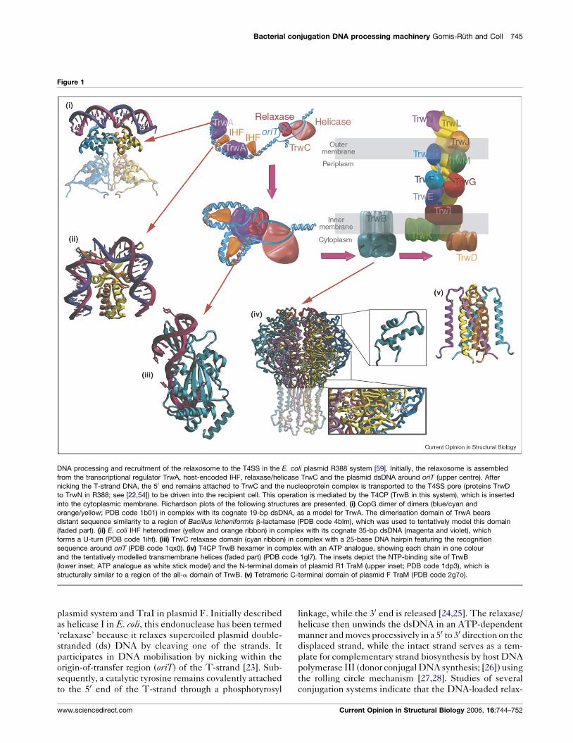
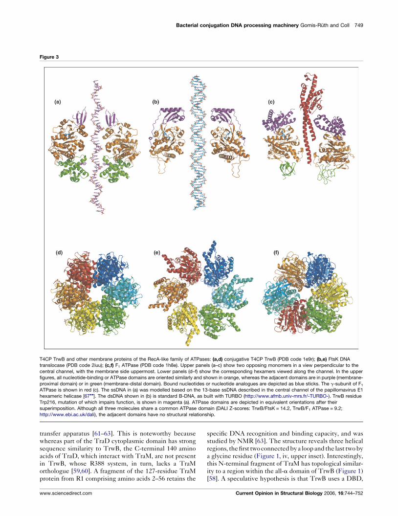
Figure 2
TrwC relaxase domain and related proteins. (a) E. coli R388 TrwC relaxase domain Y18F mutant (white/orange) in complex with a 27-base ssDNA
(green) that mimics the recognised oriT sequence (PDB code 2cdm). The central five-stranded antiparallel b sheet shared with other ORBD
proteins is shown in orange. A water molecule occupies the metal site (red sphere) and is coordinated by three histidine residues (orange sticks)
from two parallel strands. (b) E. coli F TraI in complex with 10-base ssDNA (PDB code 2a0i); colour coding as in (a). The red sphere indicates
a metal ion assigned as Mg2+. (c) Catalytic domain of the adeno-associated virus type 5 Rep protein (PDB code 1m55); ribbon colour coding as in (a).
Current Opinion in Structural Biology 2006, 16:744–752 www.sciencedirect.com

Bacterial conjugation DNA processing machinery Gomis-Ruth and Coll 747
ase/helicase pilots the passage of the T-strand through the
T4SS into the recipient cell, where activity of this key
protein has been detected [29–31].
The two main activities of the relaxase/helicase are
localised in distinct domains; the relaxase (or transester-
ification) and helicase activities are ascribed to the
N-terminal and C-terminal domains, respectively. The
structure of the N-terminal domain of TrwC and TraI
(Figures 1 and 2a,b) shows a two-layer a/b plate or open
sandwich core comprising a central antiparallel five-
stranded b sheet and two long helices on one face of
the sheet [32��,33��]. The active site is located on the
opposite face in an extended and narrow crevice, whose
floor is paved by the b sheet and whose walls are shaped
by two further a helices and loops. The active site pocket
contains the catalytic tyrosine (Tyr18 in TrwC) respon-
sible for T-strand DNA cleavage and a divalent metal ion,
whose identity in vivo remains unclear. The metal ion is
coordinated by two histidines (His161 and His163 in
TrwC) embedded in the short consensus sequence
HXH (single-letter amino acid code; X is any residue)
and a further distal histidine (His150 in TrwC). These
three metal ligands are positioned on two adjacent b
strands of the sheet (Figure 2a,g). TrwC has been co-
crystallised with a 25-base oligonucleotide encompassing
the R388 oriT sequence upstream of the cleavage site
[33��]. The DNA adopts a hairpin structure, mimicking
one arm of the extruded cruciform of plasmid oriT,
followed by a segment in an extended conformation
and a sharp U-turn just before entering the active site
pocket (Figure 1). The protein recognises this oligonu-
cleotide through unique structural features, including
the N-terminal methionine, which is trapped in a hydro-
phobic cage formed by the DNA bases of the U-turn.
More recent studies of TraI and TrwC in complex
with 10- and 27-base oligonucleotides, respectively
(Figure 2a,b), reveal that the scissile phosphate is directly
coordinated to the metal ion [34,35]. Accordingly, the
latter could participate in catalysis, either by polarising
the phosphate and facilitating nucleophilic attack by the
catalytic tyrosine or by stabilising the pentacoordinate
reaction intermediate [36].
The overall fold of the TrwC/TraI relaxase domain
bears a structural resemblance to viral Rep proteins
[36,37], the DNA-binding domain (DBD) of replication
(Figure Legend 2 Continued) The structure depicts a catalytic zinc ion (purp
sequence and an additional glutamate residue (orange sticks). (d) N-termina
curl virus Sardinia; ribbon colour coding as in (a). The putative cation-binding
DBD (PDB code 2tbd); ribbon colour coding as in (a). (f) Bovine papillomavi
fragment (PDB code 1ksy). For clarity, only half of the dimeric protein–opera
DNA strand depicted in yellow. (g) Topological scheme for the central five-s
order may vary between different ORBD proteins because circular permutat
catalytic site (a–d) bind a divalent cation through two histidine residues emb
(strand V in TrwC) and a histidine or glutamate residue from the strand adjace
DNA polymerase I (left) and TrwC (right), showing the domains characteristic
www.sciencedirect.com
initiation protein E1 from papillomavirus [38] and the
origin-of-replication DBD of SV40 large T-antigen [39]
(Figure 2c–f). All these proteins bind to viral or plasmid
DNA near the origin-of-replication (or transfer). They are
grouped into the same topological family in the SCOP
database [40], adopting the origin-of-replication binding
domain (ORBD) fold. They have a central antiparallel
five-stranded b sheet and two diagonally crossing ‘back’
helices in common (Figure 2g). However, the presence of
other structural elements, in particular sequence- and
structure-specific DNA-binding substructures, differs
greatly and their functions vary considerably. Whereas
Rep proteins and TrwC/TraI are transesterification
enzymes and share the HXH motif for metal binding
and the catalytic tyrosine (the third metal-binding residue
may be either a histidine or a glutamate, see Figure 2a–
d,g), the DBDs of E1 and the SV40 T-antigen have only a
DNA-binding function and lack the catalytic residues and
active site pocket (Figure 2e,f). Furthermore, the basic
fold of the TrwC/TraI relaxase domain is more distantly
related to that of DNA polymerases, although in the
former only palm and finger subdomains are recognisable
(Figure 2h). Superimposition of the relaxase domain and
the Klenow fragment of DNA polymerase I [41] shows
that the catalytic residues of relaxase in the palm sub-
domain spatially coincide with those of the polymerase,
despite the differing functions of these proteins.
Transcriptional regulatorA protein named TrwA in plasmid R388 has a dual role: it
enhances the relaxase/helicase activity of TrwC and
represses the operon that jointly encodes TrwA, TrwB
and TrwC within dtr [42]. The equivalent protein in the F
system, TraY, has been shown to likewise enhance the
relaxase/helicase activity of TraI and to regulate the
expression of tra genes [43]. TrwA/TraY displays an
N-terminal DBD, which recognises two palindromic sites
near oriT, and a C-terminal dimerisation domain. In the
absence of experimental structures, sequence analyses
and alignments predict a ribbon-helix-helix motif within
the DBD, as found in the Arc/MetJ/CopG superfamily of
transcriptional repressors [44]. CopG is a small 45-residue
transcriptional repressor that regulates plasmid replica-
tion and recognises cognate dsDNA as a dimer of dimers.
A homology model of the DBD of TrwA/TraY based on
the CopG crystal structure [45] is shown in Figure 1. Each
dimer penetrates the major groove of DNA using an
le sphere) coordinated by two histidine residues from the HXH consensus
l DBD of the replication initiation apoprotein from tomato yellow leaf
residues [same as in (c)] are shown as orange sticks. (e) SV40 T-antigen
rus replication initiation protein E1 in complex with a 21-bp dsDNA
tor complex is shown; ribbon colour coding as in (a), with the second
tranded antiparallel b sheet found in ORBD proteins. Strand numbering
ion has occurred in some cases. Those family members harbouring a
edded in a short HXH consensus sequence in the central strand
nt to the right (strand IV in TrwC). (h) Overall topological similarity between
of polymerases, namely the thumb, the palm and the fingers.
Current Opinion in Structural Biology 2006, 16:744–752
748 Proteins
antiparallel b ribbon made up of the ribbon-helix-helix
motifs of the constituent protomers. CopG curves its
cognate DNA by more than 1208 per protein tetramer
[46]. TrwA/TraY could act similarly, thereby contributing
to the changes in DNA bending required for proper
relaxase activity within the relaxosome.
Integration host factorIHF is a heterodimeric E. coli protein made up of two
structurally equivalent �10 kDa subunits that are �30%
identical in sequence. It recognises a specific DNA
sequence and assists in many prokaryotic processes that
require special DNA axis distortion, such as replication,
transcriptional regulation and several site-specific recom-
bination events. The protein dimerises through an
N-terminal segment consisting of two consecutive a
helices and a three-stranded antiparallel b sheet
(Figure 1). Between the second and third of these strands,
a DBD with the shape of a b ribbon is inserted and curls
around the minor groove of a nicked 35-bp dsDNA [47].
This introduces a sharp bend into the double helix in the
form of an �1608 U-turn. Most of the bending is due to
two large kinks mediated by prolines at the tip of each b
ribbon. Together with the transcriptional regulator, IHF
contributes to producing the DNA curvature required for
optimal relaxosome binding and T-strand DNA nicking
[48].
Coupling proteinTransport of the relaxosome to the T4SS pore requires
the assistance of a T4CP — an indispensable homohexa-
meric nucleoside triphosphate (NTP)-dependent integral
membrane protein [49��,50–53]. T4CPs include TrwB
from R388, TraD from F, TraG from several other Gram-
negative plasmids, archetypal VirD4 from A. tumefacienspTi and related proteins [22,54]. The first 70 residues of
TrwB form its transmembrane domain. The structure of
the remaining 48 kDa cytosolic domain resembles an
elongated orange segment (Figures 1 and 3a) [55]. It
contains an all-a domain with seven helices facing the
cytoplasm and an NTP-binding domain attached to the
inner membrane. The latter domain mainly comprises a
central twisted b sheet flanked by helices on both sides.
Six TrwB protomers associate to produce a pole-flattened
spherical particle of roughly 110 A diameter. A central
channel runs from the cytosolic pole shaped by the all-a
domains to the membrane-proximal pole, ending at the
pore formed by the transmembrane domains (Figure 1).
The NTP-binding site of TrwB is positioned at the
interface between vicinal monomers (Figure 1, iv, lower
inset), is shallow and accessible to bulk solvent.
T4CPs belong to the RecA-like family of ATPases [40],
which includes F1 ATPase, part of the mitochondrial
membrane proton-driven molecular machine for ATP
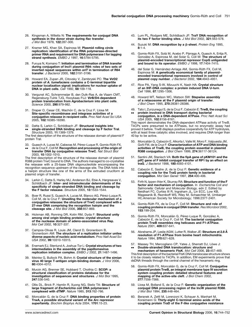
synthesis [56]. Superposition of the NTP-binding domains
of TrwB and F1 ATPase indicates that these two domains
Current Opinion in Structural Biology 2006, 16:744–752
are oriented similarly with respect to the membrane
(Figure 3). Both proteins have a central channel, occupied
in the ATP synthase by the rotary g-subunit. In TrwB, the
channel is ‘empty’ and�20 A wide along its entire length,
except at the cytosolic pole, where it decreases in diameter
to form a gate of �8 A. The membrane-proximal pole of
TrwB is formed by b strands, whereas a fully helical
domain forms the membrane-proximal pole of F1 ATPase.
A very recent study of the dsDNA translocase FtsK [57��]indicated that this hexameric helicase is the closest
structural relative to TrwB determined so far. FtsK, also
a membrane protein, acts in the last stage of chromosomal
segregation at the closing division septum in eubacteria. A
three-dimensional X-ray structure of soluble FtsK shows
that each monomer has three domains (a, b and g). The b
domain is the NTP-binding domain, which is similar to
that of TrwB, whereas the a domain differs greatly from
the part that corresponds topologically in TrwB
(Figure 3). The g domain is disordered in the FtsK crystal
structure and thus is not observed. The FtsK hexamer has
a central channel of �30 A, wider than that of TrwB and
large enough to accommodate, in this case, a dsDNA
helix, as shown by electron micrographs [57��].
Structures of several TrwB hexamers with phosphate or
nucleotides bound to the active site have been reported to
show small movements that propagate to the central
channel [58]. De la Cruz and collaborators have recently
demonstrated that substitution of Trp216, located in the
channel wall and far from the active site (Figure 3a), by an
alanine impairs conjugation and hinders ATP hydrolysis
[49��]. The same authors also demonstrated that TrwB is
a DNA-dependent ATPase. Although no direct image of
any DNA–T4CP complex is available and no conclusive
DNA tracking experiments have been performed to date,
all data, in particular the structural homologies described
above, suggest that the coupling protein is a molecular
motor: a DNA translocase whose role is to pump ssDNA
across the inner bacterial membrane during conjugal
transfer.
Ancillary transfer proteinSeveral plasmids, such as F and R1, contain within their
tra region a gene encoding protein TraM, which is
required for T-strand processing complementing relax-
ase/helicase, IHF and the transcriptional regulator. Other
plasmids, such as R388, lack a TraM orthologue [59].
TraM is a 14 kDa cytoplasmic DNA-binding protein that
binds to three sites within oriT and, in this manner,
represses its own gene promoter. It might be a constituent
of the relaxosome and/or could signal to the cell that the
mating process is completed; however, it is not essential
for the nicking reaction. Other studies claim that TraM
enhances relaxase activity and that it specifically interacts
with the T4CP of plasmid R1, TraD [60]. If this were the
case, TraM may link the relaxosome with the DNA
www.sciencedirect.com
Bacterial conjugation DNA processing machinery Gomis-Ruth and Coll 749
Figure 3
T4CP TrwB and other membrane proteins of the RecA-like family of ATPases: (a,d) conjugative T4CP TrwB (PDB code 1e9r); (b,e) FtsK DNA
translocase (PDB code 2iuu); (c,f) F1 ATPase (PDB code 1h8e). Upper panels (a–c) show two opposing monomers in a view perpendicular to the
central channel, with the membrane side uppermost. Lower panels (d–f) show the corresponding hexamers viewed along the channel. In the upper
figures, all nucleotide-binding or ATPase domains are oriented similarly and shown in orange, whereas the adjacent domains are in purple (membrane-
proximal domain) or in green (membrane-distal domain). Bound nucleotides or nucleotide analogues are depicted as blue sticks. The g-subunit of F1
ATPase is shown in red (c). The ssDNA in (a) was modelled based on the 13-base ssDNA described in the central channel of the papillomavirus E1
hexameric helicase [67��]. The dsDNA shown in (b) is standard B-DNA, as built with TURBO (http://www.afmb.univ-mrs.fr/-TURBO-). TrwB residue
Trp216, mutation of which impairs function, is shown in magenta (a). ATPase domains are depicted in equivalent orientations after their
superimposition. Although all three molecules share a common ATPase domain (DALI Z-scores: TrwB/FtsK = 14.2, TrwB/F1 ATPase = 9.2;
http://www.ebi.ac.uk/dali), the adjacent domains have no structural relationship.
transfer apparatus [61–63]. This is noteworthy because
whereas part of the TraD cytoplasmic domain has strong
sequence similarity to TrwB, the C-terminal 140 amino
acids of TraD, which interact with TraM, are not present
in TrwB, whose R388 system, in turn, lacks a TraM
orthologue [59,60]. A fragment of the 127-residue TraM
protein from R1 comprising amino acids 2–56 retains the
www.sciencedirect.com
specific DNA recognition and binding capacity, and was
studied by NMR [63]. The structure reveals three helical
regions, the first two connected by a loop and the last two by
a glycine residue (Figure 1, iv, upper inset). Interestingly,
this N-terminal fragment of TraM has topological similar-
ity to a region within the all-a domain of TrwB (Figure 1)
[58]. A speculative hypothesis is that TrwB uses a DBD,
Current Opinion in Structural Biology 2006, 16:744–752

750 Proteins
similar to that of TraM, embedded in its structure to
contact the relaxosome. More recently, structural analysis
of a C-terminal fragment encompassing the TraD-binding
segment (residues 58–127) of TraM from plasmid F
revealed that four protomers interact to form a compact
eight-helix bundle (Figure 1, v). The N-terminal helices of
each protomer interact to form a central, parallel four-
stranded coiled coil, whereas each C-terminal helix packs
in an antiparallel arrangement around the periphery of the
structure [64��]. Oligomerisation produces a central shaft
surrounded by the inner N-terminal helices, where four
protonated glutamate residues provided by each of the
protomers create a central solvent-mediated ring. Depro-
tonation of this acidic residue relaxes the TraM structure
and this affects interactions with TraD [64��].
ConclusionsBacterial conjugation, an early-discovered pathway for
lateral gene transfer and the main process responsible
for the spread of antibiotic resistance, has been a ‘black
box’. Structural biology is now making a dramatic contri-
bution to unveiling the molecular machinery underlying
this complicated protein–DNA transfer mechanism. Struc-
tural analyses of the components of the T4SS transport
apparatus are currently underway (reviewed in [65�]). Also,
the structures of the main players in DNA processing and
membrane recruitment are being solved. Many questions
remain to be answered and the results of the structural
analyses will lead to new ones. For example, what is the
structure of the helicase domain of the relaxase and how
does it move processively along DNA? If the coupling
protein is the DNA translocase, what is the role of the other
T4SS ATPases also associated with the inner side of the
membrane [66]? Some of them may be involved in protein
transport (of the piloting relaxase?), but how is this com-
patible with the T-strand DNA being threaded through the
hexameric coupling protein, which traverses the inner
membrane? What are the precise mutual interactions of
the components within the relaxosome and of those with
the coupling protein? In addition to ingenious functional
studies, the answer to these questions will require further
structural efforts, including the determination of the struc-
tures of more protein–DNA and protein–protein–DNA
complexes, and ultimately the relaxosome itself.
AcknowledgementsThis work was supported by the Ministerio de Educacion y Ciencia of Spain(grants GEN2003-20642, BIO2003-00132 and BFU2005-06758/BMC) andthe Generalitat de Catalunya (grant 2005SGR-00280 and the Centre deReferencia en Biotecnologia). AG Blanco is acknowledged for assistancewith Figure 1 and Jan Lowe for providing the FtsK coordinates.
References and recommended readingPapers of particular interest, published within the annual period ofreview, have been highlighted as:
� of special interest�� of outstanding interest
1. Ochman H, Lawrence JG, Groisman EA: Lateral gene transfer andthe nature of bacterial innovation. Nature 2000, 405:299-304.
Current Opinion in Structural Biology 2006, 16:744–752
2. Boucher Y, Douady CJ, Papke RT, Walsh DA, Boudreau ME,Nesbo CL, Case RJ, Doolittle WF: Lateral gene transfer and theorigins of prokaryotic groups. Annu Rev Genet 2003, 37:283-328.
3. Chen I, Christie PJ, Dubnau D: The ins and outs of DNA transferin bacteria. Science 2005, 310:1456-1460.
4. Durrenberger MB, Villiger W, Bachi T: Conjugational junctions:morphology of specific contacts in conjugating Escherichiacoli bacteria. J Struct Biol 1991, 107:146-156.
5. Lederberg J: Infectious history. Science 2000, 288:287-293.
6. Lanka E, Wilkins BM: DNA processing reactions in bacterialconjugation. Annu Rev Biochem 1995, 64:141-169.
7. Tatum EL, Lederberg J: Gene recombination in Escherichia coli.Nature 1946, 158:558-658.
8. Hayes W: The Genetics of Bacteria and Their Viruses. New York:John Wiley & Sons; 1964.
9. Datta N, Hedges RW: Trimethoprim resistance conferredby W plasmids in Enterobacteriaceae. J Gen Microbiol 1972,72:349-355.
10. Scott JR, Churchward GG: Conjugative transposition.Annu Rev Microbiol 1995, 49:367-397.
11. Ton-That H, Schneewind O: Assembly of pili on the surfaceof Corynebacterium diphtheriae. Mol Microbiol 2003,50:1429-1438.
12. Grohmann E, Muth G, Espinosa M: Conjugative plasmid transferin Gram-positive bacteria. Microbiol Mol Biol Rev 2003,67:277-301.
13. Christie PJ: Type IV secretion: the Agrobacterium VirB/D4 andrelated conjugation systems. Biochim Biophys Acta 2004,1694:219-234.
14. Silverman PM: Towards a structural biology of bacterialconjugation. Mol Microbiol 1997, 23:423-429.
15. Christie PJ, Atmakuri K, Krishnamoorthy V, Jakubowski S,Cascales E: Biogenesis, architecture, and function of bacterialtype IV secretion systems. Annu Rev Microbiol 2005,59:451-485.
16.�
Backert S, Meyer TF: Type IV secretion systems and theireffectors in bacterial pathogenesis. Curr Opin Microbiol 2006,9:207-217.
A very recent review of T4SS.
17. Ziegelin G, Furste JP, Lanka E: TraJ protein of plasmid RP4binds to a 19-base pair invert sequence repetition within thetransfer origin. J Biol Chem 1989, 264:11989-11994.
18. Christie PJ, Vogel JP: Bacterial type IV secretion: conjugationsystems adapted to deliver effector molecules to host cells.Trends Microbiol 2000, 8:354-360.
19. Christie PJ: Type IV secretion: intercellular transfer ofmacromolecules by systems ancestrally related toconjugation machines. Mol Microbiol 2001, 40:294-305.
20. Lawley TD, Klimke WA, Gubbins MJ, Frost LS: F factorconjugation is a true type IV secretion system. FEMS MicrobiolLett 2003, 224:1-15.
21. Ding Z, Atmakuri K, Christie PJ: The outs and ins of bacterialtype IV secretion substrates. Trends Microbiol 2003, 11:527-535.
22. Gomis-Ruth FX, Sola M, de la Cruz F, Coll M: Coupling factors inmacromolecular type-IV secretion machineries. Curr PharmDes 2004, 10:1551-1565.
23. Fekete RA, Frost LS: Mobilization of chimeric oriT plasmids by Fand R100-1: role of relaxosome formation in defining plasmidspecificity. J Bacteriol 2000, 182:4022-4028.
24. Wilkins B, Lanka E: DNA processing and replication duringplasmid transfer between gram-negative bacteria. In BacterialConjugation. Edited by Clewell DB. Plenum Press; 1993:105-135.
25. Llosa M, Gomis-Ruth FX, Coll M, de la Cruz F: Bacterialconjugation: a two-step mechanism for DNA transport.Mol Microbiol 2002, 45:1-8.
www.sciencedirect.com
Bacterial conjugation DNA processing machinery Gomis-Ruth and Coll 751
26. Kingsman A, Willetts N: The requirements for conjugal DNAsynthesis in the donor strain during flac transfer.J Mol Biol 1978, 122:287-300.
27. Kramer MG, Khan SA, Espinosa M: Plasmid rolling circlereplication: identification of the RNA polymerase-directedprimer RNA and requirement for DNA polymerase I for laggingstrand synthesis. EMBO J 1997, 16:5784-5795.
28. Furuya N, Komano T: Initiation and termination of DNA transferduring conjugation of IncI1 plasmid R64: roles of two sets ofinverted repeat sequences within oriT in termination of R64transfer. J Bacteriol 2000, 182:3191-3196.
29. Howard EA, Zupan JR, Citovsky V, Zambryski PC: The VirD2protein of A. tumefaciens contains a C-terminal bipartitenuclear localization signal: implications for nuclear uptake ofDNA in plant cells. Cell 1992, 68:109-118.
30. Vergunst AC, Schrammeijer B, den Dulk-Ras A, de Vlaam CMT,Regensburg-Tuınk TJG, Hooykaas PJJ: VirB/D4-dependentprotein translocation from Agrobacterium into plant cells.Science 2000, 290:979-982.
31. Draper O, Cesar CE, Machon C, de la Cruz F, Llosa M:Site-specific recombinase and integrase activities of aconjugative relaxase in recipient cells. Proc Natl Acad Sci USA2005, 102:16385-16390.
32.��
Datta S, Larkin C, Schildbach JF: Structural insights intosingle-stranded DNA binding and cleavage by F factor Tral.Structure 2003, 11:1369-1379.
The first description of the structure of the relaxase domain of plasmid Fprotein TraI.
33.��
Guasch A, Lucas M, Cabezas M, Perez-Luque R, Gomis-Ruth FX,de la Cruz F, Coll M: Recognition and processing of the origin oftransfer DNA by conjugative relaxase TrwC. Nat Struct Biol2003, 10:1002-1010.
The first description of the structure of the relaxase domain of plasmidR388 protein TrwC bound to DNA. The authors managed to co-crystallisethe relaxase with a 25-base DNA oligonucleotide encompassing therecognition sequence upstream of the cleavage site. The DNA folds intoa hairpin structure like one of the arms of the extruded cruciform atplasmid origin of transfer.
34. Larkin C, Datta S, Harley MJ, Anderson BJ, Ebie A, Hargreaves V,Schildbach JF: Inter- and intramolecular determinants of thespecificity of single-stranded DNA binding and cleavage bythe F factor relaxase. Structure 2005, 13:1533-1544.
35. Boer R, Russi S, Guasch A, Lucas M, Blanco AG, Perez-Luque R,Coll M, de la Cruz F: Unveiling the molecular mechanism of aconjugative relaxase: the structure of TrwC complexed with a27-mer DNA comprising the recognition hairpin and thecleavage site. J Mol Biol 2006, 358:857-869.
36. Hickman AB, Ronning DR, Kotin RM, Dyda F: Structural unityamong viral origin binding proteins: crystal structureof the nuclease domain of adeno-associated virus Rep.Mol Cell 2002, 10:327-337.
37. Campos-Olivas R, Louis JM, Clerot D, Gronenborn B,Gronenborn AM: The structure of a replication initiator unitesdiverse aspects of nucleic acid metabolism. Proc Natl Acad SciUSA 2002, 99:10310-10315.
38. Enemark EJ, Stenlund A, Joshua-Tor L: Crystal structures of twointermediates in the assembly of the papillomavirusreplication initiation complex. EMBO J 2002, 21:1487-1496.
39. Meinke G, Bullock PA, Bohm A: Crystal structure of the simianvirus 40 large T-antigen origin-binding domain. J Virol 2006,80:4304-4312.
40. Murzin AG, Brenner SE, Hubbard T, Chothia C: SCOP: astructural classification of proteins database for theinvestigation of sequences and structures. J Mol Biol 1995,247:536-540.
41. Ollis DL, Brick P, Hamlin R, Xuong NG, Steitz TA: Structure oflarge fragment of Escherichia coli DNA polymerase Icomplexed with dTMP. Nature 1985, 313:762-766.
42. Moncalian G, de la Cruz F: DNA binding properties of proteinTrwA, a possible structural variant of the Arc repressorsuperfamily. Biochim Biophys Acta 2004, 1701:15-23.
www.sciencedirect.com
43. Lum PL, Rodgers ME, Schildbach JF: TraY DNA recognition ofits two F factor binding sites. J Mol Biol 2002, 321:563-578.
44. Suzuki M: DNA recognition by a b-sheet. Protein Eng 1995,8:1-4.
45. Gomis-Ruth FX, Sola M, Acebo P, Parraga A, Guasch A, Eritja R,Gonzalez A, Espinosa M, del Solar G, Coll M: The structure ofplasmid-encoded transcriptional repressor CopG unligandedand bound to its operator. EMBO J 1998, 17:7404-7415.
46. del Solar G, Hernandez-Arriaga AM, Gomis-Ruth FX, Coll M,Espinosa M: A genetically economical family of plasmid-encoded transcriptional repressors involved in control ofplasmid copy number. J Bacteriol 2002, 184:4943-4951.
47. Rice PA, Yang S-W, Mizuuchi K, Nash HA: Crystal structureof an IHF-DNA complex: a protein induced DNA U-turn.Cell 1996, 87:1295-1306.
48. Howard MT, Nelson WC, Matson SW: Stepwise assemblyof a relaxosome at the F plasmid origin of transfer.J Biol Chem 1995, 270:28381-28386.
49.��
Tato I, Zunzunegui S, de la Cruz F, Cabezon E: TrwB, the couplingprotein involved in DNA transport during bacterialconjugation, is a DNA-dependent ATPase. Proc Natl Acad SciUSA 2005, 102:8156-8161.
This report demonstrates the DNA-dependent ATPase activity of TrwB.T4CPs were presumed to be ATPases, but no enzymatic assay hadproved it before. TrwB displays positive cooperativity for ATP hydrolysis,with at least three catalytic sites involved, and requires DNA longer than40 bp to be active.
50. Moncalian G, Cabezon E, Alkorta I, Valle M, Moro F, Valpuesta JM,Goni FM, de la Cruz F: Characterization of ATP and DNA bindingactivities of TrwB, the coupling protein essential in plasmidR388 conjugation. J Biol Chem 1999, 274:36117-36124.
51. Santini JM, Stanisch VA: Both the fipA gene of pKM101 and thepifC gene of F inhibit conjugal transfer of RP1 by an effect ontraG. J Bacteriol 1998, 180:4093-4101.
52. Cabezon E, Sastre JI, de la Cruz F: Genetic evidence of acoupling role for the TraG protein family in bacterialconjugation. Mol Gen Genet 1997, 254:400-406.
53. Firth N, Ippen-Ihler K, Skurray RA: Structure and function of the Ffactor and mechanism of conjugation. In Escherichia Coli andSalmonella: Cellular and Molecular Biology, edn 2. Edited byNeidhart FC, Curtiss III R, Ingraham JL, Lin ECC, Low KB,Magasanik B, Reznikoff WS, Riley M, Schaechter M, UmbargerHC.American Society for Microbiology; 1996:2377-2401.
54. Gomis-Ruth FX, de la Cruz F, Coll M: Structure and role ofcoupling proteins in conjugal DNA transfer. Res Microbiol 2002,153:199-204.
55. Gomis-Ruth FX, Moncalian G, Perez-Luque R, Gonzalez A,Cabezon E, de la Cruz F, Coll M: The bacterial conjugationprotein TrwB resembles ring helicases and F1-ATPase.Nature 2001, 409:637-641.
56. Abrahams JP, Leslie AGW, Lutter R, Walker JE: Structure at 2.8 Aresolution of F1-ATPase from bovine heart mitochondria.Nature 1994, 370:621-628.
57.��
Massey TH, Mercogliano CP, Yates J, Sherratt DJ, Lowe J:Double-stranded DNA translocation: structure andmechanism of hexameric FtsK. Mol Cell 2006, 23:457-469.
This first description of the bacterial FtsK DNA translocase structure showsit to be closely related to T4CPs. In addition, EM experiments prove thatdsDNA threads through the central channel of the hexameric ring.
58. Gomis-Ruth FX, Moncalian G, de la Cruz F, Coll M: Conjugativeplasmid protein TrwB, an integral membrane type IV secretionsystem coupling protein: detailed structural features andmapping of the active-site cleft. J Biol Chem 2002,277:7556-7566.
59. Llosa M, Bolland S, de la Cruz F: Genetic organization of theconjugal DNA processing region of the IncW plasmid R388.J Mol Biol 1994, 235:448-464.
60. Beranek A, Zettl M, Lorenzoni K, Schauer A, Manhart M,Koraimann G: Thirty-eight C-terminal amino acids of thecoupling protein TraD of the F-like conjugative resistance
Current Opinion in Structural Biology 2006, 16:744–752
752 Proteins
plasmid R1 are required and sufficient to confer bindingto the substrate selector protein TraM. J Bacteriol 2004,186:6999-7006.
61. Disque-Kochem C, Dreiseikelmann B: The cytoplasmicDNA-binding protein TraM binds to the inner membraneprotein TraD in vitro. J Bacteriol 1997, 179:6133-6137.
62. Kupelwieser G, Schwab M, Hogenauer G, Koraimann G,Zechner EL: Transfer protein TraM stimulates TraI-catalyzedcleavage of the transfer origin of plasmid R1 in vivo. J Mol Biol1998, 275:81-94.
63. Stockner T, Plugariu C, Koraimann G, Hogenauer G, Bermel W,Prytulla S, Sterk H: Solution structure of the DNA-bindingdomain of TraM. Biochemistry 2001, 40:3370-3377.
64.��
Lu J, Edwards RA, Wong JJ, Manchak J, Scott PG, Frost LS,Glover JN: Protonation-mediated structural flexibility inthe F conjugation regulatory protein, TraM. EMBO J 2006,25:2930-2939.
Current Opinion in Structural Biology 2006, 16:744–752
The first structure of the C-terminal domain of the conjugative ancillaryprotein TraM is presented. Four protonated glutamic acid residuesstabilise the helical bundle tetrameric quaternary structure, leading theauthors to propose that deprotonation of TraM is a mechanism for thedownregulation of conjugation.
65.�
Remaut H, Waksman G: Structural biology of bacterialpathogenesis. Curr Opin Struct Biol 2004, 14:161-170.
A comprehensive structural review of T4SS.
66. Savvides SN, Yeo HJ, Beck MR, Blaesing F, Lurz R, Lanka E,Buhrdorf R, Fischer W, Haas R, Waksman G: VirB11 ATPases aredynamic hexameric assemblies: new insights into bacterialtype IV secretion. EMBO J 2003, 22:1969-1980.
67.��
Enemark EJ, Joshua-Tor L: Mechanism of DNA translocation ina replicative hexameric helicase. Nature 2006, 442:270-2755.
The first structure of a ring helicase, showing a ssDNA segment in thecentral channel. The ssDNA adopts a helical conformation with most ofthe bases stacked.
www.sciencedirect.com