Embed Size (px)
Citation preview
Current Pharmaceutical Design, 2004, 10, 3873-3884 3873
1381-6128/04 $45.00+.00 © 2004 Bentham Science Publishers Ltd.
Structural Features of the Interleukin-10 Family of Cytokines
Alexander Zdanov*
Macromolecular Crystallography Laboratory, Center for Cancer Research, National Cancer Institute at Frederick,Frederick, MD 21702-1201, USA
Abstract: The interleukin-10 (IL-10) family of cytokines includes IL-10, a number of its viral gene homologs, and eightrecently discovered cellular cytokines (IL-19, IL-20, IL-22, IL-24, IL-26, IFN-λ1, IFN-λ2, IFN-λ3). IL-10 is anintercalated dimer consisting of two six-helix bundle domains. Signal transduction occurs when each domain of IL-10binds to two receptor chains, IL-10R1 and IL-10R2. Viral homologs use the same IL-10 receptor system, while cellularhomologs use their own receptors: three long receptor chains (IL-20R1, IL-22R1 and IFN-λ1R1) and two short receptorchains (IL-20R2 and IL-10R2). Most of the cellular homologs belong to the IL-19 subfamily of cytokines including IL-19,IL-20, IL-22 and IL-24. It is likely that IFN-λ1, IFN-λ2, and IFN-λ3 also belong to the same subfamily. All these proteinsare monomers in solution. Crystal structures of IL-19 and IL-22 show that the molecules consist of seven helices (A-G)forming a seven-helix bundle with compact hydrophobic core inside. Structures of complexes of IL-10 and CMVIL-10with an extracellular domain of high affinity receptor IL-10R1 (sIL-10R1) showed that ligand/receptor interactions are ofmostly polar nature, with two hydrophobic patches around receptor residues Tyr43 and Phe143 at the top and bottom ofthe interface. The location and structure of the binding site for the second receptor chain are still unknown. It has alsobeen shown that in the case of IL-19 and IL-20, IL-20R2 rather than IL-20R1 is a high-affinity receptor chain. Thisreview summarizes all published three-dimensional structures of the cytokines representing the IL-10 family of homologs,including the IL-19 subfamily and their interaction with appropriate receptors.
Key Words: Cytokines, interleukin-10, ligand/receptor interactions, helix bundle, signal transduction.
INTRODUCTION
Interleukin-10 (IL-10) (reviewed in ref. [1]) is afascinating cytokine first identified by its ability to stopimmune response by inhibiting production of a number ofcytokines. Due to its suppressor and inhibitor ability, IL-10was first called a cytokine synthesis inhibitory factor [2, 3].IL-10 also plays a role in proliferation and differentiation ofB cells, T cells and mast cells. Based on its immunomodu-lating functions, IL-10 has been considered an attractivecandidate for therapeutic applications for treatment of acuteand chronic inflammation, autoimmunity, cancer and infec-tious disease (reviewed in ref. [4]). Biologically functionalhuman IL-10 (hIL-10) is a 36 kDa dimer [2, 5, 6] consistingof two 160 amino acid residue-long polypeptide chains [7].Initiation of the signal transduction occurs when IL-10 bindsto two receptor chains, IL-10R1 [8] and IL-10R2 [9]. Bothchains consist of extracellular, transmembrane and intra-cellular/cytoplasmic domains (the cytoplasmic domain ofreceptor chain 1 is much longer than that of chain 2), andbelong to the class II or interferon receptor family [10, 11],characterized by the presence of two particular disulfidebridges and the absence of the so-called “WSXWS” motif inthe C-terminal part of the extracellular domain.
Subsequently, a number of viral and cellular gene homo-logs of IL-10 have been discovered (reviewed in references[12-16]). This family of the homologs, which is now called
*Address correspondence to this author at the Macromolecular Crystallo-graphy Laboratory, Center for Cancer Research, National Cancer Institute atFrederick, Frederick, MD 21702-1201, USA; Tel: (301)846-5344; Fax:(301)846-7101; E-mail: [email protected]
an IL-10 family, can be divided into two major groups: viralhomologs and cellular homologs.
Viral homologs were found in the genome of Epstein-Barr virus [3, 7, 17] (EBV), equine herpesvirus type 2 [18](EHV2), Orf parapoxvirus [19, 20] (OV), human and simiancytomegaloviruses [21, 22] (CMV), and Yaba-like diseasevirus [23] (YV). Although the amino acid identity of theseproteins with hIL-10 varies between 23% and 85% (Fig. 1),they are dimers having three-dimensional structures verysimilar to IL-10, as has been shown by the crystal structuresof EBVIL-10 and CMVIL-10 [24, 25]. The discovery ofviral homologs of human IL-10 suggested that the analog ofIL-10 could be used by parasites to escape/monitor hostimmune response, this is likely the case, however, noexperimental evidence for the role of viral IL-10s in thisprocess has been published yet.
Cellular homologs (Table 1), including IL-19, [26] IL-20,[27] IL-22 [28, 29], IL-24 [30], IL-26 [31], IFN-λ1, IFN- λ2,and IFN- λ3 (IL-29, IL-28A, IL-28B) [32, 33], have a muchlower amino acid sequence similarity with hIL-10 and differfrom both IL-10 and from each other in their biologicalfunctions. Most of them are monomers, having structuressomewhat similar to the structure of the IL-10 domain [34,35]; the only exception is IL-26, which probably is a dimer[13, 31].
It is certain that the classification of cytokines (reviewedin ref. [36]) needs and can be improved since consecutivenumbering is not the best way to do it. One of the ways toclassify proteins is based on their aggregation state andthree-dimensional structure. Based on the crystal structure ofIL-19 [34] and IL-22 [35], it has been suggested that a new

3874 Current Pharmaceutical Design, 2004, Vol. 10, No. 31 Alexander Zdanov
IL-19 subfamily of IL-10 homologs could be introduced[34]. The homologs will possess certain common structuralfeatures: the aggregation state is a monomer, belong to the
group of long chain cytokines [37, 38] and instead of thelong helix A, they have two short helices, A and B, separatedby a short β-strand.
________Helix A_________hIL-10 ....SPGQGT...QSENSC...THFPGNLPNMLRDLRDAFSRVKTFFQMKDQLDNL.LEBVIL-10 .................QC...DNFP....QMLRDLRDAFSRVKTFFQTKDEVDNL.LOVIL-10 ....EYEESE...EDKQQCGSSSNFPASLPHMLRELRAAFGKVKTFFQMKDQLNSM.LEHV2IL-10 ....DNKYDS...ESGDDC...PTLPTSLPHMLHELRAAFSRVKTFFQMKDQLDNM.LCMVIL-10 SEEAKPATTTTIKNTKPQC...R..PEDYATRLQDLRVTFHRVKPTLQR.EDDYSV.WYVIL-10 ..............SLN.C...GIEH....NELNNIKNIFFKVRNVVQADDVDHNLRI
__Helix B_ _______Helix C_________ ______________HelixHIL-10 LKESLLEDFKGYLGCQALSEMIQFYLEEVMPQAENQDPDI.KAHVNSLGENLKTLRLREBVIL-10 LKESLLEDFKGYLGCQALSEMIQFYLEEVMPQAENQDPEA.KDHVNSLGENLKTLRLROVIL-10 LTQSLLDDFKGYLGCQALSEMIQFYLEEVMPQAENHGPDI.KEHVNSLGEKLKTLRLREHV2IL-10 LDGSLLEDFKGYLGCQALSEMIQFYLEEVMPQAENHSTDQEKDKVNSLGEKLKTLRVRCMVIL-10 LDGTV...VKGCWGCSVMDWLLRRYLEIVFPAGDHVYPGL.KTELHSMRSTLESIYKDYVIL-10 LTPALLNNITVSETCFFIYDMFELYLNDVFVKYTN..TALKLNILKSLSSVANNFLAI
_D__ ____Helix E___ ____________Helix F________hIL-10 LRRCHRFLPCENKSKAVEQVKNAFNKLQEKGIYKAMSEFDIFINYIEAYMTMKIRNEBVIL-10 LRRCHRFLPCENKSKAVEQIKNAFNKLQEKGIYKAMSEFDIFINYIEAYMTIKAR.OVIL-10 LRRCHRFLPCENKSKAVEQVKRVFNMLQERGVYKAMSEFDIFINYIESYMTTKM..EHV2IL-10 LRRCHRFLPCENKSKAVEQVKSAFSKLQEKGVYKAMSEFDIFINYIEAYMTTKMKNCMVIL-10 MRQCPLLG.C.GDKSVISRLSQEAERKSDNGTRKGLSELDTLFSRLEEYLHSR.K.YVIL-10 FNKVKKRRVKKNNVNVLEIKKLLLIDNNCK...KLFSEIDIFLTWVMA....KI..
Fig. (1). Amino acid sequence alignment of hIL-10 and viral IL-10s. Amino acid residues identical to hIL-10 are shown in bold, residues ofhIL-10 involved in binding with sIL-10R1 are underlined. Helices A-F are marked based on the crystal structure of hIL-10.
Table 1. Cellular Homologs of Human IL-10
Cytokine (suggest. name) % identity with hIL-10 Aggregation state Receptor Biological role
IL-10 100 Dimer IL-10R1/IL-10R2 Immunosuppressive,anti- inflammatory
IL-19 (IL-10F1) 21 Monomer IL-20R1/IL-20R2 Immune response
IL-20 (IL-10F2) 29 Monomer IL-20R1/IL-20R2 IL-22R1/IL-20R2
Skin differentiation
IL-22 (IL-10F3) 26 Monomer IL-22R1/IL-10R2IL-22BP
Acute phase responsein hepatocytes
IL-24 (IL-10F4) 24 Monomer IL-20R1/IL-20R2IL-22R1/IL-20R2
Growth inhibition ofdifferent tumor types
IL-26 (IL-10F5) 27 Dimer IL-20R1/IL-10R2 Immune response
IFN-λ1 (IL-10F6) Low Monomer IFN- λ1R1/IL-10R2 Antiviral
IFN- λ2 (IL-10F7) Low Monomer IFN- λ1R1/IL-10R2 Antiviral
IFN- λ3 (IL-10F8) Low Monomer IFN- λ1R1/IL-10R2 Antiviral
IL-10 Family of Cytokines Current Pharmaceutical Design, 2004, Vol. 10, No. 31 3875
The purpose of this review is two fold. First, to highlightwhat is known about the structure and functions of thisdiverse family of cytokines and related proteins. Secondly,as many IL-10 related proteins have been discovered, it isnow appropriate to suggest a nomenclature system thatwould allow us to differentiate the related proteins while stillmaking their structural relationships clear.
MOLECULAR STRUCTURE OF IL-10 AND ITSVIRAL HOMOLOGS
Structure of Human IL-10
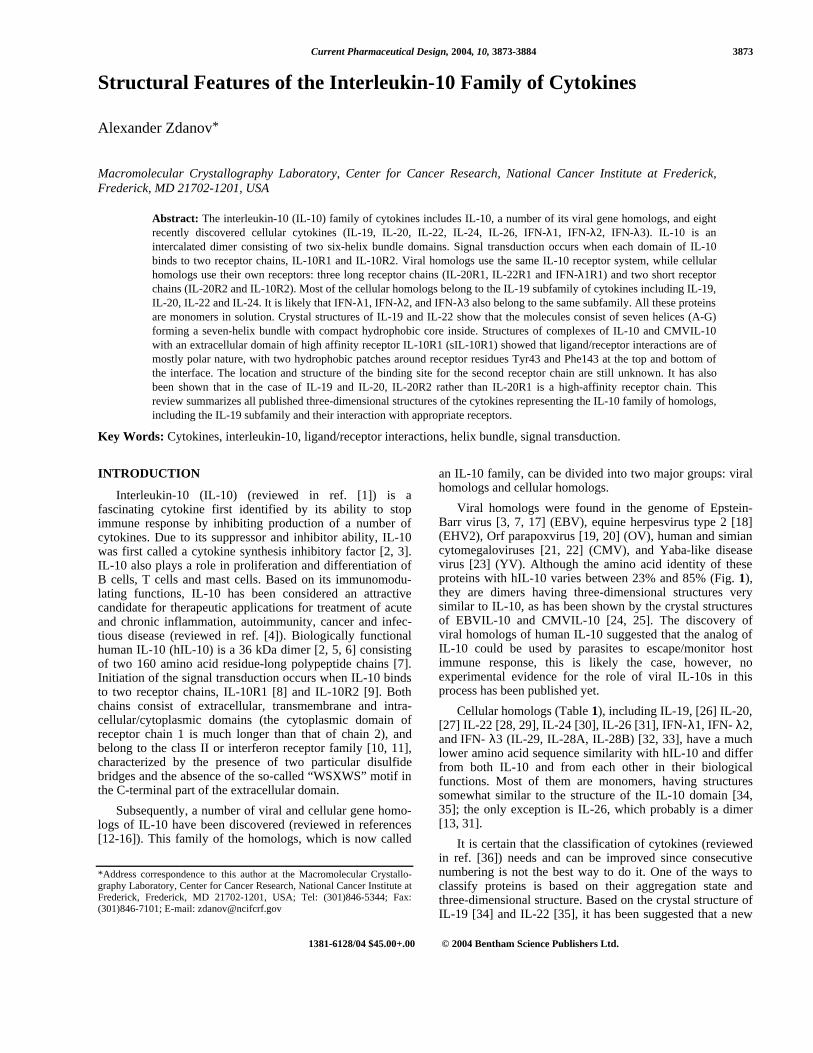
IL-10 is an intercalated dimer of two subunits [39-41]consisting of six amphipathic helices A-F (Fig. 2a). Thepolypeptide chains of each subunit contribute to both parts ofthe dimer. Helices A-D of one subunit form a distinctive six-helix domain with helices E’ and F’ through 180o rotationaround twofold axis (Fig. 2b). In addition, helices A, C, D,F’ and A’, C’, D’ and F of each domain form a left-handedfour-helix bundle, which was found in all helical cytokines[42]. The structure of the IL-10 subunit is stabilized by twointramolecular disulfide bridges (Fig. 2), Cys12-Cys108 andCys62-Cys114. Disulfides hold together helices A, C and D,forming a frame with a long depression in the middle. Theinternal surface of the frame is very hydrophobic; therefore,when amphipathic helices E’ and F’ cover the depression(Fig. 1b), almost all (86%) hydrophobic residues of IL-10are involved in formation of the intradomain hydrophobiccore. Domains are kept together by two flexible polypeptidelinks, separated by 15 Å from each other, allowing somedegree of freedom to change the elbow angle between thedomains. A comparison of structures of hIL-10 crystallizedin different crystal forms [39, 41] and EBVIL-10 [24], whichis a very close homolog of hIL-10 (85% identity), indicatedthat the elbow angle may change easily, even due to adifferent crystal packing.
Monomeric IL-10
Exchange of structural elements between molecularaggregates is known as “domain swapping.” A recent review[43] on domain swapping identified about 40 proteins,including IL-10, of known crystal structure which satisfycertain conditions for swapping. Theoretically, IL-10 haseverything in order to swap helices E’-F’ with E-F, whichwould lead to a formation of monomeric IL-10; however, inpractice the disulfide bridge Cys62-Cys114 restricts possibleconformations of loop DE, making it insufficiently long toallow the swapping without either reducing the disulfide orserious distortions of the structure [39, 41]. To overcome theproblem, Josephson et al. [44] extended loop DE betweenAsn116 and Lys117 by insertion of an additional six aminoacids (GlyGlyGlySerGlyGly) and showed that the expressedprotein folded as a monomer. The crystal structure ofmonomeric IL-10 [45] confirmed that it is very similar to asingle domain of the IL-10 dimer. Further analysis showedthat it was able to form a 1:1 complex with IL-10 receptor 1(IL-10R1); although affinity was 60-fold reduced but theprotein still retained biological activity, which was 10-foldlower as compared with the dimeric wild-type IL-10. Therewas also a report [46] on the stability studies of the IL-10dimer, which showed that dimer/monomer transition induced
either by 1.6M guanidine hydrochloride or pH 2.5 did takeplace and that the monomer under such extreme conditionsstill retained 80% to 89% of its helical structure.
Viral IL-10s
Since all viral homologs imitate at least a subset [47] ofthe biological function of IL-10, they initiate signaltransduction through the IL-10 receptor system, by bindingthe high-affinity receptor chain IL-10R1 and the low-affinitychain IL-10R2, although the affinity of particular viral IL-10may vary. For example, EBVIL-10 binding affinity towardIL-10R1 is about 1000-fold lower than that of human IL-10[47], while CMVIL-10 has approximately the same bindingaffinity toward IL-10R1 as hIL-10 [25]. Yaba-like disease
a).
b).
Fig. (2). a.) Stereo diagram of IL-10 monomer, hydrophobicresidues marked in red (pdb code 2ILK); b.) Stereo diagram of IL-10 dimer, monomers are shown in violet and green. Disulfidebonds are in yellow. All figures are made with program RIBBONS[84].
3876 Current Pharmaceutical Design, 2004, Vol. 10, No. 31 Alexander Zdanov
virus homolog of IL-10 may be an exception among viralhomologs of hIL-10, reference [15] quoted unpublished dataspecifying that this factor binds IL-20R1 and IL-20R2receptor chains; however, since the homology of Yaba-likedisease virus homolog is higher for IL-24 than for IL-10(27% vs 23%), then this may actually be a viral homolog ofIL-24, not of IL-10.
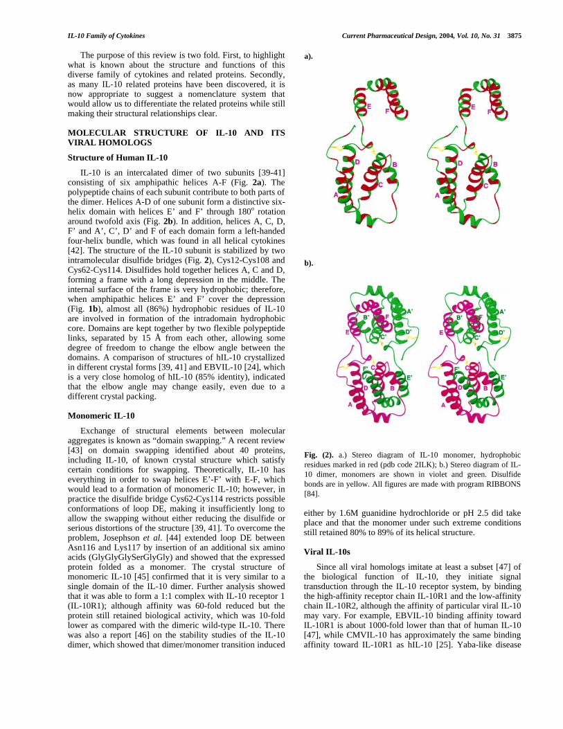
Two crystal structures of viral IL-10s have beendetermined to date, EBVIL-10 [24] at resolution 1.9 Å andCMVIL-10 [25] at resolution 2.7 Å; the latter structure hasbeen solved in complex with IL-10 soluble receptor 1 (sIL-10R1). The amino acid sequence identity of each proteinwith hIL-10 is quite different, 85% for EBV and 27% forCMV IL-10 (Fig. 1); because of that, the proteins havesimilar in general yet different in details three-dimensionalstructures (Fig. 3), which are similar to IL-10. The subunit ofEBVIL-10 consists of six helices, A-F, while the monomerof CMVIL-10 has only five helices. Because of threeresidues deletion in the area of short helix B, it is substitutedby two extended β-turns. Thus, each domain of CMVIL-10is formed by five helices; A, C, D of one subunit and E’, F’coming from another subunit. The dimer of CMVIL-10 isadditionally stabilized by a disulfide bond Cys59-Cys59’between the two subunits, making interdomain elbow angle130o and restricting possible hinge movements of thedomains. Unlike CMVIL-10, both human and EBVIL-10elbow angles are in the range of about 90o, although they canchange a few degrees even upon different packing ofmolecules in the crystals. The most significant difference ofthe amino acid sequences of EBVIL-10 and hIL-10 is foundat the N-terminus. Because of the four residue deletion (17-20, hIL-10 numbering) the first disulfide bridge Cys12-Cys108 is shifted 6.1 Å away from its position in hIL-10,affecting the conformation of the surrounding areas,particularly that of loop DE [24]. Conformation of the N-terminus and loop DE in the structure of CMVIL-10 is alsodifferent from that of hIL-10 [25]. It was also noticed beforethat loop AB is likely to change its conformation uponinteraction with the receptor [24]. This part of the structurewas found relatively flexible in various crystal forms of hIL-10 [39-41] and adopts different conformation in EBVIL-10,in spite of the fact that amino acid sequence identity in thisarea is quite high [24]. Since the conformation of this loop inboth receptor-bound hIL-10 and CMVIL-10 is the same andit is different from EBVIL-10, it is very likely that loop ABadopts its “active” conformation only upon interaction withthe receptor.
LIGAND/SOLUBLE IL-10 RECEPTOR COMPLEXES
Stoichiometry of Ternary and Intermediate/BinaryComplexes
It is commonly accepted that since IL-10R1 is the high-affinity receptor of IL-10, it should bind a ligand first [8],forming a binding site for the second receptor. Thesubsequent binding of the second receptor (IL-10R2) [9]completes the signaling ternary complex. Unfortunately, IL-10R2 is a low-affinity receptor [9, 48], and it is difficult, ifnot impossible, to obtain a stable ternary complex capable tocrystallize. However, the binary intermediate complexes ofboth hIL-10 and CMVIL-10 with soluble receptor sIL-10R1
are quite stable and both were crystallized [49]. It was alsoshown that in solution IL-10 and sIL-10R1 form a complexconsisting of two IL-10 dimers and four molecules of sIL-10R1 [50, 51]; therefore, the question of the exactstoichiometry of the signaling ternary complex is also open.
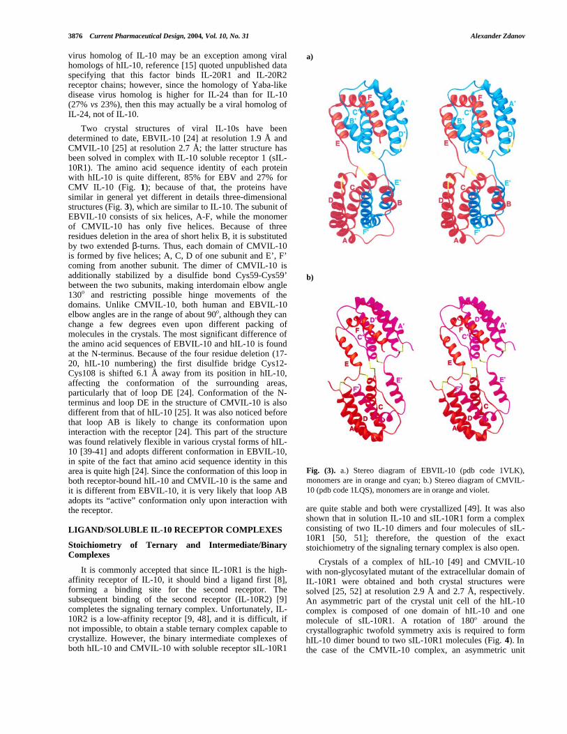
Crystals of a complex of hIL-10 [49] and CMVIL-10with non-glycosylated mutant of the extracellular domain ofIL-10R1 were obtained and both crystal structures weresolved [25, 52] at resolution 2.9 Å and 2.7 Å, respectively.An asymmetric part of the crystal unit cell of the hIL-10complex is composed of one domain of hIL-10 and onemolecule of sIL-10R1. A rotation of 180o around thecrystallographic twofold symmetry axis is required to formhIL-10 dimer bound to two sIL-10R1 molecules (Fig. 4). Inthe case of the CMVIL-10 complex, an asymmetric unit
a)
b)
Fig. (3). a.) Stereo diagram of EBVIL-10 (pdb code 1VLK),monomers are in orange and cyan; b.) Stereo diagram of CMVIL-10 (pdb code 1LQS), monomers are in orange and violet.
IL-10 Family of Cytokines Current Pharmaceutical Design, 2004, Vol. 10, No. 31 3877
contains a CMVIL-10 dimer bound to two sIL-10R1molecules. The receptor molecules interacting with the sameIL-10 dimer do not interact with each other; the distancebetween their C-termini at the points of likely entrance intocell membrane is 110 Å and 105 Å for hIL-10 and CMVIL-10 complexes respectively.
Fig. (4). Stereo diagram of hIL-10/sIL-10R1 complex (pdb code1J7V) receptor molecules are in orange, IL-10 dimer has the samecolor code as in figure 1b.
Structure of Receptor Bound hIL-10
The structure of hIL-10 bound to its receptor ispractically the same as found for free hIL-10, even the smallchange in the interdomain angle falls in the range of valuesfound previously for human and viral IL-10s. The r.m.s.deviation for the Cα atoms of the parts of the moleculeforming the helices is only 0.6 Å. Loops AB and DE, as wellas N and C-termini, are more flexible and their conformationshave greater differences. hIL-10 was previously crystallized
in different crystal forms and at different temperatures; it isinteresting that the structure of free hIL-10 crystallized intrigonal crystals and determined at temperature 100 K (pdbcode 2ilk [40]) is the closest to the receptor-bound hIL-10structure [52]. The reason for that is the unique packing ofthe molecules in the crystal unit cell, where symmetryrelated molecules of hIL-10 may serve as surrogates of thereceptor molecules.
Structure of sIL-10R1
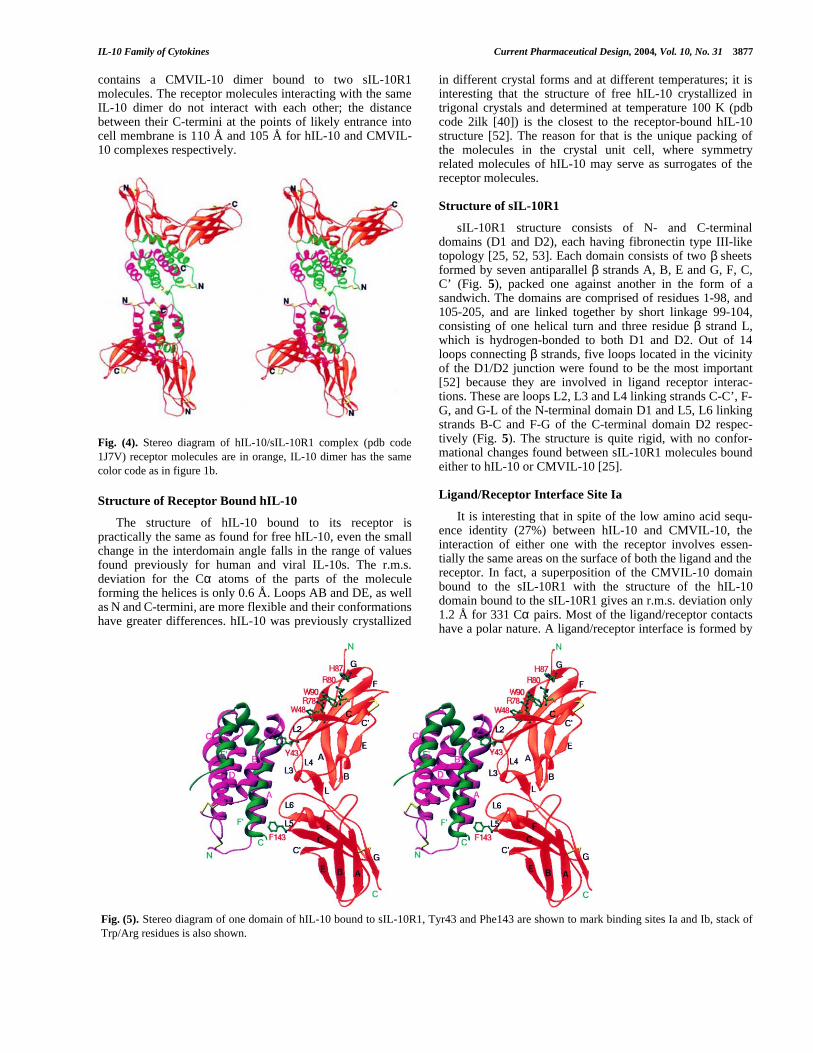
sIL-10R1 structure consists of N- and C-terminaldomains (D1 and D2), each having fibronectin type III-liketopology [25, 52, 53]. Each domain consists of two β sheetsformed by seven antiparallel β strands A, B, E and G, F, C,C’ (Fig. 5), packed one against another in the form of asandwich. The domains are comprised of residues 1-98, and105-205, and are linked together by short linkage 99-104,consisting of one helical turn and three residue β strand L,which is hydrogen-bonded to both D1 and D2. Out of 14loops connecting β strands, five loops located in the vicinityof the D1/D2 junction were found to be the most important[52] because they are involved in ligand receptor interac-tions. These are loops L2, L3 and L4 linking strands C-C’, F-G, and G-L of the N-terminal domain D1 and L5, L6 linkingstrands B-C and F-G of the C-terminal domain D2 respec-tively (Fig. 5). The structure is quite rigid, with no confor-mational changes found between sIL-10R1 molecules boundeither to hIL-10 or CMVIL-10 [25].
Ligand/Receptor Interface Site Ia
It is interesting that in spite of the low amino acid sequ-ence identity (27%) between hIL-10 and CMVIL-10, theinteraction of either one with the receptor involves essen-tially the same areas on the surface of both the ligand and thereceptor. In fact, a superposition of the CMVIL-10 domainbound to the sIL-10R1 with the structure of the hIL-10domain bound to the sIL-10R1 gives an r.m.s. deviation only1.2 Å for 331 Cα pairs. Most of the ligand/receptor contactshave a polar nature. A ligand/receptor interface is formed by
Fig. (5). Stereo diagram of one domain of hIL-10 bound to sIL-10R1, Tyr43 and Phe143 are shown to mark binding sites Ia and Ib, stack ofTrp/Arg residues is also shown.
3878 Current Pharmaceutical Design, 2004, Vol. 10, No. 31 Alexander Zdanov
residues originating from helix A, interhelical loop AB andhelix F’ of the IL-10 and loops L2-L6 of the sIL-10R1, and itcan be clustered into two interacting sites Ia and Ib [25, 52].
Site Ia includes the C-terminal part of helix A, loop AB,and the middle part of helix F on the IL-10 side and loopsL2-L4 of the receptor, while site Ib includes the N-terminaland middle part of helix A, the C-terminal part of helix F ofthe IL-10, and loops L5-L6 of the receptor. Site Ia is theprimary binding site, accounting for about 67% of the totalburied surface of the interface [52]. It is centered aroundreceptor residues Tyr43, Arg76 and Arg96, which makemost of the interactions with IL-10. Tyr43 buries in theinterface the most surface area of any residue involved in it(105 Å2 and 110 Å2 for hIL-10 and CMVIL-10 complexes,respectively) [25, 52]. In the complex with hIL-10, itshydroxyl group forms hydrogen bonds with the main chaincarbonyl oxygen of Asn45, and side chains of Lys138 andGlu142, while its aromatic ring penetrates a hydrophobiccavity made by side chains of Leu46 and Ile145 of IL-10 andaliphatic parts of Arg76 and Arg96 of the receptor (Fig. 5).In the CMV IL-10 complex, the side chain of Tyr43 rotatesabout 110o around the Cα-Cβ bond and in its new positionmakes bifurcated hydrogen bonds with the carboxyl oxygenof Asp42 of CMVIL-10 and a side-chain nitrogen of Arg76of sIL-10R1. Its aromatic group is involved in a hydrophobic“face to edge” interaction with the side chain of Tyr44 (Aspin hIL-10) and Val46 (Leu in hIL-10) of the ligand andLeu41 and aliphatic parts of Arg76 and Arg96 of thereceptor. The guanidino groups of Arg76 and Arg96 makeextensive hydrogen bonds with the ligand. In the hIL-10complex, the side chain of Arg76 adopts two alternativeconformations: in the first one, it interacts with Asp44 andGln42; in the second conformation, it interacts with mainchain carbonyl oxygen of Gln38, and through bridging waterWat103 with Gln42. In the CMVIL-10 complex, only thesecond conformer of Arg76 is possible, because of the sidechain of Tyr44 of CMVIL-10, which takes the space of thefirst conformer. Except for the interaction with Wat103,which is absent in the CMVIL-10 complex, Arg76 makessimilar contacts with the ligand: main chain oxygen of Gln38and hydroxyl of Tyr44 (Asp44). Arg96 has slightly differentconformations in hIL-10 and CMVIL-10 complexes,affecting its interactions with the corresponding IL-10. In thehIL-10 complex, the NH1 atom makes hydrogen bonds toGln38 and the carbonyl oxygen of Ser141, while its NH2interacts with the carboxyl group of Asp144, and throughbridging water Wat71 with Gln38 and Lys34. There is also abridging water Wat88 in the site Ia mediating interactions ofhIL-10 Lys34 with the carbonyl oxygen of Arg96, mainchain nitrogen and hydroxyl group of Ser98 of the receptor.It is interesting that out of three water molecules involved inthe ligand/receptor interface site Ia of the hIL-10 complex,Wat88 and Wat103 certainly belong to the receptor mole-cule, while Wat71 is likely to belong to free hIL-10, since ithas a counterpart there—water molecule Wat260 (pdb entry2ILK); the distance between the positions of Wat71 andWat260 is only 1.7 Å, when free and receptor-bound IL-10molecules are superimposed. In the CMVIL-10 complex, theNH1 atom of Arg96 makes a hydrogen bond to the carbonyloxygen of Asn73 of the receptor, NH2 atom makes ahydrogen bond to the carbonyl of Ser141 and a weak
hydrogen bond to the hydroxyl of Thr145, while the NEatom is hydrogen-bonded to the carboxyl of Asp144. Thereis also a possibility of an ionic interaction of the guanidinogroup with Glu142 (the shortest distance between the sidechains of Glu142 and Arg96 is 4.3 Å). There are no watermolecules in the vicinity of Arg96 in the CMVIL-10/sIL-10R1 interface.
Ligand/Receptor Interface Site Ib
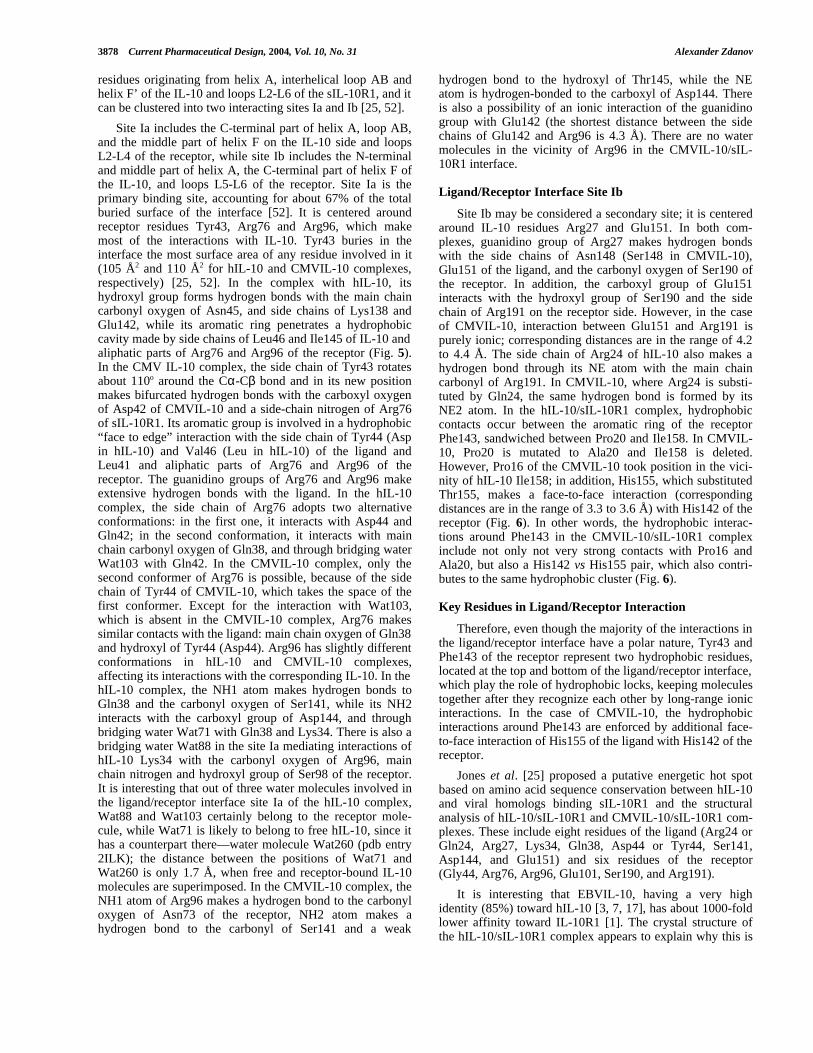
Site Ib may be considered a secondary site; it is centeredaround IL-10 residues Arg27 and Glu151. In both com-plexes, guanidino group of Arg27 makes hydrogen bondswith the side chains of Asn148 (Ser148 in CMVIL-10),Glu151 of the ligand, and the carbonyl oxygen of Ser190 ofthe receptor. In addition, the carboxyl group of Glu151interacts with the hydroxyl group of Ser190 and the sidechain of Arg191 on the receptor side. However, in the caseof CMVIL-10, interaction between Glu151 and Arg191 ispurely ionic; corresponding distances are in the range of 4.2to 4.4 Å. The side chain of Arg24 of hIL-10 also makes ahydrogen bond through its NE atom with the main chaincarbonyl of Arg191. In CMVIL-10, where Arg24 is substi-tuted by Gln24, the same hydrogen bond is formed by itsNE2 atom. In the hIL-10/sIL-10R1 complex, hydrophobiccontacts occur between the aromatic ring of the receptorPhe143, sandwiched between Pro20 and Ile158. In CMVIL-10, Pro20 is mutated to Ala20 and Ile158 is deleted.However, Pro16 of the CMVIL-10 took position in the vici-nity of hIL-10 Ile158; in addition, His155, which substitutedThr155, makes a face-to-face interaction (correspondingdistances are in the range of 3.3 to 3.6 Å) with His142 of thereceptor (Fig. 6). In other words, the hydrophobic interac-tions around Phe143 in the CMVIL-10/sIL-10R1 complexinclude not only not very strong contacts with Pro16 andAla20, but also a His142 vs His155 pair, which also contri-butes to the same hydrophobic cluster (Fig. 6).
Key Residues in Ligand/Receptor Interaction
Therefore, even though the majority of the interactions inthe ligand/receptor interface have a polar nature, Tyr43 andPhe143 of the receptor represent two hydrophobic residues,located at the top and bottom of the ligand/receptor interface,which play the role of hydrophobic locks, keeping moleculestogether after they recognize each other by long-range ionicinteractions. In the case of CMVIL-10, the hydrophobicinteractions around Phe143 are enforced by additional face-to-face interaction of His155 of the ligand with His142 of thereceptor.
Jones et al. [25] proposed a putative energetic hot spotbased on amino acid sequence conservation between hIL-10and viral homologs binding sIL-10R1 and the structuralanalysis of hIL-10/sIL-10R1 and CMVIL-10/sIL-10R1 com-plexes. These include eight residues of the ligand (Arg24 orGln24, Arg27, Lys34, Gln38, Asp44 or Tyr44, Ser141,Asp144, and Glu151) and six residues of the receptor(Gly44, Arg76, Arg96, Glu101, Ser190, and Arg191).
It is interesting that EBVIL-10, having a very highidentity (85%) toward hIL-10 [3, 7, 17], has about 1000-foldlower affinity toward IL-10R1 [1]. The crystal structure ofthe hIL-10/sIL-10R1 complex appears to explain why this is
IL-10 Family of Cytokines Current Pharmaceutical Design, 2004, Vol. 10, No. 31 3879
so. The hydrophobic pocket made by hIL-10 residues Pro20and Ile158 for receptor side chain of Phe143 does not existin EBVIL-10: residues 17-20 are deleted, and Ile158 of hIL-10 is substituted by Ala. Because of the deletions at the N-terminus, the conformation of the main chain in this area isquite different and in order for Phe143 of the receptor to getinto even weak hydrophobic contacts with IL-10, some localconformational changes on the ligand side have to occur. Inaddition, the conformation of the loop AB involved information of the site Ia is also different from what was foundin the structure of EBVIL-10 [24]. These conformationalchanges would inevitably require additional energeticexpense, lowering ligand/receptor affinity.
WSXWS Motif
Class I receptors have “WSXWS” motif in the C-terminal part of their extracellular domain, usually it islocated in the β bulge preceding strand G. Indole rings of thetryptophans are oriented toward strand F and are intercalatedwith guanidino groups of two arginines coming from eitherstrand C as in the structure of prolactin and erythropoietinreceptors [54, 55] or strand F as in the structures of growthgormone and IL-4 receptors [56, 57] creating a stack ofTrp/Arg/Trp/Arg residues forming an extended Π-cationsystem. It is interesting that Trp40, Arg78, Trp90 and Arg80of the IL-10R1 form similar stack of Trp/Arg residuesextended by Leu41 and His87 (Fig. 5). Unlike class Ireceptors, it is located in the N-terminal domain of the sIL-10R1 in the vicinity of a “WSXWS”-like motif consisting ofresidues His87, Ser88, Asn89, Trp90 and Thr91 [52]. Therole of the motif has been extensively studied by muta-genesis of the erythropoietin receptor [58, 59]. The obtainedresults have shown that the “WSXWS” motif was importantfor the passage of the receptor from endoplasmic reticulumto the Golgi apparatus.
Possible IL-10R2 Binding Sites
As mentioned above, in the solution hIL-10/sIL-10R1complex is formed by two dimers of IL-10 and fourmolecules of the sIL-10R1 [50]; that is, 2:4 stoichiometry. It
is obvious that in vivo, on the cell membrane the signalingcomplex could be different; nevertheless, it could also be2:4. An asymmetric part of the crystal unit cell of the hIL-10/sIL-10R1 complex contains one monomer of hIL-10 andone molecule of sIL-10R1. A complex of hIL-10 dimerbound to two sIL-10R1 (1:2) is formed because of thecrystallographic symmetry; if we continue this process onestep further and apply translational symmetry along unit cellaxis b, we can generate a 2:4 complex. This kind of complexis stabilized in the crystals [52] by interactions of the D1domain of one molecule of the 1:2 complex with IL-10 andthe D2 domain of the symmetry-mate molecule of the 1:2complex. Josephson et al. [52] suggested that this could bethe complex which was seen in the solution [50, 60] and itgave them a clue of how the signaling ternary complex, acomplex of hIL-10 with both IL-10R1 and IL-10R2, may beorganized. Since the key amino acids involved in the hIL-10/sIL-10R1 interactions in site Ia and Ib are mostlyconserved between IL-10R1 and IL-10R2, they proposed anidea that at the first step both IL-10R1 and IL-20R2 maybind hIL-10 by using the same site I, so that there will be amixture of high affinity hIL-10/IL-10R1 1:2 complexes andlow-affinity hIL-10/IL-10R2 1:2 complexes. In the next step,these complexes interact with each other to produce a ternary(2:4) complex similar to one generated in the crystalsthrough translational symmetry, and that one could be thesignaling complex [60]. It is clear that when a receptor isbound to the membrane, then even weak interactions couldbecome meaningful and from this point of view the eventsdescribed above could be real. However, while in solutionthe hIL-10/sIL-10R1 complex was found to be 2:4 with ahigh degree of probability, the situation in the crystal is quitedifferent. Crystal packing of IL-10/sIL-10R1 is such thatligand molecules make an infinite number of layers parallelto the plane ab, separated by parallel layers of the sIL-10R1molecules. If we assume that the 1:2 complex lying on thetwofold symmetry axis is a minimal unit, then millions ofsuch units interact with each other, creating not just 2:4 but acontinuous number of infinite layers of 1:2 moleculesinteracting exactly the same way as in the 2:4 molecule.Thus, if 1:2 molecules are favored in solution to form the 2:4
Fig. (6). Stereo diagram of interface site Ib of CMVIL-10/sIL-10R1 complex (pdb code 1LQS), CMVIL-10 residues are shown in green,receptor residues are in red, atom color code is: C-green, N-blue, O-red; hydrogen bonds are shown as pink dash lines.

3880 Current Pharmaceutical Design, 2004, Vol. 10, No. 31 Alexander Zdanov
complexes found in the crystal, they inevitably must formhigher aggregates in solution as well. Besides, a similarcomplex [25] of CMV IL-10 with the same non-glycosilatedmutant of sIL-10R1 has an absolutely different crystalpacking, and even though the 1:2 complex is very similar tohIL-10/sIL-10R1 both in terms of the structure of thereceptor and its interaction with the ligand [25], there is no2:4 CMV IL-10/sIL-10R1 complex found in solution.Therefore, it appears that the questions of the position of theIL-10R2 binding site and the structure of the ternary hIL-10/IL-10R1/IL-10R2 complex still remain to be answered.
IL-19 SUBFAMILY OF CYTOKINES
All members of the IL-19 subfamily of cytokines (IL-19,IL-20, IL-22, IL-24) are monomers in solution [34, 35, 61,62], although at high concentration some aggregation maytake place [35]. Despite their relatively low amino acidsequence identity with hIL-10, their structures, as will beseen below, are similar to the structure of one domain ofhIL-10. Biological functions of these factors, particularly fortheir therapeutic applications, are still to be studied;however, some of the proteins have already attracted consi-derable interest. For instance, IL-24 (formerly known asMDA-7) has shown profound antiproliferative and cytotoxiceffects in a wide variety of human tumor cell lines [62-69],and it is now becoming a subject of growing number ofstudies.
Crystal Structure of IL-19
A molecule of IL-19 is a monomer made up of sevenamphipathic helices A-G of different lengths (Fig. 7a),forming a unique seven-helix bundle with an extensiveinternal hydrophobic core. Three disulfide bridges located onthe top of the bundle make the polypeptide chain frameworkquite rigid. Helices B, D, E and G make a four-helix bundle,which is a characteristic feature of all helical cytokines [42].The position of helix A, covering the top of the molecule, isstabilized by the disulfide bridge Cys10-Cys103, linking itcovalently to the C terminus of helix E. The second and thirddisulfide bridges, Cys57-Cys109 and Cys58-Cys111, holdtogether the N terminus of helix D, interhelical loop EF, andthe N terminus of helix F. The C-terminal strand 154-159 isbent along the surface of the molecule and makes hydrogenbonds with the short interhelical strand AB. These twoparallel strands form a short β-sheet, never seen previouslyin helical cytokines (Fig. 7a).
Crystal Structure of IL-22
The crystal structure of IL-22 [35, 70] is topologicallyvery similar to IL-19 (Fig. 7b), although the sequencesimilarity between IL-19 and IL-22 is 36%, which is not ashigh as with IL-20 or IL-24. The structure also consists ofseven α-helices A-G, which are packed as a seven-helixbundle having an extensive hydrophobic core inside. Asuperposition of the structures of IL-19 and IL-22 [35]results in r.m.s. deviation of 1.7 Å for 123 pairs of Cα-atoms. The main difference is in the position of the disulfidebridges and lengths of the loops. While the disulfide Cys10-Cys103 of IL-19 corresponds to Cys7-Cys99 of IL-22, theyare also significantly shifted (2.5–5.5 Å for the respective
Cα coordinates). Cys57 of IL-19 is equivalent in sequence toCys56 of IL-22; their Cα coordinates are only 3.3 Å apart.However, their disulfide partners are different: in IL-19,Cys57 makes a disulfide bond to Cys109 (loop EF); in IL-22, Cys56 makes a bond to Cys145, which is the C terminusof helix G. A similar variability of the disulfides haspreviously been reported for short-chain helical cytokines[38]. Therefore, in the IL-19 structure, the seven-helixbundle is stabilized by disulfides holding together N-theterminus of helix A with the C terminus of helix E, and the Nterminus of helix D with both loop EF and the N terminus ofhelix F; while in IL-22, the first disulfide similar to IL-19also holds together the N terminus of helix A and the Cterminus of helix E, but the second disulfide is between theN terminus of helix D and the C terminus of helix G.
Comparison of IL-19 and IL-22 with IL-10
The superposition of IL-19 and IL-22 with one domain ofIL-10 gives an r.m.s. deviation between the positions of Cαatoms 1.7 Å and 1.9 Å [35], respectively. The maindifferences are in the area of the first 21 residues of the IL-10 domain, helix C, interhelical loops, orientation of helix E
a)
b)
Fig. (7). a.) Stereo diagram of IL-19 (pdb code 1N1F); b.) Stereodiagram of IL-22 (pdb code 1MR4).

IL-10 Family of Cytokines Current Pharmaceutical Design, 2004, Vol. 10, No. 31 3881
(helix D of IL-10) relative to the rest of the helical bundle,with the r.m.s. deviation at its N-terminus about 1 Å,increasing to 3.8 Å at the C terminus, and the C-terminus ofhelix G (helix F of the IL-10). Therefore, the generalarchitecture of these molecules is very much alike, althoughthe orientation of the new helix A and the short β-sheet inthe IL-19 make the overall shape of the molecule morecompact and smooth.
Interleukin-20
BestFit [71] comparison of IL-19 and IL-20 gives 44.1%sequence identity and 52.4% similarity without any gaps ordeletions, starting in the sequence at the position corres-ponding to the N terminus of helix A of IL-19. Thesuperposition of the sequences of IL-19, IL-20 and IL-10shows a remarkable degree of similarity in the positions ofhydrophobic residues and of the cysteines involved in theformation of the disulfide bridges. In fact, all three disulfidebonds present in IL-19 are also preserved in IL-20. Takentogether, we must conclude that the three-dimensionalstructure of IL-20 must be similar to IL-19, and it is nosurprise that these two cytokines share their receptors. Theonly obvious difference between IL-19 and IL-20, besidestheir N termini, is expected at the C terminus, in the regionof the C-terminal β-strand of IL-19. There is no such strandin IL-20 since Glu157, the last residue of IL-20, correspondsto His153 of IL-19, which is the last residue in the helix G.
Interleukin-24
Sequence identity between IL-19 and IL-24 (MDA-7) isslightly lower, 31%, with 40% similarity. However, allo-cation of similar and hydrophobic amino acid residuesconfirms that the three-dimensional structure of IL-24 islikely to be similar to that of IL-19. Interestingly, IL-24 hasonly two cysteines (Cys16 and Cys63), corresponding toCys10 and Cys57 of IL-19. Since these two cysteines areinvolved in making separate disulfide bonds, and IL-24 is amonomer, this must indicate that the N terminus of IL-24should have a unique conformation, which is likely to bringCys16 into the proximity of Cys63 to form a disulfide bridgebetween them. Because of that, the position of short helix Arelative to the rest of the helical bundle may be different andmay also affect binding to the receptor. Another structuralfeature, which is highly conserved between these cytokines,is a salt bridge formed between Lys27 and Asp143 andlocated on the surface of the molecule. This bridge is strictlyconserved in IL-19, IL-10, IL-22 and IL-24, with a mutationto Arg in IL-20.
Putative Ligand/Receptor Complexes
Cellular homologs, including the IL-19 subfamily,employ different from IL-10 receptor systems, although theyvery often overlap and IL-10R2 is sometimes utilized as thesecond receptor chain, but IL-10R1 is never used. IL-20R1,IL-20R2 [27, 72, 73], IL-22R1 [29, 74] and IFN-λ1R1 [32,33] have been discovered, along with their ligands. Exceptfor IL-20R2, all other newly discovered receptors have longcytoplasmic domains; in fact, because of that, they are calledthe first receptor chains. Therefore, eight cytokines signal byusing one of three first receptor chains in combination with
one of two second receptor chains (IL-20R2 or IL-10R2). IL-22 also has a natural soluble receptor or IL-22 bindingprotein (IL-22BP), which is also a type II receptor, but itlacks both the transmembrane and intracellular domains [75-77] and binds IL-22 on its own.
It was shown that IL-19, IL-20 and IL-24 signal throughthe same two chains, IL-20R1 and IL-20R2 [61, 78]; inaddition, IL-20 and IL-24 signal through the pair IL-22R1/IL-20R2 [65, 72, 73] and IL-22 uses IL-22R1 and IL-10R2 [28, 74]. It was shown recently that despite what hadbeen commonly accepted, IL-20R2 is a high-affinity receptorchain in the case of IL-19 and IL-20. It binds the ligand first[61], forming a binding site for the long receptor chain IL-20R1, which binds the second. It is very likely that IL-24also binds to IL-20R2 first. Since the three-dimensionalstructure of these cytokines is similar to the structure of onedomain of IL-10 and all receptors belong to the same family,it is reasonable to assume that receptor binding sites shouldbe also somewhat similar. The simple superposition of IL-19onto one domain of IL-10 bound to sIL-10R1 [52] allowed tomark the IL-19 surface with amino acid residues which maypotentially interact with the receptor [34]; these includedhelix B, loop BC and helix G. No crystal structures of any ofthe IL-19 subfamily ligand/receptor complexes are availableat present; however, a model of the complex of IL-22 withsIL-22R1, generated on the basis of the crystal structure ofthe complex of hIL-10/sIL-10R1, was published [79]. Notonly was the sIL-22R1 structure generated in this model, butIL-22 was modeled, too, since no crystal structure of IL-22was at that time available. Nevertheless, amino acid residueswhich might be involved in specific contacts with thereceptor were identified and they were essentially the sameas the ones found with the help of the crystal structure of IL-22 [35]. It is interesting to note that intermolecular interfacein a dimer of IL-22 molecules in the asymmetric part of thecrystal unit cell included a receptor binding site with one ofthe arginines playing the role of Arg-96 of the receptor [35],making extensive hydrogen bonds similar to those found inthe hIL-10/sIL-10R1 complex.
Interleukin-26
IL-26 appears to be the only cellular homolog of IL-10which could be a dimer [31]. Amino acid sequence analysis[71] of IL-26 gives 31% identity and 45% similarity withhIL-10, which is slightly higher than with IL-19 (27% and35%), with IL-22 (27% and 38%), or with IL-24 (30% and37%). It is obvious that its structure should be similar to IL-10; however, it still remains to be seen to what degree.Another feature of IL-26 is that it has a very high isoelectricpoint of 10.8, which is quite unusual for the family of IL-10homologs, which more often have isoelectric points in therange of 7 to 8. It is not clear why IL-26 needs to be posi-tively charged. The protein was also for a while a “mysterycytokine” because no receptor system was identified for it.Recently it has been shown1 that IL-26 shares receptors withother members of the IL-10 family. IL-26 employs IL-20R1as the first receptor chain and IL-10R2 as the second one; 1 Faruk Sheik, Vitaliy V. Baurin, Anita Lewis-Antes, Nital K. Shah, Sergey V.Smirnov, Shubha Anantha, Harold Dickensheets, Laure Dumoutier, Jean-ChristopheRenauld, Alexander Zdanov, Raymond P. Donnelly and Sergei V. Kotenko, J. ofImmunology, in press.
3882 Current Pharmaceutical Design, 2004, Vol. 10, No. 31 Alexander Zdanov
none of the other cellular homologs of IL-10 use thisparticular receptor pair. However, the question of whether ornot IL-26 belongs to the IL-19 subfamily is still open.
NEW ARRIVALS, IFN- λ1, IFN- λ2, IFN- λ3 (IL-29, IL-28A AND IL-28B)
IFN-λ1, IFN-λ2, IFN-λ3 (IL-29, IL-28A and IL-28B) arerecently discovered cytokines, having low sequence identitywith IL-10 (10%-13%); nevertheless, all three wererecognized as likely to belong to the family of IL-10homologs [32, 33]. Sequence identity between IFN-λ1 andIFN-λ2 is 81%, and between IFN-λ2 and IFN-λ3, 96%. Theproteins are monomers in solution (unpublished data) andsignal through a newly discovered receptor chain, IFN-λ1R1,in pair with IL-10R2 [32, 33]. In fact, sequence identitybetween IFN-λs and IFN-α2 is slightly higher [33],particularly in terms of the positions of cysteine residues,which implies that IFN-λ1, IFN-λ2 and IFN-λ3 may havethe same disulfide bridges as IFN-α2 (IFN-α2 and IFN-λ1have five cysteines, IFN-λ2 and IFN-λ3 have seven). Thethree-dimensional structure of IFN-λ1, IFN-λ2 or IFN-λ3has not yet been determined; however, it is possible that itwill be similar to IL-19 subfamily, since the first helix ofIFN-α2 [80] is shorter than that of IL-10 and somewhatsimilar to helix B of IL-19.
A NEW NOMENCLATURE FOR CELLULARHOMOLOGS OF IL-10
As it has been already discussed, the classification ofcytokines certainly needs to be improved, particularly in thecases of families of homologs, like IL-10. Since viralhomologs of IL-10 always imitate the function and thestructure of IL-10, there is no necessity to change anythingwith their names. In other words, EBVIL-10 or “the name ofthe virus” plus “IL-10” is clear enough, however, cellularhomologs of IL-10 could be renamed in a way clearlyshowing what family of proteins or homologs they belong in.One of the way to do it has been suggested a couple yearsago for the IL-1 family [81], and it can be easily adapted forIL-10 family. The name of each cellular homolog would beIL-10Fn, where IL-10 portion stays for IL-10, then letter “F”for the word “family” and number “n” is an order number ofthe publication/discovery of particular protein. For example,IL-19 would be IL-10F1, IL-20 would be IL-10F2 and so on.Suggested new names of all cellular homologs of IL-10 areshown in Table 1 in brackets as suggested names. The namesof the corresponding receptors could be formed similarly asit was before, by adding letter “R” and chain number at theend of the name of the ligand, for example, IL-20R1 wouldbecome IL-10F2R1 and so on. IL-19 subfamily of cytokineswould become IL-10F1 subfamily, which potentially mayinclude all cellular homologs of IL-10 provided their three-dimensional structures satisfy the described above criteria.
CONCLUSIONS: CORRELATION OF STRUCTUREWITH FUNCTION
IL-10 is an intercalated dimer which initiates signaltransduction through binding to two receptor chains in asequential order. Initially, IL-10 binds IL-10R1, having along cytoplasmic domain; as a result, the binding site for the
second receptor chain, IL-10R2, is formed and its bindingcompletes creation of the signaling complex.
The IL-10 family of cytokines can be divided into twomajor groups: viral and cellular homologs of IL-10. Viralhomologs, designed to imitate the IL-10 function, bind to thesame receptor system as IL-10 and necessarily must have athree-dimensional structure very similar to IL-10. Crystalstructures of the complexes of hIL-10/sIL-10R1 andCMVIL-10/sIL-10R1 have proved that receptor moiety doesnot change its structure upon different ligand binding; how-ever, ligands may encounter local conformational changes,particularly in the areas of the N-terminus and the inter-helical loop AB. Most of the contacts in the ligand/receptorinterface are of a polar nature, although two areas aroundreceptor amino acid residues Tyr43 and Phe143 serve ashydrophobic locks after ligand/receptor recognition hasoccurred. Since some of the viral homologs of IL-10 possessonly a subset of the activities of IL-10, it is not quite clearhow that can be accomplished with the help of the samereceptor system. It might be suggested that affinity modula-tion may play important role in a case of somewhat lowerexpression of IL-10R1, since IL-10R2 is constitutivelyexpressed in most tissues [82, 83]. However, the binding siteand the mode of binding for the second receptor chain, IL-10R2, still remain to be found.
Most of the cellular members of the IL-10 family belongto the IL-19 subfamily, including IL-19, IL-20, IL-22 andIL-24. Even though the crystal structures of IFN-λ1, IFN-λ2and IFN- λ3 have not been determined, it is likely that theyalso belong to the IL-19 subfamily. However, classificationfor IL-26 is not clear because of its aggregation state, eventhough the receptor system for this cytokine overlaps withthe members of IL-19 subfamily. All eight members (Table1) of cellular homologs of IL-10 signal with the help of threelong chain receptor chains (first receptors) and two shortreceptor chains (second receptors), in addition, IL-20 and IL-24 each use two different combinations of these receptors.Since the biological functions of these cytokines aredifferent, an idea of tissue-specific expression of appropriatereceptors is quite plausible. Size exclusion chromatographystudies showed that IL-20R2 but not IL-20R1 is a high-affinity receptor of IL-19 and IL-20, and formation of asignaling ternary complex is likely to be a two-stepprocedure: initially, a ligand binds to IL-20R2, forming thebinding site for IL-20R1, the binding of which completes thefinal complex. The high-affinity receptor binding sites,including helix B, loop BC and helix G, on the surface of IL-19 and IL-22 were marked, based on the known crystalstructure of IL-10/sIL-10R1.
ACKNOWLEDGEMENT
I would like to thank Dr. Alexander Wlodawer forcritical reading of the manuscript and for helpful discussions.
REFERENCES
References 85-87 are related articles recently published inCurrent Pharmaceutical Design.
[1] Moore KW, de Waal MR, Coffman RL, O'Garra A. Interleukin-10and the interleukin-10 receptor. Annu Rev Immunol 2001; 19: 683.

IL-10 Family of Cytokines Current Pharmaceutical Design, 2004, Vol. 10, No. 31 3883
[2] Fiorentino DF, Bond MW, Mosmann TR. Two types of mouse Thelper cell: IV. Th2 clones secrete a factor that inhibits cytokineproduction by Th1 clones. J Exp Med 1989; 170: 2081.
[3] Moore KW, Vieira P, Fiorentino DF, Trounstine ML, Khan TA,Mosmann TR. Homology of cytokine synthesis inhibitory factor(IL-10) to the Epstein Barr Virus gene BCRFI. Science 1990; 248:1230.
[4] Asadullah K, Sterry W, Volk HD. Interleukin-10 therapy--reviewof a new approach. Pharmacol Rev 2003; 55: 241.
[5] Tan JC, Indelicato SR, Narula SK, Zavodny PJ, Chou C-C.Characterization of interleukin-10 receptors on human and mousecells. J Biol Chem 1993; 268: 21053.
[6] Windsor WT, Syto R, Tsarbopoulos A, Zhang R, Durkin J,Baldwin S, et al. Disulfide bond assignments and secondarystructure analysis of human and murine interleukin 10.Biochemistry 1993; 32: 8807.
[7] Vieira P, Malefyt RW, Dang M-N, Johnson KE, Kastelein R,Fiorentino DF, et al. Isolation and expression of human cytokinesynthesis inhibitory factor cDNA clones: Homology to Epstein-Barr virus open reading frame BCRFI. Proc Natl Acad Sci USA1991; 88: 1172.
[8] Liu Y, Wei SHY, Ho ASY, Malefyt RW, Moore KW. Expressioncloning and characterization of a human IL-10 receptor. J Immunol1994; 152: 1821.
[9] Kotenko SV, Krause CD, Izotova LS, Pollack BP, Wu W, Pestka S.Identification and functional characterization of a second chain ofthe interleukin-10 receptor complex. EMBO J 1997; 16: 5894.
[10] Bazan JF. Structural design and molecular evolutuion of a cytokinereceptor superfamily. Proc Natl Acad Sci USA 1990; 87: 6934.
[11] Ho ASY, Liu Y, Khan TA, Hsu D-H, Bazan JF, Moore KW. Areceptor for interleukin 10 is related to interferon receptors. ProcNatl Acad Sci USA 1993; 90: 11267.
[12] Kotenko SV. The family of IL10 related cytokines and theirreceptors: related, but to what extent? Cytokine Growth Factor Rev2002; 13: 223.
[13] Fickenscher H, Hor S, Kupers H, Knappe A, Wittmann S, Sticht H.The interleukin-10 family of cytokines. Trends Immunol 2002; 23:89.
[14] Dumoutier L, Renauld JC. Viral and cellular interleukin-10 (IL-10)-related cytokines: from structures to functions. Eur CytokineNetw 2002; 13: 5.
[15] Renauld JC. Class II cytokine receptors and their ligands: keyantiviral and inflammatory modulators. Nat Rev Immunol 2003; 3:667.
[16] Ozaki K, Leonard WJ. Cytokine and cytokine receptor pleiotropyand redundancy. J Biol Chem 2002; 277: 29355.
[17] Hsu DH, de Waal Malefyt R, Fiorentino DF, Dang MN, Vieira P,de Vries J, et al . Expression of interleukin-10 activity by Epstein-Barr virus protein BCRF1. Science 1990; 250: 830.
[18] Rode HJ, Janssen W, Rosen-Wolff A, Bugert JJ, Thein P, BeckerY, et al. The genome of equine herpesvirus type 2 harbors aninterleukin 10 (IL10)-like gene. Virus Genes 1993; 7: 111.
[19] Fleming SB, McCaughan CA, Andrews AE, Nash AD, Mercer AA.A homolog of interleukin-10 is encoded by the poxvirus orf virus. JVirol 1997; 71: 4857.
[20] Fleming SB, Haig DM, Nettleton P, Reid HW, McCaughan CA,Wise LM, et al. Sequence and functional analysis of a homolog ofinterleukin-10 encoded by the parapoxvirus orf virus. Virus Genes2000; 21: 85.
[21] Kotenko SV, Saccani S, Izotova LS, Mirochnitchenko OV, PestkaS. Human cytomegalovirus harbors its own unique IL-10 homolog(cmvIL-10). Proc Natl Acad Sci USA 2000; 97: 1695.
[22] Lockridge KM, Zhou SS, Kravitz RH, Johnson JL, Sawai ET,Blewett EL, et al . Primate cytomegaloviruses encode and expressan IL-10-like protein. Virol 2000; 268: 272.
[23] Lee HJ, Essani K, Smith GL. The genome sequence of Yaba-likedisease virus, a yatapoxvirus. Virol 2001; 281: 170.
[24] Zdanov A, Schalk-Hihi C, Menon S, Moore KW, Wlodawer A.Crystal structure of Epstein-Barr virus protein BCRF1, a homologof cellular interleukin-10. J Mol Biol 1997; 268: 460.
[25] Jones BC, Logsdon NJ, Josephson K, Cook J, Barry PA, WalterMR. Crystal structure of human cytomegalovirus IL-10 bound tosoluble human IL-10R1. Proc Natl Acad Sci USA 2002; 99: 9404.
[26] Gallagher G, Dickensheets H, Eskdale J, Izotova LS,Mirochnitchenko OV, Peat JD, et al. Cloning, expression andinitial characterization of interleukin-19 (IL- 19), a novel
homologue of human interleukin-10 (IL-10). Genes Immun 2000;1: 442.
[27] Blumberg H, Conklin D, Xu WF, Grossmann A, Brender T,Carollo S, et al . Interleukin 20: discovery, receptor identification,and role in epidermal function. Cell 2001; 104: 9.
[28] Dumoutier L, Van Roost E, Colau D, Renauld JC. Humaninterleukin-10-related T cell-derived inducible factor: molecularcloning and functional characterization as an hepatocyte-stimulating factor. Proc Natl Acad Sci USA 2000; 97: 10144.
[29] Xie MH, Aggarwal S, Ho WH, Foster J, Zhang Z, Stinson J, et al.Interleukin (IL)-22, a novel human cytokine that signals throughthe interferon receptor-related proteins CRF2-4 and IL-22R. J BiolChem 2000; 275: 31335.
[30] Jiang H, Su ZZ, Lin JJ, Goldstein NI, Young CS, Fisher PB. Themelanoma differentiation associated gene mda-7 suppresses cancercell growth. Proc Natl Acad Sci USA 1996; 93: 9160.
[31] Knappe A, Hor S, Wittmann S, Fickenscher H. Induction of a novelcellular homolog of interleukin-10, AK155, by transformation of Tlymphocytes with herpesvirus saimiri. J Virol 2000; 74: 3881.
[32] Kotenko SV, Gallagher G, Baurin VV, Lewis-Antes A, Shen M,Shah NK, et al. IFN-lambdas mediate antiviral protection through adistinct class II cytokine receptor complex. Nature Immunol 2003;4: 69.
[33] Sheppard P, Kindsvogel W, Xu W, Henderson K, Schlutsmeyer S,Whitmore TE, et al. IL-28, IL-29 and their class II cytokinereceptor IL-28R. Nature Immunol 2003; 4: 63.
[34] Chang C, Magracheva E, Kozlov S, Fong S, Tobin G, Kotenko S,et al. Crystal structure of interleukin-19 defines a new subfamily ofhelical cytokines. J Biol Chem 2003; 278: 3308.
[35] Nagem RAP, Colau D, Dumoutier L, Renauld J-C, Ogata C,Polikarpov I. Crystal structure of recombinant human interleukin-22. Structure 2002; 10: 1051.
[36] Schein CH. The shape of the messenger: using protein structureinformation to design novel cytokine-based therapeutics. CurrPharm Des 2002; 8: 2113.
[37] Sprang SR, Bazan JF. Cytokine structural taxonomy andmechanisms of receptor engagement. Curr Opin Struct Biol 1993;3: 815.
[38] Rozwarski DA, Gronenborn AM, Clore GM, Bazan JF, Bohm A,Wlodawer A, et al . Structural comparisons among the short-chainhelical cytokines. Structure 1994; 2: 159.
[39] Zdanov A, Schalk-Hihi C, Gustchina A, Tsang M, Weatherbee J,Wlodawer A. Crystal structure of interleukin-10 reveals thefunctional dimer with an unexpected topological similarity tointerferon γ. Structure 1995; 3: 591.
[40] Zdanov A, Schalk-Hihi C, Wlodawer A. Crystal structure of humaninterleukin-10 at 1.6 Å resolution and a model of a complex withits soluble receptor. Protein Sci 1996; 5: 1955.
[41] Walter MR, Nagabhushan TL. Crystal structure of interleukin 10reveals an interferon gamma- like fold. Biochemistry 1995; 34:12118.
[42] Presnell SR, Cohen FE. Topological distribution of four-α-helixbundles. Proc Natl Acad Sci USA 1989; 86: 6592.
[43] Liu Y, Eisenberg D. 3D domain swapping: as domains continue toswap. Protein Sci 2002; 11: 1285.
[44] Josephson K, DiGiacomo R, Indelicato SR, Iyo AH, NagabhushanTL, Parker MH, et al. Design and analysis of an engineered humaninterleukin-10 monomer. J Biol Chem 2000; 275: 13552.
[45] Josephson K, Jones BC, Walter LJ, DiGiacomo R, Indelicato SR,Walter MR. Noncompetitive antibody neutralization of IL-10revealed by protein engineering and x-ray crystallography.Structure (Camb) 2002; 10: 981.
[46] Syto R, Murgolo NJ, Braswell EH, Mui P, Huang E, Windsor WT.Structural and biological stability of the human interleukin 10homodimer. Biochemistry 1998; 37: 16943.
[47] Liu Y, de Waal Malefyt R, Briere F, Parham C, Bridon JM,Banchereau J, et al. The EBV IL-10 homolog is a selective agonistwith impaired binding to the IL-10 receptor. J Immunol 1997; 158:605.
[48] Spencer SD, Di Marco F, Hooley J, Pitts-Meek S, Bauer M, RyanAM, et al . The orphan receptor CRF2-4 is an essential subunit ofthe interleukin 10 receptor. J Exp Med 1998; 187: 571.
[49] Josephson K, McPherson DT, Walter MR. Purification,crystallization and preliminary X-ray diffraction of a complexbetween IL-10 and soluble IL-10R1. Acta Crystallogr D BiolCrystallogr 2001; 57: 1908.
3884 Current Pharmaceutical Design, 2004, Vol. 10, No. 31 Alexander Zdanov
[50] Tan JC, Braun S, Rong H, DiGiacomo R, Dolphin E, Baldwin S, etal. Characterization of recombinant extracellular domain of humaninterleukin-10 receptor. J Biol Chem 1995; 270: 12906.
[51] Hoover DM, Schalk-Hihi C, Chou CC, Menon S, Wlodawer A,Zdanov A. Purification of receptor complexes of interleukin-10stoichiometry and the importance of deglycosylation in theircrystallization. Eur J Biochem 1999; 262: 134.
[52] Josephson K, Logsdon NJ, Walter MR. Crystal structure of the IL-10/IL-10R1 complex reveals a shared receptor binding site.Immunity 2001; 15: 35.
[53] Bork P, Holm L, Sander C. The Immunoglobulin Fold. Structuralclassification, sequence patterns and common core. J Mol Biol1994; 242: 309.
[54] Somers W, Ultsch M, de Vos AM, Kossiakoff AA. The X-raystructure of a growth hormone-prolactin receptor complex. Nature1994; 372: 478.
[55] Livnah O, Stura EA, Johnson DL, Middleton SA, Mulcahy LS,Wrighton NC, et al. Functional mimicry of a protein hormone by apeptide agonist: the EPO receptor complex at 2.8 A. Science 1996;273: 464.
[56] de Vos AM, Ultsch M, Kossiakoff AA. Human growth hormoneand extracellular domain of its receptor: Crystal structure of thecomplex. Science 1992; 255: 306.
[57] Hage T, Sebald W, Reinemer P. Crystal structure of theinterleukin-4/receptor alpha chain complex reveals a mosaicbinding interface. Cell 1999; 97: 271.
[58] Hilton DJ, Watowich SS, Katz L, Lodish HF. Saturationmutagenesis of the WSXWS motif of the erythropoietin receptor. JBiol Chem 1996; 271: 4699.
[59] Hilton DJ, Watowich SS, Murray PJ, Lodish HF. Increased cellsurface expression and enhanced folding in the endoplasmicreticulum of a mutant erythropoietin receptor. Proc Natl Acad SciUSA 1995; 92: 190.
[60] Walter MR. Strucure of interleukin-10/interleukin-10R1 complex:a paradigm for class 2 cytokine activation. Immunol Res 2002; 26:303.
[61] Pletnev S, Magracheva E, Kozlov S, Tobin G, Kotenko SV,Wlodawer A, et al. Characterization of the recombinantextracellular domains of human interleukin-20 receptors and theircomplexes with interleukin-19 and interleukin-20. Biochemistry2003; In press:
[62] Sauane M, Gopalkrishnan RV, Sarkar D, Su ZZ, Lebedeva IV,Dent P, et al. MDA-7/IL-24: novel cancer growth suppressing andapoptosis inducing cytokine. Cytokine Growth Factor Rev 2003;14: 35.
[63] Sauane M, Gopalkrishnan RV, Lebedeva I, Mei MX, Sarkar D, SuZZ, et al. Mda-7/IL-24 induces apoptosis of diverse cancer celllines through JAK/STAT-independent pathways. J Cell Physiol2003; 196: 334.
[64] Su ZZ, Lebedeva IV, Sarkar D, Gopalkrishnan RV, Sauane M,Sigmon C, et al. Melanoma differentiation associated gene-7, mda-7/IL-24, selectively induces growth suppression, apoptosis andradiosensitization in malignant gliomas in a p53-independentmanner. Oncogene 2003; 22: 1164.
[65] Ramesh R, Mhashilkar AM, Tanaka F, Saito Y, Branch CD, SiegerK, et al. Melanoma Differentiation-associated Gene 7/Interleukin(IL)-24 Is a Novel Ligand That Regulates Angiogenesis via the IL-22 Receptor. Cancer Res 2003; 63: 5105.
[66] Mhashilkar AM, Stewart AL, Sieger K, Yang HY, Khimani AH,Ito I, et al. MDA-7 negatively regulates the beta-catenin and PI3Ksignaling pathways in breast and lung tumor cells. Mol Ther 2003;8: 207.
[67] Yacoub A, Mitchell C, Brannon J, Rosenberg E, Qiao L,McKinstry R, et al. MDA-7 (interleukin-24) inhibits theproliferation of renal carcinoma cells and interacts with freeradicals to promote cell death and loss of reproductive capacity.Mol Cancer Ther 2003; 2: 623.
[68] Lebedeva IV, Su ZZ, Sarkar D, Fisher PB. Restoring apoptosis as astrategy for cancer gene therapy: focus on p53 and mda-7. SeminCancer Biol 2003; 13: 169.
[69] Chen J, Chada S, Mhashilkar A, Miano JM. Tumor suppressorMDA-7/IL-24 selectively inhibits vascular smooth muscle cellgrowth and migration. Mol Ther 2003; 8: 220.
[70] Nagem RA, Lucchesi KW, Colau D, Dumoutier L, Renauld JC,Polikarpov I. Crystallization and synchrotron X-ray diffractionstudies of human interleukin-22. Acta Crystallogr D BiolCrystallogr 2002; 58: 529.
[71] Genetics Computer Group. Program Manual for the GCG Package,Ver. 8. Madison, WI 1994.
[72] Dumoutier L, Leemans C, Lejeune D, Kotenko SV, Renauld JC.Cutting edge: STAT activation by IL-19, IL-20 and mda-7 throughIL-20 receptor complexes of two types. J Immunol 2001; 167:3545.
[73] Wang M, Tan Z, Zhang R, Kotenko SV, Liang P. Interleukin 24(MDA-7/MOB-5) signals through two heterodimeric receptors, IL-22R1/IL-20R2 and IL-20R1/IL-20R2. J Biol Chem 2002; 277:7341.
[74] Kotenko SV, Izotova LS, Mirochnitchenko OV, Esterova E,Dickensheets H, Donnelly RP, et al. Identification of the functionalinterleukin-22 (IL-22) receptor complex: the IL-10R2 chain (IL-10Rbeta ) is a common chain of both the IL-10 and IL-22 (IL-10-related T cell-derived inducible factor, IL-TIF) receptor complexes.J Biol Chem 2001; 276: 2725.
[75] Kotenko SV, Izotova LS, Mirochnitchenko OV, Esterova E,Dickensheets H, Donnelly RP, et al. Identification, cloning, andcharacterization of a novel soluble receptor that binds IL-22 andneutralizes its activity. J Immunol 2001; 166: 7096.
[76] Dumoutier L, Lejeune D, Colau D, Renauld JC. Cloning andcharacterization of IL-22 binding protein, a natural antagonist ofIL-10-related T cell-derived inducible factor/IL-22. J Immunol2001; 166: 7090.
[77] Xu W, Presnell SR, Parrish-Novak J, Kindsvogel W, Jaspers S,Chen Z, et al. A soluble class II cytokine receptor, IL-22RA2, is anaturally occurring IL-22 antagonist. Proc Natl Acad Sci USA2001; 98: 9511.
[78] Parrish-Novak J, Xu W, Brender T, Yao L, Jones C, West J, et al.Interleukins 19, 20, and 24 signal through two distinct receptorcomplexes. Differences in receptor-ligand interactions mediateunique biological functions. J Biol Chem 2002; 277: 47517.
[79] Logsdon NJ, Jones BC, Josephson K, Cook J, Walter MR.Comparison of interleukin-22 and interleukin-10 soluble receptorcomplexes. J Interferon Cytokine Res 2002; 22: 1099.
[80] Radhakrishnan R, Walter LJ, Hruza A, Reichert P, Trotta PP,Nagabhushan TL, et al . Zinc mediated dimer of human interferon-alpha 2b revealed by X-ray crystallography. Structure 1996; 4:1453.
[81] Sims JE, Nicklin MJ, Bazan JF, Barton JL, Busfield SJ, Ford JE, etal. A new nomenclature for IL-1-family genes. Trends Immunol2001; 22: 536.
[82] Gibbs VC, Pennica D. CRF2-4: isolation of cDNA clones encodingthe human and mouse proteins. Gene 1997; 186: 97.
[83] Lutfalla G, Gardiner K, Uze G. A new member of the cytokinereceptor gene family maps on chromosome 21 at less than 35 kbfrom IFNAR. Genomics 1993; 16: 366.
[84] Carson M. RIBBONS 4.0. J Appl Crystallogr 1991; 24: 958.[85] Amati L, Caradonna L, Magrone T, Mastronardi ML, Cuppone R,
Cozzolongo R, et al . Modifications of the immune responsivenessin patients with hepatitis C virus infection following treatment withIFN-alpha/ribavirin. Curr Pharm Design 2002; 8(11): 981-93.
[86] Takano H, Ohtsuka M, Akazawa H, Toko H, Harada M, HasegawaH, et al. Pleiotropic effects of cytokines on acute myocardialinfarction: G-CSF as a novel therapy for acute myocardialinfarction. Curr Pharm Design 2003; 9(14): 1121-7.
[87] Yang X. Role of cytokines in Chlamydia trachomatis protectiveimmunity and immunopathology. Curr Pharm Design 2003; 9(1):67-73.