Embed Size (px)
Citation preview

lable at ScienceDirect
Biomaterials 44 (2015) 155e172
Contents lists avai
Biomaterials
journal homepage: www.elsevier .com/locate/biomater ia ls
Curcumin-conjugated magnetic nanoparticles for detecting amyloidplaques in Alzheimer's disease mice using magnetic resonanceimaging (MRI)
Kwok Kin Cheng a, Pui Shan Chan a, Shujuan Fan b, Siu Ming Kwan c, King Lun Yeung c,Yì-Xi�ang J. W�ang d, Albert Hee Lum Chow a, *, Ed X. Wu b, Larry Baum a, *
a School of Pharmacy, Faculty of Medicine, The Chinese University of Hong Kong, Shatin, N. T., Hong Kong Special Administrative Regionb Laboratory of Biomedical Imaging and Signal Processing, Departments of Electrical and Electronic Engineering, Medicine and Anatomy, The Universityof Hong Kong, Pokfulam, Hong Kong Special Administrative Regionc Department of Chemical and Biomolecular Engineering, The Hong Kong University of Science and Technology, Clear Water Bay, N.T.,Hong Kong Special Administrative Regiond Department of Imaging and Interventional Radiology, Faculty of Medicine, The Chinese University of Hong Kong, Shatin, N.T.,Hong Kong Special Administrative Region
a r t i c l e i n f o
Article history:Received 4 September 2014Accepted 16 December 2014Available online 12 January 2015
Keywords:Alzheimer's diseaseCurcuminIron oxide nanoparticlesAmyloid plaquesTg2576 miceMRI
* Corresponding author. School of Pharmacy, TheKong, Shatin, N.T., Hong Kong Special Adminis39436833, þ852 39436829; fax: þ852 26035295.
E-mail addresses: [email protected] (A.H.Lhk (L. Baum).
http://dx.doi.org/10.1016/j.biomaterials.2014.12.0050142-9612/© 2014 Elsevier Ltd. All rights reserved.
a b s t r a c t
Diagnosis of Alzheimer's disease (AD) can be performed with the assistance of amyloid imaging. Thecurrent method relies on positron emission tomography (PET), which is expensive and exposes people toradiation, undesirable features for a population screening method. Magnetic resonance imaging (MRI) ischeaper and is not radioactive. Our approach uses magnetic nanoparticles (MNPs) made of super-paramagnetic iron oxide (SPIO) conjugated with curcumin, a natural compound that specifically binds toamyloid plaques. Coating of curcumin-conjugated MNPs with polyethylene glycol-polylactic acid blockcopolymer and polyvinylpyrrolidone by antisolvent precipitation in a multi-inlet vortex mixer producesstable and biocompatible curcumin magnetic nanoparticles (Cur-MNPs) with mean diameter <100 nm.These nanoparticles were visualized by transmission electron microscopy and atomic force microscopy,and their structure and chemistry were further characterized by X-ray diffraction, thermogravimetricanalysis, X-ray photoelectron spectroscopy, time-of-flight secondary ion mass spectrometry, and Fouriertransform infrared spectroscopy. Cur-MNPs exhibited no cytotoxicity in either MadineDarby caninekidney (MDCK) or differentiated human neuroblastoma cells (SH-SY5Y). The Papp of Cur-MNPs was1.03 � 10�6 cm/s in an in vitro bloodebrain barrier (BBB) model. Amyloid plaques could be visualized inex vivo T2*-weighted magnetic resonance imaging (MRI) of Tg2576 mouse brains after injection of CureMNPs, and no plaques could be found in non-transgenic mice. Immunohistochemical examination ofthe mouse brains revealed that Cur-MNPs were co-localized with amyloid plaques. Thus, CureMNPs havethe potential for non-invasive diagnosis of AD using MRI.
© 2014 Elsevier Ltd. All rights reserved.
1. Introduction
Worldwide, dementia afflicts about 40 million patients andcosts approximately US$0.7 trillion annually [1,2]. Alzheimer'sdisease (AD) comprises 60e80% of all dementia cases. AD is a
Chinese University of Hongtrative Region. Tel.: þ852
. Chow), [email protected].
progressive neurodegenerative disorder with no means yet knownfor prevention or cure. The disease initially affects cognitive abili-ties, and in the advanced stage, patients lose motor function andrequire the assistance of others to perform basic activities. Eventhough the causes of AD have not been clearly identified, brainatrophy, especially around the hippocampal region, is commonlyfound in AD patients. Soluble amyloid b (Ab) is normally secreted bybrain cells and then cleared from the brain, but in abnormal con-ditions Ab aggregates as oligomers and amyloid fibrils. These formplaques, which attract reactive astrocytes and microglia. Aggre-gates of Ab may contribute to neuronal damage [3e5]. A potential

K.K. Cheng et al. / Biomaterials 44 (2015) 155e172156
difficulty in preventing irreversible brain degeneration in AD, evenif effective drugs are developed, is the subtlety of the cognitivechanges that occur early in the disease. At the time of diagnosis, ADis usually already at a mild to moderate stage. Current treatmentscannot stop progression of the dementia, and therefore recenttherapeutic approaches are shifting to early intervention thatmighthalt neurodegeneration before irreversible damage accumulates.This requires more sensitive diagnostic techniques. Since amyloid bplaques can be found in the brain before any cognitive changes[6e8], early AD diagnosis or screening may be aided by methods ofvisualizing Ab plaques in vivo, and several studies have been re-ported in mouse models [9,10].
Current brain imaging of plaques in humans is based on positronemission tomography (PET) [11e13]. In 2012, the FDA first approveda commercially available radiotracer for PET imaging of amyloidplaques in the brain [14]. However, PET scans are limited by lowspatial resolution such that individual plaques cannot be visualized.Other drawbacks of PET are high cost, limited availability, andexposure to radiation.
In contrast, MRI does not involve ionizing radiation, and it hassuperior spatial resolution and is widely available in the clinicalsetting. However, MRI without contrast agent or amyloid specificprobes was reported to lack the sensitivity for early AD detection. Inan AD mouse model, only large and mature senile plaques could bevisualized, and even those required a high magnetic field (9.4 T)and very long scanning time (several hours) [15e17]. A probe thatcould specifically bind to Ab plaques and visualize them in MRImight make MRI a feasible early detection tool for AD.
Recent studies [18e22] show that an amyloid binding tracerconjugated with ultrasmall superparamagnetic iron oxide (USPIO),co-injected with bloodebrain barrier (BBB) permeability enhancer(mannitol), can bind to Ab plaques and enable their visualization byMRI in AD transgenic mice. However, the amyloid b specific tracerthat was used, Ab1-42 peptide, may be neurotoxic and couldinitiate further Ab deposition. Another potential difficulty was thatthe USPIO-Ab1-42 could not penetrate the BBB, thus mannitol wasneeded to induce temporary opening of the BBB. These factors mayhinder application of USPIO-Ab1-42 in future clinical trials. Theauthors have introduced a new nano formulation using a less toxicfragment of Ab peptide [23] and co-linking with polyethyleneglycol (PEG) to replace co-injection of mannitol [24]. The Ab pep-tide and PEG require a special chemical linker (EDC/NHS) to attachto the USPIO surface. The coupling process involves additional stepsand chemicals that might introduce cytotoxic effects. As the Abconjugated USPIOs were not stabilized or shielded by non-immunogenic polymer, they may have a tendency to agglomerateand be cleared by the RES and spleen filtering [25,26].
In this project, we developed a novel curcumin-conjugated SPIOstabilized by amphiphilic block copolymer (PEG-PLA). The PEGlayer can prolong the circulation of nanoparticles in the blood andenhance their penetration through the BBB [27e32]. Curcumin is anatural product extracted from turmeric, the root of the Curcumalonga plant. It has been studied for potential treatment of variousdiseases, such as cancer, AD and inflammation [33e36]. Curcumindemonstrated safety in a human clinical trial which resulted in noadverse chronic effect during 6 months of high dose consumption(4 g/day) [37]. It also possesses the ability to bind both amyloid bplaques (Ki ¼ 0.2 nM) [38,39] and iron [40] by different regions ofthe molecule. Curcumin naturally binds to the SPIO surface byintermolecular hydrogen bonds, without the need for chemicallinkers. Therefore, curcumin is a good candidate for locating amy-loid plaques in AD brain. SPIO is a clinically approved contrast agentfor MRI, and its safety profile has been well established [41e43]. Itshortens the T2 and T2* signals and provides darker color ascompared to unlabeled tissues [44]. It provides a high signal to
noise ratio and detection sensitivity. Using a multi-inlet vortexmixer (MIVM) and flash nanoprecipitation, curcumin conjugatediron oxide complex selectively partitioned inside the core of a PEG-PLA block copolymer layer in milliseconds. Due to the high energygenerated during the rapid mixing, and due to the presence of ahydrophilic barrier on their surfaces, the curcumin magneticnanoparticles (CureMNPs) were prevented from aggregating[45e47] and were recovered as stable nanoparticles with meandiameter of <100 nm.
In the current project, CureMNPs were subjected to a series ofcharacterization studies to determine their size, morphology,structure, and chemistry. These techniques included dynamic lightscattering (DLS), atomic force microscopy (AFM), Fourier transforminfrared spectroscopy (FTIR), thermogravimetric analysis (TGA), X-ray diffraction (XRD), transmission electron microscopy (TEM), X-ray photoelectron spectroscopy (XPS), and time-of-flight secondaryion mass spectrometery (ToF-SIMS). Tests were carried out in vitroon MadineDarby canine kidney (MDCK) and differentiated humanneuroblastoma (SH-SY5Y) cells for cytotoxicity and on an MDCKcell monolayer model for bloodebrain barrier penetration ability.Optimized CureMNPs were injected into Tg2576 (Tg) AD mice andage-matched controls and then were subjected to ex-vivo T2* MRI.After MRI scanning, all harvested brains were sectioned, andimmunohistochemical staining was applied to sections to correlatewith MRI results.
2. Materials and methods
2.1. Materials
Iron (II) sulfate heptahydrate (�99%), iron (III) chloride hexahydrate (�99%), po-tassium thiocyanate (�99%), sodium hydroxide (�98%, anhydrous pellets), ammo-nium persulfate (�98%), polyethylene glycol (MW: 4000, Pharmacopeial grade) and L-ascorbic acid (�99%) were purchased from Sigma-Aldrich Co. (St Louis, MO, USA).Curcumin (purity > 99%) was synthesized by and purchased from Yung Zip Chemical(Taiwan). PEG-PLA (2 ke8 k) block copolymer was purchased from SRI BiomaterialsInc (USA). Polyvinylpyrrolidone (PVP), BP grade, was from Wing Hing ChemicalCompany Ltd. (Hong Kong). Organic solvents and reagents used, dimethylformamide(DMF) (reagent grade 99%), ethyl acetate (EA) (reagent grade 99%) and hydrochlorideacid (HCl) (analytical grade 37%) were purchased from RCI Labscan Ltd. All chemicalsand solvents were used as received. Purified water from a Millipore Direct Q3 ultra-pure water system was used throughout the experiments.
2.2. Synthesis of superparamagnetic nanoparticles
SPIO nanoparticles were synthesized by the reverse co-precipitationmethod [48],which produced smaller crystals than did normal co-precipitation. The iron solutionwasmadewith 1.35 g of FeCl3,6H2O and 0.7 g of FeSO4,7H2O (2mol Fe3þ: 1mol Fe2þ)dissolved in 20 ml of deoxygenated purified water in a 50 ml round bottom flask in anitrogen environment and stirred at 900 rpm by a magnetic stirrer. 1.3 g of sodiumhydroxide was dissolved in 20 ml of deoxygenated purified water, and the solutionwas added drop-wise into the iron solution while stirring at 1200 rpm. Dark blackprecipitate was formed while the NaOH was mixed with the iron solution. After theaddition of all sodium hydroxide solution, the mixture was kept stirring at the samespeed overnight under nitrogen. The resulting black precipitate was washed fourtimes by water, re-suspended in 30 ml DMF, and stored at 4�C.
2.3. Adsorption isotherm of curcumin and iron oxide
The study is based on a published protocol [49] and is briefly described as fol-lows. Iron oxide at pH 5.0 in DMF was mixed with curcumin in DMF at initial con-centrations of 2 mg/ml iron oxide and 0.015e1 mg/ml curcumin. The mixture wascontinuously shaken for 24 h at room temperature while it reached equilibrium. Theadsorption capacity of iron oxide for curcumin, qe (mg/mg), can be calculated fromthe mass balance by Eq. (1).
qe ¼ ðC0 � CeÞ Vm
(1)
C0 is the initial curcumin concentration in solution (mg/ml), and Ce is the cur-cumin concentration in solutionwhen the mixture system reaches equilibrium (mg/ml). V is the total solution volume of the mixture (ml), andm is the total mass of ironoxide. The dependent variables, qe and Ce, were found through the experiments, anddata were fit to the Langmuir adsorption model (Eq. (6)) in order to determine theadsorption behavior and maximum uptake of curcumin onto iron oxidenanoparticles.
K.K. Cheng et al. / Biomaterials 44 (2015) 155e172 157
2.4. Synthesis of curcumin magnetic nanoparticles (Cur-MNPs)
2.4.1. Preparation of curcumin conjugated PEG-supported SPIO nanoparticlesSince pure MNPs tend to agglomerate and form larger particles, a small amount
of PEG is needed to prevent this and enhance dispersibility of MNPs. An aliquot ofMNP solution was ultrasonicated (Misonix Microson XL 2000) at amplitude 10 for2 min. PEG in DMF was then added to 4% (mole ratio with iron oxide), and ultra-sonication was resumed for another 3 min. The desired amount of curcumin wasdissolved in DMF, added to the PEG-supported SPIO mixture, and sonicated foranother 5 min [50]. The solution was then adjusted to a slightly acidic pH (~5.5)using ascorbic acid. The amount of curcumin loaded onto SPIO was calculated basedon the adsorption isotherm results. The maximum mole ratio of curcumin to ironoxide for which curcumin could form a monolayer surface was estimated at 0.05:1,and this ratio was defined as 1X loading, while 4 times this ratio was denoted as 4X(0.2:1), and 8 times was denoted as 8X (0.4:1). The whole process was performed ina nitrogen purged environment, and the particles were stored at 4�C in order toprevent further oxidation of MNPs. The particle size and zeta potential weremonitored by a dynamic light scattering (DLS) analyzer (DelsaNano C, BeckmanCoulter, USA) throughout the process.
2.4.2. Coating of curcumin conjugated MNPs with polymersCurcumin conjugated MNPs were stabilized by coating with amphiphilic poly-
mers, which were deposited on nanoparticles by using the antisolvent principle.Details of the theory and procedures were as described previously with somemodifications [51]. Rapid mixing was performed in a multi-inlet vortex mixer(MIVM)with four inlet streams [47]. Two of the inlet streamswere de-ionized water,one was organic solvent, and one was PVP in water (concentration ¼ 0.43 mg/ml).The organic stream consisted of 80.4 mg of curcumin conjugated SPIO dispersed inDMF together with 160.8 mg stabilizer, 2 K (PEG) e 10 K (PLA) block copolymer. Thestream of PVP in water was injected with curcumin conjugated SPIO at a 0.8:1 massratio. The injection speeds were 5 ml/min for the organic stream and one of thewater streams, and 45 ml/min for the PVP stream and the remaining water stream.The flow rates were digitally controlled by an infusion pump (Harvard Apparatus,PHD 2000, USA). The size distribution and zeta potential of nanoparticles weremonitored using a dynamic light scattering (DLS) analyzer (DelsaNano analyzerfrom Beckman Coulter, USA).
2.4.3. Removal of free solvent and curcuminOrganic solvent was removed from the Cur-MNP suspension by dialysis. The
suspension was placed inside a polymer dialysis bag (molecular mass cut-off be-tween 6 kDa and 8 kDa) and completely immersed for 24 h in a tank of de-ionizedwater stirred at 600 rpm at 4�C. Thewater was changed every 8 h. Since the pore sizeof the polymer membrane was much smaller than Cur-MNP particles, only organicsolvent, DMF, and non-encapsulated free curcumin diffused out of the dialysis bag.After dialysis, the nanoparticle size distribution and zeta potential were measuredby DLS analyzer.
2.4.4. Curcumin and MNP loading and encapsulation efficiencyThe protocols for measuring curcumin and MNP in the final product are similar
to those previously described [51]. Cur-MNP particles were extracted by ultrafil-tration. A 0.5 ml sample of Cur-MNP suspension from each of two different stages(freshly made and after dialysis) of the process was placed in the inner tube of a0.5 ml ultrafiltration tube device (Amicon Ultra-0.5 centrifugal filter 30 K) with a30 kDa cut-off. The filtration tube was centrifuged at 16,000 g for 15 min. The so-lution, together with non-encapsulated curcumin or MNPs, was filtered from theinner to the outer tube, while Cur-MNPs were larger and were retained. The totalcurcumin and MNP concentrations of the filtrate (solution filtered out), theconcentrate (retained inside the inner tube), and the unfiltered Cur-MNP suspensionwere measured using UV spectrophotometry at 420 nm for curcumin and 490 nmfor MNP [52,53]. The encapsulation efficiency was calculated by Eq. (2).
Eð%Þ ¼ unfiltered drug e free drug in filtrateunfiltered drug
� 100 (2)
For the MNP and curcumin loading, only one product was tested and lyophi-lized: after-dialysis Cur-MNPs (8X). The suspensionwas frozen at�80�C overnightprior to freeze drying at �40�C and 50 mbar in a vacuum dryer (Labconco FreeZone12L) for 1 day. Dried Cur-MNPs were weighed and tested using a UV spectro-photometer as described previously for determining curcumin or MNP content perunit mass of dried powder. The loading of curcumin and MNP was calculated byEq. (3).
Curcumin or MNP loading�w =w%
�¼ mass of curcumin or MNP in dried powder
total mass of dried powder
� 100
(3)
2.5. Characterization of Cur-MNPs formulation
2.5.1. Physical characterizationLyophilized Cur-MNPs were tested by X-ray diffraction (XRD), Fourier transform
infrared (FTIR) spectroscopy, and thermogravimetric analysis (TGA). XRD wasmeasured using a powder X-ray diffractometer system (PW1830, Philips, Almelo,The Netherlands). Powder sample was evenly dispersed on a sample holder of about2 mm thickness. Samples were subjected to Cu radiation at l ¼ 0.154056 Å andoperated at 40 kV and 50 mA. The measurements were performed at 2q from 10 to70� at step size of 0.02� and a dwell time of 2 s per step.
FTIR spectra were measured using an FTIR spectrophotometer (Spectrum BX,MA, USA). Sample was mixed with KBr in a 1:50 mass ratio, and the mixture wascompressed into a thin pellet by a hydraulic compressor. Each sample was scanned64 times at 4 cm�1 resolution at wave numbers ranging from 4000 to 400 cm�1 at aninterval of 2 cm�1.
In TGA (TGA 7, Perkin Elmer Corporation, USA) experiments, 5e8 mg of eachsample was carefully placed in an open pen. The weight loss was measured at aheating schedule of 10�C/min from 30�C to 700�C under nitrogen purge at 20 ml/min.
2.5.2. Morphology and particle size of SPIO core and Cur-MNPsA transmission electron microscope (TEM) (Tecnai F20, FEI Corporation, Oregon,
USA) was employed to examine the SPIO core size. The nanoparticle suspension(250 mg/ml) was diluted 10 times by water, and 1e2 ml of droplets carefully sprayedon 200 mesh formvar/carbon coated copper TEM grids (Ted Pella, Inc., Redding, CA,USA). The sprayed grid was air dried for one day before being examined under amicroscope. The TEM images were processed, and the SPIO core size was measuredby TEM imaging & analysis offline software (FEI company version 4.2 SP1). The sizeand morphology of Cur-MNPs were obtained using a tapping mode atomic forcemicroscope (TM-AFM, Dimension 3100, Digital Instruments, USA). Cur-MNP sus-pensionwas diluted as for TEM and was finely sprayed on a glass wafer to air dry for24 h. The TM-AFM was operated using a cantilever of probes (length 125mm ± 10 mm)with spring constant of 40 N/m and at an average resonant frequency of300 kHz. A range of set-point ratios from 0.6 to 0.9 was set for image collection.
2.5.3. Surface coating analysis of Cur-MNPs
2.5.3.1. Bottom-up approach by X-ray photoelectron spectroscopy (XPS). To quantifythe composition of the surface coat (depth 5e10 nm) [54], XPS is an advancedapproach, giving details of the atomic content of the surface materials. For samplepreparation, 300 ml of each nanosuspension was spread on a pre-cleaned Si waferand dried overnight. XPS spectra of samples were obtained using a Kratos Axis UltraDLD multi-technique surface analysis system (Shimadzu, Japan) equipped with amonochromatic A1 Ka X-ray (1486.6 eV) source at a detection angle of 90� . Allsamples were subjected to a survey scan (0e1220 eV) for initial detection andcondition optimization before actual measurement scan. Then, specific atomic scansfor Fe2p (700e740 eV), C1s (272e300 eV), and N1s (390e408 eV) were performed ata pass energy of 40 eV.
2.5.3.2. Top-down approach by time-of-flight secondary ion mass spectrometry (ToF-SIMS). Sputter depth profiling by ToF-SIMS is a quantitative technique to examinecomposition changes along the nanoscale depth of nanoparticles [55,56]. We usedthis method to study the layers of coating beneath the surface of Cur-MNPs. Allsamples were first dispersed in either acetone or water, and the solutions weredropped on a pre-cleaned Si wafer and then air dried for 24 h prior to mass spec-trometry analysis. The specific secondary mass spectrum of each sample and rawmaterials such as PVP, PEG-PLA block copolymer, curcumin and SPIO were acquiredusing a ToF-SIMS 5 (ION-TOF GmbH, Münster, Germany) equipped with a Bi clusterprimary ion source. The experiments were performed under ultra-high vacuumconditions, and Bi3þ primary ion was used with analysis energy at 25 keV andcurrent at 0.3 pA. For the sputtering condition, the energy employed was 10 keV, andthe current was 0.5 nA. The ion sputtering rate was 3 Å/s, and the depth profileranged from 0.5 to 180 nm. The positive secondary ion mass spectra of each samplewere used to compare the depth profile difference between samples.
2.6. In vitro test of Cur-MNPs
MDCK wild type cells were generously given by Prof. P. Borst (The NetherlandsCancer Institute, Amsterdam, The Netherlands). SH-SY5Y cells were kindly suppliedby Prof. Mary Waye (The Chinese University of Hong Kong, Hong Kong). Both cellswere cultured and maintained in monolayer conditions in Dulbecco's ModifiedEagle's Medium (DMEM) supplemented with 10% fetal bovine serum (FBS) and 1%penicillin-streptomycin (10,000 U penicillin/ml and 10 mg streptomycin/ml) (PS), allsupplied by Invitrogen (USA), at 90% relative humidity and 37�C in an atmosphere of5% CO2 in 55 cm2 cell culture dishes (Greiner Bio-one, Germany).When cells reached~80% confluence, subculture was performed by trypsinization with 0.25% trypsin-EDTA (Invitrogen, USA). Three days prior to the cytotoxicity test, 25 mM retinoicacid (SigmaeAldrich, USA) was added to SH-SY5Y cells to induce differentiation andpromote neurite outgrowth [57].
K.K. Cheng et al. / Biomaterials 44 (2015) 155e172158
2.6.1. Cell viability testCell viability was tested using 3-(4,5-dimethylthiazol-2-yl)-2,5-
diphenyltetrazolium bromide (MTT) assays [58]. Briefly, cells were seeded onto aflat-bottom 96-well plate (Corning Life Science, USA) at a density of 1.5 � 104 cells/well. Seeded plates were incubated at 37�C for 24 h. The medium was withdrawnfrom each well and replaced with 150 ml of desired concentrations of each sample inmedium: pure MNPs (120, 75, 30, 15, 6, 3 mg/ml), curcumin (40, 20, 8, 4, 2, 1 mg/ml),8X Cur-MNPs (167, 125, 50, 25, 10, 5 mg/ml), 1X Cur-MNPs (167, 125, 50, 25, 10, 5 mg/ml) and blank nanoparticles (particles formed using block copolymer and PVPwithout curcumin conjugated iron oxide core) (334, 250, 100, 50, 20, 10 mg/ml). Cellculture medium was used as negative control. All samples were dispensed in trip-licate for 4 h incubation at 37�C. Then, all mediumwaswithdrawn and replacedwith170 ml MTT solution for 2 h. Finally, all MTT solution was removed, and 200 ml ofDMSOwas added to each well to dissolve the remaining cells. MTTmetabolites weremeasured by microplate spectrophotometer (Multiskan FC microplate photometer,Thermo Scientific, USA) at 595 nm. The cell viability was calculated by Eq. (4)
Viability ð%Þ ¼ Absorbancetested cells � AbsorbanceblankAbsorbancenegative control � Absorbanceblank
� 100% (4)
The inter- or intra-group results were further analyzed by one-way ANOVA, withp < 0.05 considered to be significant.
2.6.2. MDCK cell monolayer permeability assayThe experimental setup was based on one previously reported [51], with some
modifications. Briefly, MDCK cells were cultured, seeded onto Transwell™ (CorningInc. model 3412) permeable supports, and grown for four days to reach confluence.To check that the monolayer did not leak, transendothelial electrical resistance(TEER) was measured using chopstick electrodes. 8X Cur-MNPs, 8X Cur-MNPs (nopolymer coating), 1X Cur-MNPs, 1X Cur-MNPs (no polymer coating) and pure MNPswere tested for transport across the cell monolayer. Initially, 1.5 ml of each sample atconcentration 167 mg/ml was loaded in each apical compartment, and 2.6 ml of plainDMEMwas placed in the basal compartment. Samples of 500 ml were collected fromeach basal well at 0, 30, 60, 90, 120, 150 and 180 min while the Transwell™ discswere incubated in a CO2-supplied incubator at 37�C. The cell monolayer andpermeable membrane were collected at the end of the experiment (180 min). Theconcentration of MNPs was measured using UV spectrophotometry.
The apparent permeability coefficient (Papp) was calculated using Eq. (5).
Papp ¼
�dQ=dT
�
A � Cd0(5)
The rate of appearance of the drug in the basal well is dQ/dT. The area of the filtermembrane is A, while Cd0 is the initial drug concentration on the donor side (apicalwell).
2.7. In vivo test in AD mouse model
Tg2576 transgenic mice [59] were used for in vivo tests throughout the study.Mice purchased from Taconic Farms were bred by the Laboratory Animal ServicesCentre of The Chinese University of Hong Kong. The protocols of the animalexperiment were approved by the Department of Health of the government of theHong Kong Special Administrative Region, China. Animal experiments were con-ducted in full compliance with local, national, ethical, and regulatory principles andlocal licensing regulations, per the spirit of the Association for Assessment andAccreditation of Laboratory Animal Care (AAALAC) International's expectations foranimal care and use/ethics committees (http://www.aaalac.org/education/module_1.cfm).
For ex vivo MRI scanning and immunohistology analysis, Cur-MNPs wereinjected into 4 Tg2576 mice (18 months old) and 2 age-matched wild-type control
Table 1Properties of Cur-MNPs at different curcumin loading ratios and stages.
Curcumin magnetic nanoparticle formulations/different production stages
Mean particlediameter ± SD [nm]a
Polydispersindex ± SD
Unformulated curcumin (no polymeric stabilizer) e e
Iron oxide nanoparticles (MNPs) 221.8 ± 38 0.25 ± 0.02Cur-MNPs (1X) freshly maded 72.7 ± 1.0 0.17 ± 0.02Cur-MNPs (1X) after dialysisd 255.6 ± 3.9 0.29 ± 0.01Cur-MNPs (4X) freshly maded 69.7 ± 1.7 0.23 ± 0.01Cur-MNPs (4X) after dialysisd 231.8 ± 15 0.24 ± 0.04Cur-MNPs (8X) freshly maded 94.0 ± 2.1 0.14 ± 0.02Cur-MNPs (8X) after dialysisd 93.4 ± 3.0 0.14 ± 0.03
a Average of the median of three runs (30 measurements/run).b Average of two runs (30 measurements/run).c Average of three different batches of samples.d Preloaded curcumin to iron oxide mole ratio: 1X ¼ 0.05:1, 4X ¼ 0.2:1, 8X ¼ 0.4:1.
mice. Mice were anesthetized (5 ml/kg body weight) with a mixture of 100 mg/mlketamine and 10 mg/ml xylazine in phosphate-buffered saline (PBS). Cur-MNPsuspension was diluted with PBS (pH 7.4) to a dose of 0.17 mmol Fe/kg of bodyweight immediately before injection. A total of 150 ml of diluted Cur-MNPs wasinjected through the right femoral vein.
2.7.1. Ex vivo MRI brain imagingAll mice were sacrificed 4 h after intravenous injection of Cur-MNPs, and then
their brains were extracted. The 4 h timeframe was based on another experiment, inwhich Cur-MNPs were injected into wild-type control mice, and their iron contentwas measured in different organs such as liver, kidneys and brain. The results (datanot shown) showed that the maximum uptake of iron in brain was between 3 and5 h, in agreement with a previous report [24]. Mice were first anesthetized andperfused with ice-cold PBS via the right ventricle of the heart. Then the extractedbrains were soaked in 4% paraformaldehyde at 4�C overnight. Four brains per batchwere fixed inside a plastic tube using 1% agarose gel prior to 7-T MRI imaging on anMRI PharmaScan® 70/16e7.0 T (300MHz 1H) (Bruker Biospin GmbH, Germany). Theimaging parameters were set as follows: TR/TE ¼ 464 ms/25 ms, FA ¼ 20� ,FOV ¼ 2 cm � 2 cm, Matrix ¼ 400 � 400, in-plane resolution ¼ 50 microns, slicethickness ¼ 0.55 mm, NEX ¼ 80, imaging time ¼ 180 min.
The MRI images were analyzed by ImageJ software (version 1.47v), where theproportion of the area occupied by labeled Ab amyloid plaques in eight serial sec-tions of each mouse brain was measured by a blinded operator. The results wereexamined by one-way ANOVA (SPSS version 20, IBM) followed by Bonferroni post-hoc test. P < 0.05 represents a significant difference between compared groups.
2.7.2. ImmunohistologyAfter ex vivo imaging, brains were removed from the agarose gel and soaked in
30% sucrose at 4�C until the tissue sank. Brain tissues were sectioned at 40 mmthickness using a Cryotome™ FSE cryostat and were mounted on Superfrost® Plusglass slides (Thermo Fisher Scientific, USA). The sections were double stained inorder to locate iron oxide and b-amyloid deposition, using red alkaline phosphatasesubstrate kit (Vector Laboratories, USA) for b-amyloid and Prussian blue stain foriron oxide. All sections were first stained overnight with a combination of mono-clonal anti-Ab antibodies, 4G8 and 6E10 (Covance, USA), each at a dilution of 1:50.Then, the immunolabeled sections were treated with alkaline phosphatase labeledgoat anti-mouse IgG secondary antibodies (SigmaeAldrich, USA) at 1:50 dilution for1 h. Sections were further treated with a working solution of alkaline phosphatasesubstrate for 30 min in the dark to ensure better color development. Sections wererinsed with water and further stained by Prussian blue. Briefly, a mixture of 10%potassium ferrocyanide and 20% hydrochloric acid solution was applied to eachsection for 1 h. Sections were rinsed with water and coverslipped with mountingmedium (Permount™, Fisher Scientific, USA). The sections were examined underbright or fluorescence view (ex/em, 450e490/520) of a high power microscope(Axiophot 2, Carl Zeiss AG, Germany) in order to locate the iron oxide, amyloid bplaques, and curcumin.
3. Results
3.1. Particle size distribution of Cur-MNPs
Table 1 and Fig. 1A show the mean hydrodynamic particle size,distribution and polydispersity index (PDI) for Cur-MNPs usingdifferent amounts of conjugated curcumin, either freshly made orafter dialysis. Results revealed that MNPs without curcumin had alargemean particle size (~222 nm). For 1X and 4X curcumin loading
itya
Zeta potential ± SD [mV]b Entrapment efficiencyof iron oxide ± SD [%]c
Entrapment efficiencyof curcumin ± SD [%]c
�17.3 ± 0.32 e e
7 �10.71 ± 7.2 e e
5 �1.03 ± 0.28 97.7 ± 2.7 98.8 ± 0.119 �22.93 ± 0.87 62.5 ± 3.8 60.9 ± 1.31 �2.27 ± 2.2 92.9 ± 1.2 94.9 ± 0.668 �9.97 ± 5.8 81.0 ± 0.1 77.5 ± 0.741 �0.01 ± 0.01 99.9 ± 1.6 99.0 ± 0.690 �0.389 ± 0.13 90.8 ± 1.4 92.8 ± 0.29
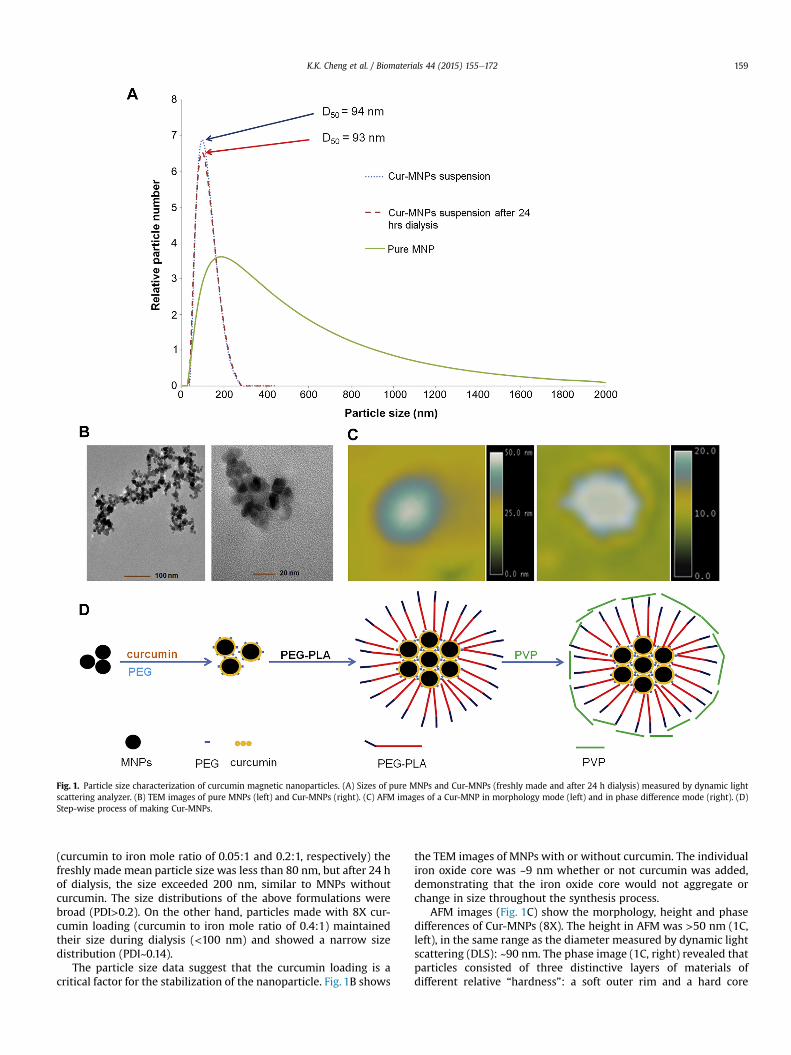
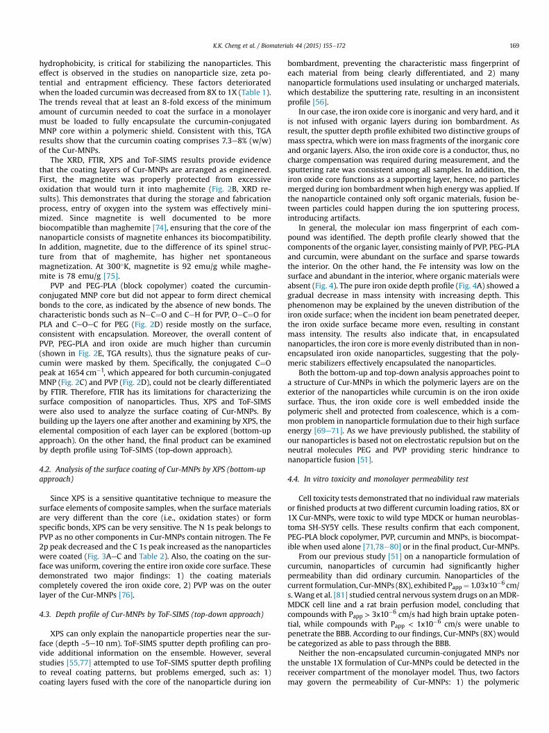
Fig. 1. Particle size characterization of curcumin magnetic nanoparticles. (A) Sizes of pure MNPs and Cur-MNPs (freshly made and after 24 h dialysis) measured by dynamic lightscattering analyzer. (B) TEM images of pure MNPs (left) and Cur-MNPs (right). (C) AFM images of a Cur-MNP in morphology mode (left) and in phase difference mode (right). (D)Step-wise process of making Cur-MNPs.
K.K. Cheng et al. / Biomaterials 44 (2015) 155e172 159
(curcumin to iron mole ratio of 0.05:1 and 0.2:1, respectively) thefreshly made mean particle size was less than 80 nm, but after 24 hof dialysis, the size exceeded 200 nm, similar to MNPs withoutcurcumin. The size distributions of the above formulations werebroad (PDI>0.2). On the other hand, particles made with 8X cur-cumin loading (curcumin to iron mole ratio of 0.4:1) maintainedtheir size during dialysis (<100 nm) and showed a narrow sizedistribution (PDI~0.14).
The particle size data suggest that the curcumin loading is acritical factor for the stabilization of the nanoparticle. Fig. 1B shows
the TEM images of MNPs with or without curcumin. The individualiron oxide core was ~9 nm whether or not curcumin was added,demonstrating that the iron oxide core would not aggregate orchange in size throughout the synthesis process.
AFM images (Fig. 1C) show the morphology, height and phasedifferences of Cur-MNPs (8X). The height in AFM was >50 nm (1C,left), in the same range as the diameter measured by dynamic lightscattering (DLS): ~90 nm. The phase image (1C, right) revealed thatparticles consisted of three distinctive layers of materials ofdifferent relative “hardness”: a soft outer rim and a hard core
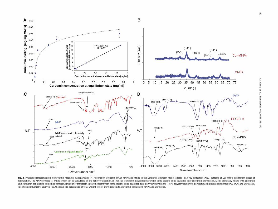
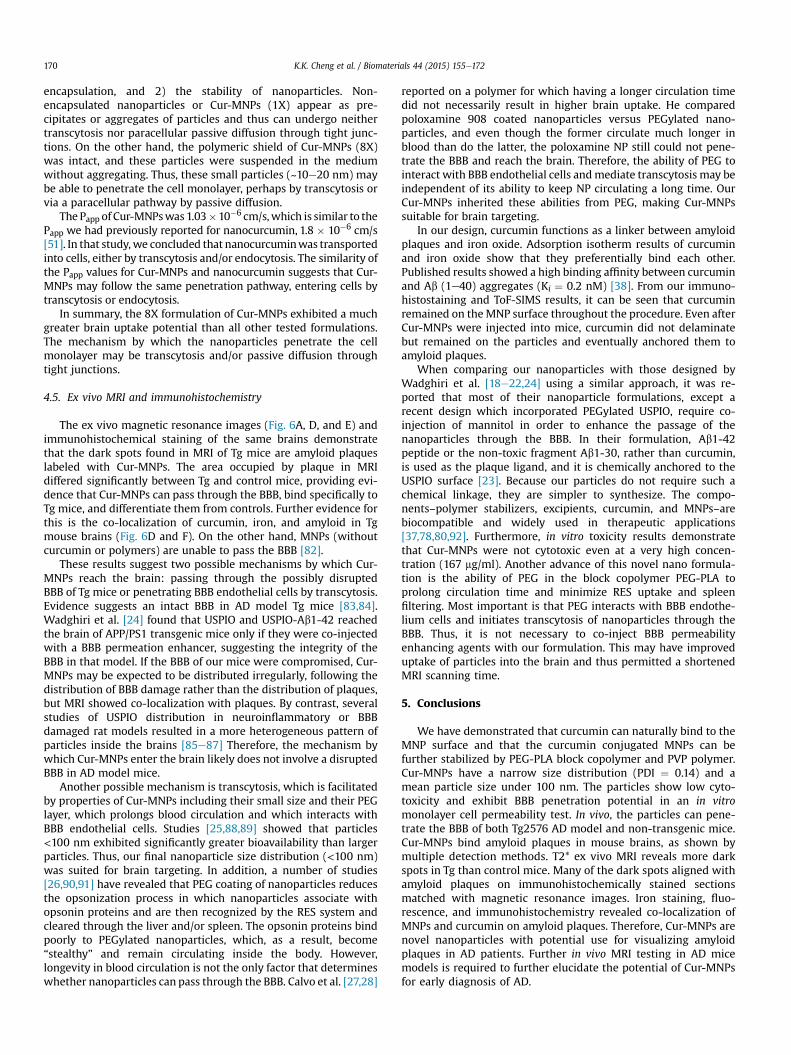
Fig. 2. Physical characterization of curcumin-magnetic nanoparticles. (A) Adsorption isotherm of Cur-MNPs and fitting to the Langmuir isotherm model (inset). (B) X-ray diffraction (XRD) patterns of Cur-MNPs at different stages offormulation. The MNP core size is ~9 nm, which can be calculated by the Scherrer equation. (C) Fourier transform infrared spectra with some specific bond peaks for pure curcumin, pure MNPs, MNPs physically mixed with curcuminand curcumin conjugated iron oxide complex. (D) Fourier transform infrared spectra with some specific bond peaks for pure polyvinylpyrrolidone (PVP), polyethylene glycol polylactic acid diblock copolymer (PEG-PLA) and Cur-MNPs.(E) Thermogravimetric analysis (TGA) shows the percentage of total weight loss of pure iron oxide, curcumin conjugated MNPs and Cur-MNPs.
K.K.Cheng
etal./
Biomaterials
44(2015)
155e172
160
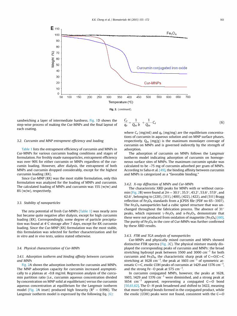
Fig. 2. (continued).
K.K. Cheng et al. / Biomaterials 44 (2015) 155e172 161
sandwiching a layer of intermediate hardness. Fig. 1D shows thestep-wise process of making the Cur-MNPs and the final layout ofeach coating.
3.2. Curcumin and MNP entrapment efficiency and loading
Table 1 lists the entrapment efficiency of curcumin and MNPs inCur-MNPs for various curcumin loading conditions and stages offormulation. For freshly made nanoparticles, entrapment efficiencywas over 90% for either curcumin or MNPs regardless of the cur-cumin loading. However, after dialysis, the entrapment of bothMNPs and curcumin dropped considerably, except for the highestcurcumin loading (8X).
Since Cur-MNP (8X) was the most stable formulation, only thisformulation was analyzed for the loading of MNPs and curcumin.The calculated loading of MNPs and curcumin was 15% (w/w) and8% (w/w), respectively.
3.3. Stability of nanoparticles
The zeta potential of fresh Cur-MNPs (Table 1) was nearly zerobut became quite negative after dialysis, except for high curcuminloading (8X). Correspondingly, some degree of particle precipita-tion was found at 4�C storage after 7 days, except for 8X curcuminloading. Since the Cur-MNP (8X) formulation was the most stable,this formulation was selected for further characterization and forin vitro and in vivo tests, unless stated otherwise.
3.4. Physical characterization of Cur-MNPs
3.4.1. Adsorption isotherm and binding affinity between curcuminand MNPs
Fig. 2A shows the adsorption isotherm for curcumin and MNPs.The MNP adsorption capacity for curcumin increased asymptoti-cally to a plateau at ~0.8 mg/ml. Regression analysis of the curcu-min partition ratio (i.e., curcumin aqueous concentration dividedby concentration onMNP solid at equilibrium) versus the curcuminaqueous concentration at equilibrium for the Langmuir isothermmodel (Fig. 2A inset) produced high linearity (R2 ¼ 0.996). TheLangmuir isotherm model is expressed by the following Eq. (6):
Ceqe
¼ 1Qm b
þ 1Qm
Ce (6)
where Ce (mg/ml) and qe (mg/mg) are the equilibrium concentra-tions of curcumin in aqueous solution and on MNP surface phases,respectively. Qm (mg/g) is the maximum monolayer coverage ofcurcumin on MNPs and is governed indirectly by the strength ofadsorption.
The adsorption of curcumin on MNPs follows the Langmuirisotherm model indicating adsorption of curcumin on homoge-neous surface sites of MNPs. The maximum curcumin uptake wascalculated to be ~75 mg of curcumin adsorbed per gram of MNPs.According to Saha et al. [49], the binding affinity between curcuminand MNPs is categorized as a “favorable binding.”
3.4.2. X-ray diffraction of MNPs and Cur-MNPsThe characteristic XRD peaks for MNPs with or without curcu-
min (Fig. 2B) were found at 2W¼ 30.1�, 35.5�, 43.2�, 53.6�, 57.0�, and62.6�, belonging to (220), (311), (400), (422), (422), and (511) Braggreflection of Fe3O4 standards from a JCPDS file (PDF no 65e3107).The Fe3O4 nanoparticles had a cubic spinel structure that was un-changed throughout the fabrication process. The absence of 31�
peaks, which represent g-Fe2O3 and a-Fe2O3, demonstrate thatthese were not produced from oxidation of magnetite (Fe3O4) [60].The purity of Fe3O4 in the core of Cur-MNPs was further confirmedby these XRD results.
3.4.3. FTIR and TGA analysis of nanoparticlesCur-MNPs and physically mixed curcumin and MNPs showed
distinctive FTIR spectra (Fig. 2C). The physical mixture mainly dis-played the corresponding peaks of curcumin and MNPs: the broadstretching hydroxyl peak between 3500 and 3000 cm�1 for bothcurcumin and Fe3O4, the characteristic sharp peak of C]O/C]Cstretching at 1628 cm�1, the peak at 1603 cm�1 of symmetric ar-omatic C]C, enolic COH peaks of curcumin at 1429 and 1376 cm�1,and the strong FeeO peak at 575 cm�1.
In curcumin conjugated MNPs, however, the peaks at 1628,1603, 1429 and 1376 cm�1 were diminished, and a strong peak at1654 cm�1 appeared, representing a conjugated C]O bond[50,61,62]. The OeH peak broadened and shifted to 3422, meaningthat more hydroxyl bonds formed in the conjugated product, whilethe enolic (COH) peaks were not found, consistent with the C]O
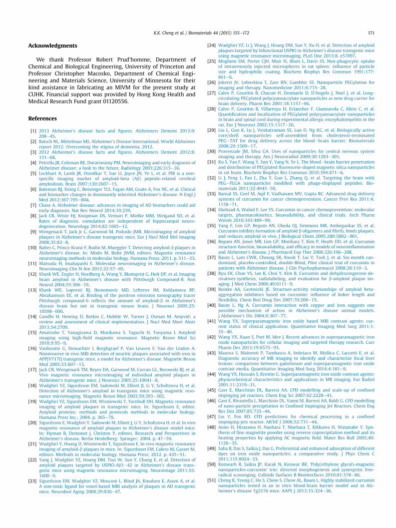
Fig. 3. Surface coating analysis of Cur-MNPs by bottom-up approach (using X-ray photoelectron spectroscopy [XPS]). Binding energy of each element is shown and compared for (A)Pure MNPs. (B) Curcumin conjugated PEG supported MNPs. (C) Cur-MNPs.
K.K. Cheng et al. / Biomaterials 44 (2015) 155e172162
and COH in the central part of curcumin molecules participating informing C]O conjugated bonds with Fe3O4 by hydrogen bonding.The FeeO peak was not altered and remained intact during theprocess.
Fig. 2D shows the spectra of the final, polymer-coated product,Cur-MNPs, and the polymers PVP and PEG-PLA (2ke10k). The Cur-MNPs inherited the characteristic peaks of PVP, PEG, and PLA, suchas NeC]O at 1654 and CeH at 2955 cm�1 for PVP, OeC]O at
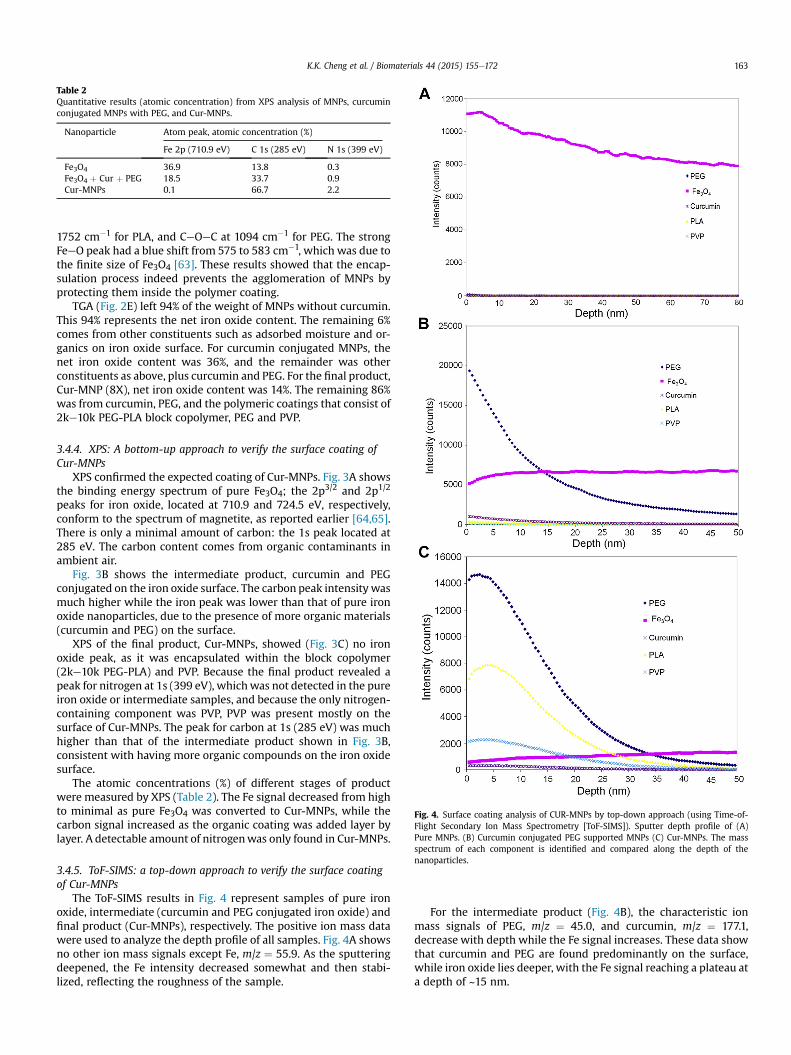
Table 2Quantitative results (atomic concentration) from XPS analysis of MNPs, curcuminconjugated MNPs with PEG, and Cur-MNPs.
Nanoparticle Atom peak, atomic concentration (%)
Fe 2p (710.9 eV) C 1s (285 eV) N 1s (399 eV)
Fe3O4 36.9 13.8 0.3Fe3O4 þ Cur þ PEG 18.5 33.7 0.9Cur-MNPs 0.1 66.7 2.2
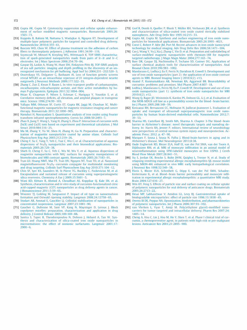
Fig. 4. Surface coating analysis of CUR-MNPs by top-down approach (using Time-of-Flight Secondary Ion Mass Spectrometry [ToF-SIMS]). Sputter depth profile of (A)Pure MNPs. (B) Curcumin conjugated PEG supported MNPs (C) Cur-MNPs. The massspectrum of each component is identified and compared along the depth of thenanoparticles.
K.K. Cheng et al. / Biomaterials 44 (2015) 155e172 163
1752 cm�1 for PLA, and CeOeC at 1094 cm�1 for PEG. The strongFeeO peak had a blue shift from 575 to 583 cm�1, which was due tothe finite size of Fe3O4 [63]. These results showed that the encap-sulation process indeed prevents the agglomeration of MNPs byprotecting them inside the polymer coating.
TGA (Fig. 2E) left 94% of the weight of MNPs without curcumin.This 94% represents the net iron oxide content. The remaining 6%comes from other constituents such as adsorbed moisture and or-ganics on iron oxide surface. For curcumin conjugated MNPs, thenet iron oxide content was 36%, and the remainder was otherconstituents as above, plus curcumin and PEG. For the final product,Cur-MNP (8X), net iron oxide content was 14%. The remaining 86%was from curcumin, PEG, and the polymeric coatings that consist of2ke10k PEG-PLA block copolymer, PEG and PVP.
3.4.4. XPS: A bottom-up approach to verify the surface coating ofCur-MNPs
XPS confirmed the expected coating of Cur-MNPs. Fig. 3A showsthe binding energy spectrum of pure Fe3O4; the 2p3/2 and 2p1/2
peaks for iron oxide, located at 710.9 and 724.5 eV, respectively,conform to the spectrum of magnetite, as reported earlier [64,65].There is only a minimal amount of carbon: the 1s peak located at285 eV. The carbon content comes from organic contaminants inambient air.
Fig. 3B shows the intermediate product, curcumin and PEGconjugated on the iron oxide surface. The carbon peak intensity wasmuch higher while the iron peak was lower than that of pure ironoxide nanoparticles, due to the presence of more organic materials(curcumin and PEG) on the surface.
XPS of the final product, Cur-MNPs, showed (Fig. 3C) no ironoxide peak, as it was encapsulated within the block copolymer(2ke10k PEG-PLA) and PVP. Because the final product revealed apeak for nitrogen at 1s (399 eV), whichwas not detected in the pureiron oxide or intermediate samples, and because the only nitrogen-containing component was PVP, PVP was present mostly on thesurface of Cur-MNPs. The peak for carbon at 1s (285 eV) was muchhigher than that of the intermediate product shown in Fig. 3B,consistent with having more organic compounds on the iron oxidesurface.
The atomic concentrations (%) of different stages of productwere measured by XPS (Table 2). The Fe signal decreased from highto minimal as pure Fe3O4 was converted to Cur-MNPs, while thecarbon signal increased as the organic coating was added layer bylayer. A detectable amount of nitrogenwas only found in Cur-MNPs.
3.4.5. ToF-SIMS: a top-down approach to verify the surface coatingof Cur-MNPs
The ToF-SIMS results in Fig. 4 represent samples of pure ironoxide, intermediate (curcumin and PEG conjugated iron oxide) andfinal product (Cur-MNPs), respectively. The positive ion mass datawere used to analyze the depth profile of all samples. Fig. 4A showsno other ion mass signals except Fe, m/z ¼ 55.9. As the sputteringdeepened, the Fe intensity decreased somewhat and then stabi-lized, reflecting the roughness of the sample.
For the intermediate product (Fig. 4B), the characteristic ionmass signals of PEG, m/z ¼ 45.0, and curcumin, m/z ¼ 177.1,decrease with depth while the Fe signal increases. These data showthat curcumin and PEG are found predominantly on the surface,while iron oxide lies deeper, with the Fe signal reaching a plateau ata depth of ~15 nm.
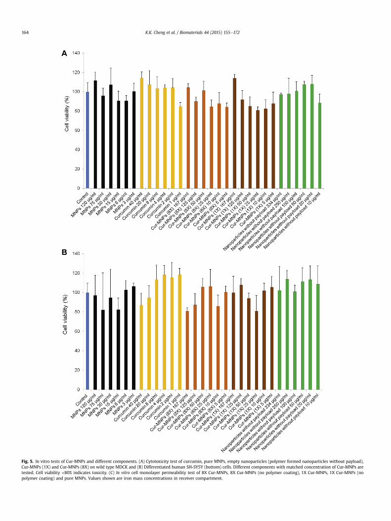
Fig. 5. In vitro tests of Cur-MNPs and different components. (A) Cytotoxicity test of curcumin, pure MNPs, empty nanoparticles (polymer formed nanoparticles without payload),Cur-MNPs (1X) and Cur-MNPs (8X) on wild type MDCK and (B) Differentiated human SH-SY5Y (bottom) cells. Different components with matched concentration of Cur-MNPs aretested. Cell viability <80% indicates toxicity. (C) In vitro cell monolayer permeability test of 8X Cur-MNPs, 8X Cur-MNPs (no polymer coating), 1X Cur-MNPs, 1X Cur-MNPs (nopolymer coating) and pure MNPs. Values shown are iron mass concentrations in receiver compartment.
K.K. Cheng et al. / Biomaterials 44 (2015) 155e172164
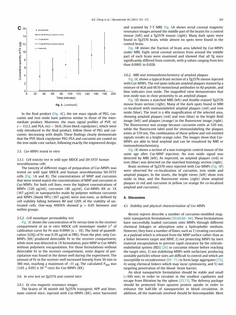
Fig. 5. (continued).
K.K. Cheng et al. / Biomaterials 44 (2015) 155e172 165
In the final product (Fig. 4C), the ion mass signals of PEG, cur-cumin and iron oxide have patterns similar to those of the inter-mediate product. Moreover, the mass signal profiles of PVP, m/z ¼ 112.1, and PLA, m/z ¼ 56.0, (from block copolymer), which wereonly introduced in the final product, follow those of PEG and cur-cumin: decreasing with depth. These findings clearly demonstratethat the PVP, block copolymer PEG-PLA and curcumin are coated onthe iron oxide core surface, following exactly the engineered design.
3.5. Cur-MNPs tested in vitro
3.5.1. Cell toxicity test in wild type MDCK and SH-SY5Y humanneuroblastoma cells
The toxicity of different stages of preparation of Cur-MNPs wastested on wild type MDCK and human neuroblastoma SH-SY5Ycells (Fig. 5A and B). The concentrations of MNP and curcuminthat were tested match the concentrations of MNP and curcumin inCur-MNPs. For both cell lines, even the highest concentrations ofMNPs (120 mg/ml), curcumin (40 mg/ml), Cur-MNPs 8X or 1X(167 mg/ml) or nanoparticles made by polymer without curcuminand MNPs (blank NPs) (167 mg/ml) were non-toxic, as defined bycell viability falling between 80 and 120% of the viability of un-treated cells. One-way ANOVA showed p > 0.05 between andwithin groups.
3.5.2. Cell monolayer permeability testFig. 5C shows the concentration of Fe versus time in the receiver
compartment of an in vitro MDCK cell monolayer model (r2 ofcalibration curve for Fe was 0.9999 (n ¼ 10). The limit of quantifi-cation (LOQ) of Fe was 0.39 mg/ml in PBS). From the plot, only Cur-MNPs (8X) produced detectable Fe in the receiver compartment,while nonewas detected in 1X formulation, pureMNP or Cur-MNPswithout polymeric encapsulation. For those formulations withoutdetectable Fe in the receiver compartment, some degree of pre-cipitation was found in the donor well during the experiment. Theamount of Fe in the receiver well increased linearly from 30 min to180 min, reaching a maximum of ~11 mg. The calculated Papp was(1.03 ± 0.05) x 10�6 cm/s for Cur-MNPs (8X).
3.6. In vivo test on Tg2576 and control mice
3.6.1. Ex vivo magnetic resonance imagesThe brains of 18 month old Tg2576 transgenic APP and litter-
mate control mice, injected with Cur-MNPs (8X), were harvested
and scanned by 7-T MRI. Fig. 6A shows serial coronal magneticresonance images around the middle part of the brains for a controlmouse (left) and a Tg2576 mouse (right). Many dark spots werefound in Tg2576 brain, while almost no spots were found in thecontrol brain.
Fig. 6B shows the fraction of brain area labeled by Cur-MNPsunder MRI. Eight serial coronal sections from around the middlepart of each brain were examined and showed that all Tg micesignificantly differed from controls, with p values ranging from lessthan 0.0001 to 0.028.
3.6.2. MRI and immunohistochemistry of amyloid plaquesFig. 6C shows a typical brain section of a Tg2576 mouse injected
with Cur-MNPs. The red spots indicate amyloid plaques stained by amixture of 4G8 and 6E10 monoclonal antibodies to Ab peptide, andblue indicates iron oxide. The magnified view demonstrates thatiron oxide was in close proximity to an amyloid plaque.
Fig. 6D shows a matched MRI (left) and double-stained Tg2576mouse brain section (right). Many of the dark spots found in MRIco-localized with immunolabeled amyloid plaques (red) and ironoxide (blue). The inset is a 40x magnification of the selected area,showing amyloid plaques (red) and iron (blue) in the bright fieldimage (left) and plaques (orange) in the fluorescent image (right).The fluorescence was orange because curcumin emits at 520 nm,while the fluorescent label used for immunolabeling the plaquesemits at 570 nm. The combination of these yellow and red emittedsignals results in a bright orange color. The images show that Cur-MNPs are able to bind amyloid and can be visualized by MRI orimmunohistochemistry.
Fig. 6E shows a section of a non-transgenic control mouse of thesame age after Cur-MNP injection. No iron oxide signal wasdetected by MRI (left). As expected, no amyloid plaques (red) oriron (blue) was detected on the matched histology section (right).
Brain sections of Tg2576 mice injected with Cur-MNPs (Fig. 6F)were observed for co-localization of curcumin, iron oxide andamyloid plaques. In the insets, the bright views (left) show ironoxide in blue, and the fluorescent views (right) show amyloidplaques in red and curcumin in yellow (or orange for co-localizedamyloid and curcumin).
4. Discussion
4.1. Stability and physical characterization of Cur-MNPs
Recent reports describe a number of curcumin-modified mag-netic nanoparticle formulations [50,60,66e68]. These formulationshave successfully loaded curcumin onto MNPs through differentchemical linkages or adsorption onto a hydrophobic medium.However, they have a number of flaws, such as 1) treating curcuminas a payload which is released from the MNP surface rather than asa linker between target and MNP, 2) not protecting MNPs by inertmaterial encapsulation to prevent rapid clearance by the reticulo-endothelial system (RES) [26] or curcumin release before reachingthe target sites, 3) not stabilizing MNPs with surfactant, producingunstable particles whose sizes are difficult to control and which aresusceptible to recoalescence [69e71] to form large aggregates [72],4) using chemical linkers which may incur cytotoxicity, and 5) nottargeting penetration of the bloodebrain barrier.
An ideal nanoparticle formulation should be stable and small(<100 nm) in order to circulate in the smallest capillaries andescape from filtration by the spleen [25,73]. The delivery packageshould be protected from opsonin protein uptake in order toenhance the half-life of nanoparticles in blood circulation. Inaddition, all the materials involved should be biocompatible. Most

K.K. Cheng et al. / Biomaterials 44 (2015) 155e172166
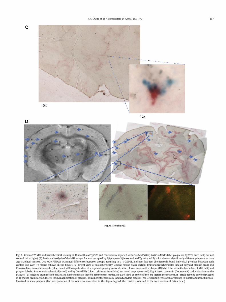
Fig. 6. (continued).
Fig. 6. Ex vivo T2* MRI and histochemical staining of 18 month old Tg2576 and control mice injected with Cur-MNPs (8X). (A) Cur-MNPs label plaques in Tg2576 mice (left) but notcontrol mice (right). (B) Statistical analysis of the MRI images for area occupied by Ab plaques (%) in control and Tg mice. All Tg mice showed significantly different plaque area thanage-matched controls. One way ANOVA examined differences between groups, resulting in p < 0.0001, and post-hoc test (Bonferroni) found individual p values between eachcontrol and each Tg mouse (shown in the figure). (C) Bright view of histochemically labeled mouse brain section. Immunohistochemically labeled amyloid plaques (red) andPrussian blue stained iron oxide (blue). Inset: 40X magnification of a region displaying co-localization of iron oxide with a plaque. (D) Match between the black dots of MRI (left) andplaques labeled immunohistochemically (red) and by Cur-MNPs (blue). Left inset: iron (blue) anchored on plaques (red). Right inset: curcumin (fluorescent) co-localization on theplaques. (E) Matched brain section of MRI and histochemically labeled aged control mouse. No dark spots or amyloid/iron are seen in the sections. (F) Triple-labeled amyloid plaquesin Tg mouse brain section. Insets: 100X magnification of plaques. Immunohistochemically labeled amyloid plaques (red), curcumin (yellow fluorescence in insets) and iron (blue) co-localized in some plaques. (For interpretation of the references to colour in this figure legend, the reader is referred to the web version of this article.)
K.K. Cheng et al. / Biomaterials 44 (2015) 155e172 167
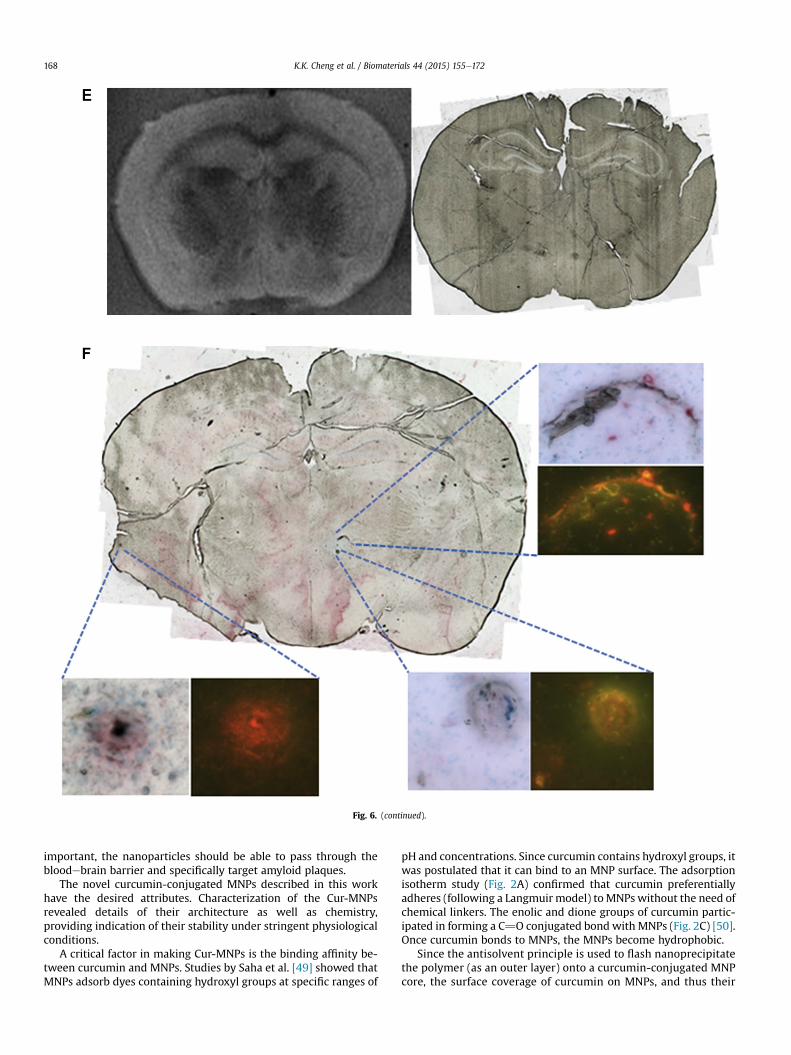
Fig. 6. (continued).
K.K. Cheng et al. / Biomaterials 44 (2015) 155e172168
important, the nanoparticles should be able to pass through thebloodebrain barrier and specifically target amyloid plaques.
The novel curcumin-conjugated MNPs described in this workhave the desired attributes. Characterization of the Cur-MNPsrevealed details of their architecture as well as chemistry,providing indication of their stability under stringent physiologicalconditions.
A critical factor in making Cur-MNPs is the binding affinity be-tween curcumin and MNPs. Studies by Saha et al. [49] showed thatMNPs adsorb dyes containing hydroxyl groups at specific ranges of
pH and concentrations. Since curcumin contains hydroxyl groups, itwas postulated that it can bind to an MNP surface. The adsorptionisotherm study (Fig. 2A) confirmed that curcumin preferentiallyadheres (following a Langmuir model) toMNPs without the need ofchemical linkers. The enolic and dione groups of curcumin partic-ipated in forming a C]O conjugated bond with MNPs (Fig. 2C) [50].Once curcumin bonds to MNPs, the MNPs become hydrophobic.
Since the antisolvent principle is used to flash nanoprecipitatethe polymer (as an outer layer) onto a curcumin-conjugated MNPcore, the surface coverage of curcumin on MNPs, and thus their
K.K. Cheng et al. / Biomaterials 44 (2015) 155e172 169
hydrophobicity, is critical for stabilizing the nanoparticles. Thiseffect is observed in the studies on nanoparticle size, zeta po-tential and entrapment efficiency. These factors deterioratedwhen the loaded curcuminwas decreased from 8X to 1X (Table 1).The trends reveal that at least an 8-fold excess of the minimumamount of curcumin needed to coat the surface in a monolayermust be loaded to fully encapsulate the curcumin-conjugatedMNP core within a polymeric shield. Consistent with this, TGAresults show that the curcumin coating comprises 7.3e8% (w/w)of the Cur-MNPs.
The XRD, FTIR, XPS and ToF-SIMS results provide evidencethat the coating layers of Cur-MNPs are arranged as engineered.First, the magnetite was properly protected from excessiveoxidation that would turn it into maghemite (Fig. 2B, XRD re-sults). This demonstrates that during the storage and fabricationprocess, entry of oxygen into the system was effectively mini-mized. Since magnetite is well documented to be morebiocompatible than maghemite [74], ensuring that the core of thenanoparticle consists of magnetite enhances its biocompatibility.In addition, magnetite, due to the difference of its spinel struc-ture from that of maghemite, has higher net spontaneousmagnetization. At 300�K, magnetite is 92 emu/g while maghe-mite is 78 emu/g [75].
PVP and PEG-PLA (block copolymer) coated the curcumin-conjugated MNP core but did not appear to form direct chemicalbonds to the core, as indicated by the absence of new bonds. Thecharacteristic bonds such as NeC¼O and CeH for PVP, OeC¼O forPLA and CeOeC for PEG (Fig. 2D) reside mostly on the surface,consistent with encapsulation. Moreover, the overall content ofPVP, PEG-PLA and iron oxide are much higher than curcumin(shown in Fig. 2E, TGA results), thus the signature peaks of cur-cumin were masked by them. Specifically, the conjugated C¼Opeak at 1654 cm�1, which appeared for both curcumin-conjugatedMNP (Fig. 2C) and PVP (Fig. 2D), could not be clearly differentiatedby FTIR. Therefore, FTIR has its limitations for characterizing thesurface composition of nanoparticles. Thus, XPS and ToF-SIMSwere also used to analyze the surface coating of Cur-MNPs. Bybuilding up the layers one after another and examining by XPS, theelemental composition of each layer can be explored (bottom-upapproach). On the other hand, the final product can be examinedby depth profile using ToF-SIMS (top-down approach).
4.2. Analysis of the surface coating of Cur-MNPs by XPS (bottom-upapproach)
Since XPS is a sensitive quantitative technique to measure thesurface elements of composite samples, when the surface materialsare very different than the core (i.e., oxidation states) or formspecific bonds, XPS can be very sensitive. The N 1s peak belongs toPVP as no other components in Cur-MNPs contain nitrogen. The Fe2p peak decreased and the C 1s peak increased as the nanoparticleswere coated (Fig. 3AeC and Table 2). Also, the coating on the sur-face was uniform, covering the entire iron oxide core surface. Thesedemonstrated two major findings: 1) the coating materialscompletely covered the iron oxide core, 2) PVP was on the outerlayer of the Cur-MNPs [76].
4.3. Depth profile of Cur-MNPs by ToF-SIMS (top-down approach)
XPS can only explain the nanoparticle properties near the sur-face (depth ~5e10 nm). ToF-SIMS sputter depth profiling can pro-vide additional information on the ensemble. However, severalstudies [55,77] attempted to use ToF-SIMS sputter depth profilingto reveal coating patterns, but problems emerged, such as: 1)coating layers fused with the core of the nanoparticle during ion
bombardment, preventing the characteristic mass fingerprint ofeach material from being clearly differentiated, and 2) manynanoparticle formulations used insulating or uncharged materials,which destabilize the sputtering rate, resulting in an inconsistentprofile [56].
In our case, the iron oxide core is inorganic and very hard, and itis not infused with organic layers during ion bombardment. Asresult, the sputter depth profile exhibited two distinctive groups ofmass spectra, which were ion mass fragments of the inorganic coreand organic layers. Also, the iron oxide core is a conductor, thus, nocharge compensation was required during measurement, and thesputtering rate was consistent among all samples. In addition, theiron oxide core functions as a supporting layer, hence, no particlesmerged during ion bombardment when high energy was applied. Ifthe nanoparticle contained only soft organic materials, fusion be-tween particles could happen during the ion sputtering process,introducing artifacts.
In general, the molecular ion mass fingerprint of each com-pound was identified. The depth profile clearly showed that thecomponents of the organic layer, consisting mainly of PVP, PEG-PLAand curcumin, were abundant on the surface and sparse towardsthe interior. On the other hand, the Fe intensity was low on thesurface and abundant in the interior, where organic materials wereabsent (Fig. 4). The pure iron oxide depth profile (Fig. 4A) showed agradual decrease in mass intensity with increasing depth. Thisphenomenon may be explained by the uneven distribution of theiron oxide surface; when the incident ion beam penetrated deeper,the iron oxide surface became more even, resulting in constantmass intensity. The results also indicate that, in encapsulatednanoparticles, the iron core is more evenly distributed than in non-encapsulated iron oxide nanoparticles, suggesting that the poly-meric stabilizers effectively encapsulated the nanoparticles.
Both the bottom-up and top-down analysis approaches point toa structure of Cur-MNPs in which the polymeric layers are on theexterior of the nanoparticles while curcumin is on the iron oxidesurface. Thus, the iron oxide core is well embedded inside thepolymeric shell and protected from coalescence, which is a com-mon problem in nanoparticle formulation due to their high surfaceenergy [69e71]. As we have previously published, the stability ofour nanoparticles is based not on electrostatic repulsion but on theneutral molecules PEG and PVP providing steric hindrance tonanoparticle fusion [51].
4.4. In vitro toxicity and monolayer permeability test
Cell toxicity tests demonstrated that no individual rawmaterialsor finished products at two different curcumin loading ratios, 8X or1X Cur-MNPs, were toxic to wild type MDCK or human neuroblas-toma SH-SY5Y cells. These results confirm that each component,PEG-PLA block copolymer, PVP, curcumin and MNPs, is biocompat-ible when used alone [71,78e80] or in the final product, Cur-MNPs.
From our previous study [51] on a nanoparticle formulation ofcurcumin, nanoparticles of curcumin had significantly higherpermeability than did ordinary curcumin. Nanoparticles of thecurrent formulation, Cur-MNPs (8X), exhibited Papp¼ 1.03ⅹ10�6 cm/s.Wang et al. [81] studied central nervous system drugs on anMDR-MDCK cell line and a rat brain perfusion model, concluding thatcompounds with Papp > 3ⅹ10�6 cm/s had high brain uptake poten-tial, while compounds with Papp < 1ⅹ10�6 cm/s were unable topenetrate the BBB. According to our findings, Cur-MNPs (8X) wouldbe categorized as able to pass through the BBB.
Neither the non-encapsulated curcumin-conjugated MNPs northe unstable 1X formulation of Cur-MNPs could be detected in thereceiver compartment of the monolayer model. Thus, two factorsmay govern the permeability of Cur-MNPs: 1) the polymeric
K.K. Cheng et al. / Biomaterials 44 (2015) 155e172170
encapsulation, and 2) the stability of nanoparticles. Non-encapsulated nanoparticles or Cur-MNPs (1X) appear as pre-cipitates or aggregates of particles and thus can undergo neithertranscytosis nor paracellular passive diffusion through tight junc-tions. On the other hand, the polymeric shield of Cur-MNPs (8X)was intact, and these particles were suspended in the mediumwithout aggregating. Thus, these small particles (~10e20 nm) maybe able to penetrate the cell monolayer, perhaps by transcytosis orvia a paracellular pathway by passive diffusion.
The Papp of Cur-MNPswas1.03�10�6 cm/s,which is similar to thePapp we had previously reported for nanocurcumin, 1.8 � 10�6 cm/s[51]. In that study,we concluded that nanocurcuminwas transportedinto cells, either by transcytosis and/or endocytosis. The similarity ofthe Papp values for Cur-MNPs and nanocurcumin suggests that Cur-MNPs may follow the same penetration pathway, entering cells bytranscytosis or endocytosis.
In summary, the 8X formulation of Cur-MNPs exhibited a muchgreater brain uptake potential than all other tested formulations.The mechanism by which the nanoparticles penetrate the cellmonolayer may be transcytosis and/or passive diffusion throughtight junctions.
4.5. Ex vivo MRI and immunohistochemistry
The ex vivo magnetic resonance images (Fig. 6A, D, and E) andimmunohistochemical staining of the same brains demonstratethat the dark spots found in MRI of Tg mice are amyloid plaqueslabeled with Cur-MNPs. The area occupied by plaque in MRIdiffered significantly between Tg and control mice, providing evi-dence that Cur-MNPs can pass through the BBB, bind specifically toTg mice, and differentiate them from controls. Further evidence forthis is the co-localization of curcumin, iron, and amyloid in Tgmouse brains (Fig. 6D and F). On the other hand, MNPs (withoutcurcumin or polymers) are unable to pass the BBB [82].
These results suggest two possible mechanisms by which Cur-MNPs reach the brain: passing through the possibly disruptedBBB of Tg mice or penetrating BBB endothelial cells by transcytosis.Evidence suggests an intact BBB in AD model Tg mice [83,84].Wadghiri et al. [24] found that USPIO and USPIO-Ab1-42 reachedthe brain of APP/PS1 transgenic mice only if they were co-injectedwith a BBB permeation enhancer, suggesting the integrity of theBBB in that model. If the BBB of our mice were compromised, Cur-MNPs may be expected to be distributed irregularly, following thedistribution of BBB damage rather than the distribution of plaques,but MRI showed co-localization with plaques. By contrast, severalstudies of USPIO distribution in neuroinflammatory or BBBdamaged rat models resulted in a more heterogeneous pattern ofparticles inside the brains [85e87] Therefore, the mechanism bywhich Cur-MNPs enter the brain likely does not involve a disruptedBBB in AD model mice.
Another possible mechanism is transcytosis, which is facilitatedby properties of Cur-MNPs including their small size and their PEGlayer, which prolongs blood circulation and which interacts withBBB endothelial cells. Studies [25,88,89] showed that particles<100 nm exhibited significantly greater bioavailability than largerparticles. Thus, our final nanoparticle size distribution (<100 nm)was suited for brain targeting. In addition, a number of studies[26,90,91] have revealed that PEG coating of nanoparticles reducesthe opsonization process in which nanoparticles associate withopsonin proteins and are then recognized by the RES system andcleared through the liver and/or spleen. The opsonin proteins bindpoorly to PEGylated nanoparticles, which, as a result, become“stealthy” and remain circulating inside the body. However,longevity in blood circulation is not the only factor that determineswhether nanoparticles can pass through the BBB. Calvo et al. [27,28]
reported on a polymer for which having a longer circulation timedid not necessarily result in higher brain uptake. He comparedpoloxamine 908 coated nanoparticles versus PEGylated nano-particles, and even though the former circulate much longer inblood than do the latter, the poloxamine NP still could not pene-trate the BBB and reach the brain. Therefore, the ability of PEG tointeract with BBB endothelial cells andmediate transcytosis may beindependent of its ability to keep NP circulating a long time. OurCur-MNPs inherited these abilities from PEG, making Cur-MNPssuitable for brain targeting.
In our design, curcumin functions as a linker between amyloidplaques and iron oxide. Adsorption isotherm results of curcuminand iron oxide show that they preferentially bind each other.Published results showed a high binding affinity between curcuminand Ab (1e40) aggregates (Ki ¼ 0.2 nM) [38]. From our immuno-histostaining and ToF-SIMS results, it can be seen that curcuminremained on theMNP surface throughout the procedure. Even afterCur-MNPs were injected into mice, curcumin did not delaminatebut remained on the particles and eventually anchored them toamyloid plaques.
When comparing our nanoparticles with those designed byWadghiri et al. [18e22,24] using a similar approach, it was re-ported that most of their nanoparticle formulations, except arecent design which incorporated PEGylated USPIO, require co-injection of mannitol in order to enhance the passage of thenanoparticles through the BBB. In their formulation, Ab1-42peptide or the non-toxic fragment Ab1-30, rather than curcumin,is used as the plaque ligand, and it is chemically anchored to theUSPIO surface [23]. Because our particles do not require such achemical linkage, they are simpler to synthesize. The compo-nents–polymer stabilizers, excipients, curcumin, and MNPs–arebiocompatible and widely used in therapeutic applications[37,78,80,92]. Furthermore, in vitro toxicity results demonstratethat Cur-MNPs were not cytotoxic even at a very high concen-tration (167 mg/ml). Another advance of this novel nano formula-tion is the ability of PEG in the block copolymer PEG-PLA toprolong circulation time and minimize RES uptake and spleenfiltering. Most important is that PEG interacts with BBB endothe-lium cells and initiates transcytosis of nanoparticles through theBBB. Thus, it is not necessary to co-inject BBB permeabilityenhancing agents with our formulation. This may have improveduptake of particles into the brain and thus permitted a shortenedMRI scanning time.
5. Conclusions
We have demonstrated that curcumin can naturally bind to theMNP surface and that the curcumin conjugated MNPs can befurther stabilized by PEG-PLA block copolymer and PVP polymer.Cur-MNPs have a narrow size distribution (PDI ¼ 0.14) and amean particle size under 100 nm. The particles show low cyto-toxicity and exhibit BBB penetration potential in an in vitromonolayer cell permeability test. In vivo, the particles can pene-trate the BBB of both Tg2576 AD model and non-transgenic mice.Cur-MNPs bind amyloid plaques in mouse brains, as shown bymultiple detection methods. T2* ex vivo MRI reveals more darkspots in Tg than control mice. Many of the dark spots aligned withamyloid plaques on immunohistochemically stained sectionsmatched with magnetic resonance images. Iron staining, fluo-rescence, and immunohistochemistry revealed co-localization ofMNPs and curcumin on amyloid plaques. Therefore, Cur-MNPs arenovel nanoparticles with potential use for visualizing amyloidplaques in AD patients. Further in vivo MRI testing in AD micemodels is required to further elucidate the potential of Cur-MNPsfor early diagnosis of AD.
K.K. Cheng et al. / Biomaterials 44 (2015) 155e172 171
Acknowledgments
We thank Professor Robert Prud'homme, Department ofChemical and Biological Engineering, University of Princeton andProfessor Christopher Macosko, Department of Chemical Engi-neering and Materials Science, University of Minnesota for theirkind assistance in fabricating an MIVM for the present study atCUHK. Financial support was provided by Hong Kong Health andMedical Research Fund grant 01120556.
References
[1] 2013 Alzheimer's disease facts and figures. Alzheimers Dement 2013;9:208e45.
[2] Batsch NL, Mittelman MS, Alzheimer's Disease International. World Alzheimerreport 2012: Overcoming the stigma of dementia. 2012.
[3] 2012 Alzheimer's disease facts and figures. Alzheimers Dement 2012;8:131e68.
[4] Petrella JR, Coleman RE, Doraiswamy PM. Neuroimaging and early diagnosis ofAlzheimer disease: a look to the future. Radiology 2003;226:315e36.
[5] Lockhart A, Lamb JR, Osredkar T, Sue LI, Joyce JN, Ye L, et al. PIB is a non-specific imaging marker of amyloid-beta (Ab) peptide-related cerebralamyloidosis. Brain 2007;130:2607e15.
[6] Bateman RJ, Xiong C, Benzinger TLS, Fagan AM, Goate A, Fox NC, et al. Clinicaland biomarker changes in dominantly inherited Alzheimer's disease. N Engl JMed 2012;367:795e804.
[7] Chase A. Alzheimer disease: advances in imaging of AD biomarkers could aidearly diagnosis. Nat Rev Neurol 2014;10:239.
[8] Jack CR, Wiste HJ, Knopman DS, Vemuri P, Mielke MM, Weigand SD, et al.Rates of diagnosis. cumulation are independent of hippocampal neuro-degeneration. Neurology 2014;82:1605e12.
[9] Wengenack T, Jack Jr C, Garwood M, Poduslo JMR. Microimaging of amyloidplaques in Alzheimer's disease transgenic mice. Eur J Nucl Med Mol Imaging2008;35:82e8.
[10] Baltes C, Princz-Kranz F, Rudin M, Mueggler T. Detecting amyloid-b plaques inAlzheimer's disease. In: Modo M, Bulte JWM, editors. Magnetic resonanceneuroimaging methods in molecular biology. Humana Press; 2011. p. 511e33.
[11] Matsuda H, Imabayashi E. Molecular neuroimaging in Alzheimer's disease.Neuroimaging Clin N Am 2012;22:57e65.
[12] Klunk WE, Engler H, Nordberg A, Wang Y, Blomqvist G, Holt DP, et al. Imagingbrain amyloid in Alzheimer's disease with Pittsburgh Compound-B. AnnNeurol 2004;55:306e19.
[13] Klunk WE, Lopresti BJ, Ikonomovic MD, Lefterov IM, Koldamova RP,Abrahamson EE, et al. Binding of the positron emission tomography tracerPittsburgh compound-b reflects the amount of amyloid-b in Alzheimer'sdisease brain but not in transgenic mouse brain. J Neurosci 2005;25:10598e606.
[14] Gandhi H, Hewing D, Botkin C, Hubble W, Turner J, Osman M. Amyvid: areview and assessment of clinical implementation. J Nucl Med Meet Abstr2013;54:2709.
[15] Amatsubo T, Yanagisawa D, Morikawa S, Taguchi H, Tooyama I. Amyloidimaging using high-field magnetic resonance. Magnetic Reson Med Sci2010;9:95e9.
[16] Vanhoutte G, Dewachter I, Borghgraef P, Van Leuven F, Van der Linden A.Noninvasive in vivo MRI detection of neuritic plaques associated with iron inAPP[V717I] transgenic mice, a model for Alzheimer's disease. Magnetic ResonMed 2005;53:607e13.
[17] Jack CR, Wengenack TM, Reyes DA, Garwood M, Curran GL, Borowski BJ, et al.Vivo magnetic resonance microimaging of individual amyloid plaques inAlzheimer's transgenic mice. J Neurosci 2005;25:10041e8.
[18] Wadghiri YZ, Sigurdsson EM, Sadowski M, Elliott JI, Li Y, Scholtzova H, et al.Detection of Alzheimer's amyloid in transgenic mice using magnetic reso-nance microimaging. Magnetic Reson Med 2003;50:293e302.
[19] Wadghiri YZ, Sigurdsson EM, Wisniewski T, Turnbull DH. Magnetic resonanceimaging of amyloid plaques in transgenic mice. In: Sigurdsson E, editor.Amyloid proteins: methods and protocols methods in molecular biology.Humana Press Inc.; 2004. p. 365e79.
[20] Sigurdsson E, Wadghiri Y, Sadowski M, Elliott J, Li Y, Scholtzova H, et al. In vivomagnetic resonance of amyloid plaques in Alzheimer's disease model mice.In: Hyman B, Demonet J, Christen Y, editors. Research and Perspectives inAlzheimer's disease. Berlin Heidelberg: Springer; 2004. p. 47e59.
[21] Wadghiri Y, Hoang D, Wisniewski T, Sigurdsson E. In vivo magnetic resonanceimaging of amyloid-b plaques in mice. In: Sigurdsson EM, Calero M, Gasset M,editors. Methods in molecular biology. Humana Press; 2012. p. 435e51.
[22] Yang J, Wadghiri YZ, Hoang DM, Tsui W, Sun Y, Chung E, et al. Detection ofamyloid plaques targeted by USPIO-Ab1e42 in Alzheimer's disease trans-genic mice using magnetic resonance microimaging. Neuroimage 2011;55:1600e9.
[23] Sigurdsson EM, Wadghiri YZ, Mosconi L, Blind JA, Knudsen E, Asuni A, et al.A non-toxic ligand for voxel-based MRI analysis of plaques in AD transgenicmice. Neurobiol Aging 2008;29:836e47.
[24] Wadghiri YZ, Li J, Wang J, Hoang DM, Sun Y, Xu H, et al. Detection of amyloidplaques targeted by bifunctional USPIO in Alzheimer's disease transgenic miceusing magnetic resonance microimaging. PLoS One 2013;8. e57097.
[25] Moghimi SM, Porter CJH, Muir IS, Illum L, Davis SS. Non-phagocytic uptakeof intravenously injected microspheres in rat spleen: influence of particlesize and hydrophilic coating. Biochem Biophys Res Commun 1991;177:861e6.
[26] Jokerst JV, Lobovkina T, Zare RN, Gambhir SS. Nanoparticle PEGylation forimaging and therapy. Nanomedicine 2011;6:715e28.
[27] Calvo P, Gouritin B, Chacun H, Desmaele D, D'Angelo J, Noel J, et al. Long-circulating PEGylated polycyanoacrylate nanoparticles as new drug carrier forbrain delivery. Pharm Res 2001;18:1157e66.
[28] Calvo P, Gouritin B, Villarroya H, Eclancher F, Giannavola C, Klein C, et al.Quantification and localization of PEGylated polycyanoacrylate nanoparticlesin brain and spinal cord during experimental allergic encephalomyelitis in therat. Eur J Neurosci 2002;15:1317e26.
[29] Liu L, Guo K, Lu J, Venkatraman SS, Luo D, Ng KC, et al. Biologically activecore/shell nanoparticles self-assembled from cholesterol-terminatedPEGeTAT for drug delivery across the bloodebrain barrier. Biomaterials2008;29:1509e17.
[30] Provenzale JM, Silva GA. Uses of nanoparticles for central nervous systemimaging and therapy. Am J Neuroradiol 2009;30:1293e301.
[31] Ku S, Yan F, Wang Y, Sun Y, Yang N, Ye L. The bloodebrain barrier penetrationand distribution of PEGylated fluorescein-doped magnetic silica nanoparticlesin rat brain. Biochem Biophys Res Commun 2010;394:871e6.
[32] Li J, Feng L, Fan L, Zha Y, Guo L, Zhang Q, et al. Targeting the brain withPEGePLGA nanoparticles modified with phage-displayed peptides. Bio-materials 2011;32:4943e50.
[33] Bansal SS, Goel M, Aqil F, Vadhanam MV, Gupta RC. Advanced drug deliverysystems of curcumin for cancer chemoprevention. Cancer Prev Res 2011;4:1158e71.
[34] Shehzad A, Wahid F, Lee YS. Curcumin in cancer chemoprevention: moleculartargets, pharmacokinetics, bioavailability, and clinical trials. Arch PharmWeinh 2010;343:489e99.
[35] Yang F, Lim GP, Begum AN, Ubeda OJ, Simmons MR, Ambegaokar SS, et al.Curcumin inhibits formation of amyloid b oligomers and fibrils, binds plaques,and reduces amyloid in vivo. J Biological Chem 2005;280:5892e901.
[36] Begum AN, Jones MR, Lim GP, Morihara T, Kim P, Heath DD, et al. Curcuminstructure-function, bioavailability, and efficacy in models of neuroinflammationand Alzheimer's disease. J Pharmacol Exp Ther 2008;326:196e208.
[37] Baum L, Lam CWK, Cheung SK, Kwok T, Lui V, Tsoh J, et al. Six-month ran-domized, placebo-controlled, double-Blind, Pilot clinical trial of curcumin inpatients with Alzheimer disease. J Clin Psychopharmacol 2008;28:110e3.
[38] Ryu EK, Choe YS, Lee K, Choi Y, Kim B. Curcumin and dehydrozingerone de-rivatives:synthesis, radiolabeling, and evaluation for b-amyloid plaque im-aging. J Med Chem 2006;49:6111e9.
[39] Reinke AA, Gestwicki JE. Structure-activity relationships of amyloid beta-aggregation inhibitors based on curcumin: influence of linker length andflexibility. Chem Biol Drug Des 2007;70:206e15.
[40] Baum L, Ng A. Curcumin interaction with copper and iron suggests onepossible mechanism of action in Alzheimer's disease animal models.J Alzheimer's Dis 2004;6:367e77.
[41] Wang YX. Superparamagnetic iron oxide based MRI contrast agents: cur-rent status of clinical application. Quantitative Imaging Med Surg 2011;1:35e40.
[42] Wang YX, Xuan S, Port M, Idee J. Recent advances in superparamagnetic ironoxide nanoparticles for cellular imaging and targeted therapy research. CurrPharm Des 2013;19:6575e93.
[43] Maurea S, Mainenti P, Tambasco A, Imbriaco M, Mollica C, Laccetti E, et al.Diagnostic accuracy of MR imaging to identify and characterize focal liverlesions: comparison between gadolinium and superparamagnetic iron oxidecontrast media. Quantitative Imaging Med Surg 2014;4:181e9.
[44] Wang YX, Hussain S, Krestin G. Superparamagnetic iron oxide contrast agents:physicochemical characteristics and applications in MR imaging. Eur Radiol2001;11:2319e31.
[45] Gavi E, Marchisio DL, Barresi AA. CFD modelling and scale-up of confinedimpinging jet reactors. Chem Eng Sci 2007;62:2228e41.
[46] Gavi E, Rivautella L, Marchisio DL, Vanni M, Barresi AA, Baldi G. CFD modellingof nano-particle precipitation in Confined Impinging Jet Reactors. Chem EngRes Des 2007;85:735e44.
[47] Liu Y, Fox RO. CFD predictions for chemical processing in a confinedimpinging-jets reactor. AIChE J 2006;52:731e44.
[48] Aono H, Hirazawa H, Naohara T, Maehara T, Kikkawa H, Watanabe Y. Syn-thesis of fine magnetite powder using reverse coprecipitation method and itsheating properties by applying AC magnetic field. Mater Res Bull 2005;40:1126e35.
[49] Saha B, Das S, Saikia J, Das G. Preferential and enhanced adsorption of differentdyes on iron oxide nanoparticles: a comparative study. J Phys Chem C2011;115:8024e33.
[50] Konwarh R, Saikia JP, Karak N, Konwar BK. ‘Poly(ethylene glycol)-magneticnanoparticles-curcumin’ trio: directed morphogenesis and synergistic free-radical scavenging. Colloids Surfaces B Biointerfaces 2010;81:578e86.
[51] Cheng K, Yeung C, Ho S, Chow S, Chow AL, Baum L. Highly stabilized curcuminnanoparticles tested in an in vitro blood-brain barrier model and in Alz-heimer's disease Tg2576 mice. AAPS J 2013;15:324e36.
K.K. Cheng et al. / Biomaterials 44 (2015) 155e172172
[52] Gupta AK, Gupta M. Cytotoxicity suppression and cellular uptake enhance-ment of surface modified magnetic nanoparticles. Biomaterials 2005;26:1565e73.
[53] Koppolu B, Rahimi M, Nattama S, Wadajkar A, Nguyen KT. Development ofmultiple-layer polymeric particles for targeted and controlled drug delivery.Nanomedicine 2010;6:355e61.
[54] Bascom WD, Chen W. Effect of plasma treatment on the adhesion of carbonfibers to thermoplastic polymers. J Adhesion 1991;34:99e119.
[55] Szymczak W, Menzel N, Kreyling WG, Wittmaack K. TOF-SIMS characterisa-tion of spark-generated nanoparticles made from pairs of IreIr and IreCelectrodes. Int J Mass Spectrom 2006;254:70e84.
[56] Gaspar DJ, Laskin A, Wang W, Hunt SW, Finlayson-Pitts BJ. TOF-SIMS analysisof sea salt particles: imaging and depth profiling in the discovery of an un-recognized mechanism for pH buffering. Appl Surf Sci 2004;231e232:520e3.
[57] Ossovskaya VS, Dolganov G, Basbaum AI. Loss of function genetic screensreveal MTGR1 as an intracellular repressor of b1 integrin-dependent neuriteoutgrowth. J Neurosci Methods 2009;177:322e33.
[58] Zhang C, Zuo Z, Kwan P, Baum L. In vitro transport profile of carbamazepine,oxcarbazepine, eslicarbazepine acetate, and their active metabolites by hu-man P-glycoprotein. Epilepsia 2011;52:1894e904.
[59] Hsiao K, Chapman P, Nilsen S, Eckman C, Harigaya Y, Younkin S, et al.Correlative memory deficits, ab elevation, and amyloid plaques in transgenicmice. Science 1996;274:99e103.
[60] Yallapu MM, Othman SF, Curtis ET, Gupta BK, Jaggi M, Chauhan SC. Multi-functional magnetic nanoparticles for magnetic resonance imaging and cancertherapy. Biomaterials 2011;32:1890e905.
[61] Namduri H, Nasrazadani S. Quantitative analysis of iron oxides using Fouriertransform infrared spectrophotometry. Corros Sci 2008;50:2493e7.
[62] Zhao X, Jiang T, Wang L, Yang H, Zhang S, Zhou P. Interaction of curcumin withZn(II) and Cu(II) ions based on experiment and theoretical calculation. J MolStruct 2010;984:316e25.
[63] Ma M, Zhang Y, Yu W, Shen H, Zhang H, Gu N. Preparation and character-ization of magnetite nanoparticles coated by amino silane. Colloids SurfPhysicochem Eng Asp 2003;212:219e26.
[64] Cheng F, Su C, Yang Y, Yeh C, Tsai C, Wu C, et al. Characterization of aqueousdispersions of Fe3O4 nanoparticles and their biomedical applications. Bio-materials 2005;26:729e38.
[65] Shieh D, Cheng F, Su C, Yeh C, Wu M, Wu Y, et al. Aqueous dispersions ofmagnetite nanoparticles with NH3
þ surfaces for magnetic manipulations ofbiomolecules and MRI contrast agents. Biomaterials 2005;26:7183e91.
[66] Tran LD, Hoang NMT, Mai TT, Tran HV, Nguyen NT, Tran TD, et al. Nanosizedmagnetofluorescent Fe3O4ecurcumin conjugate for multimodal monitoringand drug targeting. Colloids Surf Physicochem Eng Asp 2010;371:104e12.
[67] Chin SF, Iyer KS, Saunders M, St Pierre TG, Buckley C, Paskevicius M, et al.Encapsulation and sustained release of curcumin using superparamagneticsilica reservoirs. Chemistry A Eur J 2009;15:5661e5.
[68] Wani KD, Kitture R, Ahmed A, Choudhari AS, Koppikar SJ, Kale SN, et al.Synthesis, characterization and in vitro study of curcumin-functionalized citricacid-capped magnetic (CCF) nanoparticles as drug delivery agents in cancer.J Bionanoscience 2011;5:59e65.
[69] Wooster TJ, Golding M, Sanguansri P. Impact of oil type on nanoemulsionformation and Ostwald ripening stability. Langmuir 2008;24:12758e65.
[70] Studart AR, Amstad E, Gauckler LJ. Colloidal stabilization of nanoparticles inconcentrated suspensions. Langmuir 2007;23:1081e90.
[71] Gaucher G, Dufresne M, Sant VP, Kang N, Maysinger D, Leroux J. Blockcopolymer micelles: preparation, characterization and application in drugdelivery. J Control Release 2005;109:169e88.
[72] Santra S, Tapec R, Theodoropoulou N, Dobson J, Hebard A, Tan W. Syn-thesis and characterization of silica-coated iron oxide nanoparticles inmicroemulsion: the effect of nonionic surfactants. Langmuir 2001;17:2900e6.
[73] Gref R, Domb A, Quellec P, Blunk T, Müller RH, Verbavatz JM, et al. Synthesisand characterization of silica-coated iron oxide coated sterically stabilizednanospheres. Adv Drug Deliv Rev 1995;16:215e33.
[74] Gupta AK, Gupta M. Synthesis and surface engineering of iron oxide nano-particles for biomedical applications. Biomaterials 2005;26:3995e4021.
[75] Corot C, Robert P, Id�ee JM, Port M. Recent advances in iron oxide nanocrystaltechnology for medical imaging. Adv Drug Deliv Rev 2006;58:1471e504.
[76] Cao J, Wang Y, Yu J, Xia J, Zhang C, Yin D, et al. Preparation and radiolabeling ofsurface-modified magnetic nanoparticles with rhenium-188 for magnetictargeted radiotherapy. J Magn Magn Mater 2004;277:165e74.
[77] Baer DR, Gaspar DJ, Nachimuthu P, Techane SD, Castner DG. Application ofsurface chemical analysis tools for characterization of nanoparticles. AnalBioanal Chem 2010;396:983e1002.
[78] Mandarano G, Lodhia J, Eu P, Ferris N, Davidson R, Cowell S. Development anduse of iron oxide nanoparticles (part 2): the application of iron oxide contrastagents in MRI. Biomed Imaging Interv J 2010;6(2). e13.
[79] Anand P, Kunnumakkara AB, Newman RA, Aggarwal BB. Bioavailability ofcurcumin: problems and promises. Mol Pharm 2007;4:807e18.
[80] Lodhia J, Mandarano G, Ferris NJ, Eu P, Cowell SF. Development and use of ironoxide nanoparticles (part 1): synthesis of iron oxide nanoparticles for MRI2010;6(2). e12.
[81] Wang Q, Rager JD, Weinstein K, Kardos PS, Dobson GL, Li J, et al. Evaluation ofthe MDR-MDCK cell line as a permeability screen for the bloodebrain barrier.Int J Pharm 2005;288:349e59.
[82] Kenzaoui BH, Bernasconi CC, Hofmann H, Juillerat-Jeanneret L. Evaluation ofuptake and transport of ultrasmall superparamagnetic iron oxide nano-particles by human brain-derived endothelial cells. Nanomedicine 2012;7:39e53.
[83] Sharma HS, Castellani RJ, Smith MA, Sharma A. Chapter 3-The bloodebrainbarrier in Alzheimer's disease: novel therapeutic targets and nanodrug de-livery. In: Harris RA, Jenner P, editors. International review of neurobiologynew perspectives of central nervous system injury and neuroprotection. Ac-ademic Press; 2012. p. 47e90.
[84] Marques F, Sousa J, Sousa N, Palha J. Blood-brain-barriers in aging and inAlzheimer's disease. Mol Neurodegener 2013;8:38.
[85] Oude Engberink RD, Blezer ELA, Hoff EI, van der Pol SMA, van der Toorn A,Dijkhuizen RM, et al. MRI of monocyte infiltration in an animal model ofneuroinflammation using SPIO-labeled monocytes or free USPIO. J CerebBlood Flow Metab 2007;28:841e51.
[86] Xu S, Jordan EK, Brocke S, Bulte JWM, Quigley L, Tresser N, et al. Study ofrelapsing remitting experimental allergic encephalomyelitis SJL mouse modelusing MION-46L enhanced in vivo MRI: early histopathological correlation.J Neurosci Res 1998;52:549e58.
[87] Floris S, Blezer ELA, Schreibelt G, D€opp E, van der Pol SMA, Schadee-Eestermans IL, et al. Bloodebrain barrier permeability and monocyte infil-tration in experimental allergic encephalomyelitis: a quantitative MRI study.Brain 2004;127:616e27.
[88] Win KY, Feng S. Effects of particle size and surface coating on cellular uptakeof polymeric nanoparticles for oral delivery of anticancer drugs. Biomaterials2005;26:2713e22.
[89] Desai MP, Labhasetwar V, Amidon GL, Levy RJ. Gastrointestinal uptake ofbiodegradable microparticles: effect of particle size 1996;13:1838e45.
[90] Owens III DE, Peppas NA. Opsonization, biodistribution, and pharmacokineticsof polymeric nanoparticles. Int J Pharm 2006;307:93e102.
[91] van Vlerken L, Vyas T, Amiji M. Poly(ethylene glycol)-modified nano-carriers for tumor-targeted and intracellular delivery. Pharm Res 2007;24:1405e14.
[92] Cheng A, Hsu C, Lin J, Hsu M, Ho Y, Shen T, et al. Phase I clinical trial of cur-cumin, a chemopreventive agent, in patients with high-risk or pre-malignantlesions. Anticancer Res 2001;21:2895e900.