Embed Size (px)
Citation preview
Livia Bohman 1
Anthony Mancuso 1
Jerome Thompson2
William Hanafee 1
Received April 2 , 1980; accepted after revision June 24 , 1980.
, Department of Radiology, UCLA Center for the Health Sciences, Los Angeles, CA 90024. Address reprint requests to W. Hanafee.
2 Department of Surgery, Division of Head and Neck Surgery, UCLA Center for the Health Sciences, Los Angeles, CA 90024.
This article appears in November / December 1980 AJNR and January 1981 AJR.
AJNR 1 :513-520, November / Oecember 1980 0195-6108/ 80/ 0016-0513 $00.00 © American Roentgen Ray Society
CT Approach to Benign Nasopharyngeal Masses
5 13
The physical characteristics of the fascial planes of the nasopharynx provide a basis for categorizing growth patterns of the more common benign nasopharyngeal masses. lymphoid hyperplasias are confined to the surface by the very dense pharyngobasilar fascia that lies beneath the submucosa. It takes a very aggressive process to cross this fascial plane. More laterally throughout the paranasopharyngeal space the loose areolar nature of the buccopharyngeal fascia permits benign tumors in this space to assume a spherical configuration. The carotid sheath is also a loose areolar arrangement that permits free movement of the carotid artery in the neck. Juvenile angiofibromas permeate natural foramina, displace bony septa , and extend widely but do not invade the carotid sheath. Neurogenic tumors and paragangliomas are intimately associated with contents of the carotid sheath; therefore, they obliterate the low density regions surrounding the carotid vessels.
The clinical management of benign nasopharyngeal conditions differs vastly from that of malignancies of the same area. The diagnostic process involves the same studies, starting with clinical examination, plain radiographic studies, and ending in biopsy [1 , 2]. The origin and growth patterns of benign tumors are sufficiently characteristic as demonstrated by computed tomography (CT) scanning to permit the definitive diagnosis in certain clinical settings [3]. The purpose of this report is to review the anatomic considerations that aid in the CT differentiation of benign lesions from their malignant counterparts .
Anatomy
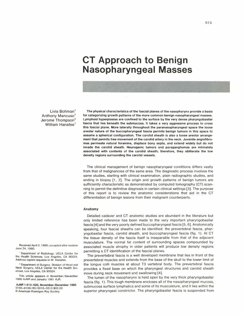
Detailed cadaver and CT anatomic studies are abundant in the literature but only limited reference has been made to the very important pharyngobasilar fascia [4] and the very poorly defined buccopharyngeal fasc ia [5, 6]. Anatomically speaking, four fascial sheaths can be identified: the prevertebral fasc ia, pharyngobasilar fascia , carotid sheath, and buccopharyngeal fasc ia (fig . 1). At CT the tissue density of the fascia itself is inseparable from that of the adjacent musculature . The normal fat content of surrounding spaces compounded by associated muscle atrophy in older patients will produce low density regions permitting a CT identification of the fasc ial planes.
The prevertebral fascia is a well developed membrane that lies in front of the prevertebral muscles and extends from the base of the skull to the lower limit of the longus colli muscles at about T3 vertebral body. The prevertebral fascia provides a fi xed base on which the pharyngeal structures and carotid sheath move during neck movement and swallowing [4].
The lumen of the nasopharynx is held open by the very thick pharyngobasilar fascia (fig . 1). This tough membrane encloses all of the nasopharyngeal mucosa, submucosa surface lymphatics and some of its musculatu re, and it lies within the superior pharyngeal constrictor. The pharyngobasilar fascia is suspended from
514 BOHMAN ET AL. AJNR: 1 , November I December 1980
A B
the base of the skull, its superior margin extending well above the top of the superior pharyngeal constrictor. The levator veli palatini muscle lies to the mucosal side of the fascia; the tensor veli palatini and its tendon lie external to it. The eustachian tube and levator veli palatini pass through a small gap between the base of the skull and the pharyngobasilar fascia; some of the levator's fibers actually arise from the cartilaginous part of the tube. However, the major origin of the veli palatini muscles is from the base of the skull. Posteriorly , the fascia attaches to the anterior margin of the carotid foramen protecting the carotid sheath and then reflects medially over the prevertebral fascia .
In the nasopharynx the buccopharyngeal fascia is formed from the epimysium of the superior pharyngeal constrictor. This fascia is sparse and loosely applied, permitting the motion that takes place during swallowing. The buccopharyngeal fascia, although poorly formed, is the medial boundary of the paranasopharyngeal space. Similarly , its lateral counterpart, a reflection of the deep cervical fascia covering the parotid gland (deep lobe) and pterygoid muscles (fig . 1), forms the lateral boundary of the paranasopharyngeal space. The space itself is a loose network of fibrofatty tissue
Fig. 1.-A is about 2 cm caudal to B. Positions of fasciae (not seen on CT scans) indicated by: solid white line, pharyngobasilar fascia; dashed black line, prevertebral fascia; dotted white line, buccopharyngeal fascia. Parapharyngeal space (PS) lies within buccopharyngeal fascia. Superior pharyngeal constrictor (not visible) lies deep to pharyngobasilar fascia (arrow) . Retropharyngeal (potential) space lies between prevertebral and pharyngobasilar fascias. Carotid sheath structures (C = carotid ; J = jugular) lie medial to styloid process (S) . Superficial airway landmarks include eustach ian tube orifice (E) and torus tubarius (T). Pharyngeal musculature lies deeper: tensor palati (TP) and levator palati (LP) ; superior constrictor not visualized. Although deep fasciae are not seen, low density paranasopharyngeal space defined by fasciae lies between pharyngeal muscles and lateral (LPM) and medial pterygoids (MPM).
whose primary function is to allow free movement of the muscles of mastication independent from the swallowing act. At the same time the contraction and relaxation of the muscle bundles do not collapse the nasopharyngeal airway. The fat content of the paranasopharyngeal space allows one to easily identify it as a low density tissue plane lying between the pterygoid and pharyngeal musculature. Inferiorly the buccopharyngeal fascia is continuous with the covering of the pharynx and esophagus [5].
The infratemporal fossa lies lateral to the paranasopharyngeal space . The infratemporal fossa is bound laterally by the zygomatic arch . Within this space are most of the mandible, pterygoid , masseter, and parts of the temporalis muscle and deep lobe of the parotid gland .
Other spaces defined by these fascial planes are important because their contents determine the cell of origin of some tumors. A potential space, the retropharyngeal space, exists between the pharyngobasilar fascia and the prevertebral fascia . This space contains two chains of lymph nodes lying to either side of midline posteriorly. Laterally the carotid sheath forms a posterolateral boundary to the retropharyngeal space. Within the carotid sheath lies the carotid
AJNR:1, November/ December 1980 BENIGN NASOPHARYNGEAL MASSES 515
TABLE 1: Differentiating Features of Benign Nasopharyngeal Lesions
No. Diagnosis
Patients Status of Fascial Planes
Superficial lesions:
Adenoid problems
Circumscribed carotid sheath lesions: Neuromas (high) Carotid body tumors (low) .. ... . .... .
Subtotal
Paranasopharyngeal space masses: Group A:
Parapharyngeal cysts Granulomas Salivary gland tumors Atypical Iymphoepithelial lesions
Group B:
Angiofibroma
Subtotal
Infiltrating lesions:
Postop mass Parotid abscess Mucormycosis ..
Subtotal
Total
3
3
4
5
7
1 2
4
18
All deep planes normal ; surface anatomy pliable
Carotid sheath obliterated
Arise deep to buccopharyneal fascia (carotid sheath preserved), well defined ; at least partly bordered by surround-ing lucency
Cross and / or arise deep to buccopharyngeal fascia (carotid sheath preserved) with bony septa displaced
Nonpliable surface anatomy-may cross all fascial planes
• Scans of these lesions were reviewed but are not included in this report.
vessels, sympathetic chains, and the vagus and proximal parts of XI and XII cranial nerves together with major deep lymphatic chains intimately associated with the jugular vein .
Materials and Methods
CT of the nasopharynx was performed on about 1 00 patients during a 2'/2 year period. Of these, 18 instances of pathologically verified benign nasopharyngeal conditions were found and analyzed retrospectively . These included five juvenile angiofibromas, three neuromas, one carotid body tumor, one chronic granuloma, one case of atypicallymphoepitheloid lesion, two unusual adenoids, one pickwickian syndrome, and four infections or surgical distortions of the nasopharynx. The infections consisted of one case of mucormycosis and two of parotid infections. The surgical case demonstrated postoperative edema after excision of a mixed tumor of the parotid gland (table 1).
Using an EMI 5005 body scanner, 8 mm sections were taken at 0.5 or 1.0 cm intervals through the nasopharynx with the head in a neutral position (canthomeatal line perpendicular to the table top). Scans were obtained both with and without contrast enhancement in most cases. About six to eight sections were taken to cover from the base of the skull through the tonsillar bed. In benign lesions the examination was extended to encompass the inferior pole of the mass, whereas in malignant lesions the examination was carried to
the level of the hyoid bone looking for evidence of lymph node metastasis. Physiologic maneuvers were occasionally useful in demonstrating pliability of the nasopharyngeal walls or resolving problems of minor surface asymmetry. These maneuvers included opening the mouth or blowing out against pursed lips and the occluded nostrils to distend the fossa of Rosenmuller [7] . Coronal sections or overlapping axial sections were occasionally quite helpful in elucidating minor asymmetries.
The scans were carefu lly reviewed for evidence of distortion of deep and superficial planes, mucosal pliability, and symmetry between the two sides. Clinical corre lation was obtained with all of our cases. All tumors had biopsy or excision for confirmation .
Results
Lesions in our patients fell into four categories based on the relation of the processes to the fascial planes (table 1).
Superficial Lesions
Three histologically different entities represent the gamut of benign superficial lesions. Two patients had lymphoid mucosal mass lesions with associated airway asymmetry that were atypical in appearance and / or in the age at
516 BOHMAN ET AL. AJNR: 1 , November/ December 1980
A B
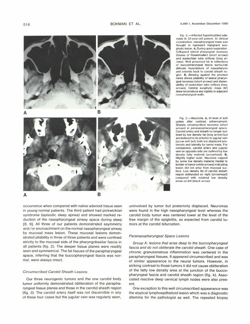
occurrence when compared with native adenoid tissue seen in young normal patients. The third patient had pickwickian syndrome (episodic sleep apnea) and showed marked reduction of the nasopharyngeal airway space during sleep [8 , 9]. All three of our patients demonstrated asymmetry and / or encroachment on the normal nasopharyngeal airway by mucosal mass lesion. These mucosal lesions demonstrated pliability in three of three patients and were confined strictly to the mucosal side of the pharyngobasilar fascia in all patients (fig. 2) . The deeper tissue planes were readily seen and symmetrical. The fat tissues of the parapharyngeal space, inferring that the buccopharyngeal fascia was normal, were always intact.
Circumscribed Carotid Sheath Lesions
Our three neurogenic tumors and the one carotid body tumor uniformly demonstrated obliteration of the parapharyngeal tissue planes and those in the carotid sheath region (fig . 3). The carotid artery itself was not discernible in any of these four cases but the jugular vein was regularly seen,
Fig. 2. -lnfected hypertrophied adenoids in 32-year-old patient. At clinical examination , nasopharyngeal mass was thought to represent malignant exophytic lesion. A, During Quiet respiration. Collapsed lateral pharyngeal recesses (fossas of Rosenmuller) (short arrows) and eustachian tube orifices (long arrows). Well preserved fat in reflections of buccopharyngeal fascia surrounds delicate musculature of nasopharynx and extends back to carotid sheath region. B, Blowing against the pinched nares shows pliability of lateral pharyngeal recesses (short arrows) and distensibility of eustachian tube orifices (Ion" arrows). Central exophytic mass (M) does not produce any rigidity to adjacent nasopharyngeal walls.
Fig . 3.-Neuroma. A, At level of soft palate after contrast enhancement. Sharply circumscribed neuroma (short arrows) in paranasopharyngeal space. Carotid artery and sheath no longer outlined by low density fat (long arrow) but are believed to lie anterior to jugular vein (arrow with tail); both are displaced posteriorly and laterally by tumor mass. For comparison, carotid artery and jugular vein on opposite side are outlined by low density fatty material (arrowhead). B, Slightly higher scan. Neuroma capped by some low density material medial to border of tumor (white arrows) indicating lesion did not arise from mucosal surface. Low density fat of carotid sheath region obliterated on right (arrowhead) compared with retained low density areas on left (black arrow).
uninvolved by tumor but posteriorly displaced . Neuromas were found in the high nasopharyngeal level whereas the carotid body tumor was centered lower at the level of the free margin of the epiglottis, as expected from carotid tumors at the carotid bifurcation .
Paranasopharyngeal Space Lesions
Group A: lesions that arise deep to the buccopharyngeal fascia and do not obliterate the carotid sheath. One case of chronic granulomatous inflammation was centered in the parapharyngeal tissues. It appeared circumscribed and was of similar appearance to the neural tumors. However, in striking contrast to those tumors it did not cause obliteration of the fatty low density area at the junction of the buccopharyngeal fascia and carotid sheath region (fig. 4). Associated reactive deep cervical lymph nodes were also present.
One exception to this well circumscribed appearance was the atypical Iymphoepithelioid lesion which was a diagnostic dilemma for the pathologist as well. The repeated biopsy
AJNR: 1 , November/ December 1980 BENIGN NASOPHARYNGEAL MASSES 517
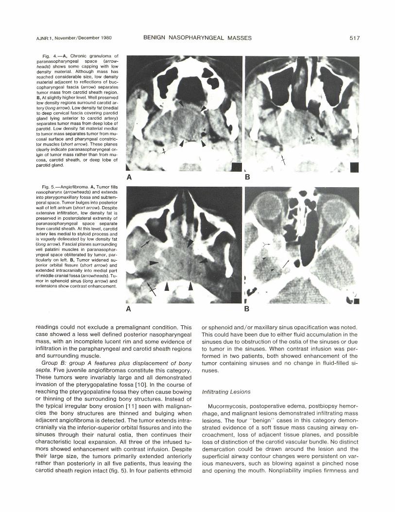
Fig . 4.-A, Chronic granuloma of paranasopharyngeal space (arrowheads) shows some capping with low density material. Although mass has reached considerable size, low density material adjacent to reflections of buccopharyngeal fascia (arrow) separates tumor mass from carotid sheath region . B, At slightly higher level. Well preserved low density reg ions surround carotid artery (long arrow). Low density fat (medial to deep cervical fascia covering parotid gland lying anterior to carotid artery) separates tumor mass from deep lobe of parotid. Low density fat material medial to tumor mass separates tumor from mucosal surface and pharyngeal constrictor muscles (short arrow). These planes clearly indicate paranasopharyngeal origin of tumor mass rather than from mucosa, carotid sheath, or deep lobe of parotid gland.
Fig. S.-Angiofibroma. A, Tumor fills nasopharynx (arrowheads) and extends into pterygomaxillary fossa and subtemporal space. Tumor bulges into posterior wall of left antrum (short arrow). Despite extensive infiltration, low density fat is preserved in posterolateral extremity of paranasopharyngeal space separate from carotid sheath. At this level, carotid artery lies medial to styloid process and is vaguely delineated by low density fat (long arrow). Fascial planes surrounding veli palatini musc les in paranasopharyngeal space obliterated by tumor, particularly on left . B, Tumor widened superior orbital fissure (short arrow) and extended intracranially into medial part of middle cranial fossa (arrowheads) . Tumor in sphenoid sinus (long arrow) and extensions show contrast enhancement.
A
readings could not exclude a premalignant condition . This case showed a less well defined posterior nasopharyngeal mass, with an incomplete lucent rim and some evidence of infiltration in the parapharyngeal and carotid sheath regions and surrounding muscle.
Group B: group A features plus displacement of bony septa. Five juvenile angiofibromas constitute this category. These tumors were invariably large and all demonstrated invasion of the pterygopalatine fossa [10]. In the course of reaching the pterygopalatine fossa they often cause bowing or thinning of the surrounding bony structures. Instead of the typical irregular bony erosion [11] seen with malignancies the bony structures are thinned and bulging when adjacent angiofibroma is detected. The tumor extends intracranially via the inferior-superior orbital fissures and into the sinuses through their natural ostia, then continues their characteristic local expansion. All three of the infused tumors showed enhancement with contrast infusion . Despite their large size, the tumors primarily extended anteriorly rather than posteriorly in all five patients, thus leaving the carotid sheath region intact (fig. 5). In four patients ethmoid
B
or sphenoid and / or maxillary sinus opacification was noted. This could have been due to either fluid accumulation in the sinuses due to obstruction of the ostia of the sinuses or due to tumor in the sinuses. When contrast infusion was performed in two patients, both showed enhancement of the tumor containing sinuses and no change in fluid-filled sinuses.
Infiltrating Lesions
Mucormycosis , postoperative edema, postbiopsy hemorrhage, and malignant lesions demonstrated infiltrating mass lesions. The four " benign " cases in this category demonstrated evidence of a soft tissue mass causing airway encroachment, loss of adjacent tissue planes, and possible loss of distinction of the carotid vascular bundle . No distinct demarcation could be drawn around the lesion and the superficial airway contour changes were persistent on various maneuvers, such as blowing against a pinched nose and opening the mouth. Nonpliability implies firmness and

518 BOHMAN ET AL. AJNR :1 , November/ December 1980
induration of the tissues, These CT patterns were identical to those found with malignant masses in the same regions ,
Discussion
Several methods are sometimes required to examine the nasopharynx, The monocular vision available to the clinician makes determinations of submucosal bulging extremely difficult to determine, Plain films of the nasopharynx are valuable for screening and to show bone invasion. Alterations in the normal nasopharyngeal airway patterns may provide valuable clues for selecting those patients for other investigation . In our hands, tomography of the nasopharynx and " routine " angiography of nasopharyngeal masses have largely been supplanted by CT scanning . Only under unusual circumstances will pluridirectional tomography or positive contrast nasopharyngograms add to the information that is much more easily obtained by CT [12, 13]. CT scanning of the nasopharynx will enjoy an expanded role in the investigation of the nasopharynx [7].
Because of their great vascularity, juvenile angiofibromas constitute a special problem and are usually handled on an individual basis. In general, large lesions are best examined by angiography as a separate procedure. If the lesion is confined to the nasopharynx and pterygopalatine fossa, angiography may be performed immediately before surgery and followed by embolization to control bleeding . In more advanced lesions, superior orbital fissure widening may suggest intracranial extension that is sometimes confirmed by CT; these patients usually receive angiography to verify the advanced nature of the lesion and are managed with radiation therapy or close clinical observation.
Within the limitations imposed by an 18 sec scan time we were stil l able to make some use of physiologic maneuvers to show pliability of the mucosal surfaces with benign lesions. These movements were limited to distension of the lateral recesses of the nasopharynx by having the patient blow against pinched nostrils or by opening and closi ng the mouth. Remembering that the pharyngobasilar fascia is very dense and lies inside the superior pharyngeal constrictor, it is easy to understand that adenoidal and mucosal aberrations do not involve the deep fascial planes. Of considerable interest might be the study of muscular function or velopalatine closure and for opening and closing the eustachian tube. The more sophisticated techniques possible with 1-3 sec scanners will add a new dimension to the study of deep infil trations about the parapharyngeal and paranasopharyngeal spaces. Even so, differentiation of malignant lesions and very aggressive inflammatory processes from the more benign circumscribed lesions can usually be made with a relative degree of certainty provided the basic facts of the patient's clinical condition are available. Malignant tumors, life-threatening fungal infections, and postoperative edema or hemorrhage characteristically produce obliteration of the deep fascial planes extending over large areas (fig. 6). The tissues become rigid and fail to distend with physiologic maneuvers. This is in sharp contradistinction to the deformation of the mucosal surfaces with lymphoid hyperplasia
and the preservation of specific deep fascial planes with the benign neoplasms.
It is not surprising to find the low density areas of the carotid sheath to be obliterated in the neurogenic tumors. The term neurogenic is used quite loosely to include neuromas as well as paragangliomas. Whether the tumors arise from the sympathetic chain or cranial nerves IX-XI is not crucial since all are located within the carotid sheath and would be expected to obliterate these fascial planes. The displaced jugular vein can be identified since it tends to course along the posterior and lateral margin of the carotid sheath and is less tightly bound in the sheath's connective tissue than the artery. The clinical management of carotid body tumors is vastly different from that of the less vascular neuromas arising in the sympathetic chain or cranial nerves. The external carotid artery may be sacrificed during surgery for carotid body tumors to control bleeding under some circumstances . It is conceivable that a carotid body tumor that extends well up into the pharynx could produce some difficulty in differential diagnosis [14]. However the extension of the tumor low in the neck to the level of the hyoid bone should provide the clue that one is dealing with a carotid body tumor rather than a circumscribed neuroma.
Although not considered here, benign mixed tumors of the deep lobe of the parotid may be seen as parapharyngeal masses [6]. In the scans we have reviewed these lesions show a circumscribed growth patiern with preservation of some of the low density regions about the carotid sheath. This is to be expected because the parotid gland is enveloped in the deep cervical fascia, which prevents invasion of the carotid sheath; however, the loose areolar arrangement of the buccopharyngeal fascia allows expansion into the paranasopharyngeal space. Despite reaching appreciable size, these tumors retain a low density " cap" along their medial border [15]. Parotid lesions did not enhance on intravenous contrast injection whereas all three of our neuroma patients showed enhancement and staining about the periphery of the tumor mass [16]. Although not specific, low density central regions within neuromas are frequently encountered [1 5].
Angiofibromas are benign tumors that freely cross and obliterate the anterior extents of the buccopharyngeal fascia. In all five of our patients the tumor involved the pterygopalatine fossa and produced an indentation on the posterior wall of the antrum. Despite the tendency of the tumors to permeate through natural foramina and to extend into the nasal cavity and paranasal sinuses, they did not extend as far laterally as the carotid sheath, and the low density regions surrounding the carotid vessels were preserved in every instance. Thus, if an infiltrating nasopharyngeal mass is seen in a boy or young man that crosses the buccopharyngeal fascia but does not involve the carotid sheath, it is highly suspicious that the lesion is a juvenile angiofibroma. If contrast produces enhancement of the lesion, the pterygopalatine fossa is widened [17], the posterior wall of the antrum is indented [18, 19], and the mass has bled, the radiologic diagnosis of a juvenile angiofibroma is secure (fig . 4) without the necessity of carotid angiography.
AJNR:1, November/ December 1980 BENIGN NASOPHARYNGEAL MASSES 519
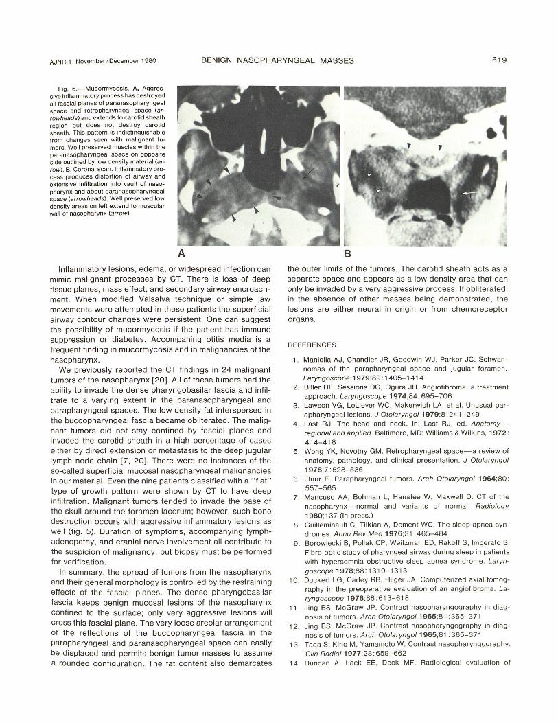
Fig . 5.-Mucormycosis. A, Aggressive inflammatory process has destroyed all fascial planes of paranasopharyngeal space and retropharyngeal space (arrowheads) and extends to carotid sheath region but does not destroy carotid sheath . This pattern is indistinguishable from changes seen with malignant tumors. Well preserved muscles within the paranasopharyngeal space on opposite side outlined by low density material (arrow) . B, Coronal scan. Inflammatory process produces distortion of airway and extensive infiltration into vault of nasopharynx and about paranasopharyngeal space (arrowheads). Well preserved low density areas on left extend to muscular wall of nasopharynx (arrow) .
Inflammatory lesions, edema, or widespread infection can mimic malignant processes by CT. There is loss of deep tissue planes, mass effect, and secondary airway encroachment. When modified Val salva technique or simple jaw movements were attempted in these patients the superficial airway contour changes were persistent. One can suggest the possibility of mucormycosis if the patient has immune suppression or diabetes. Accompaning otitis media is a frequent finding in mucormycosis and in malignancies of the nasopharynx.
We previously reported the CT findings in 24 malignant tumors of the nasopharynx (20). All of these tumors had the ability to invade the dense pharyngobasilar fascia and infiltrate to a varying extent in the paranasopharyngeal and parapharyngeal spaces . The low density fat interspersed in the buccopharyngeal fascia became obliterated. The malignant tumors did not stay confined by fascial planes and invaded the carotid sheath in a high percentage of cases either by direct extension or metastasis to the deep jugular lymph node chain [7 , 20). There were no instances of the so-called superficial mucosal nasopharyngeal malignancies in our material. Even the nine patients classified with a " flat " type of growth pattern were shown by CT to have deep infiltration . Malignant tumors tended to invade the base of the skull around the foramen lacerum; however, such bone destruction occurs with aggressive inflammatory lesions as well (fig . 5) . Duration of symptoms, accompanying lymphadenopathy, and cranial nerve involvement all contribute to the suspicion of malignancy, but biopsy must be performed for veri fication .
In summary, the spread of tumors from the nasopharynx and their general morphology is controlled by the restraining effects of the fascial planes. The dense pharyngobasilar fascia keeps benign mucosal lesions of the nasopharynx confined to the surface; only very aggressive lesions will cross this fascial plane. The very loose areolar arrangement of the reflections of the buccopharyngeal fascia in the parapharyngeal and paranasopharyngeal space can easily be displaced and permits benign tumor masses to assume a rounded configuration . The fat content also demarcates
B the outer limits of the tumors. The carotid sheath acts as a separate space and appears as a low density area that can only be invaded by a very aggressive process . If obliterated, in the absence of other masses being demonstrated , the lesions are either neural in origin or from chemoreceptor organs.
REFERENCES
1. Maniglia AJ , Chandler JR, Goodwin WJ , Parker JC. Schwannomas of the parapharyngeal space and jugular foramen. Laryngoscope 1979;89: 1405-1 4 14
2. Biller HF, Sessions DG , Ogura JH . Angiofibroma: a treatment approach. Laryngoscope 1974;84 : 695-70 6
3. Lawson VG , LeLiever WC, Makerwich LA, et al. Unusual parapharyngeal lesions. J Otolaryngo/1979; 8: 24 1 - 249
4 . Last RJ . The head and neck. In: Last RJ , ed. Anatomyregional and applied. Baltimore, MD: Willi ams & Wilkins, 1972: 414-41 8
5. Wong YK, Novotny GM. Retropharyngeal space- a review of anatomy, pathology, and clinical presentation. J Otolaryngol 1978;7 : 5 28- 536
6. Fluur E. Parapharyngeal tumors. Arch Otolaryngol 1964;80: 557-565
7. Mancuso AA , Bohman L, Hanafee W, Maxwell D. CT of the nasopharynx-normal and variants of normal. Radio logy 1980;13 7 (In press.)
8. Guilleminault C, Tilk ian A, Dement WC. The sleep apnea syndromes. Annu Rev Med 1976 ;3 1 :465-484
9 . Borowieck i B, Pollak CP, Weitzman ED, Rakoff S, Imperato S. Fibro-optic study of pharyngeal airway during sleep in patients with hypersomnia obstructive sleep apnea syndrome. Laryn
goscope 1978;88: 13 1 0 -1 3 13 10. Duckert LG, Carley RB, Hilger JA. Computeri zed axial tomog
raphy in th e preoperative evaluation of an angiofibroma. Laryngoscope 1978;88: 6 13 - 618
11 . Jing BS, McGraw JP. Contrast nasopharyngography in diagnosis of tumors. Arch Oto/aryngo/1 965 ;81 :365- 371
12. Jing BS, McGraw JP. Contrast nasopharyngography in diagnosis of tumors. Arch Oto/aryngo/1965; 81 :365-37 1
13. Tada S, Kino M, Yamamoto W. Contrast nasopharyngography . Clin Radio/1977 ;28: 659- 662
14 . Duncan A, Lack EE , Deck MF. Radiological evaluation of
520 BOHMAN ET AL. AJNR:1, November / December 1980
paragang liomas of the head and neck. Radiology 1979;1 32: 99-105
15. Som PM , Bi ller HF. The combined CT sialogram . Radiology 1980;35 :387-390
16. Miller EM , Norman D. The role of computed tomography in the evaluation of neck masses. Radiology 1979; 133: 145-149
17. Weinstein MA, Levine H, Duchesneau PM , Tucker HM . Diagnosis of juvenil e angiofibroma by computed tomography. Radiology 1978; 126: 703- 705
18 . Miller WE, Holman CB, Dockerty M. Roentgenologic manifestations of malignant tumors of the nasopharynx. AJR 1969;106:813-823
19. Holman CB, Miller WE. Juvenile nasopharyngeal fibroma: roentgenologic characteristics. AJR 1965;94 : 292-298
20. Hanafee WN, Mancuso AA , Jenkins HA, Winter J . Computerized tomography scanning of the temporal bone. Ann Otol Rhinol Laryngo/1979;88:72 1-728