Embed Size (px)
Citation preview

Korean J Radiol 7(3), September 2006 173
CT Analysis of the Anterior Mediastinumin Idiopathic Pulmonary Fibrosis andNonspecific Interstitial Pneumonia
Objective: We wanted to determine whether the amount and shape of theanterior mediastinal fat in the patients suffering with usual interstitial pneumonia(UIP) or nonspecific interstitial pneumonia (NSIP) was different from those of thenormal control group.
Materials and Methods: We selected patients who suffered with UIP (n = 26)and NSIP (n = 26) who had undergone CT scans. Twenty-six controls wereselected from individuals with normal CT findings and normal pulmonary functiontests. All three groups (n = 78) were individually matched for age and gender. Theamounts of anterior mediastinal fat, and the retrosternal anteroposterior (AP) andtransverse dimensions of the anterior mediastinal fat were compared by one-wayanalysis of variance and Bonferroni’s test. The shapes of the anterior medi-astinum were compared using the Chi-square test. Exact logistic regressionanalysis and polychotomous logistic regression analysis were employed toassess whether the patients with NSIP or UIP had a tendency to show a convexshape of their anterior mediastinal fat.
Results: The amount of anterior mediastinal fat was not different among thethree groups (p = 0.175). For the UIP patients, the retrosternal AP dimension ofthe anterior mediastinal fat was shorter (p = 0.037) and the transverse dimensionof the anterior mediastinal fat was longer (p = 0.001) than those of the normalcontrol group. For the NSIP patients, only the transverse dimension was signifi-cantly longer than those of the normal control group (p < 0.001). The convexshape of the anterior mediastinum was predictive of NSIP (OR = 19.7, CI 3.32 , p < 0.001) and UIP (OR = 24.42, CI 4.06 , p < 0.001).
Conclusion: For UIP patients, the retrosternal AP and transverse dimensionsare different from those of normal individuals, whereas the amounts of anteriormediastinal fat are similar. UIP and NSIP patients have a tendency to have a convex shape of their anterior mediastinal fat.
he anterior mediastinum is mainly composed of fat tissue and it is adynamic compartment that readily adapts to various pathologicconditions (1). Changes in the shape of the extrapleural fat and anterior
mediastinum can be seen in localized inflammatory diseases such as tuberculosis andempyema, and this can also occur as a consequence of chronic fibrotic diseases of thelung and pleura (2 4). Radiation fibrosis or lobectomy can also cause focal or diffusethickening of the mediastinum (4, 5). We have recently observed in our clinicalpractice that the mediastinal fat in patients with usual interstitial pneumonia (UIP) ornonspecific interstitial pneumonia (NSIP) was thicker than that in normal individualsand it tended to be convex in shape. Idiopathic pulmonary fibrosis is known to causemediastinal widening and it can be mistaken for lymphadenopathy caused by tumor or
Chang Hyun Lee, MD1
Hyun Ju Lee, MD1
Kyu Ri Son, MD1
Eun Ju Chun, MD1
Kun Young Lim, MD1
Jin Mo Goo, MD1
Jung-Gi Im, MD1
Jeong Nam Heo, MD2
Jae-Woo Song, MD2
Index terms:Thorax, CT Lung, interstitial disease Mediastinum
Korean J Radiol 2006;7:173-179Received March 28, 2006; accepted after revision May 19, 2006.
1Department of Radiology and theInstitute of Radiation Medicine, SeoulNational University College of Medicine,Clinical Research Institute, Seoul NationalUniversity Hospital, Seoul 110-744;2Department of Radiology, Asan MedicalCenter, University of Ulsan College ofMedicine, Seoul 138-736, Korea
This study is supported by KISTEP, theMinistry of Science and Technology,Korea.
Address reprint requests to:Hyun Ju Lee, MD, Department ofRadiology, Seoul National UniversityCollege of Medicine, 28, Yeongeon-dong,Jongno-gu, Seoul 110-744, KoreaTel. (822) 2072-1861Fax. (822) 743-6385e-mail: [email protected]
T
infection (4). To the best of our knowledge, CT analysis ofthe amount and shape of the anterior mediastinal fat inpatients with UIP or NSIP have not been describedpreviously.
The purpose of this study was to determine whether theamount and shape of the anterior mediastinal fat inpatients with UIP or NSIP are different from those of anormal control group.
MATERIALS AND METHODS
PatientsFrom January 1999 to December 2004, we identified 43
patients with a definitive diagnosis of NSIP and 115patients with a definitive diagnosis of UIP by searching thepatient records at our hospital and at another teachinghospital. Over the same period, 252 individuals wereidentified who had no abnormal lesion on high-resolutionCT (HRCT) and they had normal pulmonary function tests,so they were selected as controls. To avoid any bias due toage or gender, the UIP, NSIP and control subjects wereindividually matched for age and gender. Finally, 26patients with NSIP, 26 patients with UIP and 26 controlswere selected. Each group was comprised of 20 femalesand six males. For each of the three groups, the ages werematched within two years (age range: 40 77 years, meanage: 56.3).
Nonspecific interstitial pneumonia was diagnosed in 26patients from lung wedge resections by consensus betweena panel of experienced pathologists who worked in confer-ence, and the diagnosis was based on the criteria
developed by the American Thoracic Society andEuropean Respiratory Society (ATS/ERS) (6). Thehistologic features of NSIP included a cellular pattern (n =3), a fibrosing pattern (n = 10), and both patterns (n = 13).UIP was also diagnosed from lung resections in five casesor from a combination of the typical HRCT findings (asdescribed by the ATS) and from the clinical information in21 cases (6).
Two authors (C.H.L. and K.R.S.) reviewed the clinicalhistories and the underlying medical conditions. Toexclude other factors that might change the amount andshape of the anterior mediastinal fat, patients with ahistory of tuberculosis, empyema, connective tissuediseases, exposure to organic or inorganic dust or toxicfumes, Cushing disease, asthma or any other diseasesrequiring steroid medication were excluded from the studypopulation. Patients who had been treated with steroidbefore CT were excluded for the same reason. Our institu-tional review board did not require approval of our studyor informed consents for the patients’ records or imagereviews.
High-Resolution CT High-Resolution CT was performed in all patients. The
scans were 1 1.5 mm thick/section, and they werereconstructed using a high spatial frequency algorithm.Scans were obtained at 10 mm intervals in the supineposition at end inspiration. The HRCT scans were obtainedwith a variety of scanners (Somatom Plus-4 scanner,Siemens Medical Systems, Erlangen, Germany; SomatomPlus-S scanner, Siemens Medical Systems, Erlangen,
Lee et al.
174 Korean J Radiol 7(3), September 2006
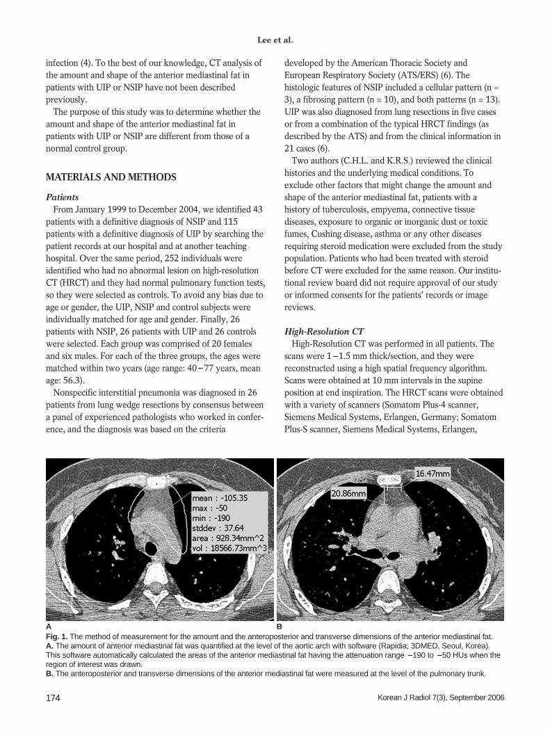
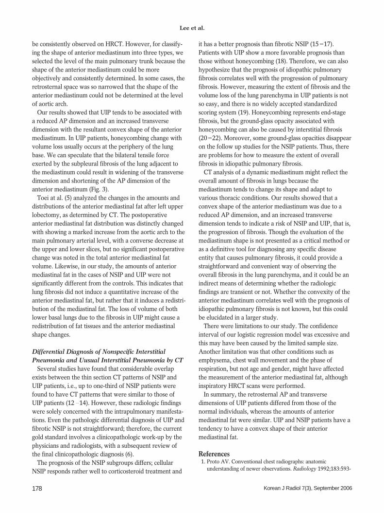
Fig. 1. The method of measurement for the amount and the anteroposterior and transverse dimensions of the anterior mediastinal fat. A. The amount of anterior mediastinal fat was quantified at the level of the aortic arch with software (Rapidia; 3DMED, Seoul, Korea).This software automatically calculated the areas of the anterior mediastinal fat having the attenuation range 190 to 50 HUs when theregion of interest was drawn. B. The anteroposterior and transverse dimensions of the anterior mediastinal fat were measured at the level of the pulmonary trunk.
A B
Germany; and Hi-Speed Advantage, GE Medical Systems,Milwaukee, WI). No intravenous contrast material wasused.
CT Image AnalysisThe CT scan Digital Imaging Communications in
Medicine (DICOM) format was used for the quantitativemeasurements. The amount of anterior mediastinal fat wasdetermined from scans at the level of the aortic arch, and itwas quantified using Rapidia software (3DMED, Seoul,Korea) (Fig. 1A). This software automatically calculates theareas of anterior mediastinal fat with the attenuation range
of 190 to 50 HUs. This attenuation threshold for theanterior mediastinal fat measurement was modified fromthe value used for the standardized abdominal fatmeasurement on CT by Yoshizumi et al. (7).
The shape of the anterior mediastinal fat was categorizedas concave, flat or convex at the level of the mainpulmonary trunk rather than determining the shape of theanterior mediastinal fat at the level of aortic arch becausethe amount of anterior mediastinal fat at the level of aorticarch was insufficient for the purposes of classifying it insome cases. The anteroposterior (AP) (from the posteriorwall of the sternum to the anterior wall of the ascending
Anterior Mediastinal Changes on CT Scans in IPF and NSIP
Korean J Radiol 7(3), September 2006 175
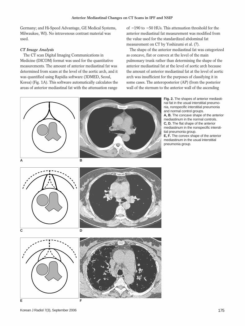
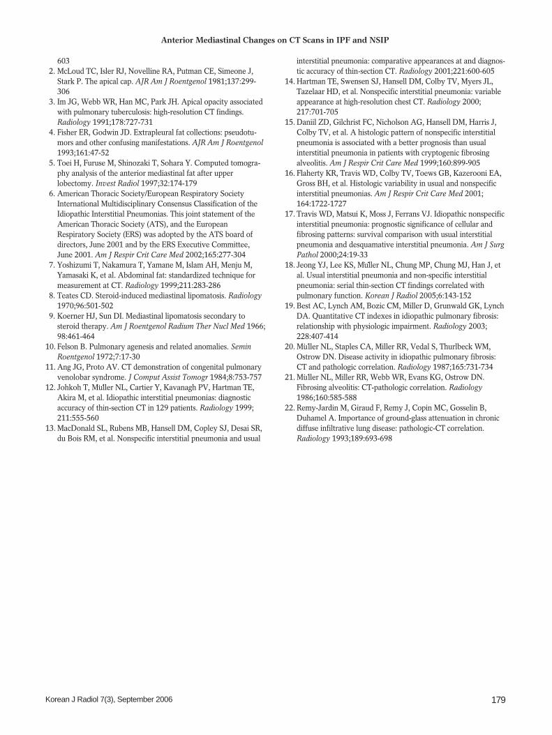
Fig. 2. The shapes of anterior mediasti-nal fat in the usual interstitial pneumo-nia, nonspecific interstitial pneumoniaand normal control groups. A, B. The concave shape of the anteriormediastinum in the normal controls. C, D. The flat shape of the anteriormediastinum in the nonspecific intersti-tial pneumonia group. E, F. The convex shape of the anteriormediastinum in the usual interstitialpneumonia group.
A B
C D
E F
aorta) and transverse dimensions (the width of theposterior wall of the sternum in contact with the anteriormediastinal fat) of the anterior mediastinal fat at the levelof the main pulmonary trunk were also measured (Fig.1B). Two radiologists evaluated the shape of anteriormediastinal fat by consensus (Fig. 2).
Statistical Analysis All the analyses were performed with SAS System
software (version 9.0, SAS Institute, Cary, NC). The 261:1:1 matched sets were analyzed, and the amounts ofanterior mediastinal fat, the retrosternal AP and transversedimensions of the anterior mediastinum, and the weightsand body mass indexes (BMIs) were compared using one-way analysis of variance (ANOVA). Bonferroni post hoctest was also performed. The shapes of the anteriormediastinal fat were compared using the Chi-square test.The conditional exact logistic regression model was used toassess whether convexity of the anterior mediastinum, asdetermined by CT, was a risk factor for pulmonaryfibrosis. The potential confounding factors includingweight, BMI, the AP and transverse dimensions and theamount of fat in the anterior mediastinum, were catego-rized (the weight was divided for 10 kg intervals and theother factors were divided into four groups in reference tothe control). Because weight was the most importantconfounding factor, the crude odds ratios and adjustedodds ratios for weight with the corresponding 95%
confidence intervals (CI) were calculated. A polychoto-mous logistic regression test model was used to calculatethe overall p-values for the UIP, NSIP and control groups.P values of < 0.05 were considered to indicate statisticallysignificant differences.
RESULTS
Comparisons of the 78 matched study subjects aresummarized in Table 1. No significant differences in age,weight and BMI were observed among the three groups.The amount of anterior mediastinal fat was not differentamong the three groups (p = 0.175). The retrosternal APdimension (p = 0.037) and transverse dimension (p <0.001) of the anterior mediastinal fat were significantlydifferent among the three groups. Bonferroni post hoc testshowed that the UIP retrosternal AP dimension wasshorter (p = 0.037) and the transverse dimension waslonger (p = 0.001) than that in the normal control group.For the NSIP group, only the transverse dimension wassignificantly longer than that in the normal control group(p < 0.001). However, no significant difference in the APand transverse dimensions of the anterior mediastinal fatwas observed between the NSIP and UIP groups.
The shapes of anterior mediastinum were significantlydifferent among the three groups (p < 0.001) (Table 2).The convex shape of the anterior mediastinum was predic-tive of NSIP (OR = 19.7, CI 3.32 , p < 0.001) and UIP
Lee et al.
176 Korean J Radiol 7(3), September 2006
Table 1. Comparisons of the Variables in the Three Groups
Groupsp-value
Variables Normal NSIP UIP (Mean SD) (Mean SD) (Mean SD) Normal:NSIP Normal:UIP NSIP:UIP Normal:NSIP:UIP
Weight (kg) 64.8 13.1 61.3 8.50 059.1 11.40 < 0.818 0.210 1.0 < 0.187Body mass index (kg/m2) 24.4 3.30 24.6 3.20 23.4 3.60 < 1.0 1.0 1.0 < 0.649Amount of anterior
502.8 350.7 746.9 594.4 661.4 441.6<
0.200 0.691 1.0<
0.175mediastinal fat
AP dimension 19.9 6.50 16.4 7.90 14.8 6.70 < 0.237 0.037* 1.0 < 0.037*Transverse dimension 7.8 8.5 27.1 180. 25.2 19.1 < 0.001* 0.001* 1.0 < 0.001*
Note. * p-value < 0.05; Data are presented as means SDs; AP dimension = Anteroposterior dimension; Statistical results were obtained by the one way analysis of variance test and the Bonferroni test.
Table 2. Different Shapes of Anterior Mediastinum in the Three Groups
GroupsShape
Normal (n/ %) NSIP (n/ %) UIP (n/ %)p-value
Concave 25 96.2 07 26.9 02 07.7 < 0.001*Flat 01 03.8 05 19.2 02 07.7 < 0.001*Convex 00 00.0 14 53.8 22 84.6 < 0.001*
Note. * p-value < 0.05; Statistical result was obtained by the Chi-square test
(OR = 24.42, CI 4.06 , p < 0.001). When the data wereadjusted for weight, a convex shape was also predictive forNSIP (adjusted OR = 17.16, CI 2.89 , p < 0.002) andUIP (adjusted OR = 32.64, CI 5.71 , p < 0.002). TheUIP patients were also found to have a higher likelihood ofa convex shape of the anterior mediastinum than werethose patients with NSIP (OR = 3.96, CI 0.74 39.79, p <0.001; adjusted OR = 6.11, CI 0.82 275.26, p < 0.002).
DISCUSSION
Although mediastinal widening in idiopathic pulmonaryfibrosis have been previously described, no objective CTanalysis has been reported (4). Thus, this is the first studyto investigate the use of CT images to quantitatively andqualitatively analyze the mediastinal morphologies inpulmonary fibrosis.
Changes in the Shape of the Anterior Mediastinum inVarious Pathologic Conditions
The mediastinum is composed primarily of fatty tissuethat directly contacts the lungs bilaterally. Therefore, theshape of the mediastinum readily adapts to the changes inlung pathology (1). Mediastinal widening can be seen inthe setting of idiopathic pulmonary fibrosis (4). Accordingto the results of our study, idiopathic interstitial pneumo-nias such as UIP and NSIP can change the shape of theanterior mediastinal fat.
Mediastinal changes have been reported in variouspathologic conditions. Mediastinal lipomatosis refers to theaccumulation of excess fat, and this is usually associatedwith corticosteroid administration (8, 9). For those patients
with UIP, the first line treatment is corticosteroid; thus,mediastinal widening can be the result of steroid medica-tion or fibrous scarring. However, because we excludedthe patients who had previously received corticosteroidmedications, its effect could be ignored in the presentstudy.
In patients with a treated neoplasm, any mediastinalbulge that develops suggests extension or recurrence oftumor. However, if the patient has been irradiated, thenmediastinal widening can be due to adjacent lung scarring,which retracts the mediastinal pleura laterally and draws infat. Other causes of lung scarring can similarly widen themediastinum (4). For example, localized lung or pleuralscarring caused by lung infection can tether the pleura andthicken the overlying fat in the chest wall or mediastinum(2, 3). The acquired causes of mediastinal rotation includelung resection and atelectasis (4). Proto (1) have alsoreported that a retrosternal band is a common finding onthe lateral radiographs of patients with an abnormally lowlung volume (congenital or acquired) on one side.Retrosternal soft tissues develop because the diminishedlung volume pulls the mediastinum toward the affectedside and this draws mediastinal fat anterolaterally in frontof the lung. Before the advent of CT, this retrosternal bandwas attributed to excessive areolar tissue or to anaccessory hemidiaphragm (10), but CT has shown that theactual cause of this retrosternal band is mediastinalrotation and mediastinal fat displacement (11).
Mediastinal Changes and its MechanismWe measured the amount of mediastinal fat at the level
of the aortic arch, which is where most of the fat tissue can
Anterior Mediastinal Changes on CT Scans in IPF and NSIP
Korean J Radiol 7(3), September 2006 177
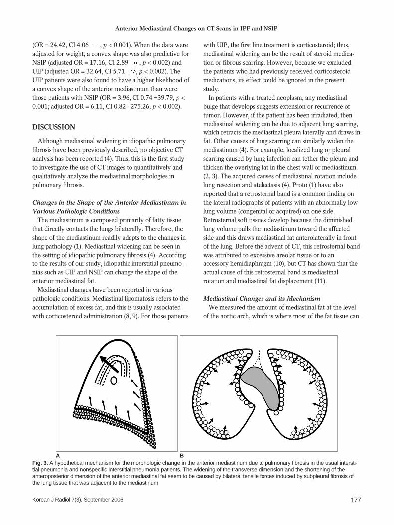
Fig. 3. A hypothetical mechanism for the morphologic change in the anterior mediastinum due to pulmonary fibrosis in the usual intersti-tial pneumonia and nonspecific interstitial pneumonia patients. The widening of the transverse dimension and the shortening of theanteroposterior dimension of the anterior mediastinal fat seem to be caused by bilateral tensile forces induced by subpleural fibrosis ofthe lung tissue that was adjacent to the mediastinum.
A B
be consistently observed on HRCT. However, for classify-ing the shape of anterior mediastinum into three types, weselected the level of the main pulmonary trunk because theshape of the anterior mediastinum could be moreobjectively and consistently determined. In some cases, theretrosternal space was so narrowed that the shape of theanterior mediastinum could not be determined at the levelof aortic arch.
Our results showed that UIP tends to be associated witha reduced AP dimension and an increased transversedimension with the resultant convex shape of the anteriormediastinum. In UIP patients, honeycombing change withvolume loss usually occurs at the periphery of the lungbase. We can speculate that the bilateral tensile forceexerted by the subpleural fibrosis of the lung adjacent tothe mediastinum could result in widening of the transversedimension and shortening of the AP dimension of theanterior mediastinum (Fig. 3).
Toei at al. (5) analyzed the changes in the amounts anddistributions of the anterior mediastinal fat after left upperlobectomy, as determined by CT. The postoperativeanterior mediastinal fat distribution was distinctly changedwith showing a marked increase from the aortic arch to themain pulmonary arterial level, with a converse decrease atthe upper and lower slices, but no significant postoperativechange was noted in the total anterior mediastinal fatvolume. Likewise, in our study, the amounts of anteriormediastinal fat in the cases of NSIP and UIP were notsignificantly different from the controls. This indicates thatlung fibrosis did not induce a quantitative increase of theanterior mediastinal fat, but rather that it induces a redistri-bution of the mediastinal fat. The loss of volume of bothlower basal lungs due to the fibrosis in UIP might cause aredistribution of fat tissues and the anterior mediastinalshape changes.
Differential Diagnosis of Nonspecific InterstitialPneumonia and Uusual Interstitial Pneumonia by CT
Several studies have found that considerable overlapexists between the thin section CT patterns of NSIP andUIP patients, i.e., up to one-third of NSIP patients werefound to have CT patterns that were similar to those ofUIP patients (12 14). However, these radiologic findingswere solely concerned with the intrapulmonary manifesta-tions. Even the pathologic differential diagnosis of UIP andfibrotic NSIP is not straightforward; therefore, the currentgold standard involves a clinicopathologic work-up by thephysicians and radiologists, with a subsequent review ofthe final clinicopathologic diagnosis (6).
The prognosis of the NSIP subgroups differs; cellularNSIP responds rather well to corticosteroid treatment and
it has a better prognosis than fibrotic NSIP (15 17).Patients with UIP show a more favorable prognosis thanthose without honeycombing (18). Therefore, we can alsohypothesize that the prognosis of idiopathic pulmonaryfibrosis correlates well with the progression of pulmonaryfibrosis. However, measuring the extent of fibrosis and thevolume loss of the lung parenchyma in UIP patients is notso easy, and there is no widely accepted standardizedscoring system (19). Honeycombing represents end-stagefibrosis, but the ground-glass opacity associated withhoneycombing can also be caused by interstitial fibrosis(20 22). Moreover, some ground-glass opacities disappearon the follow up studies for the NSIP patients. Thus, thereare problems for how to measure the extent of overallfibrosis in idiopathic pulmonary fibrosis.
CT analysis of a dynamic mediastinum might reflect theoverall amount of fibrosis in lungs because themediastinum tends to change its shape and adapt tovarious thoracic conditions. Our results showed that aconvex shape of the anterior mediastinum was due to areduced AP dimension, and an increased transversedimension tends to indicate a risk of NSIP and UIP, that is,the progression of fibrosis. Though the evaluation of themediastinum shape is not presented as a critical method oras a definitive tool for diagnosing any specific diseaseentity that causes pulmonary fibrosis, it could provide astraightforward and convenient way of observing theoverall fibrosis in the lung parenchyma, and it could be anindirect means of determining whether the radiologicfindings are transient or not. Whether the convexity of theanterior mediastinum correlates well with the prognosis ofidiopathic pulmonary fibrosis is not known, but this couldbe elucidated in a larger study.
There were limitations to our study. The confidenceinterval of our logistic regression model was excessive andthis may have been caused by the limited sample size.Another limitation was that other conditions such asemphysema, chest wall movement and the phase ofrespiration, but not age and gender, might have affectedthe measurement of the anterior mediastinal fat, althoughinspiratory HRCT scans were performed.
In summary, the retrosternal AP and transversedimensions of UIP patients differed from those of thenormal individuals, whereas the amounts of anteriormediastinal fat were similar. UIP and NSIP patients have atendency to have a convex shape of their anteriormediastinal fat.
References1. Proto AV. Conventional chest radiographs: anatomic
understanding of newer observations. Radiology 1992;183:593-
Lee et al.
178 Korean J Radiol 7(3), September 2006
6032. McLoud TC, Isler RJ, Novelline RA, Putman CE, Simeone J,
Stark P. The apical cap. AJR Am J Roentgenol 1981;137:299-306
3. Im JG, Webb WR, Han MC, Park JH. Apical opacity associatedwith pulmonary tuberculosis: high-resolution CT findings.Radiology 1991;178:727-731
4. Fisher ER, Godwin JD. Extrapleural fat collections: pseudotu-mors and other confusing manifestations. AJR Am J Roentgenol1993;161:47-52
5. Toei H, Furuse M, Shinozaki T, Sohara Y. Computed tomogra-phy analysis of the anterior mediastinal fat after upperlobectomy. Invest Radiol 1997;32:174-179
6. American Thoracic Society/European Respiratory SocietyInternational Multidisciplinary Consensus Classification of theIdiopathic Interstitial Pneumonias. This joint statement of theAmerican Thoracic Society (ATS), and the EuropeanRespiratory Society (ERS) was adopted by the ATS board ofdirectors, June 2001 and by the ERS Executive Committee,June 2001. Am J Respir Crit Care Med 2002;165:277-304
7. Yoshizumi T, Nakamura T, Yamane M, Islam AH, Menju M,Yamasaki K, et al. Abdominal fat: standardized technique formeasurement at CT. Radiology 1999;211:283-286
8. Teates CD. Steroid-induced mediastinal lipomatosis. Radiology1970;96:501-502
9. Koerner HJ, Sun DI. Mediastinal lipomatosis secondary tosteroid therapy. Am J Roentgenol Radium Ther Nucl Med 1966;98:461-464
10. Felson B. Pulmonary agenesis and related anomalies. SeminRoentgenol 1972;7:17-30
11. Ang JG, Proto AV. CT demonstration of congenital pulmonaryvenolobar syndrome. J Comput Assist Tomogr 1984;8:753-757
12. Johkoh T, Muller NL, Cartier Y, Kavanagh PV, Hartman TE,Akira M, et al. Idiopathic interstitial pneumonias: diagnosticaccuracy of thin-section CT in 129 patients. Radiology 1999;211:555-560
13. MacDonald SL, Rubens MB, Hansell DM, Copley SJ, Desai SR,du Bois RM, et al. Nonspecific interstitial pneumonia and usual
interstitial pneumonia: comparative appearances at and diagnos-tic accuracy of thin-section CT. Radiology 2001;221:600-605
14. Hartman TE, Swensen SJ, Hansell DM, Colby TV, Myers JL,Tazelaar HD, et al. Nonspecific interstitial pneumonia: variableappearance at high-resolution chest CT. Radiology 2000;217:701-705
15. Daniil ZD, Gilchrist FC, Nicholson AG, Hansell DM, Harris J,Colby TV, et al. A histologic pattern of nonspecific interstitialpneumonia is associated with a better prognosis than usualinterstitial pneumonia in patients with cryptogenic fibrosingalveolitis. Am J Respir Crit Care Med 1999;160:899-905
16. Flaherty KR, Travis WD, Colby TV, Toews GB, Kazerooni EA,Gross BH, et al. Histologic variability in usual and nonspecificinterstitial pneumonias. Am J Respir Crit Care Med 2001;164:1722-1727
17. Travis WD, Matsui K, Moss J, Ferrans VJ. Idiopathic nonspecificinterstitial pneumonia: prognostic significance of cellular andfibrosing patterns: survival comparison with usual interstitialpneumonia and desquamative interstitial pneumonia. Am J SurgPathol 2000;24:19-33
18. Jeong YJ, Lee KS, Muller NL, Chung MP, Chung MJ, Han J, etal. Usual interstitial pneumonia and non-specific interstitialpneumonia: serial thin-section CT findings correlated withpulmonary function. Korean J Radiol 2005;6:143-152
19. Best AC, Lynch AM, Bozic CM, Miller D, Grunwald GK, LynchDA. Quantitative CT indexes in idiopathic pulmonary fibrosis:relationship with physiologic impairment. Radiology 2003;228:407-414
20. Muller NL, Staples CA, Miller RR, Vedal S, Thurlbeck WM,Ostrow DN. Disease activity in idiopathic pulmonary fibrosis:CT and pathologic correlation. Radiology 1987;165:731-734
21. Muller NL, Miller RR, Webb WR, Evans KG, Ostrow DN.Fibrosing alveolitis: CT-pathologic correlation. Radiology1986;160:585-588
22. Remy-Jardin M, Giraud F, Remy J, Copin MC, Gosselin B,Duhamel A. Importance of ground-glass attenuation in chronicdiffuse infiltrative lung disease: pathologic-CT correlation.Radiology 1993;189:693-698
Anterior Mediastinal Changes on CT Scans in IPF and NSIP
Korean J Radiol 7(3), September 2006 179