Embed Size (px)
Citation preview

Diego Hipolito, MS4 5/20/19
Cryoablation for Stage I RCC Case Presentation

Focused patient history and workup
• 48 yo F with history of Right RCC s/p cryoablation on 6/11/18 at OSH who presents for repeat procedure due to high suspicion for residual tumor post-ablation seen on CT. She complains of mild right sided flank pain, but denies hematuria or increased urinary frequency. No evidence of disease progression was seen on imaging.
• PE: unremarkable at time of presentation
• Labs: CBC and BMP unremarkable; Cr stable at 0.73
RCC Workup
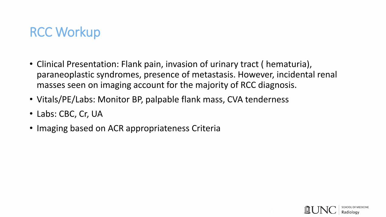
• Clinical Presentation: Flank pain, invasion of urinary tract ( hematuria), paraneoplastic syndromes, presence of metastasis. However, incidental renal masses seen on imaging account for the majority of RCC diagnosis.
• Vitals/PE/Labs: Monitor BP, palpable flank mass, CVA tenderness
• Labs: CBC, Cr, UA
• Imaging based on ACR appropriateness Criteria

ACR Appropriateness Criteria for Renal Masses

Imaging Modalities
• CT w/ and w/o contrast: 1st line modality for evaluation of renal mass and helps to characterize both solid and cystic lesions. The CT should include 3 phases, non-contrast imaging followed by contrast-enhanced imaging in corticomedullary and nephrographic phases, in order to properly characterize renal masses. If a lesion is suspicious and warrants further evaluation, patient should undergo biopsy.
• US: Alternative imaging modality to characterize renal masses. It is usually sufficient as a standalone modality to characterize category I renal cysts using the Bosniak classification of cystic renal masses by CT.
• MRI: Helpful to evaluate for tumor growth as it evaluates for invasion into the collecting system or the vessels better than the other modalities.

Imaging studies from PACS 1
Hypodense 3.8 cm lesion on R-kidney consistent with post-ablation changes

Imaging studies from PACS 2
Hyperdense rim of tissue surrounding post-ablation zone suspicious for residual RCC

Imaging studies from PACS 4

Patient treatment
• RFA and cryoablation are minimally invasive procedures that use temperature extremes to produce tumor cell death. Currently, both RFA and cryoablation are approved by the FDA to treat Stage I RCC (T1aN0M0), tumor ≤4 cm in greatest dimension, limited to the kidney.
• Indications: • Small incidentally detected RCC (< 4 cm)
• Poor surgical candidates
• Need for nephron-sparring treatment
• Patient previously underwent cryotherapy but the ablation margin was not large enough to appropriately treat the entire tumor. Given no disease progression and residual tumor being < 4 cm, patient underwent repeat cryoablation

Cryoablation
How does it work?
• Cryoablation leads to ice formation within the extracellular space creating an osmotic gradient. As a result, the osmolarity of the extracellular space is greater than that of the intracellular space, which triggers movement of free water into the extracellular space and leads to cellular dehydration. This leads to cellular membrane rupture and, ultimately, local tissue ischemia.
What factors to consider when treating RCC?
• Tumor size: considered one of the most important prognostic factors when determining treatment outcome. Tumors less than 3 cm can be usually treated with a single session; whereas, tumors bigger in size may require multiple sessions because the tumor margins may fall outside the probe’s complete kill zone.
• Location: Tumors along the periphery of the renal parenchyma tend to have better treatment outcomes. These tumors tend to have less vascularity given their location and may be treated with a single session.

Imaging studies from PACS 4

Complications of Cryoablation
Hematoma adjacent to site of cryoablation Hydro-dissection to
prevent bowel injury

Imaging Modalities for Characterizing Renal Masses
CT non-con/con MRI US
Sensitivity 88% 90% 46%
Specificity 75% 75% 12%
Radiation exposure 10-30 mSv None None
Cost $1,392 $2,611 $428

Management of Incidental Renal Mass on CT/US

Renal Mass on US

Renal masses on CT
A

BC
DE
Test Yourself/Questions
• What is the 1st line imaging modality to characterize a renal mass? • CT with and without contrast
• When should you refer a patient for cryoablation? • Renal mass is less than 4 cm and is confined to the renal parenchyma • Patient is a poor surgical candidate • Need for nephron-sparring treatment
• US can be used to evaluate what category of renal masses based on the Bosniak criteria? • Category 1

References• Vogel, Christian. "Imaging in Suspected Renal-Cell Carcinoma: Systematic Review." Clinical Genitourinary Cancer, vol. 17,
no. 2, 04/2019, pp. e345-e355, doi:10.1016/j.clgc.2018.07.024.
• de Miranda, Christiana Maia Nobre Rocha et al. “Bosniak classification of renal cystic lesions according to multidetector computed tomography findings.” Radiologia brasileira vol. 47,2 (2014): 115-21. doi:10.1590/S0100-39842014000200015
• Higgins, J. C. "Evaluation of Incidental Renal and Adrenal Masses." American Family Physician, vol. 63, no. 2, 01/2001, pp. 288-94, 299,
• Tatli, Servet, et al. "Percutaneous Cryoablation Techniques and Clinical Applications." Diagnostic and Interventional Radiology 16.1 (2010): 90-5. ProQuest. Web. 19 May 2019.
• Heilbrun, Marta E. "ACR Appropriateness Criteria Indeterminate Renal Mass."Journal of the American College of Radiology, vol. 12, no. 4, 04/2015, pp. 333-341, doi:10.1016/j.jacr.2014.12.012.
• Vikram, Raghunandan. "ACR Appropriateness Criteria Renal Cell Carcinoma Staging."Journal of the American College of Radiology, vol. 13, no. 5, 05/2016, pp. 518-525, doi:10.1016/j.jacr.2016.01.021.
• Casalino, David D. "ACR Appropriateness Criteria Post-Treatment Follow-Up of Renal Cell Carcinoma." Journal of the American College of Radiology, vol. 11, no. 5, 05/2014, pp. 443-449, doi:10.1016/j.jacr.2014.01.023.
• Nahum Goldberg S, Dupuy DE. Image-guided radiofrequency tumor ablation: challenges and opportunities--part I. J VascInterv Radiol 2001; 12:1021.
• Atkins, Michael B., Jerome P. Richie, and Sonali Sonali Shah. "Clinical Manifestations, Evaluation, and Staging of Renal Cell Carcinoma." UpToDate. N.p., 18 Nov. 2018. Web.
• Morgan, Matt A., and Daniel J. Bell. "Renal Cyst | Radiology Reference Article." Radiopaedia Blog RSS. N.p., n.d. Web. 20 May 2019.