Embed Size (px)
Citation preview

JOURNAL OF VIROLOGY, Nov. 1994, p. 7497-75060022-538X/94/$04.00+0Copyright (C 1994, American Society for Microbiology
Crucial Sequences within the Epstein-Barr Virus TP1 Promoterfor EBNA2-Mediated Transactivation and Interaction of
EBNA2 with Its Responsive ElementCHARLOTTE MEITINGER,* LOTHAR J. STROBL, GABRIELE MARSCHALL,
GEORG W. BORNKAMM, AND URSULA ZIMBER-STROBLInstitut fur Klinische Molekularbiologie und Tumorgenetik im Forschungszentrum
ftir Umwelt und Gesundheit, GSF, Munich, Germany
Received 23 May 1994/Accepted 29 July 1994
EBNA2 is one of the few genes of Epstein-Barr virus which are necessary for immortalization of humanprimary B lymphocytes. The EBNA2 protein acts as a transcriptional activator of several viral and cellulargenes. For the TPI promoter, we have shown previously that an EBNA2-responsive element (EBNA2RE)between -258 and -177 relative to the TPI RNA start site is necessary and sufficient for EBNA2-mediatedtransactivation and that it binds EBNA2 through a cellular factor. To define the critical cis elements within thisregion, we cloned EBNA2RE mutants in front of the TPI minimal promoter fused to the reporter gene forluciferase. Transactivation by EBNA2 was tested by transfection of these mutants in the absence and presence
of an EBNA2 expression vector into the established B-cell line BL41-P3HR-1. The analysis revealed that twoidentical 11-bp motifs and the region 3' of the second 11-bp motif are essential for transactivation by EBNA2.Methylation interference experiments indicated that the same cellular factor in the absence of EBNA2 bindseither one (complex I) or both (complex III) 11-bp motifs with different affinities, giving rise to two differentspecific protein-DNA complexes within the left-hand 54 bp of EBNA2RE. A third specific complex was shownpreviously to be present only in EBNA2-expressing cells and to contain EBNA2. Analysis of this EBNA2-containing complex revealed the same protection pattern as for complex III, indicating that EBNA2 interactswith DNA through binding of the cellular protein to the 11-bp motifs. Mobility shift assays with the differentmutants demonstrated that one 11-bp motif is sufficient for binding the cellular factor, whereas for binding ofEBNA2 as well as for efficient transactivation by EBNA2, both 11-bp motifs are required.
Epstein-Barr virus (EBV), a widespread human herpesvirus,is the causative agent of infectious mononucleosis, a self-limiting lymphoproliferative disorder. This virus is also associ-ated with several malignancies, including Burkitt's lymphoma(20), nasopharyngeal carcinoma (18), Hodgkin's disease (4,39), and lymphomas in immune-deficient individuals (22, 24).EBV has a dual tropism; the virus is produced in epithelialcells of the oropharynx, whereas B lymphocytes are nonpro-ductively infected. Infection of B lymphocytes in vitro leads tothe growth of permanently proliferating cell lines. In theselymphoblastoid cell lines, the virus is episomally maintained ina high copy number (32), although only a limited number ofEBV genes are expressed, including six nuclear antigens,EBNA1, -2, -3a, -3b, -3c, and LP, and three membraneproteins, LMP (LMP1), TP1 (LMP2A), and TP2 (LMP2B)(28).EBNA2, a phosphorylated polypeptide of 487 amino acids,
is absolutely necessary for the establishment of B-cell immor-talization. EBV strain P3HR-1, containing a 6.6-kb deletionencompassing the EBNA2 gene and part of the EBNA-LPgene, has lost the ability to transform B cells (7, 26). Afterreintroduction of the EBNA2 gene by homologous recombina-tion into the P3HR-1 genome, a transformation-competentvirus could be reconstituted (13, 25). EBNA2 is necessary notonly for B-cell immortalization in vitro but also for B-celltumor growth in vivo in SCID (severe combined immunodefi-
* Corresponding author. Mailing address: Institut fur KlinischeMolekularbiologie und Tumorgenetik im Forschungszentrum fur Um-welt und Gesundheit, GSF, Marchioninistr. 25, 81377 Munich, Ger-many. Phone: 89-7099510. Fax: 89-7099500.
ciency) mice (12). In natural EBV isolates, two alleles of theEBNA2 gene code for the two proteins EBNA2A andEBNA2B, which have 57% homology (2, 17, 46). Virusescarrying the EBNA2A gene have higher transforming capacitythan EBNA2B-expressing strains (13, 36).
It is likely that EBNA2 contributes to B-cell immortalizationby its ability to transactivate cellular and viral genes. Itincreases transcription of the B-cell activation markers CD21and CD23 and of the tyrosine kinase c-fgr (8, 14, 29, 42). Inaddition, EBNA2 can transactivate the promoters of the viralgenes LMP, TP1, and TP2 and the BamHI-C promoter, whichis necessary for transcription of the nuclear antigens (1, 21, 23,27, 40, 44, 45, 48). Furthermore, EBNA2 has been shown totransactivate the human immunodeficiency virus type 1 longterminal repeat (38). After in vitro infection of B cells, EBNA2and EBNA-LP are the first viral genes to be expressed (3, 37).EBNA2 subsequently induces expression of the EBNA2-re-sponsive viral and cellular genes. Therefore, elucidation of themechanism by which EBNA2 is able to transactivate othergenes will contribute to the understanding of the immortaliza-tion process.
Until now, the mechanism of EBNA2-mediated transactiva-tion has been poorly understood. An acidic transactivationdomain with similarities to that of the herpes simplex virustransactivator VP16 could be mapped at the C terminus (10,11). Recently, we could demonstrate EBNA2 interaction withthe TP1 promoter via a cellular protein (47). Ling et al. (33)have demonstrated an indirect interaction of a glutathione-S-transferase-EBNA2 fusion protein with the BamHI-C-pro-moter.To further elucidate the function of EBNA2, we chose the
7497
Vol. 68, No. 11
on February 18, 2018 by guest
http://jvi.asm.org/
Dow
nloaded from

7498 MEITINGER ET AL.
TP1 promoter, which contains an 81-bp element between -258and -177 relative to the TPI RNA start site, responsible forEBNA2-mediated transactivation (EBNA2RE) (47). The ele-ment contains a duplicated 1 1-bp motif with homologies to theEBNA2-responsive regions of the LMP, BamHI-C, and CD23promoters. The TP1 promoter is particularly well suited forstudying the mechanism of EBNA2 action for several reasons.This promoter is tightly regulated and is, in contrast to theLMP1 promoter (15), strictly dependent on EBNA2 in Blymphocytes. Compared with the BamHI-C and LMP promot-ers, EBNA2 appears to interact more stably with theEBNA2RE of the TP1 promoter, thus allowing the visualiza-tion of the EBNA2-containing complex in gel shift assays withEBNA2-positive nuclear cell extracts. In contrast, it was notpossible to demonstrate the presence of EBNA2 in protein-DNA complexes formed at the EBNA2REs of the BamHI-Cor LMP promoter (27, 31, 33). Addition of the glutathione-S-transferase-EBNA2 fusion protein is necessary to detect bind-ing of EBNA2 to its responsive element at the BamHI-Cpromoter (33).A detailed knowledge of the crucial DNA sequences con-
ferring EBNA2 responsiveness is a prerequisite for under-standing the mechanism of EBNA2-mediated transactivation.We therefore determined the sequences within the EBNA2REof the TP1 promoter which are protected by DNA-bindingproteins. This study revealed that the two 11-bp elements arecontacted by a cellular protein with different affinities. Inaddition, we present a detailed analysis of the sequences vitalfor EBNA2-mediated transactivation and binding to itsEBNA2RE through a cellular protein.
MATERLILS AND METHODS
Cell lines and culture conditions. The cell line M-ABA is amarmoset lymphoblastoid cell line transformed by a virusoriginally derived from a nasopharyngeal carcinoma (16).BL41-P3HR-1 and BL41-B95-8 were obtained after infectionof the EBV-negative Burkitt's lymphoma cell line BL41 withvirus strains P3HR-1 and B95-8, respectively (8).
All cell lines were grown in RPMI 1640 medium supple-mented with 10% fetal calf serum, 2 mM glutamine, penicillin(100 U/ml), and streptomycin (100 pLg/ml). Cultures wereincubated at 37°C in 5% CO2. Cells were diluted 1:3 with freshmedium twice a week.
Plasmids. For cloning of mutated or deleted forms of the81-bp EBNA2RE in front of the TP1 minimal promoter, wehave inserted a polylinker containing an SnaBI and NcoI site inthe StuI site of pTP1-LUC/-45 (pGa59/19) (47). pGa59/19 wasdigested with SnaBI and NcoI and ligated with oligonucleo-tides containing an SnaBI and NcoI site at their ends, respec-tively. The accuracy of the cloned oligonucleotides was con-trolled by sequencing. Sequencing reactions were carried outwith a sequencing kit (US Biochemicals). The EBNA2 expres-sion vector pU294-6 has been described previously (47).
Oligonucleotides. The positions of the oligonucleotides usedin relation to the EBV genomic sequence according to Baer etal. (5) are as follows: 040, 166281 to 166320; 054, 166236 to166289; 060, 166241 to 166300; 080, 166241 to 166320;O5'-del, 166253 to 166320; 01, 166236 to 166268; and 02,166259 to 166289.Transfection of cells. Electroporation of cells was carried
out in a Bio-Rad gene pulser by the method of Cann et al. (9).Briefly, 107 cells with a viability of more than 90% were washedonce and resuspended in 0.25 ml of RPMI 1640 mediumsupplemented with 10% fetal calf serum. Cells were placed onice in a 4-mm Gene Pulser cuvette, and 20 ,ug of the corre-
sponding DNA was added. BL41-P3HR-1 cells were electro-porated at 250 V and 960 ,uF; BL41-B95-8 were done at 260 Vand 960 pRF. Ten minutes after electroporation, the cells wereresuspended in 10 ml of warm RPMI with 20% fetal calfserum.
Luciferase assay. Cells were harvested 48 h after transfec-tion, washed once in ice-cold phosphate-buffered saline (PBS),and resuspended in 100 RI of 91 mM K2HP04-9 mMKH2PO4-1 mM dithiothreitol (DTT)-1% Triton X-100 (pH7.8). After 15 min on ice, debris was removed by centrifugationat 14,000 x g for 10 min. Then, 10 RI of the supernatant wasmixed with 350 RI of 25 mM glycylglycine (pH 7.8)-5 mMATP-15 mM MgSO4-100 [lI of 11 mM luciferin-0.5 M Tris-HCl (pH 7.8). The bioluminescence in relative light units(RLU) was measured with a Lumat LB9501 (Berthold, Wild-bach, Germany).
Nuclear extract preparation. Nuclear extracts were pre-pared by a modification of the method of Dignam et al. (19).The pellet (3 x 10⁷ to 5 x 10⁷ cells) was washed oncein ice-cold PBS, resuspended with 3 to 4 volumes of 10mM HEPES (N-2-hydroxyethylpiperazine-N'-2-ethanesulfonicacid, pH 7.9)-10 mM KCl-1.5 mM MgCl2-5 mM DTT-0.5mM phenylmethylsulfonyl fluoride (PMSF) (buffer A) andincubated on ice for 1 h. Lysis of cells was achieved by 10 to 20strokes with a Dounce homogenizer and was microscopicallycontrolled by trypan blue staining. Nuclei were pelleted for 10s at maximal speed in an Eppendorf minifuge, washed once inbuffer A, resuspended in 3 volumes of buffer B (20 mMHEPES [pH 7.9], 20% glycerol, 420 mM NaCl, 1.5 mM MgCl2,0.2 mM EDTA, 5 mM DTT, 0.5 mM PMSF) and incubated onice for 30 min. Nuclei were removed by centrifugation at14,000 x g for 20 min. The supernatant was stored in liquidnitrogen.
Radioactively labeled probes. Luciferase constructs contain-ing wild-type, mutated, or deleted forms of the 81-bpEBNA2RE in front of the TPI minimal promoter were di-gested with SnaBI and NcoI. The 81-bp or mutated fragmentswere isolated, labeled with Klenow polymerase in the presenceof [32P]dCTP (3,000 Ci/mmol), and purified on a nondenatur-ing polyacrylamide gel.
Gel shift analysis. Binding reactions were performed in avolume of 20 ,u containing 5 RI of binding buffer (41 mMHEPES [pH 7.9], 200 mM KCl, 4 mM EDTA, 1.6% Ficoll, 4mM DTT, 0.5 mM PMSF), 2 RI of poly(dI-dC) (1 mg/ml), 2 [lIof bovine serum albumin (10 mg/ml), 5 ,ug of protein extract,and 0.1 to 0.2 ng of radioactively labeled DNA. After incuba-tion at room temperature for 30 min, the reaction productswere separated on a 4% polyacrylamide gel. In competitionand supershift experiments, either unlabeled oligonucleotideor 1 pI of tissue culture supernatant containing monoclonalantibodies was added to the reaction mixture. Anti-EBNA2monoclonal antibody Rll (rat immunoglobulin G2a [IgG2a]),which recognizes EBNA2A (29a), was used. Anti-mouse CD45(rat IgG2a) was used as an isotype control antibody. Gel shiftanalysis in the presence of sodium deoxycholate was per-formed as described by Baeuerle and Baltimore (6).
Methylation interference. The methylation interference as-say is based on a G reaction of the Maxam-Gilbert sequencingreaction (34). About 100 ng of a prepared DNA fragment,labeled at one end only with Klenow polymerase in thepresence of [32P]dCTP (3,000 Ci/mmol), was methylated to-gether with 1 ,ug of unspecific carrier DNA by adding 1 pA ofdimethyl sulfate in 200 RI of 50 mM sodium cacodylate (pH7.0)-i mM EDTA and incubating for 3 min at 20°C. Thereaction was timed to yield approximately one modification perDNA molecule and was stopped by adding 50 [lI of 1.5 M
J. VIROL.
on February 18, 2018 by guest
http://jvi.asm.org/
Dow
nloaded from

ANALYSIS OF EBV TP1 PROMOTER 7499
sodium acetate (pH 7.0)-i M 2-mercaptoethanol. MethylatedDNA samples were precipitated with ethanol, washed, driedunder vacuum, and resuspended in water. A gel shift assay as
described above was performed with the modified DNA. Thefree oligonucleotide and DNA-protein complexes were excisedfrom the polyacrylamide gel and isolated by electroelution.The recovered DNA was cleaved at the methylated positionsby heating at 90°C in 100 ,ul of 1:10 freshly diluted piperi-dine for 30 min. Positions of cleavages were determined byseparating samples on an 8% denaturating polyacrylamidegel.
RESULTS
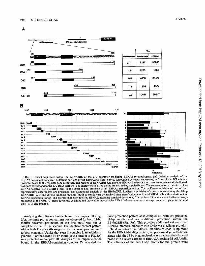
Both 11-bp motifs of EBNA2RE are important for EBNA2-mediated transactivation. For the TP1 promoter, it was shownthat an 81-bp element (EBNA2RE) located between positions-258 to -178, corresponding to the RNA start site, issufficient for transactivation by EBNA2 (47). To identify thesequences within EBNA2RE that are responsible for transac-tivation, we made deletion mutants of the 81-bp element.Plasmids containing these deletions, which are linked to theluciferase (LUC) reporter gene under the control of the TP1minimal promoter, were constructed (Fig. 1A) and transientlytransfected into EBNA2-negative BL41-P3HR-1 cells with or
without the EBNA2A expression vector pU294-6. A cytomeg-alovirus enhancer-human T-cell lymphotropic virus long ter-minal repeat-luciferase construct was used as the positivecontrol. One of four representative experiments is shown inFig. 1A. Cotransfection of the EBNA2 expression vector witha plasmid containing the entire EBNA2RE induced 27.7-foldluciferase expression, whereas a construct containing only 54bp of the 5' part of EBNA2RE (-262 to -209) was not ableto mediate transactivation by EBNA2. Analysis of an elon-gated construct (-257 to -198) resulted in an approximately10-fold activation by EBNA2. An oligonucleotide containingthe 3' part of the EBNA2RE sequence (-217 to - 178) lackingthe two 11-bp motifs was incapable of mediating transactiva-tion. A deletion at the 5' end of EBNA2RE, which removes
nearly all of the first 11-bp motif (-245 to 178), resulted ina dramatic decrease in induction by EBNA2. This indicatesthat the crucial sequences lie somewhere between -257 and-198. However, full EBNA2 transactivation was only obtain-able with the complete sequence.
Analyzing EBNA2RE in greater detail, we created variousTP1 promoter-luciferase constructs carrying consecutive sub-stitutions of four to six nucleotides in the EBNA2RE. Theseconstructs were tested in the luciferase assay after cotransfec-tion in BL41-P3HR-1 cells with or without an EBNA2 expres-
sion plasmid (Fig. 1B). A representative experiment presentingthe original luciferase activities of the constructs in the absenceand presence of EBNA2 is given in Fig. 1C. EBNA2 inducibil-ity was dramatically decreased in constructs mutH, mutI, andmutK, which have mutations within the second 11-bp motifConstructs containing mutations within the first 11-bp motifalso showed a decrease in EBNA2 inducibility (mutB, mutC,and mutD), although to a lesser extent. This indicates that thenecessary sequences for EBNA2-mediated transactivation liewithin the 11-bp motifs. The core sequence appears to be1-CGTGGGAAA-9, since substitution of the last two bases(10-AT-11) of the 11-bp motif had no effect on EBNA2inducibility (mutL and mutE). Substitutions outside these11-bp motifs did not significantly impair induction by EBNA2.Only three plasmids (mutN, mutP, and mutQ) containingnucleotide substitutions within the 3' part of the EBNA2REwere slightly less inducible by EBNA2.
To confirm these data, we transiently transfected the con-structs into EBNA2-positive BL41-B95-8 cells and comparedthe luciferase activities with those obtained after transfectioninto EBNA2-negative BL41-P3HR-1 cells with exogeneouslyadded EBNA2. The results were virtually the same for both(data not shown). To corroborate that the induction ratesresult from an EBNA2 effect and not from other gene productsupregulated by EBNA2, we transiently transfected the wild-type construct together with the EBNA2A expression vectorinto EBV-negative DG75 cells, leading to an induction ratecomparable to that in the experiment carried out with theEBV-positive cell line BL41-P3HR-1 (data not shown).The experiments revealed that EBNA2-mediated transacti-
vation of the TP1 promoter requires both 11-bp motifs and atleast 20 bp downstream of the second 11-bp motif. The 14-bpspacer between the two 11-bp motifs appears not to be relevantfor transactivation by EBNA2.
Specific protein-DNA complexes within the EBNA2RE areconfined to the left-hand 54 bp. To analyze specific DNA-protein interactions within the EBNA2RE, we performed gelretardation assays. The results of an electrophoretic mobilityshift assay (EMSA) with the -257 to -178 wild-type sequenceas a radioactive probe and the nuclear extracts from EBNA2A-positive M-ABA cells are shown in Fig. 2. The EMSA revealedfour major complexes (I, II, III, and IV) and two weakercomplexes (* and **). The wild-type 80-bp oligonucleotidesuccessfully competed in protein-DNA binding for all com-plexes except complex II. The 54-bp -262 to -209 oligonu-cleotide, lacking the 3' part of the EBNA2RE, could alsocompete for binding. All specific complexes are therefore dueto protein-DNA interactions within the left-hand 54-bp se-quence containing both 11-bp motifs. In accordance withearlier results (47), EBNA2 must be a component of complexIV, since addition of the monoclonal anti-EBNA2A antibodyRi1 (29a) but not the anti-CD45 isotype control caused asupershift of complex IV.
Cellular EBNA2-binding protein binds to the two 11-bpmotifs with different affinities. To analyze which nucleotides ofthe EBNA2RE are involved in the formation of protein-DNAcomplexes I, III, and IV, methylation interference experimentswere performed. The 54-bp -262 to -209 wild-type fragmentwas used as a probe with M-ABA nuclear extracts. ComplexesI, III, and IV and the free oligonucleotide were recovered aftergel retardation. In Fig. 3A, the methylation interference pat-tern is shown. Analysis of complex I revealed a pronouncedunderrepresentation of the bands corresponding to threeconsecutive guanines within the second 11-bp motif and anintensification of the band corresponding to the first guanineresidue. This indicates that in complex I, the second 11-bpmotif is predominantly occupied by a cellular protein. How-ever, overrepresentation of the band representing the firstguanine within both 11-bp motifs indicates, to a lesser extent,the protection of the first 11-bp motif as well.To determine whether each 11-bp motif is in itself sufficient
to bind the cellular protein, we performed methylation inter-ference experiments with mutants of the 80-bp wild-typeoligonucleotide in which the first or second 11-bp motif wasdisplaced (Fig. 3B and C). In both mutants tested (llmut/2 andllmut/1), the unmutated 11-bp motif was protected, whereasthe mutated motif was not. We therefore conclude thatcomplex I formed with the wild-type oligonucleotide is actuallya mixture of complexes, in which either the first or thesecond 11-bp motif is protected. The cellular protein seemsto have a higher affinity for the second 11-bp motif than forthe first, thus predominantly protecting the second 11-bpmotif.
VOL. 68, 1994
on February 18, 2018 by guest
http://jvi.asm.org/
Dow
nloaded from
7500 MEITINGER ET AL.
A ,~~~~~~~~~~~~~~~~~~~~~~~~~IsI I-
-257 -178
I=CaGclaA G C 1WTTCAGTWXACAGAC TGTGTGAN3TTGTCAGA
-257 -178
-209
-198060
-217 -1781
-178
-240 -2201,
-200 -178
TCA4
TATC--C A
§CTGG -AACTT-
GTCACTACTA
CTGAC---TGACC -
---TCGGT-GACACT
iu 1W0fold induction
FIG. 1. Crucial sequences within the EBNA2RE of the TP1 promoter mediating EBNA2 responsiveness. (A) Deletion analysis of theEBNA2-dependent enhancer. Different portions of the EBNA2RE were cloned, surrounded by vector sequences, in front of the TP1 minimalpromoter fused to the reporter gene luciferase. The regions of EBNA2RE contained in different luciferase constructs are schematically indicated.Positions correspond to the TPJ RNA start site. The characteristic 1 1-bp motifs are marked by stippled boxes. The constructs were transfected intoEBNA2-negative BL41-P3HR-1 cells in the absence and presence of an EBNA2 expression vector. The luciferase activities of one of fourrepresentative experiments are presented. (B) Mutational analysis of the EBNA2RE. Luciferase activities of constructs containing the 80-bpEBNA2RE (WT) and various scanning mutants (mutB to mutS) were determined after transfection into BL41-P3HR-1 cells with and without an
EBNA2 expression vector. The average induction rates by EBNA2, including standard deviations, from at least 13 independent luciferase assays
are shown at the right. (C) Basal luciferase activities and those after induction by EBNA2 of one representative experiment are given for the wildtype (WT) and mutants.
Analyzing the oligonucleotide bound in complex III (Fig.3A), the same protection pattern was observed for both 11-bpmotifs; however, protection of the first motif was not as
complete as that of the second. The identical contact patternwithin both 11-bp motifs suggests that the same protein bindsto both elements. Unlike that seen in complex I, an additionalguanine 3' of the second 11-bp motif (at the bottom of Fig. 3A)was protected in complex III. Analysis of the oligonucleotidebound in the EBNA2-containing complex IV revealed the
same protection pattern as in complex III, with two protected11-bp motifs and no additional protection within theEBNA2RE (Fig. 3A). This provides additional evidence thatEBNA2 interacts indirectly with DNA via a cellular protein.To demonstrate the different affinities of each 11-bp motif
for the EBNA2-binding protein, we performed gel retardationassays with the 54-bp oligonucleotide as a radioactively labeledprobe with nuclear extracts of EBNA2A-positive M-ABA cells.The affinities of the two 11-bp motifs for the protein were
080
0541 _
RLU
040
05'- del
Fold Induction Bsawl acthty + EBNA
27.7 1227 33988
1.0 1233 1201
9.5 4050 38477
1.3 1826 2374
2.9 10454 30317
B-257
wr
Muh BMut CMut DMut EMtlt FMutGMud HMulMut KMut LMut MMut N
MutOMut PMill 0Mull RMut S
-,= I
I m
M...
J. VIROL.
.257
-245
on February 18, 2018 by guest
http://jvi.asm.org/
Dow
nloaded from

ANALYSIS OF EBV TP1 PROMOTER 7501
CRLU
Basal actvity + EBNA2
wT 3602 92952
B 20418 174135
C 7081 40782
D 18614 147392
E 5274 68846
F 11486 302440
G 3854 99516
H 18145 19306
l 16909 11601
K 2703 2777
L 2524 95766
M 2153 316446
N 6198 56144
O 1415 32410
P 7486 61541
o 1813 22245
R 3022 41136
S 2874 47952
FIG. 1-Continued.
analyzed by addition of a 5- to 125-fold molar excess ofunlabeled oligonucleotides 01 or 02, which contain the firstand second 11-bp motif and adjacent sequences, respectively(Fig. 4). Addition of unlabeled oligonucleotides in 5- to125-fold molar excess revealed that 02 competed more effi-ciently than did 01. These results correspond to our data frommethylation interference analysis and demonstrate a greateraffinity of the cellular protein for the second 11-bp motif thanfor the first.
Interaction of EBNA2 with the cellular protein is disruptedby sodium deoxycholate treatment. The fact that EBNA2 bindsindirectly to DNA via a cellular factor led us to questionwhether this interaction could be destroyed by deoxycholate, adetergent which usually inhibits protein-protein interactionsmore readily than protein-DNA interactions. Therefore, the54-bp fragment from -209 to -262 was end labeled andincubated with EBNA2A-positive M-ABA cell extracts in thepresence or absence of 0.4% deoxycholate (Fig. 5). Withoutdeoxycholate, all three specific complexes (I, III, and IV) wereformed in which binding of the labeled probe could beinhibited by the addition of a specific, unlabeled oligonucleo-tide. Addition of a monoclonal anti-EBNA2A antibody re-
xq xt xe x
WA*WA
4
fr ,probe
FIG. 2. Analysis of DNA-protein interactions within the TPJ pro-moter. EMSA with nuclear extracts of EBNA2A-positive M-ABAcells, which were incubated with the 32P-labeled 80-bp wild-typeEBNA2RE from positions -178 to -257 relative to the TPJ transcrip-tion start site. For competition, a 100-fold molar excess of theunlabeled oligonucleotides from -178 to -257 (80bp-oligo) or from-209 to -262 (54bp-oligo) was added to the gel shift reactions. Toshow that EBNA2 is a component of complex IV, the monoclonalanti-EBNA2A antibody Rll was added. The monoclonal anti-mouseCD45 antibody of the same isotype as the anti-EBNA2A Rl1 antibodywas used as a negative control. Complexes were separated on a 4%polyacrylamide gel. The positions of complexes I, II, III, IVA, IV*, and** are shown.
sulted in a supershift of complex IV. However, in the presenceof 0.4% deoxycholate, only complexes I and III, not complexIV, were formed. Therefore, the interaction ofEBNA2 and thecellular protein by formation of complex IV is sensitive todeoxycholate treatment.
Binding of the EBNA2-interacting protein to both 11-bpmotifs is a prerequisite for detection of EBNA2 in the complex.To determine which structures within the EBNA2RE arenecessary to allow detection of a specific EBNA2-containingcomplex, EMSAs were performed with different scanningmutants of the 80-bp fragment (see Fig. 1B) as radioactivelylabeled probes and extracts of EBNA2A-positive M-ABAcells. To demonstrate the binding specifity and to verify thepresence of EBNA2 in the complexes, EMSAs were performedfor each mutant in the presence and absence of excess com-petitor DNA and in the presence and absence of monoclonalanti-EBNA2 and control antibodies (data not shown). Theresults with oligonucleotides mutB to mutM are shown in Fig.6. Except for mutL, all oligonucleotides carrying mutationswithin one of the two 11-bp motifs have fully or partially lostthe ability to form EBNA2-containing complex IV. In theEMSAs with oligonucleotides mutC, mutH, and mutl, neithercomplex III nor complex IV could be detected, whereas inEMSAs with oligonucleotides mutB, mutD, mutE, and mutK,complex III was reduced. Oligonucleotides mutD, mutE, andmutK have not totally lost their ability to form complex IV.
VOL. 68, 1994
on February 18, 2018 by guest
http://jvi.asm.org/
Dow
nloaded from

7502 MEITINGER ET AL.
A
_ ^ ~~~~~~GC* sc, cPCG
- ~~~~~~a-& A
AC
aiv i4 d* G
TI
AA
T
aGG
1, 41kv G
A
v*Co **G
A
A
T
C-257 -178
I~~~~~~~~~~~~
GACrTGTGGO TGGGCGGMGGGCACatgtctcgctQ4GTTCCAGG.ll...................11muV2
GACTaIgItcIcgctIgGGG &CGGMGGGCACCG=TGAM1GTICCAGG.l.....l1mut1
B
X ~~G's00 A// ~~Ta
I T
Gw.- W. *
_0 AA
ITI~~~~G_*
... ~~GG
AG
GCAC
1 1 mut/2
'9/
G/ G
,. / ~G/ G
,j, t / A
X.! / G
w w; ~GAC
_.. GT
..G
*~~ ~ ~~~~ *
\A
a* \ A
. ~~~G1 1 mut/1
FIG. 3. Methylation interference analysis of the EBNA2RE. (A)Methylation interference assay with wild-type EBNA2RE from -209to -262 as the radioactively labeled probe incubated with EBNA2A-positive M-ABA cell crude nuclear extract. The protected guanines areshown for complexes I, III, and IV in comparison with the free probe.Shown on the right-hand side is the sequence of the EBNA2RE frompositions -260 to -216 relative to the TPJ RNA start site. The 11-bpmotifs are indicated by boxes. Protected sequences are marked withasterisks. (B) Methylation interference assay with llmut/2 andllmut/1 as radioactively labeled probes and crude nuclear extractsprepared from EBNA2A-positive M-ABA cells. Free probe andretarded oligonucleotide (complex I) were analyzed on an 8% poly-acrylamide gel. Protected guanines are marked by asterisks. At theright-hand side of the autoradiograms, the crucial sequences areshown. The 11-bp motifs contained in llmut/2 (left) and llmut/1(right) are marked by boxes. (C) Schematic illustration of the oligo-nucleotides used for methylation interference analysis. WT, 80-bpwild-type oligonucleotide; llmut/2 and llmut/1, mutated forms inwhich the second and the first 11-bp motif is displaced, respectively.The mutated sequences of llmut/2 and llmut/1 are indicated bylowercase letters; the 11-bp motifs are marked by stippled boxes.Positions correspond to the TPJ RNA start site.
This is indicated by a faint band representing complex IV*upon stabilization of the complex with the monoclonal anti-EBNA2A antibody. Oligonucleotides mutated within the14-bp spacer between the two 11-bp motifs or 3' of the second11-bp motif showed the normal EMSA pattern. Therefore, thedata for mutN to mutS are not included in Fig. 6. The twoweaker complexes (* and **) were detected with all mutantstested.We conclude that the essential sequences for DNA-protein
interactions lie within the 11-bp motifs. Binding of the cellularprotein to both 11-bp motifs is necessary to visualize complexIII and the EBNA2-containing complex IV, whereas for com-plex I formation, binding of one 11-bp motif is sufficient.
DISCUSSION
EBNA2 plays a central role in B-cell immortalization. Itmost likely participates in this process by its ability to transac-tivate several cellular and viral genes. Further elucidation ofthe molecular mechanism of EBNA2-mediated transactivationis therefore required and should include analysis of theEBNA2-responsive regions of different EBNA2-inducible viraland cellular promoters.We present here a detailed analysis of the EBNA2RE of the
TP1 promoter. This element had previously been localized tothe region from -258 to -178 relative to the RNA start site.We first generated a series of deletion mutants of the
J. VIROL.
40kim, ui-
on February 18, 2018 by guest
http://jvi.asm.org/
Dow
nloaded from

ANALYSIS OF EBV TP1 PROMOTER 7503
+ + + + + + M-ABA NE+ + + a-EBNA2
+ + 100 x competition+ + 0.4% DOC
_ _ * x ..-
aIVA*
-IVA
0 5 12,5 26 60 126
-fold molar competitor
=102 _01
FIG. 4. Competition assays demonstrating the higher affinity of thesecond 11-bp motif for the cellular protein interacting with EBNA2.An EMSA was performed with nuclear extracts from EBNA2A-positive M-ABA cells, which were incubated with the 32P-labeled 54-bpoligonucleotide. Unlabeled oligonucleotides 01 and 02, representingthe first and the second 11-bp motif and adjacent sequences, respec-
tively, were added to the reaction mixture in stepwise increasingamounts from 5- to 125-fold molar excess. The amounts of specificallyshifted oligonucleotide (complex I) were quantified with the Fuji-BASsystem and compared with the uncompeted lanes (100%) to determinerelative binding of the wild-type 54-bp oligonucleotide.
EBNA2RE fused to the luciferase reporter gene and trans-fected them into BL41-P3HR-1 cells in the presence andabsence of EBNA2. Deletion analysis revealed that the regionbetween -262 and -209, which is sufficient to target EBNA2to its response element (47), cannot mediate transactivation byEBNA2. By transfecting an elongated TPI promoter-luciferaseconstruct, additional sequences required for transactivationwere localized between positions -209 and -198. Transfectionof TP1 promoter-luciferase constructs carrying 4- or 5-bpsubstitutions allowed us to narrow down more precisely whichsequences within the EBNA2RE are crucial for EBNA2-mediated transactivation.The most important sequence motifs for EBNA2-mediated
transactivation within the 80-bp EBNA2RE are the duplicated1 1-bp motifs, since the integrity of both motifs is necessary forfull EBNA2 responsiveness. Mutations in either 11-bp motifnot only impaired transactivation by EBNA2, but also seemedto increase the basal activity of the constructs in the absence ofEBNA2. More work is needed to establish that this is indeed a
specific effect of the 11-bp motif on basal transcription anddoes not reflect experimental variation. Scanning mutations ofthe 3' portion of EBNA2RE had little or no effect on theEBNA2 inducibility of reporter constructs. This is in contrastto the effect of 3' deletions, which completely abolishedEBNA2 inducibility. There are several possibilities to explainthis discrepancy. First, if the sequence requirements for the 3'region are less stringent, mutation of only a few bases may bemore easily tolerated. Second, if we assume that the five CAGtriplets observed in the EBNA2RE between -212 and -178are important for EBNA2 responsiveness, mutation of onlyone of them might still allow transactivation by EBNA2. Thesame argument holds true if we assume that several elementsunrelated to the CAG repeat cooperate synergistically forEBNA2 transactivation. Sequences between -198 and -178are not absolutely required for EBNA2 responsiveness but dointensify the EBNA2 effect; a luciferase construct containingthe EBNA2RE from positions -257 to -178 resulted in an
At*s 6e "II
El .- free oligo
FIG. 5. Disruption of the interaction between EBNA2 and thecellular factor by sodium deoxycholate (DOC). Nuclear extracts (NE)of M-ABA cells (EBNA2A positive) were incubated with the labeled54-bp fragment from -209 to -262 with and without 0.4% deoxy-cholate. Competition was performed by adding a 100-fold molar excess
of the unlabeled 54-bp oligonucleotide. That EBNA2 is a componentof complex IV was shown by addition of the monoclonal anti-EBNA2Aantibody Rll. Protein-DNA complexes were separated on a 4%polyacrylamide gel.
approximately 28-fold induction by EBNA2, whereas theshorter construct, containing EBNA2RE from positions -257to -198, conferred only an approximately 10-fold induction.Sequences intensifying the EBNA2 effect could be locatedbetween positions -198 and -189, since scanning mutantsmutP and mutQ showed a lower inducibility by EBNA2. Sincefull EBNA2 responsiveness could only be detected after trans-fection with a luciferase construct containing the whole 80-bpelement, it is most likely that the EBNA2RE element reflectsan enhancer composed of several different regulatory ele-ments. This is in accord with the other EBNA2-responsiveregions described so far. Both in the BamHI-C and in the LMPpromoters, it could be demonstrated that different regions are
necessary for EBNA2 responsiveness (21, 23, 27, 41).To analyze whether the same sequences necessary for trans-
activation are also involved in protein-DNA interactions, westudied specific protein-DNA interactions within theEBNA2RE. With a radioactively labeled probe from positions-257 to -178, five specific complexes (I, III, IV, *, and **)could be detected. All specific complexes could be inhibited byan unlabeled probe spanning the EBNA2RE from positions-262 to -209, implying that all observed protein-DNA inter-actions are taking place at the 5' part of the EBNA2RE.Although sequences between -189 and -209 participate intransactivation by EBNA2, we were not able to detect protein-DNA complexes binding to this region. It is possible thatadditional protein-DNA complexes exist but, because of insta-bility, cannot be detected in the standard EMSA with thewhole 80-bp element. We are now using DNase I and exonu-
clease footprinting in the search for additional protein-DNA
100
VOL. 68, 1994
on February 18, 2018 by guest
http://jvi.asm.org/
Dow
nloaded from

7504 MEITINGER ET AL.
(IV) (IV) eV Vl aS (iv) IV Il
.u IU
WT MutB MutC MutD MutE MutF MutG MutH Muti MutK MutL MutM
WT GACTCGTC3GGAAAATG3GG-C-G-G-AGGGCACCGTGGGAAAATAFTACCAG
TCAGA TCGO
rGTG CC3-I-
TATC ACAAT
IOITA
CGTC AACW
C3Tc
MutB MutC MutD MutE MutF MutC3 MutH MutI MutK MutL MutM
FIG. 6. Analysis of protein-DNA interactions in mutants of the EBNA2RE. EMSAs with the different scanning mutants of the EBNA2RE from-178 to -257 as radioactively labeled probes and nuclear extracts from EBNA2A-positive M-ABA cells are shown with (+) and without (-)addition of the monoclonal anti-EBNA2A antibody Rll. A 4% polyacrylamide gel was used for separating protein-DNA complexes and the freeoligonucleotide. On each gel, an EMSA reaction with wild-type oligonucleotide was run to class the complexes. At the bottom of the figure, baseexchanges of the different mutants are indicated compared with the wild-type (WT) sequence. The 11-bp motifs are marked by shaded boxes.
complexes formed in the 3' part of the EBNA2RE. Yetanother possibility is that, in fact, no additional protein-DNAcomplexes exist and that structural constraints, for example,DNA bending, influence the activity of the EBNA2-dependentenhancer. DNA bending may be particularly important infacilitating or modulating looping between regulatory elements(35).
Analysis of the different DNA-protein complexes in meth-ylation interference assays revealed that in complex I, the threeconsecutive guanines of the second 11-bp motif are predomi-nantly protected by a cellular protein. Full protection of thefirst 11-bp motif was only visible if the second motif wasmutated by base-pair substitutions. This indicates that the11-bp motifs are necessary not only for transactivation but alsofor protein-DNA interaction. The cellular protein binding tothis motif has a higher affinity for the second than for the first11-bp motif, pointing to the contribution of adjacent sequencesto the affinity of binding. The functional transactivation datacorroborate the finding that the second 11-bp motif is moreimportant, since mutations within the second motif resulted ina more dramatic decrease in transactivation by EBNA2 thanmutations within the first. The higher binding affinity of thesecond 11-bp motif was confirmed by competition assays withthe 54-bp oligonucleotide (-262 to -209) as the radioactivelylabeled probe and an excess of unlabeled oligonucleotidesrepresenting the first or the second 11-bp motif and adjacentsequences. The oligonucleotide carrying the second 11-bpmotif was the significantly better competitor. In complexes IIIand IV, both 1 1-bp motifs were protected, although completeprotection could only be detected at the second motif. TheEBNA2-containing complex IV revealed a protection patternidentical to that seen with complex III, which is devoid of
EBNA2. We have, therefore, as yet no evidence that EBNA2creates contact sites to the DNA itself adjacent to the sequencerecognized by the cellular protein. This is different from theinteraction of herpes simplex virus immediate-early proteinVP16 (Vmw65) with Oct-1, since VP16 seems to interactdirectly, albeit very weakly, with DNA (30). However, a moresophisticated footprint analysis must be carried out to providea definite answer to this question.
Analysis of different scanning mutants in EMSAs empha-sized the importance of the 11-bp motifs. Nearly all oligonu-cleotides containing mutations within one of the two 11-bpmotifs were able to form complex I but failed partly orcompletely to create complexes III and IV (except mutL). Onlythe oligonucleotides mutD, mutE, and mutK, carrying muta-tions within the second half of the 11-bp motif, were able toform complex III and complex IV. Formation of these com-plexes became visible only after addition of the monoclonalanti-EBNA2 antibody, indicating a very weak protein-DNAinteraction, which is stabilized in the presence of the anti-EBNA2 antibody. All mutants carrying base-pair substitutionsoutside the 11-bp motifs showed no difference in shift patterncompared with the wild type, demonstrating that only the11-bp motifs are necessary for targeting EBNA2 to its responseelement. Examination of these mutants showed that for de-tecting complex III and, in particular, complex IV binding ofthe cellular protein to both 11-bp motifs is absolutely required,whereas occupation of one 11-bp motif is sufficient for forma-tion of complex I.Sequence homologies of the 11-bp motif are found in several
EBNA2-responsive promoters, such as LMP (41), BamHI-C(40), and CD23 (43). Sequence comparison of the differentpromoters suggests that the core sequence GTGGGAA is
-_mIVA* aIVA i
J. VIROL.
on February 18, 2018 by guest
http://jvi.asm.org/
Dow
nloaded from

ANALYSIS OF EBV TP1 PROMOTER 7505
essential for EBNA2 responsiveness. This may indicate that acommon cellular protein binds to all EBNA2REs of differentpromoters and serves as an adaptor molecule between DNAand EBNA2. Purification of this protein by affinity chromatog-raphy with the 11-bp motif as the probe is currently in progress.Identification of this protein will provide a clearer insight intothe mechanism of EBNA2-mediated transactivation. In con-trast to the TP1 promoter, all other EBNA2-responsive pro-moters known so far contain only one copy of the commonmotif. For the TPI promoter, we could clearly demonstrate theimportance of both 11-bp motifs for EBNA2-mediated trans-activation as well as for detection of EBNA2 in the protein-DNA complexes formed at the EBNA2RE. Our data providean explanation of why EBNA2 has not yet been detected inprotein-DNA complexes formed at the EBNA2RE of theBamHI-C and LMP promoters. They do not explain, however,why the BamHI-C and LMP promoters can be transactivatedby EBNA2. Given the structural organization of the BamHI-Cand LMP promoters, there is no doubt that one motif must besufficient in some instances to mediate the contact to EBNA2and EBNA2 inducibility. If so, the interaction may be too labileto be detected.There are several possibilities to explain the appearance of
two 1 1-bp motifs within the TP1 promoter versus a single motifwithin all the other EBNA2-responsive promoters so farcharacterized. First, the TP1 promoter has developed a system,perhaps by DNA duplication, resulting in two identical 11-bpmotifs, which leads to more efficient transactivation byEBNA2. To test this hypothesis, comparative studies of theEBNA2 inducibility of the TP1 promoter and other promotersshould be performed. Functional transactivation studies of theEBNA2RE within the TP1 promoter revealed three regionsimportant for optimal EBNA2-mediated transactivation: both11-bp motifs and the 3' portion. If we assume that twoelements generally suffice for EBNA2-mediated transactiva-tion and that the presence of a third element has a synergisticeffect, the presence of only one 11-bp motif in other EBNA2-inducible promoters may be compensated for by anothersequence element which could possibly bind yet anothercellular protein. This more complex composition of promotersand the involvement of different factors in its regulation mayexplain the strict EBNA2 dependence of the TP1 promoter inB cells as opposed to that seen with the LMP promoter, which,depending on the cellular background, can be activated with-out EBNA2 (15), perhaps by overriding the EBNA2-depen-dent regulation by another factor which interacts with thepromoter.
ACKNOWLEDGMENTSWe thank Charles Hall for critical reading of the manuscript, E.
Kremmer for providing the monoclonal antibodies, I. Werner fordoing excellent photographic work, and all our colleagues in thelaboratory for many helpful discussions and comments.
This work was supported by Die Deutsche Forschungsgemeinschaft(Forschergruppe Virus-Zellwechselwirkung; Fa 138/3-7).
REFERENCES1. Abbot, S. D., M. Rowe, K. Cadwallader, A. Ricksten, J. Gordon, F.
Wang, L. Rymo, and A. B. Rickinson. 1990. Epstein-Barr virusnuclear antigen 2 induces expression of the virus-encoded latentmembrane protein. J. Virol. 64:2126-2134.
2. Adldinger, H. K., H. Delius, U. K. Freese, J. Clarke, and G. W.Bornkamm. 1985. A putative transforming gene of Jijoye virusdiffers from that of Epstein-Barr virus prototypes. Virology 141:221-234.
3. Allday, M. J., D. H. Crawford, and B. E. Griffin. 1989. Epstein-Barr virus latent gene expression during the initiation of B-cell
immortalization. J. Gen. Virol. 70:1755-1764.4. Anagnostopoulos, I., H. Herbst, G. Niedobitek, and H. Stein. 1989.
Demonstration of monoclonal EBV genomes in Hodgkin's diseaseand Ki-1-positive anaplastic large cell lymphoma by combinedSouthern blot and in situ hybridization. Blood 74:810-816.
5. Baer, R., A. T. Bankier, M. D. Biggin, P. L. Deininger, P. J. Farrell,T. J. Gibson, G. Hatfull, G. S. Hudson, S. C. Satchwell, C. Seguin,P. S. Tuffnell, and B. G. Barrell. 1984. DNA sequence andexpression of the B95-8 Epstein-Barr virus genome. Nature (Lon-don) 310:207-211.
6. Baeuerle, P. A., and D. Baltimore. 1988. Activation of DNA-binding activity in an apparently cytoplasmic precursor of theNF-KB transcription factor. Cell 53:211-217.
7. Bornkamm, G. W., J. Hudewentz, U. K. Freese, and U. Zimber.1982. Deletion of the nontransforming Epstein-Barr virus strainP3HR-1 causes fusion of the large internal repeat to the DSLregion. J. Virol. 43:952-968.
8. Calender, A., M. Billaud, J. P. Aubry, J. Banchereau, M. Vuil-laume, and G. M. Lenoir. 1987. Epstein-Barr virus (EBV) inducesexpression of B-cell activation markers on in vitro infection ofEBV-negative B-lymphoma cells. Proc. Natl. Acad. Sci. USA84:8060-8064.
9. Cann, A. J., Y. Koyanagi, and I. S. Y. Chen. 1988. High efficiencytransfection of primary human lymphocytes and studies of geneexpression. Oncogene 3:123-128.
10. Cohen, J. I. 1992. A region of herpes simplex virus VP16 cansubstitute for a transforming domain of Epstein-Barr virus nuclearprotein 2. Proc. Natl. Acad. Sci. USA 89:8030-8034.
11. Cohen, J. I., and E. Kieff. 1991. An Epstein-Barr virus nuclearprotein 2 domain essential for transformation is a direct transcrip-tional activator. J. Virol. 65:5880-5885.
12. Cohen, J. I., G. R. Picchio, and D. E. Mosier. 1992. Epstein-Barrvirus nuclear protein 2 is a critical determinant for tumor growthin SCID mice and for transformation in vitro. J. Virol. 66:7555-7559.
13. Cohen, J. I., F. Wang, J. Mannick, and E. Kief. 1989. Epstein-Barrvirus nuclear protein 2 is a key determinant of lymphocytetransformation. Proc. Natl. Acad. Sci. USA 86:9558-9562.
14. Cordier, M., A. Calender, M. Billaud, U. Zimber, G. Rousselet, 0.Pavlish, J. Banchereau, T. Tursz, G. Bornkamm, and G. M.Lenoir. 1990. Stable transfection of Epstein-Barr virus (EBV)nuclear antigen 2 in lymphoma cells containing the EBV P3HR1genome induces expression of B-cell activation molecules CD21and CD23. J. Virol. 64:1002-1013.
15. Cordier-Bussat, M., A. Calender, M. Vuillaume, G. W.Bornkamm, and G. M. Lenoir. 1993. Expression of the Epstein-Barr virus (EBV) latent membrane protein is tightly regulated,independently of EB nuclear antigen 2 and of EBV integration orcopy number. Virus Res. 27:55-69.
16. Crawford, D. H., M. A. Epstein, G. W. Bornkamm, B. G. Achong,S. Finerty, and J. L. Thompson. 1979. Biological and biochemicalobservations on isolates of EB virus from the malignant epithelialcells of two nasopharyngeal carcinomas. Int. J. Cancer 24:294-302.
17. Dambaugh, T., K. Hennessy, L. Chamnankit, and E. Kieff. 1984.U2 region of Epstein-Barr virus DNA may encode Epstein-Barrnuclear antigen 2. Proc. Natl. Acad. Sci. USA 81:7632-7636.
18. Desgranges, C., H. Wolf, G. de The, K. Shanmugaratnam, N.Cammoun, R. Ellouz, G. Klein, K. Lennert, N. Munoz, and H. zurHausen. 1975. Nasopharyngeal carcinoma. X. Presence of Ep-stein-Barr genomes in separated epithelial cells of tumours inpatients from Singapore, Tunisia and Kenya. Int. J. Cancer16:7-15.
19. Dignam, J. D., R. M. Lebovitz, and R. G. Roeder. 1983. Accuratetranscription initiation by RNA polymerase II in a soluble extractfrom isolated mammalian nuclei. Nucleic Acids Res. 11:1475-1489.
20. Epstein, M. A., and B. G. Achong. 1983. The relationship of theEB-virus to Burkitt's lymphoma, p. 322-327. In M. A. Epstein andB. G. Achong (ed.), The Epstein-Barr virus. Springer-Verlag,Berlin.
21. Fahraeus, R., A. Jansson, A. Ricksten, A. Sjoblom, and L. Rymo.1990. Epstein-Barr virus-encoded nuclear antigen 2 activates theviral latent membrane protein promoter by modulating the activity
VOL. 68, 1994
on February 18, 2018 by guest
http://jvi.asm.org/
Dow
nloaded from

7506 MEITINGER ET AL.
of a negative regulatory element. Proc. Natl. Acad. Sci. USA87:7390-7394.
22. Freter, C. E. 1990. Acquired immunodeficiency syndrome-associ-ated lymphomas. J. Natl. Cancer Inst. Monogr. 10:45-54.
23. Ghosh, D., and E. Kief. 1990. cis-acting regulatory elements nearthe Epstein-Barr virus latent-infection membrane protein tran-scriptional start site. J. Virol. 64:1855-1858.
24. Gratama, J. W., M. M. Zutter, J. Minarovits, M. A. Oosterveer, E. D.Thomas, G. Klein, and I. Ernberg. 1991. Expression of Epstein-Barrvirus-encoded growth-transformation-associated proteins in lympho-proliferations of bone-marrow transplant recipients. Int. J. Cancer47:188-192.
25. Hammerschmidt, W., and B. Sugden. 1989. Genetic analysis ofimmortalizing functions of Epstein-Barr virus in human B lympho-cytes. Nature (London) 340:393-397.
26. Jeang, K. T., and S. D. Hayward. 1983. Organization of theEpstein-Barr virus DNA molecule. III. Location of the P3HR-1deletion junction and characterization of the NotI repeat units thatform part of the template for an abundant 12-O-tetrade-canoylphorbol-13-acetate-induced mRNA transcript. J. Virol. 48:135-148.
27. Jin, X. W., and S. H. Speck. 1992. Identification of critical ciselements involved in mediating Epstein-Barr virus nuclear antigen2-dependent activity of an enhancer located upstream of the viralBamHI C promoter. J. Virol. 66:2846-2852.
28. Kieff, E., and D. Liebowitz. 1990. Epstein-Barr virus and itsreplication, p. 1889-1920. In B. N. Fields, D. M. Knipe, R. M.Chanock, M. S. Hirsch, J. L. Melnick, T. P. Monath, and B.Roizman (ed.), Virology. Raven Press Ltd., New York.
29. Knutson, J. C. 1990. The level of c-fgr RNA is increased byEBNA-2, an Epstein-Barr virus gene required for B-cell immor-talization. J. Virol. 64:2530-2536.
29a.Kremmer et al. Unpublished data.30. Kristie, T. M., and P. A. Sharp. 1990. Interactions of the Oct-1
POU subdomains with specific DNA sequences and with the HSVtrans-activator protein. Genes Dev. 4:2383-2396.
31. Laux, G., F. Dugrillon, C. Eckert, B. Adam, U. Zimber-Strobi, andG. W. Bornkamm. 1994. Identification and characterization of anEpstein-Barr virus nuclear antigen 2-responsive cis element in thebidirectional promoter region of latent membrane protein andterminal protein 2 genes. J. Virol. 68:6947-6958.
32. Lindahl, T., A. Adams, G. Bjursell, G. W. Bornkamm, C. Kaschka-Dierich, and U. Jehn. 1976. Covalently closed circular duplexDNA of Epstein-Barr virus in a human lymphoid cell line. J. Mol.Biol. 102:511-530.
33. Ling, P. D., D. R. Rawlins, and S. D. Hayward. 1993. TheEpstein-Barr virus immortalizing protein EBNA-2 is targeted toDNA by a cellular enhancer-binding protein. Proc. Natl. Acad. Sci.USA 90:9237-9241.
34. Maxam, A., and W. Gilbert. 1980. Sequencing end-labeled DNAwith base-specific chemical cleavages. Methods Enymol. 65:499-560.
35. Ptashne, M. 1986. Gene regulation by proteins acting nearby andat a distance. Nature (London) 322:697-701.
36. Rickinson, A. B., L. S. Young, and M. Rowe. 1987. Influence of theEpstein-Barr virus nuclear antigen EBNA 2 on the growth phe-notype of virus-transformed B cells. J. Virol. 61:1310-1317.
37. Rooney, C., J. G. Howe, S. H. Speck, and G. Miller. 1989. Influenceof Burkitt's lymphoma and primary B cells on latent gene expres-sion by the nonimmortalizing P3HR-1 strain of Epstein-Barr virus.J. Virol. 63:1531-1539.
38. Scala, G., I. Quinto, M. R. Ruocco, M. Mallardo, C. Ambrosino, B.Squitieri, P. Tassone, and S. Venuta. 1993. Epstein-Barr virusnuclear antigen 2 transactivates the long terminal repeat of humanimmunodeficiency virus type 1. J. Virol. 67:2853-2861.
39. Staal, S. P., R. Ambinder, W. E. Beschorner, G. S. Hayward, andR. Mann. 1989. A survey of Epstein-Barr virus DNA in lymphoidtissue: frequent detection in Hodgkin's disease. Am. J. Clin.Pathol. 91:1-5.
40. Sung, N. S., S. Kenney, D. Gutsch, and J. S. Pagano. 1991.EBNA-2 transactivates a lymphoid-specific enhancer in theBamHI-C promoter of Epstein-Barr virus. J. Virol. 65:2164-2169.
41. Tsang, S. F., F. Wang, K. M. Izumi, and E. Kieff. 1991. Delineationof the cis-acting element mediating EBNA-2 transactivation oflatent infection membrane protein expression. J. Virol. 65:6765-6771.
42. Wang, F., C. D. Gregory, M. Rowe, A. B. Rickinson, D. Wang, M.Birkenbach, H. Kikutani, T. Kishimoto, and E. Kieff. 1987.Epstein-Barr virus nuclear antigen 2 specifically induces expres-sion of the B-cell activation antigen CD23. Proc. Natl. Acad. Sci.USA 84:3452-3456.
43. Wang, F., H. Kikutani, S. F. Tsang, T. Kishimoto, and E. Kieff.1991. Epstein-Barr virus nuclear protein 2 transactivates a cis-acting CD23 DNA element. J. Virol. 65:4101-4106.
44. Wang, F., S. F. Tsang, M. G. Kurilla, J. I. Cohen, and E. Kieff.1990. Epstein-Barr virus nuclear antigen 2 transactivates latentmembrane protein LMP1. J. Virol. 64:3407-3416.
45. Woisetschlaeger, M., X. W. Jin, C. N. Yandava, L. A. Furmanski,J. L. Strominger, and S. H. Speck. 1991. Role for the Epstein-Barrvirus nuclear antigen 2 in viral promoter switching during initialstages of infection. Proc. Natl. Acad. Sci. USA 88:3942-3946.
46. Zimber, U., H. K. Adldinger, G. M. Lenoir, M. Vuillaume, M. V.Knebel-Doeberitz, G. Laux, C. Desgranges, P. Wittmann, U. K.Freese, U. Schneider, and G. W. Bornkamm. 1986. Geographicalprevalence of two types of Epstein-Barr virus. Virology 154:56-66.
47. Zimber-Strobl, U., E. Kremmer, F. Grasser, G. Marschall, G.Laux, and G. W. Bornkamm. 1993. The Epstein-Barr virus nuclearantigen 2 interacts with an EBNA2 responsive cis-element of theterminal protein 1 gene promoter. EMBO J. 12:167-175.
48. Zimber-Strobl, U., K. 0. Suentzenich, G. Laux, D. Eick, M.Cordier, A. Calender, M. Billaud, G. M. Lenoir, and G. W.Bornkamm. 1991. Epstein-Barr virus nuclear antigen 2 activatestranscription of the terminal protein gene. J. Virol. 65:415-423.
J. VIROL.
on February 18, 2018 by guest
http://jvi.asm.org/
Dow
nloaded from