Embed Size (px)
Citation preview
Application Note
Cross-Competition or Epitope Binning Assays on the Octet® RH96 SystemRashi Takkar 1, Amrita Yadav 1, Sriram Kumaraswamy 1, Sindy Liao-Chan2, Jan-Willem Theunissen2
1. Sartorius, Fremont, CA
2. Igenica, Inc., Burlingame, CA
Correspondence
Email: [email protected]
Abstract
Early identification of antibody candidates with desired affinity and dissociation kinetics, binding epitopes, and critical quality attributes such as glycosylation profiles can be vital for avoiding later-phase failures caused by selecting non-ideal leads. The epitope binning process enables antibody candidates that bind similar epitope regions in the antigen into bins. Octet® Bio-Layer Interferometry platforms enable epitope binning analysis using cross-competition assays where competitive binding of antibody pairs are assessed. These assays can be easily extended to characterizing large antibody libraries with high-throughput Octet® platforms and specifically developed epitope binning software analysis tools to evaluate large antibody data sets. Octet® epitope binning assays can also accommodate multiple assay orientations that enable analysis of multivalent antigens and antibodies in crude or hybridoma supernatants. This application note describes Octet® system capabilities and guides into developing epitope binning assays and data analysis.
March 15, 2021
Keywords or phrases:Octet, Bio-Layer Interferometry, epitope binning, antibody, mAb, cross competition assays, hybridoma, phage, ELISA, in-tandem, classical sandwich, premix, binChart, matrix generation, binding, kinetics, affinity
Find out more: www.sartorius.com
2
Introduction
Epitope binning is a term used to describe segmentation of a panel of monoclonal antibodies (mAbs) into bins based upon the antigen region, or epitope, bound by each antibody. This grouping is performed using cross competi-tion assays, in which the competitive binding of antibody pairs to a specific antigen is characterized. If the antigen binding of one mAb prevents the binding of another, then these mAbs are considered to bind to similar or overlap-ping epitopes. Conversely, if binding of a mAb to the anti-gen does not interfere with the binding of another, then they are considered to bind to distinct, non-overlapping epitopes. Two criteria must be fulfilled in order to assign mAbs into the same bin. First, all mAbs in the same bin should block each other’s ability to bind the antigen. Sec-ond, all mAbs in the same bin should have similar blocking profiles when paired with other mAbs in the panel.
In early drug development, cross-competition assays are used to characterize hundreds of antibody clones and can be performed with hybridoma supernatants, phage lysates or purified samples. Because mAbs in different bins bind to distinct epitopes and display diverse functional charac-teristics, epitope binning studies can increase the likeli-hood of choosing a lead antibody with the desired biological activity. Cross-competition assays also are per-formed to identify mAbs that bind similar epitopes to a previously characterized mAb as in the generation of bio-similars or biobetters. These assays may also be useful in selecting reagents for sandwich or ELISA-type assays, such as those used for biomarker testing or pharmacody-namic assays, to identify good antibody pairs that bind to the antigen simultaneously.
Octet® RH96 Instrument
Octet® systems are ideally suited to run cross-competition assays, with their combination of assay speed, versatility of assay design and parallel, independent biosensor format. The working principle is based on Bio-Layer Interferometry (BLI), a label-free technology that measures molecular interactions in real time for the purpose of quantitation and kinetic analy-sis. Binding analyses can be performed in standard 96-well, half area 96-well, standard 384-well or tilted-bottom 384-well microplates depending on throughput requirements and available sample volume. 384-well tilted-bottom micro-plates (Sartorius, Part No. 18-5076) only require 40 µL of sample or reagent per well.
Table of ContentsIntroduction . . . . . . . . . . . . . . . . . . . . . . . . . . . . . . . . . . . . . . . . . . . . . . . .2
Octet® RH96 Instrument . . . . . . . . . . . . . . . . . . . . . . . . . . . . . . . . . .2
Advantages Over ELISA and SPR . . . . . . . . . . . . . . . . . . . . . . . . .3
Developing Cross-Competition/Binning Assays . . . . . . . . .4
Choosing the appropriate assay format . . . . . . . . . . . . . . . . .4
In-tandem assay . . . . . . . . . . . . . . . . . . . . . . . . . . . . . . . . . . . . . . . . .4
Choosing the right biosensor . . . . . . . . . . . . . . . . . . . . . . . . .5
Capture using affinity tags-based approach . . . . . . . . . .5
Streptavidin-based approach . . . . . . . . . . . . . . . . . . . . . . . . .5
Amine-reactive coupling (AR2G)-based approach . . .6
Antigen loading or antigen immobilization . . . . . . . . . . .6
Hydration of biosensors . . . . . . . . . . . . . . . . . . . . . . . . . . . . . . .6
Baselines . . . . . . . . . . . . . . . . . . . . . . . . . . . . . . . . . . . . . . . . . . . . . . . 7
Ensuring the antigen is active . . . . . . . . . . . . . . . . . . . . . . . . . 7
Association . . . . . . . . . . . . . . . . . . . . . . . . . . . . . . . . . . . . . . . . . . . .8
Ensuring complete self-blocking . . . . . . . . . . . . . . . . . . . . . .8
Classical sandwich and premix assays . . . . . . . . . . . . . . . . . . .9
Choosing the right biosensor . . . . . . . . . . . . . . . . . . . . . . . . .9
Amine-reactive coupling (AR2G)-based approach . . .9
Capture-based approach . . . . . . . . . . . . . . . . . . . . . . . . . . . 10
Streptavidin-based approach . . . . . . . . . . . . . . . . . . . . . . . 10
Antibody loading or antibody immobilization . . . . . . 10
Hydration of biosensors . . . . . . . . . . . . . . . . . . . . . . . . . . . . . 10
Baseline . . . . . . . . . . . . . . . . . . . . . . . . . . . . . . . . . . . . . . . . . . . . . 10
Ensuring mAbs are active . . . . . . . . . . . . . . . . . . . . . . . . . . . . 11
Association . . . . . . . . . . . . . . . . . . . . . . . . . . . . . . . . . . . . . . . . . . . 11
Classical sandwich . . . . . . . . . . . . . . . . . . . . . . . . . . . . . . . . . . . . 11
Ensuring complete self-blocking . . . . . . . . . . . . . . . . . . . . . 11
Premix . . . . . . . . . . . . . . . . . . . . . . . . . . . . . . . . . . . . . . . . . . . . . . . . . 11
Cross reactivity between antibodies . . . . . . . . . . . . . . . . . 12
Biosensor Regeneration . . . . . . . . . . . . . . . . . . . . . . . . . . . . . . . . . . 12
Running Cross-Competition/Epitope Binning Assays. . . 13
In-tandem . . . . . . . . . . . . . . . . . . . . . . . . . . . . . . . . . . . . . . . . . . . . . . . 15
Classical sandwich . . . . . . . . . . . . . . . . . . . . . . . . . . . . . . . . . . . . . . 16
Premix . . . . . . . . . . . . . . . . . . . . . . . . . . . . . . . . . . . . . . . . . . . . . . . . . . . 16
Data Analysis (Version 8.0 or Higher) . . . . . . . . . . . . . . . . . . . . 16
Analysis post-data export . . . . . . . . . . . . . . . . . . . . . . . . . . . . . . .18
Conclusion . . . . . . . . . . . . . . . . . . . . . . . . . . . . . . . . . . . . . . . . . . . . . . . 22
References . . . . . . . . . . . . . . . . . . . . . . . . . . . . . . . . . . . . . . . . . . . . . . . 23
3
re-racked for maximum flexibility and operational cost savings. Furthermore, specialized software tools are available for epitope binning analysis, including matrix generation, BinChart, and normalization of the binning traces. More information on the Octet® RH96 and other Octet® systems can be found on the Sartorius website.
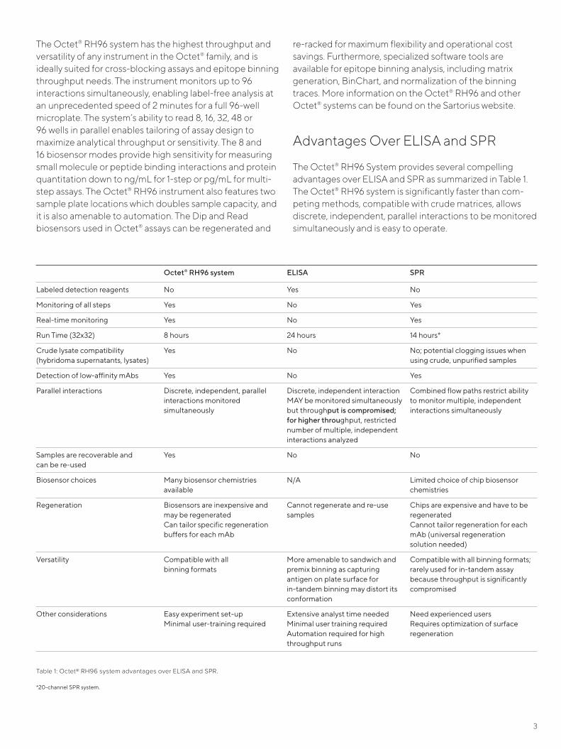
Advantages Over ELISA and SPR
The Octet® RH96 System provides several compelling advantages over ELISA and SPR as summarized in Table 1. The Octet® RH96 system is significantly faster than com-peting methods, compatible with crude matrices, allows discrete, independent, parallel interactions to be monitored simultaneously and is easy to operate.
The Octet® RH96 system has the highest throughput and versatility of any instrument in the Octet® family, and is ideally suited for cross-blocking assays and epitope binning throughput needs. The instrument monitors up to 96 interactions simultaneously, enabling label-free analysis at an unprecedented speed of 2 minutes for a full 96-well microplate. The system’s ability to read 8, 16, 32, 48 or 96 wells in parallel enables tailoring of assay design to maximize analytical throughput or sensitivity. The 8 and 16 biosensor modes provide high sensitivity for measuring small molecule or peptide binding interactions and protein quantitation down to ng/mL for 1-step or pg/mL for multi-step assays. The Octet® RH96 instrument also features two sample plate locations which doubles sample capacity, and it is also amenable to automation. The Dip and Read biosensors used in Octet® assays can be regenerated and
Octet® RH96 system ELISA SPR
Labeled detection reagents No Yes No
Monitoring of all steps Yes No Yes
Real-time monitoring Yes No Yes
Run Time (32x32) 8 hours 24 hours 14 hours*
Crude lysate compatibility (hybridoma supernatants, lysates)
Yes No No; potential clogging issues when using crude, unpurified samples
Detection of low-affinity mAbs Yes No Yes
Parallel interactions Discrete, independent, parallel interactions monitored simultaneously
Discrete, independent interaction MAY be monitored simultaneously but throughput is compromised; for higher throughput, restricted number of multiple, independent interactions analyzed
Combined flow paths restrict ability to monitor multiple, independent interactions simultaneously
Samples are recoverable and can be re-used
Yes No No
Biosensor choices Many biosensor chemistries available
N/A Limited choice of chip biosensor chemistries
Regeneration Biosensors are inexpensive and may be regenerated Can tailor specific regeneration buffers for each mAb
Cannot regenerate and re-use samples
Chips are expensive and have to be regeneratedCannot tailor regeneration for each mAb (universal regeneration solution needed)
Versatility Compatible with all binning formats
More amenable to sandwich and premix binning as capturing antigen on plate surface for in-tandem binning may distort its conformation
Compatible with all binning formats; rarely used for in-tandem assay because throughput is significantly compromised
Other considerations Easy experiment set-upMinimal user-training required
Extensive analyst time neededMinimal user training requiredAutomation required for high throughput runs
Need experienced usersRequires optimization of surface regeneration
Table 1: Octet® RH96 system advantages over ELISA and SPR.
*20-channel SPR system.
4
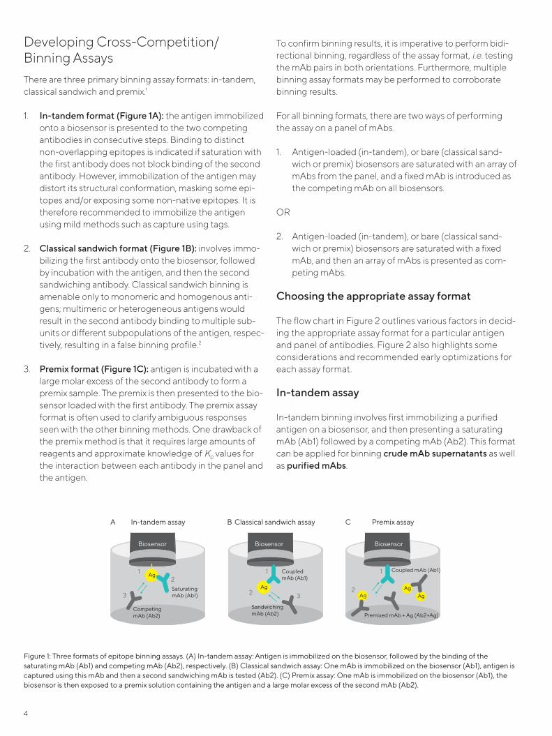
Developing Cross-Competition/Binning AssaysThere are three primary binning assay formats: in-tandem, classical sandwich and premix.1
1. In-tandem format (Figure 1A): the antigen immobilized onto a biosensor is presented to the two competing antibodies in consecutive steps. Binding to distinct non-overlapping epitopes is indicated if saturation with the first antibody does not block binding of the second antibody. However, immobilization of the antigen may distort its structural conformation, masking some epi-topes and/or exposing some non-native epitopes. It is therefore recommended to immobilize the antigen using mild methods such as capture using tags.
2. Classical sandwich format (Figure 1B): involves immo-bilizing the first antibody onto the biosensor, followed by incubation with the antigen, and then the second sandwiching antibody. Classical sandwich binning is amenable only to monomeric and homogenous anti-gens; multimeric or heterogeneous antigens would result in the second antibody binding to multiple sub-units or different subpopulations of the antigen, respec-tively, resulting in a false binning profile.2
3. Premix format (Figure 1C): antigen is incubated with a large molar excess of the second antibody to form a premix sample. The premix is then presented to the bio-sensor loaded with the first antibody. The premix assay format is often used to clarify ambiguous responses seen with the other binning methods. One drawback of the premix method is that it requires large amounts of reagents and approximate knowledge of KD values for the interaction between each antibody in the panel and the antigen.
To confirm binning results, it is imperative to perform bidi-rectional binning, regardless of the assay format, i.e. testing the mAb pairs in both orientations. Furthermore, multiple binning assay formats may be performed to corroborate binning results.
For all binning formats, there are two ways of performing the assay on a panel of mAbs.
1. Antigen-loaded (in-tandem), or bare (classical sand-wich or premix) biosensors are saturated with an array of mAbs from the panel, and a fixed mAb is introduced as the competing mAb on all biosensors.
OR
2. Antigen-loaded (in-tandem), or bare (classical sand-wich or premix) biosensors are saturated with a fixed mAb, and then an array of mAbs is presented as com-peting mAbs.
Choosing the appropriate assay format
The flow chart in Figure 2 outlines various factors in decid-ing the appropriate assay format for a particular antigen and panel of antibodies. Figure 2 also highlights some considerations and recommended early optimizations for each assay format.
In-tandem assay
In-tandem binning involves first immobilizing a purified antigen on a biosensor, and then presenting a saturating mAb (Ab1) followed by a competing mAb (Ab2). This format can be applied for binning crude mAb supernatants as well as purified mAbs.
Biosensor
A In-tandem assay B Classical sandwich assay C Premix assay
SaturatingmAb (Ab1)
CompetingmAb (Ab2)
Ag12
3
Biosensor
Coupled mAb (Ab1)
SandwichingmAb (Ab2)
Ag
1
2 3
Biosensor
AgAg
Ag
1
2
Coupled mAb (Ab1)
Premixed mAb + Ag (Ab2+Ag)
Figure 1: Three formats of epitope binning assays. (A) In-tandem assay: Antigen is immobilized on the biosensor, followed by the binding of the saturating mAb (Ab1) and competing mAb (Ab2), respectively. (B) Classical sandwich assay: One mAb is immobilized on the biosensor (Ab1), antigen is captured using this mAb and then a second sandwiching mAb is tested (Ab2). (C) Premix assay: One mAb is immobilized on the biosensor (Ab1), the biosensor is then exposed to a premix solution containing the antigen and a large molar excess of the second mAb (Ab2).
5
Fc Capture (AHC), Anti-Mouse Fc Capture (AMC) and Nickel-NTA (NTA). Alternatively, custom biosensors can be made by immobilizing an antibody against a specific motif or tag on the antigen, onto the biosensor. Once an optimized regeneration protocol has been developed, these biosensors may be regenerated and reused to reduce assay costs.
Streptavidin-based approachIn vivo site-specific biotinylation methods that introduce one streptavidin binding site at a specific location on the antigen are recommended. However, in-vitro biotinylation of the antigen can be performed if necessary. For detailed biotinylation protocols for protein ligands with Streptavidin Biosensors, refer to Technical Note 28, Biotinylation of Pro-tein for Immobilization onto Streptavidin Biosensors. Streptavidin Biosensors (SA) are regenerable to the level of the immobilized antigen.
Choosing the right biosensorFor in-tandem binning studies, it is critical to choose a biosensor that maintains the structure and activity of the immobilized antigen. For a complete list of biosensors, please refer to the Biosensor Selection Guide.
Capture using affinity tags-based approachCapture biosensors are pre-immobilized with a high affinity capture antibody or protein that binds to the antigen via a known motif or tag. Since the tag or the motif has been engineered at a specific antigen location, the cap-ture-based approach allows the most favorable orientation with the least structural distortion of the antigen. There-fore, the capture-based approach is the preferred method for antigen immobilization for in-tandem binning studies. The interaction between the antigen and the bio-sensor should be stable i.e., with low or minimal dissociation. Some ready-to-use biosensors available for tagged anti-gens include Anti-GST, HIS2, Anti-Penta His, Anti-Human
Monomer
Purified
Crude(Hybridoma supes, phage lysates) Purified
Crude(Hybridoma supes,phage lysates)
Multimer
Antigen
mAbs
Binningformat
ClassicalsandwichPremix In-tandem
Binningformat
Classicalsandwich In-tandem
mAbs
Binningformat
Premix In-tandem
Binningformat
In-tandem
Binning format considerations
Recommendedearly optimizationexperiments
Premix Classical Sandwich In-tandem• Ag concentration must be at
or greater than KD
• Premixed mAbs must be in large molar excess over Ag
• Block unsaturated biosensor surface when AHC/AMC Biosensors used
• Approx. KD required from kinetic screen
• Ensure no cross-reactivity between mAbs
• Ensure mAbs still active post coupling to SA or AR2G
• Ensure complete self-block
• Ag should not dissociate rapidly from biosensor coupled mAbs
• Block unsaturated biosensor surface when AHC/AMC Biosensors used
• Weak affinity/High off-rate mAbs coupled to biosensor may lead to ambiguous data
• Ensure no cross-reactivity between mAbs
• Ensure mAbs still active post coupling to SA or AR2G
• Ensure complete self-block
• Ag may be loaded on biosensor using tags or directly (AR2G)
• First mAb must saturate and remain bound to Ag to prevent “free Ag” from binding to competing Ab
• Weak affinity/high off-rate mAbs presented as saturating Abs may lead to ambiguous data
• Direct coupling of Ag to biosensor with AR2G may lead to Ag inactivation — ensure Ag still active
• Ensure complete self-block
Figure 2: Assay format flow chart.
6
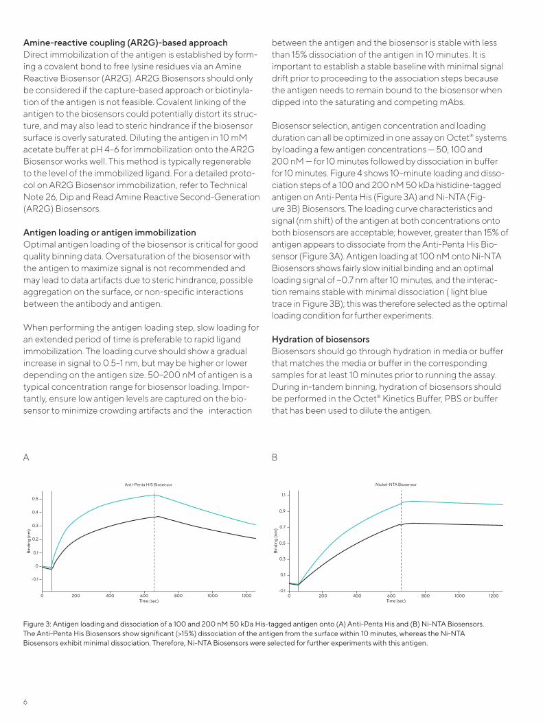
between the antigen and the biosensor is stable with less than 15% dissociation of the antigen in 10 minutes. It is important to establish a stable baseline with minimal signal drift prior to proceeding to the association steps because the antigen needs to remain bound to the biosensor when dipped into the saturating and competing mAbs.
Biosensor selection, antigen concentration and loading duration can all be optimized in one assay on Octet® systems by loading a few antigen concentrations — 50, 100 and 200 nM — for 10 minutes followed by dissociation in buffer for 10 minutes. Figure 4 shows 10–minute loading and disso-ciation steps of a 100 and 200 nM 50 kDa histidine-tagged antigen on Anti-Penta His (Figure 3A) and Ni-NTA (Fig-ure 3B) Biosensors. The loading curve characteristics and signal (nm shift) of the antigen at both concentrations onto both biosensors are acceptable; however, greater than 15% of antigen appears to dissociate from the Anti-Penta His Bio-sensor (Figure 3A). Antigen loading at 100 nM onto Ni-NTA Biosensors shows fairly slow initial binding and an optimal loading signal of ~0.7 nm after 10 minutes, and the interac-tion remains stable with minimal dissociation ( light blue trace in Figure 3B); this was therefore selected as the optimal loading condition for further experiments.
Hydration of biosensors Biosensors should go through hydration in media or buffer that matches the media or buffer in the corresponding samples for at least 10 minutes prior to running the assay. During in-tandem binning, hydration of biosensors should be performed in the Octet® Kinetics Buffer, PBS or buffer that has been used to dilute the antigen.
Amine-reactive coupling (AR2G)-based approachDirect immobilization of the antigen is established by form-ing a covalent bond to free lysine residues via an Amine Reactive Biosensor (AR2G). AR2G Biosensors should only be considered if the capture-based approach or biotinyla-tion of the antigen is not feasible. Covalent linking of the antigen to the biosensors could potentially distort its struc-ture, and may also lead to steric hindrance if the biosensor surface is overly saturated. Diluting the antigen in 10 mM acetate buffer at pH 4–6 for immobilization onto the AR2G Biosensor works well. This method is typically regenerable to the level of the immobilized ligand. For a detailed proto-col on AR2G Biosensor immobilization, refer to Technical Note 26, Dip and Read Amine Reactive Second-Generation (AR2G) Biosensors.
Antigen loading or antigen immobilizationOptimal antigen loading of the biosensor is critical for good quality binning data. Oversaturation of the biosensor with the antigen to maximize signal is not recommended and may lead to data artifacts due to steric hindrance, possible aggregation on the surface, or non-specific interactions between the antibody and antigen.
When performing the antigen loading step, slow loading for an extended period of time is preferable to rapid ligand immobilization. The loading curve should show a gradual increase in signal to 0.5–1 nm, but may be higher or lower depending on the antigen size. 50–200 nM of antigen is a typical concentration range for biosensor loading. Impor-tantly, ensure low antigen levels are captured on the bio-sensor to minimize crowding artifacts and the interaction
Figure 3: Antigen loading and dissociation of a 100 and 200 nM 50 kDa His-tagged antigen onto (A) Anti-Penta His and (B) Ni-NTA Biosensors. The Anti-Penta His Biosensors show significant (>15%) dissociation of the antigen from the surface within 10 minutes, whereas the Ni-NTA Biosensors exhibit minimal dissociation. Therefore, Ni-NTA Biosensors were selected for further experiments with this antigen.
Bin
ding
(nm
)
Time (sec)0 200 400 600 800 1000 1200
-0.1
0.1
0.2
0.3
0.4
0.5
0
Bin
ding
(nm
)
Time (sec)0 200 400 600 800 1000 1200
-0.1
0.1
0.3
0.5
0.7
0.9
1.1
Anti-Penta HIS Biosensor
Nickel-NTA Biosensor
Bin
ding
(nm
)
Time (sec)0 200 400 600 800 1000 1200
-0.1
0.1
0.2
0.3
0.4
0.5
0
Bin
ding
(nm
)
Time (sec)0 200 400 600 800 1000 1200
-0.1
0.1
0.3
0.5
0.7
0.9
1.1
Anti-Penta HIS Biosensor
Nickel-NTA Biosensor
A B

7
BaselinesIn all binning assays, there are usually three baseline steps. The three baseline steps for in-tandem binning are: 1) the step prior to antigen loading (sensor check), 2) the step before the association of the saturating mAb (Ab1), and 3) the step preceding the association of the competing mAb (Ab2) (Figure 4). A baseline should be established using buffer or media matched to the one used in the subse-quent loading and the two association steps to mitigate any non-specific binding and drift from buffer effects. Baseline steps are performed to remove any unbound antigen and Ab1 from the biosensor.
If mAbs are present in cell culture supernatants, then the baseline steps preceding the mAb binding steps should be performed in mock-transfected supernatants, spent media or low IgG FBS supplemented in growth media. Some biosensors, such as Streptavidin and Ni-NTA, may be more prone to non-specific binding by free biotin or irrelevant histidine-containing proteins present in super-natants. In these cases, performing 2–5 minute baselines in spent or FBS supplemented media becomes vital.
If binning is performed with purified mAbs, then the base-line steps preceding the association of mAbs should be done in the same buffer that the mAbs are diluted in, either Octet® Kinetics Buffer or PBS. When binning puri-fied mAbs, non-specific binding typically is minimal. How-
ever, if some non-specific binding is observed, then blocking reagents such as BSA (up to 1–2%) and/or non-ionic detergents such as Tween-20 (up to 0.05%) may be added to the buffer.
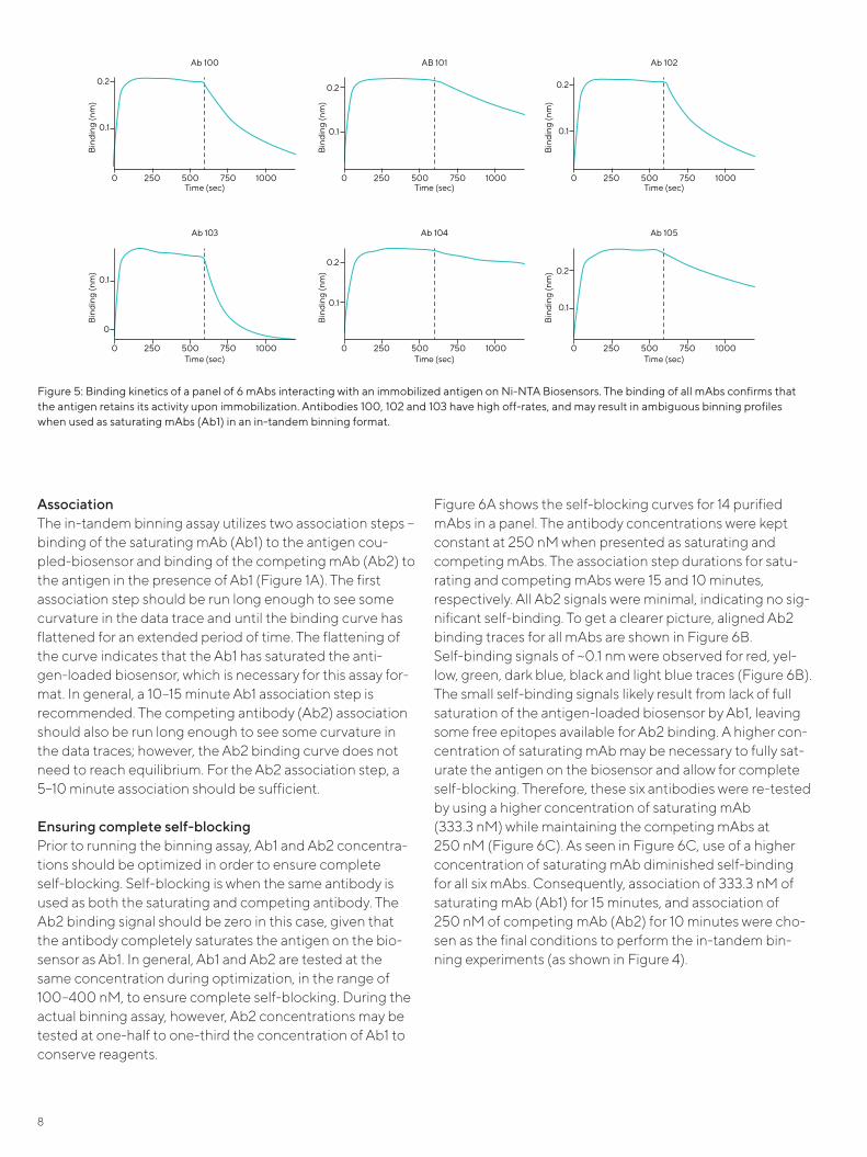
Ensuring the antigen is active Coupling of the antigen to the biosensor surface occasion-ally can alter the antigen’s structural conformation, reveal-ing non-native epitopes and/or masking epitopes. Before proceeding to binning studies, it is important to ensure that the antigen remains active and able to bind the mAb panel following immobilization. Antigen activity can be confirmed via a kinetic screening assay performed in the same format as the binning assay.
For this kinetic screen, antigen loading is followed by asso-ciation with 200–350 nM mAbs for 10–15 minutes, and then a 10–15 minute dissociation step. This screening assay can also be used to flag mAbs with weak affinities to the anti-gen. Weak affinity mAbs are unreliable when presented as saturating mAbs (Ab1); they will dissociate quickly and expose free antigen to competing mAbs (Ab2). The binning data for these mAbs will be more reliable when they are pre-sented as competing mAbs (Ab2). Figure 5 shows a kinetic screen of a panel of six mAbs. In this panel, antibodies 100, 102 and 103 exhibit high off-rates and may produce ambig-uous binning profiles when used as saturating mAbs (Ab1) in an in-tandem assay format.
Figure 4: Representative data from an in-tandem binning assay. The arrows indicate the three baselines for this assay format: before antigen loading (sensor check), after antigen loading before binding of the saturating mAb (Ab1) and before binding of competing mAb (Ab2). The baseline steps remove any unbound antigen/ antibody from the biosensor, and the baseline after Ab1 can also be used to flag for mAbs with fast off-rates.
Bin
ding
(nm
)
Time (sec)
0
1.0
2.0
3.0
4.0
2000 2200 2400 2600 2800 3000 3200
Baseline Baseline BaselineAntigen Saturating mAb (Ab1) Competing mAb (Ab2)
8
AssociationThe in-tandem binning assay utilizes two association steps – binding of the saturating mAb (Ab1) to the antigen cou-pled-biosensor and binding of the competing mAb (Ab2) to the antigen in the presence of Ab1 (Figure 1A). The first association step should be run long enough to see some curvature in the data trace and until the binding curve has flattened for an extended period of time. The flattening of the curve indicates that the Ab1 has saturated the anti-gen-loaded biosensor, which is necessary for this assay for-mat. In general, a 10–15 minute Ab1 association step is recommended. The competing antibody (Ab2) association should also be run long enough to see some curvature in the data traces; however, the Ab2 binding curve does not need to reach equilibrium. For the Ab2 association step, a 5–10 minute association should be sufficient.
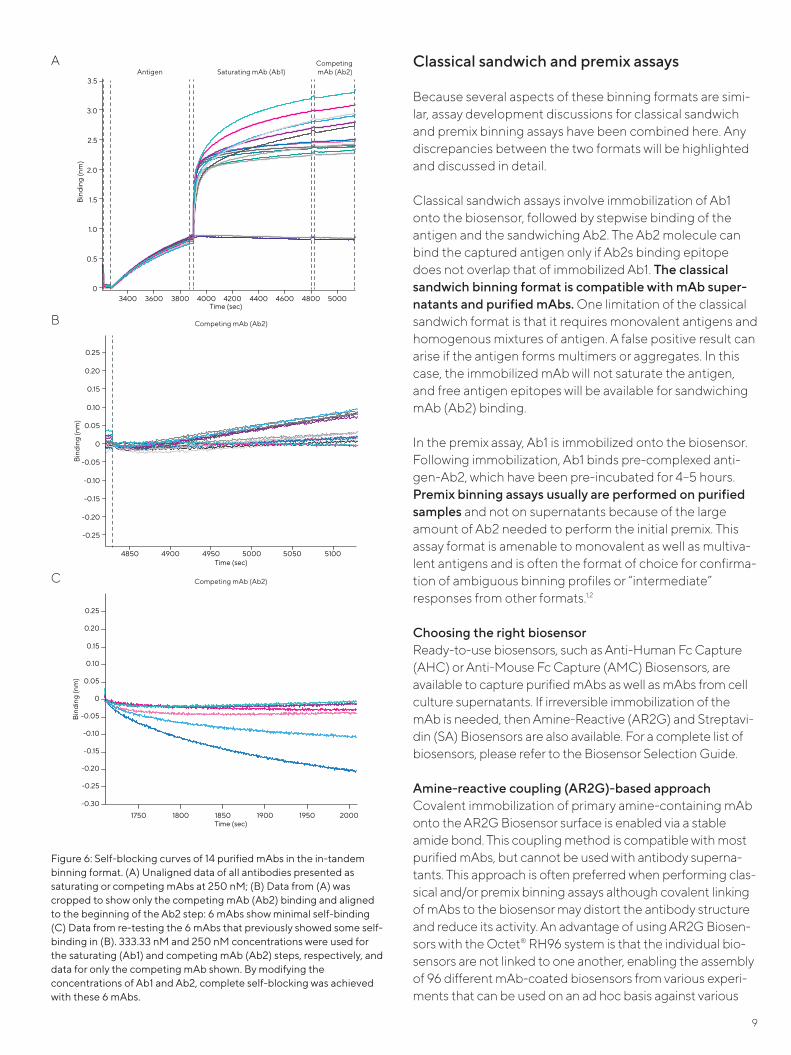
Ensuring complete self-blockingPrior to running the binning assay, Ab1 and Ab2 concentra-tions should be optimized in order to ensure complete self-blocking. Self-blocking is when the same antibody is used as both the saturating and competing antibody. The Ab2 binding signal should be zero in this case, given that the antibody completely saturates the antigen on the bio-sensor as Ab1. In general, Ab1 and Ab2 are tested at the same concentration during optimization, in the range of 100–400 nM, to ensure complete self-blocking. During the actual binning assay, however, Ab2 concentrations may be tested at one-half to one-third the concentration of Ab1 to conserve reagents.
Figure 6A shows the self-blocking curves for 14 purified mAbs in a panel. The antibody concentrations were kept constant at 250 nM when presented as saturating and competing mAbs. The association step durations for satu-rating and competing mAbs were 15 and 10 minutes, respectively. All Ab2 signals were minimal, indicating no sig-nificant self-binding. To get a clearer picture, aligned Ab2 binding traces for all mAbs are shown in Figure 6B. Self-binding signals of ~0.1 nm were observed for red, yel-low, green, dark blue, black and light blue traces (Figure 6B). The small self-binding signals likely result from lack of full saturation of the antigen-loaded biosensor by Ab1, leaving some free epitopes available for Ab2 binding. A higher con-centration of saturating mAb may be necessary to fully sat-urate the antigen on the biosensor and allow for complete self-blocking. Therefore, these six antibodies were re-tested by using a higher concentration of saturating mAb (333.3 nM) while maintaining the competing mAbs at 250 nM (Figure 6C). As seen in Figure 6C, use of a higher concentration of saturating mAb diminished self-binding for all six mAbs. Consequently, association of 333.3 nM of saturating mAb (Ab1) for 15 minutes, and association of 250 nM of competing mAb (Ab2) for 10 minutes were cho-sen as the final conditions to perform the in-tandem bin-ning experiments (as shown in Figure 4).
Figure 5: Binding kinetics of a panel of 6 mAbs interacting with an immobilized antigen on Ni-NTA Biosensors. The binding of all mAbs confirms that the antigen retains its activity upon immobilization. Antibodies 100, 102 and 103 have high off-rates, and may result in ambiguous binning profiles when used as saturating mAbs (Ab1) in an in-tandem binning format.
Bin
ding
(nm
)
Time (sec)
Ab 100
0 500 1000250 750
0.1
0.2
Bin
ding
(nm
)
Time (sec)
AB 101
0 500 1000250 750
0.1
0.2
Bin
ding
(nm
)
Time (sec)
Ab 102
0 500 1000250 750
0.1
0.2B
indi
ng (n
m)
Time (sec)
Ab 103
0 500 1000250 750
0.1
0B
indi
ng (n
m)
Time (sec)
Ab 104
0 500 1000250 750
0.1
0.2
Bin
ding
(nm
)
Time (sec)
Ab 105
0 500 1000250 750
0.1
0.2
9
Classical sandwich and premix assays
Because several aspects of these binning formats are simi-lar, assay development discussions for classical sandwich and premix binning assays have been combined here. Any discrepancies between the two formats will be highlighted and discussed in detail.
Classical sandwich assays involve immobilization of Ab1 onto the biosensor, followed by stepwise binding of the antigen and the sandwiching Ab2. The Ab2 molecule can bind the captured antigen only if Ab2s binding epitope does not overlap that of immobilized Ab1. The classical sandwich binning format is compatible with mAb super-natants and purified mAbs. One limitation of the classical sandwich format is that it requires monovalent antigens and homogenous mixtures of antigen. A false positive result can arise if the antigen forms multimers or aggregates. In this case, the immobilized mAb will not saturate the antigen, and free antigen epitopes will be available for sandwiching mAb (Ab2) binding.
In the premix assay, Ab1 is immobilized onto the biosensor. Following immobilization, Ab1 binds pre-complexed anti-gen-Ab2, which have been pre-incubated for 4–5 hours. Premix binning assays usually are performed on purified samples and not on supernatants because of the large amount of Ab2 needed to perform the initial premix. This assay format is amenable to monovalent as well as multiva-lent antigens and is often the format of choice for confirma-tion of ambiguous binning profiles or “intermediate” responses from other formats.1,2
Choosing the right biosensor Ready-to-use biosensors, such as Anti-Human Fc Capture (AHC) or Anti-Mouse Fc Capture (AMC) Biosensors, are available to capture purified mAbs as well as mAbs from cell culture supernatants. If irreversible immobilization of the mAb is needed, then Amine-Reactive (AR2G) and Streptavi-din (SA) Biosensors are also available. For a complete list of biosensors, please refer to the Biosensor Selection Guide.
Amine-reactive coupling (AR2G)-based approachCovalent immobilization of primary amine-containing mAb onto the AR2G Biosensor surface is enabled via a stable amide bond. This coupling method is compatible with most purified mAbs, but cannot be used with antibody superna-tants. This approach is often preferred when performing clas-sical and/or premix binning assays although covalent linking of mAbs to the biosensor may distort the antibody structure and reduce its activity. An advantage of using AR2G Biosen-sors with the Octet® RH96 system is that the individual bio-sensors are not linked to one another, enabling the assembly of 96 different mAb-coated biosensors from various experi-ments that can be used on an ad hoc basis against various
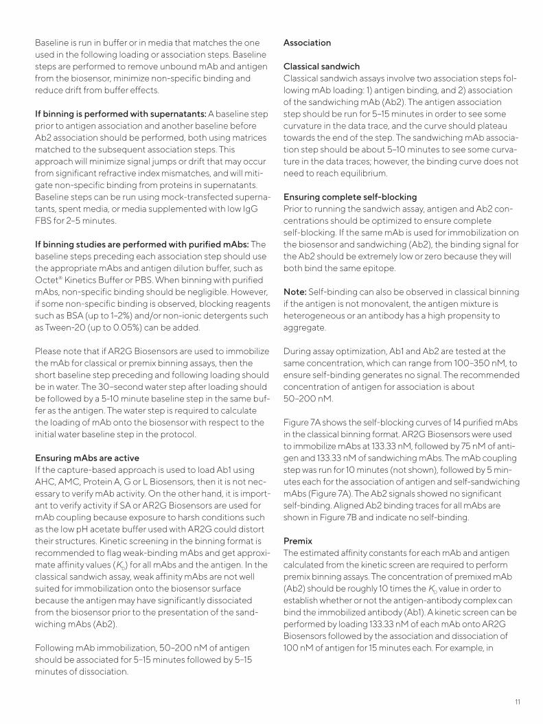
Figure 6: Self-blocking curves of 14 purified mAbs in the in-tandem binning format. (A) Unaligned data of all antibodies presented as saturating or competing mAbs at 250 nM; (B) Data from (A) was cropped to show only the competing mAb (Ab2) binding and aligned to the beginning of the Ab2 step: 6 mAbs show minimal self-binding (C) Data from re-testing the 6 mAbs that previously showed some self-binding in (B). 333.33 nM and 250 nM concentrations were used for the saturating (Ab1) and competing mAb (Ab2) steps, respectively, and data for only the competing mAb shown. By modifying the concentrations of Ab1 and Ab2, complete self-blocking was achieved with these 6 mAbs.
Bin
ding
(nm
)
Time (sec)
0
0.5
1.0
1.5
2.0
2.5
3.0
3.5
3400 3600 3800 4000 4200 4400 4600 4800 5000
Antigen Saturating mAb (Ab1)Competing mAb (Ab2)
Time (sec)4850 4900 4950 5000 5050 5100
-0.25
-0.20
-0.15
-0.10
-0.05
0
0.05
0.10
0.15
0.20
0.25
Bin
ding
(nm
)
Competing mAb (Ab2)
Bin
ding
(nm
)
Time (sec)
-0.30
-0.25
-0.20
-0.15
-0.10
-0.05
0
0.05
0.10
0.15
0.20
0.25
1750 1800 1850 1900 1950 2000
Competing mAb (Ab2)
A
B
C

10
panels of mAbs. Antibody-coupled AR2G Biosensors typi-cally are regenerable to the level of the immobilized mAb. For a detailed protocol on AR2G Biosensor immobilization, refer to Technical Note 26, Dip and Read Amine Reactive Second-Generation (AR2G) Biosensors.
Capture-based approachCapture biosensors are pre-immobilized with a high-affinity capture antibody, such as Anti-Human IgG Fc (AHC) or Anti-Mouse IgG Fc (AMC) Biosensors. These biosensors bind to mAbs via the Fc-region and enable favorable orien-tation of the mAbs on the biosensor surface. Protein A, G and L Biosensors are also available to immobilize non-hu-man and non-murine antibodies. When working with AHC, AMC, Protein A,G or L Biosensors, it is imperative to com-pletely block the biosensor surface following the immobili-zation of the first mAb. Residual unblocked sites on the biosensor surface may bind directly to the sandwiching mAb (Ab2), resulting in a false positive binning result.
For tagged mAbs, Anti-GST (GST), Anti-Penta His (HIS) or Ni-NTA (NTA) may also be used. It is important to note that if the mAb is histidine tagged in crude supernatants, then Anti-HIS may be a better option than Ni-NTA because the latter may non-specifically bind to other HIS-tagged pro-teins in supernatants. Non-specific binding can be miti-gated by optimizing the composition of the buffer of the baseline step(s), which will be discussed in detail in the fol-lowing Baseline section. All capture-based biosensors can be fully regenerated and reused multiple times to reduce assay costs.
Streptavidin-based approachIn cases where the antibodies are non-human, non-murine and untagged, the capture-based approach cannot be used. This situation often arises when working with hamster, rabbit, goat, and sheep antibodies. In such cases, antibod-ies can be biotinylated and immobilized using Streptavidin Biosensors. Streptavidin-biotin interactions are rapid, and stable. These biosensors can be regenerated to the level of the immobilized mAb. For detailed protocol for biotinyla-tion of protein ligands for use on Streptavidin Biosensors, refer to Sartorius Technical Note 28, Biotinylation of Protein for Immobilization onto Streptavidin Biosensors.
Antibody loading or antibody immobilization When performing the mAb loading step, slow loading for an extended period of time is recommended rather than rapid mAb immobilization that may lead to uneven loading. The loading curve should show a gradual increase in signal and should reach 0.5–1 nm for most mAbs. 50–200 nM of mAbs are typically immobilized on AHC, AMC, SA and AR2G Biosensors.
MAb immobilization on SA or AR2G Biosensors is stable due to effectively irreversible interactions between SA-bio-tin, and amine groups-free lysine residues, respectively. Hence, the baseline following loading should be stable.
When capture-based biosensors such as AHC or AMC are used, the initial binding curve should reach saturation. To ensure that no free binding sites remain after biosensor loading, a blocking step following mAb loading should always be performed. The general rule is that if all mAbs are the same isotype, then blocking with 50 µg/mL of irrelevant IgG of the same isotype for 5–10 minutes should be suffi-cient. The blocking step should be followed by a 2–4 minute wash to ensure that any loosely-bound antibody is removed prior to antigen association. The biosensors should be dipped back into the same antibody wells to confirm that no additional self-binding is observed. If the mAbs in the panel have varied or unknown isotypes, blocking with 50 µg/mL of polyclonal IgG for 10 minutes followed by a 2–4 minute buffer wash may help. A longer blocking step may also help, especially if the polyclonal IgG is not highly representative of the sandwiching mAb isotype(s).
A stable baseline with minimal signal drift following mAb immobilization should be established before proceeding to the next association step as the Ab1 needs to remain bound to the biosensor when it is dipped into antigen and the sandwiching antibody.
Hydration of biosensors Hydration of biosensors should be performed for at least 10 minutes in media or buffer that matches the media or buffer in the corresponding samples. For complex samples, such as hybridoma supernatants or phage lysates, hydration of biosensors may be performed for a longer period of time (~30 mins to 1 hour). In classical and premix binning with purified mAbs, hydration of biosensors should be done in the buffer that was used to dilute the mAbs, which is usually Octet® Kinetics Buffer or PBS. If binning is being performed with mAb supernatants, then hydration should be per-formed in mock-transfected supernatants, spent media or low IgG FBS supplemented in growth media.
Baseline There are three baseline steps in classical sandwich binning: 1) the step prior to mAb loading (sensor check), 2) the step before the association of the antigen, and 3) the step pre-ceding the association of the sandwiching mAb. There are two baseline steps in premix: 1) the step before mAb immo-bilization (sensor check) and 2) the step prior to the binding of the complex (antigen plus sandwiching mAb).
11
Baseline is run in buffer or in media that matches the one used in the following loading or association steps. Baseline steps are performed to remove unbound mAb and antigen from the biosensor, minimize non-specific binding and reduce drift from buffer effects.
If binning is performed with supernatants: A baseline step prior to antigen association and another baseline before Ab2 association should be performed, both using matrices matched to the subsequent association steps. This approach will minimize signal jumps or drift that may occur from significant refractive index mismatches, and will miti-gate non-specific binding from proteins in supernatants. Baseline steps can be run using mock-transfected superna-tants, spent media, or media supplemented with low IgG FBS for 2–5 minutes.
If binning studies are performed with purified mAbs: The baseline steps preceding each association step should use the appropriate mAbs and antigen dilution buffer, such as Octet® Kinetics Buffer or PBS. When binning with purified mAbs, non-specific binding should be negligible. However, if some non-specific binding is observed, blocking reagents such as BSA (up to 1–2%) and/or non-ionic detergents such as Tween-20 (up to 0.05%) can be added.
Please note that if AR2G Biosensors are used to immobilize the mAb for classical or premix binning assays, then the short baseline step preceding and following loading should be in water. The 30–second water step after loading should be followed by a 5-10 minute baseline step in the same buf-fer as the antigen. The water step is required to calculate the loading of mAb onto the biosensor with respect to the initial water baseline step in the protocol.
Ensuring mAbs are active If the capture-based approach is used to load Ab1 using AHC, AMC, Protein A, G or L Biosensors, then it is not nec-essary to verify mAb activity. On the other hand, it is import-ant to verify activity if SA or AR2G Biosensors are used for mAb coupling because exposure to harsh conditions such as the low pH acetate buffer used with AR2G could distort their structures. Kinetic screening in the binning format is recommended to flag weak-binding mAbs and get approxi-mate affinity values (KD) for all mAbs and the antigen. In the classical sandwich assay, weak affinity mAbs are not well suited for immobilization onto the biosensor surface because the antigen may have significantly dissociated from the biosensor prior to the presentation of the sand-wiching mAbs (Ab2).
Following mAb immobilization, 50–200 nM of antigen should be associated for 5–15 minutes followed by 5–15 minutes of dissociation.
Association
Classical sandwichClassical sandwich assays involve two association steps fol-lowing mAb loading: 1) antigen binding, and 2) association of the sandwiching mAb (Ab2). The antigen association step should be run for 5–15 minutes in order to see some curvature in the data trace, and the curve should plateau towards the end of the step. The sandwiching mAb associa-tion step should be about 5–10 minutes to see some curva-ture in the data traces; however, the binding curve does not need to reach equilibrium.
Ensuring complete self-blockingPrior to running the sandwich assay, antigen and Ab2 con-centrations should be optimized to ensure complete self-blocking. If the same mAb is used for immobilization on the biosensor and sandwiching (Ab2), the binding signal for the Ab2 should be extremely low or zero because they will both bind the same epitope.
Note: Self-binding can also be observed in classical binning if the antigen is not monovalent, the antigen mixture is heterogeneous or an antibody has a high propensity to aggregate.
During assay optimization, Ab1 and Ab2 are tested at the same concentration, which can range from 100–350 nM, to ensure self-binding generates no signal. The recommended concentration of antigen for association is about 50–200 nM.
Figure 7A shows the self-blocking curves of 14 purified mAbs in the classical binning format. AR2G Biosensors were used to immobilize mAbs at 133.33 nM, followed by 75 nM of anti-gen and 133.33 nM of sandwiching mAbs. The mAb coupling step was run for 10 minutes (not shown), followed by 5 min-utes each for the association of antigen and self-sandwiching mAbs (Figure 7A). The Ab2 signals showed no significant self-binding. Aligned Ab2 binding traces for all mAbs are shown in Figure 7B and indicate no self-binding.
PremixThe estimated affinity constants for each mAb and antigen calculated from the kinetic screen are required to perform premix binning assays. The concentration of premixed mAb (Ab2) should be roughly 10 times the KD value in order to establish whether or not the antigen-antibody complex can bind the immobilized antibody (Ab1). A kinetic screen can be performed by loading 133.33 nM of each mAb onto AR2G Biosensors followed by the association and dissociation of 100 nM of antigen for 15 minutes each. For example, in
12
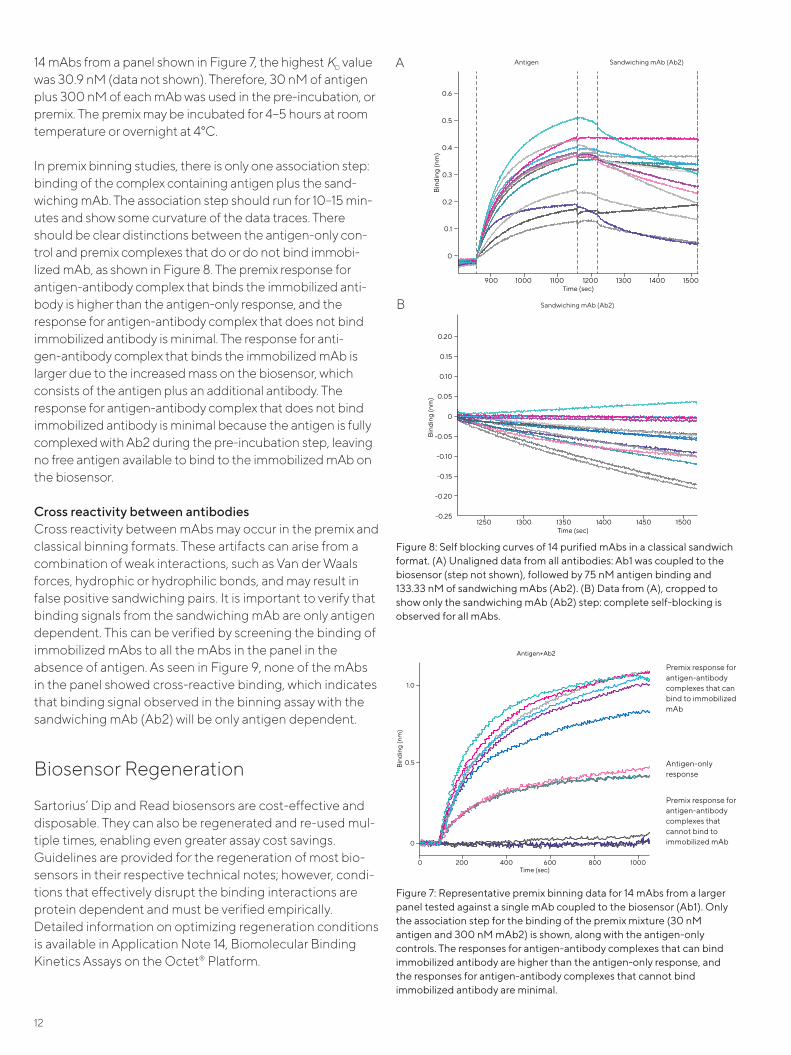
14 mAbs from a panel shown in Figure 7, the highest KD value was 30.9 nM (data not shown). Therefore, 30 nM of antigen plus 300 nM of each mAb was used in the pre-incubation, or premix. The premix may be incubated for 4–5 hours at room temperature or overnight at 4°C.
In premix binning studies, there is only one association step: binding of the complex containing antigen plus the sand-wiching mAb. The association step should run for 10–15 min-utes and show some curvature of the data traces. There should be clear distinctions between the antigen-only con-trol and premix complexes that do or do not bind immobi-lized mAb, as shown in Figure 8. The premix response for antigen-antibody complex that binds the immobilized anti-body is higher than the antigen-only response, and the response for antigen-antibody complex that does not bind immobilized antibody is minimal. The response for anti-gen-antibody complex that binds the immobilized mAb is larger due to the increased mass on the biosensor, which consists of the antigen plus an additional antibody. The response for antigen-antibody complex that does not bind immobilized antibody is minimal because the antigen is fully complexed with Ab2 during the pre-incubation step, leaving no free antigen available to bind to the immobilized mAb on the biosensor.
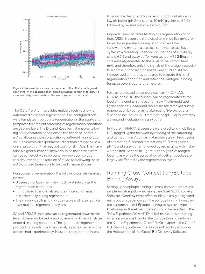
Cross reactivity between antibodies Cross reactivity between mAbs may occur in the premix and classical binning formats. These artifacts can arise from a combination of weak interactions, such as Van der Waals forces, hydrophic or hydrophilic bonds, and may result in false positive sandwiching pairs. It is important to verify that binding signals from the sandwiching mAb are only antigen dependent. This can be verified by screening the binding of immobilized mAbs to all the mAbs in the panel in the absence of antigen. As seen in Figure 9, none of the mAbs in the panel showed cross-reactive binding, which indicates that binding signal observed in the binning assay with the sandwiching mAb (Ab2) will be only antigen dependent.
Biosensor Regeneration
Sartorius’ Dip and Read biosensors are cost-effective and disposable. They can also be regenerated and re-used mul-tiple times, enabling even greater assay cost savings. Guidelines are provided for the regeneration of most bio-sensors in their respective technical notes; however, condi-tions that effectively disrupt the binding interactions are protein dependent and must be verified empirically. Detailed information on optimizing regeneration conditions is available in Application Note 14, Biomolecular Binding Kinetics Assays on the Octet® Platform.
Figure 7: Representative premix binning data for 14 mAbs from a larger panel tested against a single mAb coupled to the biosensor (Ab1). Only the association step for the binding of the premix mixture (30 nM antigen and 300 nM mAb2) is shown, along with the antigen-only controls. The responses for antigen-antibody complexes that can bind immobilized antibody are higher than the antigen-only response, and the responses for antigen-antibody complexes that cannot bind immobilized antibody are minimal.
Time (sec)
Antigen+Ab2
Bin
ding
(nm
)
0
0.5
1.0
0 200 400 600 800 1000
Premix response for antigen-antibody complexes that can bind to immobilized mAb
Antigen-only response
Premix response for antigen-antibody complexes that cannot bind to immobilized mAb
Figure 8: Self blocking curves of 14 purified mAbs in a classical sandwich format. (A) Unaligned data from all antibodies: Ab1 was coupled to the biosensor (step not shown), followed by 75 nM antigen binding and 133.33 nM of sandwiching mAbs (Ab2). (B) Data from (A), cropped to show only the sandwiching mAb (Ab2) step: complete self-blocking is observed for all mAbs.
Bin
ding
(nm
)
Time (sec)
0
0.1
0.2
0.3
0.4
0.5
0.6
900 1000 1100 1200 1300 1400 1500
Antigen Sandwiching mAb (Ab2)
Bin
ding
(nm
)
-0.25
-0.20
-0.15
-0.10
-0.05
0
0.05
0.10
0.15
0.20
Time (sec)1250 1300 1350 1400 1450 1500
Sandwiching mAb (Ab2)
A
B
13
The Octet® platform provides multiple tools to allow for automated biosensor regeneration. Pre-configured soft-ware templates incorporate regeneration in the assays and templates for efficient screening of regeneration conditions are also available. The Dip and Read format enables tailor-ing of regeneration conditions to the needs of individual mAbs, allowing the incorporation of different regeneration solutions within an experiment, rather than having to use a universal solution that may not work for all mAbs. This main-tains a higher number of active coupled mAbs than what can be achieved with a universal regeneration solution, thereby lowering the attrition of mAbs and allowing these mAb-coupled biosensors to be used in more studies.3
For successful regeneration, the following conditions must be met: - Biosensor surface chemistry must be stable under the
regeneration conditions. - Immobilized ligand-analyte protein interaction must dissociate fully during regeneration. - The immobilized ligand must be stable and retain activity over multiple regeneration cycles.
SA and AR2G Biosensors can be regenerated down to the level of the immobilized ligand by removing bound analytes under disrupting conditions. The appropriate regeneration protocol for a particular ligand-analyte protein pair must be determined experimentally. Most antibody-protein interac-
tions can be disrupted by a series of short incubations in low pH buffer (pH 2–4), such as 10 mM glycine, (pH 2–3), followed by neutralization in assay buffer.
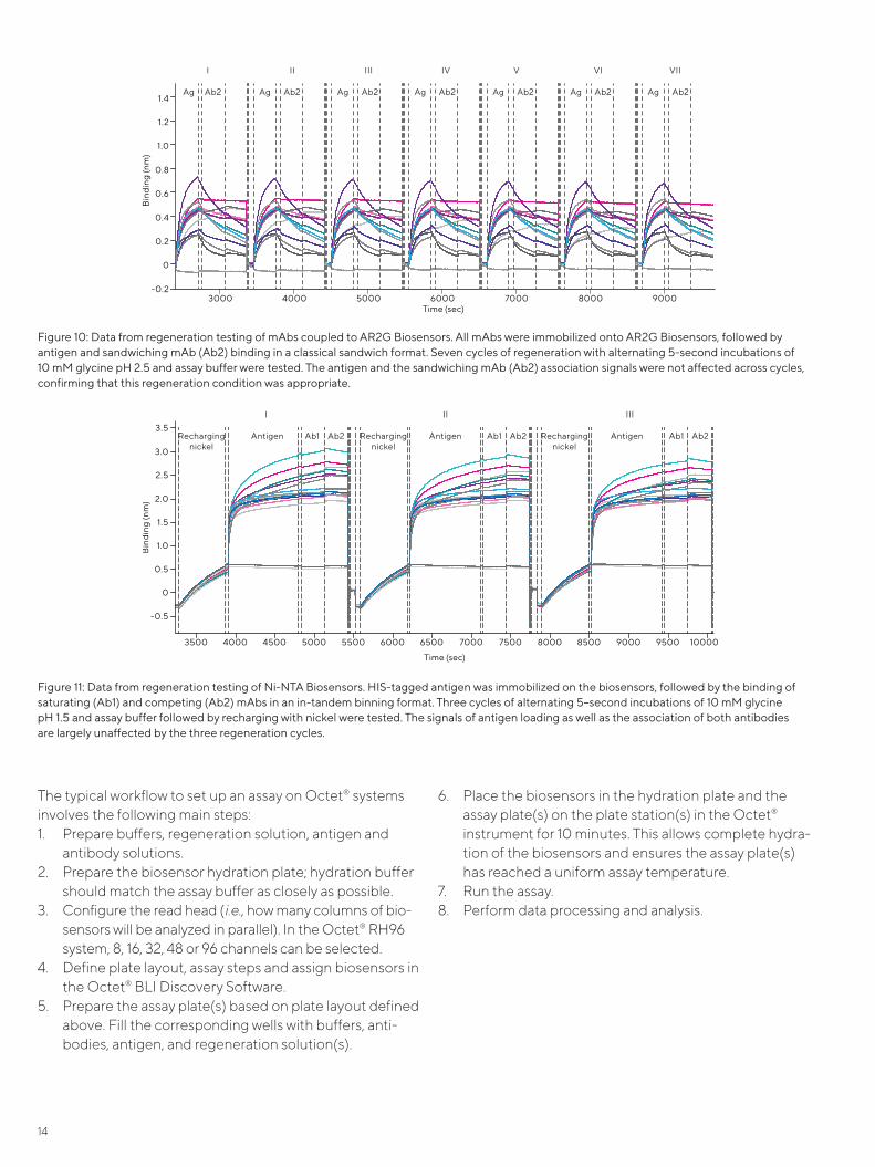
Figure 10 demonstrates testing of a regeneration condi-tion: AR2G Biosensors were used to immobilize mAbs fol-lowed by sequential binding of antigen and the sandwiching mAbs in a classical sandwich assay. Seven cycles of alternating 5-second incubations of 10 mM gly-cine pH 2.5 and assay buffer were tested. AR2G Biosen-sors were regenerated to the level of the immobilized mAb and therefore only the signals of the antigen associa-tion and self-sandwiching mAbs were studied. All the immobilized antibodies appeared to tolerate the harsh regeneration condition and retain their antigen-binding for up to seven regeneration cycles.
For capture-based biosensors, such as AMC, FLAG, Ni-NTA, and AHC, the surface can be regenerated to the level of the original surface chemistry. The immobilized ligand and the subsequent molecules are removed during regeneration by performing alternating 3–5 cycles of a 5-second incubation in 10 mM glycine (pH 1–2) followed by a 5-second incubation in assay buffer.
In Figure 11, Ni-NTA Biosensors were used to immobilize a HIS-tagged ligand followed by binding of the saturating and competing mAbs in an in-tandem assay. Three cycles of alternating 5-second incubations of 10 mM glycine pH 1.5 and assay buffer followed by recharging with nickel were tested. As seen in Figure 11, the signals of antigen loading as well as the association of both antibodies are largely unaffected by the regeneration cycles.
Running Cross-Competition/Epitope Binning AssaysSetting up an epitope binning or cross-competition assay is simple and straightforward using the Octet® BLI Discovery Software. Octet® systems offer flexibility in assay design and many options depending on the epitope binning format and the instrument used. Epitope binning assays are a type of kinetics assay, therefore “Kinetics” should be selected in the “New Experiment Wizard”. Detailed instructions on setting up an assay can be found in the Epitope Binning section in the Kinetic Experiments: Octet® RH96 chapter in the Octet® BLI Discovery Software User Guide (v8.0 or higher) under the Help section of the Octet® BLI Discovery Software.
Figure 9: Representative data for the panel of 14 mAbs tested against each other in the absence of antigen in a classical sandwich format. No cross reactivity between the mAbs was observed in this panel.
Bin
ding
(nm
)
Time (sec)
-0.05
0
0.05
0.10
0.15
0.20
0 200 400100 300 500
Sandwiching mAb (Ab2)
14
The typical workflow to set up an assay on Octet® systems involves the following main steps:1. Prepare buffers, regeneration solution, antigen and
antibody solutions.2. Prepare the biosensor hydration plate; hydration buffer
should match the assay buffer as closely as possible.3. Configure the read head (i.e., how many columns of bio-
sensors will be analyzed in parallel). In the Octet® RH96 system, 8, 16, 32, 48 or 96 channels can be selected.
4. Define plate layout, assay steps and assign biosensors in the Octet® BLI Discovery Software.
5. Prepare the assay plate(s) based on plate layout defined above. Fill the corresponding wells with buffers, anti-bodies, antigen, and regeneration solution(s).
6. Place the biosensors in the hydration plate and the assay plate(s) on the plate station(s) in the Octet® instrument for 10 minutes. This allows complete hydra-tion of the biosensors and ensures the assay plate(s) has reached a uniform assay temperature.
7. Run the assay.8. Perform data processing and analysis.
Figure 10: Data from regeneration testing of mAbs coupled to AR2G Biosensors. All mAbs were immobilized onto AR2G Biosensors, followed by antigen and sandwiching mAb (Ab2) binding in a classical sandwich format. Seven cycles of regeneration with alternating 5-second incubations of 10 mM glycine pH 2.5 and assay buffer were tested. The antigen and the sandwiching mAb (Ab2) association signals were not affected across cycles, confirming that this regeneration condition was appropriate.
Bin
ding
(nm
)
-0.2
0
0.2
0.4
0.6
0.8
1.0
1.2
1.4
Time (sec)3000 4000 5000 6000 7000 8000 9000
I II III IV V VI VII
Ag Ab2 Ag Ab2 Ag Ab2 Ag Ab2 Ag Ab2 Ag Ab2 Ag Ab2
Figure 11: Data from regeneration testing of Ni-NTA Biosensors. HIS-tagged antigen was immobilized on the biosensors, followed by the binding of saturating (Ab1) and competing (Ab2) mAbs in an in-tandem binning format. Three cycles of alternating 5–second incubations of 10 mM glycine pH 1.5 and assay buffer followed by recharging with nickel were tested. The signals of antigen loading as well as the association of both antibodies are largely unaffected by the three regeneration cycles.
Bin
ding
(nm
)
-0.5
0
0.5
1.0
1.5
2.0
2.5
3.0
3.5
Time (sec)
3500 4000 4500 5000 5500 6000 6500 7000 7500 8000 8500 9000 9500 10000
Rechargingnickel
Antigen Ab1
Ab2
Rechargingnickel
Antigen Ab1
Ab2
Rechargingnickel
Antigen Ab1
Ab2
I II III
15
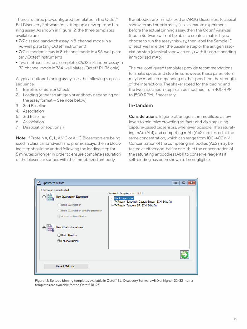
There are three pre-configured templates in the Octet® BLI Discovery Software for setting up a new epitope bin-ning assay. As shown in Figure 12, the three templates available are: - 7x7 classical sandwich assay in 8-channel mode in a
96-well plate (any Octet® instrument) - 7x7 in-tandem assay in 8-channel mode in a 96-well plate (any Octet® instrument) - Two method files for a complete 32x32 in-tandem assay in 32-channel mode in 384-well plates (Octet® RH96 only)
A typical epitope binning assay uses the following steps in sequence:1. Baseline or Sensor Check2. Loading (either an antigen or antibody depending on
the assay format — See note below) 3. 2nd Baseline4. Association 5. 3rd Baseline 6. Association 7. Dissociation (optional)
Note: If Protein A, G, L, AMC or AHC Biosensors are being used in classical sandwich and premix assays, then a block-ing step should be added following the loading step for 5 minutes or longer in order to ensure complete saturation of the biosensor surface with the immobilized antibody.
If antibodies are immobilized on AR2G Biosensors (classical sandwich and premix assays) in a separate experiment before the actual binning assay, then the Octet® Analysis Studio Software will not be able to create a matrix. If you choose to run the assay this way, then label the Sample ID of each well in either the baseline step or the antigen asso-ciation step (classical sandwich only) with its corresponding immobilized mAb.
The pre-configured templates provide recommendations for shake speed and step time; however, these parameters may be modified depending on the speed and the strength of the interactions. The shaker speed for the loading and the two association steps can be modified from 400 RPM to 1500 RPM, if necessary.
In-tandem
Considerations: In general, antigen is immobilized at low levels to minimize crowding artifacts and via a tag using capture-based biosensors, whenever possible. The saturat-ing mAb (Ab1) and competing mAb (Ab2) are tested at the same concentration, which can range from 100–400 nM. Concentration of the competing antibodies (Ab2) may be tested at either one-half or one-third the concentration of the saturating antibodies (Ab1) to conserve reagents if self-binding has been shown to be negligible.
Figure 12: Epitope binning templates available in Octet® BLI Discovery Software v8.0 or higher. 32x32 matrix templates are available for the Octet® RH96.

16
Controls: When running epitope binning in the in-tandem format, it is imperative to have a control in which anti-gen-loaded biosensors are dipped into either media or buffer instead of Ab1, and then into all competing antibod-ies (Ab2) to determine “maximal” binding of each Ab2 to the antigen in the absence of Ab1.
Classical sandwich
Considerations: Ab1 should be immobilized onto the biosen-sor at an optimal concentration (50–200 nM) with binding signals of 0.5–1 nm for all mAbs. Antibodies with high antigen affinity should be coupled to the biosensor for reliable data; immobilization of weak affinity mAbs may result in the disso-ciation of antigen prior to incubation with Ab2. This binning format is unsuitable for multivalent antigens or heteroge-neous mixtures of antigen as all the binding sites on the anti-gen may not be fully saturated with the biosensor coupled antibody. It is essential to verify that there is no cross reactiv-ity between the immobilized and the sandwiching mAbs or between the sandwiching mAb and the biosensor surface. The sandwiching mAb (Ab2) concentration is usually the same as coupled mAb (Ab1) concentration.
Controls: The no-antigen control may be run to ensure that there is no cross-reactivity between antibodies and the Ab2 signal is only antigen dependent. Another important control in this format is immobilization of an irrelevant antibody, preferably of the same isotype and species as other cou-pling mAbs, followed by antigen binding, and Ab2 binding, with the goal of identifying any Ab2 with exposed “sticky” epitopes that may facilitate non-specific binding.
Premix
Considerations: All Ab1 molecules should be immobilized onto the biosensor at an optimal concentration (50–200 nM) with binding signals of 0.5–1 nm. The concentra-tion of premixed mAb (Ab2) should be in excess of the antigen, at roughly 10 times the KD value. Antigen plus Ab2 should be premixed for 4–5 hours at room temperature or overnight at 4°C to ensure full saturation of the antigen with Ab2. It also should be confirmed that cross reactivity between mAbs is negligible.
Controls: Premix assays always should have an anti-gen-only control, meaning antigen incubated with no Ab2. If antigen+Ab2 complex can bind the immobilized mAb, then it would have a higher signal than the antigen-only control whereas if antigen+Ab2 complex cannot bind the immobilized mAb, then the signal will be diminished as there will be no antigen available to bind Ab1 coupled to
the biosensor. If an intermediate binding response is observed, then the premixed mAb (Ab2) may be titrated to verify that blocking is dose-dependent. All premixes should be exposed to an irrelevant antibody-coupled bio-sensor to identify premixes with exposed “sticky” epitopes that may facilitate non-specific binding.
The recommended temperature for running assays is 30°C for optimal results. Working at a few degrees above ambient temperature is required to maintain a consistent tempera-ture through the course of the assay.
Data Analysis (Version 8.0 or Higher)
After an epitope binning experiment is complete, start an Octet® Analysis Studio Software (v8.0 or higher) session. Detailed information on how to load and process epitope binning data, and create a matrix is outlined in the Epitope Binning section in the Octet® Analysis Studio Software User Guide under the “Help” section of the Analysis Stu-dio Software.
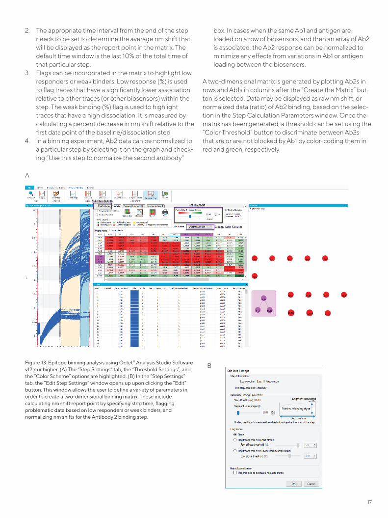
When the software is asked to “Process Epitope Binning Data” in the Processing tab, it searches for repeating sequences of baseline, loading and association steps and then overlays the traces in a new Binning tab. In some cases, there is an additional step(s) at the beginning of the assay, such as “sensor check” that does not repeat and will therefore prevent the software from overlaying the traces. Then, the “Steps to Skip” button under “Binning Parame-ters” in the Binning tab can be used to ignore that addi-tional step(s) while looking for repeating sequences (Figure 13A); the software will now be able to overlay the binning results.
After the traces have been overlaid, four aspects need to be addressed in order to generate a two-dimensional matrix. These can all be performed in the Step Calculation Parame-ters window (Figure 13B).
1. The relevant steps need to be added to the Step Calcu-lation Table (Figure 13A): this can be done by selecting the steps in the graph and clicking the “Add Step Calcu-lation Parameters” button. This opens the Step Calcula-tion Parameters window (Figure 13B), wherein the various steps needs to be assigned as either “First Antibody”, “Second Antibody” or “None” (baseline or antigen).
Note: If the mAb loading onto AR2G Biosensors was done in a separate experiment, then the step labeled with the corresponding immobilized mAb sample ID during acquisi-tion should be assigned as the “First Antibody”.
17
2. The appropriate time interval from the end of the step needs to be set to determine the average nm shift that will be displayed as the report point in the matrix. The default time window is the last 10% of the total time of that particular step.
3. Flags can be incorporated in the matrix to highlight low responders or weak binders. Low response (%) is used to flag traces that have a significantly lower association relative to other traces (or other biosensors) within the step. The weak binding (%) flag is used to highlight traces that have a high dissociation. It is measured by calculating a percent decrease in nm shift relative to the first data point of the baseline/dissociation step.
4. In a binning experiment, Ab2 data can be normalized to a particular step by selecting it on the graph and check-ing “Use this step to normalize the second antibody”
box. In cases when the same Ab1 and antigen are loaded on a row of biosensors, and then an array of Ab2 is associated, the Ab2 response can be normalized to minimize any effects from variations in Ab1 or antigen loading between the biosensors.
A two-dimensional matrix is generated by plotting Ab2s in rows and Ab1s in columns after the “Create the Matrix” but-ton is selected. Data may be displayed as raw nm shift, or normalized data (ratio) of Ab2 binding, based on the selec-tion in the Step Calculation Parameters window. Once the matrix has been generated, a threshold can be set using the “Color Threshold” button to discriminate between Ab2s that are or are not blocked by Ab1 by color-coding them in red and green, respectively.
Figure 13: Epitope binning analysis using Octet® Analysis Studio Software v12.x or higher. (A) The “Step Settings” tab, the “Threshold Settings”, and the “Color Scheme” options are highlighted. (B) In the “Step Settings” tab, the “Edit Step Settings” window opens up upon clicking the “Edit” button. This window allows the user to define a variety of parameters in order to create a two-dimensional binning matrix. These include calculating nm shift report point by specifying step time, flagging problematic data based on low responders or weak binders, and normalizing nm shifts for the Antibody 2 binding step.
A
B

18
Matrix data can be copied and pasted into external analysis programs, such as Microsoft® Excel®. Alternately, matrix data along with the graph of the overlaid traces can be exported by clicking the “Export to CSV File” button. It must be noted that the color-coding based on threshold is not exported with the matrix, and must be set manually using conditional formatting as outlined below.
An additional level of analysis can be done on the binning data with the Octet® Analysis Studio Software. If the user is interested in comparing the initial slope of the Ab2 binding curves in the presence and absence of Ab1, then the Ab2 association step can be selected and quantitated using Quantitate Selected Step in the Processing tab. This step will now open in the quantitation module of the software, and the initial slope can be calculated under the “Binding Rate Equation”. The initial slopes of the binding curves are often compared if the binding profile of Ab2 to the antigen changes in the presence of Ab1, which may be the case if Ab1 is an allosteric regulator that causes a conformational change of the antigen upon binding.
Version 12 adds new quantitative fitting model classifiers for advanced users. The model classifiers (Fig 14) help choose the best fit for the binding interactions and overlaying fitted traces on top of the raw binding traces.
The Legacy model classifier chooses a fitting model (single exponential, double exponential) based on maximum slope of the raw binding traces.
The Best Fit model classifier looks to minimize residuals – the difference between raw binding traces and fitted traces. If the difference is zero between different models (single exponential, double exponential), the Best Fit model classi-fier will choose the simpler of the two models.
The Single Exponential model classifier forces fitting of raw binding traces with the single exponential mathematical model. This classifier might be best suited for known 1:1 binding interactions.
Analysis post-data export
Following data export, a threshold should be defined to differentiate between Ab2s that are or are not blocked by Ab1. For all binning assay formats, the threshold generally is set as the highest self-binding signal in the panel. Some considerations need to be taken into account to make sure the self-binding threshold is appropriate.
- In-tandem: The Ab2 binding signal may be normalized by dividing it by the Ab2-only control signal (no Ab1) to make a clear distinction between Ab2s that are or are not blocked by Ab1. Following normalization, an appropriate self-binding threshold may be used. - Classical sandwich and Premix: Prior to setting the threshold, ensure that signals from all Ab2s in the no-antigen control and in the irrelevant IgG-coupled biosensor control are minimal. If these signals are lower than the highest self-binding signal, then the self-binding threshold can be considered appropriate. If not, one could consider adjusting the threshold.
Once the threshold is set, conditional formatting can be performed on the matrix in Excel. Conditional formatting uses color to visually identify Ab2s that are or are not blocked by Ab1 as shown in Figure 15.
Further analysis of the raw nm shift or normalized data in the matrix can be performed using the PEARSON function.5 Pearson correlation coefficients are calculated for all the mAb rows relative to the first mAb row, and then the row with the highest correlation coefficient is placed below the first row. This process is repeated until placement of all the
Figure 14: A screenshot of the Advanced Settings options for choosing among model classifiers.

19
rows are sorted and is also applied to the mAb columns. It is important to note that the two antibodies in the topmost row and leftmost column will affect the ordering of the anti-bodies upon Pearson sorting. When large binning experi-ments are conducted, Pearson correlation coefficients may only be calculated for the competing mAbs (Ab2s) in rows. After Pearson sorting, the Ab1s in columns can be re-ar-ranged such that all self-binding pairs are on a diagonal in the matrix (highlighted in red in Figure 16). More details about this methodology can be found in Liao-Chan et al.4
In addition, binning data can be analyzed by importing it into the statistical computing program R (The R Project for Statistical Computing: www.r-project.org) for generating a clustergram with the pvclust package.6 Similar analyses can be carried out with other software packages (for example, Partek). Prior to Pearson sorting, the normalized data in Figure 15 was analyzed with pvclust to generate a clustergram shown in Figure 18. The y-axis of the cluster-gram, referred to as Height, is a measure of dissimilarity between antibodies.A manual Height cut-off value is used to define bins in a clustergram. Care must be taken when setting the Height cut-off value; a Height cut-off value that is too high will result in bins containing highly dissimi-lar mAbs, a Height cut-off that is too low will cause similar mAbs to segregate into different bins. Furthermore, AU (Approximate Unbiased) p-values computed using multi-scale bootstrap resampling can be used to confirm bin assignments. An AU p-value cut-off above 95 implies the hypothesis that this bin does not exist is rejected with a 0.05 significance level. At a Height cut-off of 0.2 in Fig-ure 18, six distinct bins emerged. Hierarchical cluster assignment is also listed in the right-most column in Fig-ure 16. More details about this methodology can be found in Liao-Chan et al.4
Epitope binning facilitates identification of a few mAbs from each bin for more detailed characterization in low-through-put functional assay formats. Per our binning data in Fig-ures 16 and 18, two or three antibodies from each of the six bins and the unassigned mAbs were taken forward. Advancing a small and diverse set of mAbs potentially facili-tates identification of a lead antibody molecule that targets a functional epitope of the antigen and provides the desired biological outcome.
The Octet® Analysis Studio Software version 12 has a brand new feature called the BinChart which bins the screened antibodies based on the binding threshold set during anal-yses. This threshold can be adjusted to analyze varying bin-ning of antibodies as a result of the changing threshold in Figure 17.
Commonly used similar metrics between two sets of obser-vations A and B are available in the new binning software. We suggest to start with “Maximum” or “Pearson” but you may start with “Euclidean”. In general, the merges and splits are determined in a greedy manner. The results of hierarchi-cal clustering are usually presented in a dendrogram. More details for all clustering formulas are summarized in “Hierar-chical Clustering Formulas” in the “Data Analysis HT User Guide” which you can find in the HT software. The choice of an appropriate metric will influence the shape of the clus-ters. Except for the special case of single-linkage, none of the algorithms (except exhaustive search) can be guaran-teed to find the optimum solution.
In order to decide which clusters should be combined (for agglomerative), or where a cluster should be split (for divi-sive), a measure of dissimilarity between sets of observa-tions is required. In most methods of hierarchical clustering, this is achieved by use of an appropriate metric (a measure of distance between pairs of observations), and a linkage criterion which specifies the dissimilarity of sets as a function of the pairwise distances of observations in the sets. Linkage criterion determines the distance between sets of observations as a function of the pairwise distances between observations. Some commonly used linkage criteria between two sets of observations A and B are also available and are summarized in “Hierarchical Clustering Formulas” in the “Octet® Analysis Studio Soft-ware User Guide”.
The optimized Report Tab can be used to create exportable reports with any number of data points. New features add capability to modify and sort data tables, and flexibility for font settings.

20
Figure 15: A two-dimensional matrix for an in-tandem binning assay of 43 anti-DPEP1 mAbs. A matrix was created in Octet® Analysis Studio Software v8.0 by plotting Ab2s in rows and Ab1s in columns. Then, this dataset was exported into Microsoft Excel, normalized and color-coded based on a threshold (27) using conditional formatting to discriminate between Ab2s that were blocked (red) or were not blocked (green) by Ab1. Data normalization was conducted by division of the Ab2 signal by the Ab2-only (no Ab1) signal and multiplication by 100. Data set from Liao-Chan et al.4
Figure 16: A two-dimensional matrix for a panel of 43 anti-DPEP1 mAbs profiled in an in-tandem binning assay. The saturating antibodies (Ab1s) and the competing antibodies (Ab2s) are listed in columns and rows, respectively. The Ab2 binding signal was divided by the Ab2-only signal (no Ab1) and then multiplied by 100. Pearson correlation coefficients for the Ab2s are listed in the second-to-last most column. Note that the Pearson correlation coefficients are only calculated for the Ab2s, with all the self-binding pairs on the diagonal with normalized binding responses highlighted in red. A blue gradient is applied to normalized data between 0 (blue) and 75 (white) to highlight blocking. Figure adapted from Liao-Chan et al.4
ACluster: E F B CD
B
A
R48C2−5−04
R48C2−34−126
R48C2−34−30
R48C2−5−109
R48C2−34−04
R48C2−34−43
R48C2−34−09
R48C2−34−69
R48C1−67−21
R48C1−67−43
R48C2−5−12
R48C1−67−03
R48C1−67−08
R48C1−67−02
R48C1−67−34
R48C2−5−02
R48C1−67−44
R48C2−34−08
R48C1−67−88
R48C2−5−03
R48C1−67−37
R48C1−67−09
R48C1−67−41
R48C1−67−04
R48C1−67−53
R48C1−67−20
R48C2−34−45
R48C1−67−46
R48C1−67−40
R48C2−34−01
R48C2−34−06
R48C2−5−13
R48C2−5−14
R48C2−34−80
R48C1−67−18
R48C1−67−19
R48C1−67−89
R48C1−67−33
R48C1−67−90
R48C1−67−49 R48C2−5−01
R48C2−34−42
R48C2−34−75
0.0
0.2 0.2
0.4
0.6
0.8
1.0
1.2
Height
# 1 2 3 4 5 6 7 8 9 10 11 12 13 14 15 16 17 18 19 20 21 22 23 24 25 26 27 28 29 30 31 32 33 34 35 36 37 38 39 40 41 42 43
mA
b2 \
mA
b1
R48
C2-
34-6
9
R48
C1-
67-2
0
R48
C2-
34-4
5
R48
C1-
67-5
3
R48
C1-
67-4
0
R48
C2-
34-0
1
R48
C1-
67-4
6
R48
C1-
67-0
8
R48
C1-
67-0
2
R48
C1-
67-8
8
R48
C2-
5-03
R48
C1-
67-4
4
R48
C2-
34-0
8
R48
C2-
5-02
R48
C1-
67-3
4
R48
C1-
67-0
3
R48
C1-
67-4
1
R48
C1-
67-0
9
R48
C1-
67-3
7
R48
C1-
67-4
3
R48
C1-
67-2
1
R48
C1-
67-0
4
R48
C2-
5-12
R48
C2-
34-0
9
R48
C2-
34-1
26
R48
C2-
34-3
0
R48
C2-
5-04
R48
C2-
34-4
3
R48
C2-
34-0
4
R48
C2-
5-10
9
R48
C2-
5-13
R48
C2-
5-14
R48
C2-
5-01
R48
C1-
67-1
8
R48
C1-
67-8
9
R48
C1-
67-3
3
R48
C1-
67-4
9
R48
C1-
67-9
0
R48
C1-
67-1
9
R48
C2-
34-8
0
R48
C2-
34-4
2
R48
C2-
34-7
5
R48
C2-
34-0
6
Pear
son
Full-
pairw
ise
bios
enso
r cl
uste
r
1 R48C2-34-69 7 12 10 7 12 9 11 9 6 11 7 10 6 6 18 9 4 6 3 7 2 6 5 11 45 33 7 7 12 29 41 14 34 54 66 33 70 72 43 49 41 30 95 A2 R48C1-67-20 4 8 5 3 10 4 8 6 3 6 2 7 1 1 12 2 3 2 0 7 1 5 -2 4 54 36 3 1 5 21 39 18 43 85 83 45 71 73 49 47 32 23 95 0.97 A3 R48C2-34-45 12 18 12 11 16 11 17 11 8 13 6 13 9 6 23 11 7 9 6 12 8 9 4 11 55 39 7 6 12 24 42 18 46 85 80 48 68 74 47 48 32 28 91 1.00 A4 R48C1-67-53 9 12 8 6 12 9 11 10 6 13 5 11 7 4 20 8 3 6 7 9 5 6 3 9 50 30 5 5 11 26 33 16 37 82 70 30 61 69 41 37 29 23 83 0.99 A5 R48C1-67-40 7 6 5 4 8 4 10 5 0 10 -2 5 2 -1 20 2 5 10 3 2 1 3 -1 3 52 31 2 1 2 41 35 13 35 98 90 34 55 79 44 43 26 19 90 0.99 A6 R48C2-34-01 11 15 9 9 12 8 21 14 6 20 6 11 6 3 30 9 10 15 7 8 6 8 2 8 57 37 6 4 9 38 36 17 35 94 85 37 66 67 47 48 29 24 87 0.99 A7 R48C1-67-46 9 9 5 7 11 6 11 5 3 11 1 8 3 1 21 6 1 6 9 11 6 5 -1 6 49 30 2 1 10 22 29 9 31 102 95 40 67 71 44 34 26 19 88 0.98 A8 R48C1-67-08 10 16 10 10 12 6 18 10 4 12 9 15 4 8 22 9 5 6 13 14 8 9 2 9 47 35 11 10 16 28 46 27 47 88 87 45 62 72 38 31 36 28 79 0.98 A9 R48C1-67-02 13 19 12 12 16 12 19 13 8 17 8 15 9 8 27 12 5 8 11 15 10 11 4 15 61 40 10 9 14 34 61 41 67 96 94 37 69 88 42 44 39 33 104 0.98 A
10 R48C1-67-88 9 15 11 7 17 9 11 7 5 10 9 14 7 7 20 8 4 5 5 12 6 10 4 10 60 41 10 6 13 22 58 40 62 78 78 45 65 75 41 43 40 31 90 0.99 A11 R48C2-5-03 14 22 19 14 25 16 16 12 11 18 13 21 14 13 28 16 10 10 9 19 11 16 6 16 63 45 16 11 20 27 61 41 59 74 75 44 66 74 43 47 45 39 106 0.99 A12 R48C1-67-44 8 13 7 6 12 6 10 8 4 9 3 9 5 1 17 5 3 4 1 6 3 6 0 6 54 37 4 3 7 22 60 45 71 83 74 37 69 70 41 44 30 23 102 0.98 A13 R48C2-34-08 13 21 14 14 18 10 12 7 8 14 8 16 11 7 20 11 8 9 6 13 6 12 4 12 52 34 8 7 14 21 65 48 70 87 75 41 71 65 41 47 31 27 107 1.00 A14 R48C2-5-02 16 23 17 15 22 17 15 13 13 19 12 19 13 9 27 15 11 13 11 15 10 16 8 17 61 44 13 9 19 25 55 33 57 79 80 46 71 79 40 52 43 39 81 0.97 A15 R48C1-67-34 2 8 5 3 10 3 7 5 0 3 2 6 0 1 11 0 -1 1 2 6 3 4 -3 2 58 36 2 -1 6 14 50 29 53 73 73 36 63 65 34 42 28 23 58 0.99 A16 R48C1-67-03 11 13 10 8 12 10 12 7 6 8 7 11 7 6 18 6 10 16 4 10 10 10 3 9 49 31 6 4 9 48 52 34 53 88 94 35 67 85 40 44 37 30 86 0.96 A17 R48C1-67-41 12 19 14 11 18 13 16 17 10 16 10 16 11 9 23 13 7 8 6 13 5 12 7 14 61 40 10 11 17 30 29 12 24 88 100 40 66 98 49 45 42 36 101 0.94 A18 R48C1-67-09 9 14 11 8 13 9 13 14 6 12 8 12 8 8 20 10 5 6 5 10 5 10 5 11 49 36 8 8 14 29 21 10 15 91 88 42 65 85 46 44 39 30 79 0.99 A19 R48C1-67-37 11 20 21 10 25 19 13 13 18 18 18 20 19 17 29 22 11 12 12 19 12 19 12 22 59 37 19 18 22 32 33 17 21 69 71 38 60 61 53 49 35 33 66 0.97 A20 R48C1-67-43 11 19 13 12 19 11 14 9 7 15 5 14 7 6 26 10 7 8 4 10 6 10 2 12 61 53 7 -1 8 25 34 6 15 68 71 36 76 74 41 54 45 33 108 0.94 A21 R48C1-67-21 16 27 19 17 24 17 22 18 14 20 15 22 16 13 29 16 12 13 8 18 12 17 10 19 75 58 16 6 13 33 34 19 20 59 61 37 72 83 42 51 48 38 103 0.99 A22 R48C1-67-04 11 12 7 9 12 8 14 7 4 14 4 10 6 3 19 7 5 7 3 5 4 8 1 8 45 31 4 4 12 16 30 13 33 90 56 27 55 53 32 36 28 19 77 0.90 A23 R48C2-5-12 10 21 24 12 30 22 18 10 20 22 21 28 20 20 30 21 9 10 12 27 14 25 12 25 50 39 20 15 26 31 55 29 49 76 72 37 59 63 51 43 39 35 85 0.94 A24 R48C2-34-09 13 20 14 11 18 9 19 -1 5 14 5 15 6 3 27 6 7 7 0 9 0 10 -2 10 32 17 5 4 14 17 61 32 49 66 65 37 73 82 32 23 41 33 103 0.93 A25 R48C2-34-126 15 20 16 8 14 7 8 3 5 17 6 13 4 7 26 7 6 9 2 5 0 7 5 7 10 15 11 9 18 10 30 5 15 16 29 11 44 37 19 23 23 24 52 0.87 E26 R48C2-34-30 21 22 20 8 15 7 2 -8 1 22 3 11 1 4 31 -2 5 8 -4 4 -8 3 2 2 12 13 7 4 21 -4 27 -9 18 -8 8 -5 33 40 9 9 18 15 50 0.85 E27 R48C2-5-04 14 31 27 16 31 23 19 12 16 24 19 29 21 19 35 21 12 14 12 24 13 24 9 28 35 37 20 14 26 25 69 37 62 21 26 -2 37 30 20 16 20 25 28 0.49 unassigned28 R48C2-34-43 19 23 15 17 21 16 22 15 14 16 13 18 12 10 25 13 13 14 0 14 8 15 7 16 52 40 13 9 17 33 63 40 66 50 54 28 21 18 39 64 45 38 111 0.48 F29 R48C2-34-04 17 17 12 11 21 15 14 11 12 18 6 16 11 5 27 8 12 17 2 9 6 14 4 13 55 37 7 7 11 27 66 38 61 53 59 24 13 16 37 58 50 34 120 0.99 F30 R48C2-5-109 4 10 9 4 15 1 5 -4 0 -1 8 8 -3 8 10 -1 5 7 6 10 3 10 2 0 44 25 8 21 33 13 18 0 4 61 71 24 57 71 32 34 6 13 3 0.27 unassigned31 R48C2-5-13 21 29 62 44 79 62 1 -25 47 21 25 63 42 25 40 23 11 -6 -4 -3 -4 75 46 29 15 7 25 9 17 29 27 -6 2 28 35 13 156 139 19 -1 6 10 11 0.43 D32 R48C2-5-14 25 44 70 54 86 70 19 -5 49 61 52 68 57 47 77 40 18 14 9 18 11 84 54 45 27 24 44 24 40 32 43 14 22 39 46 31 117 100 34 20 17 21 21 0.94 D33 R48C2-5-01 96 46 84 79 114 101 39 21 89 155 44 80 72 51 170 42 29 28 7 7 5 84 67 60 78 39 34 44 61 34 16 7 8 73 96 35 72 96 40 46 9 12 15 0.70 unassigned34 R48C1-67-18 14 65 110 85 117 126 -26 -40 110 201 57 93 88 63 219 52 72 94 57 32 106 97 84 79 65 -20 40 0 4 39 41 15 33 23 30 6 12 -3 24 4 9 15 11 0.63 unassigned35 R48C1-67-89 30 44 96 47 75 91 39 23 53 96 42 69 65 54 104 33 79 85 55 25 32 77 59 59 41 18 33 -4 3 14 31 9 24 17 18 0 6 0 18 5 1 7 7 0.86 B36 R48C1-67-33 48 50 89 64 84 91 41 32 61 88 57 82 72 63 94 46 67 84 68 48 64 81 70 72 66 38 47 2 9 26 42 15 36 30 34 13 11 5 33 12 10 15 15 0.96 B37 R48C1-67-49 87 49 93 67 123 105 46 26 69 87 53 84 76 64 108 55 88 111 100 56 82 102 74 74 76 53 43 7 14 40 48 16 45 56 60 27 10 17 43 28 13 12 27 0.94 B38 R48C1-67-90 72 43 80 76 100 89 38 23 76 78 44 73 68 53 86 44 77 87 80 42 57 79 64 56 69 39 37 8 16 47 59 35 53 43 44 21 11 8 39 27 16 17 25 0.96 B39 R48C1-67-19 27 49 74 48 79 55 52 35 46 57 36 48 37 40 71 15 84 88 53 33 40 57 45 30 30 26 27 1 15 3 37 3 11 21 25 3 18 0 25 3 11 20 9 0.81 unassigned40 R48C2-34-80 66 35 65 52 76 67 27 12 41 51 43 56 45 45 63 36 62 72 66 43 56 65 52 40 59 31 37 51 62 31 36 12 29 26 33 13 11 4 27 7 28 23 68 0.66 unassigned41 R48C2-34-42 82 31 59 59 79 74 33 17 53 45 37 60 53 42 62 42 48 45 53 41 49 65 48 52 68 36 34 54 66 29 22 7 12 43 46 19 22 27 37 47 8 12 8 0.70 C42 R48C2-34-75 73 20 48 51 71 62 26 14 38 35 30 47 44 33 52 31 34 33 42 30 39 53 38 40 64 32 27 47 62 21 13 2 5 39 43 16 17 18 32 43 4 6 4 0.99 C43 R48C2-34-06 119 82 20 113 38 16 99 90 13 62 34 16 24 28 83 13 67 50 7 75 88 16 29 21 122 115 36 10 22 56 24 8 13 25 30 46 52 13 28 122 13 20 12 0.25 unassigned
Supplementary Fig. 4
100100 68 9795100100 87 10098 9674 859687 918085 87 9596 9886 9183 1008199 9998 8994
7878 78
629960
98
81
86
au
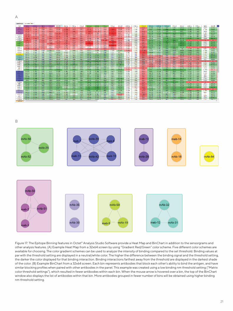
21
Figure 17: The Epitope Binning features in Octet® Analysis Studio Software provide a Heat Map and BinChart in addition to the sensorgrams and other analysis features. (A) Example Heat Map from a 32x64 screen by using “Gradient Red/Green” color scheme. Five different color schemes are available for choosing. The color gradient schemes can be used to analyze the intensity of binding compared to the set threshold. Binding values at par with the threshold setting are displayed in a neutral/white color. The higher the difference between the binding signal and the threshold setting, the darker the color displayed for that binding interaction. Binding interactions farthest away from the threshold are displayed in the darkest shade of the color. (B) Example BinChart from a 32x64 screen. Each bin represents antibodies that block each other’s ability to bind the antigen, and have similar blocking profiles when paired with other antibodies in the panel. This example was created using a low binding nm threshold setting (“Matrix color threshold settings”), which resulted in fewer antibodies within each bin. When the mouse arrow is hovered over a bin, the top of the BinChart window also displays the list of antibodies within that bin. More antibodies grouped in fewer number of bins will be obtained using higher binding nm threshold setting.
A
B
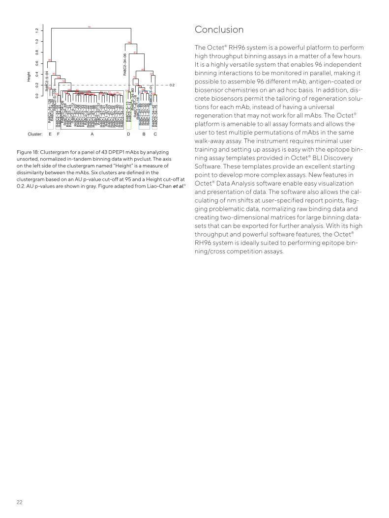
22
Conclusion
The Octet® RH96 system is a powerful platform to perform high throughput binning assays in a matter of a few hours. It is a highly versatile system that enables 96 independent binning interactions to be monitored in parallel, making it possible to assemble 96 different mAb, antigen-coated or biosensor chemistries on an ad hoc basis. In addition, dis-crete biosensors permit the tailoring of regeneration solu-tions for each mAb, instead of having a universal regeneration that may not work for all mAbs. The Octet® platform is amenable to all assay formats and allows the user to test multiple permutations of mAbs in the same walk-away assay. The instrument requires minimal user training and setting up assays is easy with the epitope bin-ning assay templates provided in Octet® BLI Discovery Software. These templates provide an excellent starting point to develop more complex assays. New features in Octet® Data Analysis software enable easy visualization and presentation of data. The software also allows the cal-culating of nm shifts at user-specified report points, flag-ging problematic data, normalizing raw binding data and creating two-dimensional matrices for large binning data-sets that can be exported for further analysis. With its high throughput and powerful software features, the Octet® RH96 system is ideally suited to performing epitope bin-ning/cross competition assays.
Figure 18: Clustergram for a panel of 43 DPEP1 mAbs by analyzing unsorted, normalized in-tandem binning data with pvclust. The axis on the left side of the clustergram named “Height” is a measure of dissimilarity between the mAbs. Six clusters are defined in the clustergram based on an AU p-value cut-off at 95 and a Height cut-off at 0.2. AU p-values are shown in gray. Figure adapted from Liao-Chan et al.4
ACluster: E F B CD
B
A
R48C2−5−04
R48C2−34−126
R48C2−34−30
R48C2−5−109
R48C2−34−04
R48C2−34−43
R48C2−34−09
R48C2−34−69
R48C1−67−21
R48C1−67−43
R48C2−5−12
R48C1−67−03
R48C1−67−08
R48C1−67−02
R48C1−67−34
R48C2−5−02
R48C1−67−44
R48C2−34−08
R48C1−67−88
R48C2−5−03
R48C1−67−37
R48C1−67−09
R48C1−67−41
R48C1−67−04
R48C1−67−53
R48C1−67−20
R48C2−34−45
R48C1−67−46
R48C1−67−40
R48C2−34−01
R48C2−34−06
R48C2−5−13
R48C2−5−14
R48C2−34−80
R48C1−67−18
R48C1−67−19
R48C1−67−89
R48C1−67−33
R48C1−67−90
R48C1−67−49 R48C2−5−01
R48C2−34−42
R48C2−34−75
0.0
0.2 0.2
0.4
0.6
0.8
1.0
1.2
Height
# 1 2 3 4 5 6 7 8 9 10 11 12 13 14 15 16 17 18 19 20 21 22 23 24 25 26 27 28 29 30 31 32 33 34 35 36 37 38 39 40 41 42 43
mA
b2 \
mA
b1
R48
C2-
34-6
9
R48
C1-
67-2
0
R48
C2-
34-4
5
R48
C1-
67-5
3
R48
C1-
67-4
0
R48
C2-
34-0
1
R48
C1-
67-4
6
R48
C1-
67-0
8
R48
C1-
67-0
2
R48
C1-
67-8
8
R48
C2-
5-03
R48
C1-
67-4
4
R48
C2-
34-0
8
R48
C2-
5-02
R48
C1-
67-3
4
R48
C1-
67-0
3
R48
C1-
67-4
1
R48
C1-
67-0
9
R48
C1-
67-3
7
R48
C1-
67-4
3
R48
C1-
67-2
1
R48
C1-
67-0
4
R48
C2-
5-12
R48
C2-
34-0
9
R48
C2-
34-1
26
R48
C2-
34-3
0
R48
C2-
5-04
R48
C2-
34-4
3
R48
C2-
34-0
4
R48
C2-
5-10
9
R48
C2-
5-13
R48
C2-
5-14
R48
C2-
5-01
R48
C1-
67-1
8
R48
C1-
67-8
9
R48
C1-
67-3
3
R48
C1-
67-4
9
R48
C1-
67-9
0
R48
C1-
67-1
9
R48
C2-
34-8
0
R48
C2-
34-4
2
R48
C2-
34-7
5
R48
C2-
34-0
6
Pear
son
Full-
pairw
ise
bios
enso
r cl
uste
r
1 R48C2-34-69 7 12 10 7 12 9 11 9 6 11 7 10 6 6 18 9 4 6 3 7 2 6 5 11 45 33 7 7 12 29 41 14 34 54 66 33 70 72 43 49 41 30 95 A2 R48C1-67-20 4 8 5 3 10 4 8 6 3 6 2 7 1 1 12 2 3 2 0 7 1 5 -2 4 54 36 3 1 5 21 39 18 43 85 83 45 71 73 49 47 32 23 95 0.97 A3 R48C2-34-45 12 18 12 11 16 11 17 11 8 13 6 13 9 6 23 11 7 9 6 12 8 9 4 11 55 39 7 6 12 24 42 18 46 85 80 48 68 74 47 48 32 28 91 1.00 A4 R48C1-67-53 9 12 8 6 12 9 11 10 6 13 5 11 7 4 20 8 3 6 7 9 5 6 3 9 50 30 5 5 11 26 33 16 37 82 70 30 61 69 41 37 29 23 83 0.99 A5 R48C1-67-40 7 6 5 4 8 4 10 5 0 10 -2 5 2 -1 20 2 5 10 3 2 1 3 -1 3 52 31 2 1 2 41 35 13 35 98 90 34 55 79 44 43 26 19 90 0.99 A6 R48C2-34-01 11 15 9 9 12 8 21 14 6 20 6 11 6 3 30 9 10 15 7 8 6 8 2 8 57 37 6 4 9 38 36 17 35 94 85 37 66 67 47 48 29 24 87 0.99 A7 R48C1-67-46 9 9 5 7 11 6 11 5 3 11 1 8 3 1 21 6 1 6 9 11 6 5 -1 6 49 30 2 1 10 22 29 9 31 102 95 40 67 71 44 34 26 19 88 0.98 A8 R48C1-67-08 10 16 10 10 12 6 18 10 4 12 9 15 4 8 22 9 5 6 13 14 8 9 2 9 47 35 11 10 16 28 46 27 47 88 87 45 62 72 38 31 36 28 79 0.98 A9 R48C1-67-02 13 19 12 12 16 12 19 13 8 17 8 15 9 8 27 12 5 8 11 15 10 11 4 15 61 40 10 9 14 34 61 41 67 96 94 37 69 88 42 44 39 33 104 0.98 A
10 R48C1-67-88 9 15 11 7 17 9 11 7 5 10 9 14 7 7 20 8 4 5 5 12 6 10 4 10 60 41 10 6 13 22 58 40 62 78 78 45 65 75 41 43 40 31 90 0.99 A11 R48C2-5-03 14 22 19 14 25 16 16 12 11 18 13 21 14 13 28 16 10 10 9 19 11 16 6 16 63 45 16 11 20 27 61 41 59 74 75 44 66 74 43 47 45 39 106 0.99 A12 R48C1-67-44 8 13 7 6 12 6 10 8 4 9 3 9 5 1 17 5 3 4 1 6 3 6 0 6 54 37 4 3 7 22 60 45 71 83 74 37 69 70 41 44 30 23 102 0.98 A13 R48C2-34-08 13 21 14 14 18 10 12 7 8 14 8 16 11 7 20 11 8 9 6 13 6 12 4 12 52 34 8 7 14 21 65 48 70 87 75 41 71 65 41 47 31 27 107 1.00 A14 R48C2-5-02 16 23 17 15 22 17 15 13 13 19 12 19 13 9 27 15 11 13 11 15 10 16 8 17 61 44 13 9 19 25 55 33 57 79 80 46 71 79 40 52 43 39 81 0.97 A15 R48C1-67-34 2 8 5 3 10 3 7 5 0 3 2 6 0 1 11 0 -1 1 2 6 3 4 -3 2 58 36 2 -1 6 14 50 29 53 73 73 36 63 65 34 42 28 23 58 0.99 A16 R48C1-67-03 11 13 10 8 12 10 12 7 6 8 7 11 7 6 18 6 10 16 4 10 10 10 3 9 49 31 6 4 9 48 52 34 53 88 94 35 67 85 40 44 37 30 86 0.96 A17 R48C1-67-41 12 19 14 11 18 13 16 17 10 16 10 16 11 9 23 13 7 8 6 13 5 12 7 14 61 40 10 11 17 30 29 12 24 88 100 40 66 98 49 45 42 36 101 0.94 A18 R48C1-67-09 9 14 11 8 13 9 13 14 6 12 8 12 8 8 20 10 5 6 5 10 5 10 5 11 49 36 8 8 14 29 21 10 15 91 88 42 65 85 46 44 39 30 79 0.99 A19 R48C1-67-37 11 20 21 10 25 19 13 13 18 18 18 20 19 17 29 22 11 12 12 19 12 19 12 22 59 37 19 18 22 32 33 17 21 69 71 38 60 61 53 49 35 33 66 0.97 A20 R48C1-67-43 11 19 13 12 19 11 14 9 7 15 5 14 7 6 26 10 7 8 4 10 6 10 2 12 61 53 7 -1 8 25 34 6 15 68 71 36 76 74 41 54 45 33 108 0.94 A21 R48C1-67-21 16 27 19 17 24 17 22 18 14 20 15 22 16 13 29 16 12 13 8 18 12 17 10 19 75 58 16 6 13 33 34 19 20 59 61 37 72 83 42 51 48 38 103 0.99 A22 R48C1-67-04 11 12 7 9 12 8 14 7 4 14 4 10 6 3 19 7 5 7 3 5 4 8 1 8 45 31 4 4 12 16 30 13 33 90 56 27 55 53 32 36 28 19 77 0.90 A23 R48C2-5-12 10 21 24 12 30 22 18 10 20 22 21 28 20 20 30 21 9 10 12 27 14 25 12 25 50 39 20 15 26 31 55 29 49 76 72 37 59 63 51 43 39 35 85 0.94 A24 R48C2-34-09 13 20 14 11 18 9 19 -1 5 14 5 15 6 3 27 6 7 7 0 9 0 10 -2 10 32 17 5 4 14 17 61 32 49 66 65 37 73 82 32 23 41 33 103 0.93 A25 R48C2-34-126 15 20 16 8 14 7 8 3 5 17 6 13 4 7 26 7 6 9 2 5 0 7 5 7 10 15 11 9 18 10 30 5 15 16 29 11 44 37 19 23 23 24 52 0.87 E26 R48C2-34-30 21 22 20 8 15 7 2 -8 1 22 3 11 1 4 31 -2 5 8 -4 4 -8 3 2 2 12 13 7 4 21 -4 27 -9 18 -8 8 -5 33 40 9 9 18 15 50 0.85 E27 R48C2-5-04 14 31 27 16 31 23 19 12 16 24 19 29 21 19 35 21 12 14 12 24 13 24 9 28 35 37 20 14 26 25 69 37 62 21 26 -2 37 30 20 16 20 25 28 0.49 unassigned28 R48C2-34-43 19 23 15 17 21 16 22 15 14 16 13 18 12 10 25 13 13 14 0 14 8 15 7 16 52 40 13 9 17 33 63 40 66 50 54 28 21 18 39 64 45 38 111 0.48 F29 R48C2-34-04 17 17 12 11 21 15 14 11 12 18 6 16 11 5 27 8 12 17 2 9 6 14 4 13 55 37 7 7 11 27 66 38 61 53 59 24 13 16 37 58 50 34 120 0.99 F30 R48C2-5-109 4 10 9 4 15 1 5 -4 0 -1 8 8 -3 8 10 -1 5 7 6 10 3 10 2 0 44 25 8 21 33 13 18 0 4 61 71 24 57 71 32 34 6 13 3 0.27 unassigned31 R48C2-5-13 21 29 62 44 79 62 1 -25 47 21 25 63 42 25 40 23 11 -6 -4 -3 -4 75 46 29 15 7 25 9 17 29 27 -6 2 28 35 13 156 139 19 -1 6 10 11 0.43 D32 R48C2-5-14 25 44 70 54 86 70 19 -5 49 61 52 68 57 47 77 40 18 14 9 18 11 84 54 45 27 24 44 24 40 32 43 14 22 39 46 31 117 100 34 20 17 21 21 0.94 D33 R48C2-5-01 96 46 84 79 114 101 39 21 89 155 44 80 72 51 170 42 29 28 7 7 5 84 67 60 78 39 34 44 61 34 16 7 8 73 96 35 72 96 40 46 9 12 15 0.70 unassigned34 R48C1-67-18 14 65 110 85 117 126 -26 -40 110 201 57 93 88 63 219 52 72 94 57 32 106 97 84 79 65 -20 40 0 4 39 41 15 33 23 30 6 12 -3 24 4 9 15 11 0.63 unassigned35 R48C1-67-89 30 44 96 47 75 91 39 23 53 96 42 69 65 54 104 33 79 85 55 25 32 77 59 59 41 18 33 -4 3 14 31 9 24 17 18 0 6 0 18 5 1 7 7 0.86 B36 R48C1-67-33 48 50 89 64 84 91 41 32 61 88 57 82 72 63 94 46 67 84 68 48 64 81 70 72 66 38 47 2 9 26 42 15 36 30 34 13 11 5 33 12 10 15 15 0.96 B37 R48C1-67-49 87 49 93 67 123 105 46 26 69 87 53 84 76 64 108 55 88 111 100 56 82 102 74 74 76 53 43 7 14 40 48 16 45 56 60 27 10 17 43 28 13 12 27 0.94 B38 R48C1-67-90 72 43 80 76 100 89 38 23 76 78 44 73 68 53 86 44 77 87 80 42 57 79 64 56 69 39 37 8 16 47 59 35 53 43 44 21 11 8 39 27 16 17 25 0.96 B39 R48C1-67-19 27 49 74 48 79 55 52 35 46 57 36 48 37 40 71 15 84 88 53 33 40 57 45 30 30 26 27 1 15 3 37 3 11 21 25 3 18 0 25 3 11 20 9 0.81 unassigned40 R48C2-34-80 66 35 65 52 76 67 27 12 41 51 43 56 45 45 63 36 62 72 66 43 56 65 52 40 59 31 37 51 62 31 36 12 29 26 33 13 11 4 27 7 28 23 68 0.66 unassigned41 R48C2-34-42 82 31 59 59 79 74 33 17 53 45 37 60 53 42 62 42 48 45 53 41 49 65 48 52 68 36 34 54 66 29 22 7 12 43 46 19 22 27 37 47 8 12 8 0.70 C42 R48C2-34-75 73 20 48 51 71 62 26 14 38 35 30 47 44 33 52 31 34 33 42 30 39 53 38 40 64 32 27 47 62 21 13 2 5 39 43 16 17 18 32 43 4 6 4 0.99 C43 R48C2-34-06 119 82 20 113 38 16 99 90 13 62 34 16 24 28 83 13 67 50 7 75 88 16 29 21 122 115 36 10 22 56 24 8 13 25 30 46 52 13 28 122 13 20 12 0.25 unassigned
Supplementary Fig. 4
100100 68 9795100100 87 10098 9674 859687 918085 87 9596 9886 9183 1008199 9998 8994
7878 78
629960
98
81
86
au

23
References
1. Exploring Blocking Assays using Octet, ProteOn, and Biacore Biosensors, Abdiche YN, et al., Anal Biochem, 386, 172–180, 2009.
2. Label-free Epitope Binning Assays of Monoclonal Antibodies Enable the Identification of Antigen Heterogeneity, Abdiche YN, et al., J Immunol Methods, 382, 101–116, 2012.
3. High-Throughput Epitope Binning Assays on Label-free Array-based Biosensors can Yield Exquisite Epitope Discrimination that Facilitates the Selection of Monoclonal Antibodies with Functional Activity, Abdiche YN, et al., PLoS One, 9(3):e92451, 2014.
4. Monoclonal Antibody Binding-site Diversity Assessment with a Cell-based Clustering Assay, Liao-Chan S, et al., J Immunol Methods, 405, 1–14, 2014.
5. Epitope Binning of Murine Monoclonal Antibodies by a Multiplexed Pairing Assay, Miller PL, et al., J Immunol Methods, 365, 118–25, 2011.
6. Pvclust: an R Package for Assessing the Uncertainty in Hierarchical Clustering, Suzuki R and Shimodaira H, Bioinformatics, 22, 1540–1542, 2006.

GermanySartorius Lab Instruments GmbH & Co. KG Otto-Brenner-Strasse 20 37079 GoettingenPhone +49 551 308 0
USASartorius Corporation565 Johnson AvenueBohemia, NY 11716Phone +1 888 OCTET 75 Or +1 650 322 1360
For further contacts, visit www.sartorius.com/octet-support
Specifications subject to change without notice. Copyright Sartorius Lab Instruments GmbH & Co. KG. For Research Use Only.AN-4016 Rev E