Embed Size (px)
Citation preview

Institute of Medical Microbiology and Hygiene
University Hospital Ulm
Head of Institute: Prof. Dr. Steffen Stenger
Cre-recombinase based genetic
manipulation and the regulation of
hemolysis in Streptococcus
anginosus
Dissertation submitted in partial fulfillment of the requirements for the degree of
„Doctor rerum naturalium” (Dr. rer. nat.) of the International Graduate School in
Molecular Medicine Ulm
Richard Bauer
Lindenberg im Allgäu
2018

Current dean:
Prof. Dr. Thomas Wirth
Thesis Advisory Committee:
- First supervisor: Prof. Dr. Barbara Spellerberg
- Second supervisor: Prof. Dr. Christian Sinzger
- Third supervisor: Dr. Shaynoor Dramsi
External Reviewer:
Prof. Dr. Marcus Fulde
Prof. Dr. Holger Rohde
Day doctorate awarded:
20th February 2019

Results gained in my thesis have previously been published in the following
publications:
Bauer, R., Mauerer, S., Grempels, A. & Spellerberg, B. The competence system of
Streptococcus anginosus and its use for genetic engineering. Mol Oral Microbiol 33,
194-202, doi:10.1111/omi.12213 (2018).
Bauer, R., Mauerer, S. & Spellerberg, B. Regulation of the beta-hemolysin gene
cluster of Streptococcus anginosus by CcpA. Sci Rep 8, 9028, doi:10.1038/s41598-
018-27334-z (2018).

Table of contents
Table of contents
Abbreviations ...................................................................................................................................... I
1 Introduction ................................................................................................................................ 1
1.1 Phylogeny of the Streptococcus anginosus group ................................................................. 1
1.2 Clinical importance of the Streptococcus anginosus group ................................................... 2
1.3 Phenotypic characteristics and identification of the Streptococcus anginosus group ............ 3
1.4 Virulence related factors in the Streptococcus anginosus group ........................................... 4
1.5 Streptolysin S and Streptolysin S related gene clusters......................................................... 7
1.6 Catabolite repression in Gram-positive bacteria................................................................. 10
1.7 Competence system in the genus Streptococcus ................................................................ 14
1.8 Aim of the work ................................................................................................................. 17
2 Material and Methods ............................................................................................................... 18
2.1 Material ............................................................................................................................. 18
Bacterial strains ......................................................................................................... 18 2.1.1
Plasmids..................................................................................................................... 21 2.1.2
Oligonucleotides ........................................................................................................ 22 2.1.3
Instruments ............................................................................................................... 24 2.1.4
Consumables ............................................................................................................. 27 2.1.5
Chemicals and reagents ............................................................................................. 28 2.1.6
Solutions .................................................................................................................... 31 2.1.7
Enzymes..................................................................................................................... 32 2.1.8
Kits ............................................................................................................................ 32 2.1.9
Software .................................................................................................................... 33 2.1.10
2.2 Methods ............................................................................................................................ 33
Bacterial strains and growth conditions...................................................................... 33 2.2.1
General DNA techniques ............................................................................................ 34 2.2.2
CSP based transformation of S. anginosus .................................................................. 36 2.2.3
Site-directed mutagenesis in S. anginosus .................................................................. 36 2.2.4
Construction of complementation strains .................................................................. 38 2.2.5
Promoter reporter assay ............................................................................................ 38 2.2.6
Hemolysis assay ......................................................................................................... 39 2.2.7
Expression and purification of His-tagged CcpA .......................................................... 40 2.2.8

Table of contents
Electrophoretic mobility shift assay ............................................................................ 40 2.2.9
Bioinformatical and statistical analysis ....................................................................... 41 2.2.10
3 Results ...................................................................................................................................... 42
3.1 Cre-lox based genetic engineering in S. anginosus ............................................................. 42
Competence system in S. anginosus ........................................................................... 42 3.1.1
CSP-induced transformation kinetics in S. anginosus .................................................. 45 3.1.2
Combining CSP and Cre-recombinase for markerless gene deletion ........................... 46 3.1.3
Optimization of transformation efficiencies in S. anginosus........................................ 51 3.1.4
3.2 CcpA regulates the SLS expression in S. anginosus ............................................................. 53
SLS expression is under the control of catabolite repression ...................................... 53 3.2.1
CcpA negatively affects SLS expression ....................................................................... 54 3.2.2
S. anginosus ∆ccpA maintains hemolytic activity in the presence of glucose .... 56 3.2.3
Mutation of cre abolishes glucose dependent SLS repression ..................................... 58 3.2.4
CcpA binds to creC in vitro.......................................................................................... 61 3.2.5
3.3 Role of a CovR homolog in the regulation of SLS in S. anginosus ........................................ 62
In silico analysis and genetic organization of the CovR homolog ................................. 62 3.3.1
A covR* integration mutant shows increased hemolysis ............................................. 65 3.3.2
Markerless covR* deletion mutant shows type strain hemolytic behavior .................. 66 3.3.3
4 Discussion ................................................................................................................................. 68
5 Summary ................................................................................................................................... 79
6 References ................................................................................................................................ 80
7 List of Publications .................................................................................................................. 107
8 Acknowledgements ................................................................................................................. 108

I
Abbreviations
Abbreviations
aa Amino acid
ABC ATP binding cassette
AmpR Ampicillin resistance
ATP Adenosine triphosphate
ATCC American Type Culture Collection
bp Base pair
CcpA Catabolite control protein A
CCR Carbon catabolite repression
CF Cystic fibrosis
cfb CAMP-factor gene
CFU Colony Forming Unit
ClpP ATP-dependent Clp protease proteolytic subunit
CmR Chloramphenicol resistance
com competence
ComX Competence-specific sigma factor
CovR Control of virulence Regulator
CPS Capsular polysaccharide
cre Catabolite responsive element
C-source Carbon-source
CSP Competence stimulating peptide
DTT Dithiothreitol
EI Enzyme I
EII Enzyme II
E. coli Escherichia coli
EDTA Ethylenediaminetetraacetic acid

II
Abbreviations
EGFP Enhanced Green Fluorescent Protein
EMSA Electrophoretic mobility shift assay
EryR Erythromycin resistance
FBP Fructose-1,6-bisphosphate
FBS Fetal bovine serum
G6P Glucose-6-phosphate
Gal Galactose
GAS Group A streptococcus (S. pyogenes)
GBS Group B streptococcus
genomosubsp. genomosubspecies
Glc Glucose
gnd 6-phosphogluconate dehydrogenase gene
Hly+ Hemolysis positive
His Histidine
HGT Horizontal gene transfer
HPr Histidine-containing Protein
HPr-His-P Histidine phosphorylated Histidine-containing Protein
HPrK HPr kinase/phosphorylase
IPTG Isopropyl β-D-1-thiogalactopyranoside
LB Lysogeny Broth
Lmb Laminin-binding protein
L. monocytogenes Listeria monocytogenes
lox Locus of crossing-over
M Molecular weight marker
MALDI-TOF MS Matrix-assisted laser desorption/ionization time-of-flight mass spectrometry
MEP Mobile Element Protein
MFI Mean fluorescence intensity

III
Abbreviations
MLSA Multilocus sequence analysis
NMR Nuclear magnetic resonance
OD Optical density
OE-PCR Overlap extension PCR
ori Origin of replication
PCR Polymerase chain reaction
PBS Phosphate-Buffered Saline
PEG Polyethylene glycol
PMNLs Polymorphonuclear leukocytes
poly(dI-dC) Poly(deoxyinosinic-deoxycytidylic) acid
PRD PTS-regulatory domain
PTS Phosphoenolpyruvate-carbohydrate phosphotransferase system
rec Recombinase
RitR Repressor of iron transport
RT Reverse transcriptase
SAG Streptococcus anginosus group
sag Streptolysin S associated gene
S. anginosus Streptococcus anginosus
S. aureus Staphylococcus aureus
SB Sheep blood
S. constellatus Streptococcus constellatus
SDS-PAGE Sodium dodecyl sulfate polyacrylamide gel electrophoresis
S. dysgalactiae Streptococcus dysgalactiae
S. intermedius Streptococcus intermedius
SLS Streptolysin S
S. milleri Streptococcus milleri
S. mitis Streptococcus mitis

IV
Abbreviations
S. mutans Streptococcus mutans
S. oralis Streptococcus oralis
spc Spectinomycin resistance gene
SpcR Spectinomycin resistance
spe Streptococcal pyrogenic exotoxin genes
speC Streptococcal pyrogenic exotoxin C gene
speG Streptococcal pyrogenic exotoxin G gene
S. pneumoniae Streptococcus pneumoniae
S. pyogenes Streptococcus pyogenes (GAS)
S. sanguinis Streptococcus sanguinis
subsp. subspecies
TBE TRIS borate-EDTA Buffer
TCS Two-component system
THY Todd Hewitt Broth with 5% yeast extract
TOMM Thiazole/oxazole-modified microcins
tufA Translation elongation factor EF-Tu 1 gene
WT Wild type
XIP SigmaX inducing peptide

1
Introduction
1 Introduction
Streptococcus anginosus belongs to the Streptococcus anginosus group (SAG) that
also includes the closely related species Streptococcus constellatus and
Streptococcus intermedius. The bacteria of this group share some characteristics,
including a small colony phenotype on blood agar plates (< 0.5 mm) and a specific
butterscotch-like smell [30], but they show an inconsistent hemolysis behavior with all
three hemolytic phenotypes (α-, β- and γ-hemolysis) being present in different strains.
Nevertheless, the SAG was assigned to the viridans group of streptococci [49].
Bacteria of the SAG can be isolated as commensals of mucosal membranes in the
oral cavity, the urogenital tract and the gastrointestinal tract [213]. However,
difficulties to culture and identify the SAG correctly lead to an underestimation of the
pathogenic potential of these bacteria in older investigations [163,174]. Nowadays, an
increasing number of epidemiological data highlight the clinical importance of these
bacteria and they show that the SAG species have to be considered as etiological
agents of a variety of different infections [101,157,163].
1.1 Phylogeny of the Streptococcus anginosus group
The species in the SAG passed through several taxonomic changes. They were
previously denominated as Streptococcus MG-intermedius and Streptococcus
anginosus-constellatus [50] or as the ʹStreptococcus milleriʹ group, although this
designation was not included in the approved lists of bacterial names [176].
Additionally it was proposed that the whole group constitutes a single species
[36,205]. Meanwhile, the SAG was demonstrated to consist of three distinct species
(S. anginosus, S. constellatus, S. intermedius) which is the currently accepted
taxonomic status [22,91,212,214]. Combining multilocus sequence analysis (MLSA),
the sequence of the 16S rRNA gene and phenotypic traits further demonstrated the
presence of seven distinct clusters within the SAG (S. intermedius, S. constellatus
subsp. constellatus, S. constellatus subsp. pharyngis, S. constellatus subsp.
viborgensis, S. anginosus subsp. anginosus, S. anginosus subsp. whileyi, S.

2
Introduction
anginosus genomosubsp. AJ1) [70,91,214]. While the first complete genome
sequences of SAG members published in 2013 substantiate the presence of these
clusters in the SAG [141], the MLSA of clinical S. anginosus isolates collected in
Vellore (India) revealed an additional cluster in S. anginosus which was proposed to
be denominated as S. anginosus genomosubsp. vellorensis and contains the
S. anginosus type strain SK52 [14]. This demonstrates that the taxonomy of the SAG
will most likely encounter further changes and the current view of the taxonomic
status needs continuing verification which will be facilitated by the increasing amounts
of genome data that can be generated by next-generation sequencing techniques.
1.2 Clinical importance of the Streptococcus anginosus group
SAG species can frequently be isolated from invasive infections, including sepsis and
abscesses of the central nervous system as well as the gastrointestinal tract
[101,104,163]. Besides, members of the SAG are reported as emerging pathogens in
cystic fibrosis (CF) patients [70,147].
In a population-based laboratory surveillance study for invasive pyogenic
streptococcal infections the incidence rate of SAG infections (8.65/100,000
population) exceeded the combined incidence rate of group A and B streptococcal
infections (7.40/100,000 population) [104]. Besides, Siegman-Igra et al. reported an
annual incidence of 8.8/10,000 hospital admissions due to SAG infections in a tertiary
care university hospital in Israel, highlighting the clinical relevance of the group [175].
In a retrospective cohort study the pyogenic potential of the SAG was further pointed
out. Among 263 patients suffering from SAG infections, 60 % were classified as
pyogenic, defined as an abscess, empyema or deep space surgical site infection. It
was thereby reported that S. intermedius was more often associated with pyogenic
infections than the rest of the SAG bacteria [33,101]. Patients with pyogenic infections
showed lower frequencies of bacteremia (8 %) than patients without pyogenic
infections (68 %) and a population based surveillance of SAG blood stream infections
reported an annual incidence of 3.7/100,000 population [105]. The SAG are thereby
also able to cause infective endocarditis and they were reported to be responsible for
9 % of endocarditis cases caused by streptococci [98]. Studies investigating the

3
Introduction
etiologic agent of invasive infections caused by β-hemolytic group C and G
streptococci further revealed that 17-19 % were due to SAG infections [27,157]. It has
to be assumed that these percentages underestimate the SAG presence as these
studies focused on β-hemolytic isolates and the majority of SAG strains is non-β-
hemolytic [212].
An association of the different species of the SAG with their clinical manifestations
has been proposed by some authors. S. anginosus showed a higher prevalence in
blood cultures, whereas S. constellatus and S. intermedius were the dominant
species in pyogenic infections [33,101,213]. However, these reports are still
controversial and the results could not be verified by similar studies [94,175].
The antimicrobial resistance profile of SAG isolates is still not a major concern in
clinical therapies [17,65,110,140,224]. But it has to be highlighted that the first
vancomycin resistant S. anginosus isolate was described in 2014 [181] emphasizing
further surveillance of the antibiotic resistance development in the SAG.
1.3 Phenotypic characteristics and identification of the Streptococcus
anginosus group
Beside the above mentioned common characteristics (small colony size and specific
odor), members of the SAG show an inconsistent hemolysis behavior and express a
variety of different Lancefield antigens [49,212]. The Lancefield antigens A, C, G and
F as well as nongroupable isolates are present in SAG. These antigens can also be
expressed in other streptococci and although the F-antigen was proposed to be
limited to SAG in the past, the detection of Streptococcus sinensis interacting with
F-antiserum finally demonstrated that the Lancefield typing is not really helpful for the
identification of SAG [26,221]. Phenotypical traits and several biochemical assays
have been proposed to identify the SAG to the species level and these characteristics
were implemented into commercial identification tests [15,187,212,214]. The majority
of these assays provide reliable results for the identification of the SAG to the species
level although some false-negative results have been reported [9,61,71]. The
developments in microbiology diagnostics further aimed to accelerate species
identification by direct bacterial profiling. The widely used matrix-assisted laser

4
Introduction
desorption/ionization time-of-flight mass spectrometry (MALDI-TOF MS) thereby
provides an accurate and rapid identification of a range of bacteria and fungi but the
results of this method concerning the identification of the SAG is still controversial.
Friedrichs et al. reported that MALDI-TOF MS is a reliable method for the
identification of viridans streptococci including SAG [58]. However, it was also
demonstrated that only 22 % of S. intermedius isolates were identified to the species
level and the discrimination of the SAG to the subspecies level is still a major obstacle
[9,222], although it has to be highlighted that the identification to the subspecies level
is actually of minor interest for diagnostic issues. For scientific purposes the correct
subspecies designation could emphasize the scientific statement and thus the use of
MLSA and whole genome sequencing approaches should be applied as gold
standard as these methods currently allow the best discrimination of the SAG to the
subspecies and even to the genomosubspecies level [14,91,141].
1.4 Virulence related factors in the Streptococcus anginosus group
The manifestation of a disease relies on virulence factors that operate in multiple
events such as colonization, internalization and invasion of host tissues as well as
dissemination and evasion of the host defense. But it has to be kept in mind that
described virulence traits often include proteins known to be involved in metabolism
or housekeeping functions [146]. Owing to the underestimation of the pathogenic
potential of the members of the SAG in the past, the knowledge about virulence
factors in these species is scarce [11] and studies so far have mainly focused on
specific virulence traits.
Adhesion
The prerequisite for the colonization of the host is the adhesion of the bacterium to
biological material. As a member of the oral microbiome, S. anginosus was
investigated for its potential to adhere to the tooth surface and to tooth-restorative
materials [216,223]. The molecules conferring the binding to these materials were not
described, but it was demonstrated that the mode of adhesion differed between

5
Introduction
Streptococcus mutans and S. anginosus as inhibitors of S. mutans adsorption did not
interfere with the binding of S. anginosus to saliva-coated hydroxyapatite beads [193].
The binding of bacteria to extracellular matrix proteins in general is not only crucial for
the colonization of a host, but also important for invasion and manifestation of a
clinical infection. In this context, S. anginosus was reported to be able to bind to
fibronectin, fibrinogen and laminin [4]. Clinical SAG isolates showed thereby a greater
fibronectin binding capability than other SAG strains and the incubation of SAG
strains with fibronectin increased the ability of the bacterium to bind to hydroxyapatite
[216,217].
In a whole genome analysis of SAG isolates numerous potential adhesins were
identified [141]. Several of these proteins contain a cell surface anchor motif (LPxTG)
and collagen-binding domains although collagen binding of S. anginosus was
demonstrated to be only minor [4]. Additional adhesion proteins, including a
fibronectin binding protein (Fbp54), a laminin binding protein (Lmb), an Internalin A
homolog, a glyceraldehyde 3-phosphate-dehydrogenase (GAPDH), the pullulanase
PulA, the pneumococcal surface adhesin A (PsaA) as well as an enolase, were
predicted with the genome analysis that could explain the genetic basis of the binding
of SAG to extracellular matrix proteins [141]. All of these predicted adhesins in SAG
are functionally characterized in different bacteria, but they were not studied in the
SAG at the molecular level so far.
Invasion and evasion of host defense
Concordantly, factors involved in invasion or evasion from the host defense are poorly
investigated and only a few in vivo analyses have been performed.
SAG isolates were reported to produce infective vegetations in an endocarditis
infection model of Wistar rats [100] and the high potential of SAG to cause pyogenic
infections insinuates that SAG strains have evolved mechanisms to inhibit their killing
by polymorphonuclear leukocytes (PMNLs). This was demonstrated by Wanahita et
al. who reported that, although SAG strains were associated in higher numbers with
PMNLs, they were killed less effectively by these cells compared to the prototypical
abscess forming bacterium Staphylococcus aureus [206].

6
Introduction
Whole genome analysis of different SAG strains further proposed four virulence
related loci that are conserved in all SAG genomes. These loci were previously
reported to be involved in invasion and evasion from host defense in other
streptococci [141]. This included a homolog of the streptococcus invasion locus of
Streptococcus pyogenes (GAS), UDP-glucose pyrophosphorlyase protein (HasC)
involved in hyaluronic acid synthesis in GAS and parts of the hemolysin operon cyl of
GBS. The forth locus is a well-investigated streptococcal virulence factor, the
capsular polysaccharide (CPS) biosynthesis operon. Capsule production was already
reported in the SAG and it was demonstrated that encapsulated S. constellatus
strains showed an increased abscess formation in a murine infection model and a
higher resistance to phagocytic killing compared to unencapsulated isolates [95].
Recently, the cps locus in the SAG was analyzed verifying that the members of the
SAG contain a cps biosynthesis operon that shows high homology to the cps operon
of Streptococcus pneumoniae and other oral streptococci [177,202]. However, a
detailed analysis of the capsule production in SAG was not performed so far.
Recently, GAS related pyrogenic virulence factors (speC, speG) were detected in S.
anginosus isolates from India [14]. These genes encode streptococcal pyrogenic
exotoxins which include superantigens [155] and they stimulate the release of high
levels of pro-inflammatory cytokines which can lead to the streptococcal toxic shock
syndrome, a life-threatening condition accompanied with multi-organ failure and high
mortality rates [78]. The high identity of the spe genes in S. anginosus and GAS,
together with the lineage independent presence of these genes in some S. anginosus
strains, strongly indicates recent horizontal gene transfer events. The publication of
Babbar et al. thereby highlighted that some S. anginosus strains acquired pyrogenic
virulence related genes that might increase their pathogenic potential [14] and thus
emphasizes the importance of the SAG as emerging pathogens.
Virulence regulation
Bacteria need to sense their environmental conditions in order to be able to adapt
their gene expression. A common bacterial mechanism to sense the environment are
two-component systems (TCS) that are known to play a key role in virulence [80].
Whole-genome analysis in SAG revealed the presence of 14 different TCS in this

7
Introduction
group [141]. Eight of these 14 TCS have orthologs in S. pneumoniae that have been
linked to virulence [194]. These include the well-investigated VicRK, CiaRH, ComDE
and BlpRH TCS which are involved in quorum sensing, competence and virulence in
S. pneumoniae [18,87,137,194]. Additionally, a control of virulence regulator (CovR)
homolog was detected in the SAG genomes [141]. CovR is a prominent transcription
factor that controls the capsule production and virulence gene expression in GAS,
GBS, Streptococcus dysgalactiae and S. mutans [45,68,103,109,182].
Overall, the pathogenicity factors of the SAG members are poorly investigated on the
molecular level although the knowledge about virulence factors in these species
starts to grow as the members of the SAG are increasingly recognized as emerging
pathogens that need further attention.
1.5 Streptolysin S and Streptolysin S related gene clusters
One exception of the scarce knowledge about virulence factors in the SAG is the
hemolysin responsible for the β-hemolytic phenotype of the S. anginosus type strain
SK52. The genetic locus of the hemolysin was identified by random mutagenesis
using the pGhost9::ISS1 vector system [10,189]. The locus is comprised of ten genes
and shows high homologies to the Streptolysin S (SLS) operon of GAS (Figure 1)
[25,139].
Figure 1: Schematic representation of the SLS operon of S. pyogenes and the homologous
SLS operon in S. anginosus SK52. Depicted are the identities [%] on the amino acid (aa)
level of the respective encoded proteins.

8
Introduction
The hemolysin operon in GAS is comprised of nine genes and these genes are
sufficient to produce the SLS [139]. The sag (Streptolysin S associated gene) operon
shows similarities to operons of class I bacteriocins and thus contains genes
encoding for a post-translational modification machinery as well as a transporter
[124]. The first gene in the operon is sagA encoding the precursor of the active
hemolysin. It encodes a 53 aa protein that contains a double glycine leader cleavage
site. SagBCD is responsible for the conversion of the prepeptide SagA to SLS [108].
This complex introduces heterocycles into SagA which are essential for the activity of
the hemolysin [39,130]. The sagGHI genes encode an ABC type transporter which is
a common mechanism for bacteriocin export [39,164]. The role of the latter two genes
sagE and sagF is still ambiguous. While SagF is predicted to be membrane
associated, SagE is a membrane spanning peptidase that was proposed to confer
immunity functions [39,108]. Strikingly, SagE only provided immunity to endogenous
production of SLS and not to the exogenous exposure to SLS, as a GAS sagE mutant
showed normal growth in the β-hemolytic zone of wild-type GAS colonies [39].
Such biosynthetic gene clusters, that encode a precursor peptide and a modification
machinery that introduces heterocycles (thiazole, oxazole and methyloxazole) into the
precursor, are widely distributed among bacteria and are designated thiazole/oxazole-
modified microcins (TOMM) [72]. Members of the TOMM can be found in other
streptococci including Streptococcus dysgalactiae subsp. equisimilis, Streptococcus
iniae and Streptococcus equi [54,59,86]. However, these microcins are also encoded
in more distantly related Gram-positive bacteria, in some Gram-negatives as well as
in archaea [108] and several members of the family possess antibacterial activity
[142,156,166,226].
The SLS in GAS is a major virulence factor that was already discovered in 1938
[196]. Occasionally, non-hemolytic GAS isolates are described which were associated
with infections, but 99% of clinical GAS isolates are hemolytic and thus it is consent
that the hemolysin in GAS plays an important role in the pathogenicity of this
bacterium [90,203,228]. The SLS enables GAS to systemically disseminate by
activating the degradation of the intercellular junctions, allowing GAS to perform
paracellular invasion [186]. Additionally, the SLS contributes to soft-tissue damage
and a non-hemolytic GAS mutant shows a reduced virulence in a murine soft-tissue

9
Introduction
infection model [23,39]. The hemolysin is further implicated in evasion of the innate
immune system by inhibiting neutrophil recruitment, by destroying neutrophils and by
activating an inflammatory programmed cell death pathway in macrophages
[52,64,132].
The identified SLS homolog in SAG is present in some strains of S. constellatus as
well as S. anginosus but in S. intermedius, encoding the human specific cytolysin
intermedilysin, it was not detected so far [136,212]. The hemolysin in SAG shares
some features with the SLS in GAS. Operons encoding the two hemolysins show high
homologies but it has to be highlighted that, although S. constellatus strains encode
one sagA gene like GAS, S. anginosus strains were reported two contain either one
copy of the gene or two sagA copies, like in the case of the S. anginosus type strain
SK52 [10,190]. Another common feature of the SLS of SAG and GAS is their
temperature dependent hemolytic activity that is only present at temperatures above
22 °C [12,28]. However, the two hemolysins also show remarkable differences. While
the SLS of GAS is able to lyse a variety of different cell types, including macrophages
and neutrophils [64,84,132,191], the hemolysin of SAG was demonstrated to be a
broad-range hemolysin able to lyse erythrocytes of different origin, but it does not
show cytolytic activity against macrophages or granulocytes [12]. Additionally, the
SLS of SAG was reported to confer cytolytic activity against HepG2 cells, a human
hepatoma cell line [190].
The mechanism how the SLS of GAS is able to lyse erythrocytes remained
controversial. Some reports suggested that the SLS induces hemolysis by a colloid-
osmotic process, e.g. by pore formation directly in the cell membrane [28,47]. The
possibility to inhibit this process by polyethylene glycol (PEG) of defined size further
strengthened this hypothesis. The use of PEG in hemolysis assays of SAG SLS, in
contrast, did not allow the prediction of a defined pore size generated by the
hemolysin in the cell membrane [10]. Recently, Higashi et al. provided evidence that
SLS leads to erythrocyte lysis through binding to Anion exchanger 1 (band 3) which
mediates the exchange of chloride and bicarbonate. The binding of SLS to this anion
channel leads to influx of chloride into the erythrocyte, followed by influx of water and
subsequent lysis of the erythrocyte [79]. Thus the SLS of GAS could exert its activity
by directly targeting membrane channels, just like other peptide toxins [48].

10
Introduction
The expression of the hemolysin in GAS is influenced by a diverse array of different
regulators (CovR, CodY, RofA, Mga) reflecting the importance of this virulence factor
in GAS [21,77,97,115]. Additionally, GAS was reported to regulate hemolysis in
connection to its carbohydrate metabolism, similarly to other streptococcal species
[173,192,197,208]. In S. anginosus, the expression of the SLS was also
demonstrated to be dependent on carbon availability, as the SLS expression was
inhibited in the presence of high glucose concentrations in the growth medium [12].
Thus, carbon catabolite control seems to be a common mechanism for the regulation
of hemolysin expression and it was demonstrated that one major factor of this
mechanism, the catabolite control protein A (CcpA), directly regulates the expression
of the Intermedilysin in S. intermedius and of the SLS in GAS [99,197].
1.6 Catabolite repression in Gram-positive bacteria
The majority of bacteria can use a variety of different substances as carbon source.
They can either be co-metabolized or the bacteria can preferentially take up and
consume the carbon source which is most easily accessible and allows the highest
energy conservation. To be able to perform this hierarchical metabolism, the bacteria
evolved complex regulatory mechanisms which are summarized as carbon catabolite
repression (CCR) [114]. Only a few pathogenic bacteria, like Chlamydiae that are
highly adapted to host niches, seem to lack these complex regulatory pathways [138].
The phosphoenolpyruvate-carbohydrate phosphotransferase system (PTS) is the key
component in CCR of Gram-negative as well as Gram-positive bacteria [43]. It
comprises a phosphorelay system that mediates the import and simultaneous
phosphorylation of sugars. The PTS systems are composed of a sugar unspecific part
that all PTS systems in the cell share and a sugar specific part. The phosphorylation
relay starts with the phosphorylation of the Enzyme I (EI) by phosphoenolpyruvate
and subsequent transfer of the phosphoryl group to the Histidine-containing Protein
(HPr) which is phosphorylated at a histidine residue (HPr-His-P). The HPr then
transfers the phosphoryl group to the substrate specific Enzyme IIA (EIIA) followed by
the phosphorylation of the Enzyme IIB (EIIB) and transport of the sugar into the cell

11
Introduction
by Enzyme IIC (EIIC). The EII complex can either be a multidomain protein consisting
of EIIABC or it can be a multiprotein complex composed of the single EII components
that can either be located in the cytosol or imbedded in the bacterial membrane [152].
Although the PTS is involved in CCR in Gram-negative and Gram-positive species,
the regulatory pathways differ in both groups. The phosphorylation state of the EIIA of
the PTS mainly confers CCR in Gram-negative species and the phosphorylation state
of the HPr is important in CCR of Gram-positive species [66]. However, the final result
of the CCR is similar, as the expression of genes that are needed for the consumption
of alternative carbon sources is reduced in the presence of a preferred sugar.
In Gram-positive species, the CCR mechanisms can be divided in global and operon
specific mechanisms (Figure 2). The operon specific mechanisms include the terms
inducer exclusion and induction prevention. The inducer exclusion describes the
mechanism that an uptake of an operon-specific inducer is inhibited. A prominent
example is the galactose exclusion in Lactobacillus brevis. In the presence of
Glucose serine-phosphorylated HPr is formed and binds to the galactose permease
thereby inhibiting the transport of galactose into the cell. As galactose is needed for
the activation of the galactose operon, the expression of this operon is blocked
(inducer exclusion, Figure 2A) [44].
The term induction prevention addresses the activity of operon-specific transcription
factors that contain PTS-regulatory domains (PRD) [184]. One of the best studied
induction prevention mechanisms occurs in the levanase operon (levDEFG and sacC)
of Bacillus subtilis which encodes a fructose-specific PTS and a levanase that
hydrolysis fructose polymers in the environment [117]. The expression of the operon
is regulated by the activator LevR which itself is regulated by HPr and the fructose-
specific EIIB (LevE) [29,40,183]. LevR contains two PRDs and the phosphorylation
state of these domains determines the LevR activity. In the presence of preferentially
metabolized sugars the concentration of HPr-His-P is low accompanied by a scarce
PRD1 phosphorylation which results in an inactive conformation of LevR (induction
prevention, Figure 2B) [118].
The global regulation of CCR in Gram-positive bacteria is primarily mediated by CcpA
in conjunction with the HPr, the HPr kinase/phosphorylase (HPrK) and the
intermediates of the glycolytic pathway fructose-1,6-bisphosphate (FBP) and glucose-

12
Introduction
6-phosphate (G6P, Figure 2C) [195,207]. CcpA is a pleiotropic transcription factor
that binds to the promoter regions of target genes, thereby mainly reducing the
expression of the genes. To be able to bind to DNA, CcpA needs to be activated by a
serine-phosphorylated HPr [167]. In the phosphorelay of the PTS HPr is
phosphorylated at a histidine at the position 15. However, this protein contains a
second phosphorylation site (serine-46) which exhibits regulatory functions. The
availability of glucose and other preferred PTS sugars leads to increased FBP levels
in the cell. High levels of FBP activate the kinase activity of the HPrK which
phosphorylates the HPr at serine-46 which in turn can activate the DNA binding
capability of CcpA [42]. CcpA binds to so called catabolite responsive elements (cre).
The sequence of the cre sites are well investigated in different species and it was
shown that they consist of highly degenerate pseudo-palindromes [131,185,219,230].
The binding of CcpA to cre is further stimulated by the presence of high levels of FBP
and G6P allowing a delicate control of the CcpA activity [168].
CCR is a genome-wide regulatory process that was reported to affect the expression
of 5-10 % of the genes in different bacteria [134,219,227]. The majority of these
genes is repressed by CcpA and encodes proteins involved in the metabolism of
alternative carbon sources. However, CcpA also activates the expression of genes
involved in glucose metabolism [195].
Furthermore, CcpA represents a link between virulence and metabolism as it is
involved in the regulation of virulence gene expression in various bacterial pathogens
[160]. Examples include the control of capsule expression in different bacteria
[62,169,218], the repression of the toxic shock syndrome toxin-1 in S. aureus and the
regulation of the toxin production of Bacillus anthracis [31,170]. In Clostridium difficile,
CcpA controls the expression of the alternative sigma factor TcdR which in turn is
responsible for the expression of high toxin levels and it represses phospholipase C
and perfringolysin expression in Clostridium perfringens which contribute to gas
gangrene formation [8,127].

13
Introduction
Figure 2: Schematic representation of carbon catabolite repression mechanisms in Gram-
positive bacteria. A: Inducer exclusion: Serine-phosphorylated HPr [HPr(Ser)-℗] inhibits
GalP (Galactose Permease) activity; In the absence of Gal (Galactose) the expression of the
gal operon is blocked. B: Induction prevention: In the absence of Glc (Glucose), histidine-
phosphorylated HPr [HPr-(His)-℗] is present. This leads to the phosphorylation of the PRD1
(PTS-regulatory domain) of LevR. High Fru (Fructose) concentrations in the environment
lead to low levels of phosphorylated LevE (fructose-specific enzyme IIB) combined with low
levels of PRD2 phosphorylation. LevR is active if PRD1 is phosphorylated and PRD2 is non-
phosphorylated which results in lev operon transcription and further Fru import by LevG
(Fructose transporter). C: Global regulation: Glucose specific PTS is depicted. In the
presence of high fructose-1,6-bisphosphate (FBP) and high ATP concentrations in the cell
the HPr kinase/phosphorylase (E) phosphorylates the HPr at a serine residue. HPr(Ser)-℗
binds and activates the catabolite control protein A (CcpA) which increases the DNA binding
capability of the regulator. This interaction is further supported by glucose-6-phosphate
(G6P) and FBP.

14
Introduction
In the genus streptococcus, numerous reports have demonstrated that CcpA controls
different virulence traits. A S. pneumoniae ccpA mutant is attenuated in murine
nasopharyngeal and lung infection models [89]. CcpA controls the acid production in
S. mutans and thus is involved in the control of the cariogenic potential of this oral
bacterium [1]. Furthermore, CcpA plays an important role in the pathogenicity of GAS.
A ccpA mutant shows a hypervirulent phenotype in murine skin and systemic
infections and CcpA seems to activate the transcription of the multiple gene regulator
of GAS (Mga) that in turn controls genes needed for adherence, internalization and
host immune evasion [5,81,99]. Additionally, CcpA directly regulates the expression
of the SLS in GAS by binding to a cre site overlapping the SLS promoter [99]. CcpA
was also investigated in the SAG and it was demonstrated that it regulates the
intermedilysin expression in S. intermedius [197].
1.7 Competence system in the genus Streptococcus
Horizontal gene transfer (HGT) is one of the major driving forces for the evolution of
the genomes in prokaryotes [7,37,151,215] and it has been proposed that over 60 %
of the pan-genomes in streptococci have been subjected to HGT [159]. Bacteria can
acquire new genetic material by transduction, conjugation and natural transformation.
The latter is the only mechanism that does not need a DNA donor and thus is
believed to be the simplest way to acquire new genetic material. Natural
transformation is widespread among different phyla and was demonstrated for some
archaea, Gram-negative as well as Gram-positive bacteria [92,112,171].
The physiological state of a bacterial cell where it is able to take up DNA from the
environment is designated competence, a process that is tightly controlled in Gram-
positive bacteria [35]. In streptococci, two different competence regulation
mechanisms have evolved independently. Species of the salivarius, pyogenic,
mutans and bovis group rely on the ComRS system whereas the SAG and the mitis
group species use the ComCDE system for competence regulation [57,119]. The
competence development in both groups is dependent on the alternative sigma factor
ComX. In the early phase of the competence induction, a secreted peptide

15
Introduction
pheromone is sensed which leads to the upregulation of the comX expression.
Subsequently, ComX induces the expression of the late phase genes which built up
the transformasome responsible for the DNA uptake [93].
Although homologs of ComX and genes encoding the transformasome are
ubiquitously distributed in streptococcal genomes, it remained puzzling for a long time
why only a subset of the streptococcal species showed natural transformation [34].
This has changed with the identification of ComR as a ComX regulator in
Streptococcus salivarius and Streptococcus thermophilus in 2010 [55]. ComR is a
stand-alone regulator that recognizes the sigmaX inducing peptide (XIP) encoded in
the comS gene [55,125]. The discovery of the ComRS as competence regulator led to
the expansion of the number of streptococcal species that are transformable and to
date at least one species from the salivarius, pyogenic, mutans and bovis group were
experimentally verified to be transformable using the XIP [41,55,122,123,135].
In contrast to the ComRS, the ComCDE system for competence development is well-
investigated in the mitis group of streptococci (Figure 3). Natural transformation was
already discovered at the beginning of the 20th century in S. pneumoniae [69] but it
took further 35 years to show that competence could be induced in non-competent
streptococci by a factor that was contained in sterile filtered supernatant of competent
bacteria [145]. The factor that lead to competence induction was demonstrated to be
heat sensitive and later turned out to be the competence stimulating peptide (CSP)
encoded in comC [73]. The competence state in S. pneumoniae develops at a
specific cell density, a mechanism that is tightly controlled by the competence
regulation operon comCDE [35,148]. Besides ComC, the precursor of the CSP, the
operon encodes the TCS ComD/E. The ComC is exported and processed by
ComA/B, an ABC transporter that cuts the ComC prepeptide after a double glycine
motif [74,85]. The extracellular CSP is recognized by the membrane-bound histidine
kinase ComD that, upon activation, phosphorylates the response regulator ComE.
The phosphorylated ComE then activates the expression of the so called early com
genes that include the comCDE and comAB operons as well as comX and comW
[120,121]. The alternative sigma factor ComX activates the expression of genes that
are responsible for the DNA uptake [107,113,150] whereas ComW was reported as
an activator and stabilizer of ComX [188,199]. The competence state develops

16
Introduction
simultaneously in the cells and it is only maintained for around 15-20 min [161,199].
This short timeframe requires a delicate competence shut-off mechanism. The cells
therefore evolved several competence exit strategies. It was demonstrated that
unphosphorylated ComE accumulates to high levels after competence induction and
it antagonizes the transcriptional activation of the phosphorylated ComE [121].
Additionally, the phosphorylated ComE is sequestered by one of the late competence
gene products (DprA) leading to the reduction of the early gene expression [129,210].
Finally, a protein that antagonizes ComX activity was assumed, although the reported
data concerning the ComW protection of ComX from proteolysis by ClpP are not
consistent [188,210].
Figure 3: Schematic representation of the competence regulation in the mitis group of
streptococci. ComC, the precursor of the CSP, is processed and exported by ComAB. High
levels of CSP lead to activation of ComD which results in ComE phosphorylation.
Phosphorylated ComE activates the transcription of the early com genes (comCDE, comAB,
comX). The alternative sigma factor ComX induces the expression of the late com genes that
built up the transformasome.

17
Introduction
While the competence system in the mitis group and especially in S. pneumoniae has
been characterized in detail as described above, the knowledge about the system in
SAG is scarce. The SAG bacteria encode the ComCDE system to control the
competence development and genes belonging to the competence regulon have
been detected in the SAG genome [119,141]. Members of the SAG were
demonstrated to encode identical CSPs which would allow interspecies
communication in this group. However, the comCDE locus was not investigated
thoroughly in the SAG and it is possible that different CSP pherotypes exist as it was
shown that horizontal gene transfer has contributed to the diversification of the
competence locus in streptococci [76,211].
1.8 Aim of the work
The aim of this work was to investigate the regulation of the SLS expression in
S. anginosus. Due to the lack of suitable genetic tools in the SAG it was necessary to
establish a site-directed mutagenesis procedure that allows fast and reliable
generation of deletion mutants in S. anginosus. The competence system of
S. anginosus was analyzed to be able to exploit this system for the mutagenesis
technique. Using the newly established method would then allow to generate targeted
deletions in the S. anginosus genome to be able to decipher the regulation of the SLS
expression at a molecular level. Regulator mutants of S. anginosus should be
analyzed in respect to their hemolytic behavior and the binding sites of identified
regulators should be characterized which should finally result in the first description of
a transcriptional regulator in the emerging pathogen S. anginosus.

18
Material and Methods
2 Material and Methods
2.1 Material
Bacterial strains 2.1.1
The bacterial strains used in this study are summarized in Table 1.
Table 1: Strains. The bacterial strains and their definitions together with their source are
summarized.
Strain Definition Source
S. anginosus
BSU 458 S. anginosus type strain SK52, ATCC33397, Hly+ ATCC
BSU 554 BSU 458 derivative, carrying pBSU409 [63]
BSU 556 BSU 458 derivative, carrying pBSU409::cfbprom [13]
BSU 805 BSU 458 derivative, carrying pBSU409::sagprom [10]
BSU 886 BSU 458 derivative; carrying pBSU409::sagprom_creA_del This study
BSU 902 BSU 458::pG+host5_covR This study
BSU 913 BSU 458 derivative; carrying pBSU409::sagprom_creB_del This study
BSU 916 BSU 458 derivative; carrying pBSU409::sagprom_creC_mut This study
BSU 924 BSU 458 derivative; ∆sagA1 This study
BSU 926 BSU 458 derivative; ∆ccpA This study
BSU 928 BSU 458 derivative; ∆ccpA, carrying pBSU409::sagprom This study
BSU 947 BSU 458::ccpA This study
BSU 948 BSU 928 derivative; ∆ccpA::ccpA This study
BSU 982 BSU 458 derivative; ∆covR This study
BSU 994 BSU 458 derivative; ∆sagBCD This study
BSU 1210 clinical isolate Ulm collection
BSU 1211 clinical isolate Ulm collection
BSU 1214 clinical isolate Ulm collection
BSU 1215 clinical isolate Ulm collection
BSU 1216 clinical isolate Ulm collection
BSU 1217 clinical isolate Ulm collection

19
Material and Methods
BSU 1227 clinical isolate Ulm collection
BSU 1289 clinical isolate Ulm collection
BSU 1303 clinical isolate Ulm collection
BSU 1306 clinical isolate Ulm collection
BSU 1307 clinical isolate Ulm collection
BSU 1308 clinical isolate Ulm collection
BSU 1310 clinical isolate Ulm collection
BSU 1312 clinical isolate Ulm collection
BSU 1313 clinical isolate Ulm collection
BSU 1317 clinical isolate Ulm collection
BSU 1318 clinical isolate Ulm collection
BSU 1319 clinical isolate Ulm collection
BSU 1323 clinical isolate Ulm collection
BSU 1324 clinical isolate Ulm collection
BSU 1326 clinical isolate Ulm collection
BSU 1327 clinical isolate Ulm collection
BSU 1328 clinical isolate Ulm collection
BSU 1329 clinical isolate Ulm collection
BSU 1330 clinical isolate Ulm collection
BSU 1331 clinical isolate Ulm collection
BSU 1332 clinical isolate Ulm collection
BSU 1334 clinical isolate Ulm collection
BSU 1336 clinical isolate Ulm collection
BSU 1338 clinical isolate Ulm collection
BSU 1339 clinical isolate Ulm collection
BSU 1344 clinical isolate Ulm collection
BSU 1345 clinical isolate Ulm collection
BSU 1346 clinical isolate Ulm collection
BSU 1351 clinical isolate Ulm collection
BSU 1354 clinical isolate Ulm collection
BSU 1355 clinical isolate Ulm collection
BSU 1358 clinical isolate Ulm collection

20
Material and Methods
BSU 1361 clinical isolate Ulm collection
BSU 1362 clinical isolate Ulm collection
BSU 1363 clinical isolate Ulm collection
BSU 1364 clinical isolate Ulm collection
BSU 1366 clinical isolate Ulm collection
BSU 1369 clinical isolate Ulm collection
BSU 1372 clinical isolate Ulm collection
BSU 1373 clinical isolate Ulm collection
BSU 1374 clinical isolate Ulm collection
BSU 1375 clinical isolate Ulm collection
BSU 1376 clinical isolate Ulm collection
BSU 1379 clinical isolate Ulm collection
BSU 1381 clinical isolate Ulm collection
BSU 1382 clinical isolate Ulm collection
BSU 1384 clinical isolate Ulm collection
BSU 1386 clinical isolate Ulm collection
BSU 1387 clinical isolate Ulm collection
BSU 1388 clinical isolate Ulm collection
BSU 1389 clinical isolate Ulm collection
BSU 1390 clinical isolate Ulm collection
BSU 1391 clinical isolate Ulm collection
BSU 1392 clinical isolate Ulm collection
BSU 1395 clinical isolate Ulm collection
BSU 1396 clinical isolate Ulm collection
BSU 1397 clinical isolate Ulm collection
BSU 1398 clinical isolate Ulm collection
BSU 1399 clinical isolate Ulm collection
BSU 1400 clinical isolate Ulm collection
BSU 1401 clinical isolate Ulm collection
BSU 1402 clinical isolate Ulm collection
BSU 1403 clinical isolate Ulm collection
BSU 1404 clinical isolate Ulm collection

21
Material and Methods
BSU 1405 clinical isolate Ulm collection
BSU 1406 clinical isolate Ulm collection
BSU 1413 clinical isolate Ulm collection
BSU 1414 clinical isolate Ulm collection
BSU 1415 clinical isolate Ulm collection
BSU 1417 clinical isolate Ulm collection
E. coli
BL21(DE3) E. coli BL21 derivative with DE3 λ prophage carrying the T7 RNA polymerase gene and lacI
q
Novagen
DH5α endA1 hsdR17 supE44 DlacU169(f80lacZDM15) recA1 gyrA96 thi-1 relA1
Boehringer
EC101 E. coli JM101 derivative with repA from pWV01 integrated into the chromosome
[106]
BSU 957 BL21 derivative, carrying pET21a-ccpA-N-His This study
Plasmids 2.1.2
The plasmids used in this study are summarized in Table 2.
Table 2: Plasmids. The plasmids, their characteristics and their source are listed.
Plasmid Characteristics Source
pAT18 lacZα, ori pUC, ori pAmβ1, EryR [201]
pAT28 lacZα, ori pUC, ori pAmβ1, SpcR [200]
pET21a pET21a; lacI, ori F1, ori pBR322, T7 promoter and terminator, Amp
R
Novagen
pGA14-Spc Replication functions of pWVO1, EryR, Spc
R [178]
pG+host5 Replication functions of pWVO1, ori pBR322; Ery
R [24]
pSET5s_PtufA-cre Replication function of pG+host3 and pUC19, Cre-
recombinase gene under control of tufA promoter, CmR
[102]
pBSU409 pAT28 derivative carrying egfp [63]
pBSU803 pBSU409::sagprom; pBSU409 derivative, egfp under the control of the SLS promoter
[10]
pBSU846 pG+host5_covR; pG
+host5 derivative carrying integration
sequence of covR This study
pBSU876 pBSU409::sagprom_creC_mut; pBSU803 derivative carrying mutations in creC
This study

22
Material and Methods
pBSU880 pBSU409::sagprom_creB_del; pBSU803 derivative carrying a deletion of creB
This study
pBSU881 pBSU409::sagprom_creA_del; pBSU803 derivative carrying a deletion of creA
This study
pBSU910 pG+host5-ccpA∆100nt; pG
+host5 derivative carrying up- and
downstream sequences of ccpA This study
pBSU919 pAT18-cre-rectufA; pAT18 derivative carrying Cre-recombinase gene under the control of tufA promoter, Ery
R
This study
pBSU956 pET21a-ccpA-N-His; pET21a derivative carrying ccpA This study
Oligonucleotides 2.1.3
The oligonucleotides used in this study are summarized in Table 3.
Table 3: Oligonucleotides. The oligonucleotides and their 5ʹ-3ʹ sequences are listed.
Name Sequence
ccpA_pGh_F1_fwd_SalI CCGATTGTCGACGGTCATTCTAGTACCTTC
ccpA_pGh_F1_rev CAGAAACTTCCTTGTTATGGAATGACAACTCCAACAGTC
ccpA_pGh_F2_fwd TAACAAGGAAGTTTCTGTTG
ccpA_pGh_F2_rev_EcoRI GGCGGCGAATTCTACCTGCGAATCATC
ccpA_integration_rev CCGAGACACTAGTATCTCAG
ccpA_integration_fwd TCAAGAGGACAGTAAGAAC
ccpA_comp_F1_rev GGGCGGTAATCCAAGCGGTC
ccpA_comp_F2_fwd CTTGGATTACCGCCCAAATG
ccpA_comp_F2_rev GTTTGCTTCTAAGCCACAAAGGTATAGAGCC
ccpA_comp_F3_fwd CTTAGAAGCAAACTTAAGAGTGTG
ccpA_comp_F3_rev CGATACAAATTCCCCGTAGGC
ccpA_comp_F4_fwd_2 CGGGGAATTTGTATCGGGGTATAATAGAAGAC
ccpA_comp_F4_rev GTCCCGCACAGACAACCAC
sagprom_fwd GGGCCCGAATTCGGTTGGATTTGATAGTAATGTACG
sagprom_rev GGGCCCGGATCCGAAGAAAATTTTAACATAGTTTG
cre2_F1_rev(creA) CCTTTGTCATGTTTTATTCACTCAGATGATAATAATTCTG
cre2_F2_fwd GTGAATAAAACATGACAAAG
cre1_F1_rev(creB) CACAAATATAACCATCATAGCATTTGAACACAC
cre1_F2_fwd GATGGTTATATTTGTGAAATAGG

23
Material and Methods
cre3_mut_fwd(creC) CGCGAAGGATCCTTTTTTATATAATGTG
cre3_mut_rev TATATAAAAAAGGATCCTTCGCGTAAAC
ccpA_N_His6_fwd GAAGACATATGCACCACCACCACCACCACAACACAGACGATACAGTAACC
ccpA_N_His6_rev GTCGGCCTCGAGTTACTTTCTTGTTGAACG
IRD700_cre_fwd(creC) IRDye700-GTTTACGCGAAAGCGCTTTTTTTATATA
cre_rev(creC) TATATAAAAAAAGCGCTTTCGCGTAAAC
cre_mut_fwd(creC_mut) GTTTACGCGAAGGATCCTTTTTTATATA
cre_mut_rev(creC_mut) TATATAAAAAAGGATCCTTCGCGTAAAC
creB_fwd TGCTATAAGAACGCGCTTTTTATTTTGTTTTAGATGGT
creB_rev ACCATCTAAAACAAAATAAAAAGCGCGTTCTTATAGCA
pAT28-2 CTCTTCGCTATTACGCCAGCT
pAT28-3 GTTGTGTGGAATTGTGAGCGG
pAT28-EGFP4 CCTTGAAGAAGATGGTGCGC
tufA_fwd_EcoRI CCCGAATTCGTGAAAATAGTCAATATA
cre_rec_rev_XbaI GCCTGCAGTCTAGACGATAAGCTTCTAATC
sagA1_del_F1_fwd CTTTACTTGTCGCACGGAAC
sagA1_del_F1_rev GCATACATTATACGAACGGTACATAGTTTGTCCTCCTTATAAAATG
sagA1_del_F2_fwd TATAATGTATGCTATACGAACGGTAAATAATTTTTCTTAACTG
sagA1_del_F2_rev ATTGCCGTGCTTCGTCTC
lox66_spec_rev TACCGTTCGTATAATGTATGCTATACGAAGTTATATGCCTGCAGGTCGATTTTCG
lox71_spec_fwd TACCGTTCGTATAGCATACATTATACGAAGTTATTTAAATGGCATTGGTACCC
response_fwd AAATCCCTTGTCCAATCA
sagB_control_rev CAGTTGCTTCCCCACTAAC
sagA0_fwd GTTCAACAACTGGATCCGTA
sagBCD_F1_rev GCATACATTATACGAACGGTAAATGAACTTTCTAACTAC
sagBCD_F2_fwd TATAATGTATGCTATACGAACGGTAATGAATTTGTACACCCTATG
sagBCD_F2_rev CCCATTAGAAGATGATTGAG
sag_pre_control_fwd GATTCCCTAACTTCTATC
sagBCD_int_test_rev AAGGTAAGACACGCAAGG
comC_fwd AGAGCATTCGCCTTCTAAGC
comC_rev GCAAATTGTAGCAATCATACT
RT_csrR_operon_fwd TTCGTCAAGCCCTCTACTTC

24
Material and Methods
RT_csrR_operon_rev GCAAAGCGTTGGATTTCCTC
csrR_fwd_SalI AGATGCGTCGACGTCTGCGAGAGCTAATC
csrR_rev_EcoRI CTCCTTGAATTCTCCACACGCGTTCTAAC
csrR_Integration_fwd AGTGGCGTTGGCTGTTCAAG
csrR_Integration_rev TAGGGAAATACGCCCATTAG
covR_inverse_fwd CCGTGCGTGGTGTTGGTTATG
covR_inverse_rev GCCCCGCTATATCAATTTTAC
covR_test_rev TCAGTAATTCTCGCTTAGTG
covR_F1_fwd CGTAATCCAGATCTTGCAAAC
covR_F1_rev GCATACATTATACGAACGGTACCGAAGCAGGCTTTATCACAC
covR_F2_fwd TATAATGTATGCTATACGAACGGTAATCGGAATCTTCGGATTG
covR_lox_int_test_fwd CTATTTGGCGTGCTGGTTG
covR_lox_int_test_rev CTTGCTTGCAAATTGTAATAG
Instruments 2.1.4
Instrument Supplier
Absorbance Plate reader Tecan infinite M Nano
Tecan Trading AG, Männedorf, Switzerland
Centrifuge
Microfuge 3S-R Heraeus Sepatech GmbH, Hanau, Germany
Variofuge 3.0R Heraeus Sepatech GmbH, Hanau, Germany
Heraeus Fresco 17 Thermo Fisher Scientific, Waltham, Massachusetts
Flow cytometer: FACS-Calibur II Beckton Dickinson, Heidelberg, Germany
Laminar flow work bench
MSC-Advantage Thermo Fisher Scientific, Waltham, Massachusetts
HERA safe Heraeus Instruments, Hanau, Germany

25
Material and Methods
Freezers
-20 °C (Bosch economic) Robert Bosch GmbH, Stuttgart, Germany
-20 °C (CNP 4758 20A/001) Liebherr-International Deutschland GmbH, Biberach an der Riß, Germany
-70 °C (Herafreeze HFU B Series) Heraeus Sepatech GmbH, Osterode, Germany
-70 °C (V86-500.1) Ewald Innovationstechnik GmbH, Rodenberg, Germany
Gel Documentation System Bio Doc Analyse
Biometra GmbH, Goettingen Germany
Gel dryer Model 543 Bio-Rad Laboratories Inc., Hercules, California
Heating block
Thermomixer 5436 Eppendorf AG, Hamburg, Germany
Thermomixer comfort Eppendorf AG, Hamburg, Germany
Incubator
Hereaus BBD 6220 Thermo Fisher Scientific, Waltham, Massachusetts
Certomat® HK Sartorius AG, Goettingen, Germany
Magnetic stirrer: MR3002 Heidolph, Leverkusen, Germany
Mikrowave seaWAVE®-real time-microbiology
SHARP, Hamburg, Germany
Multipette Eppendorf AG, Hamburg, Germany
Odyssey Clx near-infrared fluorescence imaging system
LI-COR Corporate Offices, Lincoln, Nebraska
Pipetteboy IBS Integra Biosciences, Fernwald, Germany
Pipettes: 10 µl, 100 µl 200 µl, 1000μl Eppendorf AG, Hamburg, Germany
Photometer
Ultraspec 3000 AMERSHAM, Pharmacia Biotech, NewYork

26
Material and Methods
UV-1800 Spectrophotometer Shimazu Corporation, Kyoto, Japan
pHenomenalTM pH-Meter VWR International, Radnor, Pennsylvania
Ribolyser (cell disrupter, FastPrep FP120) Qbiogene Inc., Carlsbad, California
SDS gel system
PowerPacTM Basic Bio-Rad Laboratories Inc., Hercules, California
MiniProtein® Tetrasystem Bio-Rad Laboratories Inc., Hercules, California
Trans-Blot® TurboTM Transfersystem Bio-Rad Laboratories Inc., Hercules, California
Shaker Certomat® Sartorius AG, Goettingen, Germany
Sonifier 450 Branson Ultrasonics, Danbury, Connecticut
Thermocycler
TRIO-Thermoblock Biometra GmbH, Goettingen, Germany
TProfessional Thermocycler Biometra GmbH, Goettingen, Germany
Personal cycler Biometra GmbH, Goettingen, Germany
T3 Thermocycler Biometra GmbH, Goettingen, Germany
Vortexer
REAX 2000 Heidolph, Nürnberg, Germany
Vortex-Gene2 VWR International GmbH, Darmstadt, Germany
Water bath Julabo 5 Julabo GmbH, Seelbach, Germany
Weighing scales
Kern 470 Kern und Sohn GmbH, Balingen-Frommern, Germany
AG 204 Mettler, Toledo, Spain

27
Material and Methods
Consumables 2.1.5
Consumable Supplier
Amicon®-Ultra Centrifugal Filters Ultracel®-30K
Merck Millipore, Cork. Ireland
Eppendorf tubes (0.5 ml and 1.5ml) Eppendorf AG, Hamburg, Germany
Falcon® (15 ml and 50 ml) Corning Science, Tamaulipas, Mexico
Falcon® polystyrene round-bottom tube 5 ml
Corning Science, Tamaulipas, Mexico
Glass beads (150-212 μm) Sigma-Aldrich, St. Louis, Missouri
Glass beads (3mm) Merck KGaA, Darmstadt, Germany
Glass bottles 50 ml, 100 ml, 1000 ml Schott, Mainz, Germany
Inoculation loops Greiner Bio-One GmbH, Frickenhausen, Germany
Inoculation needles Greiner Bio-One GmbH, Frickenhausen, Germany
Inoculation tube 13 ml Sarstedt AG & Co., Nümbrecht, Germany
Microbank® Pro-Lab diagnostics, Richmond Hill, Canada
Micro-Touch® Nitra-Tex® gloves Ansell Healthcare Europe, Brussels, Belgium
Petri dishes Sarstedt AG & Co., Nümbrecht, Germany
Pipette tips (10 μl; 100μl; 200 μl; 1000 μl) Nerbe plus GmbH, Winsen, Germany
Polystyrene cuvettes Sarstedt AG & Co., Nümbrecht, Germany
Ribolyser tubes (2 ml) with caps Sarstedt AG & Co., Nümbrecht, Germany
Serological pipettes 5 ml, 10 ml, 25ml Corning Science, Tamaulipas, Mexico
96-well plate conical bottom Greiner Bio-One GmbH, Frickenhausen, Germany

28
Material and Methods
96-well plate flat bottom Thermo Fisher Scientific, Waltham, Massachusetts
Chemicals and reagents 2.1.6
Chemical Supplier
Acrylamide/Bis Solution Serva Electrophoresis GmbH, Heidelberg, Germany
Agarose Sigma-Aldrich, St. Louis, Missouri
Ammonium persulfate Sigma-Aldrich, St. Louis, Missouri
Ampicillin Merck KGaA, Darmstadt, Germany
Antifect® N liquid Schülke und Mayr GmbH, Norderstedt, Germany
Anti-Mouse IgG-Alkaline Phosphatase Sigma-Aldrich, St. Louis, Missouri
Bacto™ Agar Becton Dickinson GmbH, Heidelberg, Germany
Bacto™ Tryptone Becton Dickinson GmbH, Heidelberg, Germany
Bacto™ Yeast Extract Becton Dickinson GmbH, Heidelberg, Germany
Bovine serum albumin Sigma-Aldrich, St. Louis, Missouri
Calcium Chloride Sigma-Aldrich, St. Louis, Missouri
CDP-Star Roche Diagnostics GmbH, Mannheim, Germany
Chloramphenicol Sigma-Aldrich, St. Louis, Missouri
Deoxyribonucleotides (dNTPs) Roche Diagnostics GmbH, Mannheim, Germany
Di-potassium hydrogen phosphate Merck KGaA, Darmstadt, Germany
Dithiothreitol (DTT) Merck KGaA, Darmstadt, Germany
DNA-Ladder 1kb Plus Invitrogen Corporation, Carlsbad, California
29
Material and Methods
Double-distilled water Universität Ulm-Klinikum Apotheke, Ulm, Germany
Distilled water Fresenius Kabi Deutschland GmbH, Bad Homburg, Germany
Erythromycin Serva Electrophoresis GmbH, Heidelberg, Germany
Ethanol Sigma-Aldrich, St. Louis, Missouri
Ethidiumbromide AppliChem GmbH, Darmstadt, Germany
Ethylenediaminetetraacetic acid (EDTA) Fluka Biochemika, Steinheim, Germany
FACS-Clean Becton Dickinson GmbH, Heidelberg, Germany
FACS-Rinse Becton Dickinson GmbH, Heidelberg, Germany
Fetal bovine serum Superior Biochrom AG, Berlin, Germany
Fructose Sigma-Aldrich, St. Louis, Missouri
Galactose Carl Roth GmbH, Karlsruhe, Germany
GIBCO® Dulbecco's Phosphate-Buffered Saline (DPBS) 1x
Invitrogen GmbH, Darmstadt, Germany
Glucose Sigma-Aldrich, St. Louis, Missouri
Glycerol Sigma-Aldrich, St. Louis, Missouri
Glycine Labochem international, Einhausen, Germany
Hydrochloric acid Merck KGaA, Darmstadt, Germany
Hydrogen peroxide 30% Otto Fischar GmbH & Co.KG, Saarbrücken, Germany
Isopropyl β-D-1-thiogalactopyranoside (IPTG)
Carl Roth GmbH, Karlsruhe, Germany
Loading buffer 6x Fermentas GmbH, St. Leon-Rot, Germany
Magnesium chloride Sigma-Aldrich, St. Louis, Missouri

30
Material and Methods
Magnesium sulphate Sigma-Aldrich, St. Louis, Missouri
Maltose Sigma-Aldrich, St. Louis, Missouri
β-Mercaptoethanol Sigma-Aldrich, St. Louis, Missouri
Methanol Sigma-Aldrich, St. Louis, Missouri
Milk powder Carl Roth GmbH, Karlsruhe, Germany
Penta-HisTM Antibody Qiagen, Hilden, Germany
Phosphoric acid Merck KGaA, Darmstadt, Germany
Poly(deoxyinosinic-deoxycytidylic) acid (poly [dI-dC])
Sigma-Aldrich, St. Louis, Missouri
Potassium chloride Merck KGaA, Darmstadt, Germany
Potassium dihydrogen phosphate Merck KGaA, Darmstadt, Germany
Prestained SDS-PAGE Standards, Low Range
Bio-Rad Laboratories Inc., Hercules, California
RNAprotect® Bacterial Reagent Qiagen, Hilden, Germany
Roti®-Blue Carl Roth GmbH, Karlsruhe, Germany
Sodium chloride AppliChem GmbH, Darmstadt, Germany
Sodium dodecyl sulphate (SDS) Sigma-Aldrich, St. Louis, Missouri
Sodium hydroxide (NaOH) Merck KGaA, Darmstadt, Germany
Sodium phosphate-dibasic Sigma-Aldrich, St. Louis, Missouri
Spectinomycin Sigma-Aldrich, St. Louis, Missouri
Tetramethylethylenediamine (TEMED) Sigma-Aldrich, St. Louis, Missouri
TODD-Hewitt Broth Oxoid GmbH, Wesel, Germany
TRIS borate-EDTA Buffer (TBE) Sigma-Aldrich, St. Louis, Missouri
Trizma® base Sigma-Aldrich, St. Louis, Missouri
Tryptone soya agar with sheep blood Oxoid GmbH, Wesel, Germany
Tween® 20 Sigma-Aldrich, St. Louis, Missouri

31
Material and Methods
Solutions 2.1.7
Lysogeny Broth Miller (LB-Miller)
10 g l-1 NaCl
10 g l-1 Trypton
5 g l-1 yeast extract
Todd Hewitt Broth with 5% yeast extract (THY)
36.4 g l-1 Todd Hewitt Broth
5 g l-1 yeast extract
Induction Buffer pH 7.0
100 mM KH2PO4
2 mM MgSO4,
30 mM maltose
Potassium phosphate buffer 0.1 M, pH 7.0
10.71 g l-1 K2HPO4
5.24 g l-1 KH2PO4
Tris-buffered saline (TBS) pH 7.3
8 g l-1 NaCl
1.21 g l-1 Trizma® base
Transfer buffer
25 mM Na2HPO4
Blocking buffer
1x TBS
3 % BSA
Alkaline Phosphate Buffer
12.11 g l-1 Trizma® base
5.84 g l-1 NaCl
5 mM MgCl2
32
Material and Methods
Enzymes 2.1.8
Enzyme Supplier
DNase I Roche Diagnostics GmbH, Mannheim, Germany
Expand Long DNA Polymerase Mix Roche Diagnostics GmbH, Mannheim, Germany
Ligase (T4 DNA) Thermo Fisher Scientific, Waltham, Massachusetts
Lysozym Roche Diagnostics GmbH, Mannheim, Germany
Mutanolysin Sigma-Aldrich, St. Louis, Missouri
Proteinase K Sigma-Aldrich, St. Louis, Missouri
Restriction endonucleases BamHI, EcoRI, MluI, SalI, SspI, XbaI
Roche Diagnostics GmbH, Mannheim, Germany
Taq DNA Polymerase Roche Diagnostics GmbH, Mannheim, Germany
Kits 2.1.9
Kit Supplier
Expand Long Template PCR system Roche Diagnostics GmbH, Mannheim, Germany
GenEluteTM Bacterial Genomic DNA Kits Sigma-Aldrich, St. Louis, Missouri
MinElute® Reaction Cleanup Kit Qiagen, Hilden, Germany
NucleoSpin® Gel and PCR Clean-up Macherey-Nagel GmbH&Co. KG, Düren, Germany
NucleoSpin® Plasmid Macherey-Nagel GmbH&Co. KG, Düren, Germany
Protino® Ni-TED 1000 Packed Columns Macherey-Nagel GmbH&Co. KG, Düren, Germany
Qiaprep® Spin Miniprep kit Qiagen, Hilden, Germany

33
Material and Methods
Qiagen® OneStep RT-PCR Kit Qiagen, Hilden, Germany
QubitTM dsDNA BR system Thermo Fisher Scientific, Waltham, Massachusetts
Qubit® RNA HS system Thermo Fisher Scientific, Waltham, Massachusetts
Quick StartTM Bradford Protein Assay Bio-Rad Laboratories Inc., Hercules, California
Rapid DNA Ligation Kit Thermo Fisher Scientific, Waltham, Massachusetts
RNeasy® Mini Kit Qiagen, Hilden, Germany
Software 2.1.10
Software Provider
BPROM - Prediction of bacterial promoters
http://www.softberry.com/berry.phtml
Cell Quest Pro BD Biosciences, Heidelberg, Germany
CLC Main Workbench v7.0 http://www.clcbio.com
Image Studio Version 5.2 LI-COR Corporate Offices, Lincoln, Nebraska
GenBank database http://www.ncbi.nlm.nih.gov/
Kyoto Encyclopedia of Genes and Genomes
https://www.kegg.jp/
NCBI BLAST http://blast.ncbi.nlm.nih.gov/
2.2 Methods
Bacterial strains and growth conditions 2.2.1
The S. anginosus and Escherichia coli strains used in this study are summarized in
Table 1. E. coli strains were incubated aerobically at 37 °C in LB-Miller medium.
E. coli DH5α served as host for pAT28 and pGA14-Spc plasmids and E. coli EC101

34
Material and Methods
was used as cloning host for pAT18, pG+host5 and pSET5s_PtufA-cre plasmids.
Heterologous expression of proteins was performed in E. coli BL21 with plasmid
pET21. The growth medium was supplemented with the appropriate antibiotic if
necessary (spectinomycin 100 µg ml-1, erythromycin 400 µg ml-1, ampicillin 100 µg
ml-1). S. anginosus was grown in THY broth or incubated on sheep blood agar pales
(TSA+SB) at 37 °C and 5 % CO2 in the presence of appropriate antibiotics.
S. anginosus strains carrying pAT28 or pGA14-Spc derivatives were incubated on
THY agar plates supplemented with 120 µg ml-1 spectinomycin and strains carrying
pAT18 were grown on sheep blood agar plates overlaid with a final concentration of
5 µg ml-1 erythromycin. S. anginosus harboring p+Ghost5 plasmid replicating at 30 °C
was incubated in the presence of 5 µg ml-1 erythromycin and strains with
chromosomal integration of pG+host5 were incubated with 1 µg ml-1 erythromycin. For
long-term storage, the bacteria were frozen at -70 °C in Microbank® vials.
General DNA techniques 2.2.2
Genomic DNA and plasmid DNA were isolated in standard procedures according to
the instructions of the Kit manufacturers. The DNA concentration of samples was
determined using the QubitTM dsDNA BR system. Polymerase Chain Reaction (PCR)
was performed with Taq-Polymerase or Expand Long Template PCR system
following the protocols of the manufacturers. Briefly, the PCR with Taq-Polymerase
was conducted with an initial denaturation step of 3 min at 94 °C, 30 amplification
cycles of 1 min 94 °C, 30 s 45-62 °C, 1-3 min 72 °C and a final elongation step of
7 min at 72 °C. The Expand Long Template PCR system was performed with an initial
2 min denaturation step at 94 °C, 10 amplification cycles of 10 s 94°C, 45-60 °C 30 s,
68 °C 2-3 min followed by 20 amplification cycles of 15 s at 94 °C, 45-60 °C 30 s,
68 °C 2-3 min and a final elongation of 7 min at 68 °C. The elongation time in the
second amplification cycle was increased by 20 s per cycle. The annealing
temperature as well as the elongation time varied depending on the predicted primer
annealing temperature and the size of the PCR product. The primers used in this
study are listed in Table 3.

35
Material and Methods
Inverse PCR was performed to analyze the downstream region of the covR* gene.
The genomic DNA of the S. anginosus SK52 strain was digested with different
restriction endonucleases and the resulting DNA fragments were ligated.
Subsequently, the ligated DNA products were used as a template in a PCR with
outward directed primers that bind directly downstream of the restriction
endonuclease recognition sequence. The obtained PCR products were then
sequenced by GATC Biotech AG (Konstanz, Germany).
Construction of plasmids was performed following standard procedures. Briefly, PCR
was conducted to obtain the insert flanked by restriction endonuclease recognition
sequences. The desired plasmid and the insert were digested with the respective
restriction endonuclease and ligated using the Rapid DNA Ligation Kit. Ligation
mixtures were transformed in E. coli using the heat shock procedure and the
transformants were selected on LB-Miller agar containing the appropriate antibiotic.
The construction of the desired plasmid derivatives was verified by restriction
endonuclease digestion, PCR and sequencing. Standard electroporation based
transformation of S. anginosus was performed as described by Ricci et al. [158]. This
method was applied to transform plasmids into S. anginosus as long as it is not stated
differently.
A reverse transcriptase PCR (RT-PCR) was performed to investigate the operon
structure of the covR* gene. Therefore, the RNA was extracted from S. anginosus
SK52 with the Qiagen RNeasy® Mini Kit according to the protocol of the manufacturer
with the following modifications. After growth of the bacteria to an optical density at
600 nm (OD600nm) of 0.5 in 100 ml THY broth, the bacterial cells were pelleted and
resuspended in 1 ml THY. 500 µl aliquots of the bacterial suspension were mixed with
1 ml RNAprotect® Bacterial Reagent followed by vortexing and an incubation of 5 min
at room temperature. After centrifugation the pellets were resuspended in 350 µl RLT
Buffer supplemented with 10 µl β-Mercaptoethanol. Subsequently, the mixture was
transferred to Lysing Matrix B tubes and the bacterial cells were lysed with a
Ribolyser. After centrifugation, the supernatant was transferred to QIAshredder
columns and the protocol of the manufacturer was continued. The isolated RNA was
subjected to a DNase digestion and the RNA concentration was determined using the

36
Material and Methods
Qubit® RNA HS system. A total of 2 ng RNA was further used in the RT-PCR with the
Qiagen® OneStep RT-PCR Kit following the instructions of the manufacturer.
CSP based transformation of S. anginosus 2.2.3
The CSP based transformation of S. anginosus strains was carried out as described
previously [19]. Briefly, the CSP-1 (DSRIRMGFDFSKLFGK) and CSP-2
(DRRDPRGMIGIGKKLFG) were synthesized by the Core facility functional
peptidomics of the University of Ulm (UPEP, Ulm, Germany). The CSP induced
transformation kinetics was investigated with the plasmid pAT28. Therefore, THY
broth was mixed with the respective CSP (final concentration 500 ng ml-1) and
inoculated with the respective S. anginosus strain to an OD600nm of 0.02. The mixture
was split into 1 ml aliquots and incubated at 37° C. In 10 min intervals the plasmid
(final concentration 1 µg ml-1) was added to the aliquots followed by a 1 h
regeneration time at 37°C. Afterwards the bacteria were plated on THY agar plates
with and without spectinomycin to be able to calculate the transformation efficiency
(transformation efficiency [%] = [Colony Forming units (CFU) transformants / total
CFU] *100). The same procedure was performed to analyze the uptake of linear DNA
(sagA1-lox_Spc). For the optimization of the transformation procedure the same set
up was used with varying DNA and CSP concentrations. Additionally, parts of the
experiments were performed with THY broth supplemented with 10 % Fetal Bovine
Serum (FBS).
Site-directed mutagenesis in S. anginosus 2.2.4
In the course of the study integration as well as deletion mutants of S. anginosus
were constructed. Integration mutagenesis was performed in case of covR* using the
temperature sensitive pG+host5 system. Therefore a 500 bp fragment of covR* was
ligated into pG+host5 resulting in pG+host5_covR. The plasmid was transformed into
S. anginosus SK52 and the transformants were incubated directly at 39 °C.
Integration mutants were screened on sheep blood agar plates containing 1 µg ml-1
erythromycin and the desired integration was verified by PCR and sequencing.

37
Material and Methods
For the generation of deletion mutants in S. anginosus SK52, two different
procedures were applied. The temperature sensitive pG+host5 vector system was
used for the generation of the S. anginosus ∆ccpA strain [24]. The up- and
downstream region of the target were amplified by PCR and fused in an overlap-
extension PCR (OE-PCR). The resulting DNA fragment was cloned into the pG+host5
plasmid leading to the formation of pG+host5-ccpA∆100nt. The deletion vector was
transformed into the S. anginosus SK52 with the CSP based transformation
procedure and the regeneration of the transformation mixture was performed at 39 °C
for 3 h to directly obtain S. anginosus clones that contain the chromosomally
integrated plasmid. The transformants were selected on sheep blood agar plates
supplemented with 1 µg ml-1 erythromycin and incubated for 2 days at 37 °C. After the
verification by PCR that the erythromycin resistant clones integrated the plasmid at
the desired locus, the induction of the loss of the chromosomally integrated plasmid
was performed by incubation of the bacteria at 30 °C for 8 h. Single colonies were
tested for their erythromycin susceptibility followed by the verification of the desired
100 bp deletion by PCR and sequencing.
The second procedure relies on the CSP based transformation of linear DNA
fragments as described previously [19]. A linear DNA fragment consisting of the
upstream region of the target, a spectinomycin resistance gene (spc) flanked by two
lox sites and the downstream region of the target were constructed by OE-PCR. This
DNA fragment is then transformed with the CSP based procedure into the
S. anginosus SK52 strain leading to the integration of the construct and the loss of
the target sequence. The resulting transformants were selected on THY agar plates
supplemented with 120 µg ml-1 spectinomycin and the desired integration of the
deletion construct was verified by PCR. Subsequently, the Cre-recombinase was
introduced into the transformants. Therefore, the pAT18-cre-rectufA plasmid was
constructed by amplification of the recombinase gene and the tufA promoter from the
pSET5s_PtufA-cre vector and subsequent ligation of the PCR product in the plasmid
pAT18. The pAT18-cre-rectufA plasmid was introduced into the S. anginosus clones
harboring the deletion fragment integrated in the desired locus. The regeneration of
the transformation mixture was performed at 37 °C for 4 h which allows the
expression of the recombinase. The Cre-recombinase recognizes the lox66 and lox71

38
Material and Methods
site and excises the spc resulting in the formation of the lox72 site. After verification of
the spc excision by PCR and sequencing, the pAT18-cre-rectufA plasmid was removed
by incubating the bacteria for 16 h in THY broth without antibiotic pressure. Finally,
the erythromycin sensitive clones were analyzed by PCR for the loss of the plasmid.
Construction of complementation strains 2.2.5
Chromosomal complementation of the ccpA gene at its native locus was performed in
this study. A linear DNA fragment was constructed by OE-PCR that consists of the
upstream region of ccpA, the ccpA gene harboring a silent point mutation, an
erythromycin resistance gene and the downstream sequence of ccpA. The
complementation fragment was transformed into the S. anginosus ∆ccpA strain by
CSP based transformation to construct S. anginosus ∆ccpA::ccpA. As additional
control, the same complementation fragment was introduced into the S. anginosus
wild type strain resulting in the formation of S. anginosus::ccpA. The transformation
mixtures were plated on sheep blood agar supplemented with 1 µg ml-1 erythromycin
and the integration and correct complementation of the ccpA gene was verified by
PCR and sequencing with primers binding up- and downstream of the integration site.
Promoter reporter assay 2.2.6
The SLS promoter activity was determined using an EGFP reporter system as
previously described [10]. Briefly, the S. anginosus SK52 strain carrying derivatives of
pBSU409 were grown at 37 °C in THY medium supplemented with glucose, fructose
or galactose with a concentration of 0 mM, 5 mM, 12.5 mM and 25 mM. After 16 h
growth, the relative mean fluorescence intensity (MFI) of the bacteria was determined
using a fluorescence-activated cell sorting machine. The influence of CcpA on the
SLS promoter activity was investigated by transformation of the pBSU409::sagprom
plasmid into S. anginosus ∆ccpA and subsequent measurement of the EGFP
expression as described above.
To be able to characterize the influence of the predicted cre sites on the SLS
promoter activity, we constructed pBSU409 derivatives carrying mutated SLS

39
Material and Methods
promoters. The creA and creB site were deleted by OE-PCR and the resulting PCR
products were cloned into pBSU409 leading to the generation
pBSU409::sagprom_creA_del and pBSU409::sagprom_creB_del. The creC site was
mutated by OE-PCR and the mutated SLS promoter was subsequently cloned into
pBSU409 resulting in pBSU409::sagprom_creC_mut. The plasmid harboring the
mutated SLS promoters were transformed into the S. anginosus type strain and the
SLS promoter activity was determined in the presence of different glucose
concentrations in the growth medium as described above. The S. anginosus type
strain carrying pBSU409 was used as negative control and the S. anginosus
pBSU409::cfbprom carrying the CAMP-factor promoter of S. agalactiae served as
positive control during the course of the experiment [63]. The graphs in the study
depict the MFI of the different strains normalized to the obtained values of the positive
control measured in the presence of indicated supplementations.
Hemolysis assay 2.2.7
The hemolysis assay was performed to quantify the hemolytic activity of S. anginosus
against human erythrocytes. Healthy adult volunteers gave written informed consent
to donate blood for the use in the study and the ethics committee of the University of
Ulm approved this procedure (Certificate No. 23/13). The methods were carried out in
accordance with the regulations. Peripheral blood was collected and the hemolysis
assay was performed as previously described with some modifications [10]. Bacterial
cultures were grown for 16 h in THY medium supplemented with 0 mM, 5 mM,
12.5 mM and 25 mM glucose and the cells were treated as previously described.
A two-times serial dilution of the cell suspension was performed and 100 µl of each
dilution was mixed with 100 µl of a 0.5 % erythrocyte solution. The mixture was
incubated at 37 °C for 1 h in a 96-well plate with conical bottom. The cells were
centrifuged and 100 µl of the supernatant was transferred to a 96-well plate with flat
bottom followed by the measurement of the absorption at 540 nm which corresponds
to the measurement of free hemoglobin. As a negative control served PBS and
ddH2O was used as a positive control.

40
Material and Methods
For the visualization of the hemolytic activity of S. anginosus ∆sagA1 and S.
anginosus ∆sagBCD strain a hemolytic droplet assay was performed. A total of 10 µl
of a bacterial culture (OD600nm 0.5) was spotted on a sheep blood agar plate and the
hemolytic phenotype was assessed after 24 h incubation at 37 °C in 5 % CO2
atmosphere.
Expression and purification of His-tagged CcpA 2.2.8
The pET21a system and E. coli BL21(DE3) were used to express the CcpA protein of
S. anginosus SK52. The ccpA gene was amplified from chromosomal DNA with
primers introducing restriction endonuclease recognition sequences on both ends of
the PCR product. The gene was cloned into pET21a leading to the generation of
pET21a-ccpA-N-His. The N-terminal His-tagged CcpA was produced by inducing the
expression in the mid-logarithmic growth phase of E. coli BL21 with 0.1 mM isopropyl-
β-D-thiogalactopyranoside. After 8 h growth at 30 °C the cells were centrifuged and
the bacterial pellet was stored at -20 °C till further use. For the purification of CcpA
the Protino®-Ni-TED 1000 Packed Columns Kit was used following the instructions
for the purification under native conditions. Directly after the purification, the eluent
containing the heterologously expressed CcpA was subjected to a buffer exchange.
The elution buffer was exchanged to TKED buffer (100 mM Tris-HCl, 150 mM KCl,
1 mM EDTA, 0.1 mM dithiothreitol) [5] using the Amicon® Ultra Centrifugal filters. The
protein concentration was determined with the Quick StartTM Bradford Protein Assay
and the purity of the sample was verified using coomassie-stained gels of sodium
dodecyl sulfate polyacrylamide gel electrophoresis (SDS-PAGE) and western blot
using an Anti-His-tag antibody.
Electrophoretic mobility shift assay 2.2.9
The electrophoretic mobility shift assay (EMSA) was performed to verify the binding of
CcpA to a cre site in the SLS promoter. Double stranded DNA probes were therefore
generated by annealing sense and antisense oligonucleotides. Annealing was carried
out with equal molar amounts of sense and antisense oligonucleotides in Annealing
Buffer (10 mM Tris-HCl, 1 mM EDTA, 50 mM NaCl, pH 8.0). The reaction was

41
Material and Methods
performed in a PCR machine (5 min 95 °C, 70 cycles of 1 min reducing the
temperature by 1 °C per cycle) and the oligonucleotides were purchased from
metabion international AG (Planegg, Germany). The IRD700_cre_fwd oligonucleotide
was ordered with the fluorescent dye IRDye700 which was attached to the 5’ end of
the sequence. The EMSA was carried out with increasing amounts of CcpA (0-8 µg)
and a constant amount of the labeled oligonucleotide probe (5 pmol). After 30 min
incubation of each reaction at room temperature in EMSA binding buffer (20 mM Tris-
HCl, 1.5 mM dithiothreitol, 1 mM EDTA, 75 mM NaCl, 1 µg poly(dI-dC), 5 % glycerol,
pH 8.0) in the absence or presence of specific competitor DNA, the samples were run
on a native 6 % polyacrylamide gel in 0.5x TBE buffer. The migration of the DNA-
protein complexes and the free DNA probe in the gel was visualized with the Odyssey
Clx near-infrared fluorescence imaging system. The pictures were analyzed using the
Image Studio Version 5.2 with the DNA Gel Setup and the 700 nm channel with a
scan resolution of 42 µm and a focus offset of 2 mm.
Bioinformatical and statistical analysis 2.2.10
Nucleotide and protein sequences were retrieved from the GenBank database
(http://www.ncbi.nlm.nih.gov/). The Basic Local Alignment Search Tool at the NCBI
server (http://www.ncbi.nlm.nih.gov/Blast/) was used to identify homologous
sequences [6]. The further alignment of the homologous protein sequences was
performed with CLC Main Workbench v7.0 (http://www.clcbio.com) and the
information about metabolic pathways was extracted from the Kyoto Encyclopedia of
Genes and Genomes (https://www.kegg.jp/). Experimental data is represented as the
mean value ± standard deviation and the nonparametric Mann-Whitney U test was
performed considering p-values smaller than 0.05 as significant.

Results
42
3 Results
3.1 Cre-lox based genetic engineering in S. anginosus
Competence system in S. anginosus 3.1.1
Natural transformation is a widespread mechanism in bacteria that allows the uptake
of extracellular DNA [46,126]. It was discovered at the beginning of the 20th century in
S. pneumoniae [69] and it is controlled through the competence system [57]. The com
regulon in the SAG has been detected in several genome projects [119,141] but a
detailed analysis has not been performed so far.
To characterize the com genes of S. anginosus, we used the sequence of 22 genes
that were reported to be essential for the transformation of S. pneumoniae [150] and
screened for their presence in the genome of the S. anginosus type strain SK52.
Homologs for 19 of the 22 genes could be identified in S. anginosus SK52 (Table 4)
including all ʹʹearlyʹʹ genes of the regulatory cascade except for comW and comC. The
absence of comW in S. anginosus SK52 prompted us to screen all available
S. anginosus genomes in the NCBI database for the presence of a comW homolog.
Three strains (S. anginosus 1_2_62CV, S. anginosus SK1138 and S. anginosus
ChDC B695) with yet unfinished genomes contain a comW homolog with an identity
of 44 % on the amino acid level. This gene is located in a different genomic region
compared to the comW of S. pneumoniae and the up- and downstream genes do not
share homologies. In both species the gene is located next to a transposase gene.
We also failed to detect an annotated homolog of SP1808 in S. anginosus but a
sequence showing high similarities is located in an intergenic region (ANG1_0791;
ANG1_0792). The absence of a comC homolog in S. anginosus was expected as this
gene encodes the CSP which is known to be diverse to limit interspecies crosstalk.
To analyze the occurrence of yet unknown CSP pherotypes in the SAG, we used the
published comC sequence of S. anginosus SK52 [76] and searched for homologous
genes in the published SAG genomes. This led to the identification of a new CSP
pherotype which is encoded in S. anginosus ssp. whileyi strains. The CSP of the type
strain S. anginosus SK52 was designated CSP-1 and the CSP of S. anginosus ssp.
whileyi as CSP-2, respectively. The presence of two CSP variants in S. anginosus

Results
43
strains in the NCBI database prompted us to screen a collection of clinical
S. anginosus isolates for the presence of further unknown pherotypes. A total of 76
isolates were analyzed. The majority of strains encoded CSP-1 (47/76) and CSP-2
(18/76) variants. One strain possessed a comC gene identical to comC of
Streptococcus sanguinis SK36 and one isolate encoded a CSP that was already
described for Streptococcus milleri NCTC10708 (CSP-3, Figure 4) [76].
During the amplification of the comC genomic region, nine strains showed an
increased length of the PCR product. Sequencing of the entire 2000 bp PCR product
revealed that these strains contain a mobile element protein (ISL3 transposase
family) inserted in the comD gene (Figure 5).
Figure 4: Alignment of ComC. The ComC protein sequences were aligned using CLC Main
Workbench. Black arrow indicates peptidase cleavage site (double glycine leader). SK36:
ComC sequence of S. sanguinis SK36 and S. anginosus BSU1313.
Figure 5: Schematic organization of the comCDE operon of S. anginosus. Numbers of
strains with depicted organization are indicated. MEP: Mobile Element Protein. Black arrows
indicate primers used for the screening of the comC gene.

Results
44
Table 4: Com regulon in S. anginosus SK52. Genes important for transformation in S. pneumoniae TIGR4 were used to detect
homologous genes in S. anginosus SK52. The coverage [%], identity [%] and positives [%] on the amino acid level are shown
using tblastn from NCBI. a: comC no similarity between S. anginosus SK52 (ANG1_1284, comC) and S. pneumoniae TIGR4
(SP2237, comC) [19].
S. anginosus SK52 No.
Coverage [%] Identity [%] Positives [%] S. pneumoniae TIGR4 No.
S. pneumoniae gene
Function
Genes early induced by CSP in S. pneumoniae
ANG1_1859 96 55 77 SP0014 comX1 competence-specific sigma factor
ANG1_0253 96 53 67 SP0014 comX1 competence-specific sigma factor
no hit
SP0018 comW hypothetical protein, ComX Cofactor
ANG1_0682 99 66 82 SP0042 comA Transport ATP binding protein
ANG1_0683 99 34 59 SP0043 comB Transport protein ComB
ANG1_1859 96 55 77 SP2006 comX2 competence-specific sigma factor
ANG1_0253 96 53 67 SP2006 comX2 competence-specific sigma factor
ANG1_1282 98 70 86 SP2235 comE response regulator
ANG1_1283 77 48 69 SP2236 comD putative sensor histidine kinase
no hita SP2237 comC Competence stimulating peptide
Genes late induced by CSP in S. pneumoniae
ANG1_0384 100 53 69 SP0954 cilE, celA, comEA competence protein ComEA
ANG1_0383 100 53 71 SP0955 celB, comEC competence protein ComEC
ANG1_0666 100 51 68 SP0978 CPIP104, coiA competence protein CoiA
ANG1_827 99 76 87 SP1266 dprA, dalA, cilB DNA processing protein DprA
not annotated 96 56 76 SP1808 cclA, cilC Type IV prepilin peptidase
ANG1_0129 100 83 93 SP1908 ssbB, cilA single strand binding protein
ANG1_1790 91 89 94 SP1940 recA RecA protein
ANG1_0157 72 42 62 SP2047 cglG conserved domain protein
ANG1_0160 99 55 69 SP2050 cglD competence protein; comGD
ANG1_0161 93 67 76 SP2051 cglC competence protein; comGC
ANG1_0162 99 67 84 SP2052 cglB competence protein; comGB
ANG1_0163 100 75 84 SP2053 cglA, CPIP186; cilD competence protein; comGA
ANG1_0799 99 48 63 SP2207 cflB competence protein; comFB
ANG1_0800 98 72 86 SP2208 cflA, CPIP901 Helicase; comFA

Results
45
CSP-induced transformation kinetics in S. anginosus 3.1.2
To evaluate the transformation kinetic after CSP stimulation, we tested the uptake of
plasmid pAT28 after competence stimulation with CSP-1 and CSP-2 in the type strain
S. anginosus SK52 encoding CSP-1 and S. anginosus BSU1216 encoding CSP-2
(Figure 6). S. anginosus SK52 showed a rapid competence development, reaching a
maximum transformation efficiency of 0.24 % ± 0.08 % 40 min after CSP-1
stimulation followed by a fast decline in competence. The competence development
could not be stimulated by the addition of the CSP-2. In contrast to this observation,
the S. anginosus BSU1216 strain which does not encode CSP-1, showed
transformants after CSP-2 as well as CSP-1 stimulation.
Figure 6: Development of competence in S. anginosus strain SK52 (encoding CSP-1) and
S. anginosus strain BSU1216 (encoding CSP-2) following CSP-1 and CSP-2 stimulation.
Transformation efficiency was determined for the uptake of plasmid pAT28. The mean values
and standard deviations of five independent experiments are shown [19]. (Modified with
permission by © 2017 John Wiley & Sons A/S. Published by John Wiley & Sons Ltd.;
modifications: only part A of the published figure is illustrated; the figure was colorized)

Results
46
Combining CSP and Cre-recombinase for markerless gene deletion 3.1.3
The generation of deletion mutants in streptococci is currently performed by
homologous recombination leading to an exchange of the target gene with an
antibiotic resistance marker [179,189]. These methods are cumbersome and
comprise the risk for undesired polar effects due to an introduction of the antibiotic
resistance gene into the genome. To circumvent this risk, we established a method
for the construction of markerless deletion mutants in S. anginosus based on the
Cre-recombinase system which has been successfully used in other streptococcal
species [16,56,102,209].
The method relies on the generation of a linear DNA fragment that consists of 500 bp
fragments of the up- and downstream region of the target gene embracing a
spectinomycin resistance gene (spc) flanked by two lox sites (Figure 7). The
transformation of this DNA fragment was performed with the CSP leading to the
uptake of the DNA and a double cross-over event that result in the incorporation of
the spc into the desired genetic locus and the deletion of the target sequence. In a
second transformation, the Cre-recombinase encoded on plasmid pAT18-cre-rectufA
was introduced into the strain. The Cre-recombinase recognizes the lox66 and lox71
sites flanking the spc and removes the resistance gene leading to the formation of
lox72. This lox site is a poor substrate for the Cre-recombinase [2] allowing the
formation of multiple deletions in the same strain. In the last step, the pAT18-cre-
rectufA is removed by culturing the deletion strain in the absence of antibiotics and
subsequent screening for erythromycin sensitive clones.
The deletion procedure relies on the transformation of linear DNA into S. anginosus.
So far, only the transformation of plasmid DNA was tested using the CSP (3.1.2). To
demonstrate that S. anginosus SK52 can be transformed with linear DNA fragments,
the transformation efficiency of the deletion construct sagA1-lox_Spc was
investigated (Figure 8). Already at the beginning of the experiment, the cells showed
relatively high transformation capabilities with a maximum at around 30 min after
CSP-1 stimulation. The fast decline in transformation efficiency observed at 50 min
after CSP stimulation is comparable to plasmid transformation but the transformation
efficiency for linear DNA is decreased by one log in comparison to plasmid DNA.

Results
47
Figure 7: Schematic representation of the creation of markerless gene deletions in
S. anginosus. In a first step, the up- and downstream regions of the target are amplified and
fused to the spc gene flanked by lox66 and lox71 using OE-PCR. The linear deletion
construct was transformed in S. anginosus using CSP. Spectinomycin resistant clones were
selected and the integration of the deletion construct was verified by PCR using primers
binding up- and downstream of the integration site. The pAT18-cre-rectufA plasmid was
transformed into spectinomycin resistant clones leading to the excision of the deletion
construct and the formation of the lox72. Removal of the pAT18-cre-rectufA plasmid was
achieved by 16 h incubation in THY broth without antibiotics. Erythromycin sensitive clones
were selected and analyzed for the loss of the plasmid and the deletion was verified
sequencing the target locus with primers binding up- and downstream of the integration site
[19]. (Modified with permission by © 2017 John Wiley & Sons A/S. Published by John Wiley
& Sons Ltd.; modifications: the figure was colorized)

Results
48
Figure 8A: Schematic representation of single steps during creation of sagA1 deletion in
S. anginosus SK52. The SLS operon is illustrated with the respective gene designations.
1: linear deletion construct sagA1-lox_Spc; 2: S. anginosus chromosome; 3: S. anginosus
∆sagA1::lox_spc; 4: S. anginosus ∆sagA1 pAT18-cre-rectufA; 5: S. anginosus ∆sagA1.
B: Transformation efficiency of S. anginosus strain SK52 using CSP-1 and the linear deletion
construct sagA1-lox_Spc depicted in A. The mean values and standard deviations of four
independent experiments are shown [19]. (Modified with permission by © 2017 John Wiley &
Sons A/S. Published by John Wiley & Sons Ltd.; modifications: only parts of the published
figures are illustrated; the figure was colorized)

Results
49
To demonstrate the applicability of the deletion procedure we deleted sagA1 and
sagBCD in S. anginosus SK52. These genes are part of the SLS operon responsible
for the β-hemolysin production. SagA1 encodes the hemolytic prepeptide and the
proteins encoded in sagBCD are presumed to be responsible for the posttranslational
modification of SagA. These two loci were selected to demonstrate that the method
can be used to generate deletions of various sizes including small deletions (sagA1,
158 bp) and multi-gene deletions (sagBCD, 3347 bp). The single steps during the
generation of the deletion mutants were controlled via PCR with primers binding up-
and downstream of the integration sites (Figure 9A) and the final deletion strain was
verified by sequencing the target locus. The phenotype of the resulting deletion
strains was investigated in a hemolytic droplet assay demonstrating that the
S. anginosus ∆sagBCD strain is non-hemolytic and the S. anginosus ∆sagA1 strain
shows a reduced hemolysis as described previously (Figure 9B) [189].

Results
50
Figure 9A: PCR analysis of deletions mutants of S. anginosus SK52. Chromosomal DNA of
the respective strains was used as template for the PCR reactions. Primer binding up- and
downstream of the target locus were used to verify sagA1 and sagBCD deletion. The 1 kb
Plus DNA Ladder (Invitrogen) served as molecular weight marker (M). 1: S. anginosus SK52,
2: S. anginosus ∆x::lox_spc, 3: S. anginosus ∆x.
B: Analysis of the hemolytic activity of S. anginosus SK52 (WT), S. anginosus ∆sagA1 and
S. anginosus ∆sagBCD using a hemolytic droplet assay. A 10 µl droplet of the indicated
strain (OD600nm 0.5) was incubated for 24 h on sheep blood agar and the hemolytic zone was
visually assessed [19]. (Modified with permission by © 2017 John Wiley & Sons A/S.
Published by John Wiley & Sons Ltd.; modifications: the figure was colorized)

Results
51
Optimization of transformation efficiencies in S. anginosus 3.1.4
The transformation efficiencies obtained for type strain S. anginosus SK52 are high
and suitable for the generation of deletion mutants with the above described
procedure. But the transformation of clinical S. anginosus isolates turned out to be
difficult. Only 14 of 44 tested clinical isolates were transformable with the previously
described transformation method. Therefore, we aimed to optimize the transformation
of S. anginosus SK52 to apply the changed transformation conditions to clinical
S. anginosus strains. Increasing the DNA concentration up to 10 µg ml-1 in the
transformation procedure resulted in the highest transformation efficiency (Figure
10A).
Exchanging the growth medium to THY supplemented with 10 % FBS further
increased the transformation efficiency to a maximum of 0.81 % ± 0.024 (Figure 10B).
To investigate the influence of the CSP concentration on the transformation, the
assay was carried out using different CSP amounts (Figure 11).
Figure 10: Transformation efficiency of S. anginosus strain SK52 using CSP-1 (500 ng ml-1)
and plasmid pAT28 with the indicated DNA concentrations. A: Cells were grown in THY
medium. B: Cells were grown in THY supplemented with 10 % FBS. The mean values and
standard deviations of three independent experiments are shown.
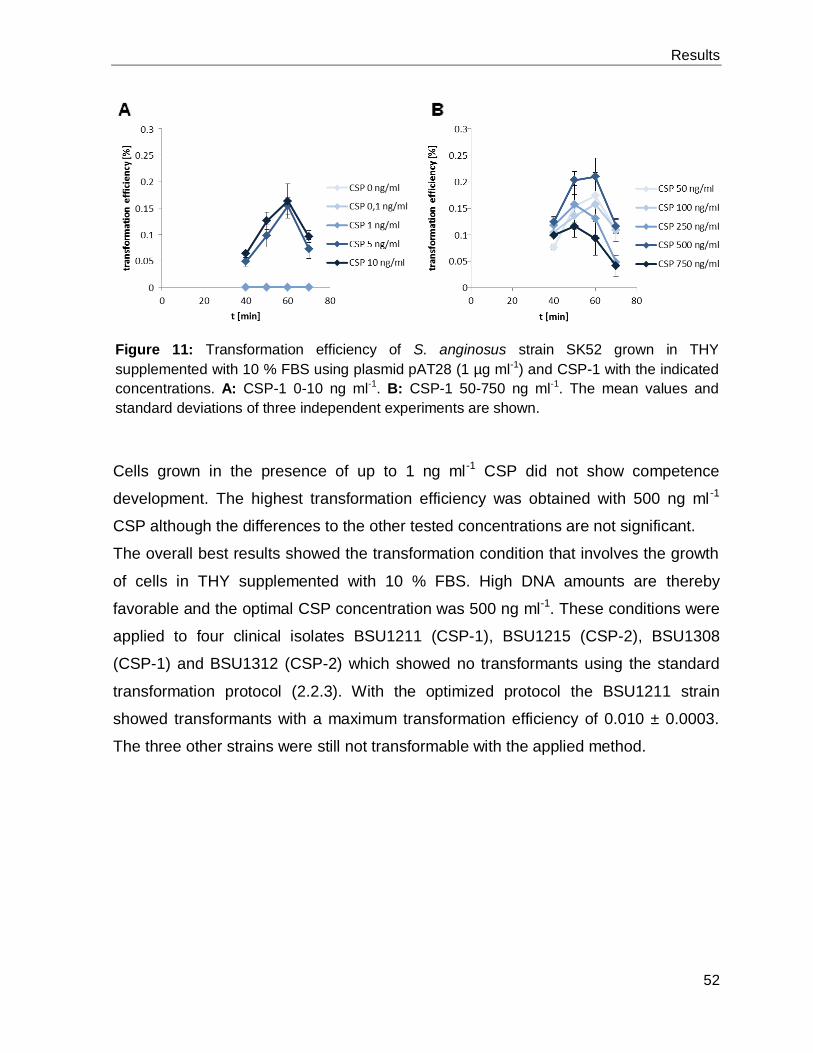
Results
52
Cells grown in the presence of up to 1 ng ml-1 CSP did not show competence
development. The highest transformation efficiency was obtained with 500 ng ml -1
CSP although the differences to the other tested concentrations are not significant.
The overall best results showed the transformation condition that involves the growth
of cells in THY supplemented with 10 % FBS. High DNA amounts are thereby
favorable and the optimal CSP concentration was 500 ng ml-1. These conditions were
applied to four clinical isolates BSU1211 (CSP-1), BSU1215 (CSP-2), BSU1308
(CSP-1) and BSU1312 (CSP-2) which showed no transformants using the standard
transformation protocol (2.2.3). With the optimized protocol the BSU1211 strain
showed transformants with a maximum transformation efficiency of 0.010 ± 0.0003.
The three other strains were still not transformable with the applied method.
Figure 11: Transformation efficiency of S. anginosus strain SK52 grown in THY
supplemented with 10 % FBS using plasmid pAT28 (1 µg ml-1) and CSP-1 with the indicated
concentrations. A: CSP-1 0-10 ng ml-1. B: CSP-1 50-750 ng ml-1. The mean values and
standard deviations of three independent experiments are shown.

Results
53
3.2 CcpA regulates the SLS expression in S. anginosus
SLS expression is under the control of catabolite repression 3.2.1
It was previously reported that the SLS expression of S. anginosus was reduced with
increasing glucose supplementations in the growth medium, the molecular
mechanisms of this effect were however not clear [12]. The glucose effect was
observed using a promoter reporter system [63] that measures the EGFP expression
which is under the control of the SLS promoter. The reduction of the SLS promoter
activity with increasing glucose concentrations indicates a catabolite repression
mechanism that controls the SLS expression. Classical CCR is mediated by the
activity of CcpA but additional alternative CCR mechanisms are described that are
independent of CcpA [38,198,229].
To further investigate the potential CCR at the SLS promoter, two additional sugars
were tested in the reporter system (Figure 12). The sugars were selected according
to their uptake mechanism and metabolism. Fructose is transported by a fructose
specific PTS allowing the analysis of the effect of the glucose specific PTS on the
SLS expression. The expression pattern, observed for cells grown with increasing
fructose supplementation in the growth medium, resembles the EGFP expression
measured with glucose indicating that proteins of the glucose specific PTS are not
involved in controlling the SLS promoter activity. The other tested sugar, galactose,
was chosen as this sugar is predicted to be taken up by a PTS and a non-PTS
(permease) transporter and it was reported to lead to only low levels of FBP in the cell
[143,144]. Increasing galactose supplementation in the growth medium did not result
in reduced SLS promoter activity further indicating a CcpA dependent CCR that
controls the SLS expression.

Results
54
CcpA negatively affects SLS expression 3.2.2
To directly elucidate the effect of CcpA on the regulation of the SLS expression, we
generated a S. anginosus ∆ccpA strain. This strain was constructed with the help of
the CSP-1 and the temperature sensitive pG+host5 vector system. It contains a
100 bp deletion in the ccpA gene that leads to a frameshift and a premature stop
codon (Figure 13A). This strain was transformed with the reporter plasmid
pBSU409::sagprom and the SLS promoter activity was analyzed in respect to
different glucose supplementations in the growth medium (Figure 13B).
Figure 12: Effect of C-source supplementation on SLS promoter activity. The activity of the
SLS promoter was determined using an EGFP reporter plasmid. The relative mean
fluorescence intensity (MFI) of S. anginosus pBSU409::sagprom grown in THY medium
supplemented with the indicated sugar concentrations is shown in comparison to the positive
control. Negative control: S. anginosus pBSU409; positive control: S. anginosus
pBSU409::cfbprom. The mean values and standard deviations of five independent
experiments are shown. Mann-Whitney-U test was performed to illustrate significant
differences to cells grown in presence of indicated glucose concentrations (p < 0.05) [20].
(Modified with permission by © CC BY 4.0, http://creativecommons.org/licenses/by/4.0/;
modifications: the figure was colorized)

Results
55
The reduced SLS promoter activity with increasing glucose concentrations in the
S. anginosus wild type was absent in S. anginosus ∆ccpA. The deletion strain
showed a constant EGFP expression over all tested glucose concentrations.
Figure 13A: Schematic representation of the 100 bp deletion in the S. anginosus ∆ccpA
strain. The site of deletion is marked with black lines and the deleted base pairs in bold. The
100 bp deletion results in a frameshift and a premature stop codon (*).
B: Effect of glucose supplementation on SLS promoter activity. The activity of the SLS
promoter was determined using an EGFP reporter plasmid. The relative mean fluorescence
intensity (MFI) of cells grown in THY medium supplemented with the indicated glucose
concentrations is shown in comparison to the positive control. Negative control: S. anginosus
pBSU409; positive control: S. anginosus pBSU409::cfbprom; WT: S. anginosus type strain;
ΔccpA: S. anginosus ΔccpA strain. The mean values and standard deviations of five
independent experiments are shown. Mann-Whitney-U test was performed to illustrate
significant difference to S. anginosus pBSU409::sagprom (p < 0.05) [20]. (Modified with
permission by © CC BY 4.0, http://creativecommons.org/licenses/by/4.0/; modifications: the
figure was colorized)

Results
56
S. anginosus ∆ccpA maintains hemolytic activity in the presence of 3.2.3
glucose
A functional hemolytic assay was performed to investigate the effect of the constant
SLS expression of the S. anginosus ∆ccpA on the hemolytic activity in presence of
high glucose concentrations. Two additional strains were constructed that served as
controls in the assay. The ccpA complementation strain S. anginosus ∆ccpA::ccpA
was generated by CSP-1 transformation of S. anginosus ∆ccpA with a linear DNA
fragment that contained the ccpA gene with a silent point mutation, an erythromycin
resistance gene and the up- and downstream region of ccpA (Figure 14). To exclude
a possible effect of the erythromycin gene on the hemolytic behavior of the cells the
S. anginosus::ccpA strain served as additional control.
The S. anginosus type strain was able to lyse human erythrocytes if cells were grown
in the presence of up to 12.5 mM glucose (Figure 15). Cells that were incubated in
medium with 25 mM glucose supplementation were non-hemolytic. The S. anginosus
∆ccpA strain in contrast performed regular β-hemolysis in presence of all tested
glucose concentrations. Both complementation strains showed the same hemolytic
behavior like the parental strain verifying that CcpA is responsible for the observed
hemolytic difference of the S. anginosus ∆ccpA strain.
Figure 14: Schematic representation of the ccpA complementation strains. A linear DNA
fragment containing the upstream region of ccpA, the ccpA gene carrying a silent point
mutation, the erythromycin resistance gene (ery) and parts of ANG1_1037 was transformed
with CSP-1 in S. anginosus ∆ccpA to construct S. anginosus ∆ccpA::ccpA (::ccpA). To
exclude an effect of the integration of ery on the phenotype of the complemented strain, the
same DNA fragment was transformed in S. anginosus type strain SK52 (WT) to generate
S. anginosus::ccpA (::ccpA). *: silent point mutation.

Results
57
Figure 15: Hemolytic activity of S. anginosus strains for human erythrocytes. Bacteria were
grown in THY medium supplemented with the indicated glucose concentrations. The
hemolytic behavior of the S. anginosus type strain (A), the S. anginosus ΔccpA (B), the S.
anginosus ΔccpA::ccpA (C) and the S. anginosus::ccpA (D) is illustrated for different
bacterial cell dilutions. Positive control: ddH2O; negative control: assay buffer. The mean
values and standard deviations of five independent experiments are shown [20]. (Modified
with permission by © CC BY 4.0, http://creativecommons.org/licenses/by/4.0/; modifications:
the figure was colorized)

Results
58
Mutation of cre abolishes glucose dependent SLS repression 3.2.4
Binding of CcpA to its target sequence (cre) is reported in different Gram-positive
bacteria and consensus sequences of cre are published
(http://regprecise.lbl.gov/RegPrecise/collection_tf.jsp?collection_id=163). Additionally,
a second CcpA binding site was identified in Streptococcus suis (cre2) [219]. We
used this information to screen the promoter of the SLS operon for the presence of
potential CcpA binding sites to further investigate if CcpA directly regulates the SLS
expression by binding to the SLS promoter. Three potential cre sites were identified in
the SLS promoter allowing a maximum of three mismatches to the consensus
sequences (Figure 16).
Figure 16A: The locations of putative cre sites are illustrated in respect to the transcription
start site (arrow) of the SLS promoter. A: creA, B: creB, C: creC.
B: Comparison of the in silico predicted cre in the SLS promoter and the published
consensus cre sites. The size of the single nucleotides corresponds to their conservation.
The creB site consists of a cre homolog overlapping a potential cre2 site. Mismatches to the
consensus sequences are underlined and bold [20]. (Modified with permission by © CC BY
4.0, http://creativecommons.org/licenses/by/4.0/; modifications: the figure was colorized)

Results
59
CreA is located 167 bp upstream of the transcription start site and shows homologies
to the published cre2 site. One predicted CcpA binding site (creB) is composed of a
putative cre site overlapping a potential cre2 site and the third cre site (creC) overlaps
the promoter of the SLS operon.
To investigate the influence of the predicted cre sites on the expression of the SLS
promoter in the presence of glucose, we mutated these sites in the promoter and
performed a promoter reporter assay using three pBSU409::sagprom derivatives. The
creA and creB site were separately deleted in the promoter (Figure 17A). The deletion
of creC would destroy the bacterial promoter, thus this site was mutated in four
potentially conserved nucleotides that should be important for the binding of CcpA to
this sequence. The altered promoters were cloned in front of egfp in the reporter
plasmid and transformed into the S. anginosus type strain SK52 to measure the
promoter activity of cells grown in the presence of different glucose concentrations
(Figure 17B). Deletion of creA resulted in a similar expression pattern like in the strain
carrying the wild type promoter that showed a reduction of promoter activity with
increasing glucose supplementations. The strain with the creB deletion showed a
reduced promoter activity without glucose supplementation compared to the strain
that contains the wild type promoter and further demonstrated a constant egfp
expression for all tested glucose concentrations. In comparison to the wild type
promoter, the egfp expression was higher for cells growing in presence of 25 mM
glucose supplementation. Differing from the previous attainments, the mutation of
creC shows a constantly high promoter activity irrespective of the glucose
supplementations. This expression pattern closely resembles the promoter activity
measured in the S. anginosus ∆ccpA strain.

Results
60
Figure 17: Schematic representation of reporter plasmids with mutated SLS promoters used
to investigate the in silico predicted cre sites. WT: promoter of SLS; creA_del:
pBSU409::sagprom_creA_del (pBSU881); creB_del: pBSU409::sagprom_creB_del
(pBSU880); creC_mut: pBSU409::sagprom_creC_mut (pBSU876). c: The sequence of the
predicted cre site (upper row) and the mutations of the sequence (bold and underlined) in
pBSU409::sagprom_creC_mut (lower row) are shown.
B: Effect of glucose supplementation on the activity of mutated SLS promoters in the
S. anginosus type strain SK52. The relative mean fluorescence intensity of cells grown in
THY medium supplemented with the indicated glucose concentrations is shown in
comparison to the positive control. Positive control: S. anginosus pBSU409::cfbprom;
Negative control: S. anginosus pBSU409. The mean values and standard deviations of five
independent experiments are shown. Mann-Whitney-U test was performed to illustrate
significant difference to S. anginosus pBSU409::sagprom (p < 0.05) [20]. (Modified with
permission by © CC BY 4.0, http://creativecommons.org/licenses/by/4.0/; modifications: the
figure was colorized)

Results
61
CcpA binds to creC in vitro 3.2.5
To verify that CcpA binds to the in silico predicted creC site in the SLS promoter, we
performed EMSA experiments. We used heterologously expressed His-tagged CcpA
in the absence of HPr as several in vitro studies reported that CcpA is able to bind to
cre sites in the absence of its activator [1,5,197,219]. Increasing the amounts of CcpA
in the bandshift reaction induced a shift of the labeled creC DNA fragment
(GTTTACGCGAAAGCGCTTTTTTTATATA) demonstrating that CcpA is able to bind
to this sequence (Figure 18). The specificity of binding was tested using 500-fold
molar excess of unlabeled creC as well as unlabeled mutated creC
(GTTTACGCGAAGGATCCTTTTTTATATA). The addition of excess unlabeled creC
was able to compete for CcpA binding and shows no shift of the labeled creC. In
contrast to that the mutated creC sequence was unable to inhibit the binding of CcpA
to creC. The observation that the deletion of creB in the promoter reporter assay
resulted in a different expression pattern compared to the wild type promoter
prompted us to test also the binding of CcpA to this sequence. The use of 500-fold
excess of unlabeled creB had no effect on the CcpA binding to the creC sequence
further demonstrating the specificity of CcpA binding to creC. Hence we were able to
verify that CcpA can directly regulate the SLS expression by binding to the creC site
that we identified in the SLS promoter.
Figure 18: EMSA of creC using His-tagged CcpA. Increasing amounts of purified CcpA (0-8
µg) were used and tested for binding to labeled creC
(GTTTACGCGAAAGCGCTTTTTTTATATA). The specificity of binding was assayed
using 500-fold molar excess of unlabeled creC (A), creC_mut (B;
GTTTACGCGAAGGATCCTTTTTTATATA) and creB (C;
TGCTATAAGAACGCGCTTTTTATTTTGTTTTAGATGGT) [20].

Results
62
3.3 Role of a CovR homolog in the regulation of SLS in S. anginosus
In silico analysis and genetic organization of the CovR homolog 3.3.1
The publication of the complete genome sequence of some S. anginosus strains in
2013 provided evidence for a CovR homolog (CovR*) in SAG [141]. CovR is the
response regulator of the well-known TCS CovSR that is involved in the regulation of
virulence gene expression in GAS and GBS [53,103,128]. Additionally, CovR was
reported as regulator of the SLS operon in GAS [60]. Thus, we hypothesized a role of
the CovR* in the regulation of hemolysis in S. anginosus.
The proposed CovR* shows similarities to other streptococcal CovR proteins and
consists of an N-terminal signal receiver domain and a C-terminal OmpR-like winged
helix-turn-helix DNA binding domain. But a further blast search revealed that CovR* is
more closely related to a response regulator that can be found in S. pneumoniae,
Streptococcus oralis and Streptococcus mitis (Figure 19A). CovR of GBS and GAS
are well studied and it was reported that the phosphorylation of CovR at positions
D53 and T65 are important for the regulation of the CovR activity [82,111]. The CovR*
of S. anginosus is highly conserved in different strains (Figure 19B) but it lacks these
phosphorylation sites (Figure 19C).

Results
63
Instead of being organized in one operon with covS, covR* of S. anginosus is located
directly downstream of the 6-phosphogluconate dehydrogenase gene (gnd) and a
homolog of the sensor histidine kinase/phosphatase CovS could not be detected in a
genome wide BLAST search (Figure 20A). CovR* of the S. anginosus type strain is
the last gene of a contig in the NCBI database. Therefore, the downstream gene was
assumed to be a homolog to the covR* downstream genes located in the genome of
completely sequenced S. anginosus strains like S. anginosus C1051. All sequenced
S. anginosus strains contain a gene encoding an enzyme IIBCA component of the
glucose-specific PTS downstream of covR*. Due to the close proximity of covR* to
gnd, a RT-PCR was performed confirming that the two genes are organized in one
operon (Figure 20B).
Figure 19A: Comparison (amino acid level) of CovR* of S. anginosus to CovR of GBS, GAS
and S. mutans and a regulator of S. pneumoniae, S. oralis and S. mitis.
B: Circular phylogram showing the conservation of CovR* in different S. anginosus strains
(blue circle). CovR* was amplified and sequenced with primers binding up- and downstream
of the gene.
C: Alignment of CovR* of S. anginosus, CovR of GBS, GAS and S. mutans and a regulator
of S. pneumoniae, S. oralis and S. mitis. The first 70 amino acids are shown. Red boxes
highlight the phosphorylation sites of GBS and GAS which are important for the regulation of
CovR activity.

Results
64
Figure 20A: Organization of covR encoding genomic region in S. pyogenes M1,
S. pneumoniae R6, S. anginosus C1051 and S. anginosus type strain SK52. gnd:
6-phosphogluconate dehydrogenase; A: Spy_0334; B: Spy_0338; C: spr_0334; D: cpbF;
E: SAIN_1372; F: PTS glucose-specific EIICBA component; E* homolog of E; F* homolog of
F. Arrows indicate positions of primer used in the RT-PCR experiment.
B: Agarose gel of RT-PCR using template as illustrated (neg: H2O) proving the organization
of covR* and gnd in one operon. +: with reverse transcription step, −: without reverse
transcription step.
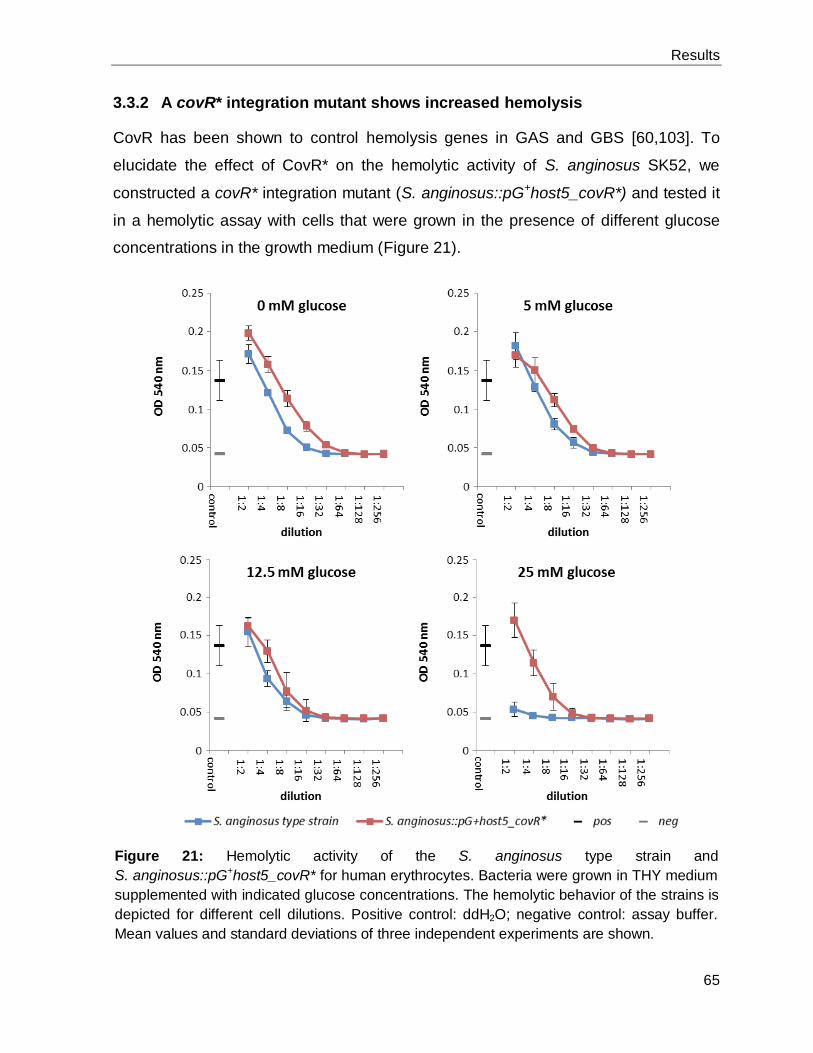
Results
65
A covR* integration mutant shows increased hemolysis 3.3.2
CovR has been shown to control hemolysis genes in GAS and GBS [60,103]. To
elucidate the effect of CovR* on the hemolytic activity of S. anginosus SK52, we
constructed a covR* integration mutant (S. anginosus::pG+host5_covR*) and tested it
in a hemolytic assay with cells that were grown in the presence of different glucose
concentrations in the growth medium (Figure 21).
Figure 21: Hemolytic activity of the S. anginosus type strain and
S. anginosus::pG+host5_covR* for human erythrocytes. Bacteria were grown in THY medium
supplemented with indicated glucose concentrations. The hemolytic behavior of the strains is
depicted for different cell dilutions. Positive control: ddH2O; negative control: assay buffer.
Mean values and standard deviations of three independent experiments are shown.

Results
66
The hemolytic activity of the S. anginosus::pG+host5_covR* strain was slightly
increased compared to the parental strain if cells have been grown in medium
supplemented with 0, 5 and 12.5 mM glucose. In the presence of 25 mM glucose, the
covR integration mutant was still able to lyse the erythrocytes whereas the type strain
showed no hemolytic activity at all.
Markerless covR* deletion mutant shows type strain hemolytic behavior 3.3.3
To exclude possible polar effects of the pG+host5 integration into the covR* locus on
the hemolytic behavior of the strain, a markerless covR* deletion mutant was
constructed using the Cre-lox based genetic manipulation procedure [19]. Therefore,
the downstream region of the covR* locus needs to be amplified to be able to
generate the deletion. This region is not available in the NCBI database, thus we
assumed that the genome of the S. anginosus type strain is organized like the other
completely sequenced S. anginosus strains. According to the available genetic
information of other S. anginosus strains, primers were designed that bind
downstream of their covR* in the gene encoding a PTS glucose-specific EIICBA
component. Several attempts to amplify this region in the S. anginosus type strain
failed (data not shown). Thus, an inverse PCR was performed verifying that the
S. anginosus type strain SK52 contains a different genetic organization downstream
of covR* (Figure 22).
Figure 22: Organization of covR* encoding genomic region in S. anginosus C1051 and
S. anginosus type strain SK52. gnd: 6-phosphogluconate dehydrogenase; E: SAIN_1372;
F: PTS glucose-specific EIICBA component; E* homolog of E; G: ANG1_1764, hypothetical
protein. F* homolog of F; Accession numbers: contig17 BAST01000017.1; contig13
BAST01000013.1; contig12 BAST01000012.

Results
67
With this knowledge, a covR* deletion mutant was constructed and its hemolytic
activity was tested (Figure 23). In contrast to the S. anginosus::pG+host5_covR*
integration mutant, the clean deletion mutant S. anginosus ∆covR* showed a similar
behavior like the S. anginosus type strain in the hemolysis assay with no hemolytic
activity of cells grown in the presence of 25 mM glucose supplementation.
Figure 23: Hemolytic activity of the S. anginosus type strain and S. anginosus ∆covR* for
human erythrocytes. Bacteria were grown in THY medium supplemented with indicated
glucose concentrations. The hemolytic behavior of the strains is depicted for different cell
dilutions. Positive control: ddH2O; negative control: assay buffer. Mean values and standard
deviations of three independent experiments are shown.

68
Discussion
4 Discussion
S. anginosus is considered a human commensal but an increasing number of reports
in recent years highlighted the clinical relevance of this species
[94,101,104,105,157,163,175]. Despite the rise of epidemiological knowledge about
S. anginosus, the knowledge about the virulence gene repertoire is scarce [11]. One
obstacle is the lack of genetic manipulation techniques that would allow fast and
reliable investigations of molecular pathogenicity mechanisms in S. anginosus. The
identification of the com regulatory system in different viridans streptococci
revolutionized the scope for genetic manipulations in these species and the presence
of competence genes in the genome of several S. anginosus strains was already
reported [76,119,141] although an analysis of the transformation machinery has not
been performed so far. In S. pneumoniae, the presence of 22 genes is required for
transformation [150]. We used the sequence of these genes to find homologs in the
genome of the S. anginosus type strain. While 19 genes were annotated in
S. anginosus, three homologs were missing (SP1808, comW and comC). SP1808
encoding a type IV prepilin peptidase is not annotated in the genome but a sequence
with high identity is located in an intergenic region indicating the presence of this
gene in S. anginosus. The second gene for which a homolog could not be detected in
S. anginosus SK52 is comW. ComW is reported as stabilizer and activator of ComX
and thus as crucial for competence development in S. pneumoniae [188,199]. The
absence of this gene in S. anginosus SK52 prompted us to screen all the available
S. anginosus genomes in the NCBI database for the presence of a comW homolog
leading to the identification of three S. anginosus isolates that contain a homolog. The
protein encoded in this gene shares only an identity of 44 % and a similarity of 63 %
on the amino acid level to ComW of S. pneumoniae. Therefore, it is tempting to
speculate that the identified homologs share the same function as ComW. This would
need further investigations by either deleting these genes in the strains or by
introducing this homologous gene into S. anginosus SK52 followed by the analysis of
the transformation behavior of the manipulated strains. Nevertheless, the absence of
a ComW homolog in S. anginosus SK52 demonstrates that this species, in contrast to
S. pneumoniae, does not need ComW for competence development. This is line with

69
Discussion
the observation that Streptococcus gordonii shows competence development [75]
although this species does also not encode a ComW homolog [119].
The absence of a homolog of the S. pneumoniae comC in S. anginosus was certainly
expected as diversity of the CSP encoded in comC is crucial to limit interspecies
crosstalk of the competence system [76,211]. We therefore used the published comC
sequence of S. anginosus SK52 [76] to screen for yet unknown CSP pherotypes in
SAG. Genetic variation in comC within the same species is common in different
viridans streptococci [3,76,154] and we were able to identify two major CSP
pherotypes in S. anginosus. According to the designation in S. pneumoniae, we
termed the pherotypes CSP-1 and CSP-2 [154]. The S. anginosus BSU1417 isolate
encoded a third CSP variant that is identical to the CSP described in S. milleri
NCTC10708 [76], which is the former designation for the S. anginosus. All three
ComC variants contain the double-glycine leader motif that allows the proteolytic
cleavage of the prepeptide by ComAB which results in the formation of the CSP [74].
One S. anginosus isolate contains a comC identical to comC of S. sanguinis SK36.
The ComC prepeptide of this strain lacks the double-glycine motif, but it was reported
that the C-terminal region (DLRGVPNPWGWIFGR) is sufficient to induce
competence [76,162]. The remaining nine S. anginosus isolates contain a Mobile
Element Protein inserted in the comD gene. This protein belongs to the ISL3
transposase family which seems to be common in S. anginosus as one completely
sequenced S. anginosus strain J4206 contains six copies of this gene located in
different genetic loci. The integration of the transposase in the comD gene probably
results in the disruption of the competence regulation. However, this needs to be
verified by further investigations.
Although the CSP was already used to transform S. anginosus SK52 [189], an
analysis of the transformation kinetics has so far not been performed. Therefore, we
investigated the uptake of plasmid DNA of cells grown in the presence of the CSP.
After CSP stimulation, the bacteria showed rapid competence development indicating
a S. pneumoniae-like regulation cascade [161,199] that controls the competence in
S. anginosus. The slower competence development using plasmid DNA in
S. anginosus SK52, with a maximum transformation efficiency after ~ 40 min
compared to S. pneumoniae (~ 20 min), could potentially be explained by the slower

70
Discussion
growth behavior of S. anginosus. The uptake of linear DNA, in contrast, could already
be observed in the beginning of the transformation experiment. One conceivable
reason for this could be the lower complexity of the linear DNA in comparison to
plasmid DNA which was reported as a factor that can affect transformation
efficiencies [93,165]. The presence of two major CSP pherotypes further prompted us
to test the cross-reactivity of the variants in the competence development. The
S. anginosus SK52 developed competence only after stimulation with its cognate
CSP-1, whereas the S. anginosus BSU1216, encoding CSP-2, showed considerable
transformation with both CSP pherotypes. At first sight, this was an unexpected
observation as the diversity of comC should limit crosstalk [76,211], but it was already
reported that some S. pneumoniae strains can also recognize other than the self-
expressed CSP variants [154,225].
By employing the gained knowledge about the competence system in S. anginosus,
we aimed to establish a fast and reliable genetic manipulation technique that would
facilitate the investigation of pathogenicity mechanisms in this species. Thereby we
focused on the generation of markerless gene deletions as gene deletions in the SAG
are currently constructed via allelic exchange of the target gene with an antibiotic
resistance marker [88,189,202]. This method comprises the risk of undesired polar
effects on the transcription of up- and downstream genes and the introduction of
multiple gene deletions in the same strain can be limited by the availability of suitable
antibiotic resistance markers. Therefore, we decided to establish a Cre-recombinase
based genetic engineering technique that circumvents these potential problems by
removing the antibiotic resistance gene from the bacterial genome. Additionally, this
method does not comprise time consuming cloning procedures which are necessary
in case of temperature sensitive vectors that are used in different Gram-positive
bacteria for the generation of markerless gene deletions [24]. The mutation technique
further relies on the transformation of linear DNA with the CSP and subsequent
removal of the resistance gene via the Cre-recombinase. Thereby the mutated loxP
sites lox66 and lox71 are fused to one lox72 site [2]. This lox site is a poor substrate
for the Cre-recombinase and thus allows the generation of multiple deletions in the
same strain. To confirm that this novel mutation technique can be applied for gene
deletions of various sizes, we deleted sagA1 (158 bp) and sagBCD (3347 bp) from
71
Discussion
the β-hemolysin gene cluster of S. anginosus SK52. The mutant strains possessed
the expected hemolytic phenotype on blood agar plates with the sagA1 mutant
showing a reduced hemolysis as this strain encodes two SagA homologs [10,189].
Transformation of clinical bacterial isolates can be challenging [133,154,220].
Therefore, we aimed to optimize the transformation conditions to be able to transform
clinical S. anginosus isolates that proved to be untransformable with the current
method. The growth of the bacteria in medium containing serum was reported to
enhance transformation frequency [149,153]. The use of 10 % FBS in the growth
medium, together with an increase of the DNA concentration in the transformation
procedure, yielded a maximal transformation efficiency of 0.81 % in the S. anginosus
type strain. The concentration of the CSP had only a minor effect on the
transformation frequency. Cells were transformable in the presence of a low level of
CSP (5 ng ml-1) which correlates to a concentration of 2.6 nM. This CSP
concentration is in the range that was previously reported to induce genetic
competence in different streptococci [73,165]. The highest transformation efficiency
was observed using 500 ng ml-1 CSP although the difference to other concentrations
was only minor. It has to be pointed out that the transformation efficiencies measured
during the investigation of the transformation kinetics (3.1.2) were higher than during
the optimization of the procedure (3.1.4) using the same transformation set up. The
only deviation was that we used a new batch of CSP in the optimization which could
be the reason for the observed difference. We applied the optimized conditions to four
clinical S. anginosus isolates that could not be transformed with the standard
procedure. Using the optimized method, one of these isolates (S. anginosus
BSU1211) could be transformed with the pAT28 plasmid. This demonstrates that
further optimization is needed and further attempts on improvements of the procedure
will concentrate on the bacterial number and growth phase that were used in the
transformation which could probably lead to further enhanced transformation
efficiencies [35,165]. However, we were able to show that the competence system in
S. anginosus can be used for genetic modifications that will facilitate to investigate
this species on the molecular level.
Molecular pathogenicity mechanisms in S. anginosus are widely unknown [11]. One
exception is the hemolysin that shows high homologies to SLS of GAS [10,12,189].

72
Discussion
It was previously reported that the expression of the SLS in S. anginosus was
reduced in the presence of increased glucose concentrations in the growth medium
[12]. This expression pattern indicates a CCR mechanism controlling the SLS
expression. To further investigate this phenomenon, we performed an EGFP
promoter reporter assay with different sugars. We decided to use fructose as this
sugar is transported by a fructose-specific PTS into the cell. Thus, through the use of
fructose, we can exclude that the parts of the glucose-specific PTS regulate the SLS
expression as it was reported that components of PTS systems can regulate gene
expression independently of CcpA [198,229]. The SLS expression measured with
increasing fructose supplementation in the growth medium resembled the expression
pattern observed with the addition of glucose. This indicates that components of the
glucose-specific PTS are not involved in the observed concentration dependent
reduction of EGFP expression and further points towards a CCR mechanism that is
regulated by CcpA. Additionally, we tested galactose in the reporter assay. This sugar
was chosen as S. anginosus encodes the Leloir as well as the tagatose 6-phosphate
pathway for galactose metabolism. The Leloir pathway starts with an
unphosphorylated galactose that is transported via a permease and is further
metabolized to FBP. For the tagatose 6-phosphate pathway, the galactose needs to
be transported via a PTS, as galactose 6-phosphate is the first metabolite in this
pathway. The presence of both galactose metabolism pathways in the genome of
S. anginosus indicates that this bacterium transports galactose by PTS and non-PTS
(permease) transporters like S. pneumoniae. For S. pneumoniae, it was shown by in
vivo 13C-NMR and 31P-NMR that cells grown in presence of galactose accumulated
only low amounts of FBP in comparison to cells grown in the presence of glucose
[143,144]. Thus galactose should not be able to activate CcpA in a comparable
manner like glucose, as high FBS levels are needed to activate the kinase activity of
the HPr kinase/phosphorylase that controls the HPr serine phosphorylation
accompanied with the CcpA activation [42]. Therefore, we should not observe a
markedly reduced EGFP expression with increasing galactose supplementation in the
growth medium and this we could demonstrate with the reporter system. The use of
fructose and galactose in summary pointed towards a CCR mechanism at the SLS
operon that is dependent on CcpA.

73
Discussion
To further corroborate the CcpA dependent CCR, we constructed a ccpA deletion
mutant by pG+host5 mutagenesis [24]. The temperature sensitive plasmid pG+host5
containing the deletion sequence for ccpA was thereby transformed with CSP-1 into
S. anginosus. The vector was directly forced to integrate into the genome by growing
the transformation mixture at 39 °C. This procedure finally allowed the construction of
a 100 bp deletion in the ccpA gene that resulted in a frameshift and a premature stop
codon. The S. anginosus ∆ccpA strain was then investigated in the reporter assay
showing that the glucose dependent reduction of the promoter activity observed in the
parental S. anginosus SK52 strain is absent in the ccpA mutant. Thus, we were able
to demonstrate that CcpA regulates the SLS expression by inhibiting transcription at
high glucose levels. During the reporter plasmid analysis we observed an increase of
the relative MFI for mutant cells grown in the absence of glucose supplementation
(Figure 13B). This could be explained by the use of the THY growth medium that
already contains about 11 mM glucose.
We further analyzed the effect of the observed expression patterns in a functional
hemolysis assay with human erythrocytes which showed that, in contrast to the
parental S. anginosus SK52 strain, the ccpA mutant was able to lyse the erythrocytes
also in the presence of high glucose concentrations. To verify that this observation
results from the ccpA mutation, we constructed the complementation strain
S. anginosus ∆ccpA::ccpA. We chose to use a chromosomal complementation as the
introduction of the ccpA gene into the mutant by a plasmid would lead to an increased
copy number of ccpA in the cell. As ccpA is a major regulator, this could have
pleiotropic effects on the global gene expression pattern. The chromosomal
complementation could be achieved by CSP driven transformation of a linear DNA
fragment containing the ccpA gene and an erythromycin resistance gene. This
complemented strain showed a similar behavior like the S. anginosus type strain and
showed hemolytic activity up to 12.5 mM glucose supplementation, but was non-
hemolytic in presence of 25 mM glucose supplementation. To exclude an effect of the
introduced erythromycin resistance gene on the expression of ccpA, we additionally
generated a S. anginosus::ccpA strain by transformation of the complementation
construct into the S. anginosus type strain. This strain also demonstrated a type strain
like behavior, finally confirming the role of CcpA in the regulation of the hemolysis.

74
Discussion
The expression of up to 10 % of genes is affected by CcpA dependent CCR in
different bacteria [134,219,227]. CcpA thereby mainly regulates genes involved in
metabolism, but it also regulates the expression of other transcription factors
[5,8,134]. This implies that the observed regulation of the SLS expression by CcpA
could be direct by binding of CcpA to the SLS promoter or indirect by CcpA regulating
the expression of an alternative transcription factor that in turn regulates the SLS
expression. To investigate these two possibilities, we screened the SLS promoter for
the presence of potential CcpA binding sites. This led to the identification of three
potential cre sites in the SLS promoter with high homologies to the published
consensus cre sites [116,219]. The predicted CcpA binding site creC overlaps the
bacterial promoter and, as the location of the cre site in respect to the transcriptional
start site determines the effect of the regulation, the position of creC would indicate
that CcpA is a strong repressor of the SLS operon [116]. We decided to determine the
effect of the putative cre sites on the glucose dependent reduction of the SLS
promoter activity by mutating these sites in the reporter plasmid pBSU409::sagprom.
The creA and creB site were separately deleted and the creC was mutated in four
potentially conserved nucleotides as the deletion of this site would probably destroy
the SLS promoter. The deletion of creA demonstrated a promoter activity with
increasing glucose supplementation that closely resembles the expression pattern
measured for the wild type promoter. Thus, this predicted cre site can be excluded as
possible CcpA binding site. The deletion of creB, in contrast, resulted in an altered
promoter activity. Without glucose supplementation, the relative MFI was decreased
in comparison to S. anginosus pBSU::sagprom and it was constant for all tested
glucose supplementations which resulted in a higher promoter activity for cells grown
with 25 mM glucose supplementation in comparison to the strain carrying the wild
type promoter. The most noticeable promoter activity alteration could be observed for
the mutation of creC. This strain did not show the glucose dependent reduction of
EGFP expression with increasing glucose concentrations in the growth medium. The
expression pattern of this strain resembles the pattern measured in the S. anginosus
∆ccpA strain. Accordingly, the creC site was identified as the most promising CcpA
binding site in the SLS promoter of S. anginosus SK52.

75
Discussion
The final verification of direct regulation of the SLS operon by CcpA was performed
by EMSA. It was previously reported that CcpA is able to bind to cre sites in the
absence of serine phosphorylated HPr and FBP in vitro [1,5,197,219]. We therefore
used heterologously expressed His-tagged CcpA alone and demonstrated that CcpA
is able to induce a shift of the creC site in a concentration dependent manner. The
binding could be inhibited using an excess of unlabeled creC and the specificity of
CcpA-creC complex formation was tested with an unlabeled mutated creC sequence
which was not able to compete for CcpA binding. Previous studies reported that more
than one cre site can be present in the promoter of a CcpA regulated gene [116,219].
As we observed in the promoter reporter assay that the strain carrying the promoter
with the deletion of the creB site showed an increased relative MFI at 25 mM glucose
supplementation compared to the S. anginosus pBSU409::sagprom strain, we
decided to test if CcpA is able to bind to this creB sequence. We therefore used an
excess of unlabeled creB in the EMSA, but this sequence could not inhibit the binding
of CcpA to the creC site, demonstrating that, under the conditions tested, CcpA does
not bind to creB. It has to be noted that we did not incorporate serine phosphorylated
HPr in our EMSA which was reported to increase the affinity of CcpA to the cre sites
[67,167] and thus we cannot rule out that CcpA might be able to bind to the creB
sequence in vivo. However, we were able to demonstrate that CcpA regulates the
SLS expression and binds to a cre site in the SLS promoter. This is the first report of
a regulator controlling a potential virulence factor in S. anginosus and it further
highlights the role of CcpA in the regulation of the interplay between carbohydrate
metabolism and virulence in Gram-positive species [160,173]. Efficient acquisition
and metabolism of nutrients is required for growth in free-living as well as in host-
associated bacteria. However, bacteria living in hostile environments have to cope
with fluctuations in the availability of carbon sources. To deal with these diverse host
environments, the bacteria have evolved virulence factors that facilitate the nutrient
acquisition. Thus, the link of pathogenesis and access to nutrition explains why
pathogenic bacteria concurrently use regulators of the metabolism to regulate
expression of virulence determinants [160,172,180]. CcpA is a well investigated
regulator in this context and it was demonstrated that the SLS of GAS is under the
control of CcpA mediated CCR [99]. It was proposed that the SLS plays a role in the

76
Discussion
secondary metabolism during infection. As a cytolysin, it may contribute to the
nutrient release from host cells that can promote the growth of the bacterium.
However, the tissue damage leads to host immune response and thus the SLS
expression in GAS is tightly controlled by CcpA as well as by other regulatory
systems (CovR, CodY, RofA, Mga) [21,77,97,115].
Although the knowledge about pathogenicity mechanisms in S. anginosus is scarce
[11], the publication of the first complete S. anginosus genome in 2013 allows the
prediction of potential virulence factors in this species [141]. Olson and colleagues
proposed in this publication that the S. anginosus C238 strain encodes a covR*
homolog. CovR is the response regulator of the two component system CovSR and
was originally identified in GAS [51,109]. It regulates the expression of virulence
related genes in GAS, GBS and S. dysgalactiae as well as in S. mutans
[45,68,103,182]. Additionally, it was reported that covR directly regulates the SLS
expression in GAS and a covSR mutant in GBS exhibited an increased hemolytic
phenotype [60,103]. Therefore we hypothesized that the covR* homolog in
S. anginosus could also be involved in the regulation of the hemolysin expression in
this species.
We therefore constructed the S. anginosus covR* integration mutant using the
pG+host5 vector system. We decided to test the hemolytic activity of this
S. anginosus::pG+host5_covR* strain grown in presence of different glucose
concentrations as the up- and the potential downstream gene of covR* are supposed
to be involved in glucose metabolism and regulators often control the expression of
genes which are located in close proximity in the genome. The upstream gene
encodes a 6-phosphogluconate dehydrogenase gene (gnd) which is part of the
pentose phosphate pathway. For the downstream gene, we assumed that the
S. anginosus type strain genome is organized like the available completely
sequenced S. anginosus genomes. This was necessary as the covR* homolog is
located at the end of contig17 of S. anginosus SK52 that is published in NCBI. The
predicted downstream gene was a PTS glucose-specific EIICBA component, further
suggesting a role of covR* in the regulation of metabolism that in turn might affect
hemolysis as already observed for the S. anginosus ∆ccpA strain. The
S. anginosus::pG+host5_covR* strain showed a slightly increased hemolytic activity

77
Discussion
for cells that were grown in the presence of up to 12.5 mM glucose supplementation
and the hemolytic activity was retained, in contrast to the parental strain, for cells
grown in medium with 25 mM glucose supplementation. This resembled the hemolytic
phenotype of the S. anginosus ∆ccpA strain.
The insertion of the pG+host5 plasmid into the covR* locus could have polar effects
on the expression of up- and downstream genes. As we established the Cre-lox
based mutagenesis tool that allows the markerless deletion of genes, we constructed
a S. anginosus covR* deletion strain and tested this strain in the hemolysis assay. For
the generation of the S. anginosus ∆covR* strain, we needed to amplify the
downstream region of covR*. It thereby turned out that the predicted downstream
gene (PTS glucose-specific EIICBA component) is not located at the supposed
position in the S. anginosus SK52 genome. Thus, we performed an inverse PCR
verifying that the S. anginosus type strain comprises a different genetic organization
at this genomic locus. The downstream gene of covR* in S. anginosus SK52 is
ANG1_1764, a hypothetical protein without predicted function. We used this
knowledge and constructed the covR* deletion strain. This S. anginosus ∆covR*
strain showed, in contrast to the S. anginosus::pG+host5_covR*, a similar hemolytic
behavior like the parental S. anginosus SK52 strain. Two possible explanations could
be the reason for the observed hemolytic difference of the deletion strain in
comparison to the integration strain. It would be conceivable that the integration of the
pG+host5 vector altered the expression level of gnd leading to a change in the carbon
flux in the cell to a dominant pentose phosphate pathway. This would result in a
decrease of ATP production and FBS concentration in the cell that could influence the
CcpA activation. On the other hand, possible undesired multiple integrations of
pG+host5 in the S. anginosus::pG+host5_covR* are conceivable which could have
effects on the hemolytic activity. However, this demonstrates that the construction of
markerless gene deletions with the Cre-lox system can prevent possible constraints.
In the meanwhile, we further investigated the covR* homolog in S. anginosus. We
showed that covR* is conserved in different clinical S. anginosus isolates, but it lacks
the phosphorylation sites that were reported for CovR in different streptococcal
species [82,83,96,111]. Instead of being transcribed in one operon with covS, the
covR* in S. anginosus is located in an operon with gnd and a CovS homolog could

78
Discussion
not be detected in the S. anginosus genome. It has to be mentioned that S. mutans
also lacks a covS homolog and the CovR in this species was reported as an orphan
regulator [32]. The covR locus in S. mutans and GAS show high similarities not only
on the amino acid level of CovR but also the covR surrounding genes in both species
are similar, favoring the description of the S. mutans gene as covR. In S. anginosus
the lack of CovS, the missing phosphorylation sites and the organization of covR* in
an operon with gnd strongly indicate that the proposed covR* homolog in
S. anginosus should not be denominated as covR. This, once again, is illustrated by
the presence of a gene in S. pneumoniae, S. mitis and S. oralis that shows a high
identity on the amino acid level to the proposed CovR* in S. anginosus. This gene
was reported as ritR (repressor of iron transport) in S. pneumoniae and thus we
suggest that the proposed covR* homolog in S. anginosus should be declared as a
ritR homolog. The ritR in S. pneumoniae is also cotranscribed with gnd, further
strengthening this nomination [204].
The increasing amount of publicly available genome data can help to understand and
predict pathogenicity mechanisms but the described data above highlight that the
experimental verification of predicted gene functions is still crucial. It finally turned out
that the predicted CovR* homolog is a RitR homolog in S. anginosus and is not
involved in the regulation of hemolysis under the tested conditions. However, as a
described regulator of iron transport and as RitR was shown to be required for lung
pathogenicity in S. pneumoniae [194], it is worth to further investigate its role in the
virulence of S. anginosus.

79
Summary
5 Summary
While S. anginosus is considered a human commensal of mucosal membranes,
increasing number of reports showed their isolation from invasive infections
highlighting the clinical relevance of this species. We aimed to establish a fast and
reliable genetic manipulation technique to be able to investigate S. anginosus on the
genetic level which would help to increase our scarce knowledge about the
pathogenicity mechanisms in this species. Therefore, we analyzed the competence
system of S. anginosus demonstrating that two major CSP pherotypes evolved in
different strains. The CSP can be used to efficiently transform the S. anginosus type
strain with plasmid as well as linear DNA, allowing the exchange of target genes with
antibiotic resistance markers that can be removed using the Cre-lox system. This
genetic manipulation technique facilitates the introduction of gene deletions in
S. anginosus as it does not rely on time consuming cloning procedures allowing the
construction of S. anginosus mutant strains within two weeks. We used the
knowledge about the competence system to investigate the role of CcpA and a
proposed CovR* homolog on the regulation of hemolysin expression. While the
proposed CovR* homolog turned out to be a RitR homolog and was not involved in
the regulation of hemolysis under the tested conditions, the CcpA was verified to
directly regulate the SLS expression. We were able to demonstrate that different
sugars had a different influence on the SLS promoter activity in an EGFP based
reporter system, indicating a CCR mediated by CcpA and we further confirmed this by
the generation of a ccpA deletion mutant. The findings could be reproduced using a
functional hemolytic assay and we verified the effect of CcpA on the SLS expression
by constructing a ccpA complementation strain. Analysis of the SLS promoter finally
led to the identification of a CcpA binding site that was verified by the promoter
reporter assay using mutated promoter sequences as well as electrophoretic mobility
shift assays. Thus, we were able to demonstrate for the first time the regulation of a
potential virulence factor in the emerging pathogen S. anginosus and we established
a Cre-lox based genetic engineering technique that will facilitate the investigation of
pathogenicity mechanisms in this species.

80
References
6 References
1 Abranches, J., Nascimento, M. M., Zeng, L., Browngardt, C. M., Wen, Z. T.,
Rivera, M. F. & Burne, R. A. CcpA regulates central metabolism and virulence
gene expression in Streptococcus mutans. J Bacteriol 190, 2340-2349,
doi:10.1128/JB.01237-07 (2008).
2 Albert, H., Dale, E. C., Lee, E. & Ow, D. W. Site-specific integration of DNA into
wild-type and mutant lox sites placed in the plant genome. Plant J 7, 649-659
(1995).
3 Allan, E., Hussain, H. A., Crawford, K. R., Miah, S., Ascott, Z. K., Khwaja, M. H.
& Hosie, A. H. Genetic variation in comC, the gene encoding competence-
stimulating peptide (CSP) in Streptococcus mutans. FEMS Microbiol Lett 268,
47-51, doi:10.1111/j.1574-6968.2006.00593.x (2007).
4 Allen, B. L., Katz, B. & Hook, M. Streptococcus anginosus adheres to vascular
endothelium basement membrane and purified extracellular matrix proteins.
Microb Pathog 32, 191-204, doi:10.1006/mpat.2002.0496 (2002).
5 Almengor, A. C., Kinkel, T. L., Day, S. J. & McIver, K. S. The catabolite control
protein CcpA binds to Pmga and influences expression of the virulence
regulator Mga in the Group A streptococcus. J Bacteriol 189, 8405-8416,
doi:10.1128/JB.01038-07 (2007).
6 Altschul, S. F., Gish, W., Miller, W., Myers, E. W. & Lipman, D. J. Basic local
alignment search tool. J Mol Biol 215, 403-410, doi:10.1016/S0022-
2836(05)80360-2 (1990).
7 Ambur, O. H., Davidsen, T., Frye, S. A., Balasingham, S. V., Lagesen, K.,
Rognes, T. & Tonjum, T. Genome dynamics in major bacterial pathogens.
FEMS Microbiol Rev 33, 453-470 (2009).
8 Antunes, A., Camiade, E., Monot, M., Courtois, E., Barbut, F., Sernova, N. V.,
Rodionov, D. A., Martin-Verstraete, I. & Dupuy, B. Global transcriptional control
by glucose and carbon regulator CcpA in Clostridium difficile. Nucleic Acids
Res 40, 10701-10718, doi:10.1093/nar/gks864 (2012).
9 Arinto-Garcia, R., Pinho, M. D., Carrico, J. A., Melo-Cristino, J. & Ramirez, M.
Comparing Matrix-Assisted Laser Desorption Ionization-Time of Flight Mass

81
References
Spectrometry and Phenotypic and Molecular Methods for Identification of
Species within the Streptococcus anginosus Group. J Clin Microbiol 53, 3580-
3588, doi:10.1128/JCM.01892-15 (2015).
10 Asam, D., Mauerer, S., Walheim, E. & Spellerberg, B. Identification of beta-
haemolysin-encoding genes in Streptococcus anginosus. Mol Oral Microbiol
28, 302-315, doi:10.1111/omi.12026 (2013).
11 Asam, D. & Spellerberg, B. Molecular pathogenicity of Streptococcus
anginosus. Mol Oral Microbiol 29, 145-155, doi:10.1111/omi.12056 (2014).
12 Asam, D., Mauerer, S. & Spellerberg, B. Streptolysin S of Streptococcus
anginosus exhibits broad-range hemolytic activity. Med Microbiol Immunol 204,
227-237, doi:10.1007/s00430-014-0363-0 (2015).
13 Aymanns, S., Mauerer, S., van Zandbergen, G., Wolz, C. & Spellerberg, B.
High-level fluorescence labeling of gram-positive pathogens. PLoS One 6,
e19822, doi:10.1371/journal.pone.0019822 (2011).
14 Babbar, A., Kumar, V. N., Bergmann, R., Barrantes, I., Pieper, D. H., Itzek, A. &
Nitsche-Schmitz, D. P. Members of a new subgroup of Streptococcus
anginosus harbor virulence related genes previously observed in
Streptococcus pyogenes. Int J Med Microbiol 307, 174-181,
doi:10.1016/j.ijmm.2017.02.002 (2017).
15 Ball, L. C. & Parker, M. T. The cultural and biochemical characters of
Streptococcus milleri strains isolated from human sources. J Hyg (Lond) 82,
63-78 (1979).
16 Banerjee, A. & Biswas, I. Markerless multiple-gene-deletion system for
Streptococcus mutans. Appl Environ Microbiol 74, 2037-2042,
doi:10.1128/AEM.02346-07 (2008).
17 Bantar, C., Fernandez Canigia, L., Relloso, S., Lanza, A., Bianchini, H. &
Smayevsky, J. Species belonging to the "Streptococcus milleri" group:
antimicrobial susceptibility and comparative prevalence in significant clinical
specimens. J Clin Microbiol 34, 2020-2022 (1996).
18 Bartilson, M., Marra, A., Christine, J., Asundi, J. S., Schneider, W. P. &
Hromockyj, A. E. Differential fluorescence induction reveals Streptococcus

82
References
pneumoniae loci regulated by competence stimulatory peptide. Mol Microbiol
39, 126-135 (2001).
19 Bauer, R., Mauerer, S., Grempels, A. & Spellerberg, B. The competence
system of Streptococcus anginosus and its use for genetic engineering. Mol
Oral Microbiol 33, 194-202, doi:10.1111/omi.12213 (2018).
20 Bauer, R., Mauerer, S. & Spellerberg, B. Regulation of the beta-hemolysin
gene cluster of Streptococcus anginosus by CcpA. Sci Rep 8, 9028,
doi:10.1038/s41598-018-27334-z (2018).
21 Beckert, S., Kreikemeyer, B. & Podbielski, A. Group A streptococcal rofA gene
is involved in the control of several virulence genes and eukaryotic cell
attachment and internalization. Infect Immun 69, 534-537,
doi:10.1128/IAI.69.1.534-537.2001 (2001).
22 Bentley, R. W., Leigh, J. A. & Collins, M. D. Intrageneric structure of
Streptococcus based on comparative analysis of small-subunit rRNA
sequences. Int J Syst Bacteriol 41, 487-494, doi:10.1099/00207713-41-4-487
(1991).
23 Betschel, S. D., Borgia, S. M., Barg, N. L., Low, D. E. & De Azavedo, J. C.
Reduced virulence of group A streptococcal Tn916 mutants that do not
produce streptolysin S. Infect Immun 66, 1671-1679 (1998).
24 Biswas, I., Gruss, A., Ehrlich, S. D. & Maguin, E. High-efficiency gene
inactivation and replacement system for gram-positive bacteria. J Bacteriol
175, 3628-3635 (1993).
25 Borgia, S. M., Betschel, S., Low, D. E. & de Azavedo, J. C. Cloning of a
chromosomal region responsible for streptolysin S production in Streptococcus
pyogenes. Adv Exp Med Biol 418, 733-736 (1997).
26 Brogan, O., Malone, J., Fox, C. & Whyte, A. S. Lancefield grouping and smell
of caramel for presumptive identification and assessment of pathogenicity in
the Streptococcus milleri group. J Clin Pathol 50, 332-335 (1997).
27 Broyles, L. N., Van Beneden, C., Beall, B., Facklam, R., Shewmaker, P. L.,
Malpiedi, P., Daily, P., Reingold, A. & Farley, M. M. Population-based study of
invasive disease due to beta-hemolytic streptococci of groups other than A and
B. Clin Infect Dis 48, 706-712, doi:10.1086/597035 (2009).

83
References
28 Carr, A., Sledjeski, D. D., Podbielski, A., Boyle, M. D. & Kreikemeyer, B.
Similarities between complement-mediated and streptolysin S-mediated
hemolysis. J Biol Chem 276, 41790-41796, doi:10.1074/jbc.M107401200
(2001).
29 Charrier, V., Deutscher, J., Galinier, A. & Martin-Verstraete, I. Protein
phosphorylation chain of a Bacillus subtilis fructose-specific
phosphotransferase system and its participation in regulation of the expression
of the lev operon. Biochemistry 36, 1163-1172, doi:10.1021/bi961813w (1997).
30 Chew, T. A. & Smith, J. M. Detection of diacetyl (caramel odor) in presumptive
identification of the "Streptococcus milleri" group. J Clin Microbiol 30, 3028-
3029 (1992).
31 Chiang, C., Bongiorni, C. & Perego, M. Glucose-dependent activation of
Bacillus anthracis toxin gene expression and virulence requires the carbon
catabolite protein CcpA. J Bacteriol 193, 52-62, doi:10.1128/JB.01656-09
(2011).
32 Chong, P., Drake, L. & Biswas, I. Modulation of covR expression in
Streptococcus mutans UA159. J Bacteriol 190, 4478-4488,
doi:10.1128/JB.01961-07 (2008).
33 Claridge, J. E., 3rd, Attorri, S., Musher, D. M., Hebert, J. & Dunbar, S.
Streptococcus intermedius, Streptococcus constellatus, and Streptococcus
anginosus ("Streptococcus milleri group") are of different clinical importance
and are not equally associated with abscess. Clin Infect Dis 32, 1511-1515,
doi:10.1086/320163 (2001).
34 Claverys, J. P. & Martin, B. Bacterial "competence" genes: signatures of active
transformation, or only remnants? Trends Microbiol 11, 161-165 (2003).
35 Claverys, J. P., Prudhomme, M. & Martin, B. Induction of competence regulons
as a general response to stress in gram-positive bacteria. Annu Rev Microbiol
60, 451-475, doi:10.1146/annurev.micro.60.080805.142139 (2006).
36 Coykendall, A. L., Wesbecher, P. M. & Gustafson, K. B. Streptococcus milleri,
Streptococcus constellatus, and Streptococcus intermedius are later synonyms
of Streptococcus anginosus. Int J Syst Bacteriol 37, 222-228 (1987).

84
References
37 Dagan, T. Phylogenomic networks. Trends Microbiol 19, 483-491,
doi:10.1016/j.tim.2011.07.001 (2011).
38 Dahl, M. K. CcpA-independent carbon catabolite repression in Bacillus subtilis.
J Mol Microbiol Biotechnol 4, 315-321 (2002).
39 Datta, V., Myskowski, S. M., Kwinn, L. A., Chiem, D. N., Varki, N., Kansal, R.
G., Kotb, M. & Nizet, V. Mutational analysis of the group A streptococcal
operon encoding streptolysin S and its virulence role in invasive infection. Mol
Microbiol 56, 681-695, doi:10.1111/j.1365-2958.2005.04583.x (2005).
40 Debarbouille, M., Martin-Verstraete, I., Klier, A. & Rapoport, G. The
transcriptional regulator LevR of Bacillus subtilis has domains homologous to
both sigma 54- and phosphotransferase system-dependent regulators. Proc
Natl Acad Sci U S A 88, 2212-2216 (1991).
41 Desai, K., Mashburn-Warren, L., Federle, M. J. & Morrison, D. A. Development
of competence for genetic transformation of Streptococcus mutans in a
chemically defined medium. J Bacteriol 194, 3774-3780,
doi:10.1128/JB.00337-12 (2012).
42 Deutscher, J., Kuster, E., Bergstedt, U., Charrier, V. & Hillen, W. Protein
kinase-dependent HPr/CcpA interaction links glycolytic activity to carbon
catabolite repression in gram-positive bacteria. Mol Microbiol 15, 1049-1053
(1995).
43 Deutscher, J., Francke, C. & Postma, P. W. How phosphotransferase system-
related protein phosphorylation regulates carbohydrate metabolism in bacteria.
Microbiol Mol Biol Rev 70, 939-1031, doi:10.1128/MMBR.00024-06 (2006).
44 Djordjevic, G. M., Tchieu, J. H. & Saier, M. H., Jr. Genes involved in control of
galactose uptake in Lactobacillus brevis and reconstitution of the regulatory
system in Bacillus subtilis. J Bacteriol 183, 3224-3236,
doi:10.1128/JB.183.10.3224-3236.2001 (2001).
45 Dmitriev, A., Mohapatra, S. S., Chong, P., Neely, M., Biswas, S. & Biswas, I.
CovR-controlled global regulation of gene expression in Streptococcus mutans.
PLoS One 6, e20127, doi:10.1371/journal.pone.0020127 (2011).
46 Dubnau, D. DNA uptake in bacteria. Annu Rev Microbiol 53, 217-244,
doi:10.1146/annurev.micro.53.1.217 (1999).

85
References
47 Duncan, J. L. & Mason, L. Characteristics of streptolysin S hemolysis. Infect
Immun 14, 77-82 (1976).
48 Dutertre, S. & Lewis, R. J. Use of venom peptides to probe ion channel
structure and function. J Biol Chem 285, 13315-13320,
doi:10.1074/jbc.R109.076596 (2010).
49 Facklam, R. What happened to the streptococci: overview of taxonomic and
nomenclature changes. Clin Microbiol Rev 15, 613-630 (2002).
50 Facklam, R. R. The major differences in the American and British
Streptococcus taxonomy schemes with special reference to Streptococcus
milleri. Eur J Clin Microbiol 3, 91-93 (1984).
51 Federle, M. J., McIver, K. S. & Scott, J. R. A response regulator that represses
transcription of several virulence operons in the group A streptococcus. J
Bacteriol 181, 3649-3657 (1999).
52 Feng, W., Minor, D., Liu, M. & Lei, B. Requirement and Synergistic Contribution
of Platelet-Activating Factor Acetylhydrolase Sse and Streptolysin S to
Inhibition of Neutrophil Recruitment and Systemic Infection by Hypervirulent
emm3 Group A Streptococcus in Subcutaneous Infection of Mice. Infect Immun
85, doi:10.1128/IAI.00530-17 (2017).
53 Firon, A., Tazi, A., Da Cunha, V., Brinster, S., Sauvage, E., Dramsi, S.,
Golenbock, D. T., Glaser, P., Poyart, C. & Trieu-Cuot, P. The Abi-domain
protein Abx1 interacts with the CovS histidine kinase to control virulence gene
expression in group B Streptococcus. PLoS Pathog 9, e1003179,
doi:10.1371/journal.ppat.1003179 (2013).
54 Flanagan, J., Collin, N., Timoney, J., Mitchell, T., Mumford, J. A. & Chanter, N.
Characterization of the haemolytic activity of Streptococcus equi. Microb
Pathog 24, 211-221, doi:10.1006/mpat.1997.0190 (1998).
55 Fontaine, L., Boutry, C., de Frahan, M. H., Delplace, B., Fremaux, C., Horvath,
P., Boyaval, P. & Hols, P. A novel pheromone quorum-sensing system controls
the development of natural competence in Streptococcus thermophilus and
Streptococcus salivarius. J Bacteriol 192, 1444-1454, doi:10.1128/JB.01251-09
(2010).

86
References
56 Fontaine, L., Dandoy, D., Boutry, C., Delplace, B., de Frahan, M. H., Fremaux,
C., Horvath, P., Boyaval, P. & Hols, P. Development of a versatile procedure
based on natural transformation for marker-free targeted genetic modification in
Streptococcus thermophilus. Appl Environ Microbiol 76, 7870-7877,
doi:10.1128/AEM.01671-10 (2010).
57 Fontaine, L., Wahl, A., Flechard, M., Mignolet, J. & Hols, P. Regulation of
competence for natural transformation in streptococci. Infect Genet Evol 33,
343-360, doi:10.1016/j.meegid.2014.09.010 (2015).
58 Friedrichs, C., Rodloff, A. C., Chhatwal, G. S., Schellenberger, W. & Eschrich,
K. Rapid identification of viridans streptococci by mass spectrometric
discrimination. J Clin Microbiol 45, 2392-2397, doi:10.1128/JCM.00556-07
(2007).
59 Fuller, J. D., Camus, A. C., Duncan, C. L., Nizet, V., Bast, D. J., Thune, R. L.,
Low, D. E. & De Azavedo, J. C. Identification of a streptolysin S-associated
gene cluster and its role in the pathogenesis of Streptococcus iniae disease.
Infect Immun 70, 5730-5739 (2002).
60 Gao, J., Gusa, A. A., Scott, J. R. & Churchward, G. Binding of the global
response regulator protein CovR to the sag promoter of Streptococcus
pyogenes reveals a new mode of CovR-DNA interaction. J Biol Chem 280,
38948-38956, doi:10.1074/jbc.M506121200 (2005).
61 Gavin, P. J., Warren, J. R., Obias, A. A., Collins, S. M. & Peterson, L. R.
Evaluation of the Vitek 2 system for rapid identification of clinical isolates of
gram-negative bacilli and members of the family Streptococcaceae. Eur J Clin
Microbiol Infect Dis 21, 869-874, doi:10.1007/s10096-002-0826-x (2002).
62 Giammarinaro, P. & Paton, J. C. Role of RegM, a homologue of the catabolite
repressor protein CcpA, in the virulence of Streptococcus pneumoniae. Infect
Immun 70, 5454-5461 (2002).
63 Gleich-Theurer, U., Aymanns, S., Haas, G., Mauerer, S., Vogt, J. &
Spellerberg, B. Human serum induces streptococcal c5a peptidase expression.
Infect Immun 77, 3817-3825, doi:10.1128/IAI.00826-08 (2009).
64 Goldmann, O., Sastalla, I., Wos-Oxley, M., Rohde, M. & Medina, E.
Streptococcus pyogenes induces oncosis in macrophages through the

87
References
activation of an inflammatory programmed cell death pathway. Cell Microbiol
11, 138-155, doi:10.1111/j.1462-5822.2008.01245.x (2009).
65 Gomez-Garces, J. L., Alos, J. I. & Cogollos, R. Bacteriologic characteristics
and antimicrobial susceptibility of 70 clinically significant isolates of
Streptococcus milleri group. Diagn Microbiol Infect Dis 19, 69-73 (1994).
66 Gorke, B. & Stulke, J. Carbon catabolite repression in bacteria: many ways to
make the most out of nutrients. Nat Rev Microbiol 6, 613-624,
doi:10.1038/nrmicro1932 (2008).
67 Gosseringer, R., Kuster, E., Galinier, A., Deutscher, J. & Hillen, W. Cooperative
and non-cooperative DNA binding modes of catabolite control protein CcpA
from Bacillus megaterium result from sensing two different signals. J Mol Biol
266, 665-676, doi:10.1006/jmbi.1996.0820 (1997).
68 Graham, M. R., Smoot, L. M., Migliaccio, C. A., Virtaneva, K., Sturdevant, D.
E., Porcella, S. F., Federle, M. J., Adams, G. J., Scott, J. R. & Musser, J. M.
Virulence control in group A Streptococcus by a two-component gene
regulatory system: global expression profiling and in vivo infection modeling.
Proc Natl Acad Sci U S A 99, 13855-13860, doi:10.1073/pnas.202353699
(2002).
69 Griffith, F. The Significance of Pneumococcal Types. J Hyg (Lond) 27, 113-159
(1928).
70 Grinwis, M. E., Sibley, C. D., Parkins, M. D., Eshaghurshan, C. S., Rabin, H. R.
& Surette, M. G. Characterization of Streptococcus milleri group isolates from
expectorated sputum of adult patients with cystic fibrosis. J Clin Microbiol 48,
395-401, doi:10.1128/JCM.01807-09 (2010).
71 Haanpera, M., Jalava, J., Huovinen, P., Meurman, O. & Rantakokko-Jalava, K.
Identification of alpha-hemolytic streptococci by pyrosequencing the 16S rRNA
gene and by use of VITEK 2. J Clin Microbiol 45, 762-770,
doi:10.1128/JCM.01342-06 (2007).
72 Haft, D. H., Basu, M. K. & Mitchell, D. A. Expansion of ribosomally produced
natural products: a nitrile hydratase- and Nif11-related precursor family. BMC
Biol 8, 70, doi:10.1186/1741-7007-8-70 (2010).

88
References
73 Havarstein, L. S., Coomaraswamy, G. & Morrison, D. A. An unmodified
heptadecapeptide pheromone induces competence for genetic transformation
in Streptococcus pneumoniae. Proc Natl Acad Sci U S A 92, 11140-11144
(1995).
74 Havarstein, L. S., Diep, D. B. & Nes, I. F. A family of bacteriocin ABC
transporters carry out proteolytic processing of their substrates concomitant
with export. Mol Microbiol 16, 229-240 (1995).
75 Havarstein, L. S., Gaustad, P., Nes, I. F. & Morrison, D. A. Identification of the
streptococcal competence-pheromone receptor. Mol Microbiol 21, 863-869
(1996).
76 Havarstein, L. S., Hakenbeck, R. & Gaustad, P. Natural competence in the
genus Streptococcus: evidence that streptococci can change pherotype by
interspecies recombinational exchanges. J Bacteriol 179, 6589-6594 (1997).
77 Heath, A., DiRita, V. J., Barg, N. L. & Engleberg, N. C. A two-component
regulatory system, CsrR-CsrS, represses expression of three Streptococcus
pyogenes virulence factors, hyaluronic acid capsule, streptolysin S, and
pyrogenic exotoxin B. Infect Immun 67, 5298-5305 (1999).
78 Herman, A., Kappler, J. W., Marrack, P. & Pullen, A. M. Superantigens:
mechanism of T-cell stimulation and role in immune responses. Annu Rev
Immunol 9, 745-772, doi:10.1146/annurev.iy.09.040191.003525 (1991).
79 Higashi, D. L., Biais, N., Donahue, D. L., Mayfield, J. A., Tessier, C. R.,
Rodriguez, K., Ashfeld, B. L., Luchetti, J., Ploplis, V. A., Castellino, F. J. & Lee,
S. W. Activation of band 3 mediates group A Streptococcus streptolysin S-
based beta-haemolysis. Nat Microbiol 1, 15004, doi:10.1038/nmicrobiol.2015.4
(2016).
80 Hoch, J. A. Two-component and phosphorelay signal transduction. Curr Opin
Microbiol 3, 165-170 (2000).
81 Hondorp, E. R. & McIver, K. S. The Mga virulence regulon: infection where the
grass is greener. Mol Microbiol 66, 1056-1065, doi:10.1111/j.1365-
2958.2007.06006.x (2007).
82 Horstmann, N., Saldana, M., Sahasrabhojane, P., Yao, H., Su, X., Thompson,
E., Koller, A. & Shelburne, S. A., 3rd. Dual-site phosphorylation of the control of

89
References
virulence regulator impacts group a streptococcal global gene expression and
pathogenesis. PLoS Pathog 10, e1004088, doi:10.1371/journal.ppat.1004088
(2014).
83 Horstmann, N., Sahasrabhojane, P., Yao, H., Su, X. & Shelburne, S. A. Use of
a Phosphorylation Site Mutant To Identify Distinct Modes of Gene Repression
by the Control of Virulence Regulator (CovR) in Streptococcus pyogenes. J
Bacteriol 199, doi:10.1128/JB.00835-16 (2017).
84 Hryniewicz, W. & Pryjma, J. Effect of streptolysin S on human and mouse T
and B lymphocytes. Infect Immun 16, 730-733 (1977).
85 Hui, F. M., Zhou, L. & Morrison, D. A. Competence for genetic transformation in
Streptococcus pneumoniae: organization of a regulatory locus with homology
to two lactococcin A secretion genes. Gene 153, 25-31 (1995).
86 Humar, D., Datta, V., Bast, D. J., Beall, B., De Azavedo, J. C. & Nizet, V.
Streptolysin S and necrotising infections produced by group G streptococcus.
Lancet 359, 124-129, doi:10.1016/S0140-6736(02)07371-3 (2002).
87 Ibrahim, Y. M., Kerr, A. R., McCluskey, J. & Mitchell, T. J. Control of virulence
by the two-component system CiaR/H is mediated via HtrA, a major virulence
factor of Streptococcus pneumoniae. J Bacteriol 186, 5258-5266,
doi:10.1128/JB.186.16.5258-5266.2004 (2004).
88 Imaki, H., Tomoyasu, T., Yamamoto, N., Taue, C., Masuda, S., Takao, A.,
Maeda, N., Tabata, A., Whiley, R. A. & Nagamune, H. Identification and
characterization of a novel secreted glycosidase with multiple glycosidase
activities in Streptococcus intermedius. J Bacteriol 196, 2817-2826,
doi:10.1128/JB.01727-14 (2014).
89 Iyer, R., Baliga, N. S. & Camilli, A. Catabolite control protein A (CcpA)
contributes to virulence and regulation of sugar metabolism in Streptococcus
pneumoniae. J Bacteriol 187, 8340-8349, doi:10.1128/JB.187.24.8340-
8349.2005 (2005).
90 James, L. & McFarland, R. B. An epidemic of pharyngitis due to a nonhemolytic
group A streptococcus at lowry air force base. N Engl J Med 284, 750-752,
doi:10.1056/NEJM197104082841403 (1971).

90
References
91 Jensen, A., Hoshino, T. & Kilian, M. Taxonomy of the Anginosus group of the
genus Streptococcus and description of Streptococcus anginosus subsp.
whileyi subsp. nov. and Streptococcus constellatus subsp. viborgensis subsp.
nov. Int J Syst Evol Microbiol 63, 2506-2519, doi:10.1099/ijs.0.043232-0
(2013).
92 Johnsborg, O., Eldholm, V. & Havarstein, L. S. Natural genetic transformation:
prevalence, mechanisms and function. Res Microbiol 158, 767-778,
doi:10.1016/j.resmic.2007.09.004 (2007).
93 Johnston, C., Martin, B., Fichant, G., Polard, P. & Claverys, J. P. Bacterial
transformation: distribution, shared mechanisms and divergent control. Nat Rev
Microbiol 12, 181-196, doi:10.1038/nrmicro3199 (2014).
94 Junckerstorff, R. K., Robinson, J. O. & Murray, R. J. Invasive Streptococcus
anginosus group infection-does the species predict the outcome? Int J Infect
Dis 18, 38-40, doi:10.1016/j.ijid.2013.09.003 (2014).
95 Kanamori, S., Kusano, N., Shinzato, T. & Saito, A. The role of the capsule of
the Streptococcus milleri group in its pathogenicity. J Infect Chemother 10,
105-109, doi:10.1007/s10156-004-0305-7 (2004).
96 Khara, P., Mohapatra, S. S. & Biswas, I. Role of CovR phosphorylation in gene
transcription in Streptococcus mutans. Microbiology 164, 704-715,
doi:10.1099/mic.0.000641 (2018).
97 Kihlberg, B. M., Cooney, J., Caparon, M. G., Olsen, A. & Bjorck, L. Biological
properties of a Streptococcus pyogenes mutant generated by Tn916 insertion
in mga. Microb Pathog 19, 299-315 (1995).
98 Kim, S. L., Gordon, S. M. & Shrestha, N. K. Distribution of streptococcal groups
causing infective endocarditis: a descriptive study. Diagn Microbiol Infect Dis
91, 269-272, doi:10.1016/j.diagmicrobio.2018.02.015 (2018).
99 Kinkel, T. L. & McIver, K. S. CcpA-mediated repression of streptolysin S
expression and virulence in the group A streptococcus. Infect Immun 76, 3451-
3463, doi:10.1128/IAI.00343-08 (2008).
100 Kitada, K., Inoue, M. & Kitano, M. Experimental endocarditis induction and
platelet aggregation by Streptococcus anginosus, Streptococcus constellatus
91
References
and Streptococcus intermedius. FEMS Immunol Med Microbiol 19, 25-32
(1997).
101 Kobo, O., Nikola, S., Geffen, Y. & Paul, M. The pyogenic potential of the
different Streptococcus anginosus group bacterial species: retrospective cohort
study. Epidemiol Infect 145, 3065-3069, doi:10.1017/S0950268817001807
(2017).
102 Koczula, A., Willenborg, J., Bertram, R., Takamatsu, D., Valentin-Weigand, P.
& Goethe, R. Establishment of a Cre recombinase based mutagenesis protocol
for markerless gene deletion in Streptococcus suis. J Microbiol Methods 107,
80-83, doi:10.1016/j.mimet.2014.09.007 (2014).
103 Lamy, M. C., Zouine, M., Fert, J., Vergassola, M., Couve, E., Pellegrini, E.,
Glaser, P., Kunst, F., Msadek, T., Trieu-Cuot, P. & Poyart, C. CovS/CovR of
group B streptococcus: a two-component global regulatory system involved in
virulence. Mol Microbiol 54, 1250-1268, doi:10.1111/j.1365-2958.2004.04365.x
(2004).
104 Laupland, K. B., Ross, T., Church, D. L. & Gregson, D. B. Population-based
surveillance of invasive pyogenic streptococcal infection in a large Canadian
region. Clin Microbiol Infect 12, 224-230, doi:10.1111/j.1469-
0691.2005.01345.x (2006).
105 Laupland, K. B., Pasquill, K., Parfitt, E. C., Dagasso, G. & Steele, L.
Streptococcus anginosus group bloodstream infections in the western interior
of British Columbia, Canada. Infect Dis (Lond), 1-6,
doi:10.1080/23744235.2017.1416163 (2017).
106 Law, J., Buist, G., Haandrikman, A., Kok, J., Venema, G. & Leenhouts, K. A
system to generate chromosomal mutations in Lactococcus lactis which allows
fast analysis of targeted genes. J Bacteriol 177, 7011-7018 (1995).
107 Lee, M. S. & Morrison, D. A. Identification of a new regulator in Streptococcus
pneumoniae linking quorum sensing to competence for genetic transformation.
J Bacteriol 181, 5004-5016 (1999).
108 Lee, S. W., Mitchell, D. A., Markley, A. L., Hensler, M. E., Gonzalez, D.,
Wohlrab, A., Dorrestein, P. C., Nizet, V. & Dixon, J. E. Discovery of a widely
92
References
distributed toxin biosynthetic gene cluster. Proc Natl Acad Sci U S A 105, 5879-
5884, doi:10.1073/pnas.0801338105 (2008).
109 Levin, J. C. & Wessels, M. R. Identification of csrR/csrS, a genetic locus that
regulates hyaluronic acid capsule synthesis in group A Streptococcus. Mol
Microbiol 30, 209-219 (1998).
110 Limia, A., Jimenez, M. L., Alarcon, T. & Lopez-Brea, M. Five-year analysis of
antimicrobial susceptibility of the Streptococcus milleri group. Eur J Clin
Microbiol Infect Dis 18, 440-444 (1999).
111 Lin, W. J., Walthers, D., Connelly, J. E., Burnside, K., Jewell, K. A., Kenney, L.
J. & Rajagopal, L. Threonine phosphorylation prevents promoter DNA binding
of the Group B Streptococcus response regulator CovR. Mol Microbiol 71,
1477-1495, doi:10.1111/j.1365-2958.2009.06616.x (2009).
112 Lipscomb, G. L., Stirrett, K., Schut, G. J., Yang, F., Jenney, F. E., Jr., Scott, R.
A., Adams, M. W. & Westpheling, J. Natural competence in the
hyperthermophilic archaeon Pyrococcus furiosus facilitates genetic
manipulation: construction of markerless deletions of genes encoding the two
cytoplasmic hydrogenases. Appl Environ Microbiol 77, 2232-2238,
doi:10.1128/AEM.02624-10 (2011).
113 Luo, P., Li, H. & Morrison, D. A. ComX is a unique link between multiple
quorum sensing outputs and competence in Streptococcus pneumoniae. Mol
Microbiol 50, 623-633 (2003).
114 Magasanik, B. Catabolite repression. Cold Spring Harb Symp Quant Biol 26,
249-256 (1961).
115 Malke, H., Steiner, K., McShan, W. M. & Ferretti, J. J. Linking the nutritional
status of Streptococcus pyogenes to alteration of transcriptional gene
expression: the action of CodY and RelA. Int J Med Microbiol 296, 259-275,
doi:10.1016/j.ijmm.2005.11.008 (2006).
116 Marciniak, B. C., Pabijaniak, M., de Jong, A., Duhring, R., Seidel, G., Hillen, W.
& Kuipers, O. P. High- and low-affinity cre boxes for CcpA binding in Bacillus
subtilis revealed by genome-wide analysis. BMC Genomics 13, 401,
doi:10.1186/1471-2164-13-401 (2012).

93
References
117 Martin-Verstraete, I., Debarbouille, M., Klier, A. & Rapoport, G. Levanase
operon of Bacillus subtilis includes a fructose-specific phosphotransferase
system regulating the expression of the operon. J Mol Biol 214, 657-671,
doi:10.1016/0022-2836(90)90284-S (1990).
118 Martin-Verstraete, I., Charrier, V., Stulke, J., Galinier, A., Erni, B., Rapoport, G.
& Deutscher, J. Antagonistic effects of dual PTS-catalysed phosphorylation on
the Bacillus subtilis transcriptional activator LevR. Mol Microbiol 28, 293-303
(1998).
119 Martin, B., Quentin, Y., Fichant, G. & Claverys, J. P. Independent evolution of
competence regulatory cascades in streptococci? Trends Microbiol 14, 339-
345, doi:10.1016/j.tim.2006.06.007 (2006).
120 Martin, B., Granadel, C., Campo, N., Henard, V., Prudhomme, M. & Claverys,
J. P. Expression and maintenance of ComD-ComE, the two-component signal-
transduction system that controls competence of Streptococcus pneumoniae.
Mol Microbiol 75, 1513-1528, doi:10.1111/j.1365-2958.2010.07071.x (2010).
121 Martin, B., Soulet, A. L., Mirouze, N., Prudhomme, M., Mortier-Barriere, I.,
Granadel, C., Noirot-Gros, M. F., Noirot, P., Polard, P. & Claverys, J. P.
ComE/ComE~P interplay dictates activation or extinction status of
pneumococcal X-state (competence). Mol Microbiol 87, 394-411,
doi:10.1111/mmi.12104 (2013).
122 Mashburn-Warren, L., Morrison, D. A. & Federle, M. J. A novel double-
tryptophan peptide pheromone controls competence in Streptococcus spp. via
an Rgg regulator. Mol Microbiol 78, 589-606, doi:10.1111/j.1365-
2958.2010.07361.x (2010).
123 Mashburn-Warren, L., Morrison, D. A. & Federle, M. J. The cryptic competence
pathway in Streptococcus pyogenes is controlled by a peptide pheromone. J
Bacteriol 194, 4589-4600, doi:10.1128/JB.00830-12 (2012).
124 McAuliffe, O., Ross, R. P. & Hill, C. Lantibiotics: structure, biosynthesis and
mode of action. FEMS Microbiol Rev 25, 285-308 (2001).
125 McIver, K. S. Stand-alone response regulators controlling global virulence
networks in streptococcus pyogenes. Contrib Microbiol 16, 103-119,
doi:10.1159/000219375 (2009).

94
References
126 Mell, J. C. & Redfield, R. J. Natural competence and the evolution of DNA
uptake specificity. J Bacteriol 196, 1471-1483, doi:10.1128/JB.01293-13
(2014).
127 Mendez, M. B., Goni, A., Ramirez, W. & Grau, R. R. Sugar inhibits the
production of the toxins that trigger clostridial gas gangrene. Microb Pathog 52,
85-91, doi:10.1016/j.micpath.2011.10.008 (2012).
128 Miller, A. A., Engleberg, N. C. & DiRita, V. J. Repression of virulence genes by
phosphorylation-dependent oligomerization of CsrR at target promoters in S.
pyogenes. Mol Microbiol 40, 976-990 (2001).
129 Mirouze, N., Berge, M. A., Soulet, A. L., Mortier-Barriere, I., Quentin, Y.,
Fichant, G., Granadel, C., Noirot-Gros, M. F., Noirot, P., Polard, P., Martin, B. &
Claverys, J. P. Direct involvement of DprA, the transformation-dedicated RecA
loader, in the shut-off of pneumococcal competence. Proc Natl Acad Sci U S A
110, E1035-1044, doi:10.1073/pnas.1219868110 (2013).
130 Mitchell, D. A., Lee, S. W., Pence, M. A., Markley, A. L., Limm, J. D., Nizet, V.
& Dixon, J. E. Structural and functional dissection of the heterocyclic peptide
cytotoxin streptolysin S. J Biol Chem 284, 13004-13012,
doi:10.1074/jbc.M900802200 (2009).
131 Miwa, Y., Nakata, A., Ogiwara, A., Yamamoto, M. & Fujita, Y. Evaluation and
characterization of catabolite-responsive elements (cre) of Bacillus subtilis.
Nucleic Acids Res 28, 1206-1210 (2000).
132 Miyoshi-Akiyama, T., Takamatsu, D., Koyanagi, M., Zhao, J., Imanishi, K. &
Uchiyama, T. Cytocidal effect of Streptococcus pyogenes on mouse
neutrophils in vivo and the critical role of streptolysin S. J Infect Dis 192, 107-
116, doi:10.1086/430617 (2005).
133 Monk, I. R., Shah, I. M., Xu, M., Tan, M. W. & Foster, T. J. Transforming the
untransformable: application of direct transformation to manipulate genetically
Staphylococcus aureus and Staphylococcus epidermidis. MBio 3,
doi:10.1128/mBio.00277-11 (2012).
134 Moreno, M. S., Schneider, B. L., Maile, R. R., Weyler, W. & Saier, M. H., Jr.
Catabolite repression mediated by the CcpA protein in Bacillus subtilis: novel
95
References
modes of regulation revealed by whole-genome analyses. Mol Microbiol 39,
1366-1381 (2001).
135 Morrison, D. A., Guedon, E. & Renault, P. Competence for natural genetic
transformation in the Streptococcus bovis group streptococci S. infantarius and
S. macedonicus. J Bacteriol 195, 2612-2620, doi:10.1128/JB.00230-13 (2013).
136 Nagamune, H., Ohnishi, C., Katsuura, A., Fushitani, K., Whiley, R. A., Tsuji, A.
& Matsuda, Y. Intermedilysin, a novel cytotoxin specific for human cells
secreted by Streptococcus intermedius UNS46 isolated from a human liver
abscess. Infect Immun 64, 3093-3100 (1996).
137 Ng, W. L., Tsui, H. C. & Winkler, M. E. Regulation of the pspA virulence factor
and essential pcsB murein biosynthetic genes by the phosphorylated VicR
(YycF) response regulator in Streptococcus pneumoniae. J Bacteriol 187,
7444-7459, doi:10.1128/JB.187.21.7444-7459.2005 (2005).
138 Nicholson, T. L., Chiu, K. & Stephens, R. S. Chlamydia trachomatis lacks an
adaptive response to changes in carbon source availability. Infect Immun 72,
4286-4289, doi:10.1128/IAI.72.7.4286-4289.2004 (2004).
139 Nizet, V., Beall, B., Bast, D. J., Datta, V., Kilburn, L., Low, D. E. & De Azavedo,
J. C. Genetic locus for streptolysin S production by group A streptococcus.
Infect Immun 68, 4245-4254 (2000).
140 Obszanska, K., Kern-Zdanowicz, I., Kozinska, A., Machura, K., Stefaniuk, E.,
Hryniewicz, W. & Sitkiewicz, I. Streptococcus anginosus (milleri) Group Strains
Isolated in Poland (1996-2012) and their Antibiotic Resistance Patterns. Pol J
Microbiol 65, 33-41 (2016).
141 Olson, A. B., Kent, H., Sibley, C. D., Grinwis, M. E., Mabon, P., Ouellette, C.,
Tyson, S., Graham, M., Tyler, S. D., Van Domselaar, G., Surette, M. G. &
Corbett, C. R. Phylogenetic relationship and virulence inference of
Streptococcus Anginosus Group: curated annotation and whole-genome
comparative analysis support distinct species designation. BMC Genomics 14,
895, doi:10.1186/1471-2164-14-895 (2013).
142 Onaka, H., Nakaho, M., Hayashi, K., Igarashi, Y. & Furumai, T. Cloning and
characterization of the goadsporin biosynthetic gene cluster from Streptomyces

96
References
sp. TP-A0584. Microbiology 151, 3923-3933, doi:10.1099/mic.0.28420-0
(2005).
143 Paixao, L., Caldas, J., Kloosterman, T. G., Kuipers, O. P., Vinga, S. & Neves,
A. R. Transcriptional and metabolic effects of glucose on Streptococcus
pneumoniae sugar metabolism. Front Microbiol 6, 1041,
doi:10.3389/fmicb.2015.01041 (2015).
144 Paixao, L., Oliveira, J., Verissimo, A., Vinga, S., Lourenco, E. C., Ventura, M.
R., Kjos, M., Veening, J. W., Fernandes, V. E., Andrew, P. W., Yesilkaya, H. &
Neves, A. R. Host glycan sugar-specific pathways in Streptococcus
pneumoniae: galactose as a key sugar in colonisation and infection [corrected].
PLoS One 10, e0121042, doi:10.1371/journal.pone.0121042 (2015).
145 Pakula, R. & Walczak, W. On the nature of competence of transformable
streptococci. J Gen Microbiol 31, 125-133, doi:10.1099/00221287-31-1-125
(1963).
146 Pancholi, V. & Chhatwal, G. S. Housekeeping enzymes as virulence factors for
pathogens. Int J Med Microbiol 293, 391-401, doi:10.1078/1438-4221-00283
(2003).
147 Parkins, M. D., Sibley, C. D., Surette, M. G. & Rabin, H. R. The Streptococcus
milleri group--an unrecognized cause of disease in cystic fibrosis: a case series
and literature review. Pediatr Pulmonol 43, 490-497, doi:10.1002/ppul.20809
(2008).
148 Pestova, E. V., Havarstein, L. S. & Morrison, D. A. Regulation of competence
for genetic transformation in Streptococcus pneumoniae by an auto-induced
peptide pheromone and a two-component regulatory system. Mol Microbiol 21,
853-862 (1996).
149 Petersen, F. C. & Scheie, A. A. Natural transformation of oral streptococci.
Methods Mol Biol 666, 167-180, doi:10.1007/978-1-60761-820-1_12 (2010).
150 Peterson, S. N., Sung, C. K., Cline, R., Desai, B. V., Snesrud, E. C., Luo, P.,
Walling, J., Li, H., Mintz, M., Tsegaye, G., Burr, P. C., Do, Y., Ahn, S., Gilbert,
J., Fleischmann, R. D. & Morrison, D. A. Identification of competence
pheromone responsive genes in Streptococcus pneumoniae by use of DNA
microarrays. Mol Microbiol 51, 1051-1070 (2004).

97
References
151 Popa, O. & Dagan, T. Trends and barriers to lateral gene transfer in
prokaryotes. Curr Opin Microbiol 14, 615-623, doi:10.1016/j.mib.2011.07.027
(2011).
152 Postma, P. W., Lengeler, J. W. & Jacobson, G. R.
Phosphoenolpyruvate:carbohydrate phosphotransferase systems of bacteria.
Microbiol Rev 57, 543-594 (1993).
153 Pozzi, G., Musmanno, R. A., Lievens, P. M., Oggioni, M. R., Plevani, P. &
Manganelli, R. Method and parameters for genetic transformation of
Streptococcus sanguis Challis. Res Microbiol 141, 659-670 (1990).
154 Pozzi, G., Masala, L., Iannelli, F., Manganelli, R., Havarstein, L. S., Piccoli, L.,
Simon, D. & Morrison, D. A. Competence for genetic transformation in
encapsulated strains of Streptococcus pneumoniae: two allelic variants of the
peptide pheromone. J Bacteriol 178, 6087-6090 (1996).
155 Proft, T., Sriskandan, S., Yang, L. & Fraser, J. D. Superantigens and
streptococcal toxic shock syndrome. Emerg Infect Dis 9, 1211-1218,
doi:10.3201/eid0910.030042 (2003).
156 Quereda, J. J., Nahori, M. A., Meza-Torres, J., Sachse, M., Titos-Jimenez, P.,
Gomez-Laguna, J., Dussurget, O., Cossart, P. & Pizarro-Cerda, J. Listeriolysin
S Is a Streptolysin S-Like Virulence Factor That Targets Exclusively
Prokaryotic Cells In Vivo. MBio 8, doi:10.1128/mBio.00259-17 (2017).
157 Reissmann, S., Friedrichs, C., Rajkumari, R., Itzek, A., Fulde, M., Rodloff, A.
C., Brahmadathan, K. N., Chhatwal, G. S. & Nitsche-Schmitz, D. P.
Contribution of Streptococcus anginosus to infections caused by groups C and
G streptococci, southern India. Emerg Infect Dis 16, 656-663,
doi:10.3201/eid1604.090448 (2010).
158 Ricci, M. L., Manganelli, R., Berneri, C., Orefici, G. & Pozzi, G.
Electrotransformation of Streptococcus agalactiae with plasmid DNA. FEMS
Microbiol Lett 119, 47-52 (1994).
159 Richards, V. P., Palmer, S. R., Pavinski Bitar, P. D., Qin, X., Weinstock, G. M.,
Highlander, S. K., Town, C. D., Burne, R. A. & Stanhope, M. J. Phylogenomics
and the dynamic genome evolution of the genus Streptococcus. Genome Biol
Evol 6, 741-753, doi:10.1093/gbe/evu048 (2014).

98
References
160 Richardson, A. R., Somerville, G. A. & Sonenshein, A. L. Regulating the
Intersection of Metabolism and Pathogenesis in Gram-positive Bacteria.
Microbiol Spectr 3, doi:10.1128/microbiolspec.MBP-0004-2014 (2015).
161 Rimini, R., Jansson, B., Feger, G., Roberts, T. C., de Francesco, M., Gozzi, A.,
Faggioni, F., Domenici, E., Wallace, D. M., Frandsen, N. & Polissi, A. Global
analysis of transcription kinetics during competence development in
Streptococcus pneumoniae using high density DNA arrays. Mol Microbiol 36,
1279-1292 (2000).
162 Rodriguez, A. M., Callahan, J. E., Fawcett, P., Ge, X., Xu, P. & Kitten, T.
Physiological and molecular characterization of genetic competence in
Streptococcus sanguinis. Mol Oral Microbiol 26, 99-116, doi:10.1111/j.2041-
1014.2011.00606.x (2011).
163 Ruoff, K. L. Streptococcus anginosus ("Streptococcus milleri"): the
unrecognized pathogen. Clin Microbiol Rev 1, 102-108 (1988).
164 Sahl, H. G. & Bierbaum, G. Lantibiotics: biosynthesis and biological activities of
uniquely modified peptides from gram-positive bacteria. Annu Rev Microbiol 52,
41-79, doi:10.1146/annurev.micro.52.1.41 (1998).
165 Salvadori, G., Junges, R., Morrison, D. A. & Petersen, F. C. Overcoming the
Barrier of Low Efficiency during Genetic Transformation of Streptococcus mitis.
Front Microbiol 7, 1009, doi:10.3389/fmicb.2016.01009 (2016).
166 Scholz, R., Molohon, K. J., Nachtigall, J., Vater, J., Markley, A. L., Sussmuth,
R. D., Mitchell, D. A. & Borriss, R. Plantazolicin, a novel microcin
B17/streptolysin S-like natural product from Bacillus amyloliquefaciens FZB42.
J Bacteriol 193, 215-224, doi:10.1128/JB.00784-10 (2011).
167 Schumacher, M. A., Allen, G. S., Diel, M., Seidel, G., Hillen, W. & Brennan, R.
G. Structural basis for allosteric control of the transcription regulator CcpA by
the phosphoprotein HPr-Ser46-P. Cell 118, 731-741,
doi:10.1016/j.cell.2004.08.027 (2004).
168 Schumacher, M. A., Seidel, G., Hillen, W. & Brennan, R. G. Structural
mechanism for the fine-tuning of CcpA function by the small molecule effectors
glucose 6-phosphate and fructose 1,6-bisphosphate. J Mol Biol 368, 1042-
1050, doi:10.1016/j.jmb.2007.02.054 (2007).

99
References
169 Seidl, K., Stucki, M., Ruegg, M., Goerke, C., Wolz, C., Harris, L., Berger-Bachi,
B. & Bischoff, M. Staphylococcus aureus CcpA affects virulence determinant
production and antibiotic resistance. Antimicrob Agents Chemother 50, 1183-
1194, doi:10.1128/AAC.50.4.1183-1194.2006 (2006).
170 Seidl, K., Bischoff, M. & Berger-Bachi, B. CcpA mediates the catabolite
repression of tst in Staphylococcus aureus. Infect Immun 76, 5093-5099,
doi:10.1128/IAI.00724-08 (2008).
171 Seitz, P. & Blokesch, M. Cues and regulatory pathways involved in natural
competence and transformation in pathogenic and environmental Gram-
negative bacteria. FEMS Microbiol Rev 37, 336-363, doi:10.1111/j.1574-
6976.2012.00353.x (2013).
172 Shelburne, S. A., Davenport, M. T., Keith, D. B. & Musser, J. M. The role of
complex carbohydrate catabolism in the pathogenesis of invasive streptococci.
Trends Microbiol 16, 318-325, doi:10.1016/j.tim.2008.04.002 (2008).
173 Shelburne, S. A., 3rd, Keith, D., Horstmann, N., Sumby, P., Davenport, M. T.,
Graviss, E. A., Brennan, R. G. & Musser, J. M. A direct link between
carbohydrate utilization and virulence in the major human pathogen group A
Streptococcus. Proc Natl Acad Sci U S A 105, 1698-1703,
doi:10.1073/pnas.0711767105 (2008).
174 Sibley, C. D., Grinwis, M. E., Field, T. R., Parkins, M. D., Norgaard, J. C.,
Gregson, D. B., Rabin, H. R. & Surette, M. G. McKay agar enables routine
quantification of the 'Streptococcus milleri' group in cystic fibrosis patients. J
Med Microbiol 59, 534-540, doi:10.1099/jmm.0.016592-0 (2010).
175 Siegman-Igra, Y., Azmon, Y. & Schwartz, D. Milleri group streptococcus--a
stepchild in the viridans family. Eur J Clin Microbiol Infect Dis 31, 2453-2459,
doi:10.1007/s10096-012-1589-7 (2012).
176 Skerman, V. B. D., McGowan, V. & Sneath, P. H. A. Approved lists of bacterial
names. Int J Syst Bacteriol 30, 225-420 (1980).
177 Skov Sorensen, U. B., Yao, K., Yang, Y., Tettelin, H. & Kilian, M. Capsular
Polysaccharide Expression in Commensal Streptococcus Species: Genetic and
Antigenic Similarities to Streptococcus pneumoniae. MBio 7,
doi:10.1128/mBio.01844-16 (2016).

100
References
178 Smith, H. E., Wisselink, H. J., Vecht, U., Gielkens, A. L. & Smits, M. A. High-
efficiency transformation and gene inactivation in Streptococcus suis type 2.
Microbiology 141 ( Pt 1), 181-188, doi:10.1099/00221287-141-1-181 (1995).
179 Smith, H. E., Damman, M., van der Velde, J., Wagenaar, F., Wisselink, H. J.,
Stockhofe-Zurwieden, N. & Smits, M. A. Identification and characterization of
the cps locus of Streptococcus suis serotype 2: the capsule protects against
phagocytosis and is an important virulence factor. Infect Immun 67, 1750-1756
(1999).
180 Somerville, G. A. & Proctor, R. A. At the crossroads of bacterial metabolism
and virulence factor synthesis in Staphylococci. Microbiol Mol Biol Rev 73, 233-
248, doi:10.1128/MMBR.00005-09 (2009).
181 Srinivasan, V., Metcalf, B. J., Knipe, K. M., Ouattara, M., McGee, L.,
Shewmaker, P. L., Glennen, A., Nichols, M., Harris, C., Brimmage, M.,
Ostrowsky, B., Park, C. J., Schrag, S. J., Frace, M. A., Sammons, S. A. &
Beall, B. vanG element insertions within a conserved chromosomal site
conferring vancomycin resistance to Streptococcus agalactiae and
Streptococcus anginosus. MBio 5, e01386-01314, doi:10.1128/mBio.01386-14
(2014).
182 Steiner, K. & Malke, H. Dual control of streptokinase and streptolysin S
production by the covRS and fasCAX two-component regulators in
Streptococcus dysgalactiae subsp. equisimilis. Infect Immun 70, 3627-3636
(2002).
183 Stulke, J., Martin-Verstraete, I., Charrier, V., Klier, A., Deutscher, J. &
Rapoport, G. The HPr protein of the phosphotransferase system links induction
and catabolite repression of the Bacillus subtilis levanase operon. J Bacteriol
177, 6928-6936 (1995).
184 Stulke, J., Arnaud, M., Rapoport, G. & Martin-Verstraete, I. PRD--a protein
domain involved in PTS-dependent induction and carbon catabolite repression
of catabolic operons in bacteria. Mol Microbiol 28, 865-874 (1998).
185 Stulke, J. & Hillen, W. Regulation of carbon catabolism in Bacillus species.
Annu Rev Microbiol 54, 849-880, doi:10.1146/annurev.micro.54.1.849 (2000).

101
References
186 Sumitomo, T., Nakata, M., Higashino, M., Jin, Y., Terao, Y., Fujinaga, Y. &
Kawabata, S. Streptolysin S contributes to group A streptococcal translocation
across an epithelial barrier. J Biol Chem 286, 2750-2761,
doi:10.1074/jbc.M110.171504 (2011).
187 Summanen, P. H., Rowlinson, M. C., Wooton, J. & Finegold, S. M. Evaluation
of genotypic and phenotypic methods for differentiation of the members of the
Anginosus group streptococci. Eur J Clin Microbiol Infect Dis 28, 1123-1128,
doi:10.1007/s10096-009-0758-9 (2009).
188 Sung, C. K. & Morrison, D. A. Two distinct functions of ComW in stabilization
and activation of the alternative sigma factor ComX in Streptococcus
pneumoniae. J Bacteriol 187, 3052-3061, doi:10.1128/JB.187.9.3052-
3061.2005 (2005).
189 Tabata, A., Nakano, K., Ohkura, K., Tomoyasu, T., Kikuchi, K., Whiley, R. A. &
Nagamune, H. Novel twin streptolysin S-like peptides encoded in the sag
operon homologue of beta-hemolytic Streptococcus anginosus. J Bacteriol 195,
1090-1099, doi:10.1128/JB.01344-12 (2013).
190 Tabata, A., Sato, Y., Maya, K., Nakano, K., Kikuchi, K., Whiley, R. A., Ohkura,
K., Tomoyasu, T. & Nagamune, H. A streptolysin S homologue is essential for
beta-haemolytic Streptococcus constellatus subsp. constellatus cytotoxicity.
Microbiology 160, 980-991, doi:10.1099/mic.0.075580-0 (2014).
191 Taketo, Y. & Taketo, A. Cytolytic effect of streptolysin S complex on Ehrlich
ascites tumor cells. J Biochem 60, 357-362 (1966).
192 Tang, Y., Wu, W., Zhang, X., Lu, Z., Chen, J. & Fang, W. Catabolite control
protein A of Streptococcus suis type 2 contributes to sugar metabolism and
virulence. J Microbiol 50, 994-1002, doi:10.1007/s12275-012-2035-3 (2012).
193 Tarsi, R., Corbin, B., Pruzzo, C. & Muzzarelli, R. A. Effect of low-molecular-
weight chitosans on the adhesive properties of oral streptococci. Oral Microbiol
Immunol 13, 217-224 (1998).
194 Throup, J. P., Koretke, K. K., Bryant, A. P., Ingraham, K. A., Chalker, A. F., Ge,
Y., Marra, A., Wallis, N. G., Brown, J. R., Holmes, D. J., Rosenberg, M. &
Burnham, M. K. A genomic analysis of two-component signal transduction in
Streptococcus pneumoniae. Mol Microbiol 35, 566-576 (2000).

102
References
195 Titgemeyer, F. & Hillen, W. Global control of sugar metabolism: a gram-positive
solution. Antonie Van Leeuwenhoek 82, 59-71 (2002).
196 Todd, E. W. The differentiation of two distinct serologic varieties of streptolysin,
streptolysin O and streptolysin S. J Pathol Bacteriol 47, 423-445 (1938).
197 Tomoyasu, T., Tabata, A., Hiroshima, R., Imaki, H., Masuda, S., Whiley, R. A.,
Aduse-Opoku, J., Kikuchi, K., Hiramatsu, K. & Nagamune, H. Role of catabolite
control protein A in the regulation of intermedilysin production by Streptococcus
intermedius. Infect Immun 78, 4012-4021, doi:10.1128/IAI.00113-10 (2010).
198 Tong, H., Zeng, L. & Burne, R. A. The EIIABMan phosphotransferase system
permease regulates carbohydrate catabolite repression in Streptococcus
gordonii. Appl Environ Microbiol 77, 1957-1965, doi:10.1128/AEM.02385-10
(2011).
199 Tovpeko, Y., Bai, J. & Morrison, D. A. Competence for Genetic Transformation
in Streptococcus pneumoniae: Mutations in sigmaA Bypass the ComW
Requirement for Late Gene Expression. J Bacteriol 198, 2370-2378,
doi:10.1128/JB.00354-16 (2016).
200 Trieu-Cuot, P., Carlier, C., Poyart-Salmeron, C. & Courvalin, P. A pair of
mobilizable shuttle vectors conferring resistance to spectinomycin for molecular
cloning in Escherichia coli and in gram-positive bacteria. Nucleic Acids Res 18,
4296 (1990).
201 Trieu-Cuot, P., Carlier, C., Poyart-Salmeron, C. & Courvalin, P. Shuttle vectors
containing a multiple cloning site and a lacZ alpha gene for conjugal transfer of
DNA from Escherichia coli to gram-positive bacteria. Gene 102, 99-104 (1991).
202 Tsunashima, H., Miyake, K., Motono, M. & Iijima, S. Organization of the
capsule biosynthesis gene locus of the oral streptococcus Streptococcus
anginosus. J Biosci Bioeng 113, 271-278, doi:10.1016/j.jbiosc.2011.10.013
(2012).
203 Turner, D. P. & Gunn, S. L. Fatal case of sepsis caused by a non-haemolytic
strain of Streptococcus pyogenes. J Clin Pathol 60, 1057,
doi:10.1136/jcp.2007.048793 (2007).

103
References
204 Ulijasz, A. T., Andes, D. R., Glasner, J. D. & Weisblum, B. Regulation of iron
transport in Streptococcus pneumoniae by RitR, an orphan response regulator.
J Bacteriol 186, 8123-8136, doi:10.1128/JB.186.23.8123-8136.2004 (2004).
205 Vandamme, P., Torck, U., Falsen, E., Pot, B., Goossens, H. & Kersters, K.
Whole-cell protein electrophoretic analysis of viridans streptococci: evidence
for heterogeneity among Streptococcus mitis biovars. Int J Syst Bacteriol 48 Pt
1, 117-125, doi:10.1099/00207713-48-1-117 (1998).
206 Wanahita, A., Goldsmith, E. A., Musher, D. M., Clarridge, J. E., 3rd, Rubio, J.,
Krishnan, B. & Trial, J. Interaction between human polymorphonuclear
leukocytes and Streptococcus milleri group bacteria. J Infect Dis 185, 85-90,
doi:10.1086/338145 (2002).
207 Warner, J. B. & Lolkema, J. S. CcpA-dependent carbon catabolite repression in
bacteria. Microbiol Mol Biol Rev 67, 475-490 (2003).
208 Watanabe, S., Shimomura, Y., Ubukata, K., Kirikae, T. & Miyoshi-Akiyama, T.
Concomitant regulation of host tissue-destroying virulence factors and
carbohydrate metabolism during invasive diseases induced by group g
streptococci. J Infect Dis 208, 1482-1493, doi:10.1093/infdis/jit353 (2013).
209 Weng, L., Biswas, I. & Morrison, D. A. A self-deleting Cre-lox-ermAM cassette,
Cheshire, for marker-less gene deletion in Streptococcus pneumoniae. J
Microbiol Methods 79, 353-357, doi:10.1016/j.mimet.2009.10.007 (2009).
210 Weng, L., Piotrowski, A. & Morrison, D. A. Exit from competence for genetic
transformation in Streptococcus pneumoniae is regulated at multiple levels.
PLoS One 8, e64197, doi:10.1371/journal.pone.0064197 (2013).
211 Whatmore, A. M., Barcus, V. A. & Dowson, C. G. Genetic diversity of the
streptococcal competence (com) gene locus. J Bacteriol 181, 3144-3154
(1999).
212 Whiley, R. A., Fraser, H., Hardie, J. M. & Beighton, D. Phenotypic
differentiation of Streptococcus intermedius, Streptococcus constellatus, and
Streptococcus anginosus strains within the "Streptococcus milleri group". J Clin
Microbiol 28, 1497-1501 (1990).
213 Whiley, R. A., Beighton, D., Winstanley, T. G., Fraser, H. Y. & Hardie, J. M.
Streptococcus intermedius, Streptococcus constellatus, and Streptococcus

104
References
anginosus (the Streptococcus milleri group): association with different body
sites and clinical infections. J Clin Microbiol 30, 243-244 (1992).
214 Whiley, R. A., Hall, L. M., Hardie, J. M. & Beighton, D. A study of small-colony,
beta-haemolytic, Lancefield group C streptococci within the anginosus group:
description of Streptococcus constellatus subsp. pharyngis subsp. nov.,
associated with the human throat and pharyngitis. Int J Syst Bacteriol 49 Pt 4,
1443-1449, doi:10.1099/00207713-49-4-1443 (1999).
215 Wiedenbeck, J. & Cohan, F. M. Origins of bacterial diversity through horizontal
genetic transfer and adaptation to new ecological niches. FEMS Microbiol Rev
35, 957-976, doi:10.1111/j.1574-6976.2011.00292.x (2011).
216 Willcox, M. D. & Knox, K. W. Surface-associated properties of Streptococcus
milleri group strains and their potential relation to pathogenesis. J Med
Microbiol 31, 259-270, doi:10.1099/00222615-31-4-259 (1990).
217 Willcox, M. D., Loo, C. Y., Harty, D. W. & Knox, K. W. Fibronectin binding by
Streptococcus milleri group strains and partial characterisation of the
fibronectin receptor of Streptococcus anginosus F4. Microb Pathog 19, 129-
137 (1995).
218 Willenborg, J., Fulde, M., de Greeff, A., Rohde, M., Smith, H. E., Valentin-
Weigand, P. & Goethe, R. Role of glucose and CcpA in capsule expression
and virulence of Streptococcus suis. Microbiology 157, 1823-1833,
doi:10.1099/mic.0.046417-0 (2011).
219 Willenborg, J., de Greeff, A., Jarek, M., Valentin-Weigand, P. & Goethe, R. The
CcpA regulon of Streptococcus suis reveals novel insights into the regulation of
the streptococcal central carbon metabolism by binding of CcpA to two distinct
binding motifs. Mol Microbiol 92, 61-83, doi:10.1111/mmi.12537 (2014).
220 Winstel, V., Kuhner, P., Krismer, B., Peschel, A. & Rohde, H. Transfer of
plasmid DNA to clinical coagulase-negative staphylococcal pathogens by using
a unique bacteriophage. Appl Environ Microbiol 81, 2481-2488,
doi:10.1128/AEM.04190-14 (2015).
221 Woo, P. C., Teng, J. L., Leung, K. W., Lau, S. K., Tse, H., Wong, B. H. & Yuen,
K. Y. Streptococcus sinensis may react with Lancefield group F antiserum. J
Med Microbiol 53, 1083-1088, doi:10.1099/jmm.0.45745-0 (2004).

105
References
222 Woods, K., Beighton, D. & Klein, J. L. Identification of the 'Streptococcus
anginosus group' by matrix-assisted laser desorption ionization--time-of-flight
mass spectrometry. J Med Microbiol 63, 1143-1147,
doi:10.1099/jmm.0.076653-0 (2014).
223 Yamamoto, K., Noda, H. & Kimura, K. Adherence of oral streptococci to
composite resin restorative materials. J Dent 17, 225-229 (1989).
224 Yamamoto, N., Kubota, T., Tohyama, M., Kanamori, S., Shinzato, T., Higa, F.,
Tateyama, M. & Saito, A. Trends in antimicrobial susceptibility of the
Streptococcus milleri group. J Infect Chemother 8, 134-137,
doi:10.1007/s101560200023 (2002).
225 Yang, Y., Koirala, B., Sanchez, L. A., Phillips, N. R., Hamry, S. R. & Tal-Gan,
Y. Structure-Activity Relationships of the Competence Stimulating Peptides
(CSPs) in Streptococcus pneumoniae Reveal Motifs Critical for Intra-group and
Cross-group ComD Receptor Activation. ACS Chem Biol 12, 1141-1151,
doi:10.1021/acschembio.7b00007 (2017).
226 Yorgey, P., Davagnino, J. & Kolter, R. The maturation pathway of microcin
B17, a peptide inhibitor of DNA gyrase. Mol Microbiol 9, 897-905 (1993).
227 Yoshida, K., Kobayashi, K., Miwa, Y., Kang, C. M., Matsunaga, M., Yamaguchi,
H., Tojo, S., Yamamoto, M., Nishi, R., Ogasawara, N., Nakayama, T. & Fujita,
Y. Combined transcriptome and proteome analysis as a powerful approach to
study genes under glucose repression in Bacillus subtilis. Nucleic Acids Res
29, 683-692 (2001).
228 Yoshino, M., Murayama, S. Y., Sunaoshi, K., Wajima, T., Takahashi, M.,
Masaki, J., Kurokawa, I. & Ubukata, K. Nonhemolytic Streptococcus pyogenes
isolates that lack large regions of the sag operon mediating streptolysin S
production. J Clin Microbiol 48, 635-638, doi:10.1128/JCM.01362-09 (2010).
229 Zeng, L. & Burne, R. A. Seryl-phosphorylated HPr regulates CcpA-independent
carbon catabolite repression in conjunction with PTS permeases in
Streptococcus mutans. Mol Microbiol 75, 1145-1158, doi:10.1111/j.1365-
2958.2009.07029.x (2010).

106
References
230 Zomer, A. L., Buist, G., Larsen, R., Kok, J. & Kuipers, O. P. Time-resolved
determination of the CcpA regulon of Lactococcus lactis subsp. cremoris
MG1363. J Bacteriol 189, 1366-1381, doi:10.1128/JB.01013-06 (2007).

107
List of publications
7 List of Publications
Shabayek, S., Bauer, R., Mauerer, S., Mizaikoff, B. & Spellerberg, B. A streptococcal
NRAMP homologue is crucial for the survival of Streptococcus agalactiae under low
pH conditions. Mol Microbiol 100, 589-606, doi:10.1111/mmi.13335 (2016).
Bauer, R., Mauerer, S., Grempels, A. & Spellerberg, B. The competence system of
Streptococcus anginosus and its use for genetic engineering. Mol Oral Microbiol 33,
194-202, doi:10.1111/omi.12213 (2018).
Bauer, R., Mauerer, S. & Spellerberg, B. Regulation of the beta-hemolysin gene
cluster of Streptococcus anginosus by CcpA. Sci Rep 8, 9028, doi:10.1038/s41598-
018-27334-z (2018).

108
Acknowledgements
8 Acknowledgements
[Content has been removed for data privacy protection reasons]


