Embed Size (px)
Citation preview

UNIVERSITA’ DEGLI STUDI DI NAPOLI “FEDERICO II”
Scuola di Medicina e Chirurgia
Dipartimento di Scienze Biomediche Avanzate
Dottorato di Ricerca in Scienze Biomorfologiche e Chirurgiche
XXIX Ciclo
Coordinatore: Prof. Alberto Cuocolo
“Craniopharyngiomas: immunohistochemical
evaluation of prognostic factors.”
Tutor Candidato
Prof. Gaetano De Rosa Dott.ssa Anna Somma
Anno Accademico
2016-2017

Index
I. Introdution pag. 2
II. Scope pag. 19
III. Materials and methods pag. 20
IV. Results pag. 23
V. Discussion pag. 26
VI. Conclusions pag. 32
VII.Bibliography pag. 33
VIII. Iconography pag. 37

2
I. Introduction: clinical and pathological aspects of craniopharyngioma
1.1 Historical background
Craniopharyngiomas (CPs) are a heterogeneous group of benign but locally aggressive epithelial
tumors arising within the sellar region, along the path of craniopharyngeal duct.
In 1847, Friedrich Zenker identified the presence of a proliferation of squamous cells at the
tuberalis pars and pars distalis of pituitary. In the coming years a number of studies were carried
out on adenohypophysis squamous epithelial cells. In 1899, the English pathologists Frederick
Mott and Max Barrett, studying different epithelial tumors of the sellar region, were the first to
hypothesize that these tumors originate from the residues of Rathke's pouch. This hypothesis was
supported by Jakob Erdheim in 1904, after a systematic study of squamous epithelial cells of
adenohypophysis. In the following years many terminologies were used until, in 1932, Harvey
Cushing introduced the term craniopharyngioma unifying thus, under a joint name, cystic and
solid tumors that originated from epithelial remnants attributed to the imperfect closure of the
ductus craniopharyngeal (1, 2).
The CPs are benign neoplasms, histologically grade I sec WHO (2007), of which there are two
subtypes: the adamantinomatous (adaCP), which is the most frequent and the papillary (papCP),
rarer. The two subtypes differ in terms of frequency, age distribution, morphological and
biological aspects and also show a different prognosis and clinical outcome. The adaCP, in
particular, is a neoplasm often clinically aggressive and associated with a poor quality of life of
patients (3).

3
1.2 Epidemiology
The CPs represent from 1.2 to 4.6% of all intracranial tumors and are the most common not
neuroepithelial malignancy in childhood, constituting, in this age group, 5-10% of all intracranial
tumors and the most frequent pituitary tumor. These tumors are more common in Nigerians (18%
of all cancers of the central nervous system) and Japanese children (annual incidence of 5.25 cases
for million people in the pediatric population). Not observed gender predilection (3). As regards
the age of onset, it recognizes a bimodal distribution in adamantinomatous subtype, with a first
peak in children between 5 and 15 years and a second peak in adults between 45 and 60 years (3-
5). The papillary subtype occurs almost exclusively in adults, with an average age of 40-55 years.
The CP may originate in any of the product segment craniopharyngeal, from the sella turcica to
hypothalamus, however, the most frequent location (95%) is suprasellar region, with a minor
component intrasellar (3, 6, 7). They have been described rare primary sites, such as at the level
of sphenoyd sinus and nasopharynx (1, 8, 9), which have their origin, likely, by ectopic pituitary
tissue pharyngeal and which must be distinguished from secondary neoplastic invasion,
characteristic of these tumors .
1.3 Clinical, biochemical and radiological aspects
Symptoms are non-specific and are related to the effect of the tumor mass in sellar region. The
majority of patients present with headache (75%) and visual disturbances (62-84%), the latter,
more frequent in adults, including diplopia, papilledema, and various patterns of vision loss (3).

4
Other symptoms include behavioral disorders (irritability, memory loss, learning delays in
children), nausea, vomiting, obesity, drowsiness and symptoms of pituitary hormone deficiency,
the latter observed in 52-87% of patients, particularly in children (3, 10).
Endocrine disorders can be caused or by destruction of pituitary tissue or interruption of the
pituitary stalk and loss of hypothalamic feedback. The hormonal deficiency include those for GH
(75%), LH / FSH (40%), ACTH (25%) and TSH (25%) (3). In children the most common
endocrine disorder is represented by the growth defects (up to dwarfism), less common premature
or delayed puberty; in adults, however, are frequent sexual dysfunctions: impotence primary in
men or secondary amenorrhea in women. Diabetes insipidus is present in more than 17% of
pediatric patients and in more than 30% of those adults (1). Finally intracranial hypertension-
related signs may be present, especially in cases with compression or invasion of the third
ventricle (3, 10).
The onset of symptoms is variable, in relation to the speed of tumor growth: some patients may
present an acute clinical picture, characterized by rapidly progressive symptoms such as morning
headaches, vomiting and double vision; others may manifest a subacute onset and slower clinical
course.
Laboratory tests show a variable level of hypopituitarism, with reduction of baseline hormone
levels or with normal hormone levels but a reduced response to stimulation. Patients with
hypopituitarism may have reduced levels of thyroid and glucocorticoid hormones. In some
patients there is a mild hyperprolactinemia caused by the interruption of the pituitary stalk and
loss of hypothalamic control (1). It’s possible an association of CP with a pituitary adenoma. In

5
case of prolactinoma it will present a more marked hyperprolactinemia (11, 12) while in case of
adenoma thyroid stimulating hormone may be occur thyrotoxicosis (13). They are described cases
of CP associated with lymphocytic hypophysitis resulting in complete failure pituitary (14).
The most frequent neuroimaging findings in patients with CP (50% of cases) are expansion or
erosion of the sellar region and calcifications of suprasellar region. Computed tomography (CT)
and magnetic resonance imaging (MRI) are the most widely used as diagnostic methods. The TC,
in fact, is able to identify calcifications with greater precision; it assesses tumoral structure,
generally cystic or solid-cystic, more rarely only solid; it defines the site of the tumor (suprasellar
in 75% of cases; suprasellar with intrasellar component in 21% of cases; fully intrasellar in 4% of
cases) and tumoral extension. MRI allows to define the extent of the particular tumor, and,
relationships with surrounding structures; it differentiates with greater accuracy the solid
component (isointense and heterogeneous signal) from the cystic (hyperintense signal in the
absence of contrast medium in T1 weighted sequences) component; it evaluates the possible
involvement of the 3rd ventricle and the optic chiasm (15-17).
1.4 Pathological anatomy
1.4.1 Macroscopy
Most CPs appear as a suprasellar mass of variable dimensions, usually more than 1 cm at the time
of diagnosis. The adamantinomatuos subtype shows a lobulated or solid or spongy aspect based
on the presence of a variable cystic component. In section, the cysts often contain
characteristically a dark brownish liquid. The macroscopic appearance reflects, also, the presence
6
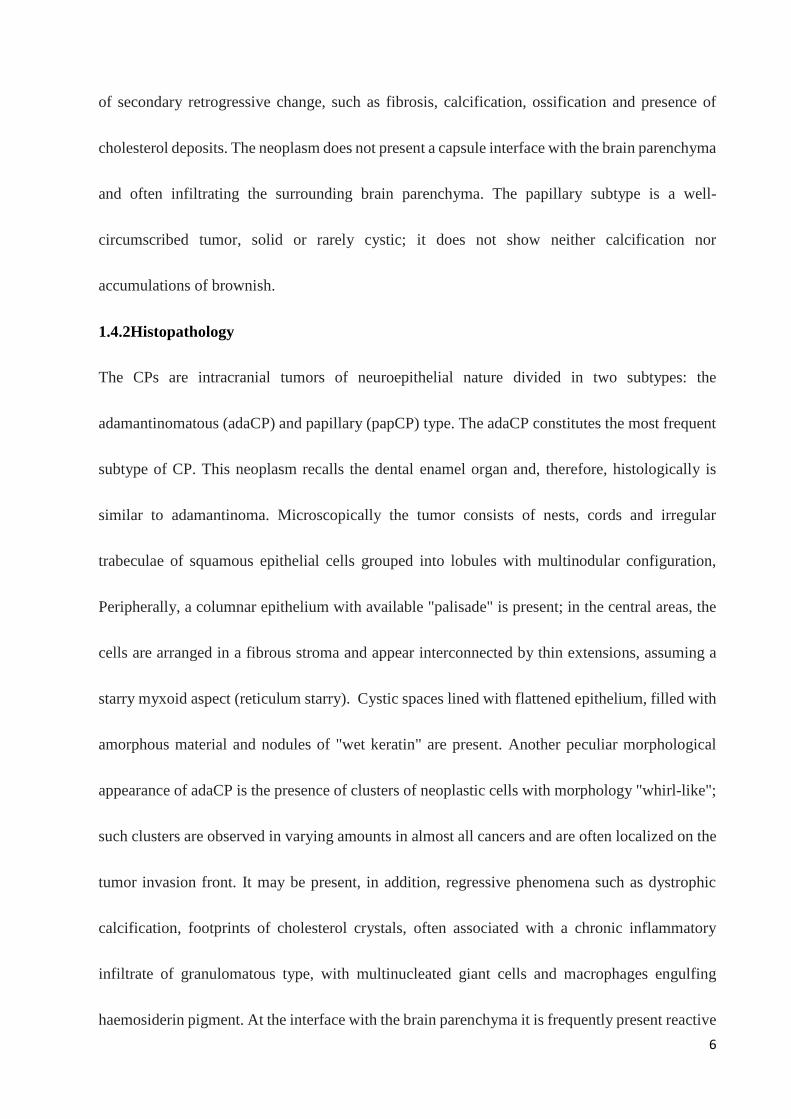
of secondary retrogressive change, such as fibrosis, calcification, ossification and presence of
cholesterol deposits. The neoplasm does not present a capsule interface with the brain parenchyma
and often infiltrating the surrounding brain parenchyma. The papillary subtype is a well-
circumscribed tumor, solid or rarely cystic; it does not show neither calcification nor
accumulations of brownish.
1.4.2Histopathology
The CPs are intracranial tumors of neuroepithelial nature divided in two subtypes: the
adamantinomatous (adaCP) and papillary (papCP) type. The adaCP constitutes the most frequent
subtype of CP. This neoplasm recalls the dental enamel organ and, therefore, histologically is
similar to adamantinoma. Microscopically the tumor consists of nests, cords and irregular
trabeculae of squamous epithelial cells grouped into lobules with multinodular configuration,
Peripherally, a columnar epithelium with available "palisade" is present; in the central areas, the
cells are arranged in a fibrous stroma and appear interconnected by thin extensions, assuming a
starry myxoid aspect (reticulum starry). Cystic spaces lined with flattened epithelium, filled with
amorphous material and nodules of "wet keratin" are present. Another peculiar morphological
appearance of adaCP is the presence of clusters of neoplastic cells with morphology "whirl-like";
such clusters are observed in varying amounts in almost all cancers and are often localized on the
tumor invasion front. It may be present, in addition, regressive phenomena such as dystrophic
calcification, footprints of cholesterol crystals, often associated with a chronic inflammatory
infiltrate of granulomatous type, with multinucleated giant cells and macrophages engulfing
haemosiderin pigment. At the interface with the brain parenchyma it is frequently present reactive

7
gliosis, with abundant fibers of Rosenthal (1, 3) (figures 1-2). The papCP is rarer than adaCP.
Histologically it is characterized by a monomorphic proliferation of well differentiated squamous
epithelial cells without maturation surface, often arranged in buds or lax pseudopapille around a
fibrovascular core, sometimes with a chronic inflammatory infiltrate of varying amounts. Unlike
the adaCP, are not observed images of "wet keratin", footprints of cholesterol crystals nor
calcifications (1, 3) (figures 3-4).
1.4.3 Ancillary techniques
The histological features of these tumors are quite characteristic and, therefore, ancillary methods
are not required for the purposes of diagnosis. However, CPs are characterized by the expression
of low and high molecular weight cytokeratins (1). In particular, the CPs show positivity for
cytokeratin 7 and cytokeratin 8 and negativity for cytokeratin 20; this immunophenotypic profile
is useful for the differential diagnosis of CP with Rathke cleft cysts.
Electron microscopy shows typical ultrastructural features of keratinizing epithelial cells, namely
the presence of numerous tonofilamenti, solid intercellular junctions and the absence of secretory
granules (1, 3, 18).
1.5 Differential diagnosis
The CPs with extensive cystic component can create problems of differential diagnosis with other
cystic lesions typical of the sellar region, in particular with epidermoid cysts and those of Rathke's

8
pouch. Epidermoid cysts are unilocular, bordered by a stratified squamous epithelium of uniform
thickness and rarely calcified. Rathke cleft cysts instead are bordered by a ciliated cubic or
columnar epithelium with occasional "goblet cells", and focal areas of squamous metaplasia. In
these cases, immunohistochemistry can aid in the differential diagnosis since the CP does not
express cytokeratin 8 nor cytokeratin 20, while both the cysts Rathke cleft that the normal cells
of the pars intermedia of the pituitary express both markers (1, 19).
It must be remembered, finally, as the infiltrative nature of CP often causes a reactive gliosis in
the surrounding brain parenchyma, where it is particularly exuberant, it should not be confused
with a pilocytic astrocytoma.
1.6 Therapy
The complete surgical excision of adaCP is the first choice of treatment, however, it is quite
difficult to practice, since it is associated with a high morbidity rate caused by endocrine
dysfunction and severe neurological deficits (20). For these reasons a less aggressive surgical
method is preferred, followed by radiotherapy on residual tumor (21). The rate of postoperative
recurrence is high and amounts to 23% and 27% respectively as a result of total resection or
subtotal resection associated with adjuvant radiotherapy, in a post-operative period of five years
(22, 23).
Currently, there is no consensus and there are no guidelines for the treatment of CPs. Surgical
removal of the CPs is performed through different approaches microsurgical. The choice of
approach depends substantially on the site of the lesion (24, 25). The evolution of surgical
9
techniques has led, in recent decades, to a net reduction in mortality and an increase in the
effectiveness of the intervention. The early mortality is, in fact, equal to 2.6% after transphenoidal
surgery and to 3.1% after transcranial surgery (26). In recurrent tumors the total removal could be
obtained in a percentage of cases lower than in primary tumors (0-25%) and is associated with
increased mortality and perioperative morbidity (10.5-24%) (27).
For cases in which no total removal is practicable (for example neoplasms of dimensions greater
than 4-5 cm or recurrent tumors), the standard approach consists in subtotal removal followed by
a traditional adjuvant radiotherapy (28).
The stereotactic radiotherapy, both in the form of radiosurgery (RS) that of fractionated
stereotactic radiotherapy (FSrt), constitutes a more accurate irradiation technique that allows a
more precise localization of the tumor with consequent reduction of the normal brain parenchyma
volume exposed to high doses of radiation therapy (29-31).
Currently there is no effective drug therapy for CP; the only treatment is surgery and / or
radiotherapy.
1.7 Prognosis
More than 85% of patients with CP survives after three years of diagnosis; mortality appears
therefore usually contained, when the tumor is treated in a appropriate time. The overall survival
rates, mainly described in pediatric series, reflecting the effects of various treatments and vary
from 83 to 96% at 5 years and 65 to 100% at 10 years, with an average of 62% at 20 years (2 ).
In adult or mixed population (adult-children) the survival rate is 54-96% at 5 years and 40 to 93%
10
at 10 years (2), with a recurrence-free survival of 60-93% at 10 years (3). The lower limits of
survival rates depend on data derived from older studies, ie before modern advances in
neurosurgical techniques, neuroimaging and radiotherapy.
The late mortality can be caused by the progressive disease with multiple recurrences, chronic
hypothalamic failure, hormone deficiency, cardiovascular diseases, convulsions (2).
The CP frequently involve a poor quality of life of affected patients, and because of the many
long-term sequelae associated with treatments (surgical and / or radiotherapy), are often
considered by clinicians as a chronic disease.
Finally, the risk of secondary cancer, as result of radiotherapy, is very low, with only a few cases
reported in the literature, including two cases of glioblastoma, a case of glioma (grade malignancy
unspecified) and a case of meningioma of the posterior cranial fossa (28, 32). The CP malignant
transformation is very rare and is characterized by metastatic dissemination via leptomeningeal
(33-35). The malignant component may show morphological features of a squamous cell
carcinoma, myoepithelial carcinoma or odontogenic ghost cell carcinoma (1).
At present there is still no consensus about the factors that may influence the biological behavior
of CP. The only established prognostic factor is the size of the tumor, with the highest survival
rates found in the size of tumors less than 3 cm (2, 36), because, as already mentioned, the
appearance of recurrence is closely related to the radicality of surgery, not practicable for the
lesions of large dimensions, with a very poor prognosis for tumors larger than 5 cm (3).
As regards the histopathological aspects, the papillary subtype is usually associated with a lower
tendency to recurrence and to a better prognosis than the adamantinomatous subtype or to mixed

11
forms (2). Furthermore, a morphological aspect significantly associated with tumor recurrence, is
constituted by the presence of clusters of neoplastic cells with morphology "whirl-like", often
present on the tumor invasion front. Even a marked reactive gliosis in the brain parenchyma
perilesional, according to some authors (36, 37) is associated with relapses.
The proliferation index (Ki-67 / MIB-1) appears to be associated with relapses, however, it is
extremely variable from case to case and has not yet been established a cut-off value (3); some
authors have described a case of CP clinically very aggressive and characterized by rapid onset
of relapse, with values of 20% and 15% respectively in the primary tumor and in the relapsed case
(36).
1.8 Pathogenesis of CP
1.8.1 Pathogenetic theories
The CP is a intracranial tumor derived from the trasformation of embryonic tissue. There are
several hypotheses about what is the tissue of origin: residues ectodermal of Rathke cleft or
embryonic epithelial residues of adenohypophysis.
The exact etiopathogenesis of these tumors is still unclear and highly controversial, however,
several genetic and molecular aspects seem to indicate a distinct pathogenesis in two subtypes.
There are, therefore, two main etiopathogenetic theories: embryonal and metaplastic (2):
- Embryonic theory: according to this theory the adaCP derives from neoplastic transformation
of squamous nests of embryonic cells left over by involution of craniopharyngeal duct. During
the process of proliferation and rotation of Rathke's pouch cells leading to the formation of the

12
adenohypophysis, the residual cells of craniopharyngeal duct are scattered in the saddle and
suprasellar region that are, in fact, the most frequent sites of CP’s onset (2, 38-40).
- Metaplastic theory: according to this theory the papCP would result from metaplasia of pituitary
cells of pars tuberalis resulting in the formation of squamous cell nests (2). This hypothesis is
supported by the finding of metaplastic squamous nest in the pituitary gland (the number of which
increases with age) and the presence of hormones in these nests. It been described a rare case of
papCP whose features suggested a possible origin from the processing of residues of a cyst of
Rathke's pouch (38).
Therefore, according to these theories, while the papCP would originate from the metaplastic
transformation of mature epithelial cells of the anterior pituitary, the adaCP derives from less
differentiated cells of Rathke's pouch, left over along the course of craniopharyngeal duct. In this
process plays a fundamental role the Wnt pathway / beta-catenin (canonical WNT pathway) (23).
1.8.2 Wnt \ beta-catenin pathway
The pathway Wnt / beta-catenin is one of the main regulators of multiple cellular processes both
in the embryo and in adults. This pathway, at the pituitary level, is involved in normal
morphogenesis and tissue differentiation and, if activated abnormally, in cancerogenesis
processes (41-43).
The Wnt proteins (Wingless Type) family consists of twenty members that act by binding to
Frizzled receptors (Fz) which present a extracellular domain N-terminal rich in cysteine that links

13
the Wnt protein, seven transmembrane helices and a short cytoplasmic tail carboxy-terminal . For
the activation of the system it is also required the presence of the co-receptor LRP (LDL-related
protein) and the formation of the ternary complex (Wnt / Fz / LRP) (44).
The activity of the Wnt pathway / beta-catenin is controlled by the stability of beta-catenin, a
transcriptional activator missing a binding domain with the DNA. In the absence of Wnt ligands,
the beta-catenin is phosphorylated at specific amino acids residues encoded by exon 3 of
CTNNB1 gene as part of a destruction complex consisting axina, complex APC (Adenomatous
Polyposis Coli) and GSK-3β (Glycogen synthase Kinase-3β) with a proteasome degradation
through pathway of ubiquitination. This prevents its stabilization and, consequently, the
activation of the Wnt pathway. The binding of Wnt proteins to their receptors Frizzled (FZL) and
co-receptor LRP cause the degradation of the destruction complex through the activation of
Dishevelled (DVL), cytoplasmic protein scaffold, thereby preventing the phosphorylation of beta-
catenin and consequently, its degradation. The stabilization of beta-catenin causes an increase of
the cytoplasmic levels of the protein and its translocation to the nucleus where it can interact with
the DNA binding transcription factors belonging to the TCF \ LEF family, thereby activating the
expression of the target genes of the pathway ( 45). Some of the activated genes are important for
cell cycle progression, such as c-myc and cyclin D1, others are inhibitors of apoptosis, such as
survivin, or-promoting genes tumor progression, such as laminin-γ2, and MMP7 (matrix
metalloproteinase-7), that encode for proteins involved in the degradation of the basement
membrane and extracellular matrix.

14
When the Wnt system is not stimulated, the transcription factors TCF / LEF remain associated
with transcriptional co-repressors Gro (Groucho) and CtBP (C-Terminal Binding Protein),
forming an inactive complex (46).
CTNNB1 mutations of the gene encoding the beta-catenin in exon 3 involve deletions or
substitutions of amino acid residues critical for its degradation by the destruction of the complex
causing, therefore, an increase in cytoplasmic levels of the protein, its expression aberrant nuclear
and, consequently, a constitutive activation of the Wnt pathway (40-45).
Studies in mice genetically modified to obtain a mutated form of beta-catenin (transcriptionally
active, but devoid of the amino acid residue encoded by exon 3 of CTNNB1 gene) have shown
that the Wnt pathway /beta-catenin is required for the terminal differentiation of the pituitary cells
in the last stages of development and should be inhibited in the early stages to allow the normal
proliferation of the progenitors of Rathke's pouch. Un upregulation of the Wnt pathway / beta-
catenin in progenitor Rathke cleft causes, in fact, pituitary hyperplasia, ectopic pituitary tissue in
the oropharyngeal cavity and perinatal death in 80% of mice; the remaining 20% survive (an
average of 11 weeks after birth) but with serious defects of growth and pituitary tumors showing
histopathological aspects similar to those of adaCP and nuclear expression of beta-catenin and
Axina2, LEF1 and BMP4, indicating the activation of the Wnt pathway (41, 47-50).
Although murine tumors differ from human ones for some aspects (absence of wet keratin or
calcifications; faster postnatal onset), suggesting the existence of species-specific differences,
mutations of the human beta-catenin are functionally equivalent to those of the models murine.

15
Mutations of the gene CTNNB1 therefore constitute the molecular hallmark of adaCP. These
mutations were observed in the majority of human adaCP (about 70% of cases), predominantly
affecting the exon 3 of the gene and, as already explained above, lead to the loss or replacement
of critical amino acid residues for the stabilization of the protein thus blocking its degradation,
nucleus-cytoplasmic accumulation (ie aberrant nuclear expression) and activation of the Wnt
pathway (51-53). However, nuclear accumulation of β-catenin is observed in more than 94% of
adaCP, suggesting that additional mechanisms, not yet known, probably of epigenetic nature, may
be involved in the stabilization of the protein (23). Furthermore, although the CTNNB1 mutation
is ubiquitous, the nuclear expression of β-catenin is observed only in small clusters of neoplastic
cells; these clusters often show a "whirl-like" morphology and are exclusively observed in adaCP,
never in papCP or other tumors of the sellar region (54, 55). Neoplastic cells of the cluster do not
express markers of proliferation (Ki-67), pituitary hormones and are negative for synaptophysin.
1.8.3 Role of stem cells in the pathogenesis of CP
The presence of tissue specific stem cells has been well documented in many organs and tissues
including, brain, gastro-intestinal tract, skin, bone marrow, and skeletal muscle. These cells are
characterized by self-regeneration capacity ( "self renewal"), by the expression of stem cell
markers (such as SOX2) and can give rise to a progeny of cells able to differentiate and to
contribute both to homeostasis, both to tissue regeneration (41, 56, 57). The presence of stem cells
in the pituitary, although long hypothesized, was only recently demonstrated in murine models

16
and in humans. The pituitary stem cells express the transcription factors SOX9 and SOX2 (the
latter only in the mouse), can differentiate into all pituitary cell lines hormone-producing and are
crucial for homeostasis and regeneration of the gland (58-59). Tissue-specific stem cells can give
rise to tumors, a concept that has given rise to the "hierarchical model" of cancer stem cell (CSC)
(60). The term CST defines a neoplastic cell capable of self-regenerate, that is to give rise to
another cell identical to itself (with tumorigenic capacity) and to generate a more differentiated
neoplastic cell population (devoid of tumorigenic capacity). Therefore tumor is present in a
minority population of CST responsible of tumor growth, maintenance of the compartment
neoplastic stem and its progeny, non-tumorigenic, which constitutes the bulk of the tumor but is
lacking in those characteristics of the CST. It is apparent how the staminal model considers the
tumor not as a homogeneous population of cells rather as a heterogeneous proliferation
characterized by a sort of "hierarchy" where the CST, though few, are the basic component of the
development and maintenance of the neoplasm.
Another feature of the CSC is constituted by the resistance to chemotherapy and radiotherapy;
this aspect may be responsible for tumor progression and relapse (61, 62).
The CSC may, therefore, represent potential targets for new therapies in oncology.
Recent studies, both mouse models and in human tumors, have investigated the presence of CSC
in pituitary tumors, especially in adaCP (23, 49, 54, 58). The results of these studies suggest that
the adaCP presented a minority population of cells with stem cell phenotype. These clusters are
17
characterized by the expression of aberrant nuclear beta-catenin and do not express the Ki-67, so
they are in a non-proliferating status.
1.8.4 Molecular alterations of papillary craniopharyngioma
The molecular markers of papCP consists of the BRAF V600E mutation, found in a higher
percentage of cases (95%), with a specificity of 100%, with activation of the MAP kinase
pathway.
The CTNNB1 mutations and BRAF V600E in the two subtypes of CP are both clonal and
mutually exclusive. These findings may have important implications in the diagnosis and
treatment of these malignancies (63).
1.8.5 Role of ataxia-telangiectasia-mutated (ATM) protein Kinase
ATM is a known tumour suppressor which is frequently mutated in a broad range of human
cancers including lung, colorectal, breast and haematopoietic cancers. ATM is a kinase activated
by autophosphorylation (p-ATM) upon DNA double-strand breaks arising from errors during
replication, byproducts of metabolism, chemotherapy or ionizing radiations. The initial scientific
interest in ATM was, however, not focused on its role in tumour development. ATM was first
described in 1995 as the gene defective in the autosomal recessive human hereditary disorder
ataxia–telangiectasia (A–T). This disease, caused by a loss of ATM function, is characterized by
progressive cerebellar degeneration, telangiectasia, immunodeficiency, genomic instability

18
cancer susceptibility and profound sensitivity to ionising radiation (IR); this hypersensitivity of
ATM-defective cells to IR represents the main interest for ATM as therapeutic target for cancer
therapy (64-65).

19
II. Scope of Work
Based on the above, the objectives of our work were as follows:
• To deepen the study of the pathobiology of the CP, in particular the role of β-catenin and stem
cell;
• To study possible factors associated with the tendency to relapse, specifically in
adamantinomatous subtype;
• To identify the prognostic and predictive markers that can be used by immunohistochemistry
method.
To achieve these objectives we have therefore decided to:
• Evaluate, using immunohistochemical method, on a selected group of CPs:
1) the expression of β-catenin;
2) the proliferation index (Ki-67);
3) the expression of stem cell markers: the CD166 (ALCAM), closely related to many types of
tumors, with different prognostic outcomes (64); CD133 (Prominin 1), whose expression is
associated with a poor response to radiotherapy (23);
4) the expression of pATM
To correlate the results obtained with the clinical aspects of neoplasms, or the presence or
absence of tumor recurrence.

20
III. Materials and methods
3.1 Case Study
45 samples of craniopharyngiomas were analyzed (table 1). They were all obtained as formalin
fixed tissue from the archive of the Department of Anatomic Pathology of the Federico II
University Hospital of Naples. Patients underwent surgery at the Department of Neurosurgery of
the same hospital. The cases were identified between 1998 and 2015 and the diagnoses were made
in accordance with the World Health Organization (WHO) classification. Only specimens
containing sufficient amounts of the respective CP subtype were taken into account.
For each patient follow-up data were available. Written informed consent was obtained from all
patients.
Of 45 patients 30 were males and 15 were females. Age ranged from 2 to 77 years. Patients had
been treated by complete surgical removal in 23 cases or by subtotal surgery followed by
radiotherapy in 22 cases. In 38 cases the diagnosis was of adamantinomatous chraniopharyngioma
(adaCP) and in 7 cases it was of papillary subtype (papCP). Of 4 recurrent cases only the primary
tumor tissue was included for statistical evaluation; in 9 cases only the tissue of the recurrence
was available (8 adaCP and 1 papCP). Therefore 13 cases were considered recurrent and 32 not
recurrent.
3.2 Immunohistochemistry
4 μm sections were used for immunohistochemistry. Sections were dewaxed in xylene, hydrated
in graded series of alcohol and subjected to heat-induced antigen retrieval. After blocking

21
endogenous peroxidase activity, the tissue was incubated with monoclonal antibodies for anti β-
catenin (clone 14, mouse; Roche Ventana, 1:100 dilution), CD166 (clone MOG/07, ab49496,
mouse; Abcam, 1:100 dilution), p-ATM (phosphoS1981)(clone ab81292;abcam; 1: 200 dilution)
and Ki67 (Clone MIB1, mouse; Dako, 1:100 dilution) and with the polyclonal antibody CD133
(rabbit; Abcam, 1:100 dilution), all for 90 minutes. Subsequently, the slices were rinsed and
incubated with the biotinylated secondary antibody, at room temperature, for 30 minutes. The
bound antibody complexes were stained for 3-5 minutes or until appropriate for microscopic
examination with diaminobenzidine, and they were then counterstained with hematoxylin (30s)
and mounted. Appropriate positive controls were chosen: mammary fibromatosis for β-catenin,
normal skin for CD166 and CD133 and normal pancreas for p-ATM. Negative control was
obtained by omitting the primary antibody.
3.3 Scoring
All the slices were reviewed using light microscopy.
β-catenin was considered as positive only in case of nuclear staining and a total immunostaining
score (IS) was calculated as the product of a Proportion score (0=0%, 1<10 %, 2=10-50%, 3=51-
80%, 4>80%) and an Intensity score (0= no signal, 1= weak signal, 2= moderate, 3= strong). The
same IS was used also for CD133 and CD166 membrane signal. The 3 markers were then divided
into two scoring groups, on the base of the IS: absent/low expression (IS=0-2) and moderate/high
expression (IS=3-12).

22
For pATM, a different scoring system was adopted because no differences in intensity were
noticed. It was based on the proportion of neoplastic cells showing a signal and on its cellular
localization (C, cytoplasmic or N, nuclear): 0=absent, 1=<10%, 2=10-30%, 3>30%.
For the evaluation of the proliferating index Ki67 (Labeling Index, L.I.), "hot spot" areas were
chosen and an average of the values on 5 adjacent fields (at least 500 neoplastic cells) was
calculated: normally highly proliferating areas were excluded, namely basal cells. A cut-off of
5% was identified because the median value was of 6%.
In some cases not all the markers could be tested because of tissue exhaustion.
3.4 Statistical evaluation
The study of association between the scoring group of each marker (absent/low or moderate/high
expression for β-catenin, CD133 and CD166 and ≤5% or >5% for Ki67) and clinico-pathological
features (age, gender, recurrence and histotype) was carried out by Fisher’s exact test. For pATM
the population was divided into positive and negative cases.
Spearman correlation test was used to examine the correlation between β-catenin and,
respectively, CD133, CD166 and Ki67 and between CD166 and Ki67.
A p value≤ 0.05 was considered statistically significant. All tests were two sided and carried out
with GraphPad Prism 5 software (GraphPad Software, La Jolla, CA, USA).

23
IV Results
4.1 Immunohistochemical expression of β-catenin
β-catenin immunoreactivity was detected at the cell membrane in most cases and in the nucleus
in 28 cases. We focused on the nuclear signal that was located in neoplastic cells forming "whirl-
like" structures and in palisading basal cells arranged around the stellate reticulum. The signal
was variable in intensity and proportion of distribution.
β-catenin immunostaining score was negative in 17 cases and in positive cases it ranged from 1
to 9, being 1, 2, 3, 4, 6 and 9 in 4, 11, 2, 7, 3 and 1 cases, respectively. Based on the calculated
IS, all specimens were subsequently divided into two different scoring groups: 32 were absent/low
(IS=0-2) and 13 were moderate/high (IS>2) (figure 5-6).
Out of 32 cases with absent/low β-catenin IS, 5 were recurrent while out of 13 with moderate/high
β-catenin IS 8 were recurrent (table 1).
4.2 Immunohistochemical expression of Ki67
Ki67 immunostaining was available in 41 cases and it showed nuclear signal with a range of
values between 1 and 25 %, a mean value of 8% and a median of 6% (table 1).
4.3 Immunohistochemical expression of CD166
CD166 was evaluated on 31 cases. The IS was negative in 7 cases and positive in 24 cases,
ranging from 1 to 6 and being respectively 1, 2, 3, 4 and 6 in 5, 5, 4, 8 and 2 cases. 17 belonged
to the absent/low (IS=0-2) scoring group while 14 to the moderate/high (IS>2) (figure 7-8).

24
The signal was mainly located at the cell membrane with diffuse distribution but sparing the whirl-
like cell clusters that showed nuclear β-catenin accumulations in adaCPs. In papCPs the
immunostaining was more homogeneous.
Out of 13 recurrent cases 3 showed a CD166-IS≤ 2, while in 10 cases the immunoscore was>2
(table 1).
4.4 Immunohistochemical expression of CD133
CD133 displayed membrane immunostaining with variable distribution in both adaCPs and
papCPs and it did not localize exclusively in cell clusters, as already described in literature (table
1) (figure 9).
4.5 Immunohistochemical expression of pATM
27 cases out of 45 showed nuclear expression of pATM: 9, 11 and 7 cases with 1, 2 and 3 IS
respectively. All papCPs showed nuclear reactivity (7/7).
In 23 out of 45 cases a cytoplasmic reactivity was detected (9, 10 and 4 cases showed 1, 2 and 3
IS respectively). Only 1/7 papCP and 16/28 adaCPs were positive (table 1).
4.6 Statistic analysis
The Fisher exact test showed a statistically significant correlation between the onset of relapsed
tumor and:
• A moderate\high level of immunoreactivity of the beta-catenin (p = 0.0039);

25
• A moderate\high level of immunoreactivity of CD166 (p = 0.0040);
• High proliferation index (Ki-67) (p = 0.0385).
We have not found a statistically significant correlation between the expression of CD133 and the
presence of recurrence (p = 0.5962) (table 2a).
With the Spearman correlation test, we found a statistically significant correlation between the
expression of:
• β-catenin and Ki-67 (p=0.0011, r=0.4903);
• β-catenin and CD166 (p=0.0002, r=0.6218).
. CD66 and Ki67 (p=0.0054, r=0.5119);
We have not found a statistically significant correlation between β-catenin and CD133 (p=0.4156,
r=0.1543).
We compared both pATM expressions, nuclear and cytoplasmic, in both CP subtypes, by Fisher's
exact test and found out that NpATM was significantly more expressed in papCPs (p=0.0313),
while CpATM was more present in adaCPs(p=0.0470). No differences were found out when we
analyzed the different scoring groups (IS1, 2 or 3).
Afterwards, we analyzed the probability of recurrence among papCPs, on the base of NpATM,
and among adaCP, on the base of CpATM. No statistically significant correlation was observed
(table 2b).

26
V Discussion
The CPs are benign epithelial tumors of the sellar region that arise from embryonic remnants of
Rathke's pouch. They are rare neoplasms (incidence of 0.5 - 2.5 per 100,000 persons / year), most
frequently in children representing, in this age group, 5-10% of intracranial tumors and the most
frequent pituitary tumors (3). There are two subtypes, the adaCP and the papCP, which differ for
histological and etiopathogenetic features, for the biological behavior and clinical outcome. The
papCP is a fairly uncommon cancer, occurs almost exclusively in adults and has a favorable
prognosis. The adaCP, the most common subtype, shows two peaks of onset, the first in children
(5-15 years) and second in adults (45-60 years) and despite being a histologically benign tumors
(grade I sec. wHO), shows a locally aggressive behavior that is manifested by a tendency to
infiltrate the surrounding brain and vital neurovascular structures (3). There are no guidelines to
treat these tumors and the choise is essentially based on two main therapeutic options, surgery
and / or radiation therapy. The goal is to remove as much tumor as possible without impairment
of the patients. However, despite the therapeutic efforts, the adaCP shows a tendency to recur
frequently, whether as a result of the total removal or subtotal removal associated with
radiotherapy (23% and 27%, respectively, in a post-operative period of five years) (22, 23). In
recurrent cases the total removal is achievable in a percentage of cases considerably lower
compared to primary tumors (0-25%) and is associated with increased mortality and morbidity
(27). Therefore, even if patients with CP show a prolonged survival (> 85% after three years of

27
diagnosis), often have a poor quality of life because of the direct effects of tumor growth and the
complications of the previous treatment.
For this reason it’s important to identify tumors with more aggressive biological behavior.
Currently we have no effective prognostic and predictive markers that can be used to discriminate
cases of neoplasms characterized by greater biological aggressiveness and hence tendency to
relapse. It is a topic debated, probably because the CPs are rare tumors whose pathogenesis is still
unclear; in fact, the role of the Wnt pathway / beta-catenin in the pathobiology of adaCP (40-54)
is well documented, whereas the emerging role of the stem cell is still controversial.
In this regard the most recent works (23, 49, 54, 58) suggest the presence of a minority of cells
with stem cell phenotype in adaCP. From these cells originate clusters of neoplastic cells that are
crucial for tumor growth and survival, by acting, probably, the production of a wide range of
growth factors, chemokines, cytokines and their receptors (54). These clusters are variables for
quantity, often show a "whorl-like" morphology, are in a state not proliferating (not express Ki-
67) and, phenotypically, are characterized by the expression of the aberrant nuclear beta-catenin.
The expression of stem cell markers in these clusters generated unclear and discordant data (49,
54). In the scientific literature beta-catenin and stem cell markers are widely studied in CP as
ethiopatogenetic factors but not for prognostic and predictive markers. The papers addressing the
topic are few and often quite inaccurate. For example, Li et al (66), in a retrospective study of
fifty CP, correlate the membrane expression of beta-catenin with a poor prognosis, defining the
pattern of expression as "aberrant", contrary to the data, now known, reporting that the aberrant

28
expression of the protein is within the nucleus; Cao et al (67) studied the immunohistochemical
expression of beta-catenin and p63 in a large enough group of sixty-six CP, comprising fifty-one
adaCP and fifteen papCP, showing that the nuclear accumulation of beta-catenin is a
morphological feature of the adamantinomatous subtype. Finally, in a more recent work, Esheba
et al (68) evaluated the immunohistochemical expression of beta-catenin in a retrospective study
of twenty CP, reporting that nuclear expression of aberrant beta-catenin is useful for the
differential diagnosis between the two histological types of CP. Also in this work the clinical data,
in particular those relating to the follow-up, are rather weak and shows no prognostic correlation.
In 2013 Prieto et al (36) described a case of adaCP characterized by the rapid onset of tumor
recurrence following surgery, performing a detailed review of the literature regarding the potential
predictive factors of tumor recurrence. In this work epidemiological (age and gender of the
patients) morphological, both macroscopic (tumor size, location, consistency, adhesion and
invasion of surrounding structures) and microscopic (histological type, presence of clusters with
"whirl-like" morphology, peri-tumoral reactive gliosis) and immunophenotypic (Ki-67 and p53)
features were evaluated. The conclusion of this paper is that there is no consensus on the factors
that may influence cancer recurrence, however, the only parameters that could suggest a closer
follow-up are the presence of a marked reactive gliosis, a high value of Ki-67 and increased
expression of p53.
Hölsken et al (23) evaluated the expression of stem cell markers (CD133 and CD44), both by
immunohistochemistry and by real time PCR, in a group of thirty-three CP, which include eight
papCP and twenty five adaCP; it was carried out, also, a correlation with the mutational status of

29
CTNNB1 and then with the aberrant nuclear expression of beta-catenin. The results obtained were
the prevalent expression of CD44 in adaCP and CD133 in papCP and the co-expression, in the
adaCP, of both stem cell markers in clusters of neoplastic cells characterized by the accumulation
of nuclear beta-catenin. The interesting aspects of this work are the following: (I) for the first
time was observed the expression of CD133 in human CP; it is well known that CD133 + cells
are less sensitive to radiotherapy, therefore the expression of such markers may be significantly
related to the onset of relapses in the CP; (II) it was demonstrated the expression of stem cell
markers in the cells cluster characterized by the accumulation of nuclear beta-catenin, suggesting
the presence of a "niche" of CSC involved in the pathogenesis of CP (69-70).
Based on the above, we decided to conduct a immunohistochemical study in a group of CPs, with
the aim to identify potential prognostic and predictive markers selecting cases more aggressive
and, therefore, at greater risk of recurrence.
We performed a retrospective study of 45 CP removed by endoscopic endonasal transphenoidal
approach. The cases were divided into groups based on clinical outcome (presence or absence of
tumor recurrence).
In 38 cases the diagnosis was of adaCP and in 7 cases it was of papCP. Of 4 recurrent cases only
the primary tumor tissue was included for statistical evaluation; in 9 cases only the tissue of the
recurrence was available (8 adaCP and 1 papCP). Therefore 13 cases were considered recurrent
tumors and 32 not.

30
In this study it was then performed an immunohistochemical evaluation of the expression of β-
catenin, the stem cell markers CD133 and CD166, pATM and rate of proliferation (Ki-67)
In our series, we observed that CD166 was expressed in CPs and it showed a membrane signal
whose immunoscore correlated with β-catenin, Ki67 and recurrence risk. It remains unclear
whether it behaves as a stem cell marker in CPs; though, it seems to spare the whirl-like cell
clusters in adaCPs, where there should be the stem cell niches. However, regardless of its role in
stemness, it can be of help, together with β-catenin, to predict recurrences in CPs. In our cohort
CD133 was expressed in both histotypes, while it was described to be almost detectable in papCPs
(11). The potential role of Ki67 as prognostic marker was confirmed. A cut-off of 5% was
established as a valid tool to effectively distinguish highly proliferative forms from those with a
low rate of proliferation. The Labeling Index was calculated on cells other than those of the basal
layer where a high proliferative activity was expected.
As well, pATM expression was assessed in the attempt of identifying potential factors that can
affect radiosensitivity in CPs. This kinase has a specific role in the DNA double-strand breaks
repair, that is exercised in the nucleus, and has a cytoplasmic function that is related to autophagy
(32).
In our series, we observed mainly a nuclear signal in papCPs and a cytoplasmic signal in adaCPs;
this allowed us to consider that pATM different expression patterns in CP subtypes might be used
as additional diagnostic and prognostic tool. No predictive role of recurrence was observed for
pATM, neither cytoplasmic nor nuclear.

31
The prevalent cytoplasmic localization of pATM in adaCPs may be the effect of a different
pathogenetic role of this kinase in the two histotypes. Further studies are needed to clear this
aspect.

32
VI Conclusions
Finally, we suggest to use a panel of immunohistochemical markers including beta-catenin, Ki-
67 and CD166, to identify ab initio cases of CPs with poor prognostic outcome, and therefore at
greater risk of recurrence. To evaluate the expression of beta-catenin and CD166 may be useful
to apply a score of immunoreactivity considering the intensity and the proportion of positive
tumor cells; for the Ki-67 we suggest a cut-off value of 5% for distinguishing the low neoplasms
from those with a high proliferative rate.
The identification of these markers is mandatory in order to draw prognostic evaluation of CPs
(biological and clinical behavior) also in regards of recurrence risk of these lesions, therefore
defining a more appropriate treatment strategy, with post-operative additional treatments.

33
VII. Bibliography
1. Asa S L. Tumors of the Pituitary Gland. AFIP Atlas of Tumor Pathology, Third Series 2011 (1st
ed).
2. Müller HL. Craniopharyngioma. Endocr Rev. 2014;35(3):513-43. 3. Louis DN, Ohgaki H, Wistler OD, Cavenee WK. WHO classification of tumours of the central
nervous system. Lyon: IARC; 2007. 4. Kolen ER, Horvai A, Perry V, Gupta N. Congenital craniopharyngioma: a role for imaging in
the prenatal diagnosis and treat- ment of an uncommontumor. Fetal Diagn Ther. 2003;18:270-274.
5. Scagliotti V, Avagliano L , Gualtieri A, Graziola F, Doi P, Chalker J, Righini A, Korbonits M, Bulfamante G, Jacques TS, Massa V, Gaston-Massuet C. Histopathology and molecular characterisation of intrauterine-diagnosed congenital craniopharyngioma. Pituitary. 2016;19:50–56.
6. Harwood-Nash DC. Neuroimaging of childhood craniopharyngioma. Pediatr Neurosurg. 1994;21 Suppl 1:2-10.
7. Karavitaki N, B.C., Warner JT, Adams CBT, Richards P, Ansorge O, Shine B. Craniopharyngiomas in children and adults: systematic analysis of 121 cases with long-term follow-up. Clin Endocrinol. 2005;62(4):397-409.
8. Lewin R, Ruffolo E, Saraceno C. Craniopharyngioma arising in the pharyngeal hypophysis. South Med J 1984;77:1519-1523.
9. Koral K, Weprin B, Rollins NK. Sphenoid sinus craniopharyngioma simulating mucocele. Acta Radiol. 2006;47: 494-496.
10. Yasargil MG, Curcic M, Kis M, Siegenthaler G, Teddy PJ, Roth P. Total removal of craniopharyngiomas. Approaches and long-term results in 144 patients. J Neurosurg. 1990;73:3-11.
11. Cusimano MD, Kovacs K, Bilbao JM, Tucker WS, Singer W. Suprasellar craniopharyngioma associated with hyperprolactinemia, pituitary lactotroph hyperplasia, and microprolactinoma. Case report. J Neurosurg. 1988;69(4):620-3.
12. Wheatley T, Clark JD, Stewart S. Craniopharyngioma with hyperprolactinaemia due to a prolactinoma. J Neurol Neurosurg Psychiatry. 1986;49(11):1305-7.
13. Yoshida A, Sen C, Asa SL, Rosenblum MK. Composite pituitary adenoma and craniopharyngioma? An unusual sellar neoplasm with divergent differentiation. Am J Surg Pathol. 2008;32(11):1736-41.
14. Puchner MJ, Lüdecke DK, Saeger W. The anterior pituitary lobe in patients with cystic craniopharyngiomas: three cases of associated lymphocytic hypophysitis. Acta Neurochir. 1994;126(1):38-43.
15. Liu A, Wang JM, Li GL, Sun YL, Sun SB, Luo B, Wang MH. Clinical and pathological analysis of benign brain tumors resected after Gamma Knife surgery. J Neurosurg. 2014;121 Suppl:179-87.
16. Mortini P, Gagliardi F, Bailo M, Spina A, Parlangeli A, Falini A, Losa M. Magnetic resonance imaging as predictor of functional outcome in craniopharyngiomas. Endocrine. 2016;51(1):148-62.
17. Pusey E, K.K., Flannigan B, Tsuruda J, Bradley WG MR. MR of craniopharyngiomas: tumor delineation and characterization. AJR Am J Roentgenol. 1987;149(2):383-8.
18. Sato K, Kubota T. Fine structure of ossification in craniopharyngiomas. Ultrastruct Pathol. 1999;23:395-399.

34
19. Xin W, Rubin MA, McKeever PE. Differential expression of cytokeratins 8 and 20 distinguishes craniopharyngioma from rathke cleft cyst. Arch Pathol Lab Med. 2002;126(10):1174-8.
20. Clark AJ, Cage TA, Aranda D, Parsa AT, Auguste KI, Gupta N. Treatment-related morbidity and the management of pediatric craniopharyngioma: a systematic review. J Neurosurg Pediatr. 2012:10(4):293–301.
21. Hofmann BM, Hollig A, Strauss C, Buslei R, Buchfelder M, Fahlbusch R. Results after treatment of craniopharyngiomas: further experiences with 73 patients since 1997. J Neurosurg. 2012;116(2):373–384.
22. Clark AJ, Cage TA, Aranda D, Parsa AT, Sun PP, Auguste KI, Gupta N A systematic review of the results of surgery and radiotherapy on tumor control for pediatric craniopharyngioma. Child’s Nerv Syst ChNS Off J Int Soc Pediatr Neurosurg. 2013; 29(2):231–238.
23. Hölsken A, Stache C, Schlaffer SM, Flitsch J, Fahlbusch R, Buchfelder M, Buslei R. Adamantinomatous craniopharyngiomas express tumor stem cell markers in cells with activated Wnt signaling: further evidence for the existence of a tumor stem cell niche? Pituitary. 2014;17(6):546-56.
24. Cavallo LM, Solari D, Esposito F, Villa A, Minniti G, Cappabianca P. The role of the endoscopic endonasal route in the management of craniopharyngiomas. World Neurosurg. 2014;82(6 Suppl):S32-40.
25. Buchfelder M, Schlaffer SM, Lin F, Kleindienst A. Surgery for craniopharyngioma. Pituitary. 2013;16:18 –25.
26. Elliott RE, Jane JA Jr, Wisoff JH. Surgical management of craniopharyngiomas in children: meta-analysis and comparison of transcranial and transsphenoidal approaches. Neurosurgery. 2011;69(3):630-43;
27. Karavitaki N. Management of craniopharyngiomas. J Endocrinol Invest. 2014;37(3):219-28. 28. Combs SE, Thilmann C, Huber PE, Hoess A, Debus J, Schulz-Ertner D. Achievement of long-
term local control in patients with craniopharyngiomas using high precision stereotactic radiotherapy. Cancer. 2007;109:2308 -2314.
29. Kanesaka N, Mikami R, Nakayama H, Nogi S, Tajima Y, Nakajima N, Wada J, Miki T, Haraoka J, Okubo M, Sugahara S, Tokuuye K. Preliminary results of fractionated stereotactic radiotherapy after cyst drainage for craniopharyngioma in adults. Int J Radiat Oncol Biol Phys. 2012;82:1356 –1360.
30. Minniti G, Esposito V, Amichetti M, Enrici RM. The role of fractionated radiotherapy and radiosurgery in the management of patients with craniopharyngioma. Neurosurg Rev. 2009;32(2):125-32.
31. Winkfield KM, Linsenmeier C, Yock TI, Grant PE, Yeap BY, Butler WE, Tarbell NJ. Surveillance of craniopharyngioma cyst growth in children treated with proton radiotherapy. Int J Radiat Oncol Biol Phys. 2009;73:716 –721.
32. Regine WF, Mohiuddin M, Kramer S. Long-term results of pediatric and adult craniopharyngiomas treated with combined surgery and radiation. Radiother Oncol. 1993;27: 13–21.
33. Frangou EM, Tynan JR, Robinson CA, Ogieglo LM, Vitali AM. Metastatic craniopharyngioma: case report and literature review. Childs Nerv Syst. 2009;25(9):1143-7.
34. Gupta K, Kuhn MJ, Shevlin DW, Wacaser LE. Metastatic craniopharyngioma. AJNR Am J Neuroradiol. 1999;20(6):1059-60.
35. Petito CK, De Girolami U, Earle KM. Craniopharyngiomas: a clinical and pathological review. Cancer. 1976;37:1944 –1952.
36. Prieto R, Pascual JM, Subhi-Issa I, Jorquera M, Yus M, Martínez R. Predictive factors for

35
craniopharyngioma recurrence: a systematic review and illustrative case report of a rapid recurrence. World Neurosurg. 2013;79(5-6):733-49.
37. Sartoretti-Schefer S, Wichmann W, Aguzzi A, Valavanis A. MR differentiation of adamantinous and squamous-papillary craniopharyngiomas. AJNR Am J Neuroradiol. 1997;18:77– 87.
38. Sato K, Oka H, Utsuki S, Kondo K, Kurata A, Fujii K. Ciliated craniopharyngioma may arise from Rathke cleft cyst. Clin Neuropathol. 2006;25:25–28.
39. Powers CJ, New KC, McLendon RE, Friedman AH, Fuchs HE. Cerebellopontine angle craniopharyngioma: case re- port and literature review. Pediatr Neurosurg. 2007;43: 158 –163.
40. Martinez-Barbera JP. Invited review: Molecular and cellular pathogenesis of adamantinomatous craniopharyngioma. Neuropathol Appl Neurobiol. 2015;41(6):721-32.
41. Clevers H, Nusse R. Wnt/β -catenin signaling and disease. Cell. 2012;149: 1192–205. 42. Huelsken J, Birchmeier W. New aspects of Wnt signaling pathways in higher vertebrates. Curr
Opin Genet Dev. 2001;11: 547–53. 43. Bejsovec A. Wnt signaling: an embarrassment of receptors. Curr Biol. 2000;14-
28;10(24):R919-22 44. Buslei R, Nolde M, Hofmann B, Meissner S, Eyupoglu IY, Siebzehnrübl F, Hahnen E, Kreutzer
J, Fahlbusch R. Common mutations of beta-catenin in adamantinomatous craniopharyngiomas but not in other tumours originating from the sellar region. Acta Neuropathol. 2005;109(6):589-97.
45. Städeli R, Hoffmans R, Basler K. Transcription under the control of nuclear Arm/beta-catenin. Curr Biol. 2006;16(10):R378-85.
46. Brinkmeier ML, Potok MA, Cha KB, Gridley T, Stifani S, Meeldijk J, Clevers H, Camper SA. TCF and Groucho- related genes influence pituitary growth and development. Mol Endocrinol. 2003;17:2152–61.
47. Zhang Z, Florez S, Gutierrez-Hartmann A, Martin JF, Amendt BA. MicroRNAs regulate pituitary development, and microRNA 26b specifically targets lymphoid enhancer factor 1 (Lef-1), which modulates pituitary transcription factor 1 (Pit-1) expression. J Biol Chem. 2010;285:34718–28.
48. Harada N, Tamai Y, Ishikawa T, Sauer B, Takaku K, Oshima M, Taketo MM, Miyoshi H, Murai N, Oshima H. Intestinal polyposis in mice with a dominant stable mutation of the beta-catenin gene. EMBO J. 1999; 18:5931–42.
49. Gaston-Massuet C, Andoniadou CL, Signore M, Jayakody SA, Charolidi N, Kyeyune R, Vernay B, Jacques TS, Taketo MM, Le Tissier P, Dattani MT, Martinez-Barbera JP. Increased Wingless (Wnt) signaling in pituitary progenitor/stem cells gives rise to pituitary tumors in mice and humans. Proc Natl Acad Sci. 2011;108: 11482–7.
50. Sekine S, Shibata T, Kokubu A, Morishita Y, Noguchi M, Nakanishi Y, Sakamoto M, Hirohashi S. Craniopharyngiomas of adamantinomatous type harbor beta-catenin gene mutations. Am J Pathol. 2002;161: 1997–2001.
51. Buslei R, Nolde M, Hofmann B, Meissner S, Eyupoglu IY, Siebzehnrubl F, Hahnen E, Kreutzer J, Fahlbusch R. Common mutations of beta-catenin in adamantinomatous craniopharyngiomas but not in other tumours originating from the sellar region. Acta Neuropathol. 2005;109:589–97.
52. Oikonomou E, Barreto DC, Soares B, De Marco L, Buchfelder M, Adams EF. Beta-catenin mutations in craniopharyngiomas and pituitary adenomas. J Neurooncol. 2005;73:205–9.
53. Andoniadou CL, Gaston-Massuet C, Reddy R, Schneider RP, Blasco MA, Le Tissier P, Jacques TS, Pevny LH, Dattani MT, Martinez-Barbera JP. Identification of novel pathways involved in
36
the pathogenesis of human adamantinomatous craniopharyngioma. Acta Neuropathol. 2012;124:259–271.
54. Larkin SJ, Ansorge O. Pathology and pathogenesis of craniopharyngiomas. Pituitary. 2013;16:9–17.
55. Clevers H, Nusse R. Wnt/beta-catenin signaling and disease. Cell. 2012;149:1192–205. 56. Van Amerongen R, Bowman AN, Nusse R. Developmental stage and time dictate the fate of
Wnt/beta-catenin- responsive stem cells in the mammary gland. Cell Stem Cell. 2012;11:387–400.
57. Andoniadou CL, Matsushima D, Mousavy Gharavy SN, Signore M, Mackintosh AI, Schaeffer M, Gaston-Massuet C, Mollard P, Jacques TS, Le Tissier P, Dattani MT, Pevny LH, Martinez-Barbera JP. Sox2(+) stem/progenitor cells in the adult mouse pituitary support organ homeostasis and have tumor-inducing potential. Cell Stem Cell. 2013;13:433–45.
58. Rizzoti K, Akiyama H, Lovell-Badge R. Mobilized adult pituitary stem cells contribute to endocrine regeneration in response to physiological demand. Cell Stem Cell. 2013;13: 419–32.
59. Garcia-Lavandeira M, Quereda V, Flores I, Saez C, Diaz-Rodriguez E, Japon MA, Ryan AK, Blasco MA, Dieguez C, Malumbres M, Alvarez CV. A GRFa2/Prop1/ stem (GPS) cell niche in the pituitary. PLoS ONE. 2009;4:e4815.
60. Nuciforo PG, Fraggetta F. Cancer stem cells: the neoplastic disease from a different view point. Pathologica. 2005;97:73-77.
61. Holsken A, Kreutzer J, Hofmann BM, Hans V, Oppel F, Buchfelder M, Fahlbusch R, Blumcke I, Buslei R. Target gene activation of the Wnt signaling pathway in nuclear beta-catenin accumulating cells of adamantinomatous craniopharyngiomas. Brain Pathol. 2009;19: 357–64.
62. Gong J, Zhang H, Xing S, Li C, Ma Z, Jia G, Hu W. High expression levels of CXCL12 and CXCR4 predict recurrence of adamanti-nomatous craniopharyngiomas in children. Cancer Biomark. 2014;14:241–51.
63. Marucci G, de Biase D, Zoli M, Faustini-Fustini M, Bacci A, Pasquini E, Visani M, Mazzatenta D, Frank G, Tallini G. Targeted BRAF and CTNNB1 next-generation sequencing allows proper classification of nonadenomatous lesions of the sellar region in samples with limiting amounts of lesional cells. Pituitary. 2015;18(6):905-11.
64. Weber A.M, Anderson Joseph Ryan ATM and ATR as therapeutic targets in cancer
Pharmacology & Therapeutics 149 (2015) 124–149 65. Bartkova, J., Horejsi, Z., Koed, K., Krämer, A., Tort, F., Zieger, K., et al. DNA damage
response as a candidate anti-cancer barrier in early human tumorigenesis. Nature (2005)
434, 864–870. 66. Li Z, Xu J, Huang S, You C. Aberrant membranous expression of β-catenin predicts poor
prognosis in patients with craniopharyngioma. Ann Diagn Pathol. 2015;19(6):403-8 67. Cao J, Lin JP, Yang LX, Chen K, Huang ZS. Expression of aberrant beta-catenin and impaired
p63 in craniopharyngiomas. Br J Neurosurg. 2010;24(3):249-56 68. Esheba GE, Hassan AA. Comparative immunohistochemical expression of β-catenin, EGFR,
ErbB2, and p63 in adamantinomatous and papillary craniopharyngiomas. J Egypt Natl Canc Inst. 2015;27(3):139-45.
69. Baumann M, Krause M, Hill R. Exploring the role of cancer stem cells in radioresistance. Nat Rev Cancer. 2008;8(7):545–554.
70. Moncharmont C, Levy A, Gilormini M, Bertrand G, Chargari C, Alphonse G, Ardail D, Rodriguez-Lafrasse C, Magne N. Targeting a cornerstone of radiation resistance: cancer stem kcell. Cancer Lett. 2012;322(2):139–147

37
VIII Iconography
Figure 1. AdaCP: cystic space lined with squamous epithelial cells mixed with nodules of "wet keratin" (E/E, 20x.)
Figure 2. AdaCP: clusters of neoplastic cells with "whirl-like" morphology (E/E, 20x).
Figure 4. PapCP: chronic inflammatory infiltrate in the axis of the papillae (E/E, 20x).
Figure 3. PapCP: monomorphic proliferation of well-differentiated squamous epithelial cells arranged in lax pseudopapille (E/E, 10x).

38
Imm
Figure 5 Immunohistochemical staining for beta catenin: immunoreactivity negative/ low in no recurrent adaCP
Figure 6Immunohistochemical staining for beta-
catenin: immunoreactivity moderate|high in recurrent
adaCP
Figure 7. Immunohistochemical staining for CD166:
immunoreactivity negative/low in not recurrent adaCP Figure 8. Immunohistochemical staining for CD166: immunoreactivity moderate/high in recurrent adaCP

39
h
Figure 9. Immunohistechemical staining for CD133: immunoreactivity moderate/high in recurrent papCP

40
Table 1. Clinico-pathologic data
*Immunoscore= intensity score x proportion score; # ada prim is the primitive tumor whose recurrence was
not inserted in this table; ada rec and pap rec are tissues from the recurrent tumor, whose primitive was not
available. RT: radiotherapy
Histotype#
Gende
r
Age β-catenin* CD133* CD166
*
Ki67 NpATM CpAT
M
RT
1 ada prim M 51 2 (1x2) 0 1(1x1) 2% 0 2 no 2 ada M 61 0 0 0 3% 1 2 no 3 ada F 13 1 (1x1) 1(1x1) 2(1x2) 2% 2 3 no 4 ada F 2 2(1x2) 0 1(1x1) 10% 0 3 yes 5 ada F 66 0 1(1x1) 0 2% 0 1 no 6 ada M 9 0 0 0 1% 0 0 yes 7 ada rec M 9 9(3x3) 4(2x2) 4(2x2) 25% 1 0 yes 8 ada rec F 4 2(2x1) 3(3x1) 3(3x1) -- 0 2 yes 9 ada F 15 1(1x1) 1(1x1) 1(1x1) 5% 1 1 no 10 pap rec M 58 0 0 2(1x2) 6% 2 2 no 11 ada rec M 63 6(2x3) 2(2x1) 6(3x2) 15% 0 2 yes 12 ada rec M 51 2(1x2) 4(2x2) 4(2x2) 5% 0 1 yes 13 pap F 47 0 6(3x2) 4(2x2) 6% 3 0 yes 14 pap M 57 0 4(2x2) 4(2x2) 5% 3 0 no 15 ada prim M 67 4 (2x2) 3(3x1) 3(3x1) 10% 0 0 yes 16 ada M 48 0 0 0 2% 0 1 yes 17 ada rec M 66 4(2x2) 2(2x1) 3(3x1) 7% 0 0 yes 18 ada M 64 0 0 2(2x1) 3% 0 0 no 19 pap M 27 0 4(2x2) 4(2x2) -- 2 0 no 20 ada M 32 3(1x3) 1(1x1) 2(1x2) 2% 2 0 no 21 ada M 57 2(2x1) 1(1x1) 0 3% 0 2 yes 22 ada M 53 2(1x2) 0 1(1x1) 6% 2 0 yes 23 ada F 43 2(2x1) 0 0 1% 3 2 yes 24 ada F 36 0 -- 0 5% 0 3 yes 25 ada prim M 62 0 0 1(1x1) 15% 2 0 yes 26 ada F 30 0 -- -- -- 1 1 yes 27 ada prim M 10 4(2x2) 0 4(2x2) 20% 2 2 no 28 ada M 63 2(2x1) -- -- 20% 1 0 no 29 pap M 49 0 0 2(1x2) 15% 2 0 no 30 ada rec F 12 4(2x2) 3(1x3) 4(2x2) n.v. 0 0 yes 31 ada F 68 4(2x2) 0 6(3x2) 10% 1 3 no 32 ada M 28 1 (1x1) -- -- 3% 2 0 yes 33 ada M 77 0 -- -- 2% 3 0 no 34 ada F 52 0 -- -- 4% 3 2 yes 35 ada rec M 12 3(1x3) 0 4(2x2) 10% 0 1 no 36 ada M 59 1 (1x1) -- -- 18% 2 0 no 37 ada M 45 2(1x2) -- -- 8% 1 0 no 38 pap M 42 2(1x2) -- -- 3% 2 0 yes 39 ada M 42 4(2x2) -- -- 6% 0 1 no 40 ada F 76 6(3x2) -- -- 15% 3 0 no 41 ada M 39 0 -- -- 20% 0 2 no 42 pap F 50 0 -- -- 7% 1 0 yes 43 ada M 58 6(2x3) -- -- 15% 0 1 no 44 ada rec M 21 4(2x2) 4(2x2) 3(1x3) 7% 1 0 yes 45 ada F 54 2(1x2) -- -- 5% 3 1 no

41
Table 2a. Examination of correlation between clinical data and immunohistochemical scores
Variables All cases
N=45
(%)
Β-catenin
IS
0-2 ≥3
p value All cases
N=30
(%)
CD133 IS
0-2 ≥3
p value
Gender 0.4917 1.0000
Males
30 (67)
20 (62.5) 10 (77)
21 (70)
15 (71) 6 (67)
Females
15 (33)
12 (37.5) 3 (23)
9 (30)
6 (29) 3 (33)
Age 0.4113 1.0000
≤ 18 y
9 (20)
5 (16) 4 (31)
9 (30)
6 (29) 3 (33)
> 18 y
36 (80)
27 (84) 9 (69)
21 (70)
15 (71) 6 (67)
Histotype 0.0895 0.1432
adaCP
38 (84)
25 (78) 13 (100)
25 (83)
19 (90) 6 (67)
papCP
7 (16)
7 (22) 0 (0)
5 (17)
2 (10) 3 (33)
Recurrence 0.0039 0.1232
Yes
13 (29)
5 (16) 8 (62)
13 (43)
7 (33) 6 (67)
No
32 (71)
27 (84) 5 (38)
17 (57)
14 (67) 3 (33)
Variables All cases
N=31
(%)
CD166
IS
0-2 ≥3
p value All cases
N=41
(%)
Ki67 L.I.
0-2 ≥3
p value
Gender 1.0000 0.4926
Males
21 (68)
11 (65) 10 (71)
29 (71)
12 (63) 17 (77)
Females
10 (32)
6 (35) 4 (29)
12 (29)
7 (37) 5 (23)
Age 0.6927 1.0000
≤ 18 y
9 (29)
4 (31) 5 (36)
7 (17)
3 (16) 4 (18)
> 18 y
22 (71)
13 (69) 9 (64)
34 (83)
16 (84) 18 (82)
Histotype 0.6358 0.6681
adaCP
26 (84)
15 (88) 11 (79)
35 (85)
17 (89) 18 (81)
papCP
5 (16)
2 (12) 3 (21)
6 (15)
2 (11) 4 (19)
Recurrence 0.0040 0.0385
Yes
13 (42)
3 (76) 10 (71)
11 (27)
2 (10) 9 (41)
No
18 (58)
14 (24) 4 (29)
30 (73)
17 (90) 13(59)

42
Table 2b. Examination of correlation between clinical data and immunohistochemical scores
Variables All
cases
N=45
(%)
NpATM
IS
0 1-3
p value All cases
N=45
(%)
CpATM
IS 0 1-3
p value
Gender 0.7477 0.0574
Males
30 (67)
13 (72) 17 (63)
30 (67)
18 (81) 12 (52)
Females
15 (33)
5 (28) 10 (37)
15 (33)
4 (19) 11 (48)
Age 0.4487 0.4591
≤ 18 y
9 (20)
5 (28) 4 (15)
9 (20)
3 (14) 6 (26)
> 18 y
36 (80)
13 (72) 23 (85)
36 (80)
19 (86) 17 (74)
Histotype 0.0313 0.0470
adaCP
28 (80)
18 (100) 20 (74)
38 (84)
16 (73) 22 (96)
papCP
7 (20)
0 (0) 7 (26)
7 (16)
6 (27) 1 (4)
Recurrence* 1.0000 1.0000
Yes
1 (14)
0 (0) 1 (17)
12 (32)
8 (32) 4(31)
No
6 (86)
1 (100) 5 (83)
26 (68)
17 (68) 9 (69)
* recurrence was evaluated only among papCP for NpATM and only among adaCP for CpATM

43