Embed Size (px)
Citation preview

CRANIAL NERVE VII
THE FACIAL NERVE

ANATOMY
Long complicated course:Cerebral cortexInternal capsuleBrainstem : nucleus in the lower PonsLeaves brainstem at cerebello-pontine angleInternal auditory meatus – Canal
CN VIICN VIIINervus intermediusInternal auditary artery and vein

ANATOMY (cont)

ANATOMY (cont)
Temporal boneLabyrinthine segmentHorizontal segment
Medial wall of middle earVertical segment
mastoid
Exits at stylomastoid foramen

ANATOMY (cont)
Turns to run through parotid gland
Divides into branches
ANATOMY (cont)
Motor supply to face and a few sensory fibres to earSecretomotor component-parasympathetic

ANATOMY (cont)
Fibres from contralateralhemisphere supply the nucleus in ponsMotor fibres from ipsilateral hemisphere supplies the portion of nucleus that innervates the forehead
UMN innervation of forehead - bilateral
GENERAL
Damage = facial weakness + cosmetic deformityLevel of damage is determined by clinical picture
UMN vs LMNDegree of recovery dependent on extent of damage
AETIOLOGY
UMN lesions = neurosurgeon/ neurologistLMN lesion = ENT surgeon
damage along pathway of nerve
CAUSES: NON TRAUMATIC
Bell’s palsy – most commonHerpes Zoster oticusTumors
Acoustic neuromaParotid tumors
Ramsey Hunt Syndrome

CAUSES: NON TRAUMATIC
InfectionsTBMastoiditisViral infections
CSOMAOM
CAUSES: CONGENITAL
TRAUMATICDifficult deliveryForcepsLarge infant

CAUSES: CONGENITAL
INHERITEDMyotonic Dystrophy
Autosomal dominantMuscle wasting + mental impairmentCNVII palsy = early sign
Albers-Schoenberg diseaseAutosomal recessive Affects bone metabolismOsteoperosis of bony canals

CAUSES: CONGENITAL
DEVELOPMENTALMoebius syndromeCharge syndromeOculo-auriculo- vertabral syndromeCongenital unilateral lower lip palsy

CAUSES: ACQUIRED
INFECTIONSRamsey Hunt SyndromeHerpes Zoster oticusOMETBMastoiditisSyphillisAIDS
CAUSES: ACQUIRED
NEOPLASTICSchwannomasAcoustic neuroma
CNVIIIParotid gland tumors

CAUSES: ACQUIRED
METABOLICDM HTPregnancyAutoimmune diseaseshypothyroidism
CAUSES: ACQUIRED
NEUROGBSMS
CAUSES: ACQUIRED
TRAUMATICSkull base #’sIatrogenic
Surgical injuriesSharp injuries

APPROACH:
TRAUMATIC AND NON-TRAUMATIC LMN
FACIAL NERVE PALSY
TRAUMATIC:
Post surgicalRequires urgent attention ? urgent surgery
Laceration to extra-temporal courseAssess:
Branches involved, how distal lesion is and degree of damage (paralysis, paresis and palsy)
Urgent referral to ENT/ plastic surgeon
TRAUMATIC (cont):
Petrous temporal bone #’s:Characteristics:
Hx of significant head traumaHaemotypanum / laceration of EAM
#’s:Longitudinal (90%)
Side blow; 20% facial nerve injuryTransverse (10%)
Frontal/occipital blow; 40% facial nerve injuryMay be bilateral, ass. with hearing loss

TRAUMATIC (cont):
Petrous temporal bone #’s…Mechanism of damage:
Bony spiculeIntraneural haematomaNeural contusionNerve transection
Possible complications:Facial nerve palsyDeafness (sensorineural/conductive)VertigoCSF leakage (otorrhoea)

TRAUMATIC (cont):
Petrous temporal bone #’s:Management: thorough neuro assessment
Immediate and complete palsy: refer to ENTCSF leakage: neurosurgical opinionSensorineural deafness and vertigo (inner ear)
Bedrest, labyrinthine sedatives and early mobilisationGuidelines for elective ENT referral:
Conductive deafness >1/12Partial or delayed facial nerve palsyAny signs of inner ear damage
NON-TRAUMATIC:
Mostly idiopathic (Bell’s)90% resolve spontaneouslyUsually no significant sequelae

NON-TRAUMATIC (cont):
EXCLUSION CRITERIA FOR BELL’S:Signs of a tumourBilateral simultaneous palsyVesiclesInvolvement of multiple motor CN’sHx or evidence of traumaEar infectionSigns of CNS lesionFacial palsy noted at birthTriad of IM (fever, sore throat, cervical LA)

BELL’S PALSY:
Unilateral facial palsyAcute onsetOther sx: pain, hearing loss85% begin to recover in 3 weeks
Usually recover fully15% recover after 3/12
Poor clinical result

BELL’S PALSY (cont):
Management: Prednisone 1mg/kg/day for 10 daysMust start within 14 days of onsetAcyclovir 400mg QID for 10 daysCorneal protection:
OintmentsEye dropsEye coverage
HERPES ZOSTER OTICUS:
Ramsay-Hunt syndromeVaricella zoster virusPoor prognosisPresents with severe otalgiaVesicles appear in 3-7 daysRx: steroids and acyclovir

ACUTE OTITIS MEDIA:
Palsy occurs in 2-3 daysRx: myringotomy and IV antibioticsIf acute mastoiditis: do mastoidectomyDo not decompress nerve
CHRONIS OTITIS MEDIA:
Acute infectious exacerbations of CSOM
IVI antibiotics and surgeryCholesteatoma
surgery
SPECIAL INVESTIGATIONS:

RADIOLOGICAL TESTS:
Not indicated for every ptHigh resolution CTMRI
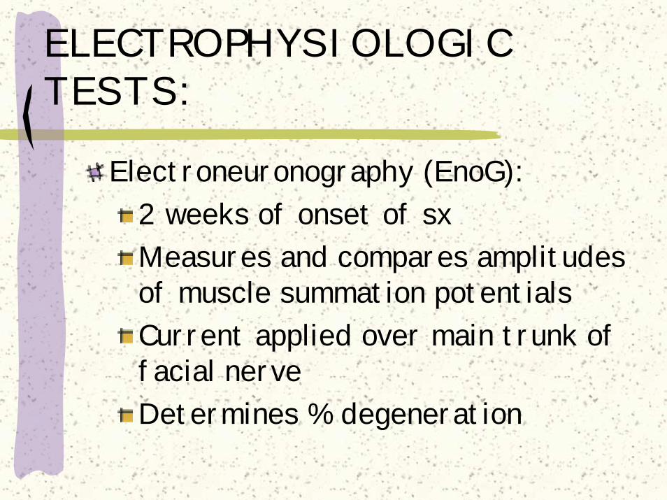
ELECTROPHYSIOLOGIC TESTS:
Electroneuronography (EnoG):2 weeks of onset of sxMeasures and compares amplitudes of muscle summation potentialsCurrent applied over main trunk of facial nerveDetermines % degeneration
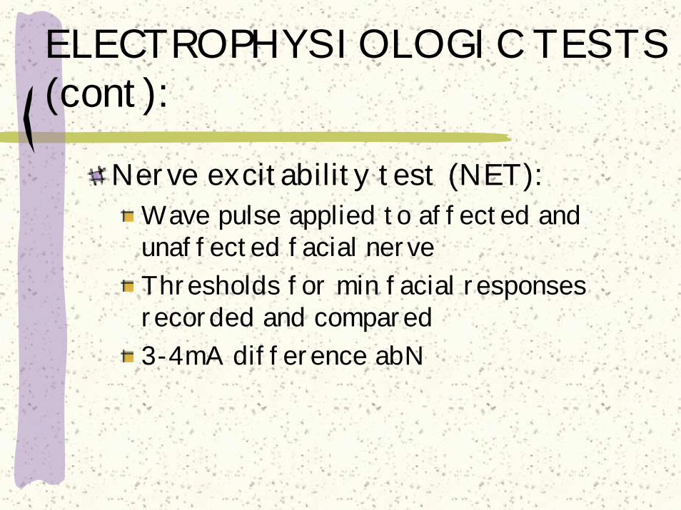
ELECTROPHYSIOLOGIC TESTS (cont):
Nerve excitability test (NET):Wave pulse applied to affected and unaffected facial nerveThresholds for min facial responses recorded and compared3-4mA difference abN

ELECTROPHYSIOLOGIC TESTS (cont):
Maximal stimulation test (MST):Stimulates ipsi- and contralateral facial musclesUse max stimulation to evaluate muscular responseSubjective observation
Electromyography (EMG):Determines muscle activity rather than nerve

ELECTROPHYSIOLOGIC TESTS (cont):
Audiometry:Evaluates conductive and SN hearing lossCo-existent in pt with CN VII palsies
Branches: Greater Superficial Petrosal Nerve:
Schirmer’s test: assess parasym innervation to lacrimal gland
Nerve to Stapedius: stapedius reflex (audiometry)Chorda tympani nerve: test for taste

CLASSIFICATION:
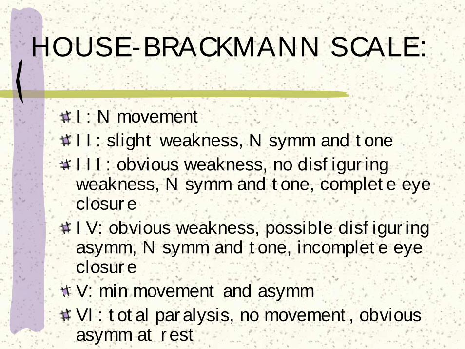
HOUSE-BRACKMANN SCALE:
I: N movementII: slight weakness, N symm and toneIII: obvious weakness, no disfiguring weakness, N symm and tone, complete eye closureIV: obvious weakness, possible disfiguring asymm, N symm and tone, incomplete eye closureV: min movement and asymmVI: total paralysis, no movement, obvious asymm at rest
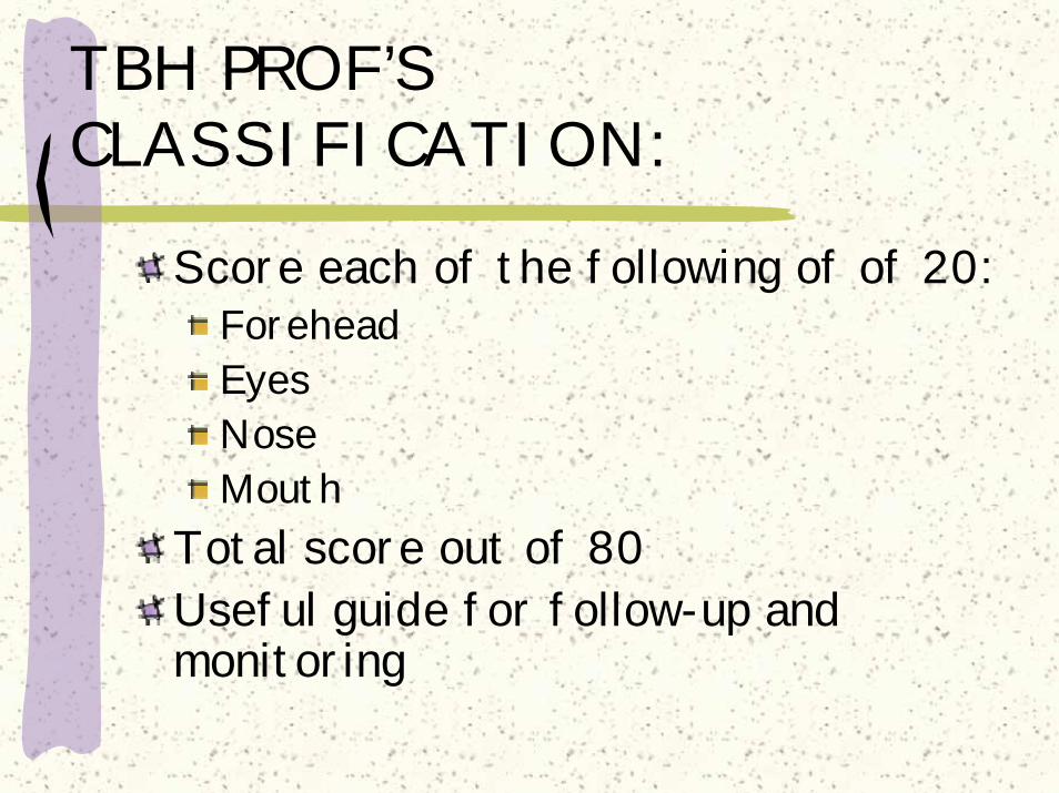
TBH PROF’S CLASSIFICATION:
Score each of the following of of 20:ForeheadEyesNoseMouth
Total score out of 80Useful guide for follow-up and monitoring

THANK YOU!!