Embed Size (px)
Citation preview
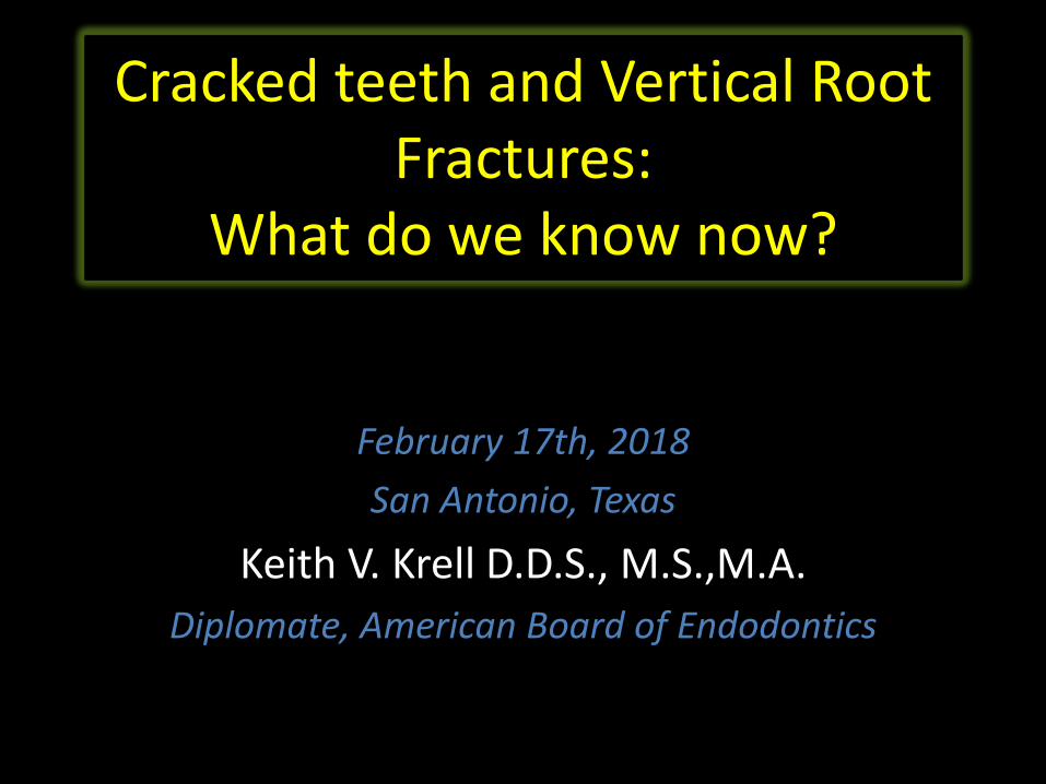
February 17th, 2018
San Antonio, Texas
Keith V. Krell D.D.S., M.S.,M.A.
Diplomate, American Board of Endodontics
Cracked teeth and Vertical Root Fractures:
What do we know now?

Dr. Rob Roda, Past AAE President
• “Cracked teeth seem to be a result of repetitive stress injury, and so the longer teeth are in use, the more likely they will become cracked. This is a modern epidemic and something we have never before as a profession had to deal with.”

What does the literature show?
Bader et.al 1995 JADA estimate 1 in 20 people fracture a tooth a yr.
Krell and Rivera 2007 JOE 796 of 8175 cases (9.7%) cracked teeth
mandibular 2nd molars (30%) > mandibular 1st
molars (29%) > maxillary 1st molars (21%)
Tsesis, et al. 2010 JOE unable to do a systematic review for VRF!

How I will proceed!
• New definitions from 2015!
• Diagnostic tests
• Etiologies
• Treatments
• Outcomes
• Prevention??

What is a “ cracked tooth” versus a root fracture?


REPORT TO THE BOARD OF DIRECTORSAnnual Meeting – May 4-5, 2015
• SPECIAL COMMITTEE ON CRACKED TOOTH INITIATIVE
•
• Louis H. Berman, Chair
• Scott L. Doyle
• Gary G. Goodell
• Keith V. Krell
• H Mark A. Odom, Board Liaison
• Helen Jameson, Staff Liaison

Cracked tooth definitions:
• AAE Glossary: A phenomenon involving posterior teeth in which fractures usually involve the marginal ridges; primarily in minimally restored mandibular first and second molars; symptoms may vary but pain to chewing and thermal sensitivity are common.
• Issues: There are a number of problems with the definition. First, a cracked tooth is not a “phenomenon.” The 2008 Colleagues for Excellence article states that, “… cracks present in teeth are findings only; they are not to be considered a pulpal or periapical diagnosis.” Second, the definition does not differentiate a crack from a fracture. Third, describing where they occur on a tooth and on what type of teeth they occur is too specific and limiting.

New Definitions from 2015!
Cracked tooth — A thin surface disruption of enamel and dentin, and possibly cementum, of unknown depth or extension.

New Definitions from 2015!
Root fracture — A fracture that exists or extends into the root, to include dentin, cementum, and possibly pulpal space, which may progress to or from the enamel.
Longitudinal fracture — A root fracture extending in the axial plane within the tooth.
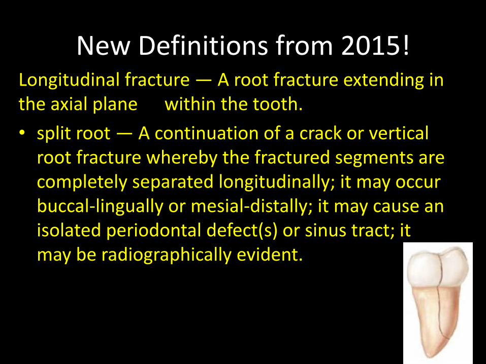
New Definitions from 2015!Longitudinal fracture — A root fracture extending in the axial plane within the tooth.
• split root — A continuation of a crack or vertical root fracture whereby the fractured segments are completely separated longitudinally; it may occur buccal-lingually or mesial-distally; it may cause an isolated periodontal defect(s) or sinus tract; it may be radiographically evident.

New Definitions from 2015!Longitudinal fracture — A root fracture extending in the axial plane within the tooth.
• vertical root fracture — A fracture in the root whereby the fractured segments are incompletely separated; it may occur buccal-lingually or mesial-distally; it may cause an isolated periodontal defect(s) or sinus tract; it may be radiographically evident.


CTS – “Cracked Tooth Syndrome”
▪ CTS – This term is a misnomer ▪ Since syndrome is defined as “A number of symptoms occurring together and characterizing a specific disease” ▪ A crack is NOT a disease nor is it a pathological entity ▪ But cracks may become portals for bacteria and subsequent diseases: o Pulpitis – reversible or irreversible o Pulp necrosis and infection the root canals o Periodontitis – lateral and/or apical

What is really the difference?
➢The difference between “cracks” and “fractures” are a matter of degrees of severity. However, the etiology may be very different.
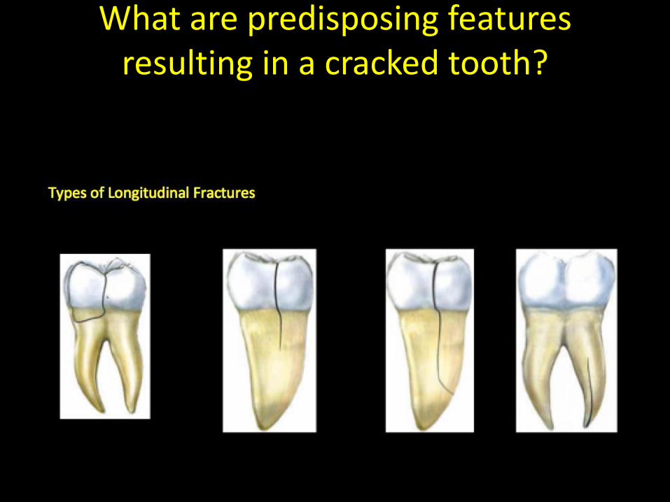
What are predisposing features resulting in a cracked tooth?

REPORT TO THE BOARD OF DIRECTORS Annual Meeting, April 4-5, 2016
• THE SPECIAL COMMITTEE ON METHODOLOGY OF CRACKED TOOTH STUDIES – Dr. Shimon Friedman, Chair
– Dr. Amir Azarpazhooh
– Dr. George Bruder
– Dr. Keith V. Krell, Dr. Isabel Mello
– Dr. Donald Nixdorf
– Dr. Robert S. Roda, Board Liaison
– Helen Jameson, Staff Liaison

Survey AAE Members’ Perceptions on RC/F
• 941 of 5204 members responded• Half the participants (49%) perceived that
occurrence of RC/F in the past decade increased compared to previous years
• Majority of participants (72%) agreed that occurrence of RC/F in root-filled teeth was a major concern in endodontics
• 58% agreed it should be a priority to revise endodontic treatment protocols so as to mitigate the risk of RC/F

How do we diagnose a cracked tooth or vertical root fracture?
Diagnostic Tests –Cracked Teeth!
Start with the dental history!
A good dental history can be important , especially, if a recent“biting event” has occurred and they “felt something pop!”
Patient comments of pain when chewing, pain upon release, or “pain when I hit the tooth a certain way” all suggest a crack.
“Fillings that keep falling out”
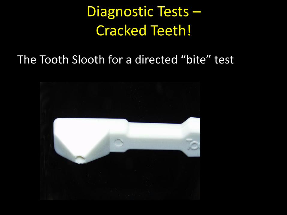
Diagnostic Tests –Cracked Teeth!
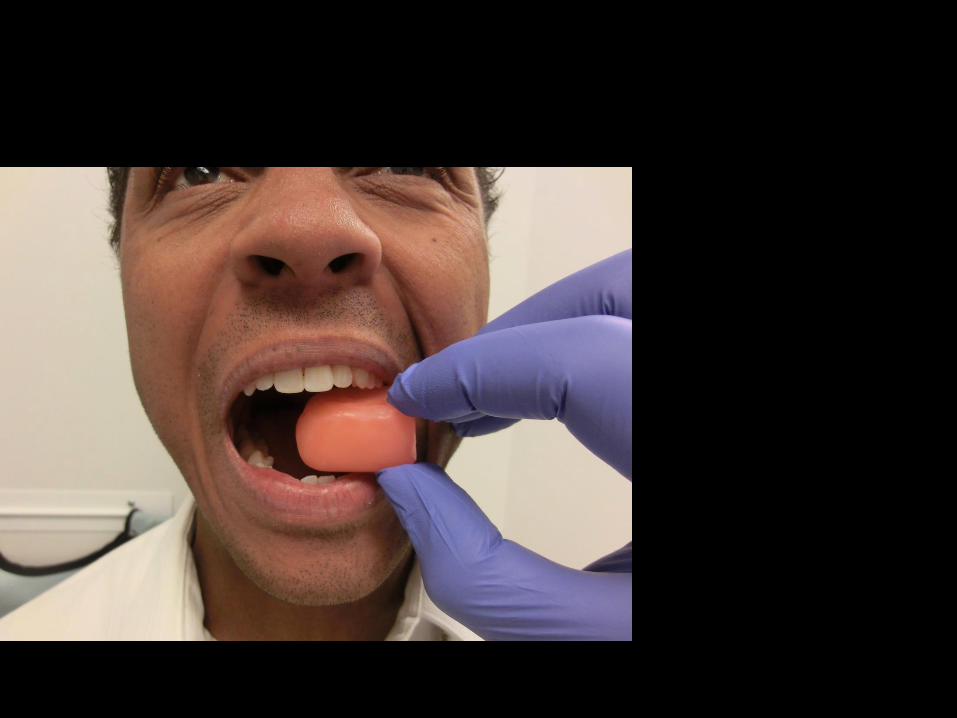
The Tooth Slooth for a directed “bite” test
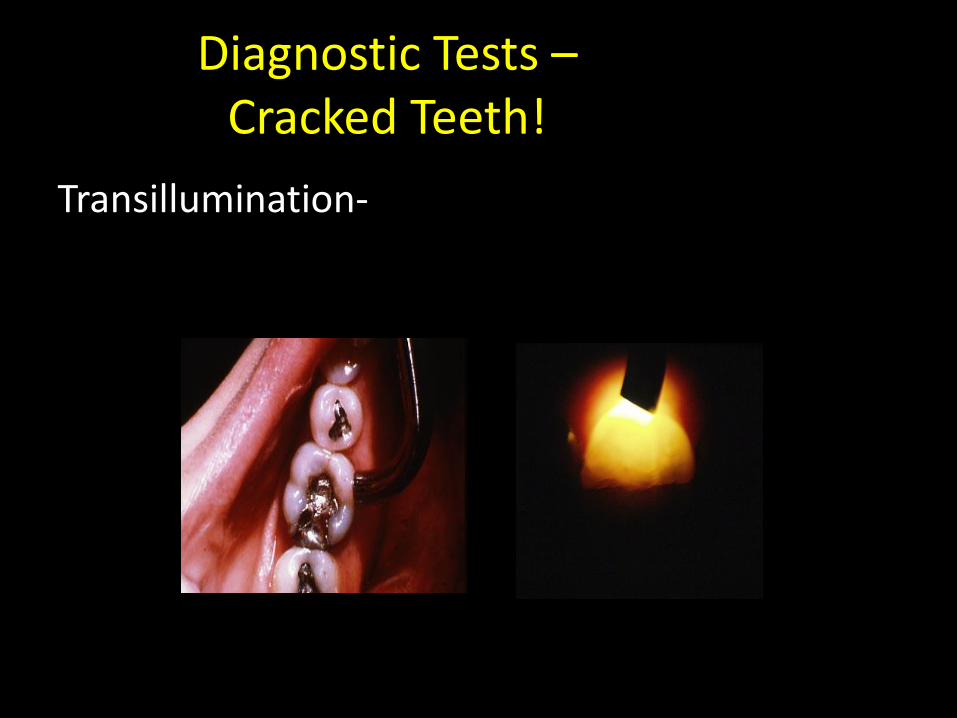
Diagnostic Tests –Cracked Teeth!
Transillumination-

Diagnostic Tests –Cracked Teeth!
Restoration Removal Dyes Probing

Diagnostic Tests –Cracked Teeth!
Magnification-

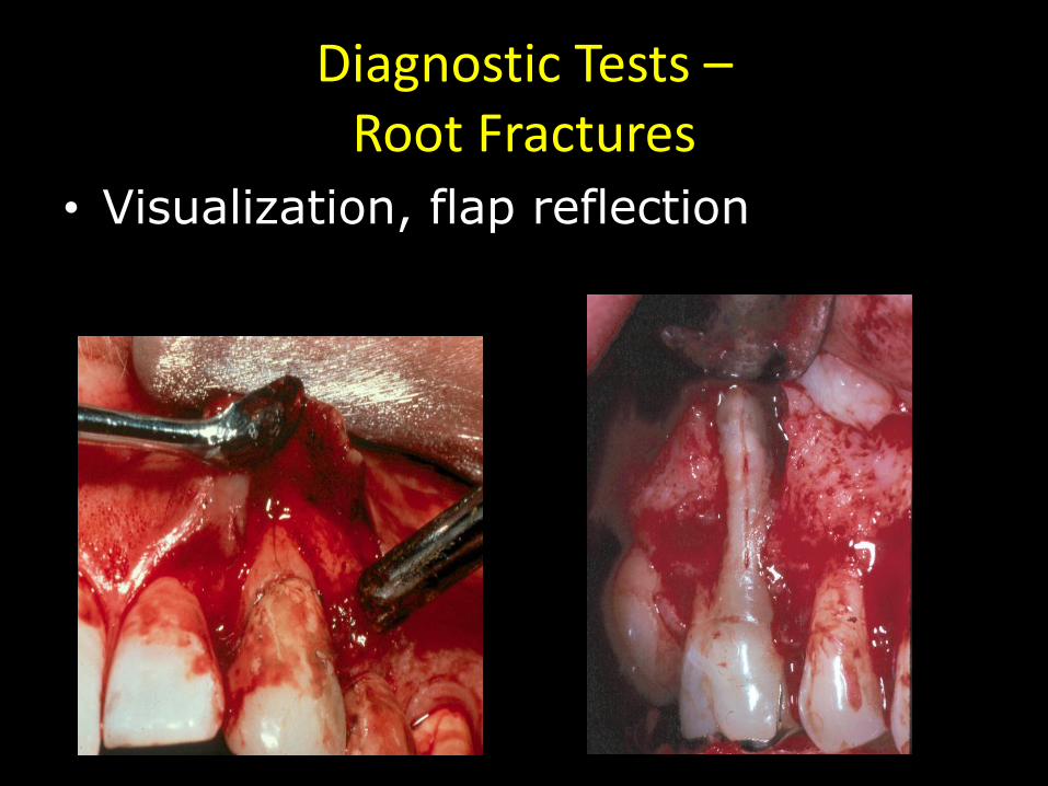
Diagnostic Tests –Root Fractures
• Location: Roots• Direction: Facial-Lingual, mesial-distal• Orientation: Root, extending coronal and apical• Symptoms: Usually none, may present as a
periodontal problem• Signs: Variable• Radiograph: Bone loss pattern• CBCT: Study dependent and load dependent• Probing: A sudden increase in probing
depth can suggest VRF when all other probingsare “normal”
• ID: Visualization, flap reflection• Treatment: Removal of the fractured root• Prognosis: Hopeless, but not all the time!
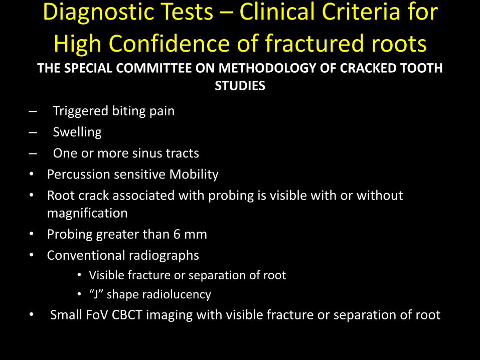
Diagnostic Tests – Clinical Criteria for High Confidence of fractured roots
THE SPECIAL COMMITTEE ON METHODOLOGY OF CRACKED TOOTH STUDIES
– Triggered biting pain
– Swelling
– One or more sinus tracts
• Percussion sensitive Mobility
• Root crack associated with probing is visible with or without magnification
• Probing greater than 6 mm
• Conventional radiographs
• Visible fracture or separation of root
• “J” shape radiolucency
• Small FoV CBCT imaging with visible fracture or separation of root

Diagnostic Tests –Root Fractures
• Radiograph: Bone loss pattern with 2-D radiographs
• “Halo” lesion, perilateral radiolucency, and angular resorption of the crestalbone…indicated a high probability of vertical root fractures in maxillary premolars. – Tamse et al. 1999 Oral Surg.

Diagnostic Tests –Root Fractures
• Radiograph: Bone loss pattern with CBCT
• More accurate than 2D (Bernardes, R. 2009. OOORE)
• Accuracy is Machine dependent (Metska, M.E. et al., 2012 JOE)
• Technique dependent- Double Scan TQ (Bruno Azevedo, 2015)


A Tridimensional Journey
Bruno Azevedo DDS MSBoard Certified OMFR


“Double Scan” Technique

Baseline Wax


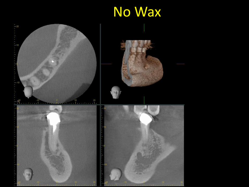
No Wax

Wax
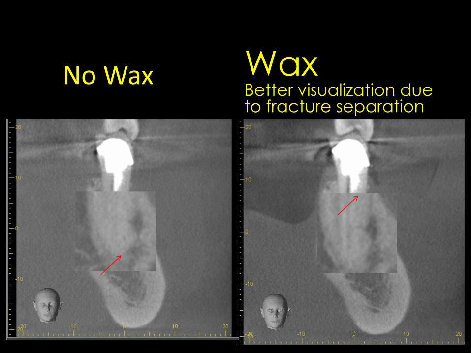
No Wax WaxBetter visualization due to fracture separation

Diagnostic Tests –Root Fractures
• Probing: A sudden increase in probing depth can suggest VRF when all other probings are “normal”
Diagnostic Tests –Root Fractures
• Visualization, flap reflection

–Thermal Cycling
–Pin Placement
–Cementation of Inlays
–Masticatory Forces
–Age!
Etiliogies for Enamel cracks

Etiologies-Cracked teeth
• Enamel Crazings-
– Most likely – Naturally occurring through normal mastication-(Rivera and Walton 2015, Endo Topics)
or– Thermocycling (Brown,’72 JDR)

Etiology
• From the definitions it is obvious there is a difference of degrees of the extension of the crack. This is determined by:
-Magnitude of Stress experienced by the tooth
-Mechanical properties of remaining tissue for resisting fracture
Arola,D et. al “Microstructure and mechanical behavior of radicular and coronal dentin”
Endodontic Topics, 2012 pp30-51

Fatigue Crack Growth
Propagation life of a material or tooth is comprised of the number of loading cycles required to propagate an existing crack to a critical length that facilitates bulk fracture.
Arola,D et. al “Microstructure and mechanical behavior of radicular and coronal dentin” Endodontic Topics,
2012 pp30-51
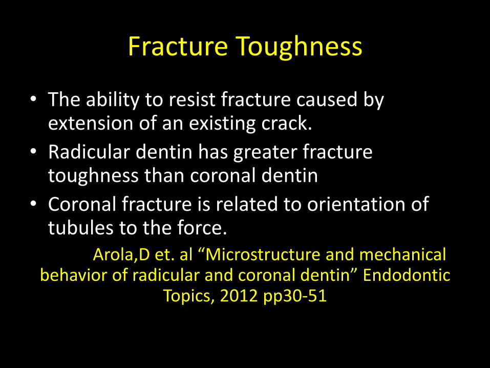
Fracture Toughness
• The ability to resist fracture caused by extension of an existing crack.
• Radicular dentin has greater fracture toughness than coronal dentin
• Coronal fracture is related to orientation of tubules to the force.
Arola,D et. al “Microstructure and mechanical behavior of radicular and coronal dentin” Endodontic
Topics, 2012 pp30-51

Age of dentin!• Age alone has been shown to be a significant
factor in dentin fracture.
• Dentin from patients < 35 vs >55 shows a 50% reduction in strength of >55
• Dentin from patients < 35 vs >55 shows 75% reduction in energy require to FX >55
• >55 has greater mineral content and avg rate of crack growth 100 X that of <35.
Arola,D et. al “Microstructure and mechanical behavior of radicular and coronal dentin” Endodontic Topics, 2012 pp30-51

Etiologies-Cracked teeth
Hydration of dentinKishen, A. and S. Vedantam (2007). "Hydromechanics in dentine: Role of dentinal tubules and hydrostatic pressure on mechanical stress-strain distribution." Dent Mater.
• External compressive loads were conteracted by the residual stresses and strains loads.
• Hydrated specimens showed greater toughness vspartially dehydrated specimens. The stress at fracture was significantly higher in Dehydrated specimens than in partially dehydrated specimens
• Strain at fracture was significantly higher in hydrated dentine specimens (p=0.037).

Etiologies-Cracked teeth
Hydration of dentinKishen, A. and S. Vedantam (2007). "Hydromechanics in dentine: Role of dentinal tubules and hydrostatic pressure on mechanical stress-strain distribution." Dent Mater.
SIGNIFICANCE: These experiments highlighted the distinct role of free water in the dentinal tubules and hydrostatic pressure on the stress-strain distribution within the bulk dentine.

Levin, L, et al. Oral and dental complications of intra-oral piercing. Dent Traumatol. 2005;21:341-3
Assessed the prevalence of oral piercing among young adults and revealed the types and rate of complications following oral piecing in 400 consecutive patients, who randomly arrived at a military dental office.
Intra-oral examination included special attention to piercing-related complications, such as tooth fractures, gingivitis, bleeding, infections, gingival recessions, etc.
A total of 389 patients, 54% males, and 46% females agreed to participate.
Of the participants 20.% reported having at least one type of oral piercing; lingual piercing was the most common.
Swelling and bleeding after piercing were reported by 41 males (51.9%) and 36 females (45.7%) participants.
57.8% were unaware of the dangers of intra-oral piercing.
Clinical examination revealed 15 fractured teeth (13.9%) with piercing.
Gingival recessions were observed in 26.6%, mostly in the mandibular incisor area.

EtiologiesCracked teeth
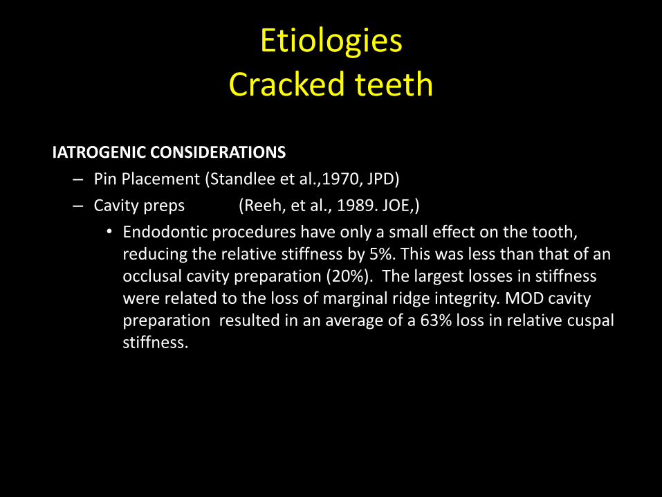
IATROGENIC CONSIDERATIONS
Orofacial piercings, Chadwick, et al. 2005. Prim. Dent. Care
EtiologiesCracked teeth
IATROGENIC CONSIDERATIONS
– Pin Placement (Standlee et al.,1970, JPD)
– Cavity preps (Reeh, et al., 1989. JOE,)
• Endodontic procedures have only a small effect on the tooth, reducing the relative stiffness by 5%. This was less than that of an occlusal cavity preparation (20%). The largest losses in stiffness were related to the loss of marginal ridge integrity. MOD cavity preparation resulted in an average of a 63% loss in relative cuspalstiffness.

EtiologiesFractured roots
– Stress from occlusal forces– Canal preparation
• Physical• Chemical
– Lateral condensation of gutta percha– Expansion of root fillings– Cementation of posts

EtiologiesFractured roots
Stress from occlusal forces
• Yang S.F., Rivera, E.M., Walton, R.E. 1995. Vertical root fracture in nonendodonticallytreated teeth. JOE.

EtiologiesFractured roots
Stress from occlusal forces– Chan C.P., et al. 1998. JOE.
EtiologiesFractured roots
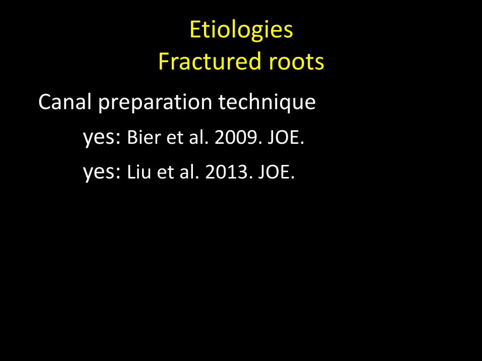
Canal preparation technique
yes: Bier et al. 2009. JOE.
yes: Liu et al. 2013. JOE.

Etiologies-Fractured roots
Canal preparation: length-control
– Adorno, C.G. et al. 2009. Int Endod. J
– Length-control is critical

EtiologiesFractured roots
Canal preparation techniqueTang, Wu, Smales. 2010. JOE.
Conclude:
• Overinstrumentation of root canals + noncircular canals and thin canal walls= Increased risk for root fracture.
• Effect from various Ni-Ti rotary files is somewhat controversial, but hand files have fewer FX
EtiologiesFractured roots
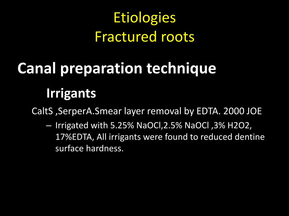
Canal preparation technique
IrrigantsCaltS ,SerperA.Smear layer removal by EDTA. 2000 JOE
– Irrigated with 5.25% NaOCl,2.5% NaOCl ,3% H2O2, 17%EDTA, All irrigants were found to reduced dentine surface hardness.

EtiologiesFractured roots
Canal preparation technique
Calcium Hydroxide,MTA NaOClWhiteJD,et al..The effect of three commonly used endodontic materials on the strength and hardness of root dentin. 2002 JOE.
After 5 weeks 32% mean decrease in strength after calcium hydroxide treatment,
33% decrease in strength after mineral trioxide aggregate treatment,
59% decrease in strength after sodium hypochlorite treatment
Etiologies-Fractured roots
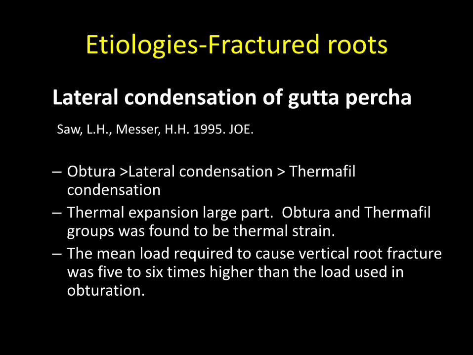
Lateral condensation of gutta perchaSaw, L.H., Messer, H.H. 1995. JOE.
– Obtura >Lateral condensation > Thermafilcondensation
– Thermal expansion large part. Obtura and Thermafilgroups was found to be thermal strain.
– The mean load required to cause vertical root fracture was five to six times higher than the load used in obturation.
Etiologies-Fractured roots
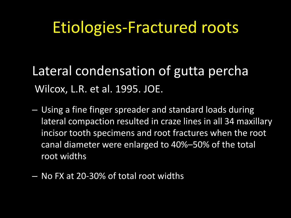
Lateral condensation of gutta perchaWilcox, L.R. et al. 1995. JOE.
– Using a fine finger spreader and standard loads during lateral compaction resulted in craze lines in all 34 maxillary incisor tooth specimens and root fractures when the root canal diameter were enlarged to 40%–50% of the total root widths
– No FX at 20-30% of total root widths

Etiologies-Fractured roots
Expansion of root fillings
The Properties of Endocal®10 and Its Potential Impact on the Structural Integrity of the Root
Goldberg, R. et al., 2004. JOE.

Etiologies-Fractured roots

Etiologies-Fractured roots
Cementation of posts
• Obermayr G. et al.1991. JPD.
• Morando G. et al. 1995. JPD.

Treatments-
• Treatment depends on the severity of the crack or fracture. “Split teeth/fractured roots” are considered “hopeless” by most.
• Treatment planning for cracked teeth and cracked roots becomes dependent on the clinical findings at the time of the “discovery”.

Treatments-Cracked Crown/Root
• Remember!! A crack is now “a thin disruption of enamel and dentin, and possibly cementum, of unknown depth or extension.”
• Ricucci D, et al. JOE 2015 concluded cracks are always colonized with bacterial biofilms. The pulp tissue response varies according to the location, direction, and extent of the crack.
• Treatment planning for cracked crowns and cracked roots becomes dependent on the clinical findings at the time of the “discovery.”
Treatments-Cracked Crown/Root
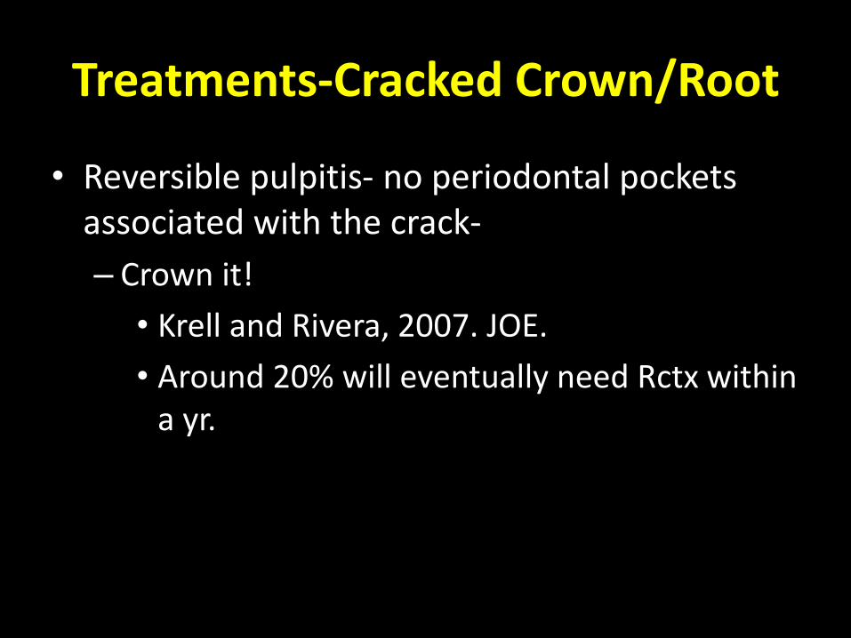
• Reversible pulpitis- no periodontal pockets associated with the crack-
– Crown it!
• Krell and Rivera, 2007. JOE.
• Around 20% will eventually need Rctx within a yr.


Treatments-Cracked Crown/Root
• Reversible pulpitis- no periodontal pockets associated with the crack
• Sedative Lining and interim restoration
• Abbott and Leow. 2009. Aust. Dent. J.
• 20 % eventually needed Rctx within 5yrs


Treatments-Cracked Crown/Root
• Reversible pulpitis- no periodontal pockets associated with the crack-
– Bonded restoration
Opdam, et al. 2008. JOE.
7 % eventually needed Rctx within 5yrs
Treatments-Cracked Crown/Root
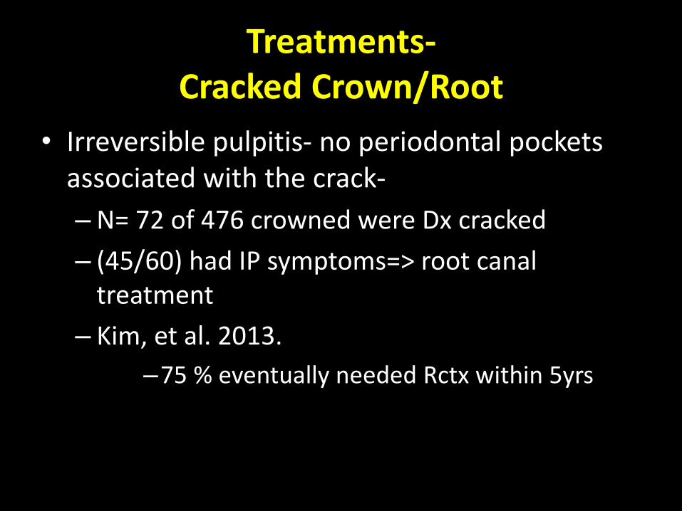
• Irreversible pulpitis- no periodontal pockets associated with the crack-
– N= 72 of 476 crowned were Dx cracked
– (45/60) had IP symptoms=> root canal treatment
– Kim, et al. 2013.
–75 % eventually needed Rctx within 5yrs

Treatments-Cracked Crown/Root
• Irreversible pulpitis/necrosis- no periodontal pockets associated with the crack-
– Resin filling, inlays or prov. crown
Kang, Kim and, Kim. 2016. JOE.
50 % eventually needed Rctx within 5yrs

Treatments-Cracked Crown/Root
• Necrosis- no or minimal restoration and periodontal pockets associated with the crack-(fracture necrosis)– Extraction!
Berman and Kuttler 2010 JOE• --Retention!• Kang et. al. 2016 JOE
– Multivariate analysis revealed a weak correlation between multiple crack directions and pulp necrosis at initial examination
Krell
Berman


Krell KV1, Caplan DJ2
1Department of Endodontics2Department of Preventive and Community Dentistry
Local AADR: February 13, 2018
University of Iowa, College of Dentistry
12-month Success ofCracked Teeth Treated with
Orthograde Root Canal Treatment
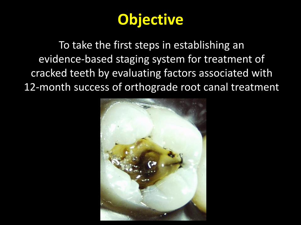
To take the first steps in establishing anevidence-based staging system for treatment of
cracked teeth by evaluating factors associated with12-month success of orthograde root canal treatment
Objective

Methods
- Secondary data (Krell private practice, 1989-2014)
- Age, sex, date of first visit (patient-level)Tooth typeRestoration (material, # restored surf)M/D marginal ridge cracks (y/n) M/D pocket depths (mm) Pulpal/periapical diagnosis
- Outcome = “success”
- Univariate, bivariate analyses (SAS Version 9.4)
tooth-level

Diagnosed
Cracked Teeth
(n=2086)
Did not return for
12-month recall (n=1026)
Not treated with Root
Canal Therapy (n=680)
Treated with
Root Canal Therapy
(n=1406)
Ineligible for regression analysis (n=17)
- Outcome “questionable” (n=13)
- Patient <18 years old (n=1)
- Wisdom tooth (n=1)
- Reversible pulpitis (n=1)
- Unknown size of restoration (n=1)
Potentially Eligible
for Analysis
(n=380)
Analyzed (n=363)
Results

Tooth, Restoration

Cracks, Pockets, Pulp / Periapical Dx

Mesial or Distal
Probing Pocket
Depth ≥5 mm?
Distal Marginal
Ridge cracked?
No
Iowa Stage IV:
8% of cases
(41% success)
Yes
Iowa Stage I:
37% of cases
(93% success)
No
Periapical
Diagnosis
CAP, SAP,
or AAA?
Yes
Iowa Stage II:
39% of cases
(84% success)
No
Yes
Iowa Stage III:
15% of cases
(69% success)

Discussion
Limitations- 1 operator, 1 city, private practice (generalizability?)- Outcome (radiographic success vs. tooth survival?)- 12-month follow-up (clinical relevance?)- Selection bias (less tx of teeth with deep pockets?)
Strengths- Large number of observations (n=363)- Single examiner (consistency over time)- Staging system easily understood by clinicians
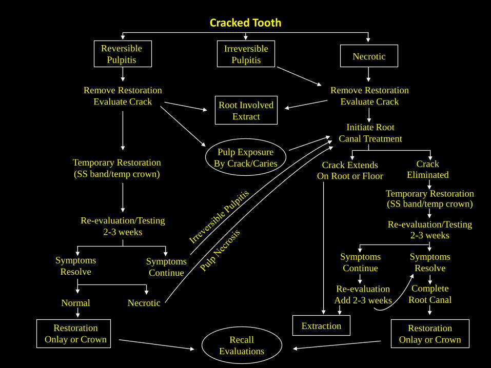
Cracked Tooth
Reversible
Pulpitis
Irreversible
Pulpitis Necrotic
Remove Restoration
Evaluate Crack
Crack
Eliminated
Extraction
Re-evaluation/Testing
2-3 weeks
Symptoms
Resolve
Complete
Root Canal
Restoration
Onlay or Crown
Root Involved
Extract
Remove Restoration
Evaluate Crack
Initiate Root
Canal Treatment
Pulp Exposure
By Crack/CariesTemporary Restoration
(SS band/temp crown)
Temporary Restoration(SS band/temp crown)
Re-evaluation/Testing
2-3 weeks
Symptoms
Resolve
Symptoms
ContinueSymptoms
Continue
Restoration
Onlay or Crown
Normal Necrotic
Re-evaluation
Add 2-3 weeks
Crack Extends
On Root or Floor
Recall
Evaluations

Treatment
Blast from the past!
JDR 2016 Vol 95, 5-6.
“Teeth even compromised because of periodontal disease or endodontic problems may have a longevity that surpasses by far that of theaverage implant (Carnevale et al. 1998; Hardtet al. 2002; Lang and Zitzmann 2012; Salvi et al. 2014; Klinge et al. 2015).”
Giannobile and Lang, 2016
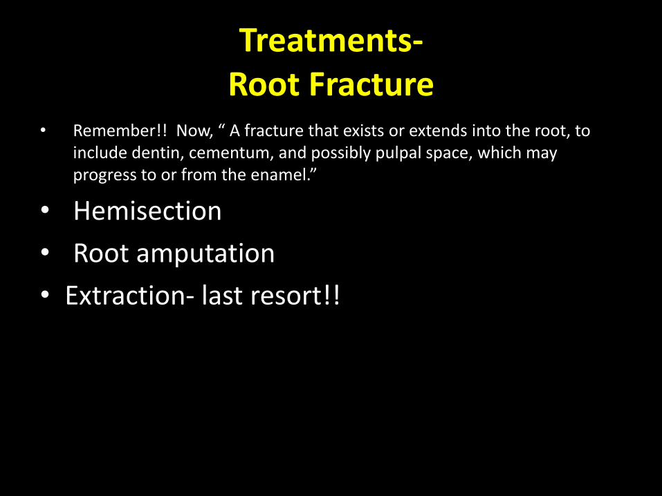
Treatments-Root Fracture
• Remember!! Now, “ A fracture that exists or extends into the root, to include dentin, cementum, and possibly pulpal space, which may progress to or from the enamel.”
• Hemisection
• Root amputation
• Extraction- last resort!!

Treatments-Root FractureHemisection
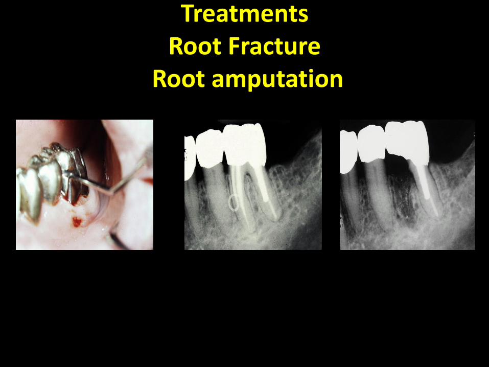
Treatments Root Fracture
Root amputation

Herodontics in the 80’s

Herodontics in the 80’s
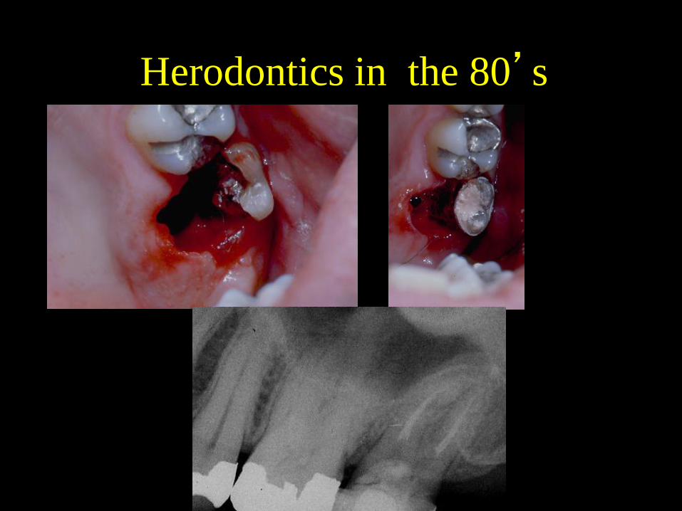
Herodontics in the 80’s

Herodontics in the 80’s
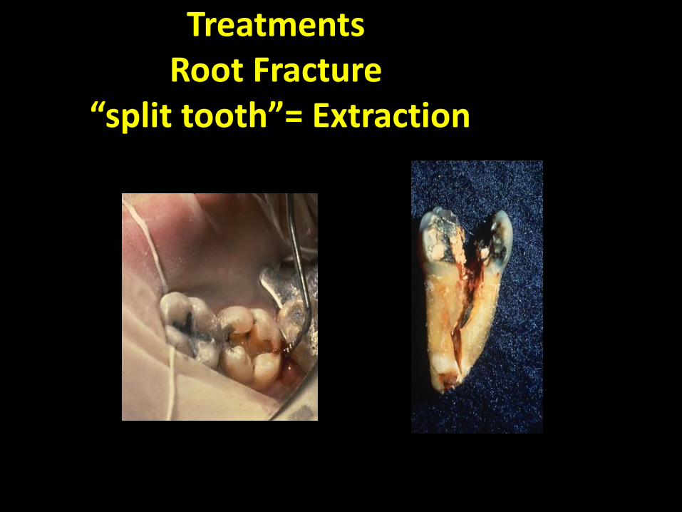
TreatmentsRoot Fracture
“split tooth”= Extraction

Outcomes- changes in thinking!
(excerpts from Dr. Hargreaves Jan, 2016)
• survival (a patient-centered outcomes) and “radiographic success” (a clinician-centered outcome)
• “levels of evidence” may be only one way to measure clinical relevance of research. An entirely alternative approach is comparative effectiveness research (also called pragmatic clinical trials)

Outcomes
• Reversible pulpitis+ restoration
– 80% survival without rctx
– Krell and Rivera, 2007 JOE
– Abbott and Leow, 2009 Aust Dent J
Outcomes
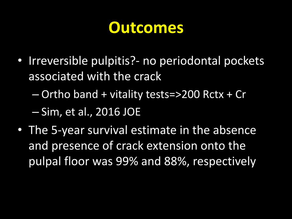
• Irreversible pulpitis?- no periodontal pockets associated with the crack
– Ortho band + vitality tests=>200 Rctx + Cr
– Sim, et al., 2016 JOE
• The 5-year survival estimate in the absence and presence of crack extension onto the pulpal floor was 99% and 88%, respectively

Outcomes-Cracked crown/root with root canal treatment
50 teeth had a 85% survival rate (2 yrs)Tan, Chen, Poon and Wong 2006 IEJ
88 teeth had a 90% survival rate (5 yrs)Kang, Kim and, Kim 2016 JOE (>6mm probings decreased survival)
Iowa Stage 1= 93% successIowa Stage 2= 84% successIowa Stage 3= 69% successIowa Stage 4= 41% success
Krell and Caplan accepted JOE 2018

Outcomes-rctx-fractured root + root resection=
92% survival over 12 yrs
Basten CH. Ammons WF Jr. Persson R. 1996 International Journal of Periodontics & Restorative Dentistry
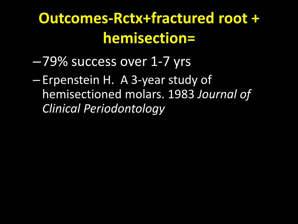
Outcomes-Rctx+fractured root + hemisection=
–79% success over 1-7 yrs– Erpenstein H. A 3-year study of
hemisectioned molars. 1983 Journal of Clinical Periodontology

Prevention???
• Physical anthropologists estimate tooth longevity without modern care to be 30-40 yrs based on enamel wear patterns.
Simon Hillson-Dental Anthropology 1996Average Life Expectancy USA Circa 1900
Men= 46.3 yrsWomen= 48.3 yrs.
Average Life Expectancy USA Circa 2016Men=76.6 yrsWomen= 81.5 yrs

Summary
• The truth is we are all potentially outliving the structural limits of our natural teeth!
• Endodontics is the specialty for definitively diagnosing cracks and fractures!
• Periodontists are re-examining their decades old shift to implants and are again realizing the need to preserve the natural dentition.

Summary
• Endodontics never abandoned the need to preserve the natural dentition and we can reassert our leadership role as the only specialty devoted to preserving the natural dentition!

Summary
• Are all fractured teeth “Hopeless”?
– Maybe “split teeth”, but multi-rooted teeth can survive, in part, if we want to put forth the effort. Let us all respond to Dr. Giannoble’s request to save the natural dentition again!
Let’s Make Endodontics Great Again!