Embed Size (px)
Citation preview

Case Presentation and Discussion on a
Patient with Right Upper Quadrant Mass
Janix M. De Guzman, MDEdgardo Penserga, MDDepartment of Surgery

General Data:20 year-oldMale
Chief Complaint:“abdominal pain”

History of the Present Illness:1 month PTA RUQ pain
vomiting
6 days PTA increased severity of painconsult – IM-ER
2 days PTA recurrence of painconsult IM-ER
Few hours PTA RUQ painConsult IM-ERReferred to Surgery
Admitted

IM-ER Diagnosis: Hepatic Abscess, Amebic, Right Hepatic Lobe
• Initial US Findings:– RUQ Mass, Hepatic in origin, Right Hepatic
Lobe, Cystic with septations
• Plain film of the Abdomen– Hepatomegaly– Ileus– Fecal Retention

• Past medical history:unremarkable
• Personal and social history unremarkable
• Family medical historyunremarkable
• Review of Systemweight loss

Physical Examination: Upon referral to Surgery
• Conscious, coherent, oriented• BP = 130/80mmHg CR = 81beats/min
RR = 20 cycles/min Temp = 36.9OC• Pink palpebral conjuctivae, anicteric
sclerae• Supple neck, no cervical
lymphadenopathy

Physical Examination:
• Symmetrical Chest Expansion, no retractions, Clear breath sounds
• Adynamic precordium, no murmur• Extremities: no edema; full and equal
pulses, no cyonosis
Physical Examination:
• Slightly Globular, Normoactive bowel sounds, soft, RUQ mass 14cm below subcostal margin RMCL with tendernessdull on percussion

Salient Features• 20 y/o, Male• Right Upper Quadrant Pain• Right Upper Quadrant Mass with
tenderness• Initial US Finding:
– RUQ Mass, Hepatic in origin, Right Hepatic Lobe, Cystic with septations

Clinical Diagnosis
A.Primary Clinical Diagnosis:Hepatic Mass, Amebic Abscess, Right Hepatic Lobe
B.Secondary Clinical Diagnosis:Simple Hepatic Cyst, Right Hepatic Lobe

RUQ Mass
Abdominal Wall Intra-abdominal
Intraperitoneal Retroperitoneal
Non-Inflammatory Inflammatory
Malignant Benign
Liver
GB and Biliary Tree
PancreasKidney
Amebic Liver Abscess
Simple Hepatic Cyst

ObservationSurgical
20%Simple Hepatic Cyst, Right Hepatic Lobe
MedicalSurgical
80%Hepatic Mass, Amebic Abscess, Right Hepatic Lobe
TREATMENTMODALITY
CERTAINTYCLINICAL DIAGNOSIS

Paraclinical Diagnostic Procedures
• Do I need additional paraclinical diagnostic procedure?
– NO.– Follow-up on official ultrasound report

Treatment
Pre-treament Diagnosis:Hepatic Mass, Amebic Abscess, Right
Hepatic Lobe

Treatment
• Goal of Treatment:> resolution of liver abscess
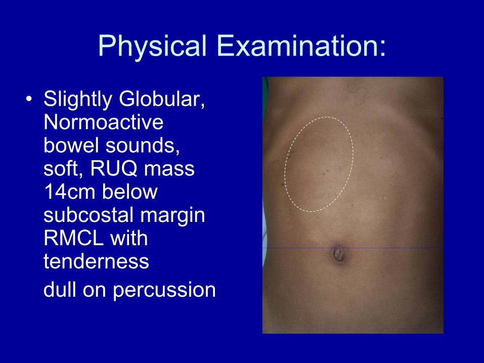
Treatment Options
yes1,500 to 5,000
Bleeding60 – 100% Success rateSurgical
yes300/dayAdverse Drug reactions90% Success RateMedical
AvailabilityCostRiskBenefitTreatment
Agreed on Medical Management by Internal Medicine.
Patient was admitted.
Patient was given Metronidazole

Course in the wards:
• 4 days after admission:– No abdominal pain– RUQ mass, 10 cm below subcostalmargin
RMCL– No tenderness
– Plan: Suggested repeat ultrasound

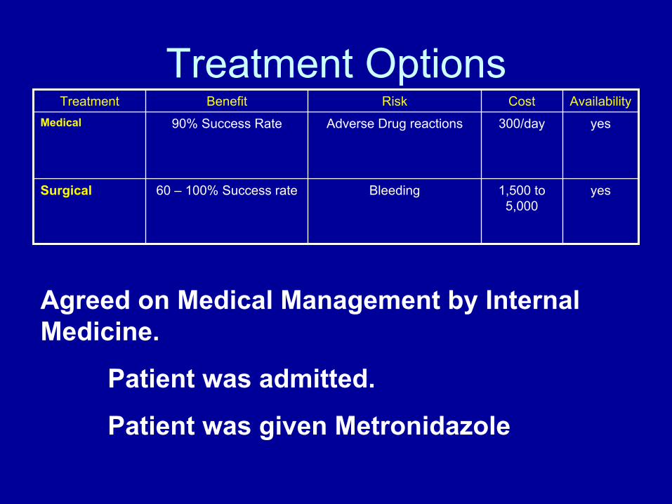
Ultrasound Report
• The liver is normal in size. The proximal intra-hepatic ducts are slightly dilated. There is a large tubular septated cystic mass measuring about 28.8cm x 10.7 cm just above the portal vein showing a bright echo 2.0cm in size with acoustic shadowing. This fluid filled mass extends to the midabdomen and partially obscured the pancreatic head.

IMPRESSION:Normal size liverContracted GallbladderT/C Choledochal Cyst with stone formationSuggest CT Scan or PTC

Further Imaging Options:
No10T+++++/-PTC
Yes5T+++++CT Scan
Yes10T++++MRI
AvailabilityCostRiskBenefitImaging Modalities
• Importance:– Confirmatory– Plan for future surgical intervention

CT Scan Report
• The liver is markedly enlarged with the tip of the right lobe extending below the lower pole of the ipsilateral kidney
• There is fairly he non-enhancing hypodensityoccupying the right lobe of the liver mesuring11.8 x 14.1 cm in its widest axial diameter. The border outline is well defined. The central biliarytree is slightly dilated. There is a small and separate hypodensity in the left lobe.

• The spleen and adrenal glands are unremarkable.
• The pancreas is displaced laterally and the size and contour of which are difficult to evaluate.
• Both kidneys are functioning without hydronephrosis.
• The opacified bowels are displaced inferiorly and laterally
• The urinary blader is fully distended and is unremarkable.
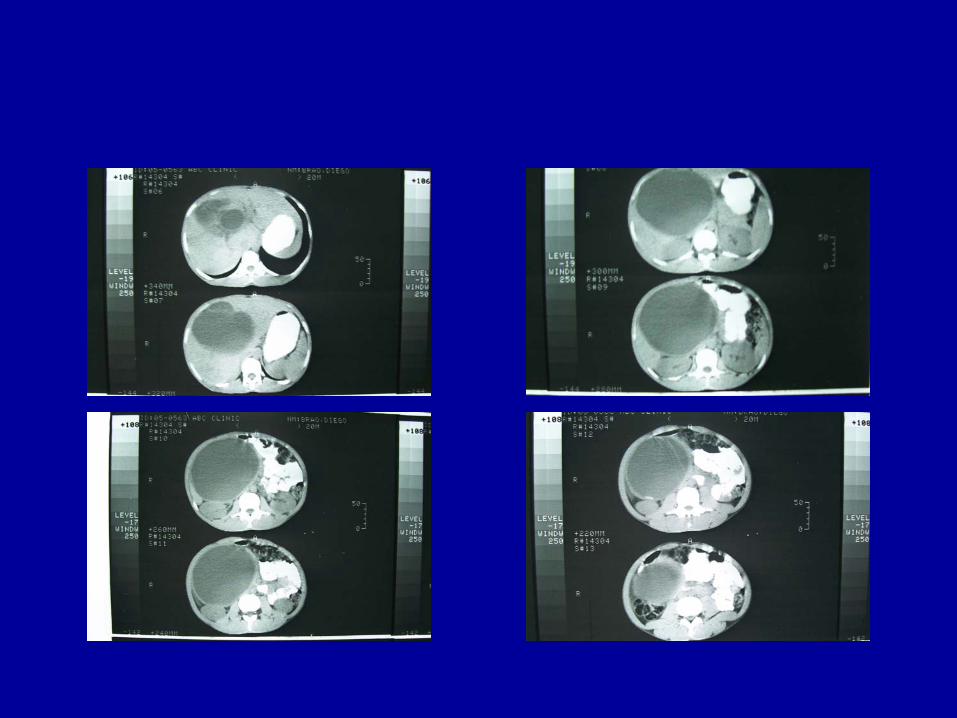

• IMPRESSION:– Hepatomegaly with a fairly large, Right
Hepatic Cyst– Dilated central biliary tree

Referred back to surgery:
Treatment:Pre-treatment Diagnosis:
Hepatic Cyst
Goal:Resolution of Mass

Treatment Options
yes5,000Biliary injurySuccess Rate 90 –100%
LaparotomyUnroofing
Yes1,500bleedingHigh recurrence rate
Diagnostic
US guidedPercutaneous
Aspiration
AvailabilityCostRiskBenefitOptions

Preoperative preparation:• Informed consent secured• Psychosocial support• Optimize patient’s health• Screen for any condition that will interfere with
treatment• Prepare materials• Patient supine• Aseptic and antiseptic technique RUQ AREA• Local anesthesia injected• French 16 needle inserted Guided ultrasonically

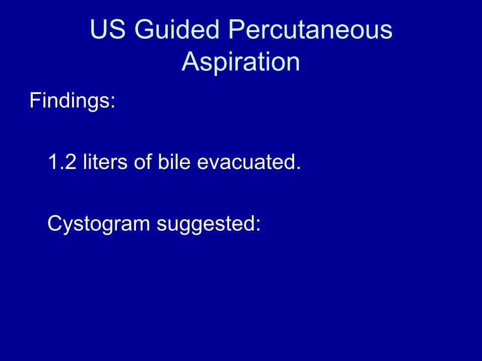
US Guided Percutaneous Aspiration
Findings:
1.2 liters of bile evacuated.
Cystogram suggested:

Cystogram
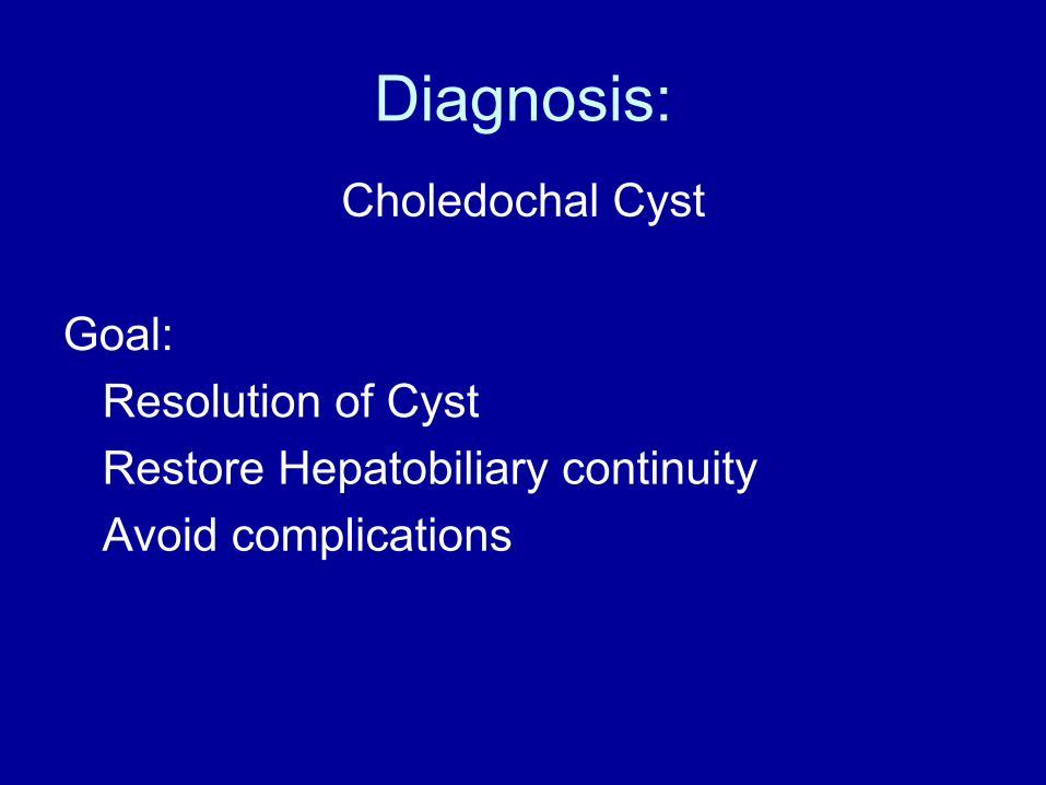
Diagnosis:Choledochal Cyst
Goal:Resolution of CystRestore Hepatobiliary continuityAvoid complications

Treatment Options
Yes7,000Injury to surrounding structures
% Revision: 0 – 1%Mortality: 2– 7%Morbidity: 2 – 8%
Cyst Excision(Roux-y
Hepaticojejunotomy)
Yes7,000Leak% Revision: 22 – 30%Mortality: 8 – 12%Morbidity: 10 – 50%
Cyst Drainage(Cyst-
enterostomy)
AvailabilityCostRiskBenefitOptions

Preoperative preparation:• Informed consent secured• Psychosocial support• Optimize patient’s health• Screen for any condition that will interfere with
treatment• Prepare materials

Operative Technique
• Position: patient supine under GA• Asepsis and antisepsis technique• Incision: Right Subcostal oblique incision 2
fingerbreadths from subcostal margin

Intra-operative Findings Noted:
• Upon opening through right subcostaloblique incision noted massive dilatation of the whole segment of extrahepatic duct containing 750 cc of bile. This was markedly adherent to the portal vein, pancreas, and retroperitoneal structures.
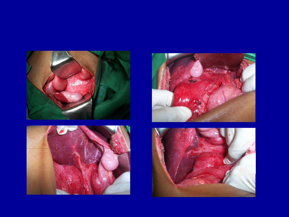

Operative Technique• Decompression done• Cyst mobilized but deemed unresectable• Jejunum mobilized for internal drainage• Jejunum transected 40cm from LOT• End of Distal portion closed and anastomosed
side to side to the cyst wall after partial cystectomy
• Proximal end of jeunum anastomosed end to side to Distal jejunum 40cm from choledochojejunostomy

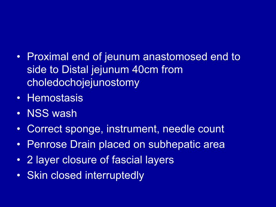
• Proximal end of jeunum anastomosed end to side to Distal jejunum 40cm from choledochojejunostomy
• Hemostasis• NSS wash• Correct sponge, instrument, needle count• Penrose Drain placed on subhepatic area • 2 layer closure of fascial layers• Skin closed interruptedly

Operative Procedure
• Partial Cystectomy; Internal Drainage Cyst-jejunostomy (Roux-Y)

Post operative Diagnosis
Choledochal CystType Ia

Discussion:
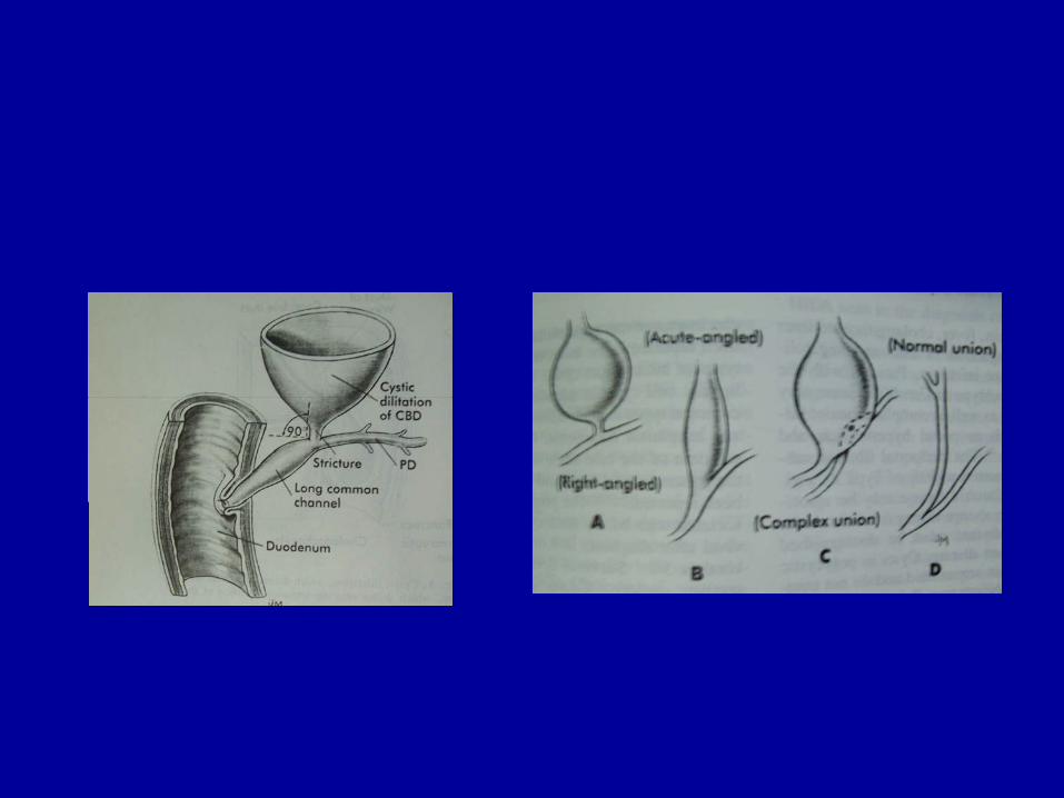
What is Choledochal Cyst?-congenital anomalies of the bile ducts -cystic dilatations of the extrahepaticbiliary tree, intrahepatic biliary radicles, or both.

Classification:
Type II
Type I
Type IV
Type III
Type V

Pathophysiology:• multifactorial• majority have APBJ more than 90% - Miyano and Yamataka
– pancreatic duct enters the common bile duct 1 cm or more proximal to where the common bile duct reaches the ampulla of Vater.
• APBJ allows pancreatic secretions and enzymes to reflux into thecommon bile duct. – alkaline conditions found in the common bile duct– pancreatic proenzymes become activated. – results in inflammation and weakening of the bile duct wall
• congenital standpoint, – defects in epithelialization and recanalization of the developing bile
ducts during organogenesis – congenital weakness of the duct wall have also been implicated.

Clinical presentation:
• Classic clinical triad - present in only 10-20% – abdominal pain (51 to 55%)– Jaundice (45 – 46%)– palpable right upper quadrant abdominal
mass (37 – 58%)

Cyst-Associated Malignancy
- 2.5% to 28%- 10.8% incidence of cholangiocarcinomawith non-cyst excision or non-operated congenital choledochal cyst
- Cholangiocarcinoma- adenocarcinoma - most common malignant cell type

– Other less common types• Adenosquamous• Squamous• Anaplastic
– Prognosis• Poor• Mean survival after diagnosis is 8 months

References:
• Alonso-Lej F, Rever WB, Pessagno DJ: Congenital choledochal cyst, with a report of 2, and an analysis of 94, cases. Int Abstr Surg 1959; 108(1): 1-30
• Todani T, Watanabe Y, Narusue M, et al: Congenital bile duct cysts: Classification, operative procedures, and review of thirty-seven cases including cancer arising from choledochalcyst. Am J Surg 1977; 134(2): 263-9
• Zheng LX, Jia HB, Wu DQ, et.al. Experience of congenital choledochal cyst in adults:treatment, surgical procedures and clinical outcome in the Second Affiliated Hospital of Harbin Medical University. J Korean Med Sci. 2004;19(6):842-7
• Cheng SP, Yang TL, Jeng KS, Liu CL, Lee JJ, Liu TP. Choledochal cyst in adults: aetiological considerations to intrahepatic involvement. ANZ J Surg. 2004 ;74(11):964-7
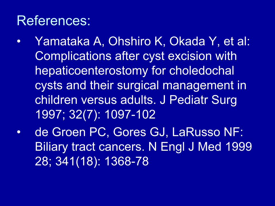
References:• Yamataka A, Ohshiro K, Okada Y, et al:
Complications after cyst excision with hepaticoenterostomy for choledochalcysts and their surgical management in children versus adults. J Pediatr Surg1997; 32(7): 1097-102
• de Groen PC, Gores GJ, LaRusso NF: Biliary tract cancers. N Engl J Med 1999 28; 341(18): 1368-78

MCQ
Direction: Choose the best answer.
1. What is the most common malignancy in the biliary duct system?
A. SquamousB. AnaplasticC. Perihilar AdenocarcinomaD. Intrahepatic AdenocatcinomaE. Botryoid sarcoma

MCQ
Direction: Choose the best answer.
1. What is the most common malignancy in the biliary duct system?
A. SquamousB. AnaplasticC. Perihilar AdenocarcinomaD. Intrahepatic AdenocatcinomaE. Botryoid sarcoma

2. Following factor/s is/are associated with higher incidence of choledochal cyst?
A. JapaneseB. MaleC. ObeseD. AmericanE. Elderly

2. Following factor/s is/are associated with higher incidence of choledochal cyst?
A. JapaneseB. MaleC. ObeseD. AmericanE. Elderly

MCR.
Direction: Write“A” if 1, 2, and 3 are valid statements.“B” if only 1 and 3 are valid statements.“C” if only 2 and 4 are valid statements.“D” if only 4 is a valid statement.“E” if all are valid statements.

3. What type as described by Todani is not included in the Alonso-Lej Classification?
1. Type Ia2. Type II3. Type III4. Type IV

3. What type as described by Todani is not included in the Alonso-Lej Classification?
1. Type Ia2. Type II3. Type III4. Type IV

3. The following factors are associated with higher incidence of choledochal cyst?
1. Race - Asian ancestry2. Age – less than 10 year old3. Sex – Female4. Nutritional Status - Obese

4. Correct statement for Percutaneous Transhepatic Cholangiography includes the following:
1.Usually has complication of significant hemobilia that requires immediate itervention2. preferred when initial US shows dilated intrahepatic ducts without extrahepatic duct dilatation3. preferred when distal common bile duct obstruction is suspected4. requires positioning of a guide wire in an intrahepatic duct

5. Babbit’s concept of concerning the cause of choledochal cyst is based on the demonstration of APBDU. What type/s of choledochal cyst is/are not associated with this concept?
1. Type I2. Type III3. Type IV4. Type V

5. Babbit’s concept of concerning the cause of choledochal cyst is based on the demonstration of APBDU. What type/s of choledochal cyst is/are not associated with this concept?
1. Type I2. Type III3. Type IV4. Type V