Embed Size (px)
Citation preview

CHA
Martinez-Conde, Macknik, Martinez, Alonso & Tse (Eds.)
Progress in Brain Research, Vol. 154
ISSN 0079-6123
Copyright r 2006 Elsevier B.V. All rights reserved
PTER 3
Covert attention increases contrast sensitivity:psychophysical, neurophysiological and
neuroimaging studies
Marisa Carrasco
Department of Psychology & Center for Neural Science, New York University, 6 Washington Pl. 8th floor, New York,NY 10003, USA
Abstract: This chapter focuses on the effect of covert spatial attention on contrast sensitivity, a basic visualdimension where the best mechanistic understanding of attention has been achieved. I discuss how modelsof contrast sensitivity, as well as the confluence of psychophysical, single-unit recording, and neuroimagingstudies, suggest that attention increases contrast sensitivity via contrast gain, an effect akin to a change inthe physical contrast stimulus. I suggest possible research directions and ways to strengthen the interactionamong different levels of analysis to further our understanding of visual attention.
Keywords: visual attention; early vision; contrast sensitivity; psychophysics; neurophysiology;neuroimaging
Our understanding of visual attention has advancedsignificantly over the last two decades thanks to anumber of factors: psychophysics research on hu-mans has systematically characterized distinct at-tentional systems, and single-unit neurophysiologicalresearch has made possible the recording of neuronalresponses in monkeys under attention-demandingtasks. The coupling of the results from these twoapproaches, as well as the findings emerging fromcombining fMRI (functional magnetic resonanceimaging) and psychophysics, have begun to providea mechanistic characterization of this fundamentalprocess, which lies at the crossroads of perceptionand cognition.
This chapter focuses on the effect of covert spatialattention on contrast sensitivity, a basic visual di-mension where the best mechanistic understandingof attention has been achieved. This is due to theexistence of models of contrast sensitivity, as well as
DOI: 10.1016/S0079-6123(06)54003-8 33
to the confluence of psychophysical, single-unit re-cording, and neuroimaging studies, all indicatingthat attention increases contrast sensitivity. Grow-ing evidence supports the idea that this effect is me-diated by contrast gain, an effect akin to a change inthe physical contrast stimulus.
In the first section, I introduce the construct ofselective attention, and discuss the idea that itarises from the high bioenergetic cost of corticalcomputation and the brain’s limited capacity toprocess information. Then I provide an overviewof the two systems of covert attention — transient(exogenous) and sustained (endogenous) — and ofthe mechanisms that underlie attentional effects —signal enhancement and external noise reduction.
The second section deals with the psychophysicaleffects of transient and sustained attention on con-trast sensitivity. After introducing some ways inwhich attention is manipulated in psychophysical

34
experiments, I discuss studies of transient attentionindicating that contrast sensitivity is increased atthe attended location across the contrast sensitivityfunction and the contrast psychometric function.Conversely, compared to a neutral condition, con-trast sensitivity is decreased at the unattended lo-cation. I then document how the effect of transientattention on appearance is consistent with its effectson performance: apparent contrast increases at theattended location and decreases at the unattendedlocation. At the end of the psychophysics section, Idiscuss a study comparing the effects of transientand sustained attention on contrast sensitivity; spe-cifically with regard to the mechanism of signal en-hancement and the contrast gain and response gainfunctions.
The third section presents neurophysiologicalstudies of visual attention. Single-unit recordingstudies in the monkey have provided detailed,quantitative descriptions of how attention altersvisual cortical neuron responses. I provide anoverview of the studies showing that attentionalfacilitation and attentional selection may comeabout by increasing contrast sensitivity in extra-striate cortex in a way comparable to increasingstimulus contrast. In addition, I discuss parallelsbetween contrast and attentional effects at theneuronal level, which advance our understandingof how effects of attention may come about.
In the fourth section, I discuss a human fMRIstudy that provides a retinotopic neuronal corre-late for the effects of transient attention on con-trast sensitivity with a concomitant behavioraleffect. This study illustrates how neuroimagingstudies, in particular fMRI, offer an intermediatelevel of analysis between psychophysics and single-unit studies.
To conclude, I discuss how models of contrastsensitivity, as well as the confluence of psycho-physical, single-unit recording, and neuroimagingstudies, suggest that attention increases contrastsensitivity via contrast gain, i.e., in such a way thatits effect is indistinguishable from a change instimulus contrast. Finally, I offer some thoughtsregarding possible research directions and ways tostrengthen the interaction among different levelsof analysis to further our understanding of visualattention.
Selective attention
Limited resources
Each time we open our eyes we are confrontedwith an overwhelming amount of information.Despite this fact, we have the clear impression ofunderstanding what we see. This requires selectingrelevant information out of the irrelevant noise,selecting the wheat from the chaff. In Funes el
Memorioso [Funes the Memoirist], Borges suggeststhat forgetting is what enables remembering andthinking; in perception, ignoring irrelevant infor-mation is what makes it possible for us to attendand interpret the important part of what we see.Attention often turns looking into seeing.
Attention allows us to select a certain locationor aspect of the visual scene and to prioritize itsprocessing. The limits on our capacity to absorbvisual information are severe. They are imposed bythe high-energy cost of the neuronal activity in-volved in cortical computation (Lennie, 2003).Neuronal activity accounts for much of the met-abolic cost of brain activity, and this cost largelydepends on the rate at which neurons producespikes (Attwell and Laughlin, 2001). The high bio-energetic cost of firing pressures the visual systemto use representational codes that rely on very fewactive neurons (Barlow, 1972). As only a smallfraction of the machinery can be engaged concur-rently, energy resources must be allocated flexiblyaccording to task demand. Given that the amountof overall energy consumption available to thebrain is constant, the average discharge rate in ac-tive neurons will determine the number of neuronsthat can be active at any time. The bioenergeticlimitations provide a neurophysiological basis forthe idea that selective attention arises from thebrain’s limited capacity to process information(Lennie, 2003).
As an encoding mechanism, attention helps thevisual system to optimize the use of valuableprocessing resources. It does so by enhancing therepresentation of the relevant locations or featureswhile diminishing the representation of the lessrelevant locations or aspects of our visual envi-ronment. The processing of sensory input is en-hanced by knowledge and assumptions of the

35
world, by the behavioral state of the organism, andby the (sudden) appearance of possibly relevantinformation in the environment.
Throughout the 19th and early 20th centuries,scientists such as Wundt, Fechner, James, andHelmholtz proposed that attention plays an im-portant role in perception. It is necessary foreffortful visual processing, and may be the ‘glue’that binds simple visual features into an object. Inthe 1980s and 1990s, cognitive psychologists de-veloped experimental paradigms to investigatewhat attention does and which perceptual proc-esses it affects (Neisser, 1967; Posner, 1980; Treis-man and Gelade, 1980). Over the last decade,cognitive neuroscientists have investigated theeffects of attention on perception using threedifferent methodological approaches. The physio-logical brain systems that underlie attention havebeen explored using two different methodologicalapproaches. One has enabled studying how andwhere attention modulates neuronal responses byusing single-unit recording; this method yields aprecise estimate of local activity, but largely ig-nores behavioral consequences. The second ap-proach has employed brain scanners (fMRIsystems) to study the human brain while engagedin attentional tasks. This has enabled the identi-fication of many of the cortical and subcorticalbrain areas involved in attention, and these exper-iments have yielded insights into the global struc-ture of the brain architecture employed inselectively processing information. A third ap-proach has focused on behavior; researchers haveused cognitive and psychophysical techniques toexplore what attention does and what perceptualprocesses it affects. More recently, they havestarted to investigate the mechanisms of visual at-tention, including how visual attention modulatesthe spatial and temporal sensitivity of early filters,and how it influences the selection of stimuli ofinterest, and its interaction with eye movements(Baldassi, Burr, Carrasco, Eckstein & Verghese,2004).
Recent studies show that attention affects earlyvisual processes such as contrast discrimination,orientation discrimination, and texture segmenta-tion — which until recently were considered tobe preattentive. Electrophysiological studies have
established that neural activity increases at at-tended locations and decreases at unattendedlocations. Consequently, we can now infer thatattention helps manage energy consumption. Usu-ally we think of the need to selectively process in-formation in cluttered displays with differentcolors and shapes (i.e., in ‘Where’s Waldo’-likedisplays). However, psychophysical evidenceshows that even with very simple displays, atten-tion is involved in distributing resources across thevisual field. Because of bioenergetic limitations,the allocation of additional resources to an at-tended location implies a withdrawal of resourcesfrom unattended locations. Indeed, we have re-cently published a study showing that when onlytwo stimuli are present in a display, compared to aneutral attentional state, attention enhances thesignal at the attended location, but impairs it at theunattended location (Pestilli and Carrasco, 2005).
Systems of covert attention: transient and sustained
Attention can be allocated by moving one’s eyestowards a location, or by attending to an area inthe periphery without actually directing one’s gazetoward it. This peripheral deployment of atten-tion, known as covert attention, aids us in mon-itoring the environment, and can informsubsequent eye movements (Posner, 1980). Manyhuman psychophysical studies as well as monkeysingle-unit recording studies have likened attentionto increasing visual salience.
A growing body of behavioral evidence demon-strates that there are two systems of covert atten-tion, which deal with facilitation and selection ofinformation: ‘sustained’ (endogenous) and ‘tran-sient’ (exogenous). The former corresponds to ourability to monitor information at a given locationat will; the latter corresponds to an automatic, in-voluntary orienting response to a location wheresudden stimulation has occurred. Experimentally,these systems can be differentially engaged by us-ing distinct cues. Symbolic cues direct sustainedattention in a goal- or conceptually- driven fashionin about 300ms, whereas peripheral cues grab at-tention in a stimulus-driven, automatic manner inabout 100ms. Whereas the shifts of attention by

36
sustained cues appear to be under conscious con-trol, it is extremely hard for observers to ignoretransient cues (Nakayama and Mackeben, 1989;Cheal and Lyon, 1991; Yantis, 1996; Giordanoet al., 2003). This involuntary transient shift occurseven when the cues are uninformative or may im-pair performance (Yeshurun and Carrasco, 1998,2000; Yeshurun, 2004; Pestilli and Carrasco,2005).
Transient and sustained attentions show somecommon perceptual effects (Hikosaka et al., 1993;Suzuki and Cavanagh, 1997), but some differencesin the mechanisms mediating increased contrastsensitivity have been reported (Lu and Dosher,2000; Ling and Carrasco, 2006). Of interest, thesesystems have different temporal characteristics anddegrees of automaticity (Nakayama andMackeben,1989; Cheal and Lyon, 1991; Yantis, 1996), whichsuggest that these systems may have evolved fordifferent purposes and at different times — thetransient system may be phylogenetically older.There is no consensus as to whether common ne-urophysiological substrates underlie sustained andtransient attention. On the one hand, all single-cellrecording studies have manipulated sustained at-tention; on the other hand, some fMRI studies havefound no difference in the brain networks mediat-ing these systems (Peelen et al., 2004); others havereported differences. For example, sustained atten-tion is cortical in nature, but transient attentionalso activates subcortical processing (Robinson andKertzman, 1995; Zackon et al., 1999), and partiallysegregated networks mediate the preparatory con-trol signals of sustained and transient attention.Sustained attention is mediated by a feedbackmechanism involving delayed reentrant feedbackfrom frontal and parietal areas (e.g., Martinezet al., 1999; Kanwisher and Wojciulik, 2000;Kastner and Ungerleider, 2000; Corbetta andShulman, 2002).
Mechanisms of covert attention: signal enhancementand external noise reduction
Although it is well established that covert attentionimproves performance in various visual tasks (e.g.,Morgan et al., 1998; Lu and Dosher, 1998, 2000;
Carrasco et al., 2000, 2001, 2002, 2004a,b; Baldassiand Burr, 2000; Baldassi and Verghese, 2002;Blanco and Soto, 2002; Cameron et al., 2002;Solomon, 2004), the nature of the attentionalmechanisms, and the stages and levels of process-ing at which they modulate visual activity are notyet well understood. Explanations of how atten-tion improves perception range from proposalsmaintaining that the deployment of attentionchanges observers’ decision criteria and reducesspatial uncertainty (Davis et al., 1983; Sperling andDosher, 1986; Kinchla, 1992; Palmer, 1994; Shiuand Pashler, 1994; Nachmias, 2002), to proposalsasserting that attention actually improves sensitiv-ity by reducing external noise (Lu and Dosher,1998; Morgan et al., 1998; Baldassi and Burr,2000; Dosher and Lu, 2000; Cameron et al., 2004)or by enhancing the signal (Bashinski and Bacha-rach, 1980; Carrasco et al., 2000, 2002; Dosherand Lu, 2000; Cameron et al., 2002; Ling andCarrasco, 2006).
The external noise reduction hypothesis main-tains that attention selects information by dimin-ishing the impact of stimuli that are outside itsfocus. Noise-limited models incorporate internalnoise arising from such sources as spatial and tem-poral uncertainty of targets and distracters, as wellas external noise resulting from distracters andmasks. Several studies have attributed attentionalfacilitation to reduction of external noise, eitherbecause a near-threshold target presented alonecould be confused with empty locations (spatialuncertainty) or because a suprathreshold targetcould be confused with suprathreshold distracters.According to these models, performance decreasesas spatial uncertainty and the number of distractersincrease, because the noise they introduce can beconfused with the target signal (Shiu and Pashler,1994; Solomon et al., 1997; Morgan et al., 1998;Baldassi and Burr, 2000; Dosher and Lu, 2000).Presumably, precues allow observers to monitoronly the relevant location(s) instead of all possibleones. This reduction of statistical noise with respectto the target location is also known as reduction ofspatial uncertainty. According to external noise re-duction, attention affects performance in a givenarea by actively suppressing the strength of repre-sentation for areas outside its locus. Some studies

37
report that attentional effects emerge when dis-tracters appear with the target (distracter exclu-sion), but not when the target is presented alone,and are more pronounced as the number of dis-tracters increases (Palmer, 1994; Shiu and Pashler,1994, 1995; Eckstein and Whiting, 1996; Foley andSchwarz, 1998; Verghese, 2001; Cameron et al.,2004). These studies assert that attention allows usto exclude distracters that differ along some rele-vant dimension from the signal by narrowing afilter that processes the stimulus.
The signal enhancement hypothesis proposesthat attention directly improves the quality of thestimulus representation of the signal within thelocus of attention enhancement (Bashinski andBacharach, 1980; Luck et al., 1996; Muller et al.,1998; Lu and Dosher, 1998; Carrasco et al., 2000,2002; Cameron et al., 2002; Ling and Carrasco,2006). In my lab, we have conducted a series ofstudies to evaluate whether signal enhancement (orinternal noise) occurs in addition to external noisereduction. An attentional benefit can be attributedwith certainty to signal enhancement only when allthe factors that according to the external noise re-duction model, are responsible for the attentionaleffects are eliminated. Presenting a suprathresholdtarget alone, without added external noise such asdistracters or local or multiple masks, and elimi-nating spatial uncertainty, have allowed us to con-clude that transient attention can increase contrastsensitivity (Carrasco et al., 2000; Cameron et al.,2002; Ling and Carrasco, 2006) and spatial reso-lution (Yeshurun and Carrasco, 1999; Carrascoet al., 2002) via signal enhancement (for a review,see Carrasco, 2005). However, it is reasonable toassume that attentional effects in visual tasks re-flect a combination of mechanisms such as signalenhancement, external noise reduction, and deci-sional factors. Indeed, under some experimentalconditions it has been shown that signal enhance-ment and noise reduction mechanisms coexist(e.g., Lu and Dosher, 2000; Carrasco et al.,2004a,b; Pestilli and Carrasco, 2005).
Neurophysiological (e.g., Luck et al., 1997;Reynolds et al., 1999, 2000; Martinez-Trujillo andTreue, 2002; Reynolds and Chelazzi, 2004), psycho-physical (Carrasco et al., 2000; Carrasco andMcElree, 2001; Cameron et al., 2002, 2004; Talgar
et al., 2004) and neuroimaging (Pinsk et al., 2004;Liu et al., 2005) studies indicate that both mecha-nisms affect the processing of visual stimuli. Single-cell studies show that attention can alter the re-sponses of V1 neurons and can result in strongerand more selective responses in both V4 and MTneurons (Motter, 1994; Desimone and Duncan,1995; McAdams and Maunsell, 1999; Reynoldsand Desimone, 1999; Treue and Martinez-Trujillo,1999). Likewise, signal enhancement is reflected inbrain-imaging studies showing that attentional mod-ulation is accompanied by stronger stimulus-evokedbrain activity, as measured by scalp potential (seereview by Hillyard and Anllo-Vento, 1998) andfMRI in both striate and extrastriate visual areas(e.g., Gandhi et al., 1999; Martinez et al., 1999;Pessoa et al., 2003; Yantis and Serences, 2003; Liuet al., 2005). All these studies support the psycho-physical finding that attention affects the quality ofsensory representation.
Psychophysical studies
Effects of transient attention on early vision
Much research has focused on the time course anddegree of automaticity of the allocation of sus-tained and transient attention. However, less isknown about the ways in which these systems, inparticular sustained attention, affect fundamentalvisual dimensions. In past research, my laboratoryhas been particularly interested in characterizingthe effects of transient attention on early visualprocesses. Given that transient attention highlightssalient changes in the environment, its default,heuristic-like operation may be to enhance thequality of the signal and to reduce the externalnoise, enabling one to react accurately and quicklyin most instances.
Indeed, we have found that transient attentionaffects spatial and temporal aspects of vision in re-markable ways. Compared to a neutral condition, itenhances contrast sensitivity (Carrasco et al., 2000;Cameron et al., 2002; Ling and Carrasco, 2006;Pestilli and Carrasco, 2005) and apparent contrast(Carrasco et al., 2004a,b) at the attended location,and decreases sensitivity (Pestilli and Carrasco,

38
2005) and apparent contrast (Carrasco et al.,2004a,b) at the unattended location. Transient at-tention also enhances spatial resolution (Yeshurunand Carrasco, 1998, 1999, 2000; Carrasco et al.,2002), and apparent spatial frequency (Gobell andCarrasco, 2005). In addition to improving discrim-inability, transient attention also speeds up infor-mation accrual (Carrasco and McElree, 2001;Carrasco et al., 2004a,b, 2006).
By improving discriminability, transient attentionenables us to selectively extract relevant informationin a noisy environment; by accelerating processing, itenables us to extract this information efficiently in adynamic environment, before potentially interferingstimuli occur. However, purportedly because of itsautomatic fashion, transient attention does notalways result in improved performance. It causesenhanced contrast sensitivity and spatial resolution;even when doing so leads to deviations from veridi-cal perception (Carrasco et al., 2004; Gobell andCarrasco, 2005), makes us more prone to perceive anillusion (Santella and Carrasco, 2003), or impairsperformance (Yeshurun and Carrasco, 1998, 2000;Talgar and Carrasco, 2002; Yeshurun, 2004).
Using fMRI, we have demonstrated a retinotop-ically specific neural correlate in striate and extra-striate areas for the enhanced contrast sensitivityengendered by transient attention (Liu et al., 2005).The attentional effect increases along the hierarchyof visual areas, from V1 to V4. Because attention canboost the signal by increasing the effective stimuluscontrast via contrast gain (Reynolds et al., 2000;Carrasco et al., 2000, 2004a,b; Martinez-Trujilloand Treue, 2002; Cameron et al., 2002; Ling andCarrasco, 2006), its effect would be more pro-nounced in extrastriate than striate areas, where thecontrast response functions get steeper, due to arealsummation across progressively larger receptive fieldsin higher areas (Sclar et al., 1990). Thus, a feedfor-ward mechanism in which attentional modulationaccumulates across sequential levels of processingcan underlie the transient attention gradient.
Manipulations of spatial covert attention
To interpret the psychophysical results reportedhere, some methodological issues need to be clarified
upfront. First, to investigate attention, it is best tokeep the task and stimuli constant across conditionsand to explicitly manipulate attention, rather thanto infer its role (unfortunately, this has often notbeen the case in attention studies). We compareperformance in conditions where attention is delib-erately directed to a given location (attendedcondition) with performance when attention is dis-tributed across the display (neutral or control con-dition), and in some cases, with performance inconditions where attention is directed to anotherlocation (unattended condition).
In cued trials, attention is directed to the targetlocation via either a transient or a sustained cue. Toeffectively manipulate transient attention and to pre-vent forward spatial masking, the transient cue ispresented�100ms before the display onset, adjacentto the location of the upcoming stimulus. In con-trast, sustained cues typically appear at the displaycenter �300ms before stimulus onset (e.g., Jonides,1981; Muller and Rabbitt, 1989; Nakayama andMackeben, 1989; Cheal and Lyon, 1991; Yantis,1996). Because �200–250ms are needed for goal-directed saccades to occur (Mayfrank et al., 1987),the stimulus-onset-asynchrony (SOA) for the sus-tained cue may allow observers to make an eyemovement toward the cued location. Thus, observ-ers’ eyes are monitored to ensure that central fixationis maintained throughout each trial.
In the neutral trials, a small disk appears in thecenter of the display (central neutral cue) or severalsmall bars appear at all possible target locations(distributed neutral cue), or lines encompass thewhole display (distributed neutral cue), indicatingthat the target is equally likely to occur at any pos-sible location. We have found that performance iscomparable with these neutral cues. The perform-ance difference between a single peripheral cue anda distributed neutral cue is comparable to thedifference between a single peripheral cue and acentral-neutral cue in a letter identification taskcontingent on contrast sensitivity (Talgar et al.,2004), an acuity task (Cameron et al., 2002), and atemporal resolution task (Yeshurun, 2004). All cuesindicate display onset, but only the transient orsustained cue provides information, with a givenprobability, about the location of the upcomingtarget.

Fig. 1. Sequence of events in a given trial. Observers perform a
2AFC orientation discrimination task on a tilted target Gabor
patch, which appears at one of eight isoeccentric locations. The tar-
get is preceded by a sustained cue (instructing observers to deploy
their attention to the upcoming target location), a transient cue
(reflexively capturing attention to the upcoming target location),
or a neutral cue (baseline). The timing (precue and interstimulus
interval (ISI)) for sustained and transient conditions differs (along
with their respective neutral conditions), in order to maximize
the effectiveness of the cues (Ling and Carrasco, 2005, Fig. 2).
39
The following are some critical methodologicalissues to be considered when using spatial cues totest for sensory effects of attention: Spatial cuesshould convey only information that is orthogonalto the task, e.g., in a discrimination task they couldindicate probable target location but not the cor-rect response (e.g., Carrasco and Yeshurun, 1998).Many experiments manipulate sustained attentionin detection tasks with cues indicating that a cer-tain location has a given probability of containingthe target (e.g., Posner, 1980). Although a highprobability encourages observers to direct theirattention to a particular location, it is hard to de-termine whether the enhanced detection is due tofacilitation of information coding at that location,to probability matching, or to a decision mecha-nism, i.e., the higher probability encourages ob-servers to assign more weight to informationextracted from that probability location (Kinchla,1992). By using a two-alternative-forced-choice(2AFC) in which the observers discriminate stimulipreceded by a cue (e.g., the orientation of a stim-ulus: left vs. right; Fig. 1), even when the cue is100% valid in terms of location, it conveys no in-formation as to the correct response. Thus, we canassess whether a cueing effect reflects changes insensory (d’), rather than decisional (criterion),processes. A second critical factor is that of spatialuncertainty. According to noise-limited models,performance decreases as spatial uncertainty in-creases, because the empty locations introducenoise that can be confused with the target signal.For instance, a spatial uncertainty effect is presentfor low-contrast pedestals but not for high-contrast pedestals (Foley and Schwarz, 1998).Uncertainty about the target location produces amore noticeable degradation at low than at highperformance levels (Pelli, 1985; Eckstein andWhiting, 1996), and uncertainty is larger for lessdiscriminable stimuli (Nachmias and Kocher,1970; Cohn, 1981; Pelli, 1985). Thus, uncertaintymodels predict that the precueing effect would begreater for low-contrast stimuli and when locali-zation performance is poor (e.g., Pelli, 1985;Eckstein and Whiting, 1996; Solomon et al., 1997;Palmer et al., 2000; Carrasco et al., 2000, 2002).
In some studies, we have explored the conditionsfor which the effect of attention can be attributed to
signal enhancement. To do so, it is necessary toensure that a performance benefit occurs underconditions that exclude all variables that the exter-nal noise reduction models hold to be responsiblefor the attentional effect. That is, the target shouldbe suprathreshold (to reduce spatial uncertainty)and presented alone, without distracters and localor multiple masks (Lu and Dosher, 1998, 2000;Carrasco et al., 2000, 2002; Cameron et al., 2002;Golla et al., 2004; Ling and Carrasco, 2006).
Many of the studies I describe in this chapterinvolve an orientation discrimination task becausethis dimension has been well characterized bothpsychophysically and neurophysiologically, and alink between these two levels of analysis has beenwell established (Regan and Beverley, 1985;De Valois and De Valois, 1988; Graham, 1989;Ringach et al., 1997). In addition, we use orien-tation discrimination to assess the effect of at-tention on stimulus contrast because performanceon this task improves with increasing contrast(Nachmias, 1967; Skottun et al., 1987; Lu andDosher, 1998; Cameron et al., 2002), and becausefMRI response increases monotonically with stimuluscontrast (Boynton et al., 1999). Moreover, the shared

40
nonlinearity between the contrast response functionand the magnitude of the attentional modulationacross different areas of the dorsal and ventral visualpathways indicate a close link between attentionalmechanisms and the mechanisms responsible forcontrast encoding (Martinez-Trujillo and Treue,2005; Reynolds, 2005).
Transient attention increases contrast sensitivity
Transient attention increases sensitivity across the
contrast sensitivity function
A number of psychophysical studies have shownthat in the presence of competing stimuli contrastsensitivity for the attended stimulus is enhanced(Solomon et al., 1997; Lee et al., 1997, 1999; Foleyand Schwartz, 1998). We assessed whether atten-tion increases sensitivity in a wide range of spatialfrequencies, spanning the contrast sensitivity func-tion. To evaluate whether increased contrast couldbe mediated by signal enhancement, we explored ifthis effect also emerges when a suprathresholdtarget is presented alone (Carrasco et al., 2000).
We compared the stimulus contrast necessaryfor observers to perform an orientation discrimi-nation task at a given performance level when thetarget location was preceded by a peripheral cueappearing adjacent to the target location, and
Fig. 2. (a) Data for two individual observers (CPT and YY) illustra
location enhances sensitivity across the contrast sensitivity function
necessary to attain the same performance level for a range of spatia
peripheral cue (bottom squares) than by a neutral cue (top squares). T
on data reported by Carrasco et al. (2000).
when it is preceded by a neutral cue appearing atfixation, which indicates that the target is equallylikely to occur at any of the eight isoeccentric lo-cations. We assessed the effect of transient atten-tion across a wide range of spatial frequencies andfound that it increases sensitivity across the con-trast sensitivity function (Fig. 2a). Less contrastwas necessary to attain the same performance levelwhen a transient cue preceded the Gabor thanwhen a neutral cue did (Fig. 2b). The results areconsistent with a signal enhancement mechanism.The display did not contain any added externalnoise; there were no distracters, or local or globalmasks, which according to the external noise re-duction model are responsible for attentionaleffects (e.g., Davis et al., 1983; Solomon et al.,1997; Morgan et al., 1998; Dosher and Lu, 2000;Lu and Dosher, 2000; Baldassi and Burr, 2000;Nachmias, 2002).
We found that a signal detection model (SDT)of external noise reduction could account for thecueing benefit in an easy discrimination task (e.g.,vertical vs. horizontal Gabor patches). However,such a model could not account for this benefitwhen location uncertainty was reduced, either byincreasing overall performance level, increasingstimulus contrast to enable fine discriminations ofslightly tilted suprathreshold stimuli, or presentinga local postmask. An SDT model that incorporates
ting that for a target of constant contrast, precueing the target
(CSF; Carrasco et al., 2000, Fig. 3). (b) The stimulus contrast
l frequencies is lower when the target location is precued by a
he contrast differences depicted in the Gabor patches are based

41
intrinsic uncertainty (the observers’ inability toperfectly use information about the elements’ spa-tial or temporal positions, sizes, or spatial fre-quencies) revealed that the cueing effect exceededthat predicted by uncertainty reduction. Thus, thecueing effect could not be explained by the merereduction of location uncertainty. Given that theattentional benefits occurred under conditions thatexclude all variables predicted by the externalnoise reduction model, the results support thesignal enhancement model of attention. The find-ing that transient attention operates via signalenhancement under low-noise conditions hasbeen corroborated using the external noise plusattention paradigm (Lu and Dosher, 1998, 2000).
Transient covert attention enhances letter
identification without affecting channel tuning
To explore how the enhancement of contrast sen-sitivity at the attended location comes about weinvestigated whether covert attention affects thetuning of a spatial frequency channel (Talgar et al.,2004) (see Fig. 3). We chose a task that isolatesa spatial frequency channel that mediates theidentification of broadband stimuli. A broadbandstimulus could be seen through channels with
Fig. 3. A schematic representation of a trial sequence. In one third
(a dot in the center of the display), in another third by a distributed–
locations), and in the remaining block by a peripheral cue (a single dot
patches were outlined in black to demarcate the locations (Talgar et
various tunings, allowing us to test for shifts ofpeak frequency of the channel as a result of di-recting covert attention. Given that observers havemultiple independent channels with various peakfrequencies, one would expect a broadband stim-ulus such as a letter to activate many channels.However, using a critical-band-masking paradigmwith unfiltered letters, the same filter tuning isfound for detection of narrowband gratings andidentification of broadband letters (Solomon andPelli, 1994). Critical-band masking of letters allowsus to test the effects of covert attention on a sin-gle spatial frequency channel using a broadbandstimulus.
In auditory detection tasks observers are ableto switch channels to avoid noise and attain alower threshold than they would without switchingchannels, a process termed off-frequency listening(Patterson and Nimmo-Smith, 1980). Correspond-ingly, in a visual task observers might be able to‘switch channels’ to use the noise-free part of thespectrum to reduce their thresholds. When nar-row-band noise is superimposed on a broadbandstimulus (e.g., a letter), an ideal observer could usethe noise-free region of the signal spectrum toperform perfectly.
of the blocks, the target was preceded by a central–neutral cue
neutral cue (a dot adjacent to each of the eight possible target
adjacent to the actual target location). Note that the eight noise
al., 2004; Fig. 1).

42
To assess whether transient covert attentionaffects the spatial frequency tuning of a singlechannel, we used a task that isolates a single spatialfrequency channel which mediates the identificationof broadband stimuli (e.g., letters), in conjunctionwith the critical-band masking paradigm (Solomonand Pelli, 1994). In particular, we investigated thefollowing two hypotheses:
First, covert attention shifts the peak frequencyof the channel. Studies dealing with acuity andhyperacuity tasks (Yeshurun and Carrasco, 1999;Carrasco et al., 2002) as well as with texture seg-mentation tasks (Yeshurun and Carrasco, 1998,2000; Talgar and Carrasco, 2002) have supportedthe hypothesis that attention increases spatial res-olution at the attended location. Hence, we hy-pothesized that the peak frequency of the channelmay shift to higher spatial frequencies when the lowportion of the letter spectrum is masked (high-passnoise), and to lower spatial frequencies when thehigher portion of the letter spectrum is masked(low-pass noise).
Second, covert attention alters the channel band-width, making it better matched to the signal. Thereis no consensus as to whether attention increasesthe selectivity of the neuronal response. Some havereported that attention narrows the tuning for ori-entation and color of neurons in V4 (Spitzer et al.,1988; Reynolds and Desimone, 1999; Reynoldset al., 2000), whereas others have found an in-creased gain but unchanged tuning for orientationin area V4 (McAdams and Maunsell, 1999), and fordirection of motion in areas MT/MST (Treue andMartinez-Trujillo, 1999). Increased contrast sensi-tivity for a grating of a given frequency could bemediated by a narrowing of the channel tuning, butincreased sensitivity for a broadband stimulus suchas a letter would arise from a widening of thebandwidth. In general, better matching the channelto the noise-normalized signal would increase sen-sitivity.
To investigate these two hypotheses, we used crit-ical-band masking of letters (Solomon and Pelli,1994) and tested the effects of covert attention on asingle spatial frequency channel using a broadbandstimulus. The target letter (N, Z, or X; presented inlow- or high-pass noise with different cut-offfrequencies) followed the transient cue at 1 of 8
locations. Distracter letters (V’s) occupied the re-maining locations. All stimuli appeared at isoeccen-tric non-cardinal locations for which contrastsensitivity is similar (Carrasco et al., 2001; Cameronet al., 2002). We measured the energy thresholdelevation for each observer at each of the low- andhigh-pass cut-off noise frequencies with both aperipheral and a neutral cue.
To quantify the attentional benefit, we used twocontrol conditions. The central-neutral cue ap-peared at the center of the display. To test for thepossibility that this cue reduces the extent of theattentional spread by attracting attention to itslocation, away from the peripheral target locations(Pashler, 1998), we also employed a distributed-neutral cue presented at all possible target loca-tions. By simultaneously stimulating the detectorsat all candidate locations, the distributed-neutralcue should also reduce uncertainty as well as anydifferences in the onset time of activation in re-sponse to the central-neutral and the peripheralcues.
We derived the power gain of the inferred filterfrom the threshold energy elevation at each noisecut-off frequency, by assuming a parabola-shapedfilter
½log Gðf Þ ¼ b0 þ b1 log f þ b2ðlog f Þ2�.
The low- and high-pass noises are additive if theirsum leads to a threshold energy elevation that isequivalent to the sum of threshold energy eleva-tions yielded by each noise alone. If observers ex-hibit channel switching and utilize the noise-freepart of the signal spectrum to perform the task,noise additivity would be violated (Majaj et al.,2002). We assume E to be linearly related to thetotal power passed through the channel filtermediating letter identification (Solomon and Pelli,1994; Majaj et al., 2002):
E ¼ E0 þ a
Z 1
0
2pfGðf ÞNðf Þdf ,
where E0 is the threshold at 0 noise, N the noisespectrum, f the spatial frequency, and G the powergain of the channel. We estimate its parameters bymaximum likelihood methods. The ratio of the E
obtained in the peripheral- and neutral-cue condi-tions is computed to quantify the attention effect.

Fig. 4. Transient attention decreases threshold (a), but does not alter a channel’s peak spatial frequency (b) or its bandwidth (c)
(Talgar et al., 2004; Adapted with permission from Talgar et al., 2004, Figs. 3, 4, and 5.)
43
We found that directing attention to the targetlocation reduces energy threshold by a factor of 2(Fig. 4a). The magnitude of the effect is consistentwith neurophysiological findings, indicating thatattention increases the effective contrast of the at-tended stimulus by a factor of 1.5 (e.g., Reynoldset al., 2000; Martinez-Trujillo and Treue, 2002;Reynolds and Desimone, 2003). Contrary to ourhypotheses, there is no change in the tuning of thechannel mediating this task, as assessed by thepeak channel frequency (Fig. 4b) and the channelbandwidth (Fig. 4c) in each condition for eachobserver. The channel characteristics are remark-ably stable; neither center frequency nor band-width was affected. The absence of channelswitching makes it clear that transient covert at-tention does not induce observers to perform thistask in a flexible way. Recently, we have reportedthat sustained attention yields the same pattern ofresults. It also increases contrast sensitivity in thistask, without affecting the channel’s center fre-quency or bandwidth (Pestilli et al., 2004). Lu andDosher (2004) have corroborated these results.
Transient attention increases sensitivity across the
contrast psychometric function
Two types of gain control mechanisms have beenconsidered in neural responses to luminance-mod-ulated stimuli — contrast gain and response gain(Sclar et al., 1989; Fig. 5). The signature of con-trast gain is a shift in the contrast response func-tion to the left. In the case of attention, this reflects
a decrease in the contrast required for the neuronto respond at the same level as in a neutral con-dition. The signature of a response gain is an in-crease in firing rate proportional with stimulusintensity. Some have supported a contrast gainmodel (Reynolds et al., 2000; Martinez-Trujilloand Treue, 2002), but others have reportedfindings consistent with a response gain model(McAdams and Maunsell, 1999; Treue andMartinez-Trujillo, 1999). How do attentionalchanges at the neural level affect the psychophys-ical contrast response functions?
We have examined the effect of transient atten-tion across a range of performance levels, fromsubthreshold to suprathreshold, when the targetwas presented alone at 1 of 8 isoeccentric locations(Cameron et al., 2002). We found that transientattention decreased the threshold of the psycho-metric function for contrast sensitivity in this ori-entation discrimination task (Fig. 6a). The resultswere consistent with a contrast gain mechanism;the effect of attention was more pronouncedwithin the dynamic range. However, the high as-ymptotic level for the neutral condition may haveprecluded the emergence of response gain.
To assess the role of spatial uncertainty inthe precue effect, we conducted two control ex-periments. First, we made the discriminationtask harder by decreasing the tilt of the targetsfrom 15 to 41. Observers required higher stimuluscontrasts to perform this discrimination task,and this in turn diminished spatial uncertainty.Even though the target contrast was higher, an
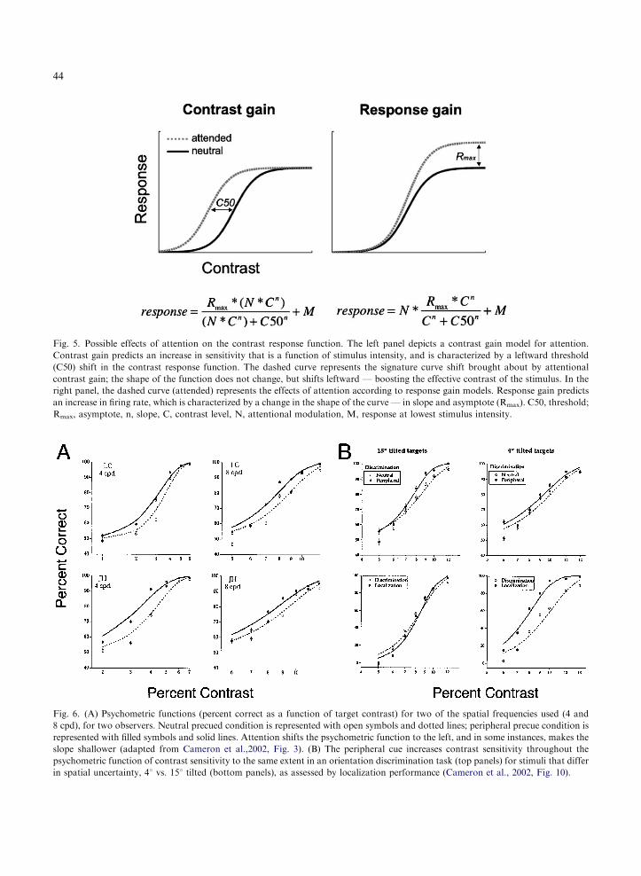
Fig. 5. Possible effects of attention on the contrast response function. The left panel depicts a contrast gain model for attention.
Contrast gain predicts an increase in sensitivity that is a function of stimulus intensity, and is characterized by a leftward threshold
(C50) shift in the contrast response function. The dashed curve represents the signature curve shift brought about by attentional
contrast gain; the shape of the function does not change, but shifts leftward — boosting the effective contrast of the stimulus. In the
right panel, the dashed curve (attended) represents the effects of attention according to response gain models. Response gain predicts
an increase in firing rate, which is characterized by a change in the shape of the curve — in slope and asymptote (Rmax). C50, threshold;
Rmax, asymptote, n, slope, C, contrast level, N, attentional modulation, M, response at lowest stimulus intensity.
Fig. 6. (A) Psychometric functions (percent correct as a function of target contrast) for two of the spatial frequencies used (4 and
8 cpd), for two observers. Neutral precued condition is represented with open symbols and dotted lines; peripheral precue condition is
represented with filled symbols and solid lines. Attention shifts the psychometric function to the left, and in some instances, makes the
slope shallower (adapted from Cameron et al.,2002, Fig. 3). (B) The peripheral cue increases contrast sensitivity throughout the
psychometric function of contrast sensitivity to the same extent in an orientation discrimination task (top panels) for stimuli that differ
in spatial uncertainty, 41 vs. 151 tilted (bottom panels), as assessed by localization performance (Cameron et al., 2002, Fig. 10).
44

45
attentional effect of similar magnitude was ob-served (Fig. 6b). In addition, to directly assess theease with which observers can localize the stimu-lus, we also performed a localization task. Whenthe target was tilted 151, discrimination and local-ization performance were tightly coupled. How-ever, when the targets were tilted 41, performanceon the localization task was much better than per-formance on the discrimination task. Notwith-standing the superior localization performance onthe 41 discrimination task, the attentional effectwas comparable for both orientation conditions.Importantly, at contrasts that yielded perfectlocalization, there was still an attentional effectin the discrimination tasks. Thus, given that weused suprathreshold stimuli, excluded all sourcesof added external noise (distracters, local and glo-bal masks) and showed experimentally that spatialuncertainty cannot explain this decrease in thresh-old, the observed attentional benefit is consistentwith a signal enhancement mechanism.
Transient attention increases contrast at the cued
location and decreases it at the uncued location
It had been proposed that very few neurons can beconcurrently engaged, but this proposition only re-cently became tractable and has now been system-atically evaluated. The calculations are astonishing— the cost of a single spike is high and severelylimits (possibly to about 1%) the number of neu-rons that can be (substantially) active concurrently(Lennie, 2003). The limited energy expenditure thatthe brain can afford necessitates machinery for thesystem to allocate energy according to task de-mand. This limited capacity entails selective atten-tion, which enables us to process effectively vastamounts of visual information by selecting relevantinformation from noise. In this study we investi-gated the possibility that covert attention helps tocontrol the expenditure of cortical computation bytrading contrast sensitivity across attended and un-attended areas of the visual field, even with impov-erished displays and simple tasks. Specifically, weassessed contrast sensitivity at both cued and un-cued locations (Pestilli and Carrasco, 2005).
There is consensus that attention improves per-formance at the attended location, but there is less
agreement regarding the fate of information that isnot directly attended, i.e., outside the focus of at-tention (Eriksen and Hoffman, 1974; Rock andGutman, 1981; Kinchla, 1992). Although mosthypotheses regarding the distribution of attentionin the visual field assume that information outsidethe attended area is not processed, many studieshave shown that information beyond the focus ofattention affects performance, indicating that it isprocessed to a certain degree (Carrasco andMcElree, 2001; Carrasco et al., 2004a,b; Cameronet al., 2004).
When manipulating attention, a cue is con-sidered valid when it indicates the target location,and it is considered invalid when it indicates anontarget location. Although assessing the effectsof attention by comparing performance in thevalid and invalid conditions is useful for distin-guishing between sensitivity-based and decisional-based explanations of the cueing effect, thiscomparison cannot determine whether such aneffect is due to an enhanced signal at the cuedlocation, a diminished signal at the uncued loca-tion, or both. To pinpoint the source of the at-tentional effect, it is necessary to compareperformance in both the valid and invalid condi-tions with a neutral condition, in which the cuedoes not indicate a stimulus location but only thetiming of the display onset (Hawkins et al., 1990;Luck et al., 1994; Carrasco and Yeshurun, 1998).
We evaluated the effect of transient attention oncontrast sensitivity at both the attended and un-attended locations. As discussed above, at the at-tended area transient attention increases sensitivityin an orientation discrimination task with an in-formative cue, i.e., when the cue indicates targetlocation but not its orientation (Lu and Dosher,1998; Carrasco et al., 2000; Cameron et al., 2002).When a peripheral cue is always valid in terms oflocation, however, some of its effect could be dueto a conceptually driven, voluntary componentof attention. To eliminate this possible contami-nation, we ensured cue unpredictability by cue-ing the target only 50% of the time, and byasking observers to report the orientation of thestimulus indicated by a response cue (a line dis-played after stimuli offset). Indeed, observerscould have entirely disregarded the cue and based

46
their responses only on the information accumu-lated during stimulus presentation and still at-tained the same overall performance level. The useof the nonpredictive cue and the response cueenabled us to isolate the purely automatic orient-ing of attention. Given that the transient periph-eral cue is thought to be automatic (Yantis andJonides, 1984; Jonides and Yantis, 1988), even anuninformative cue (which indicates neither targetlocation nor orientation) should exert an effect onperformance.
Previous studies have examined the effect of at-tention on contrast sensitivity at parafoveal loca-tions (e.g., Lee et al., 1997; Lu and Dosher, 1998,2000; Cameron et al., 2002; Solomon, 2004). Weinvestigated the effects of transient attention atboth parafoveal and peripheral locations to assesswhether the benefit and cost varied as a function ofthe distance between the attended and unattendedstimuli. Observers were asked to discriminate theorientation of 1 of 2 Gabor patches simultaneouslypresented left and right of fixation (at either 4 or 91of eccentricity). Contrast sensitivity was measuredat the cued (valid cue) and uncued (invalid cue)locations, and compared with the contrast sensi-tivity obtained at the same locations when the tar-get was preceded by a cue presented at fixation(neutral cue). Based on models of signal enhance-ment, which propose that attention directly im-proves the quality of the stimulus representation(Bashinski and Bacharach, 1980; Lu and Dosher,1998; Muller et al., 1998; Carrasco et al., 2000;Cameron et al., 2002), we hypothesized that sen-sitivity would be increased at the cued location.Based on models of distracter exclusion, whichpropose that attention allows us to exclude dis-tracters from the signal by narrowing the filterprocessing the stimulus (Davis et al., 1983; Palmer,1994; Solomon et al., 1997; Foley and Schwarz,1998; Morgan et al., 1998; Baldassi and Burr,2000), we hypothesized that sensitivity will be re-duced at the uncued location.
Following a peripheral or a central-neutral tran-sient cue, two slightly tilted Gabor patches weresimultaneously presented to the left and right offixation (Fig. 7). A response cue was presented af-ter the Gabors, indicating to the observer forwhich Gabor the orientation was to be reported,
thus defining valid and invalid trials (cue locationand response-cue match and do not match, re-spectively). We estimated contrast thresholds un-der each attention condition at each eccentricity.Usually, with invalid cue trials attention is divertedaway from the target location at stimulus onset,but observers have information regarding the tar-get location because its identity differs from thedistracter. However, in this study, observers didnot know where the target was, and they had toprocess the identity of the stimuli presented atboth locations to perform the task (Fig. 7).
To quantify the magnitude of the attentionaleffect, we calculated the ratio of the contrast sen-sitivity (1/median threshold) for valid vs. neutralcue, and invalid vs. neutral cue at both eccentric-ities. No difference between the two conditionswould yield a ratio equal to 1. A benefit in contrastsensitivity is indicated by values 41; a cost byvalues o1. All observers followed the same pat-tern of responses: values 41 for the valid:neutralratio (benefit) and values o1 for the invalid:neu-tral ratio (cost). Figure 8 (left panel) shows thevalues for one observer.
The data for individual observers were consist-ent with the overall frequency distributions. Thehistograms represent the threshold values obtainedin each cue condition at each eccentricity. Al-though the absolute contrast threshold and thespread of the distribution varied across observers,the valid cue (blue histograms) improved perform-ance and the invalid cue (red histograms) impairedperformance with respect to the neutral cue foreach individual observer at both eccentricities.Fig. 8 (right panel) illustrates the frequency distri-bution for the same observer. The same pattern ofresults, and of comparable magnitude, was ob-tained at both parafoveal and peripheral locations.
Results from all observers indicate that despitethe fact that they were told that the cue was un-informative as to the target location and orienta-tion, and despite the simplicity of the display, thereis a performance trade-off: the cue increases sen-sitivity at the cued location (benefit) and impairs itat the uncued location (cost), as compared to theneutral condition. This indicates that informationat the attended location is processed to a greaterdegree than in the neutral condition, and that
Valid
Neutral
Invalid
Threshold (% contrast)
5 8 13 20 32 5 8 13 20 32
20151050
20151050
20151050
Num
ber
of th
resh
olds
4° 9°JG
4° 9°
0.8
0.9
1
1.1
1.2
Cos
tB
enef
it
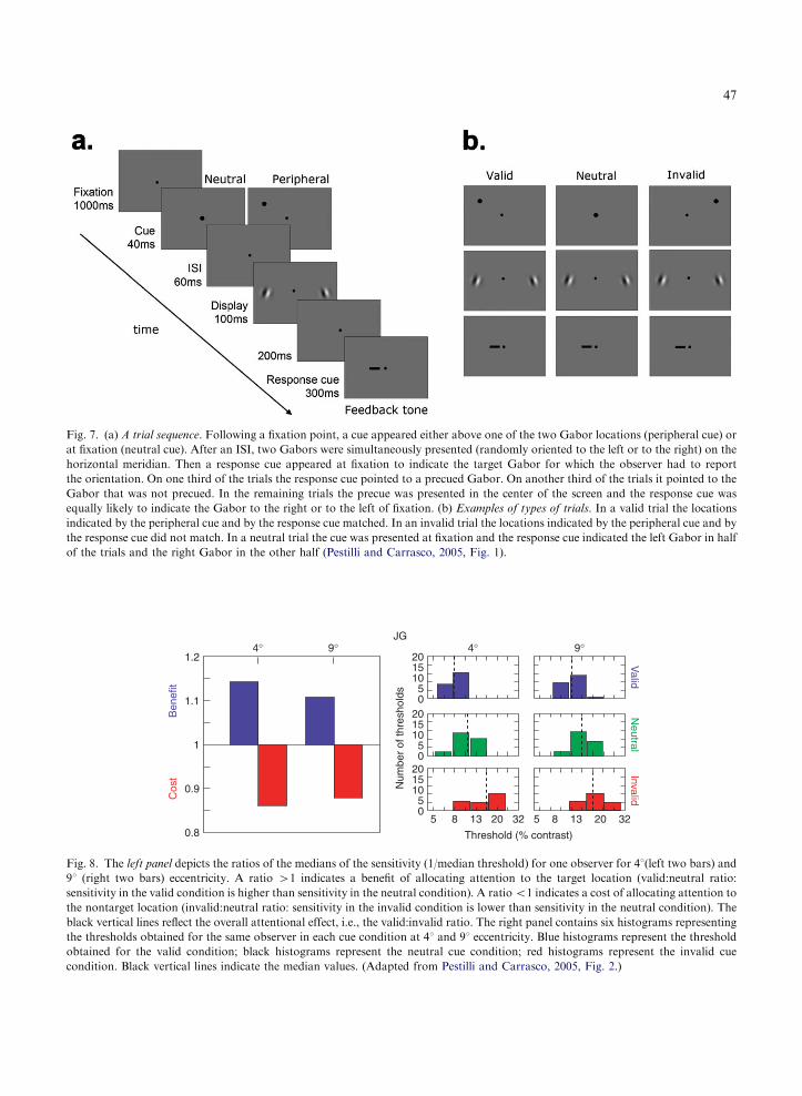
Fig. 8. The left panel depicts the ratios of the medians of the sensitivity (1/median threshold) for one observer for 41(left two bars) and
91 (right two bars) eccentricity. A ratio 41 indicates a benefit of allocating attention to the target location (valid:neutral ratio:
sensitivity in the valid condition is higher than sensitivity in the neutral condition). A ratioo1 indicates a cost of allocating attention to
the nontarget location (invalid:neutral ratio: sensitivity in the invalid condition is lower than sensitivity in the neutral condition). The
black vertical lines reflect the overall attentional effect, i.e., the valid:invalid ratio. The right panel contains six histograms representing
the thresholds obtained for the same observer in each cue condition at 41 and 91 eccentricity. Blue histograms represent the threshold
obtained for the valid condition; black histograms represent the neutral cue condition; red histograms represent the invalid cue
condition. Black vertical lines indicate the median values. (Adapted from Pestilli and Carrasco, 2005, Fig. 2.)
Fig. 7. (a) A trial sequence. Following a fixation point, a cue appeared either above one of the two Gabor locations (peripheral cue) or
at fixation (neutral cue). After an ISI, two Gabors were simultaneously presented (randomly oriented to the left or to the right) on the
horizontal meridian. Then a response cue appeared at fixation to indicate the target Gabor for which the observer had to report
the orientation. On one third of the trials the response cue pointed to a precued Gabor. On another third of the trials it pointed to the
Gabor that was not precued. In the remaining trials the precue was presented in the center of the screen and the response cue was
equally likely to indicate the Gabor to the right or to the left of fixation. (b) Examples of types of trials. In a valid trial the locations
indicated by the peripheral cue and by the response cue matched. In an invalid trial the locations indicated by the peripheral cue and by
the response cue did not match. In a neutral trial the cue was presented at fixation and the response cue indicated the left Gabor in half
of the trials and the right Gabor in the other half (Pestilli and Carrasco, 2005, Fig. 1).
47

48
information processed outside of the focus of at-tention is processed to a lesser degree. Given thatfor an ideal observer the uninformative cue wouldnot reduce uncertainty, this finding supportssensitivity-based explanations, i.e., signal enhance-ment at the cued location — the sensory represen-tation of the relevant stimuli is boosted — anddistracter exclusion at the uncued location — theinfluence of the stimuli outside the attentionalfocus is reduced.
By illustrating that transient attention can helpin managing the overall bioenergetic expenditureacross the attended and unattended locations ofthe visual field, this study provides evidence for thenotion that transient attention directs observers’attention to the cued location in an automaticfashion (Muller and Rabbitt, 1989; Nakayama andMackeben, 1989; Cheal and Lyon, 1991; Yantis,1996).
Transient attention increases apparent contrast
From recent psychophysical and neurophysiolog-ical evidence indicating that covert attention in-creases contrast sensitivity, one might infer thatattention changes contrast appearance. But doesattention alter appearance? Whether attention canactually affect the perceived intensity of a stimulushas been a matter of debate dating back to thefounding fathers of experimental psychology andpsychophysics — Helmholtz, James, and Fechner(Helmholtz, 1866/1911; James, 1890). Surprisingly,very little direct empirical evidence has beenbrought to bear on the issue (Tsal et al., 1994;Prinzmetal et al., 1997, 1998), and a number ofmethodological concerns limit the conclusions wecan draw from these studies (Carrasco et al.,2004a,b; Luck, 2004; Treue, 2004; Gobell andCarrasco, 2005).
To directly investigate this issue, Carrasco et al.,(2004a,b) implemented a novel paradigm that en-ables us to assess the effects of spatial cueing onappearance and to test subjective contrast. Thisparadigm allows one to objectively assess observers’subjective experience while circumventing method-ological limitations of previous studies, and to ad-dress other questions about phenomenologicalexperience, making it possible to study subjective
experience more objectively and rigorously (Luck,2004; Treue, 2004).
Observers were briefly presented with either aperipheral or neutral cue, followed by two Gaborpatches (tilted to the left or right) to the left andright of fixation (Fig. 9). The contrast of one of theGabors was presented at a fixed contrast (stand-ard), whereas the other varied in contrast randomlyfrom a range of values around the standard (testpatch). The orientation of each Gabor was chosenrandomly. We manipulated transient attention withan uninformative peripheral cue. We asked the ob-servers: what is the orientation of the stimulus thatis higher in contrast? These instructions emphasizedthe orientation judgment, when in fact we were in-terested in their contrast judgments; i.e., the orien-tation discrimination task served as a ‘cover story’task, which de-emphasized the fact that we wereinterested in the observers’ subjective experience.
The results showed that transient attention sig-nificantly increased perceived contrast (Fig. 10).When a Gabor was peripherally cued, the point ofsubjective equality (PSE) was shifted — the ap-parent contrast of the stimulus for which transientattention had been drawn to was higher than whenattention was not drawn there. That is to say,when observers attend to a stimulus, they perceiveit to be of significantly higher contrast than whenthey perceive the same stimulus without attention.
We conducted multiple control experiments torule out alternative accounts of these findings:(1) We increased the temporal separation betweenthe cue onset and the display onset from 120ms,the optimal time for transient attention, to 500ms,when transient attention is no longer active. Con-sistent with the quick decay of transient attentionto the cued location, this manipulation yielded nocontrast enhancement of the cued stimulus, i.e.,there is no appearance effect (data not shown).This result shows that observers were not biased toreport the orientation of a cued stimulus per se.(2) When observers are asked to report the orien-tation of the Gabor of lower contrast, they selectthe cued stimulus less often if it is of the same con-trast as the uncued stimulus (data not shown). Thisresult is consistent with the enhanced apparentcontrast of the cued stimulus observed in the mainexperiment. This control rules out the possibility

Fig. 9. (a) Sequence of events in a single trial. Each trial began with a fixation point followed by a brief neutral or peripheral cue. The
peripheral cue had equal probability of appearing on the left- or right-hand side, and was not predictive of the stimulus contrast or
orientation. The timing of this sequence maximized the effect of transient attention and precluded eye movements. (b) Task. Observers
performed a 2� 2 forced choice (2� 2 AFC) task: they were asked to indicate the orientation (left vs. right) for the stimulus that appeared
higher in contrast. In this trial, they would report the orientation for the stimulus on the right. (Carrasco et al., 2004a,b, Fig. 1.)
49
that observers report the orientation of a cuedstimulus more often simply because they find itsorientation easier to judge or are subject to sometype of cue bias.
This study provides evidence for a contrast gainmodel (Reynolds et al., 1999, 2000) in which at-tention allows for greater neuronal sensitivity,suggesting that attention changes the strength of astimulus by enhancing its effective contrast or sa-lience. It is as if attention boosts the actual stim-ulus contrast. The finding that the cue not onlyenhanced the cued stimulus’ appearance but alsoimproved the observers’ performance supports thehypothesis that the increased saliency at the targetlocation seems to be the basis of perceptual judg-ments. Many have considered the saliency map tobe the basis of perceptual judgments and a tool fordirecting gaze to potential relevant locations of thevisual environment (Itti and Koch, 2001; Treue,2004; Gobell et al., 2004; Itti, 2005; Zhaoping,2005).
Sustained attention and contrast sensitivity
Single-unit studies have evaluated the effects ofattention on the contrast response function bymanipulating sustained attention (Reynolds et al.,2000; Martinez-Trujillo and Treue, 2002). To eval-uate the similarity of the transient and sustainedsystems of attention, it is important to characterizetheir effects on early vision, and to investigatewhether the same mechanism(s) can underlie sucheffects. Recently, Ling and Carrasco (2006) ob-tained contrast psychometric functions for bothsustained and transient attention to further bridgethe gap between neurophysiological and psycho-physical results. We systematically compared sus-tained and transient covert attention using the sametask, stimuli, and observers. We tested whether asignal enhancement mechanism underlies both typesof attention. Moreover, we investigated the neuralmodel underlying signal enhancement by measuringthe psychometric functions for both sustained and

Fig. 10. Attention alters appearance. Top panel: Appearance
psychometric function. Percentage of responses in which ob-
servers reported the contrast of the test patch as higher than the
standard, plotted as a function of the physical contrast of the
test patch. Data are shown for the neutral and peripheral con-
ditions (test cued and standard cued). The standard was 22%
contrast and that is the contrast at which the test and standard
stimuli attained subjective equality (50%). Bottom panel: Effect
of covert attention on apparent contrast. If you were looking at
one of the two fixation points (black dots), and the grating to
the left of that fixation point was cued, the stimuli at both sides
of fixation would appear to have the same contrast. A cued
16% contrast grating appears as if it were 22% contrast, and a
cued 22% contrast grating appears as if it were 28% contrast.
(Adapted from Carrasco et al., 2004a,b, Figs. 4 and 5.)
50
transient attention to assess whether they have sim-ilar or different effects on the contrast responsefunction.
As mentioned above, two types of gain controlmechanisms have been considered in neural re-sponses to luminance-modulated stimuli — contrastgain and response gain (Sclar et al., 1989; see Fig. 5).We had provided evidence in support of a contrastgain mechanism for transient attention (Cameronet al., 2002). However, in that study, performanceasymptoted close to 100%, leaving little room at thehigher contrasts for a possible test of response gain.Neurophysiological studies of sustained attention
that have evaluated these two mechanisms haveavoided levels at which neural saturation occurs.Similarly, to properly compare contrast gain andresponse gain psychophysically, the psychometricfunctions should arise from a demanding task thatensures that performance on the neutral baselinecondition does not asymptote at 100%, leavingroom to test for response gain.
Observers performed a 2AFC orientation dis-crimination task on a slightly tilted Gabor patch.We first established the contrast range required tomeasure the full extent of the psychometric func-tion with an asymptote that occurs at a perform-ance level that allows room for benefit. We usedthe method of constant stimuli to measure per-formance as a function of target contrast in theneutral, transient, and sustained cue conditions. Ineach trial, a Gabor is presented in 1 of 8 possibleisoeccentric locations. The cues (sustained and itsneutral control vs. transient and its neutral con-trol) are constant throughout a block, but thespatial frequency and contrast levels are randomi-zed within each block.
Using a nested hierarchical model, for each ob-server we estimated the probability that the sameWeibull distribution can describe the data sets forboth cue conditions (sustained vs. its neutral con-trol; transient vs. its neutral control), as opposedto two separate distributions. Additionally, to testthe models of response gain vs. contrast gain, wefit the data to their respective models, along with ahybrid model of both response and contrast gain,and compare likelihoods to assess which modeldescribes the data better. Whereas response gainpredicts an increasing effect of attention with con-trast (a multiplicative effect across the psychomet-ric function), contrast gain predicts only a shift insensitivity with attention (an overall additive effectindependent of stimulus intensity). The hybridmodel predicts both a shift in sensitivity as well amultiplicatively increasing effect of attention.
Results indicate that whereas sustained attentionoperates via contrast gain (Fig. 11; top panel; char-acterized by a shift in threshold), transient attentionoperates via a mixture of contrast and response gain(bottom panel; characterized by an effect even athigh-contrasts asymptotic levels; Ling and Carrasco,2006). An uncertainty reduction model of attention

Fig. 11. Psychometric functions for sustained and transient attention. The solid line represents the fits for the neutral condition, and the
dashed line represents the fits for the precued. (a) Sustained attention consistently shifted the function to the left, having little impact on its
shape, but increasing contrast sensitivity. (b) Transient attention consistently led to an elevation in asymptote, and the fits suggest a
decrease in contrast threshold as well. Error bars correspond to mean71 standard error. (Adapted from Ling and Carrasco, 2005, Fig. 3.)
51
would predict that the attention effect should bemost prominent with low-contrast stimuli (whereuncertainty is greatest and performance would ben-efit most from uncertainty reduction) and decreasewith increasing stimulus contrast (where uncertaintyis diminished and performance would not benefitfrom uncertainty reduction). However, this was notthe case in this study. Moreover, different signatureresponses across the psychometric function emergednotwithstanding the fact that the reduction of loca-tion uncertainty is the same in both cases.
Using the external noise paradigm, Lu andDosher (1998, 2000) reported that transient covertattention seems to operate via both signal en-hancement and external noise reduction. Theyshowed that transient attention increases contrastsensitivity in conditions of low noise, indi-cative of signal enhancement, and also improves
performance in high-noise conditions, indicativeof external noise reduction. However, they haveattributed sustained attention effects only to anexternal noise reduction mechanism (Dosher andLu, 2000a,b; Lu and Dosher, 2000, 2002).
With regard to transient attention, these andprevious findings are in agreement; under low ex-ternal noise conditions, it operates via signal en-hancement. However, the results for sustainedattention are inconsistent with those reported pre-viously by Dosher and Lu. The most relevantdifference that could help reconcile the discrep-ancy lies in the amount of time observers weregiven to deploy their sustained attention. The SOAin their studies was 150ms because it has been re-ported that this time was enough for experiencedobservers to deploy sustained attention (Cheal andLyon, 1991). Perhaps this short timing precluded

52
emergence of the signal enhancement mechanism.It is possible that the observers who failed to showan effect were not trained optimally to deploy sus-tained attention within the allotted time; had theyhad longer time to deploy sustained attention aneffect could have emerged.
In a sustained attention task, using a dual-taskparadigm in which observers performed tasks underconditions of full or poor attention, evidence forpure response gain has been reported (Morroneet al., 2004). However, a subsequent psychophysicalstudy suggested that dual task, sustained attentionmay operate via a hybrid model, involving bothcontrast gain and response gain (Huang andDobkins, 2005). Whereas the dual-task paradigmhas some advantages, such as eliminating locationuncertainty reduction as an alternative explanation,it has disadvantages that may have hampered theirconclusions. Dual-task paradigms do not controlthe deployment of attention very well and makeit hard to isolate the source of possible process-ing differences (e.g., Sperling and Dosher, 1986;Pashler, 1998). The difference with the present re-sults may be due to the way in which attention wasmanipulated. First, in dual-task paradigms, atten-tion is not directed to a specific location, but theamount of resources being spread to all locationsis manipulated. Second, to manipulate attentionthose authors withdrew attention from the target,whereas we directed attention toward the target.
This study systematically compared sustainedand transient covert attention using the same task,stimuli, and observers. On the one hand, bothtypes of attention had a similar effect on perform-ance; they increased contrast sensitivity underzero-noise conditions (the display contained noth-ing to be suppressed, since there was no addedexternal noise). Hence, we conclude that both at-tentional systems can be mediated by a signal en-hancement mechanism. Furthermore, because thiseffect occurred even with very high-contrast stim-uli, it cannot be explained by uncertainty reduc-tion. On the other hand, sustained and transientattention had different effects on the contrast re-sponse function. Sustained attention enhancescontrast sensitivity strictly via contrast gain,whereas, in addition to contrast gain, transient at-tention revealed response gain.
Neurophysiological studies of attentional
modulation of apparent stimulus contrast:
attentional facilitation and selection
The development of techniques to record the ac-tivity of neurons in awake-behaving animals hasenabled researchers to probe the biological foun-dations of sustained attention. Single-unit record-ing studies in the monkey have provided detailed,quantitative descriptions of how attention altersvisual cortical neuron responses.
A number of neurophysiological studies haveshown that directing attention to a stimulus in-creases neuronal sensitivity, so that neurons re-spond to an attended stimulus much as they wouldwere its luminance increased. It is possible to relatethese findings to studies in anesthetized cats andmonkeys documenting how luminance contrastaffects neuronal responses. The same models ex-plaining contrast-dependent changes in neuronalresponse can account for contrast-dependent mod-ulation of the competitive interactions observedwhen multiple stimuli appear within a neuron’sreceptive field (for reviews see Reynolds andChelazzi, 2004; Martinez-Trujillo and Treue,2005; Reynolds, 2005).
With regard to attentional facilitation, consistentwith psychophysical findings, single-unit record-ing studies have found that spatial attention canenhance responses evoked by a single stimulus ap-pearing alone in a neuron’s receptive field (e.g.,Motter, 1993; Ito and Gilbert, 1999; McAdams andMaunsell, 1999; Reynolds et al., 2000). Reynoldset al. (2000) assessed the effects of sustained atten-tion on contrast sensitivity when a single stimulusappeared in a neuron’s receptive field. The monkey’stask was to detect a target grating that could appearat an unpredictable time at the cued location. Thetarget’s luminance contrast was randomly selectedto ensure that the monkey had to attend continuallyto the target location. The contrast response func-tion (CRF) summarizes the way in which changes instimulus contrast are translated into changes inneuronal firing rate via a nonlinear sigmoid func-tion (Fig. 5). Consistent with a contrast gain, in V4,an extrastriate visual area at an intermediate stageof the ventral processing stream, attention shiftsthe CRF horizontally with the most pronounced

Fig. 12. Response of an example neuron from area V4 as a
function of attention and stimulus contrast. (A) The contrast of
the stimulus in the receptive filed increased from 5% (bottom
panel) to 10% (middle panel) to 80% (top panel). The monkey
had to detect a grating at the attended location. On each trial,
attention was directed to either the location of the stimulus in-
side of the receptive field (solid line) or a location far away from
the receptive field (dotted line). Attention reduced the contrast
threshold to elicit a response (middle panel), but did not affect
the response at saturation contrast (top panel). (B) Averages
responses of V4 neurons while the monkey attends to the lo-
cation (thick line) or away (thin line) of the receptive field (thin
line). The horizontal line depicts the five different contrast val-
ues of the gratings presented inside the RF, which spanned the
dynamic range of the neuron. The dashed and dotted lines show
percentage and absolute difference in firing rate, respectively,
across the two attention conditions, as a function of contrast.
(Adapted with permission from Reynolds et al., 2000.)
53
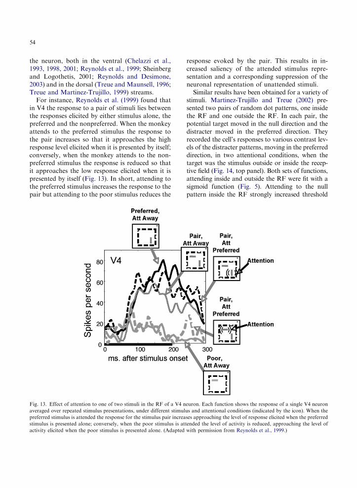
changes occurring at its dynamic range (steepest re-gion). When the grating stimulus appearing in theneuron’s receptive field was below the contrast re-sponse threshold (5% and 10% contrast), it fails toelicit a response, when unattended. However, whenthe monkey attended to its location in the RF thesame 10% contrast elicits the neuron to respond.Attention does not alter the neuronal response whenthe stimulus is above saturated contrast. Across apopulation of V4 neurons, the greatest increments infiring rate were observed at contrasts in the dynamicrange of each neuron’s CRF (Fig. 12). The findingthat similar results were found for preferred andpoor stimuli indicates that the lack of attentionaleffect at high contrast did not reflect an absolutefiring rate limit; instead, it reflected a leftward shiftin the contrast response function.
Under the conditions of this experiment, for acell to reliably detect an unattended stimulus, itscontrast needed to be 50% higher than that of theattended stimulus; i.e., attention was equivalent toabout 50% increase in contrast (Reynolds et al.,2000). This value has been corroborated by otherstudies that have also quantified spatial attentionin units of luminance contrast, including studies inMT (Martinez-Trujillo and Treue, 2002) and in V4(Reynolds and Desimone, 2003), whose estimateswere 50% and 56%, respectively. As mentionedabove, this effect of attention is indistinguishablefrom a change in stimulus contrast (see alsoMaunsell and McAdams, 2000).
Given our limited ability to process information,it is also crucial to understand how attentional se-lection of behavioral relevant stimuli from amongcompeting distracters (Wolfe, 1994; Palmer et al.,2000; Carrasco and McElree, 2001; Verghese,2001; Cameron et al., 2004) may be instantiatedat a neural level. Neuronal recordings within theextrastriate cortex have revealed a direct neuralcorrelate of attentional selection. Moran andDesimone (1985) were the first to show that thefiring rate is determined primarily by the task-relevant stimulus. This seminal study showed thatwhen two stimuli are presented within the receptivefield, the neuron’s response to the pair is greaterwhen the monkey is asked to identify the stimuluscorresponding to the neuron’s preferred color andorientation than when asked to identify the
nonpreferred stimulus. Several labs have replicatedthis observation that the attentional modulationdepends on the similarity between the attendedstimulus properties and the sensory preferences of
54
the neuron, both in the ventral (Chelazzi et al.,1993, 1998, 2001; Reynolds et al., 1999; Sheinbergand Logothetis, 2001; Reynolds and Desimone,2003) and in the dorsal (Treue and Maunsell, 1996;Treue and Martinez-Trujillo, 1999) streams.
For instance, Reynolds et al. (1999) found thatin V4 the response to a pair of stimuli lies betweenthe responses elicited by either stimulus alone, thepreferred and the nonpreferred. When the monkeyattends to the preferred stimulus the response tothe pair increases so that it approaches the highresponse level elicited when it is presented by itself;conversely, when the monkey attends to the non-preferred stimulus the response is reduced so thatit approaches the low response elicited when it ispresented by itself (Fig. 13). In short, attending tothe preferred stimulus increases the response to thepair but attending to the poor stimulus reduces the
Fig. 13. Effect of attention to one of two stimuli in the RF of a V4 n
averaged over repeated stimulus presentations, under different stimul
preferred stimulus is attended the response for the stimulus pair increa
stimulus is presented alone; conversely, when the poor stimulus is at
activity elicited when the poor stimulus is presented alone. (Adapted
response evoked by the pair. This results in in-creased saliency of the attended stimulus repre-sentation and a corresponding suppression of theneuronal representation of unattended stimuli.
Similar results have been obtained for a variety ofstimuli. Martinez-Trujillo and Treue (2002) pre-sented two pairs of random dot patterns, one insidethe RF and one outside the RF. In each pair, thepotential target moved in the null direction and thedistracter moved in the preferred direction. Theyrecorded the cell’s responses to various contrast lev-els of the distracter patterns, moving in the preferreddirection, in two attentional conditions, when thetarget was the stimulus outside or inside the recep-tive field (Fig. 14, top panel). Both sets of functions,attending inside and outside the RF were fit with asigmoid function (Fig. 5). Attending to the nullpattern inside the RF strongly increased threshold
euron. Each function shows the response of a single V4 neuron
us and attentional conditions (indicated by the icon). When the
ses approaching the level of response elicited when the preferred
tended the level of activity is reduced, approaching the level of
with permission from Reynolds et al., 1999.)
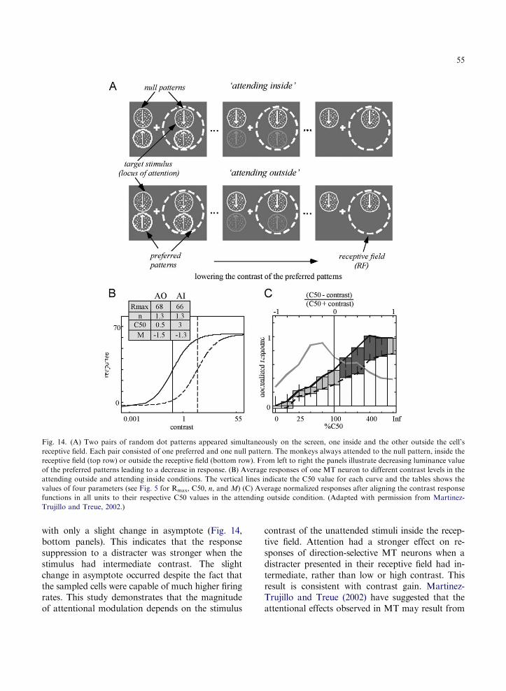
Fig. 14. (A) Two pairs of random dot patterns appeared simultaneously on the screen, one inside and the other outside the cell’s
receptive field. Each pair consisted of one preferred and one null pattern. The monkeys always attended to the null pattern, inside the
receptive field (top row) or outside the receptive field (bottom row). From left to right the panels illustrate decreasing luminance value
of the preferred patterns leading to a decrease in response. (B) Average responses of one MT neuron to different contrast levels in the
attending outside and attending inside conditions. The vertical lines indicate the C50 value for each curve and the tables shows the
values of four parameters (see Fig. 5 for Rmax, C50, n, and M) (C) Average normalized responses after aligning the contrast response
functions in all units to their respective C50 values in the attending outside condition. (Adapted with permission from Martinez-
Trujillo and Treue, 2002.)
55
with only a slight change in asymptote (Fig. 14,bottom panels). This indicates that the responsesuppression to a distracter was stronger when thestimulus had intermediate contrast. The slightchange in asymptote occurred despite the fact thatthe sampled cells were capable of much higher firingrates. This study demonstrates that the magnitudeof attentional modulation depends on the stimulus
contrast of the unattended stimuli inside the recep-tive field. Attention had a stronger effect on re-sponses of direction-selective MT neurons when adistracter presented in their receptive field had in-termediate, rather than low or high contrast. Thisresult is consistent with contrast gain. Martinez-Trujillo and Treue (2002) have suggested that theattentional effects observed in MT may result from

56
the modulation of input gain, which would be sim-ilar to a change in stimulus contrast; that theattentional effects vary nonlinearly as a function ofstimulus contrast indicates that these effects are notsimple multiplications of a cell’s response.
Likewise, Chelazzi et al. (2001) obtained a similarpattern of results while recording in area V4 ofmonkeys performing a visual search task involvingobjects (faces and houses). The finding that attend-ing to the preferred stimulus increases the responseto the pair but attending to the poor stimulus re-duces the response evoked by the pair, providessupport for attentional models positing that responsesuppression mediates the selection of one stimulusand the inhibition of the other (e.g., Desimone andDuncan, 1995; Ferrera and Lisberger, 1995; Leeet al., 1999).
The common nonlinearity between the CRF andthe magnitude of attentional modulation acrossdifferent areas of the dorsal and ventral pathwaysindicate a tight link between attentional mecha-nisms and the mechanisms responsible for contrastencoding. Indeed, it has been proposed that be-cause this tight link does not seem to exist for theencoding mechanisms of other stimulus properties(e.g., motion coherence), attention can be con-sidered a mechanism aimed at modulating stimu-lus saliency while leaving other stimulus propertiesrelatively unchanged (Martinez-Trujillo andTreue, 2002).
A similar relative enhancement or suppressionof stimulus representations can be obtained whenvarying stimulus contrast. Given that the role ofcontrast in modulating the visual response prop-erties of neurons has been well documented, andthat response suppression plays an important rolein the models developed to account for these mod-ulations, to better understand the role that atten-tion may play in selecting stimuli, it is importantto consider the role of suppression in the visualcortex (Reynolds and Chelazzi, 2004; Reynolds,2005).
Contrast changes in the visual stimulus yield mul-tiplicative changes in neuronal responses, similar tothose evoked by attention (e.g., Tolhurst, 1973; Sclarand Freeman, 1982; Treue and Martinez-Trujillo,1999). The similarity between the effects of attentionand the effects of varying stimulus contrast could
indicate that these two processes are closely relatedand probably use similar mechanisms to modulateneuronal responses. Were this the case, one would ex-pect attentional and contrast modulation of neuronalresponses to share the same properties (Martinez-Trujillo and Treue, 2005; Reynolds, 2005).
It is known that contrast modulates neuronalresponses in the following ways:
(1)
Cortical neuronal responses typically saturateas contrast increases, and this saturation-firing rate is stimulus-dependent. The dynamicrange is larger and the saturation point ishigher for a stimulus of the neuron’s preferredorientation than for a stimulus of poor butexcitatory orientation, and no response is elic-ited by a nonpreferred stimulus. Thus, in-creasing contrast leads to a multiplicativeincrease in the response (Sclar and Freeman,1982; Fig. 15A). Correspondingly, consistentwith the idea of attentional facilitation, atten-tion causes a leftward shift in the contrast re-sponse function by increasing the effectivecontrast of a stimulus (Reynolds et al., 2000;Fig. 12).(2)
Increasing the stimulus contrast characteris-tically results in a multiplicative increase inthe neurons’ tuning curves for propertiessuch as motion and orientation (Sclar andFreeman, 1982; Fig. 15B). The multiplicativeeffect of contrast on the orientation-tuningcurve is due to the fact that contrast-response functions derived for any orientat-ions can be related to each other by a gainfactor, as is the case in Fig. 12. Similarly,spatial attention causes a multiplicative in-crease in the orientation-tuning curve ofneurons in area V4, without otherwise alter-ing its shape. This increase in the gain of theorientation-tuning curve enables neuronalsignals to better distinguish the orientationof the stimulus (McAdams and Maunsell,1999). Likewise, attention multiplicativelyincreases the direction of motion-tuningcurves of neurons in area MT (Treue andMartinez-Trujillo, 1999).(3)
When two spatially superimposed gratings ofdifferent orientation appear simultaneously
Fig 15. Contrast-dependent response modulations. (A) Contrast–response functions for a stimulus of the neuron’s preferred orien-
tation (upper line), a poor but excitatory orientation (middle line), and the null orientation (bottom line). (Adapted with permission
from Sclar and Freeman, 1982.) (B) Orientation tuning curves of a second neuron, stimulus contrast varied from 10% (empty circles)
to 80% (filled circles). (Adapted with permission from Sclar and Freeman (1982.) (C) Responses of a neuron recorded in area V1 of an
anesthetized macaque. Two superimposed gratings appeared within the receptive field: one grating was of optimal orientation, the
other was of a suboptimal orientation; both gratings varied from 0% to 50% contrast. (Adapted with permission from Carandini et al.,
1997.) (D–F) The contrast gain model can account for these contrast-dependent response modulations. (See text; adapted with
permission from Reynolds, 2005.)
57
within a receptive field in V1, increasing thecontrast of one of them results in an in-creased or decreased response depending onthe neuron’s selectivity for the two stimuli. Ingeneral, increasing the contrast of the pre-ferred grating increases the response to the
pair; conversely, increasing the contrast ofthe nonpreferred grating decreases theresponse to the pair. However, the highestcontrast preferred stimulus seems to be vir-tually immune to the suppressive effect of thenonpreferred stimulus (Carandini et al., 1997;
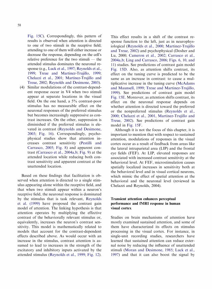
58
Fig. 15C). Correspondingly, this pattern ofresults is observed when attention is directedto one of two stimuli in the receptive field;attending to one of them will either increase ordecrease the response, depending on the cell’srelative preference for the two stimuli — theattended stimulus dominates the neuronal re-sponse (e.g., Luck et al., 1997; Reynolds et al.,1999; Treue and Martinez-Trujillo, 1999;Chelazzi et al., 2001; Martinez-Trujillo andTreue, 2002; Reynolds and Desimone, 2003).
(4)
Similar modulations of the contrast-depend-ent response occur in V4 when two stimuliappear at separate locations in the visualfield. On the one hand, a 5% contrast-poorstimulus has no measurable effect on theneuronal responses of the preferred stimulusbut becomes increasingly suppressive as con-trast increases. On the other, suppression isdiminished if the preferred stimulus is ele-vated in contrast (Reynolds and Desimone,2003; Fig. 16). Correspondingly, psycho-physical studies show that attention in-creases contrast sensitivity (Pestilli andCarrasco, 2005; Fig. 8) and apparent con-trast (Carrasco et al., 2004a,b; Fig. 9) at theattended location while reducing both con-trast sensitivity and apparent contrast at theunattended location.Based on these findings that facilitation is ob-served when attention is directed to a single stim-ulus appearing alone within the receptive field, andthat when two stimuli appear within a neuron’sreceptive field, the neuronal response is dominatedby the stimulus that is task relevant, Reynoldset al. (1999) have proposed the contrast gainmodel of attention. The linking hypothesis is thatattention operates by multiplying the effectivecontrast of the behaviorally relevant stimulus or,equivalently, increases the neuron’s contrast sen-sitivity. This model is mathematically related tomodels that account for the contrast-dependenteffects described above. As would occur with anincrease in the stimulus, contrast attention is as-sumed to lead to increases in the strength of theexcitatory and inhibitory inputs activated by theattended stimulus (Reynolds et al., 1999; Fig. 12).
This effect results in a shift of the contrast re-sponse function to the left, just as in neurophys-iological (Reynolds et al., 2000; Martinez-Trujilloand Treue, 2002) and psychophysical (Dosher andLu, 2000; Cameron et al., 2002; Carrasco et al.,2004a,b; Ling and Carrasco, 2006; Figs. 6, 10, and11) studies. See predictions of contrast gain modelFig. 15D. Also, as attention shifts contrast, itseffect on the tuning curve is predicted to be thesame as an increase in contrast: to cause a mul-tiplicative increase in the tuning curve (McAdamsand Maunsell, 1999; Treue and Martinez-Trujillo,1999). See predictions of contrast gain modelFig. 15E. Moreover, as attention shifts contrast, itseffect on the neuronal response depends onwhether attention is directed toward the preferredor the nonpreferred stimulus (Reynolds et al.,2000; Chelazzi et al., 2001, Martinez-Trujillo andTreue, 2002). See predictions of contrast gainmodel in Fig. 15F.
Although it is not the focus of this chapter, it isimportant to mention that with respect to sustainedattention, modulations of responses in the visualcortex occur as a result of feedback from areas likethe lateral intraparietal area (LIP) and the frontaleye fields (FEF). At LIP, elevated responses areassociated with increased contrast sensitivity at thebehavioral level. At FEF, microstimulation causesspatially localized increases in sensitivity both atthe behavioral level and in visual cortical neurons,which mimic the effect of spatial attention at thebehavioral and the neuronal level (reviewed inChelazzi and Reynolds, 2004).
Transient attention enhances perceptual
performance and fMRI response in human
visual cortex
Studies on brain mechanisms of attention havemostly examined sustained attention, and some ofthem have characterized its effects on stimulusprocessing in the visual cortex. For instance, insingle-unit recording studies, researchers havelearned that sustained attention can reduce exter-nal noise by reducing the influence of unattendedstimuli (Moran and Desimone, 1985; Luck et al.,1997) and that it can also boost the signal by

Fig. 16. Increasing the contrast of a poor stimulus at one location suppresses the response elicited by a fixed contrast preferred
stimulus at a second location in the receptive field of a V4 neuron. The poor stimulus’ contrast (left column) increased from 5 to 80%,
and did not elicit a clear response at any contrast. The preferred stimulus was fixed in contrast (right column). For the response to the
pair (middle column), at low contrast, the poor stimulus had no measurable effect on the response to the preferred stimulus, but as its
contrast increased (moving up the column) it became increasingly suppressive. (Adapted with permission from Reynolds and
Desimone, 2003.)
59
increasing the effective stimulus contrast (Reynoldset al., 2000; Martinez-Trujillo and Treue, 2002).Correspondingly, human electrophysiological stud-ies have provided evidence that attention can in-crease sensory gain (Johannes et al., 1995; Hillyardand Anllo-Vento, 1998), and neuroimaging studieshave shown attentional modulation of neuralactivity in many visual areas (Kanwisher andWojciulik, 2000; Kastner and Ungerleider, 2000),including the primary visual cortex (Brefczynskiand DeYoe, 1999; Gandhi et al., 1999; Martinezet al., 1999; Somers et al., 1999).
Less is known about the neural mechanism fortransient attention and its effects on stimulus
processing. Psychophysical findings demonstratingthat transient attention increases contrast sensitiv-ity (e.g., Lu and Dosher, 1998; Carrasco et al.,2000; Cameron et al., 2002; Ling and Carrasco,2006) suggest that transient attention should en-hance neural activity in early stages of visualprocessing. We tested this hypothesis by measuringbrain activity in early visual areas using rapidevent-related fMRI in conjunction with a peri-pheral cueing paradigm to manipulate transientattention (Liu et al., 2005). Participants discri-minated the orientation of one of two gratingspreceded or followed by a nonpredictive peri-pheral cue.

Fig. 17. The diagram on the left illustrates the locations of the
cue and the Gabor stimulus, which were presented in alternat-
ing blocks. Shown on the right are data from the right hem-
isphere of one participant, viewed on an inflated surface
representation of the posterior occipital cortex. Light and dark
gray depict gyral and sulcal surfaces, respectively. Brain activity
associated with the cue and the Gabor stimulus are shown in
blue and green maps, respectively. Black lines indicate the bor-
ders of early visual areas defined by the retinotopic mapping
procedure (solid line: vertical meridian, dashed line: horizontal
meridian). The asterisk indicates the foveal confluence where
borders between areas cannot be resolved. The activation of the
cue and the Gabor did not overlap in V1, V2, and V3. Activity
started to overlap in V3a and hV4, as they contain a hemifield
representation (Liu et al., , 2005, Fig. 2.)
60
A number of previous neuroimaging studies inhumans have examined the control mechanism ofattentional capture in the frontoparietal network(reviewed in Corbetta and Shulman, 2002), butthose studies have not addressed the effects oftransient attention on the stimulus representationin the visual cortex. This is perhaps due to a po-tential measurement difficulty with the peripheralcueing paradigm used to manipulate transient at-tention. In this paradigm, a cue is briefly presentedin the periphery and quickly followed by a stim-ulus nearby; the cue draws attention to the loca-tion of the upcoming stimulus. Because thespatiotemporal separation between the cue andstimulus is relatively small compared to the spa-tiotemporal resolution of imaging techniques, it isdifficult to differentiate the sensory response to thecue and attentional modulation of the stimulus-evoked response and thus to rule out an explana-tion based on sensory summation.
We circumvented this methodological limitationwith two innovations in our experimental design,involving a spatial and a temporal manipulationthat complement each other. First, to anatomicallyseparate the cue and stimulus responses we pre-sented them above and below the horizontal me-ridian, respectively (Fig. 17a). Early retinotopicareas (V1, V2, and V3) form quadrant representa-tions of the visual field (Horton and Hoyt, 1991)such that the cue and stimulus would activate theventral and dorsal partition of the visual cortex,respectively. Because V1 has a contiguous hemifieldrepresentation, we determined the distance neces-sary to separate the cue and stimulus activity withinV1. However, given that the hemifield representa-tion and larger receptive fields of higher visual areas(e.g., V3a and hV4) are likely to give rise to over-lapping activations of the cue and stimulus, andgiven subthreshold activation, imperfect image co-registration and surface reconstruction, it may notbe possible to completely isolate the cortical loca-tions activated by the cue and stimulus. Thus, inaddition to the spatial control, we took advantageof the sluggishness of the hemodynamic responseand evaluated the effect of postcue trials to controlfor the sensory effect of the cue. A postcue trial wasidentical to a precue trial, except that the temporalorder of the cue and stimulus were reversed. The
two trial types had identical amounts of visualstimulation, but the postcue does not elicit transientattention. Because of the sluggishness of the hemo-dynamic response compared to the cue-stimulus in-terstimulus-interval (50ms), a sensory responsefrom a region that responded to both the cue andstimulus could not differentiate the order of thetwo. Thus, any differential effects between the pre-cue and postcue conditions cannot be attributed topurely sensory summation of the hemodynamic re-sponse to the cue and stimulus, and must representattentional modulation.
We presented two Gabor patches simultane-ously in the periphery, one vertical and one tilted.Participants were asked to discriminate the orien-tation of the tilted Gabor (target); the verticalGabor was a distracter. We used orientation dis-crimination to assess the effect of attention onstimulus contrast because performance on thistask improves with increasing contrast (Nachmias,1967; Skottun et al., 1987; Lu and Dosher, 1998;

61
Cameron et al., 2002) and because fMRI responseincreases monotonically with stimulus contrast(Boynton et al., 1999). Each Gabor was eitherpreceded or followed by a cue that was either validor invalid. As mentioned above, the terms ‘valid’and ‘invalid’ refer to whether the cue and targetappeared on the same or on opposite sides, re-spectively. In fact, the cue was not predictive ofeither the location of the target (50% validity) orof its orientation. Participants were explicitly toldthat the cue was completely uninformative regard-ing both target location and orientation and thatthere was no benefit whatsoever in using the cue toperform the task (Carrasco et al., 2004a,b; Gobelland Carrasco, 2005).
In each experimental session, we localized cor-tical regions responding to the target stimuli andperformed region-of-interest analyses on the fMRIsignal in early visual areas V1, V2, V3, V3a, andhV4. Representative results from the localizer scanare shown in Fig. 17b on an inflated right hem-isphere. The blue and green maps depict activat-ions for the cue and the Gabor stimulus,respectively. Consistent with the known retinotop-ic organization of early visual areas, the cue andthe Gabor stimulus largely activated ventral anddorsal regions of visual cortex, respectively. Theseparation between cue and Gabor activity wasevident in V1 even though the dorsal and ventralrepresentations are contiguous in that area. Acti-vations for the cue and the Gabor remained sep-arate in dorsal V2 and V3, whereas they started tooverlap in higher areas such as V3a and hV4,which was expected as these areas contain a hemi-field representation (Tootell et al., 1997; Wadeet al., 2002).
Discrimination accuracy and reaction time (RT)were computed for each participant in each con-dition, and the group average is shown in Fig. 18a.The valid precue condition produced the highestaccuracy — the accuracy of valid precue conditionwas higher than that of the invalid precue, validpostcue and invalid postcue conditions; accu-racy did not differ for these three conditions.Correspondingly, the valid precue also yielded theshortest responses — RT in the valid precue con-dition was faster than in the invalid precue, whichin turn was faster than for the valid postcue, with
the invalid postcue being the slowest. This patternof results indicates that there was no speed-accu-racy tradeoff across different conditions.
The group-averaged estimates of the fMRI re-sponse for contralateral targets and distractersare shown in Fig. 18b. A given trial always con-tained one target (tilted Gabor) and one distracter(vertical Gabor) in opposite hemifields. The activ-ity level of different conditions did not differ sig-nificantly in V1, whereas activity for the validprecue condition was higher than for the otherconditions in extrastriate visual areas. We testedfor the possible effect of sensory summation of thecue and target, we compared valid postcue vs.invalid precue or vs. invalid postcue conditions.These comparisons yielded no significant effects inany visual area. All observers followed the samepattern of results. If a mere summation of thesensory response of the cue and target were re-sponsible for the differences among the experi-mental conditions, we should have observedsimilar levels of fMRI response for the precueand postcue trials. The finding that activity washigher for the valid precue than for the valid post-cue conditions allows us to rule out the possibilitythat the enhanced fMRI signal was due to low-level sensory effects of the cue.
Compared to control conditions, precueing thetarget location improved performance and pro-duced a larger fMRI response in correspondingretinotopic areas. This enhancement progressivelyincreased from striate to extrastriate areas. Con-trol conditions indicated that enhanced fMRI re-sponse was not due to sensory summation of cueand target signals. Thus, an uninformative precueincreases both perceptual performance and theconcomitant stimulus-evoked activity in early vis-ual areas. These results provide evidence regardingthe retinotopically specific neural correlate for theeffects of transient attention on early vision.
To further quantify the effect of transient atten-tion, we calculated an attention modulation index,similar to that used in single-unit physiology (Treueand Maunsell, 1996). A large attentional effectleads to an AMI value close to 1, and a small effectleads to an AMI value close to 0. The attentionmodulation index increased gradually from V1 toextrastriate visual areas (from about 0.05 to about

Fig. 18. Group-averaged data. (a) Behavioral results. Proportion correct (left) and reaction time (right) are shown for the four cue
conditions (V-Pre: valid precue, I-Pre: invalid precue, V-Post: valid postcue, I-Post: invalid postcue). Error bars are 1 s.e.m.
(b) Imaging results. Mean fMRI responses across participants for each cue condition and distracter are shown for each visual area.
Response was obtained from the dorsal (V1, V2, V3 and V3a) and ventral (hV4) representations of the target (the green areas in
Fig. 15). The average standard error of all time-points along a curve is shown as the error bar on the first time-point. (Adapted with
permission from Liu et al., 2005, Fig. 3.)
62

63
0.3). Larger attentional effects in higher visual areashave also been found in studies of sustained atten-tion (e.g., Kastner et al., 1999; Maunsell and Cook,2002). Such a pattern is consistent with top-downmodulation from frontal and parietal areas feedingback to the visual cortex, with diminishing effects inearlier visual areas. However, the attentional gra-dient could also be due to a feed-forward mecha-nism in which attentional modulation accumulatesacross sequential levels of processing. Whereas ithas been established that sustained attention, aconceptually driven mechanism, is mediated by afeedback mechanism (Desimone and Duncan, 1995;Kanwisher and Wojciulik, 2000; Kastner and Un-gerleider, 2000; Schroeder et al., 2001; Corbetta andShulman, 2002), a feed-forward model seems morelikely in the case of transient attention, a stimulus-driven mechanism. Such a feed-forward modelcould be implemented by steeper CRFs in extra-striate than striate areas. This higher sensitivity inextrastriate areas is due to areal summation acrossprogressively larger receptive fields in higher areas(Sclar et al., 1990). Given that attention can boostthe signal by increasing the effective stimulus con-trast (Reynolds et al., 2000; Martinez-Trujillo andTreue, 2002) via contrast gain (Reynolds et al.,2000; Cameron et al., 2002; Martinez-Trujillo andTreue, 2002; Carrasco et al., 2004a,b; Ling andCarrasco, 2006), its effect would be more pro-nounced in areas with steeper CRFs.
To conclude, these results show that transientattention increases neural activity at the retino-topic locations of the subsequent target stimulus.The spatial and temporal parameters used enabledus to rule out a pure sensory explanation of thiseffect. We demonstrated that a nonpredictive pe-ripheral cue increased both behavioral perform-ance and retinotopic-specific neural response to asubsequent stimulus. Our results are the first toprovide a retinotopically specific neural correlatefor the effects of transient attention on early visionwith a concomitant behavioral effect. The in-creased fMRI response in visual cortex broughtabout by transient attention provides the neuralcorrelate of enhanced behavioral performance inan early visual task — enhanced contrast sensitiv-ity in orientation discrimination. Previous researchin single-unit physiology (Reynolds et al., 2000;
Martinez-Trujillo and Treue, 2002) and human psy-chophysics (e.g., Lu and Dosher, 1998; Leeet al., 1999; Carrasco et al., 2000, 2004a,b; Cameronet al., 2002; Ling and Carrasco, 2006; Pestilli andCarrasco, 2005) indicate that covert attention in-creases contrast sensitivity. By supplying evidencefrom an intermediate scale of analysis — neuro-imaging — this study narrows the gap between sin-gle-unit physiology and human psychophysics ofattention.
Conclusion
As remarkable as the human visual and cognitivesystems may be, inevitably we are still limited byboth bandwidth and processing power. There is afixed amount of overall energy consumption avail-able to the brain, and the cost of cortical compu-tation is high. Attention is crucial in optimizingthe systems’ limited resources.
This chapter has focused on the effects of spatialattention on contrast sensitivity, for which the bestmechanistic understanding of visual attention hasbeen achieved due to the confluence of psycho-physical, electrophysiological, and neuroimagingstudies.
I illustrated how psychophysical studies allow usto probe the human visual system. Specifically, Idiscussed studies showing that attention enhancescontrast sensitivity, and how these studies allow usto characterize the underlying mechanisms, namelyexternal noise reduction and signal enhancement. Itis reasonable to assume that attentional modulationmay reflect a combination of mechanisms, such asreduction of external noise, reduction of spatialuncertainty, and signal enhancement (e.g., Carrascoet al., 2000, 2002; Dosher and Lu, 2000; Pestilli andCarrasco, 2005). Many of the studies conducted inmy lab have been designed to isolate and evaluatethe existence of signal enhancement; they indicatethat increased contrast sensitivity and increasedspatial resolution can be mediated by signalenhancement (Carrasco, 2005). Psychophysicalstudies characterizing the effects of transient andsustained attention have shown that covert atten-tion increases the signal across the contrast sensi-tivity function (Carrasco et al., 2000) and across the

64
contrast psychometric function (Cameron et al.,2002). Both sustained and transient attention canbe mediated by signal enhancement, as revealed bythe finding that the increased contrast sensitivityemerges under conditions of zero-external noise(Ling and Carrasco, 2006).
We have shown that the attentional effect exceedsthe effect predicted by reduction of location uncer-tainty. For instance, although location uncer-tainty is greater at low- than at high-performancelevels, the magnitude of the attentional benefit issimilar regardless of the likelihood of observersconfusing the target with blank locations. Atten-tion increases sensitivity throughout the psychomet-ric function of contrast sensitivity to the sameextent for stimuli that differ in spatial uncertainty(Cameron et al., 2002; Ling and Carrasco, 2006) andeven when localization performance indicates thatthere is no uncertainty with regard to the target lo-cation (Carrasco et al., 2000). In addition, the pres-ence of a local postmask, which reduces locationuncertainty, does not affect the magnitude of theattentional benefit (Carrasco et al., 2000).
To explore how the enhancement of contrastsensitivity at the attended location comes about weinvestigated whether covert attention affects thetuning of a spatial frequency channel. Attentionhalved the contrast threshold necessary for letteridentification. However, we found no change inthe tuning of the channel mediating letter identi-fication: covert attention did not affect the peakfrequency of the channel or the channel bandwidth(Talgar et al., 2004; Pestilli et al., 2004).
Investigating whether the enhancement of con-trast sensitivity at the attended location has a con-comitant cost at other locations, we found thatcompared to a neutral condition, an uninformativeperipheral precue improves discrimination per-formance at the cued location and impairs it at theuncued location. This was the case despite thesimplicity of the display and despite the fact thatobservers knew the cue was uninformative, theywere explicitly told that the cues contained noinformation regarding either the location or theorientation of the target (Pestilli and Carrasco,2005). The presence of a benefit and a cost reflectsthe bioenergetic limitations of the system. Thesechanges are consistent with the idea that attention
elicits two types of mechanisms: signal enhance-ment — the sensory representation of the relevantstimuli is boosted — and external noise reduction— the influence of the stimuli outside the attent-ional focus is reduced. In addition, this pattern ofresults confirms the stimulus-driven, automaticnature of transient attention. Similar results havebeen reported for contrast appearance (Carrascoet al., 2004a,b), spatial frequency appearance(Gobell and Carrasco, 2005), accuracy and tem-poral dynamics of visual search (Giordano et al.,2004), and for accuracy of letter identification(Luck and Thomas, 1999).
Pestilli and Carrasco (2005) documented theeffect of transient attention on performance in anorientation discrimination task. The effect of tran-sient attention on apparent contrast is remarkablyconsistent: compared to a neutral cue, apparentcontrast is increased at the cued location and de-creased at the other location (Carrasco et al.,2004a,b). This appearance study has been con-sidered a crucial step in completing a chain offindings that provide insights with regard to theimmediate perceptual consequences of attention(Treue, 2004). This chain is composed of neuro-physiological results indicating that: varying con-trast levels create multiplicatively scaled tuningcurves (e.g., Sclar and Freeman, 1982); attentionsimilarly scales neural responses (McAdams andMaunsell, 1999; Treue and Martinez-Trujillo,1999); attention influences contrast gain mecha-nisms (Di Russo et al., 2001; Cameron et al., 2002;Ling and Carrasco, 2006); and that attentionalmodulation and changes in stimulus contrast cre-ate identical and therefore indistinguishable mod-ulation of firing rates (Reynolds et al., 2000; Treue,2001; Martinez-Trujillo and Treue, 2002).
As mentioned above, the high bioenergetic cost offiring entails the visual system to use neural codingthat relies on very few active neurons (Barlow,1972). For many perceptual aspects, e.g., to distin-guish figure and ground, it is advantageous for thesystem to enhance contrast in an economic fashion.Treue (2004) has pointed out that much like thecenter-surround organization of visual receptivefields that serves to enhance the perceived contrastof luminance edges, attention is another tool pro-viding an organism with an optimized representation

65
of the sensory input that emphasizes relevant details,even at the expense of a faithful representationof the sensory input. Indeed, many human psycho-physical studies (e.g., Itti and Koch, 2001; Itti, 2005;Zhaoping, 2005) as well as monkey single-unitrecording studies (e.g., Reynolds and Desimone,2003; Treue, 2004) have likened attention to increas-ing visual salience.
Both sustained and transient attention can in-crease contrast sensitivity by increasing the signal;however, these attentional systems have differenteffects on the CRF: sustained attention enhancescontrast sensitivity strictly by contrast gain,whereas transient attention does so by a mixtureof contrast gain and response gain (Ling andCarrasco, 2006). Our psychophysical findings forsustained attention are consistent with single-cellstudies showing that the increased sensitivitybrought about by sustained attention is mediatedby contrast gain (e.g., Reynolds et al., 2000,Martinez-Trujillo and Treue, 2002). Obviously,comparisons between psychophysical and neuro-physiological results need to be made with caution.Whereas the results of psychophysical studies pre-sumably represent the response of the entire visualsystem, neurometric response functions are basedon the response of single neurons or groups ofneurons confined to particular regions of the visualsystem. Moreover, to date, there are no studies ofsingle-unit recordings dealing with transient atten-tion. Nevertheless, the link between psychometricand neurometric findings is tenable; for simplevisual tasks such as motion discrimination, re-sponses from single-unit recordings in MT arecapable of accounting for behavioral psychometricfunctions (Britten et al., 1992).
There are several ways in which the link of psy-chometric and neurometric functions can bestrengthened. First, it would be ideal that whilecharacterizing single-unit activity, neurophysiolog-ical studies would index behavioral effects. Sec-ond, it would be ideal to implement a paradigmthat enables the investigation of the effects oftransient attention in awake-behaving monkey todevelop a system’s model of this stimulus-drivenattentional system. The lack of single-cell studiesof transient attention is probably due to the factthat it is hard to disentangle the effect of transient
attention from a sensory cue effect. As mentionedabove, we have been able to overcome such lim-itations and isolate the effect of transient attentionin an fMRI study (Liu et al., 2005). Although themethodological challenges and the possible way toovercome them differ, meeting this challengewould significantly advance the field.
A third way to fortify the link of psycho-metric and neurometric functions is to conductneuroimaging studies, in particular fMRI, as theyprovide an intermediate level of analysis capa-ble of indexing retinotopic activity. In my opin-ion, the usefulness of fMRI studies of attention innarrowing the gap between psychophysical andelectrophysiological studies depends on our un-derstanding of the behavioral task performed dur-ing imaging, and the degree to which these studiescan provide a neural correlate for the effects ofattention on vision with a concomitant behavioraleffect.
A fourth way is to take more seriously the ideaof including biological constraints in the modelingof attention and in the generation of psychophys-ical experiments. For instance, in this chapter, Idiscussed how the wealth of knowledge regardingcontrast-dependent changes in neuronal responsecould account for contrast dependent modulationof the competitive interaction observed when mul-tiple stimuli appear within a neuron’s receptivefield. We could implement psychophysical para-digms to exploit all aspects of this parallel.
Although not the topic of this chapter, it is worthmentioning that attention speeds informationprocessing (Carrasco and McElree, 2001; Carrascoet al., 2004a,b), but the neural basis of this effect isunknown. We know that speed of processingincreases with stimulus contrast (Albrecht, 1995;Carandini et al., 1997). We also know that the effectof attention on contrast sensitivity is akin to in-creasing stimulus contrast (e.g., Reynolds et al.,2000; Carrasco et al., 2004a,b). However, increasingstimulus contrast seems to accelerate informationprocessing to a lesser degree than the speedingof processing time brought about by attention.It remains to be explored, psychophysically andneurophysiologically, to what degree the effect ofattention on contrast may mediate its effect on thespeed of information processing.

66
To close, our understanding of visual attentionhas been advanced by the integration of differentlevels of analysis and methodologies. In this chap-ter, it has been illustrated how combining knowl-edge gathered from single-unit neurophysiology,psychophysics, and neuroimaging techniques,proves useful to understanding the way in whichattention increases contrast sensitivity, in particu-lar, and how attention alters perception.
Acknowledgments
We thank the past and present lab members, inparticular Leslie Cameron, Sam Ling, TaoshengLiu, Cigdem Penpeci-Talgar, and Franco Pestilli,coauthors in the psychophysical and neuroimagingresearch described here, to Stuart Fuller, SamLing, Taosheng Liu, and Franco Pestilli, for help-ful comments on this manuscript, to Jun fukukurafor editorial assistance, and to John Reynolds andStephan Treue for allowing us to reprint figuresfrom their papers.
References
Albrecht, D.G. (1995) Visual cortex neurons in monkey and
cat: effect of contrast on the spatial and temporal phase
transfer functions. Vis. Neurosci., 12: 1191–1210.
Attwell, D. and Laughlin, S.B. (2001) An energy budget for
signaling in the grey matter of the brain. J. Cereb. Blood
Flow. Metab., 21: 1133–1145.
Baldassi, S. and Burr, D.C. (2000) Feature-based integration of
orientation signals in visual search. Vision Res., 40(10–12):
1293–1300.
Baldassi, S., Burr, D.C., Carrasco, M., Eckstein, M. and
Verghese, P. (2004) Visual attention. Vision Res., 44:
1189–1191.
Baldassi, S. and Verghese, P. (2002) Comparing integration
rules in visual search. J. Vis., 2(8): 559–570.
Barlow, H.B. (1972) Single units and sensation: a neuron doc-
trine for perceptual psychology? Perception, 1: 371–394.
Bashinski, H.S. and Bacharach, V.R. (1980) Enhancement of
perceptual sensitivity as the result of selectively attending to
spatial locations. Percept. Psychophys., 28(3): 241–248.
Blanco, M.J. and Soto, D. (2002) Effects of spatial attention on
detection and identification of oriented lines. Acta Psychol.
(Amst),, 109(2): 195–212.
Boynton, G.M., Demb, J.B., Glover, G.H. and Heeger, D.J.
(1999) Neuronal basis of contrast discrimination. Vision
Res., 39(2): 257–269.
Brefczynski, J.A. and DeYoe, E.A. (1999) A physiological cor-
relate of the ‘spotlight’ of visual attention. Nat. Neurosci.,
2(4): 370–374.
Britten, K., Shadlen, M.N., Newsome, W.T. and Movshon,
J.A. (1992) The analysis of visual motion: a comparison of
neuronal and psychophysical performance. J. Neurosci., 12:
4745–4767.
Cameron, E.L., Tai, J.C. and Carrasco, M. (2002) Covert at-
tention affects the psychometric function of contrast sensi-
tivity. Vision Res., 42(8): 949–967.
Cameron, E.L., Tai, J.C., Eckstein, M.P. and Carrasco, M.
(2004) Signal detection theory applied to three visual search
tasks: identification, yes/no detection and localization. Spat.
Vis., 17(4–5): 295–325.
Carandini, M., Heeger, D.J. and Movshon, J.A. (1997) Line-
arity and normalization in simple cells of the macaque pri-
mary visual cortex. J. Neurosci., 17: 8621–8644.
Carrasco, M. (2005) Transient covert attention increases
contrast sensitivity and spatial resolution: support for signal
enhancement. In: Itti, L., Rees, G. and Tsotsos, J.
(Eds.), Neurobiology of Attention. Elsevier, San Diego,
pp. 442–447.
Carrasco, M., Giordano, A.M. and McElree, B. (2004a) Tem-
poral performance fields: visual and attentional factors.
Vision Res., 44(11): 1351–1356.
Carrasco, M., Ling, S. and Read, S. (2004b) Attention alters
appearance. Nat. Neurosci., 7(3): 308–313.
Carrasco, M. and McElree, B. (2001) Covert attention accel-
erates the rate of visual information processing. Proc. Natl.
Acad. Sci. USA, 98(9): 5363–5367.
Carrasco, M., Penpeci-Talgar, C. and Eckstein, M.P. (2000)
Spatial covert attention increases contrast sensitivity across
the CSF: support for signal enhancement. Vision Res., 40:
1203–1215.
Carrasco, M., Talgar, C.P. and Cameron, E.L. (2001) Charac-
terizing visual performance fields: effects of transient covert
attention, spatial frequency, eccentricity, task and set size.
Spat. Vis., 15(1): 61–75.
Carrasco, M., Williams, P.E. and Yeshurun, Y. (2002) Covert
attention increases spatial resolution with or without masks:
support for signal enhancement. J. Vis., 2: 467–479.
Carrasco, M. and Yeshurun, Y. (1998) The contribution of covert
attention to the set-size and eccentricity effects in visual search.
J. Exp. Psychol. Hum. Percept. Perform., 24(2): 673–692.
Cheal, M. and Lyon, D. (1991) Central and peripheral precuing
of forced-choice discrimination. Q. J. Exp. Psychol. A,
43A(4): 859–880.
Chelazzi, L., Miller, E.K., Duncan, J. and Desimone, R. (2001)
Responses of neurons in macaque area V4 during memory-
guided visual search. Cereb. Cortex, 11: 761–772.
Chelazzi, L., Duncan, J., Miller, E.K. and Desimone, R. (1998)
Responses of neurons in inferior temporal cortex during
memory-guided visual search. J. Neurophysiol., 80:
2918–2940.
Chelazzi, L., Miller, E.K., Duncan, J. and Desimone, R. (1993)
A neural basis for visual search in inferior temporal cortex.
Nature, 363: 345–347.

67
Cohn, T.E. (1981) Absolute threshold: analysis in terms of un-
certainty. J. Opt. Soc. Am., 71(6): 783–785.
Corbetta, M. and Shulman, G.L. (2002) Control of goal-
directed and stimulus-driven attention in the brain. Nat. Rev.
Neurosci., 3(3): 201–215.
Davis, E.T., Kramer, P. and Graham, N. (1983) Uncertainty
about spatial frequency, spatial position, or contrast of visual
patterns. Percept. Psychophys., 33(1): 20–28.
Desimone, R. and Duncan, J. (1995) Neural mechanisms of
selective visual attention. Annu. Rev. Neurosci., 18: 193–222.
De Valois, R.L. and De Valois, K.K. (1988) Spatial Vision.
New York, Oxford University Press.
Di Russo, F., Spinelli, D. and Morrone, M.C. (2001) Auto-
matic gain control contrast mechanisms are modulated by
attention in humans: evidence from visual evoked potentials.
Vision Res., 41: 2435–2447.
Dosher, B.A. and Lu, Z.L. (2000a) Mechanisms of perceptual
attention in precuing of location. Vision Res., 40: 1269–1292.
Dosher, B.A. and Lu, Z.L. (2000b) Noise exclusion in spatial
attention. Psychol. Sci., 11: 139–146.
Eckstein, M.P. and Whiting, J.S. (1996) Visual signal detection in
structured backgrounds. I. Effect of number of possible spatial
locations and signal contrast. J. Opt. Soc. Am., 13(9): 1777–1787.
Eriksen, C.W. and Hoffman, J.E. (1974) Selective attention:
noise suppression or signal enhancement? Bull. Psychon.
Soc., 4(6): 587–589.
Ferrera, V.P. and Lisberger, S.G. (1995) Attention and target
selection for small pursuit eye movements. J. Neurosci., 15:
7472–7484.
Foley, J.M. and Schwartz, W. (1998) Spatial attention: effect of
position uncertainty and number of distractor patterns on the
threshold-versus-contrast function for contrast discrimina-
tion. J. Opt. Soc. Am., 15(5): 1036–1047.
Gandhi, S.P., Heeger, D.J. and Boynton, G.M. (1999) Spatial
attention affects brain activity in human primary visual cor-
tex. Proc. Natl. Acad. Sci. USA, 96(6): 3314–3319.
Giordano, A.M., McElree, B. and Carrasco, M. (2003) On the
Automaticity of Transient Attention. Annual Meeting of
Psychonomic Society, Vancouver, Canada.
Giordano, A.M., McElree, B. and Carrasco, M. (2004) On
the automaticity and flexibility of covert attention. http://
journalofvision.org/4/8/524/
Gobell, J.L. and Carrasco, M. (2005) Attention alters the ap-
pearance of spatial frequency and gap size. Psychol. Sci., 16:
644–651.
Gobell, J.L., Tseng, C.H. and Sperling, G. (2004) The spatial
distribution of visual attention. Vision Res., 44: 1273–1296.
Golla, H., Ignashchenkova, A., Haarmeier, T. and Their, P.
(2004) Improvement of visual acuity by spatial cueing: a
comparative study in human and non-human primates. Vi-
sion Res., 44(13): 1589–1600.
Graham, N. (1989) Visual Pattern Analyzers. New York, Ox-
ford University Press.
Hawkins, H.L., Hillyard, S.A., Luck, S.J., Mouloua, M.,
Downing, C.J. and Woodward, D.P. (1990) Visual attention
modulates signal detectability. J. Exp. Psychol. Hum. Per-
cept. Perform., 16(4): 802–811.
Helmholtz, H.V. (1866/1911) Treatise on Physiological Optics.
Rochester, Continuum.
Hikosaka, O., Miyauchi, S. and Shimojo, S. (1993) Focal visual
attention produces illusory temporal order and motion sen-
sation. Vision Res., 33: 1219–1240.
Hillyard, S.A. and Anllo-Vento, L. (1998) Event-related brain
potentials in the study of visual selective attention. Proc.
Natl. Acad. Sci. USA, 95(3): 781–787.
Horton, J.C. and Hoyt, W.F. (1991) The representation of the
visual field in human striate cortex. a revision of the classic
Holmes map. Arch. Ophthal., 109(6): 816–824.
Huang, L. and Dobkins, K.R. (2005) Attentional effects
on contrast discrimination in humans: evidence for
both contrast gain and response gain. Vision Res., 45:
1201–1212.
Ito, M. and Gilbert, C.D. (1999) Attention modulates contex-
tual influences in the primary visual cortex of alert monkeys.
Neuron, 22: 593–604.
Itti, L. (2005) Models of bottom-up attention and saliency. In:
Itti, L., Rees, G. and Tsotsos, J. (Eds.), Neurobiology of
Attention. Elsevier, San Diego, pp. 576–582.
Itti, L. and Koch, C. (2001) Computational modeling of visual
attention. Nat. Rev. Neurosci., 2: 194–203.
James, W. (1890) The Principles of Psychology. New York,
Henry Holt.
Johannes, S., Munte, T.F., Heinze, H.J. and Mangun, G.R.
(1995) Luminance and spatial attention effects on early visual
processing. Brain Res. Cogn. Brain Res., 2: 189–205.
Jonides, J. (1981) Voluntary vs. automatic control of the mind’s
eye’s movement. In: Long, J.B. and Baddeley, A. (Eds.), At-
tention and Performance IX. Hillsdale, NJ, Erlbaum,
pp. 187–204.
Jonides, J. and Yantis, S. (1988) Uniqueness of abrupt visual
onset in capturing attention. Percept. Psychophys., 43(4):
346–354.
Kanwisher, N. and Wojciulik, E. (2000) Visual attention: in-
sights from brain imaging. Nat. Rev. Neurosci., 1: 91–100.
Kastner, S., Pinsk, M.A., De Weerd, P., Desimone, R. and
Ungerleider, L.G. (1999) Increased activity in human visual
cortex during directed attention in the absence of visual
stimulation. Neuron, 22(4): 751–761.
Kastner, S. and Ungerleider, L.G. (2000) Mechanisms of visual
attention in the human cortex. Annu. Rev. Neurosci., 23:
315–341.
Kinchla, R.A. (1992) Attention. Annu. Rev. Psychol., 43:
711–742.
Lee, D.K., Itti, L., Koch, C. and Braun, J. (1999) Attention
activates winner-take-all competition among visual filters.
Nat. Neurosci., 2(4): 375–381.
Lee, D.K., Koch, C. and Braun, J. (1997) Spatial vision thresh-
olds in the near absence of attention. Vision Res., 37(17):
2409–2418.
Lennie, P. (2003) The cost of cortical computation. Curr. Biol.,
13(6): 493–497.
Ling, S. and Carrasco, M. (2006) Sustained and transient covert
attention enhance the signal via different contrast response
functions. Vision Res., In press.

68
Liu, T., Pestilli, F. and Carrasco, M. (2005) Transient attention
enhances perceptual performance and FMRI response in hu-
man visual cortex. Neuron, 45(3): 469–477.
Lu, Z.L. and Dosher, B.A. (1998) External noise distinguishes
attention mechanisms. Vision Res., 38(9): 1183–1198.
Lu, Z.L. and Dosher, B.A. (2000) Spatial attention: different
mechanisms for central and peripheral temporal precues?
J. Exp. Psychol. Hum. Percept. Perform., 26(5): 1534–1548.
Lu, Z.L. and Dosher, B.A. (2004) Spatial attention excludes
external noise without changing the spatial frequency tuning
of the perceptual template. J. Vis., 4: 955–966.
Lu, Z.L., Lesmes, L.A. and Dosher, B.A. (2002) Spatial attention
excludes external noise at the target location. J. Vis., 3: 312–323.
Luck, S.J. (2004) Understanding awareness: one step closer.
Nat. Neurosci., 7: 208–209.
Luck, S.J., Chelazzi, L., Hillyard, S.A. and Desimone, R. (1997)
Neural mechanisms of spatial selective attention in areas V1, V2,
and V4 of macaque visual cortex. J. Neurophysiol., 77: 24–42.
Luck, S.J., Hillyard, S.A., Mouloua, M. and Hawkins, H.L.
(1996) Mechanisms of visual-spatial attention: resource allo-
cation or uncertainty reduction? J. Exp. Psychol. Hum. Per-
cept. Perform., 22(3): 725–737.
Luck, S.J., Hillyard, S.A., Mouloua, M., Woldorff, M.G.,
Clark, V.P. and Hawkins, H.L. (1994) Effects of spatial cuing
on luminance detectability: psychophysical and electrophys-
iological evidence for early selection. J. Exp. Psychol. Hum.
Percept. Perform., 20(4): 887–904.
Luck, S.J. and Thomas, S.J. (1999) What variety of attention is
automatically captured by peripheral cues? Percept. Psycho-
phys., 61(7): 1424–1435.
Majaj, N.J., Pelli, D.G., Kurshan, P. and Palomares, M. (2002)
The role of spatial frequency channels in letter identification.
Vision Res., 42(9): 1165–1184.
Martinez, A., Anllo-Vento, L., Sereno, M.I., Frank, L.R.,
Buxton, R.B., Dubowitz, D.J., Wong, E.C., Hinrichs, H.,
Heinze, H.J. and Hillyard, S.A. (1999) Involvement of striate
and extrastriate visual cortical areas in spatial attention. Nat.
Neurosci., 2(4): 364–369.
Martinez-Trujillo, J. and Treue, S. (2002) Attentional modula-
tion strength in cortical area MT depends on stimulus con-
trast. Neuron, 35(2): 365–370.
Martinez-Trujillo, J. and Treue, S. (2005) Attentional modula-
tion of apparent stimulus contrast. In: Itti, L., Rees, G. and
Tsotsos, J. (Eds.), Neurobiology of Attention. Elsevier, San
Diego, p. 428.
Maunsell, J.H. and Cook, E.P. (2002) The role of attention in
visual processing. Philos. Trans. R. Soc. London B,
357(1424): 1063–1072.
Maunsell, J.H. and McAdams, C.J. (2000) Effects of attention
on neural response properties in visual cerebral cortex. In:
Gazzaniga, M.S. (Ed.), The New Cognitive Neurosciences
(2nd ed.). MIT Press, Cambridge, pp. 315–324.
Mayfrank, L., Kimmig, H. and Fischer, B. (1987) The role of
attention in the preparation of visually guided saccadic eye
movements in man. In: O’Regan, J.K. and Levy-Schoen, A.
(Eds.), Eye Movements: From Physiology to Cognition.
North-Holland, New York, pp. 37–45.
McAdams, C.J. and Maunsell, J.H. (1999) Effects of attention
on orientation-tuning functions of single neurons in macaque
cortical area V4. J. Neurosci., 19(1): 431–441.
Moran, J. and Desimone, R. (1985) Selective attention gates
visual processing in the extrastriate cortex. Science, 229:
782–784.
Morgan, M.J., Ward, R.M. and Castet, E. (1998) Visual search
for a tilted target: tests of spatial uncertainty models. Q. J.
Exp. Psychol. A,, 51A(2): 347–370.
Morrone, M.C., Denti, V. and Spinelli, D. (2004) Different at-
tentional resources modulate the gain mechanisms for color
and luminance contrast. Vision Res., 44: 1389–1401.
Motter, B.C. (1993) Focal attention produces spatially selective
processing in visual cortical areasV1, V2, and V4 in the pres-
ence of competing stimuli. J. Neurophysiol., 70: 909–919.
Motter, B.C. (1994) Neural correlates of attentive selection for
color or luminance in extrastriate area V4. J. Neurosci.,
14(4): 2178–2189.
Muller, H.J. and Rabbitt, P.M. (1989) Reflexive and voluntary
orienting of visual attention: time course of activation and
resistance to interruption. J. Exp. Psychol. Hum. Percept.
Perform., 15(2): 315–330.
Muller, M.M., Picton, T.W., Valdes-Sosa, P., Riera, J., Teder-
Salejarvi, W.A. and Hillyard, S.A. (1998) Effect of spatial
selective attention on the steady-state visual evoked potential
in the 20–28Hz range. Brain Res. Cogn. Brain Res., 6:
249–261.
Nachmias, J. (1967) Effect of exposure duration on visual con-
trast sensitivity with square-wave gratings. J. Opt. Soc. Am.,
57(3): 421–427.
Nachmias, J. (2002) Contrast discrimination with and without
spatial uncertainty. Vision Res., 42(1): 41–48.
Nachmias, J. and Kocher, E.C. (1970) Visual detection and
discrimination of luminance increments. J. Opt. Soc. Am.,
60(3): 382–389.
Nakayama, K. and Mackeben, M. (1989) Sustained and tran-
sient components of focal visual attention. Vision Res.,
29(11): 1631–1647.
Neisser, U. (1967) Cognitive psychology. Englewood Cliffs, NJ,
Prentice-Hall.
Palmer, J. (1994) Set-size effects in visual search: the effect of
attention is independent of the stimulus for simple tasks. Vi-
sion Res., 34(13): 1703–1721.
Palmer, J., Verghese, P. and Pavel, M. (2000) The psychophys-
ics of visual search. Vision Res., 40(10–12): 1227–1268.
Pashler, H. (1998) The Psychology of Attention. MIT Press,
Cambridge.
Patterson, R.D. and Nimmo-Smith, I. (1980) Off-frequency
listening and auditory-filter asymmetry. J. Acoust. Soc. Am.,
67: 229–245.
Peelen, M.V., Heslenfeld, D.J. and Theeuwes, J. (2004) Endog-
enous and exogenous attention shifts are mediated by the
same large-scale neural network. Neuroimage, 22(2):
822–830.
Pelli, D.G. (1985) Uncertainty explains many aspects of visual
contrast detection and discrimination. J. Opt. Soc. Am., 2(9):
1508–1532.

69
Pessoa, L., Kastner, S. and Ungerleider, L.G. (2003) Neuroim-
aging studies of attention: from modulation of sensory process-
ing to top-down control. J. Neuroscience, 23: 3990–3998.
Pestilli, F. and Carrasco, M. (2005) Attention enhances contrast
sensitivity at cued and impairs it at uncued locations. Vision
Res., 45(14): 1867–1875.
Pestilli, F., Talgar, C.P. and Carrasco, M. (2004). Sustained
attention enhances letter identification without affecting
channel tuning. http://journalofvision.org/4/8/524/
Pinsk, M.A., Doniger, G.M. and Kastner, S. (2004) Push–pull
mechanism of selective attention in human extrastriate cor-
tex. J. Neurophysiol., 92(1): 622–629.
Posner, M.I. (1980) Orienting of attention. Q. J. Exp. Psychol.
A, 32(1): 3–25.
Prinzmetal, W., Amiri, H., Allen, K. and Edwards, T. (1998)
Phenomenology of attention: I. Color, location, orientation,
and spatial frequency. J. Exp. Psychol. Hum. Percept. Per-
form., 24: 261–282.
Prinzmetal, W., Nwachuku, I., Bodanski, L., Blumenfeld, L.
and Shimizu, N. (1997) The phenomenology of attention: 2.
Brightness and contrast. Conscious. Cogn., 6: 372–412.
Regan, D. and Beverley, K.I. (1985) Postadaptation orientation
discrimination. J. Opt. Soc. Am., 2: 147–155.
Reynolds, J.H. (2005) Visual cortical circuits and spatial atten-
tion. In: Itti, L., Rees, G. and Tsotsos, J. (Eds.), Neurobi-
ology of Attention. Elsevier, San Diego, pp. 42–49.
Reynolds, J.H. and Chelazzi, L. (2004) Attentional modulation
of visual processing. Annu. Rev. Neurosci., 27: 611–647.
Reynolds, J.H., Chelazzi, L. and Desimone, R. (1999) Com-
petitive mechanisms subserve attention in macaque areas V2
and V4. J. Neurosci., 29: 1736–1753.
Reynolds, J.H. and Desimone, R. (1999) The role of neural
mechanisms of attention in solving the binding problem.
Neuron, 24: 19–29.
Reynolds, J.H. and Desimone, R. (2003) Interacting roles of
attention and visual salience in V4. Neuron, 37: 853–863.
Reynolds, J.H., Pasternak, T. and Desimone, R. (2000) Atten-
tion increases sensitivity of V4 neurons. Neuron, 26(3):
703–714.
Ringach, D.L., Hawken, M.J. and Shapley, R. (1997) Dynam-
ics of orientation tuning in macaque primary visual cortex.
Nature, 387: 281–284.
Robinson, D.L. and Kertzman, C. (1995) Covert orienting of
attention in macaques. III. Contributions of the superior
colliculus. J. Neurophysiol., 74: 713–721.
Rock, I. and Gutman, D. (1981) The effect of inattention on
form perception. J. Exp. Psychol. Hum. Percept. Perform., 7:
275–285.
Santella, D. and Carrasco, M. (2003) Perceptual consequences
of temporal disparities in the visual field: the case of the line
motion illusion (Abstract). J. Vis., 3(9): 752a.
Schroeder, C.E., Mehta, A.D. and Foxe, J.J. (2001) Determi-
nants and mechanisms of attentional control over cortical
neural processing. Front. Biosci., 6: 672–684.
Sclar, G. and Freeman, R.D. (1982) Orientation selectivity in
the cat’s striate cortex is invariant with stimulus contrast.
Exp. Brain Res., 46(3): 457–461.
Sclar, G., Lennie, P. and Depriest, D.D. (1989) Contrast adap-
tation in striate cortex of macaque. Vision Res., 29(7): 747–755.
Sclar, G., Maunsell, J.H. and Lennie, P. (1990) Coding of image
contrast in central visual pathways of the macaque monkey.
Vision Res., 30(1): 1–10.
Shiu, L.P. and Pashler, H. (1994) Neglible effect of spatial pre-
cuing on identification of single digits. J. Exp. Psychol. Hum.
Percept. Perform., 20(5): 1037–1054.
Shiu, L.P. and Pashler, H. (1995) Spatial attention and vernier
acuity. Vision Res., 35(3): 337–343.
Skottun, B.C., Bradley, A., Sclar, G., Ohzawa, I. and Freeman,
R.D. (1987) The effects of contrast on visual orientation and
spatial frequency discrimination: a comparison of single cells
and behavior. J. Neurophysiol., 57(3): 773–786.
Solomon, J.A. (2004) The effect of spatial cues on visual sen-
sitivity. Vision Res., 44(12): 1209–1216.
Solomon, J.A., Lavie, N. and Morgan, M.J. (1997) Contrast
discrimination function: spatial cuing effects. J. Opt. Soc.
Am., 14(9): 2443–2448.
Solomon, J.A. and Pelli, D.G. (1994) The visual filter mediating
letter identification. Nature, 369(6479): 395–397.
Somers, D.C., Dale, A.M., Seiffert, A.E. and Tootell, R.B.H.
(1999) Functional MRI reveals spatially specific attentional
modulation in human primary visual cortex. Proc. Natl.
Acad. Sci. USA, 96(4): 1663–1668.
Sperling, G. and Dosher, B.A. (1986) Strategy and optimization
in human information processing. In: Boff, K.R., Kaufman,
L. and Thomas, J.P. (Eds.), Handbook of Perception and
Human Performance. Wiley, New York, pp. 1–65.
Spitzer, H., Desimone, R. and Moran, J. (1988) Increased at-
tention enhances both behavioral and neuronal performance.
Science, 240: 338–340.
Suzuki, S. and Cavanagh, P. (1997) Focused attention distorts
visual space: an attentional repulsion effect. J. Exp. Psychol.
Hum. Percept. Perform., 23: 443–463.
Talgar, C.P. and Carrasco, M. (2002) Vertical meridian asym-
metry in spatial resolution: visual and attentional factors.
Psychon. Bull. Rev., 9: 714–722.
Talgar, C.P., Pelli, D.G. and Carrasco, M. (2004) Covert at-
tention enhances letter identification without affecting chan-
nel tuning. J. Vis., 4(1): 23–32.
Tolhurst, D.J. (1973) Separate channels for the analysis of the
shape and the movement of a moving visual stimulus. J.
Physiology, 231: 385–402.
Tootell, R.B., Mendola, J.D., Hadjikhani, N.K., Ledden, P.J.,
Liu, A.K., Reppas, J.B., Sereno, M.I. and Dale, A.M. (1997)
Functional analysis of V3A and related areas in human visual
cortex. J. Neuroscience, 17(18): 7060–7078.
Treisman, A.M. and Gelade, G. (1980) A feature-integration
theory of attention. Cognit. Psychol., 12(1): 97–136.
Treue, S. (2001) Neural correlates of attention in primate visual
cortex. Trends Neurosci., 24(5): 295–300.
Treue, S. (2004) Perceptual enhancement of contrast by atten-
tion. Trends Cogn. Sci., 8(10): 435–437.
Treue, S. and Martinez-Trujillo, J.C. (1999) Feature-based at-
tention influences motion processing gain in macaque visual
cortex. Nature, 399(6736): 575–579.

70
Treue, S. and Maunsell, J.H. (1996) Attentional modulation of
visual motion processing in cortical areas MT and MST.
Nature, 382(6591): 539–541.
Tsal, Y., Shalev, L., Zakay, D. and Lubow, R.E. (1994) At-
tention reduces perceived brightness contrast. Q. J. Exp.
Psychol. A, 47A: 865–893.
Verghese, P. (2001) Visual search and attention: a signal detec-
tion theory approach. Neuron, 31(4): 523–535.
Wade, A.R., Brewer, A.A., Rieger, J.W. and Wandell, B.A.
(2002) Functional measurements of human ventral occipital
cortex: retinotopy and colour. Philos. Trans. R. Soc. London
B, 357(1424): 963–973.
Wolfe, J.M. (1994) Guided search: 2.0: a revised model of visual
search. Psychon. Bull. Rev., 1: 202–238.
Yantis, S. (1996) Attentional capture in vision. In: Kramer,
A.F. and Coles, G.H. (Eds.), Converging Operations in the
Study of Visual Selective Attention. American Psychological
Association, Washington, DC, pp. 45–76.
Yantis, S. and Jonides, J. (1984) Abrupt visual onsets and se-
lective attention: evidence from visual search. J. Exp. Psy-
chol. Hum. Percept. Perform., 10(5): 601–621.
Yantis, S. and Serences, J.T. (2003) Cortical mechanisms of
space-based and object-based attentional control. Curr.
Opin. Neurobiol., 13(2): 187–193.
Yeshurun, Y. (2004) Isoluminant stimuli and red background
attenuate transient spatial attention on temporal resolution.
Vision Res., 44(12): 1375–1387.
Yeshurun, Y. and Carrasco, M. (1998) Attention improves or
impairs visual perception by enhancing spatial resolution.
Nature, 396: 72–75.
Yeshurun, Y. and Carrasco, M. (1999) Spatial attention im-
proves performance in spatial resolution tasks. Vision Res.,
39(2): 293–306.
Yeshurun, Y. and Carrasco, M. (2000) The locus of attentional
effects in texture segmentation. Nat. Neurosci., 3(6): 622–627.
Zackon, D.H., Casson, E.J., Zafar, A., Stelmach, L. and Racette,
L. (1999) The temporal order judgment paradigm: subcortical
attentional contribution under exogenous and endogenous
cueing conditions. Neuropsychologia, 37(5): 511–520.
Zhaoping, L. (2005) The primary visual cortex creates a bottom-
up saliency map. In: Itti, L., Rees, G. and Tsotsos, J. (Eds.),
Neurobiology of Attention. Elsevier, San Diego, pp. 570–575.