Embed Size (px)
Citation preview


Correspondence address:
Editor, JBUMDC, Bahria University Medical & Dental College, DHA Phase-II, Adjacent PNS Shifa
Email. [email protected], Tel: +92-021-99204685-8, Fax: +92-021-99204689.
Website www.bumdc.bahria.edu.pk/jbumdc.
Published by: Bahria University Medical & Dental College, DHA Phase-II, Adjacent PNS Shifa

The Journal of Bahria University Medical and Dental College, Karachi, Pakistan
JBUMDC ISSN 2220-7562
Biannual Journal
Editorial Board
Patron
Vice Admiral (Retd) Shahid Iqbal HI(M)
Rector Bahria University, Pakistan
Associate Editor
Iqbal Hussain
Assistant Editor
Asad Ullah Khan
Editor-in-Chief
Syed Tipu Sultan
Editor
Nasim Karim
Members Editorial Board- National
Ahmed Danyal
Akbar Waheed
Anis Jaffery
Abdul Majeed Malik
Aafia Zafar
Abid Azhar
Aneela Jaleel
Hasan Ali
Jaleel Anwar
Khalida Nasreen
Mohiuddin Alamgir
Masood Qureshi
Munawar Alam Ansari
Nasreen Amanat
Nighat Huda
Nighat Rukhsana
Qamar Jamal
Razia Korego
Shakeel Ahmed
Shaheen Moin
Sher Shah Syed
Saeeda Baig
Tahir Khadim
Ziaul Islam
Members Advisory Board
Jaffar Naqvi
Kamran Hameed
Naeem Jafarey
Fatema Jawad
Huma Qureshi
Peerzada Qasim Raza Siddiqi
Samad Shera
Members Editorial Board- International
AraTekian (USA)
Abdul Ghaffar Nagi (Malaysia)
Aamir Omair (KSA)
Farah Mansoori (KSA)
Farida Habib (KSA)
Irfanullah Siddiqi (KSA)
Mukhtiar Baig (KSA)
Sadiqa Syed (KSA)
Shamaun Razi (KSA)

CONTENTS
Editorial
Reflection as an Essential Component of Medical Education.Shaheen Moin
Review Article
Cemental Tear - Predisposing factors,Clinical Signs, Symptoms,Diagnosis and its Management.Shama Asghar
Original Articles
1.
2.
3.
4.
Measuring Patient Satisfaction Parameters: A Cross-Sectional Descriptive StudyAt PNS RAHAT Hospital Karachi.Naila Azam, Sikandar Hayat Khan
Prescribing Patterns in Hospital InpatientsNasim Karim, Sajid Abbas Jaffri, Zubair Ahmed Tirmizi
Finding Factors Causing Postdural Puncture Headache In Obstetric PatientsAfter Spinal AnaesthesiaMaqsood Ahmad, Zareen Fatima
Transpedicular Decompression And Spinal Fixation In Thoracolumbar Burst FracturesAhmed Tashfeen Ashraf
1
4
8
13
17
21
Student Corner
Frequency And Factors Associated With Headache Among People Of Various OccupationsMadiha Mohyuddin, Wajahat Lodhi, Ramsha Khan
Commentary
OSPE In Pharmacology - Students PerspectiveMehtab Munir, Talea Hoor, Nasim Karim
Case Report
Giant Parotid TumorShaukat Malik, Khalid Ashrafi, Qaiser Sajjad
JBUMDC Instruction To Authors
26
32
35
38

Reflection As An Essential Component of Medical EducationShaheen Moin
JBUMDC 2012; 2(2): 1-3 Page 1
Shaheen MoinProfessor & HeadDepartment of Medicine.BUMDC, Karachi.Email: [email protected]: August 16, 2013Accepted: September20, 2013
Medical education has taken many turns in the last 2decades. For centuries the teaching and learning of thescience and art of Medicine as a discipline has beenpedagogic. A figure of authority taught from personalexperience and knowledge, garnered in time, most of itfrom former teachers and passed on verbatim to students.There was no need for proof, experimentation, changeor the challenge of inquiry or skepticism on the part ofthe learner. Additional knowledge crept in but again wentunchallenged. This system still accounts for a large partof education or information transfer worldwide albeitwith an increasing tendency to seek a more solid basisfor the knowledge than the pronouncement of a pedagogueor the words of a pedagogic textbook. Further refinementcame when it was recognized and accepted that teachersneeded to learn how to teach hence arose the need fordeveloping departments of Medical Education. Differentteaching methodologies were developed: interactivelearning; problem based learning; problem solvinginteractive learning; evidence based learning, makingassociation maps and more significantly reflection,reflective learning and reflective practice.AMEE(Association of Medical Educators of Europe)guideline 44 defines reflection as "a meta cognitiveprocess that occurs before, during and after situationswith the purpose of developing greater understandingof both the self and the situation so that future encounterswith the situation are informed from previous encounters".Metacognition is thinking about thinking. Points forreflection are: the basis of decisions making, actionstaken or behavioral changes made, the results of theaction taken. Reflection may not result in immediateimprovement in patient care but will certainly help todevelop better decision making in an individual and ina team. Reflection can only be successful when there isexperiential learning. A child also learns by experience,we all do. The basic three stage model of reflection isDO > REVIEW > PLAN. A child touches a hot plate,feels the searing pain of burning fingers, and learns thathot plates, indeed all hot objects must never be touched.Can we learn before touching a hot plate? Can we applythe experience of getting burnt to other situations? Canwe translate experimential learning to reflection?
EDITORIAL
How do we learn from experience? According toKolb(1984) there are 4 phases; having an experience;reflection; abstract conceptualization; application. Anexample is: a patient is brought to the ER with ageneralized fit: reflection- what made him have this fit;was it a drug or injury or diet or brain disorder: he hasan insulin pen in his pocket; conceptualization - too muchinsulin; not enough food: application- reduce the doseof insulin when discharging him and make sure he carriesfood with him at all times and a card in his pocket sayingthat he is a diabetic on insulin. If application is restrictedto checking his blood sugar and giving him intravenousglucose then reflection has not helped because the situationwill occur again. In the majority of cases the situationwill be restricted to the correction of hypoglycemiaonly.Critical reflection is the process of analyzing,questioning, and reframing an experience in order tomake an assessment of it for the purposes of learning(reflective learning) and/or to improve practice (reflectivepractice).How can reflection be practiced in clinical life? Oneform of reflection is group reflection. Healthcare workersdo not work in isolation. The team that shares patientcare includes doctors from different disciplines, nurses,technicians, auxiliary workers. An input from eachmember, especially those who are not heard or involvedduring a ward round or clinical decision making session,will make the reflective session meaningful. The groupreflection is not a critique nor is it meant to apportionblame or praise.That is how it differs from a formalpostmortem or clinical audit session. An input from eachmember is meant to include personal values andobservations. The input from each member of the groupis of value as moral and social values, perceptions ofpriority are as important as clinical management. Aconclusion may or may not be reached. Some areas ofchange will usually be identified and the group can decideformally or informally whether behavior changes in thegroup or its members are needed. This may be formali.e. written down or informal i.e. communicated duringthe discussion. Every patient or clinical situation neednot be reflected on but a group member can request areflection session, which a group leader can arrange.Reflection can be a solo exercise. A person can reflecton a situation or encounter with the help of a mentor.This has the advantage that an input from the mentor canbe obtained. The individual carrying out the reflectiveexercise can maintain a journal or audio record of thesession and can use this record later to review theperformance.

JBUMDC 2012; 2(2): 1-2 Page 2
There is increasing emphasis on the use of reflection inboth undergraduate, postgraduate and continuing medicaleducation, but often the nature and intentions of reflectionare nebulous. Does reflection have a definite purpose?Will reflection be useful in the practice of medicine? Ifreflection can shape our actions in the future it has adefinite purpose. If we can use reflection to make senseof a situation or an encounter and improve our reactionto it then reflection will become a tool that can be usedto improve medical care and medical practice. What isan encounter? It is an interaction with another person orgroup of people i.e. a patient, or a cohort under study, ora group for a therapy session, pertaining to healthcare inany way, a medical encounter is said to have taken place.A medical event such as a road traffic accident, cardiacarrest, decision to turn off a ventilator is a medicalsituation.The aim of being a clinician par excellence requiresknowledge, clinical skills and renewal or updating ofknowledge. To interact with a patient and the patient'scare givers requires reflection on the part of the clinician.An essential part of the relationship between a patientand a doctor, is to preserve, respect and maintain thevalue system held by both of them. An essential part ofthe development of a doctor is to become a self-regulatedlife-long learner. Self-regulated learners use metacognitiveprocesses i.e. think about their own approach to thinking,to select, monitor and evaluate their approach to a task,hence reflection is essential. The terms used for reflection,the processes used for it are often ambiguous and anoverlap in usage occurs.A powerful shift in learning occurs when an individual'sstrongly held view of self-worth or world view changes;as the individual realizes that the learning or other skillswhich were successfully applied previously do not applyany longer. This is a phenomenon encountered by medicalstudents when they encounter their peers in college i.e.students who are equally good or better and realize thatthey are no longer the "best" student in the class bydefault and that the cognitive skills at which they excelledand which helped them enter a medical college are nolonger suff icient . These s tudents encounterembarrassment, shame, sadness, anger. Reflection willhelp them realize that the skills required to survive inmedical college are diverse and angled towards applicationand understanding.How can reflection be used in undergraduate andpostgraduate learning in a medical college?Guidedreflection, with help of a mentor who is experienced inreflective activities can be very useful. A facilitator canprovide the necessary supportive environment to enablethe individual to notice and make sense of their experience.The facilitator can provide this support through keycounselling and mentoring skills, such as non-judgmental
questioning and acceptance of differences. Attention tothe physical environment is also important, ensuring thatthe discussion can occur in privacy and is free frominterruption. To gain maximum support from reflectionthe individual must first notice that they need more thaninformation from their education. Being able to askquestions such as. Does anything surprise me about the situation?. Do I have the information or skills to deal with thissituation?. Do I need to have further information or skills to dealwith this situation, either now or in the future?The ethical and emotional effects of medical education,clinical encounters and emergency situations can beenormous. An experienced mentor with time and empathyis required. The use of portfolios, structured clinicalstorytelling are useful and it is necessary to include theability to reflect in the assessment plan can be used toimprove and include reflection in medical education. Itcan be argued that the human race would not have reachedits present state of civilization without reflection but itcan also be argued that insufficient use of reflection hasslowed the process of civilization perhaps by millennia.
REFERENCES:1.
2.
3.4.
5.
6.
7.
8.
9.
Bolton G. Reflections through the looking-glass: Thestory of a course of writing as a reflexive practitioner.Teach High Educ 1999; 4(2):193-212.DasGupta S, Charon R. Personal illness narratives:Using reflective writing to teach empathy. Acad Med2004;79:351-6.Epstein RM. Mindful practice. JAMA1999; 282:833-9.Flavell JH. Metacognition and cognitive monitoring:A new area of cognitive-developmental inquiry. AmPsychol1979 ; 34(10):906-11.Gordon MJ. Review of the validity and accuracy ofself assessments in health professions training. AcadMed. 1994; 66:762-9.Grant A, Kinnersley P, Metcalf E, Pill R, HoustonH. Students' views of reflective learning techniques:An efficacy study at a UK medical school. Med Educ2006; 40(4):379-88.Hampshire AJ, Avery AJ. What can students learnfrom studying medicine in literature? MedEduc2001;35:687-90.Henderson E, Berlin A, Freeman G, Fuller J. Twelvetips for promoting significant event analysis toenhance reflection in undergraduate medical students.Med Teach2002; 24(2):121-4.Henderson E, Hogan H, Grant A, Berlin A. Conflictand coping strategies: A qualitative study of studentattitudes to significant event analysis. Med Educ2003; 37:438-46.

JBUMDC 2012; 2(2): 1-3 Page 3
10.
11.
Lonka K, Slotte V, Halttunen M, Kurki T, TiitinenA, Vaara L, Paavonen J. Portfolios as a learning toolin obstetrics and gynaecology undergraduate training.Med Educ 2001; 35:1125-30.Niemi PM. Medical students' professional identity:Self-reflection during the pre-clinical years. MedEduc1997;31:408-15.
12. Li STT, Paterniti DA, Co JPT, West DC. SuccessfulSelf-Directed Lifelong Learning in Medicine: AConceptual Model Derived From Qualitative Analysisof a National Survey of Pediatric Residents.AcademicMedicine 2010; 85(7):1229-36.
CementalTearPredisposing Factors, Clinical Signs Symptoms, Diagnosis and its Management
Shama Asghar
JBUMDC 2012; 2(2): 4-7 Page 4
INTRODUCTION:Cementaltear is a particular kind of root surface fracturewhichis rarely observed in clinicaldentistry.1It isclassifiedasa complete or incompletedetachment of the cementum,ariseswithin the root surface along the cemento-dentinaljunction or along an incremental line.1,2 It isobservedthat cemental separation is a reason forperiodontal or periapical tissue breakdown and isfrequently associated with a periodontal pocketof variabledepth.3,4 At rest, the prevalence of cementalseparation isnot known; this maybedue to difficult recognition ofcementalfragment and limited case reports or studiesavailable in the literature.Difficulty in early diagnosis of cemental separation andits management causes severe localized periodontal andperiapical lesion with angular bony breakdown andinfluences the prognosis of teeth.5 Therefore, correctevaluation ofcemental split has great clinical importance.1Cervical cemental breakdown is different from verticalroot fracture that involves the long axis of the root andpasses through the root canal space.6,7 ,8 Thecementumdetachment occursfrequently in the mid-cervicalor in the apical root and its diagnosis can be establishedby clinical signs and symptoms, radiographic findingsand surgical examination.7,9 This article discusses theetiological factors responsible for cemental split with itsclinical and radiographic characteristics and managementapproaches.METHODOLOGYLiterature search for this review was done from January2008 to December 2012 with key words and phrases,cemental tear, perio-endo lesions, vertical root fracture,guided tissue regeneration, non-surgical periodontal
REVIEW ARTICLE
treatment etc.utilizing search engines PubMed, Medlineand Google scholar.ETIOLOGICAL FACTORSAt present,the mechanism by which cementalbreakdownoccurs are not completely understood but several etiologicfactors including age, gender, tooth type, trauma,occlusion, traumatic incident, attrition, and high brittlenessof cementum are responsible for it.10,11,12 (Table 1)Othercauses that are considered for the development of cementaltears includes, scaling and previous periodontalprocedures, tooth extraction which damage the cementumof adjacent tooth, structural flaws at the cemento-dentinaljunction.12,13
Cemental tear is more frequent in male and older patientsabove 60 years.14 Incisors are the commonly involvedteeth.4 Anatomic distributions of the teeth showedmaxillary incisors are the dominant group followedbymandibular incisorsand maxillary premolars.15 A studyreported that high occlusalforce of male patients inanterior single-rooted teeth is a predisposingfactor ofcemental split.16 During aging, physiochemical alterationof the cemento-dentinal interface, increased fibrosis andthe decreased collagen extensibility make thecementummore proneto detachment.17,18
Lin et al found in his study that endodontic therapy andpost/core placement has little link with the cementalseparation.2 He also said that Vertical root fracture hasclose relation with post placement as it is not possiblethat the stress from a post can separate the dentin anddentin- cementum junction.2 Vertical root fracture (VRF)occurs in non-vital posterior teeth (83.3%) between 40-60 years of age (55%).19,20 On the other hand,cementalsplit occurs in anterior vital teeth (65.3%) above 60 yearsof age (73.1%).21,22 Traumatic occlusion is also depictedas the major reason of cervical cemental separation.23
Noma et al observed that a collective effect of strainoriginated with repetitive loading on premolars can causecracks in the cemento-enamel junction, leading abrasionand abfraction cavities, in addition to a fracture alongthe root surfaces, aiding the development of cementalsplits.13
ABSTRACT:A cemental tear is a rare condition in which a total or partial detachment of the cementum occurs along the root surfaceat the cemento-dentinal junctionand is associated with moderate to severe periodontal attachment loss. Literature regarding this article was searched fromPub Med, Medline andGoogle during the period of Jan 2008- Dec 2012.Cemento-dentinal tear is more frequently seen in older men above 60 years, single-rooted vital ornonvital teeth, particularly the incisors and premolars areinvolved. Other significant etiological factors are traumatic occlusion, poor ability of tissuehealing due to age and structural weakness of the cementum. Its diagnosis can be confirmed by clinical signs and symptoms,(presence of localizedperiodontal pockets with exudates and localized pain) by radiographic findings(as a radiopaque fragment) and surgical inspection. The treatment ofcemental tears involves scaling and root planning, open flap debridement, bone graft, regenerative tissue guide, apical surgery and dental extraction.KEY WORDS: Cemento-dentinal junction, Apical lesion, Fracture, Cemental tear,Periodontal disease.
Shama AsgharAssistant Prof. & HeadOperative Dentistry Department,BUMDC, Karachi.E-mail: [email protected]: June 24, 2013Revised: August 28, 2013Accepted: September 10, 2013

JBUMDC 2012; 2(2): 4-7 Page 5
The length, Sizeand Site of Cemental Tear:The length of cementalfragment has a range of 3.0-6.0mm, a width of 2.0-4.0mm, and a thickness of 1.0-1.5mm.24 A report described that the thicknessof cementumaugments throughout life, so this thickened cementuminolder individuals is more susceptible to breakas comparedto adolescents.25 Light microscopic inspection of a studydiscoveredthat the detachments were frequently observedalongside the cemento-dentinal interface.26
Examination for mesio-distal site revealed that themajority ofcemental splits are on the proximal side ofroot surfaces soearly recognition in radiographs is possibleif some separation of cementum has occurred.5,27 Forapico-coronal site, Ishikawa et al described that cementalseparation were often observed in the cervical third.1Though, another study found that cemental tears presentmore frequently in the middle third (45.3%) and apicalthird (41.5%) ofroot surface as compared to cervicalthird.28 Lin HJ et al described thatcontinuous excessivestrain (such as attrition)could lead to cementumdisplacement on the thicker place (such as theapical third)or on the tensional part (such as the middle third) of ananterior single rooted tooth.As considering theunnecessary tensional forces on the posterior teeth, suchas vertical or lateral force, numbers of roots, integrity ofdentition, also add to this action.2Clinical sign symptoms and Radiographic presentation:The clinical complaintsof cemental separation are theoccurrence of localized periodontal pockets with bleedingon probing as well as localized tenderness and swellingbuttooth may response to vitality.6,18,29
Radiographic assessment is always necessary to theidentification of cemental breakdown.30 A studyrecommends that before and throughout root canalprocedure, radiographs should be cautiously observedfor the occurrence of cemental separation,particularlyfor referred cases and teeth that are not giving responseto conventional endodontic management.31
On preoperative radiograph, the detached cementumvisible as a radiopaque piece in the proximal surfacesofthe root within the periodontal ligament.5,32 However, inbuccal or lingual surfaces, this image can be covered bythe tooth root,making the diagnosis difficult.33 In thesecases, computed tomography should be taken to make adifferential identification between root fracture (Table2) and cemental split.34,35 A radiopaque foreign bodyshould be suspected to be a cemental split/tearwithradiograph or surgical examination.36
Differential diagnosis includes root fracture (particularlyin endodontically treated teeth or bridge abutments),periapical infection, periodontal abscess caused by foreignbody or incomplete instrumentation and loss of attachmentdue to cemental tears.37
Table 1. : Predisposing factors for cemental split/tearsin teeth
GenderAgeTooth type
Location
Occurs frequently in MaleAbove 60Single rooted teeth, commonly incisorsand premolars are involvedUsually on the proximal sides in themid-cervical of root surfaces
Table 2.: Difference between Cemental split/tear andRoot fracture
It is a total or partiald e t a c h m e n t o f t h ecementum primarily occursin the cementum-dentininterface.
It usually arise in old ageabove 60 years
It typically involves single-rooted teeth (incisors andpremolars)It presents in vital or non-vital teeth
Cemental tearIt involves the long axis ofthe root and pass throughthe root canal spaceIt occurs between 40- 60years.
It commonly observe inposterior teeth (molars)
It occur in non-vital teeth(RCT, post/core placedteeth)
Vertical Root fracture
Fig 1. : Detached fragment, cemental split is exposedin oral cavity.
TREATMENT APPROACHES:The fragments of cementumvisible or not to the oralcavity can initiate a localized attachment loss andnumerous management approaches have beenrecommended:,26,27
a). Scaling and root planning28
b). Open flap debridement16
c). Regenerative tissue guide and bone graft38,39,40
d). Apical surgerye). Intentional replantation,42
f). Extraction in cases of unfavorable scenario.

Nonsurgical management for periodontal diseases hasbeen advised as the first line of treatment, as scaling androot planning are successful in the resolution ofperiodontal diseases, decreasing the depth of periodontalpockets.27,28
A case reported, when part of the cementum segmentwas showing to the oral cavity and the pocket depth wasless than 4mm, only nonsurgical management was done.30
(Fig.1)Another case report mentioned that conservativeprocedure should be adopted in cases in which thecemental fragment is exposed, since it causes lessmorbidity, as well as reducing the management time andexpenditure.39 Sandeep reported a treatment of cementalsplit,removed the fragment,curettage and clean thedefectand restored with MTA and followed by applicationof Glass ionomer.16 If affected teeth in cemental tear arenonvital due to the spread of infection from the periodontalpocket through the lateral canals, first root canal treatmentshould be performed.40 In cemental breakdown caseswith periapical infection, endodontic treatment shouldbe done followed by apical surgery and removal ofcemental fragments.31 The long term prognosis of teethwith cemento-dentinal tear is poor.35 Earlier studies haverevealed that teeth treated for cemental tear with manydifferent approaches areat last extracted.40,41,42
CONCLUSION:Cemental tear is a rare type of root fracture thatusuallydemonstrates clinical features resembles the periapicalor periodontal disease.The knowledge of the clinical andradiographic features of the cementalsplit/tear is essentialin dental practice to avoid misdiagnosis and needlesstreatment of teeth with cemental tears.Dental cliniciansshould know the predisposing factor (such as age, gender,anterior teeth, and traumatic occlusion etc.) andappropriately assess the radiographs and pulp vitality ofteeth. Non surgical periodontal therapy should be anappropriate and conservative treatment modality for thisrare lesion.ACKNOWLEDGEMENT:The author is highly thankful to Prof. Nasreen Amanat,Principal Dental Section, BUMDC for guidanceand encouragement in writing this review.
REFERENCES:1.
2.
3.
JBUMDC 2012; 2(2): 4-7 Page 6
Ishikawa I, Oda S, Hayashi J, Arakawa S. Cervicalcemental tears in older patients with adultperiodontitis: case reports. J Periodontol 1996;67:15-20.Lin HJ, Chan CP, Yang CY. Cemental tears: clinicalcharacteristics and its predisposing factor. J Endod2011;37:611-8.Stewart ML, McClanahan SB. Cemental tear: a casereport. Int Endod J 2006;39:81-6.
4.
5.
6.
7.
8.
9.
10.
11.
12.
13.
14.
15.
16.
17.
18.
Chou J, Rawal YB, O'Neill JR, Tatakis DN.Cementodentinal tear: a case report with 7 yearfollow up. J Periododntol 2004; 71: 1761-6.Lin H J, Chang S H, Chang M C, Tsai Y L, ChiangC P, Chan C P et al. Clinical fracture sites,morphologic and histopathologic characteristics ofcemental tear: Role in endodontic lesions. J Endod2012;38:1058-62.Bosshardt DD, Selvig KA. Dental cementum: thedynamic tissue covering of the root. Periodontol2000;13:61-75.Haney JM, Leknes KN, Lie T, Selvig KA, Wikesj€oUME. Cemental tear related to rapidperiodontal breakdown: a case report. J Periodontol1992;63:220-4.Llena P M, Forner N L, Barbero N I. Vertical rootfracture in endodontically treated teeth: a review of25 cases. Oral Surg Oral Med Oral Pathol OralRadiolEndod 2001; 92:553-5.Leknes KN, Lie T, Selvig KA. Cemental tear: a riskfactor in periodontal attachment loss. J Periodontol1996;67:583-8.Eid AA, Komabayashi T, WatanabeE, Shiraishi T,Watanabe I. Characterization of the mineral trioxideaggregate-resin modified glass ionomer cementinterface indifferent setting conditions. J Endod2012;38:1126-9.Camargo PM, Pirih FQM, Wolinsky LE, Lekovic V,Kamrath H, White SN. Clinical repair of an osseousdefect associated with a cemental tear: a case report.Int J Periodont Restor Dent 2003;23:79-85.Tai TF, Chiang CP, Lin CP, Lin CC, Jeng JH.Persistent endodontic lesion due to complexcementodentinal tears in a maxillary central incisor:a case report. Oral Surg Oral Med Oral Pathol OralRadiol Endod 2007;103:e55-60.Noma N, Kakigawa H, Kozono Y, Yokota M.Cementum crack formation by repeated loading invitro. J Periodontol 2007;78:764-9.Lyons CT, Peacock MK, Cuenin MF, Swiec GD,Dickey DJ. Severe localized periodontal destructionassociated with cervical cemental separation. GenDent 2005;53:212-4.Tulkki MJ, Baisden MK, McClanahan SB. Cementaltear: a case report of a rare rootfracture. J Endod2006;32:1005-7.Kaur S, Kumar S, Mishra R, Gera A, Gupta H.Cemental Tear: An Un-usual Case Report. IndianJournal of Dental Sciences 2012;4:84-6.Grant D, Bernick S. The periodontium of ageinghumans. J Periodontol 1972;43:660-7.Blieden TM. Tooth-related issues. Ann Periodontol1999;4:91-7.

JBUMDC 2012; 2(2): 4-7 Page 7
19.
20.
21.
22.
23.
24.
25.
26.
27.
28.
29.
30.
Chan CP, Lin CP, Tseng SC, Jeng JH. Vertical rootfracture in endodontically versus nonendodonticallytreated teeth. A survey of 315 cases in chinesepatients. Oral Surg Oral Med Oral Pathol OralRadiolEndod 1999; 87:504-7.Tames A, Fuess Z, Lustig J, Kaplavi J. An evaluationof endodontically treated vertically fracture teeth. JEndod 1999; 25: 506-8.Marquam B. Atypical localized deep pocket due toa cemental tear: a case report. J Contemp Dent Pract2003; 4: 52-64.Leknes KN. The influence of anatomic and iatrogenicroot surface characteristics on bacterial colonizationand periodontal destruction: a review. J Periodontol1997;68:507-16.Yamamoto T, Domon T, Takahashi S, Islam MN,Suzuki R, Wakita M. The structure andfunction of the cemento-dentinal junction in humanteeth. J Periodont Res 1999;34:261-8.Brunsvold MA, Lasho DJ. Cemental tears related tosevere localized periodontal disease. PractPeriodontics Aesthet Dent 2000;12:536,539-40.Gran D A, Chase J,Bernick S. Biology of thePeriodontium in Primates of the Galago Species: I.The Normal Periodontium in Young Animals. II.Inflammatory Periodontal Disease. III. Lability ofCementum. IV. Changes in Ageing. V. Ankylosis:Types and Sequential Events. Journal ofPeriodontology 1973; 44:540-50.Holton W L, . Hancock E B ,. Pelleu G B Jr.Prevalence and Distribution of Attached Cementicleson Human Root Surfaces. Journal of Periodontology1986;57: 321-4John V, Warner NA, Blanchard SB. Periodontal-endodontic inter disciplinary treatment-a case report.Compend ContinEduc Dent 2004;25:601-6.Rotstein I, Simon JH. Diagnosis, prognosis anddecision-making in the treatment of combinedperiodontal-endodontic lesions. Periodontology 20002004;34:165-203.Harrel SK, Wright JM. Treatment of periodontaldestruction associated with a cemental tear usingminimally invasive surgery. J Periodontol2000;71:1761-6.Watanabe C, Watanabe Y, Miyauchi M, Fujita M,Watanabe Y. Multiple cemental tears. Oral Surg OralMed Oral Pathol Oral Radiol. 2012;114:365-72.
31.
32.
33.
34.
35.
36.
37.
38.
39.
40.
41.
42.
Badersten A, Nilvéus R, Egelberg J. Effect ofnonsurgical periodontal therapy. I. Moderatelyadvanced periodontitis. J Clin Periodontol 1981;8:57-72.Kuo T C, Cheng Y A, Lin C P. Clinical managementof severe root resorption. Chin Dent J 2005; 24: 59-64Severson J A, Moffett B C, Kokich V, Selipsky H.A Histologic Study of Age Changes in the AdultHuman Periodontal Joint (Ligament). Journal ofPeriodontology 1978;49: 189-200.Pauwels R, Beinsberger J , Col laer t B.SEDENTEXCT Project Consortium. Effective doserange for dental cone beam computed tomographyscanners . Eur J Radiol 2012;81:267-71.Ludlow JB, Davies-Ludlow LE, White SC. Patientrisk related to common dental radiographicexaminations: the impact of 2007 InternationalCommission on Radiological Protect ionrecommendations regarding dose calculation. JADA2008;139:1237-43.Kasaj A, Gortan KA, Briseno MB, WillershausenB. Treatment of severe localized periodontaldestruction associated with a cemental tear: a casereport and review of the literature. Gen Dent 2009;57: e 5-9.Benatti BB, Carvalho MD, Gomes BP, de Toledo S,Nociti Junior FH, Nogueira-FilhoGda R. Importanceof differential diagnosis in endodontic-periodontallesions: case reports. Gen Dent 2003;51:246-8.Cortellini P, Tonetti MS. Focus on intrabony defects:guided tissue regeneration. Periodontol 2000; 22:104-32.Müller HP. Cemental tear treated with guided tissueregeneration: a case report 3 years after initialtreatment. Quintessence Int 1999;30:111-5.Sculean A, Schwarz F, Becker J, Brecx M. Theapplication of an enamel matrix protein derivative(Emdogain) in regenerative periodontal therapy: areview. Med Princ Pract 2007;16: 167-80.Needleman IG, Worthington HV, Giedrys-Leeper E,Tucker RJ. Guided tissue regeneration for periodontalinfra-bony defects. Cochrane Database Syst Rev2006; (2): CD001724.Hsin YC, Wu CL, Lin SL, Chen CS. Treatment ofcemental tear using intentional replantation. J EndodSci 2011;21:49-54.
Measuring Patient Satisfaction Parameters: A Cross-Sectional Descriptive StudyAt PNS RAHAT Hospital Karachi.Naila Azam1, Sikandar Hayat Khan2
JBUMDC 2012; 2(2): 8-12 Page 8
INTRODUCTION:Healthcare management revolves around appropriatehuman and material resource utilization and developingworkflow patterns in line with the requirements of thepatients.1 Like various business profession and otherservices industries, health care delivery also has itsfoundations based upon public perception and demandsfrom the consumer i.e., in need patient.2 Apart from theirmedical or surgical ailments they harbor, they also needcare in a respectful way from the caregivers, qualityservice provision and a chance to comment upon whatthey want to say about services focused for their welfare.3Thus managing patients as stakeholders and incorporatingtheir views for improving service provision along withan effective healthcare utilization in public sector hasbeen identified as one of the opportunity areas forimproving performance.4 In order to improve the process,the existing practices must be evaluated to developbenchmarks and key performance indicators from whereeffective management should intervene for the sake ofimprovement.5Measuring healthcare quality and improving patientsatisfaction have become increasingly prevalent amonghealthcare providers and purchasers of healthcare.6, 7 Themeasurement of satisfaction among patients as clients isa multi-dimensional concept. Such measurement doesrequire appreciation and understanding of multiple factors,
ORIGINAL ARTICLE
which need to be socio-economically compatible andculturally relevant for any effective intervention toimprove patient's satisfaction.8 Many developed nationshave formulated systems for continuous improvementsof hospital functions based upon feedbacks from theirpatients. These feedbacks encompass various easy tounderstand and answer style questionnaires, which areused to identify areas for improvement.9 Present dayhealthcare setups suffer due to less attention being focusedon patient's associated needs: Firstly, minimal efforts arebeing implemented to create a congenial physicalatmosphere for patient stay during their visit to thehospital.10 Secondly, a patient centered managementapproach has been shown to improved satisfaction levelsamong different patients as concluded by Navipour 11
Lastly, the new dimensions in healthcare managementeven among tertiary care set up do focus on incorporatingpractices which are measurable in terms of the promisedbenefit to the patient.12
With this rationale in background, a public opinionsurvey was carried out in PNS Rahat hospital to assessthe degree of satisfaction of patients attending variousoutpatient departments. This survey was intended to serveas the measure of patient satisfaction parameter to improvehospital processes performances in line with valuablepatient's input.MATERIALS AND METHODS:The survey was conducted from January to April 2011at PNS Rahat. The hospital medical store dispensary wasidentified as the endpoint of any hospital outdoor visit.The pre-tested questionnaire was offered to randomlyselected patients reporting for acquisition of prescribedmedicines at the dispensary. They were all entitled patientsbelonging to Pakistan Navy and were requested tovoluntarily fill the form and drop it in the locked dropbox provided at the outer wall of medical store. The filled
Naila AzamAssistant Professor, CHS department,Army Medical College RawalpindiE-mail: [email protected] Hayat Khan Pathologist CMH JhelumReceived: June 15, 2012Revised: August 22, 2013Accepted: September 23, 2013
ABSTRACTObjective: To describe patient satisfaction with hospital services and staff dealing.Materials and Methods: This cross-sectional study was carried out between January to April-2011 at out-patient departments of PNS Rahat. Randomlyselected 96 patients entitled to free medical treatment were offered to voluntarily fill the pretested structured questionnaire in URDU(with mathematicalscoring for each selected satisfaction index selected) to comment on the various aspects of services offered at the hospital. The four objective satisfactionscores included: 1-seating /waiting facilities, 2-length of waiting time, 3-staff attitude and 4-Cleanliness at the outpatient departments, radiology,laboratory and pharmacy.Results: The availed mean score was 80.1 + 42.6. Out of the total possible score of 170 of the questionnaires filled. The mean patient score achievedwas 57.4 + 33.9. Patients scored less on the satisfaction indices pertaining to waiting time [Average score=4.73/10] and comfortable stay [Averagescore=6.43/10] in the waiting areas of the hospital OPDs. Patients had a higher satisfaction score on indices related to sanitation/cleanliness issues[Average score=7.52/10] and staff attitude [Average score=7.71/10].Conclusion: Prolonged waiting time and non-availability of quality stay in waiting areas of outpatient departments and diagnostic centers are thecause of lesser patient satisfaction during a patient's visit to hospital.KEYWORDS: OPDs, Diagnostics, Pharmacy, Satisfaction

JBUMDC 2012; 2(2): 8-12 Page 9
KEY TO INDICATOR
A.
B.
C.
D.
OPD attendance time scorePatient's scoreAttitude of staff scorePatient's scoreSeating area comfort scorePatient's scoreHospital cleanliness scorePatient's score
> 30 min1
Bad1
Bad1
Bad1
20-30 min4
Satisfactory4
Satisfactory4
Satisfactory4
10-20 min7
Better7
Better7
Better7
< 10 min10
Best10
Best10
Best10
SCORE
Total score PossiblePatient scorePatientPatient
AVAILEDSECUREDSECURED (%)
170
Table-I: Data scoring key for Closed ended questionnaire in Urdu.
forms were collected on daily basis by administrativestaff for coding and data entry as per the format givenin figure-1. A closed ended questionnaire in Urdu wasused as instrument designed as shown in figure-1. Thequestionnaire was developed in line with similar surveyinstruments used for studies to assist the measurementof the satisfaction of patients visiting outpatient clinicsof National Health System (NHS) general hospitals13,14.The data was entered on Microsoft Excel and analyzedby SPSS version 15. The individual scores were definedas per the scores availed in the questionnaire as per anumerical scale. The numerical scale was then definedonce data was entered into SPSS. The data was describedfor descriptive statistics, and various bar-charts wereproduced through SPSS-15 data output. Mean patient
scores were compared between genders by theIndependent sample t-test. A p-value of < 0.05 wasconsidered as significant.Operational Definitions: The various satisfactionparameters assessed during our study included following:1-OPD attendance time score, 2- Attitude of staff score,3- Seating area comfort score, and 4- Hospital cleanlinessscore. These parameters were measured as per the scalementioned in (Table-I). Total patient satisfaction scorewas 170, out of which patients were marked for totalavailed score. Individual departments including OPD,radiology, pharmacy and lab were compared for statusof various scores on a numerical scale to assess whichdepartment stands where in terms of specific satisfactionindex.
Fig-I: : Closed ended questionnaire in Urdu as distributed among patient population.
JBUMDC 2012; 2(2): 8-12 Page 10
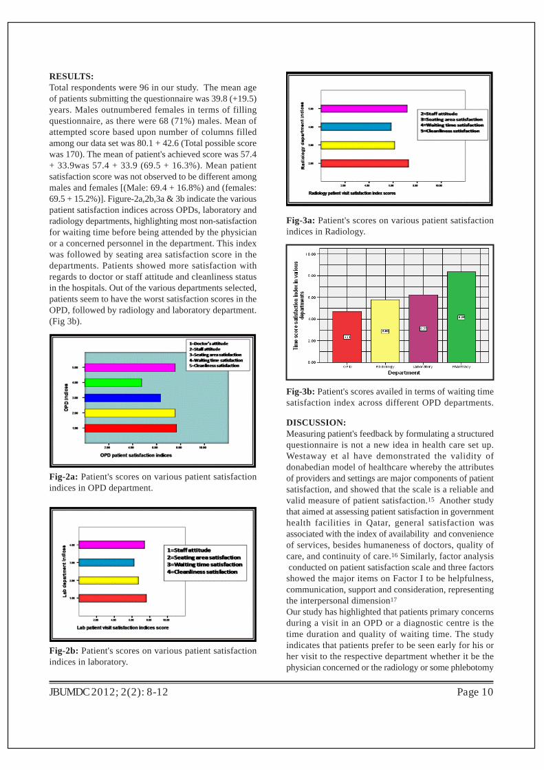
RESULTS:Total respondents were 96 in our study. The mean ageof patients submitting the questionnaire was 39.8 (+19.5)years. Males outnumbered females in terms of fillingquestionnaire, as there were 68 (71%) males. Mean ofattempted score based upon number of columns filledamong our data set was 80.1 + 42.6 (Total possible scorewas 170). The mean of patient's achieved score was 57.4+ 33.9was 57.4 + 33.9 (69.5 + 16.3%). Mean patientsatisfaction score was not observed to be different amongmales and females [(Male: 69.4 + 16.8%) and (females:69.5 + 15.2%)]. Figure-2a,2b,3a & 3b indicate the variouspatient satisfaction indices across OPDs, laboratory andradiology departments, highlighting most non-satisfactionfor waiting time before being attended by the physicianor a concerned personnel in the department. This indexwas followed by seating area satisfaction score in thedepartments. Patients showed more satisfaction withregards to doctor or staff attitude and cleanliness statusin the hospitals. Out of the various departments selected,patients seem to have the worst satisfaction scores in theOPD, followed by radiology and laboratory department.(Fig 3b).
Fig-2a: Patient's scores on various patient satisfactionindices in OPD department.
Fig-2b: Patient's scores on various patient satisfactionindices in laboratory.
Fig-3a: Patient's scores on various patient satisfactionindices in Radiology.
Fig-3b: Patient's scores availed in terms of waiting timesatisfaction index across different OPD departments.
DISCUSSION:Measuring patient's feedback by formulating a structuredquestionnaire is not a new idea in health care set up.Westaway et al have demonstrated the validity ofdonabedian model of healthcare whereby the attributesof providers and settings are major components of patientsatisfaction, and showed that the scale is a reliable andvalid measure of patient satisfaction.15 Another studythat aimed at assessing patient satisfaction in governmenthealth facilities in Qatar, general satisfaction wasassociated with the index of availability and convenienceof services, besides humaneness of doctors, quality ofcare, and continuity of care.16 Similarly, factor analysis conducted on patient satisfaction scale and three factorsshowed the major items on Factor I to be helpfulness,communication, support and consideration, representingthe interpersonal dimension17
Our study has highlighted that patients primary concernsduring a visit in an OPD or a diagnostic centre is thetime duration and quality of waiting time. The studyindicates that patients prefer to be seen early for his orher visit to the respective department whether it be thephysician concerned or the radiology or some phlebotomy

JBUMDC 2012; 2(2): 8-12 Page 11
procedure. While not much has been published locally,some evidence augmenting our findings is there in theliterature to suggest similar results.18,19,20 One more factorwhich must be appreciated is the observation that thecontent level was observed to be higher in diagnosticdepartments than in OPDs. Probable reasons include thefollowing: Firstly, the patient's are immediately taken onboard by direct interaction with the dealing staff for theintended procedure which may be suggested to improvepatient's satisfaction level. Secondly, few hospital OPDshave nurse stations added as a step before they are actuallyseen by the physician. These nurse stations do includeseveral anthropometric measurements and recording ofvital signs in details along with basic details about patient'shistory. This approach not only saves times for thephysicians but probably adds to improvement in patient'ssatisfaction level as well.21,22 Finally it highlights thatthe physician to patients statistics in primary and specialistOPDs can be enhanced to reduce the duration of timebefore they are dealt by the physicians. Examples areavailable in literature which indirectly signifies ourdiscussed concept. 23
Some studies have highlighted that staff dealing thepatient creates a major impact from patient's perspectivewith regards to patient satisfaction,24 our study hasshown the attitude of dealing staff to be lesser factor forpatient's non-satisfaction. This is an important findingand suggests that the physical environment surroundinga patient during a hospital visit has to do a lot to changehis perception and thought process. Other studies havealso highlighted the patient's surrounding's to be tailormade as per specific patient's needs as having a majorinfluence on his ideas about hospital improvement.25.Some of the weakness associated with the study must beappreciated: it is a hospital based study with a smallsample size and non probability convenient samplingwhich has its own inherent weaknesses. Secondly,Hawthrone phenomena could be a factor which couldaffect our results.The study has important clinical implications. This studybeing a descriptive study opens a Pandora box ofquestions, which challenge our routine functioning basedmainly upon decisions of management. Incorporatingpatient's input and valuable thought processes in routinefunctioning can certainly add to improve our businessprospects i.e., healthcare. Moreover, it also necessitatesthe creation of our national standards based upon realisticresource calculation regarding several healthcare resourceindicators like patients to physician statistics. It is expectedthat more studies may follow this pattern and shouldattempt to answer the questions raised by our observations.
CONCLUSION:Prolonged waiting time and non-availability of quality
stay in waiting areas of outpatient departments anddiagnostic centers are the cause of lesser patientsatisfaction during a patient's visit to hospital.
REFERENCES:1.
2.
3.
4.
5.
6.
7.
8.
9.
10.
11.
12.
13.
De Feo JA. Why are employers prodding health-care providers to adopt new management systems?Reducing the cost of health care. Clin LeadershManag Rev .2004; 18(2):80-5.Halterman S, Camero C, Maillet P. The consumer-driven approach: can it pick up where managed careleft off? Benefits Q. 2003; 19(2):13-26.Ware JE Jr, Wright WR, Snyder MK, Chu GC.Consumer perceptions of health care services:implications for academic medicine. J Med Educ1975; 50(9):839-48.Pascoe GC. Patient satisfaction in primary healthcare: A literature review and analysis. Eval ProgramPlann. 1983; 6:185-210.Schalm C. Implementing a balanced scorecard as astrategic management tool in a long-term careorganization. J Health Serv Res Policy 2008 ;13(1):8-14.Soufi G, Belayachi J, Himmich S, Ahid S. Patientsatisfaction in an acute medicine department inMorocco. BMC Health Serv Res. 2010; 10: 149-53Nguyen Thi PL, Briançon S, Empereur F, GuilleminF. Factors determining inpatient satisfaction withcare. Soc Sci Med. 2002; 54: 493-504.Messner ER. Quality of care and patient satisfactionthe improvement efforts of one emergencydepartment. Top Emerg Med 2005; 27:132-41.Peltzer K. Patient experiences and health systemresponsiveness in South Africa. BMC Health ServRes 2009; 9:117-21.Nguyen Thi PL, Briançon S, Empereur F, GuilleminF. Factors determining inpatient satisfaction withcare. Soc Sci Med 2002; 54:493-504.Navipour H, Nayeri ND, Hooshmand A, Zargar MT.An investigation into the effects of qualityimprovement method on patients' satisfaction: a semiexperimental research in Iran. Acta Med Iran 2011;49(1):38-43.Frick U, Gutzwiller FS, Maggiorini M, Christen S.A questionnaire on treatment satisfaction and diseasespecific knowledge among patients with acutecoronary syndrome. II: Insights for patient educationand quality improvement. Patient Educ Couns 2011.Aletras VH, Papadopoulos EA, and Niakas DA.Development and preliminary validation of a Greek-language outpatient satisfaction questionnaire withprincipal components and multi-trait analyses BMCHealth Serv Res 2006; 6: 66-9.

JBUMDC 2012; 2(2): 8-12 Page 12
14.
15.
16.
17.
18.
19.
Ware JE Jr, Wright WR, Snyder MK, Chu GC.Consumer perceptions of health care services:implications for academic medicine. J Med Educ1975;50(9):839-48.Westaway MS, Rheeder P, Van Zyl DG, Seager JR.Interpersonal and organizational dimensions of patientsatisfaction: the moderating effects of health status.Int J Qual Health Care. 2003; 15(4):337-44.Abdal Kareem A, Aday LA, Walker GM Jr. Patientsatisfaction in government health facilities in thestate of Qatar. J Community Health 1996; 21(5):349-58.Westaway MS, Rheeder P, van Zyl DG, Seager JR.Development and testing of a 25-item patientsatisfaction scale for black South African diabeticoutpatients. Curationis 2002; 25(3):68-75.Kisa K, Kawabata H, Itou T, Nishimoto N, MaezawaM. Survey of patient and physician satisfactionregarding patient-centered outpatient consultationsin Japan. Intern Med 2011; 50(13):1403-8.Gamroth L, Budgen C, Lougheed M. Feasibility andoutcomes of paid undergraduate student nursepositions. Nurs Leadersh (Tor Ont). 2006; 19(3):e1-14.
20.
21.
22.
23.
24.
25.
McMullen M, Netland PA.Wait time as a driver ofoverall patient satisfaction in an ophthalmologycl inic . Cl in Ophthalmol 2013;7:1655-60Chang CH, Stukel TA, Flood AB, Goodman DC.Primary care physician workforce and Medicarebeneficiaries' health outcomes. JAMA 2011;305(20):2096-104.Puri N, Gupta A, Aggarwal AK, Kaushal V.Outpatientsatisfaction and quality of health care in North Indianmedical institute. Int J Health Care Qual Assur 2012;25(8): 682-97Kelley ML, Parke B, Jokinen N, Stones M, RenaudD. Senior-friendly emergency department care: anenvironmental assessment. J Health Serv Res Policy2011; 16(1):6-12.Rao KD, Peters DH, Bandeen-Roche K. Towardspatient-centered health services in India--a scale tomeasure patient perceptions of quality. Int J QualHealth Care 2006; 18 (6):414-21.Pinto MB, Leonidas L. The impact of officecharacteristics on satisfaction with medical care: a"before and after" analysis. Health Mark Q 1994;12(2):43-54.

Prescribing Patterns in Hospital InpatientsNasim Karim1, Sajid Abbas Jaffri2, Zubair Ahmed Tirmizi3
JBUMDC 2012; 2(2): 13-16 Page 13
INTRODUCTION:Once a patient with a clinical problem has been evaluated& a diagnosis is reached the most common chosen optionis by far the drug therapy. Around the world more than50% of all medicines are prescribed, dispensed or soldinappropriately. This ineffective & inefficient use ofdrugs commonly occurs at health facilities in developing& developed countries.1 Evidence suggests that moreappropriate utilization of prescription drugs has thepotential to lower the total expenditure & improve thequality of care.2 Thus drugs are the essential tool forpreventive, curative and rehabilitation in health care.3The overuse, underuse or misuse of medicines results inwastage of scarce resources & widespread health hazards.WHO conference of experts has given a guideline to thehealth care providers in 1985,that all patients should begiven medications appropriate to their clinical needs inan adequate dose that is as per requirement of theindividual. These drugs should be administered throughan appropriate route for an adequate period of time &above all should be available at the lowest cost to thecommunity.4 Drugs are prescribed to the patient by theprescriber which in our scenario is traditionally thephysician. However in many states of America, healthcare practitioners other than MD and physicians can writeprescriptions. Licensed physician`s assistants, nurse
ORIGINAL ARTICLE
practitioners & pharmacists can prescribe medicationsunder various circumstances.5 Prescription is aprescriber`s order, a written direction to prepare, dispenseor administer a specific treatment. Moreover it is a legalorder and therefore should be dealt with great care &attention.6As per cycle of drug use (Figure-1) there are 5 phases inthe use of any drug. These are (I) diagnosis (II) prescribing(III) dispensing (IV) adherence & (V) follow-up.7
Although the physician /doctor/ prescriber has impacton all these phases but a more direct effect is seen onthe first two phases. At the level of prescribing thecommonly encountered problems areA) Under-prescribing where:Needed medications are not prescribedDosage is inadequate for treating the diseaseLength of treatment is too briefB) Incorrect prescribing where:Drug is given for incorrect diagnosisWrong drug is selected for the diagnosisPrescription is prepared improperlyAdjustment is not made for co-existing medical, geneticor other factorsC) Extravagant prescribing where:A less expensive drug can provide comparable efficacyand safety & is not givenSymptomatically treating mild conditions & divertingfunds from treating serious illnessesD) Over-prescribing where:Drug is not needed and is still givenDose is too large for any disease treatmentTreatment period is too long than actually neededE) Multiple prescribing where:Two or more medications are used when fewer wouldachieve the same effectSeveral related conditions are treated when treatment of
Nasim KarimProfessor & HeadDepartment of Pharmacology BUMDC Karachi.Email: [email protected] Abbas Jaffri Associate ProfessorDepartment of Medicine BUMDC Karachi.Zubair Ahmed Tirmizi Assistant Professor & HeadDepartment of Forensic Medicine BUMDC Karachi.Received: May 18, 2012Revised: September 12, 2013Accepted: September 15, 2013
ABSTRACT:Objective: To evaluate the prescribing patterns by an audit of prescriptions in hospital inpatients.Materials and methods: After a written informed consent from the medical ward incharge & hospital administrator 32 case notes of adult patientsdischarged from a private hospital in Malir were collected from 1st to 30th April 2012. Patients demographics, disease & prescription details (number,type, dose, route, frequency, duration of drug use, tendency of polypharmacy, cost of drugs & discharge notes) were entered in a specially designedperforma.Results: Mean age of patients was 27.18years with 14 males & 18 females. They were diagnosed to have enteric fever (10), gastroenteritis (5), RTI(4) & others (13). Average hospital stay period was 2.5 days.Total number of drugs used were 120, of which only 5 (4.17%) were prescribed by genericname. 25.83% drugs were from National Essential drug List of Pakistan (NEDLP). Mean number of drugs per patient was 9.35. Antibiotics & analgesicseach was given to 29 (90.63%) patients. Anti -ulcer drugs were given to 27(84.38%) & nebulization to 11 (34.38 %) patients without need. Averagecost of drugs per patient was 1200 rupees. None of the prescription was complete for the above mentioned parameters.Conclusion: Audit of prescribing patterns in hospital inpatients of a private setup showed irrational use of drugs.Key Words: Prescribing patterns, Private hospital, Inpatients, Rational use, Drugs
JBUMDC 2012; 2(2): 13-16 Page 14
primary condition would improve or cure the otherconditions.8It is documented that effective plan design, strategiesutilizing generic substitutions, rational prescribing & useof formulary can help manage cost while maintainingquality & customer satisfaction. Before such strategiescan be implemented prescribing patterns of cliniciansmust first be explored.9 The study of prescribing patternis a component of medical audit that does monitoringand evaluation of the prescribing practice of theprescribers as well as recommends necessarymodifications to achieve rational & cost effective medicalcare.10 This helps to evaluate & suggest modificationsin prescribing practices of medical practitioners so as tomake medical care rational & medical profession highesteemed.11 Few studies are documented in Pakistan onhospital inpatients & that too are mainly on pediatricpopulation. Present study was done to audit the prescribingpatterns in the adult inpatients of a private hospital.MATERIALS & METHODS:This pilot study was approved by IRB/ERB-BUMDCthrough letter ERC03/12. After a written informed consentfrom the medical ward incharge & hospital administrator32 case notes of patients discharged from a privatehospital in Malir, Karachi were collected from 1st Aprilto 30th April 2012. Patients demographics, disease &prescription details were entered into a specially designedperforma. Case notes of medical ward inpatients of bothgenders more than 18 years with proper diagnosis andwith or without concurrent illness were included in thestudy. Audit of the prescribing practices in these hospitalinpatients was done by determining the number, type,dose, route, frequency & duration of drug use. Tendencyof polypharmacy, cost of drugs & follow up of patientswas also ascertained. SPSS version 17 was used foranalysis of data.RESULTS:Mean age of 32 patients was 27.18 years with 14 males& 18 females. They were diagnosed to have enteric fever(10), gastroenteritis (5), RTI (4) & others (13) [Fig: 2].Total number of drugs used were 120, of which 115 drugswere prescribed by brand name and only 5 (4.17%) wereprescribed by generic name [Fig: 2]. 31(25.83%) drugswere from National Essential Drug List of Pakistan(NEDLP) [Fig: 2]. Antibiotics & analgesics were givento 29 (90.63%) patients respectively & their main routeof administration was parenteral with most injectionsgiven intravenously. Vitamin injections were given to 6(18.75%) patients. Anti -ulcer drugs were given to27(84.38%) & nebulization of Ipratropium Bromide(Atrovent) to 11 (34.38 %) patients without need that istreatment not in accordance to their respective diagnosis[Fig: 3]. Average hospital stay period was 2.5 days.Average number of drugs prescribed per patient was 9.35
and average cost of drugs per patient was 1200 rupees[Fig: 3]. None of the prescriptions was found to becomplete for route, dose, frequency & duration of druguse. Discharge notes were present in only 18(56.25%)sheets and they were also incomplete [Fig: 3]
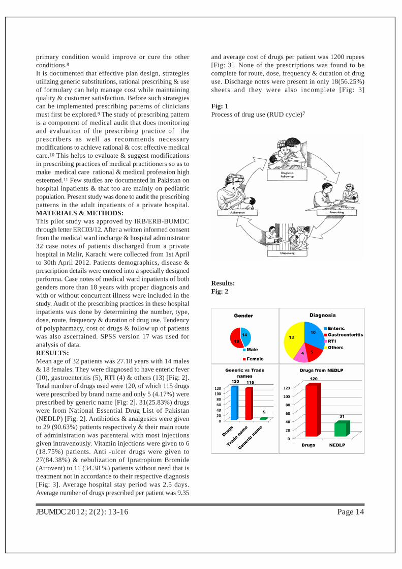
Fig: 1Process of drug use (RUD cycle)7
Results:Fig: 2

JBUMDC 2012; 2(2): 13-16 Page 15
by the generic name in contrast to 45.2% & 23.6% atHUKM, a teaching hospital owned by UniversityKebangsaan Malaysia.20 However it is said that forspecialists & consultants more options are available asthey are allowed to prescribe from both branded & genericdrug list. In our case it seems that their preferences weremore inclined towards the branded drugs.2 1
We have found an average hospital stay period of 2.5days with mean number of drugs per patient 9.35 whichis comparable to the results of Lucena.22 Polypharmacyis said to be associated with more adverse effects & lesspatient`s compliance. Average cost of drugs per patientfor a period of 2.5 days was found to be 1200 rupees thatis per day 480 rupees. This didnot include the consultant`sfee, investigation charges, hospital charges or even thefood of the patient at the hospital. Pakistan is a thirdworld country with per capita income of 7000rupees/month declared in May 2012. One can very wellimagine that even if a person is earning 1000 rupees perday that is a monthly income of 30,000 rupees will notbe able to bear these drug expenses with the simultaneousresponsibilities of the family, housing & food. Najmi 23
have documented an average cost of drugs per day to be88.36 rupees & 80% of drugs use from NEDLP in 1988.But this was 23 years back & now prices of commoditiesare gone up by many fold. Essential medicines are those that satisfy the priorityhealth care needs of the population. They are selectedwith due regard to disease prevalence, evidence onefficacy and safety, and comparative cost-effectiveness.Essential medicines are intended to be available withinthe context of functioning health systems at all times inadequate amounts, in the appropriate dosage forms, withassured quality, and at a price the individual and thecommunity can afford. We have found use of 25.83%drugs from National Essential Drug List of Pakistan(NEDLP). Antibiotics & analgesics were the mostcommon drugs prescribed to 90.63% patients respectively& the most common route for their administration wasintravenous. Vitamin injections were given to 18.75%patients. Our findings are coinciding with those of Littonwho found 28.7 % of drugs used from the Ministry ofHealth Drug List & antibiotics the most commonlyprescribed drugs.24
Mengistu has documented in his study that significantnumber of files (case sheets) were incomplete for theroute , dose, frequency, duration of drug use & dischargenotes which completely favors our findings as none ofour case sheets were complete for the above mentionedfactors .Discharge notes were found in only 56.25% casesheets & that too were incomplete. These malpracticescould result in administration of drugs through the wrongroute, unwanted shorter or longer interval of drugadministration & incorrect duration of treatment. Wewere not able to find why anti-ulcer drug injections weregiven to 84.38% & Ipratropium bromide nebulization to34.38% patients when they didn`t had any features of
DISCUSSION:The widespread use of many new and powerful drugsand the increasing recognition of adverse effects havestimulated interest in the manner in which physiciansprescribe drugs. The three main sources of informationabout the prescribing patterns of physicians are marketingresearch data, studies of general practice and monitoringof prescribing in hospitals.12 Aggressive drug marketingpromotions, lack of information on the use of drugs &drug shortages are said to be the major causes of irrationaldrug use. The rational use of drugs demands prescriptionof appropriate drugs.13 Prescribing practices of theconsultants in Karachi, the home of at least eight medicalcolleges has been documented as non-rational.14,15,16
We collected 32 case sheets of patients discharged fromthe medical ward of a private hospital in Malir, Karachifrom 1st April to 30th April. They showed mean age ofpatients 27.18 years with 14 males & 18 females. Theywere diagnosed to have enteric fever (10), gastroenteritis(5), respiratory tract infection (4) & others (13). Mengistuhas documented a similar data where case sheets of 36adults admitted to the medical ward of Jimma hospitalfrom first April 2002 to 30th May 2002 were evaluated.They had mean age of 30 years with diagnosis of TB (8),diabetes (6), cardiac disease (5) & others (17).17
By definition, a product identified by its official chemicalname rather than an advertised brand name (propriety ortrade name) is called a generic. It exerts itspharmacological effects at the same site, supposed toshow the same potency, same dosage form & samebioavailability as a brand name.18 Higher generic drugprescription rate implicates less cost on health care withsimilar efficacy in clinical results.19 In our study a totalof 120 drugs were used & only 4.17% were prescribed
Fig 3

peptic ulcer or bronchoconstriction. These might be usedto satisfy the patients high expectancies when treated byspecialist in a private set-up or to produce a feeling ofwell being within a short period. It is clear that treatmentis not co-relating with the diagnosis in these patients orvice versa.Educational, managerial & regulatory interventions torationalize the prescribing practices are the need of today& should be carried out by the government authorities& professional bodies The important thing is the safetyof an ill person which should not be compromised forthe sake of personal or industrial growth.25
CONCLUSION:Audit of prescribing patterns in adult hospital inpatientsof a private setup showed irrational use of drugs. Measuresshould be taken by the government & PMDC for:1. Standardization of therapeutic schemes.2. Prescription control sheet audits.3. Improving the knowledge of doctors through specifictrainings, printed educational materials, therapeuticmanuals & guidelines.ACKNOWLEDGEMENT:The authors are highly thankful to Prof Dr. Tipu Sultan,Principal and Dean Health Sciences, Bahria UniversityMedical & Dental College (BUMDC) for being a drivingforce behind this pilot project.REFERENCES:1.
2.
3.
4.
5.
6.
7.
8.
9.
JBUMDC 2012; 2(2): 13-16 Page 16
10.
11.
12.
13.
14.
15.
16.
17.
18.
19.
20.
21.
22.
23.
24.
25.
Falkenberg T, Tomson G. The World Bank andPharmaceuticals. Health Policy and Planning2000;15(1):52-8Copeland C. Prescription drugs: Issues of cost,coverage and quality. Employee Benefit ResearchInstitute Issue. Brief 1999;208:1-21Hafeez A, Kiani A G, ud Din S, Mohammad W.Prescription and Dispensing Practices in PublicSector Health Facilities in Pakistan: Survey Report.JPMA 2004; 54(4):187-91The Rational Use of Drugs. Report of the Conferenceof Experts. Nairobi, 25-29 November 1985Buxton ILO. Principles of prescription order writingand patient compliance In: Goodman & Gilman`sThe Pharmacological Basis of Therapeutics. Editors,Laurence L Brunton, John S Lazo, Keith L Parker.11th edition. 2006; 1777-86. McGraw Hill CompaniesUSA. Lofholm PW, Katzung BG. Rational Prescribing &Prescription Writing In: Basic & ClinicalPharmacology. Editors Katzung BG, Masters BS,Trevor AJ. 11th edition 2009; 1127-36.TATA McgrawHill India.Ndyanabangi B. RUD cycle. Rational Drug Use inART Programs: An overview. SEAM ConferenceAccra, Ghana, June 20-22, 2005Media Centre WHO. Medicines: rational use ofmedicines. Fact sheet No338 May 2010Al-Junid SM, Puteh S E W, Surianti S. Prescribingpatterns and drug cost among cardiovascular patientsin Hospital University Kebangsaan Malaysia.Med J Malaysia 2007; 62(1):59-65
Kumar MA, Nizar A, Shailaja K, Jayasutha J,Ramasamy C. A study on prescribing pattern &potential drug-drug interactions in type 2 diabetesmellitus inpatients in a tertiary care teaching hospital.Der Pharmacia Lettre 2011; 3(4):13-9Srishyla MV, Krishnamurthy M, Nagarani MA,Andrade C, Venketaraman BV. Prescription audit inan Indian hospital setting using the DD concept.Indian J Pharmacol 1994;26:23-8Stolley PD. Prescribing patterns of physicians.J chronic diseases. 1969; 22 (6-7):395-405Offerhaus O. Rational use of drugs in Balkans: aWHO Workshop. Essential Drugs Monitor.1995;20(3):38-42Siddiqi S, Hamid S, Rafique G, Chaudhry SA, AliN, Shahab S et al. Prescription practices of publicand private health care providers in Attock Districtof Pakistan. Int J Health Plann Manage 2002; 17:23-40Hussain SF, Zahid S, Khan JA, Haqqee R. Asthmamanagement by general practitioners in Pakistan.Int J Tuberc Lung Diseases 2004;8:414-7Das N, Khan AN, Badini ZA, Baloch H, Parkash J.Prescribing practices of consultants at Karachi,Pakistan. J Pak Med Assoc 2001; 51:74-7.Mengistu A. Patterns of drug utilization in inpatientdepartments, Jimma Hospital South West Ethiopia.Ethiop J Heal th Sci 2005;15(2) :139-45Chukwuani C M, Onifade M, Sumonu K. Survey ofdrug use practices and antibiotic prescribing patternat a general hospital in Nigeria. Pharm World Sci2002;24(S):188-95World Health Organization. Reducing risks,promoting healthy life. Geneva. The World HealthReport 2002Amritzal MN, Rohaizat Y, Zafar A , Saperi S, AljunidSM. Case Mix costing in University KebangsaanMalaysia Hospital. A top down approach: Costanalysis for cardiology cases. Malaysian Journal ofPublic Heal th medicine 2005;5(2):74-81 Sagardul VJ, LaCalle R M, Casado BS. Substitutionof generic for brand medicines in primary care.Factors associated to refuse the change. Aten Primaria2005; 36(9):489-93 Lucena MI, Ruiz J, Andrade RJ .The impact ofhospitalization on drug prescription. Med Clin Barc1995;104(6):211-5 Najmi MH, Hafiz RA, Khan I, Fazli FR. Prescribingpractices: an overview of three teaching hospitals inPakistan. J Pak Med Assoc 1998; 48(3):73-7Litton LM, Sisk FA, Akins ME. Managing drug cost:the perception of managed care pharmacydirectors.American Journal of managed care2000;6(7):805-14Shiwani MH. Quest of prescribing practice inPakistan. Letter to editor. JPMA2006; 56(5):249-50

Finding Factors Causing Postdural Puncture Headache In Obstetric PatientsAfter Spinal Anaesthesia
Maqsood Ahmad1, Zareen Fatima2
JBUMDC 2012; 2(2): 17-20 Page 17
INTRODUCTION:Spinal anaesthesia is widely accepted technique forCaesarean sections1 revolutionizing the practicebyproviding fewer complications since its discovery in1885 by J Leonard Corning2. The ease of performance,quick onset of dense block rendering excellent analgesiahas surpassed other techniques for obstetric anaesthesiainterms of its benefits but PDPH after subarachnoid (SAB)block is the greatest fear which has contributed to searchfor optimum spinal needles and drugs. The obstetricanaesthesia care accounts for approximately 12 % ofAmerican Society of Anesthesiologists (ASA) ClosedClaims database where post dural puncture headache wasthird in claims 3, 4.
It is pertinent to note that headache is diagnosed clinicallyand causation is multifactorial but size and shape ofspinal needles is mostly blamed.These needles have beenmodified to simplify their use and minimize complications.Needle design variables, such as diameter, tip design andorifice location, have been altered to enable rapid flowof cerebral spinal fluid (CSF) and injected medications,yet simultaneously limit dural trauma and loss of CSF.The CSF leak is one proposed mechanism which inducesreflex vasodilatation and traction on cranial contents5.This leak is directly proportional to dural hole which in
ORIGINAL ARTICLE
turn is directly proportional to needle size.Parturient ageand gender are inevitable contributing factors in spinalanaesthesia6.A popular needle in practice is 25 G Quincke Babcockwith a reported PDPH incidence of 25% 7. Needles like29 G and 30 G are available but their use is limited dueto high failure rate and technical difficulty8, 9. Theselection of needle is personal preference but 25 G isgenerally accepted. The use of pencil point needles wassuggested very early by Hart and Whitacre for reducingduraltrauma10.Various studies have demonstrated thatpencil point and smaller bore needles are beneficial 11,
12. The smaller needles have no or minimum incidenceof PDPH at the cost of common technical errors likefailed spinal and bending of needle. We have conductedthis study to search a needle which is easy to use, readilyavailable, fewer complications rate and economical.MATERIALS AND METHODS:This cross sectional observational study of 500 C sectioncases was conducted in Combined Military HospitalGujranwala and Pakistan Naval Shipping Rahat Hospitalfor 3 years by a single anaesthesia specialist using only27 G QuinckeBabcok spinal needle. After prior approvalof hospital ethical committee and written informedconsent, the procedure was explained and local anaesthesiaplain lidocaine 2% 1-2ml was injected in patientspreloaded with 1000 ml of Ringers Lactate. Sensocaine(Bupivacaine 0.75 % 2 ml hyperbaric or Bupivacaine0.5 % hyperbaricml packing of Brookes PharmaceuticalLaboratories (PVT) Pakistan) or Abocaine (Bupivacaine0.75 % hyperbaric 2 ml of Abbott Laboratories Pakistan) was used in L 2-3 / L3-4 level in sitting as well as lyingpositions. Free flow of CSF was confirmed before
Maqsood AhmadConsultant Anesthesiologist.PNS Shifa Karachi.E-mail: [email protected] Fatima Gynaecologist PNS Rahat, Karachi.Received: June 25, 2013Revised: September 25, 2013Accepted: October 1, 2013
ABSTRACT:Objective: Among various recognized factor of spinal headache the single most important causative factor is size of spinal needle. The aim of thisstudy was to determine incidence of spinal headache with 27 GQuincke Babcock spinal needle in Caesarean section patients.Materials and Methods: This observationalcross sectional study was carried out in the Combined Military Hospital Gujranwala and Pakistan NavalShipping Rahat Hospital Karachi from Jan 2011 to Jan 2013. In 500 Caesarean section (C section) cases preloaded with 1000 ml Ringers Lactate,27 G QuinckeBabcok spinal needle was used in sitting as well as left lateral position for spinal anaesthesia in all patients using local anaesthesia plainlidocaine 2% 1-2ml.In interspace L 2-3 / L3-4 eitherBupivacaine hydrochloride hyperbaric 0.75 % or 0. 5% was injected. All Caesarean cases wereincluded except contraindicated. Spinal needle Quincke Babcock 27 G alone was used.The results were presented in percentages, mean and standarddeviation.Results: A total of 500 patients of c-section were evaluated. Overall incidence of true spinal headache was 2%, failed spinal anaesthesia 4%, spinalneedle was changed in 3 %, success rate of 96 % and maternal acceptance 47.4 %. Single pricks were 59.4 % while 2-3 pricks were 40.6 % .Conclusion: Smaller spinal needle has changed the safety profile of spinal anaesthesia in C section cases by very low failure rates and true PDPHa rarity. PDPH will continue as long as dura is punctured but incidence can be decreased by different techniques.KEY WORDS: Spinal anaesthesia, Spinal needle, Caesarean section, Postdural puncture headache (PDPH).

JBUMDC 2012; 2(2): 17-20 Page 18
injection. All elective, emergencies, pregnancy inducedhypertensive, primigravida, multigravida and obese caseswere included in this study excluding unwilling andcontraindicated patients.In emergency cases C-Fuser1000 (medex, Dublin, Ohio 43016 USA) was used topreload the patients and ephedrine IV was titrated tocontrol blood pressure. Nalbuphine IV 4-6 mg was givenpost delivery and Metoclopramide 10 mg IV for nauseaand vomiting. O2 was given with facemask at a rate of4 liters if required and level of T4-6 was achieved beforestarting incision. After completion of surgery the patientwas shifted to respective wards /intensive care advisingcomplete bed rest for 24 hours. The PDPH incidence asdefined by the international headache society criteria(Table 1) after the operation till 72 hours, change ofneedle to another 27 G due to bending, successful/failedspinal block and maternal spinal acceptance was noticed.
The data was collected and presented in percentages afteranalysis.RESULTS:A total of 500 patients were evaluated.Mean age ofpatients in the study was 28.5years. Mean weight andheight was 62 kg & 155 cm respectively. Majority ofpatients were multipara 337 and more than half (300)belonged to ASA grade 1. (Table 2)Success rate in the study was 96 % with maternalacceptance rate of the procedure 47.4 %. Overallincidence of true spinal headache was found to be 2%.Failure of spinal anaesthesia was encountered in 4% ofthe patients. Spinal needle was changed in 3 % of cases.Majority of patients underwent single prick that is in59.4 % while 2-3 pricks were needed in 40.6 % of thepatients. (Table 3)
Table 1: International Society of Headache PDPH Criteria 12
••
••
•
Definition: Headache that develops within 7 days of dural puncture and disappears with 14 days.
Classic Featuresbut variablepresentation
Headache is often frontal-occipitalMost headaches do not develop immediately after dural puncture but 24-48 hoursafter the procedure with 90% of headaches presenting within 3 days.Headache is worse in the upright position and eases when supine.Pressure over the abdomen with the woman in the upright position may give transientrelief to a rise in intra abdominal pressure (Gutsche sign)
Neck stiffness, photophobia, tinnitus, visual disturbance and cranialnerve palsies.Associated symptoms
Table2: Demographic Data
ASA: American Society of Anesthesiologist grade
Age in yearsWeight in KgHeight in cmASA grade
Parity
28.5±11.562±15155.486±7.62ASA 1
300ASA 2
120ASA 3
80Multigravida
337Primigravida
163
Table 3: Outcomes of Spinal Anaesthesia
PDPH IncidenceSpinal Success RateSpinal Failure RateMaternal AcceptanceNeedle ChangedSingle prick2-3 Pricks
2 %96%4%47.4%3 %59.4 %40.6 %

JBUMDC 2012; 2(2): 17-20 Page 19
DISCUSSIONThe search of optimum spinal needle had started sincethe invention of spinal anaesthesia but PDPH is reportedwith all needles.PDPH mechanism is not clear but CSFleak is clearly associated with this headache and CSFleak is directly proportional to needle size. The backacheassociated with spinal dural puncture has nothing to doin the long run except where some damage has beendone during procedure. The backache solely associatedwith spinal anaesthesia in obstetric cases has been studiedby Kashif and Arshad13 declaring that pre anesthesiaexam should include counseling about backache as thebackache is not associated with this technique. Howeverpersisting chronic cases must be referred and epiduralabscess or hematoma is excluded 13. Pre anaesthetichistory should exclude preexisting backache or nerveinjury and many obstetric patients had preoperativebackache which is multifactorial likechange in centre ofgravity and hormones.Reportedly contributing factors for higher PDPH areneedle size, type, entrance angle, technique, no of duraltap, multiple attempts by different users, pre existingbackache history, trauma to structures especiallyperiostium and nerves. Other factor like age, weight,posture, patient's sensitivity to pain, spinal acceptanceand previous experience are contributing to maternalsatisfaction. Only PONV (post operative nausea andvomiting) and pain are best controlled in spinal anaesthesiawhereas acceptance for regional techniques is very low.Similarly backache is there despite cause is not establishedand the maternal satisfaction is very poor14 .Large bore (<25 or =25) and cutting point needles producePDPH 15,16,17,18 so their use must be discouraged. Theneedles of 27 G are studied extensively and approved inmany studies 19, 20, 21, 22 but had variable PDPH incidence.Theoretically atraumatic pencil point needles provideadvantages over cutting needles in the form ofinsignificant PDPH23, 24. The histological review on eitherneedle had proved equivocal results of neurologicaldamage/ inflammation 25. Our study is based on using27G needle for spinal anaesthesia in all elective as wellas emergency obstetric cases. In expert hands resultswith this needle in form of high success rate, low PDPH,good analgesia and fetal outcome are excellent butbackache complaints are difficult to rectify. Psychologicalfactors along with extent of structural damage arecontributing.CONCLUSION:All efforts must be exercised for gentle atraumatic spinalanaesthesia in a single attempt or minimum attemptsminus damaging nerve or bone. Smaller spinal needlehas changed the safety profile of spinal anaesthesia in Csection cases by very low failure rates and true PDPH ararity. PDPH will continue as long as dura is punctured
but incidence can be decreased by different techniques.Furthermore adoption to this needle is required afterpractice as it is soft providing better dural puncture feelthan larger bore needles. We strongly suggest alljunioranaesthesia doctors to use this needle early in theircareer for better future practice outcomes.ACKNOWLEDGEMENTS:The persons of great help who have contributed differentaspects in preparing this article while providing comfort,moral support, continuous encouragement are Dr. NabiBux of PNS Rahat, Ashraf CMA (OT), Naeem MA (OT)lV of PNS Rahat, ORA Ahmad Ali and ORA Sanaullahof Gujranwala CMH.
REFERENCES:1.
2.
3.
4.
5.
6.
7.
8.
9.
10.
Allman K G, Wilson I H. Caesarean section: spinal.In: Oxford handbook of Anaesthesia 3rd ed. OxfordUniversity Press. 2011. Chapter 32. p758.Chadwick HS. Obstetric anesthesia closed claimupdate II-ASA. Newsletter 1999:63:6.Morgan G E Jr, Mikhail M S, Murray M J. Chapter43 Obstetric anesthesia. In: Clinical Anesthesiology4th ed. Lange Medical Books/McGraw -Hill MedicalPublishing Division.2006. p 892RodriquesAM, Roy RM. Post lumbar punctureheadache. La Revue du praticien 2007; 57:4: 353-7.Chohan U, Hamdani G A. Post dural punctureheadache. JPMA 2003; 53:8-10Pal A,Acharya A, Pal N D, Dawn S, Biswas J. Dopencil-point spinal needles decrease the incidenceof postdural puncture headache in reality? Acomparative study between pencil-point 25GWhitacre and cutting-beveled 25G Quincke spinalneedles in 320 obstetric patients. Anaesthesia essaysand researches 2011; 5(2): 162-6.Kang SB, Goodnough DE, Lee YK, Olson RA,Borshof JA, Furiano MM et al. Comparison of 26-G and 27-G needles for spinal anaesthesia forambulatory surgery patients. Anesthesiology 1992;76(5):734-8.Tariq MM, Amjad I, Asghar K. Post spinal headache;comparing needles of 25 and 27 gauges for incidenceof post spinal headache. Professional Med J 2007;14(3):441-7.Shaikh JM, Memon A, Memon MA, Khan M. Postdural puncture headache after spinal anesthesia forCesarean section: A comparison of 25 g Quincke,27 g Quincke and 27 g Whitacre spinal needles. JAyub Med Coll Abbottabad 2008; 20:10-3.Vallejo MC, Mandell GL, Sabo DP, Ramanathan S.Post dural puncture headache: A randomizedcomparison of five spinal needles in obstetric patients.AnesthAnalg 2000:91:916-20.

19.
20.
21.
22.
23.
24.
25.
Behzad S, Asjad S, Ahmad A, Rashid I, Umar Z.Postdural puncture headache. Comparison betweenlumbar puncture needle no 25 G and 27 G. ProfessMed 2011;18 (1):51-56Chohan U, Hamdani G A. Postdural punctureheadache. JPMA 2003; 53:8-12Morros-Vinoles C, Perez-Cuenca MD, Cedo-Lluis,Colls C, Bueno J, Cedo-Valloba F. Comparison ofefficacy and complications of 27 G and 29 G Sprotteneedles for subarachnoid anaesthesia.RevistaEspanola de anestesiologiaryreanimacion. 2002;49(2) :448-54.RodriquesAM, Roy PM. Postdural puncture headache.La Revue du praticien. 2007; 57(4):353-7.Arendt K, Demaerschalk BM,Wingerchuk, DM,Camann W. Atraumatic lumbar puncture needles:after all these years, we are still missing the point?The neurologist 2009; 15(1): 17-20.Saenghirumvat tana R, Tant ivi tavatan K,Chumnanyech W, Tangsukkasemsun S ,Siritongtaworn P. A comparison study between newlydesigned pencil-point and cutting needles in spinalanesthesia. Journal of the Medical Association ofThailand 2008; 91(1):156-6.Steinfeld T, Nimphius W, Wurps M, Eberhart L,Vassiliou T, Kill C et al. Nerve perforation withpencil point or short beveled needles: histologicaloutcome. Actaanaesthesiologica Scandinavica2010;54(8) :993-9.
11.
12.
13.
14.
15.
16.
17.
18.
Naz F, Khan S, Begum A, Malik M, Zareen A.Complications of spinal anaesthesia in C section.Pjmhsonline.com/complicat ions of spinalanaesthesia.htmNicola J C. Effective management of the postduralpuncture headache. ATOTW 2010;18 : 181-4Kashif M, Arshad T, The causes, prevention andmanagement of post spinal backache. An overview.Anesth, Pain and Intensive care. 2013Rashad S, Abdullah J. Maternal satisfaction afterspinal anaesthesia in caesarean deliveries.JCPSP 2009; 19 (2): 77-80Bano F, Haider S, Aftab S, Sultan ST. Comparisonof 25 gauge quincke and whitacre needles forpostdural puncture headache in obstetric patients. JColl Physicians Surg Pak 2004; 14: 647-50.Angle PJ, Kronberg JE, Thompson DE, Ackerley C,Szalai JP, Duffin et al. Dural tissue trauma andcerebrospinal fluid leak after epidural needlepuncture: effect of needle design, angle and bevelorientation. Anesthesiology 2003; 99: 1376-82.RodriquesAM, Roy PM.Post-lumbar punctureheadache. Rev Prat 2007; 57: 353-7.Lambert DH, Hurley RJ, Hertwig L, Datta S. Roleof needle gauge and tip configuration in theproduction of lumbar puncture headache. Reg Anesth2003; 22: 66-72.
JBUMDC 2012; 2(2): 17-20 Page 20

Transpedicular Decompression And Spinal Fixation In Thoracolumbar Burst FracturesAhmed Tashfeen Ashraf
JBUMDC 2012; 2(2): 21-25 Page 21
INTRODUCTION:Spinal injuries have greatly increased as a result of highspeed vehicle accidents. They generally involve youngerpopulation and therefore are more tragic as it makes thepatient bed ridden in the prime of his youth. Howevermany recent advances have enabled the surgeons togreatly reduce the morbidity and improve the outcomein these injuries. A specific subset of spinal fractures areconsists of Burst fractures of thoraco-lumbar spine. Burstfractures, as defined by Denis 1, involve compressionfailure of the anterior and middle columns of the spine.Most burst fractures of the spine are associated withvarying degrees of bone fragment retropulsion into theneural canal leading to neurological deficit. Althoughburst fractures can occur at any spinal region, theiroccurrence at thoraco-lumbar region presents specificproblems as well as opportunity for neurologicalimprovement and recovery due to involvement of lowermotor neurons in injury 2. The optimal initial treatmentof thoracolumbar burst fractures continues to be stronglydebated 3,4. Although some centers choose to treat theseinjuries conservatively 5, vast majority of centers treatthem surgically. The surgical approach has varied fromanterior decompression alone or with staged posteriorfixation or posterior fixation and indirect reduction byligament taxis. The transpedicular decompression andsingle stage pedicle screw fixation is another approachto treat these fractures. This was the only approach usedin our series of patients. The transpedicular decompression
ORIGINAL ARTICLE
and fixation represents an attempt to restore the anteriorcolumn without the need for anterior decompression orstrut grafting 6. In fact it combines decompression andfixation through a single approach and avoids morbidityof anterior or combined approach. The use of pediclescrews increases the biomechanical strength of the fusedsegments more than any anterior construct alone7,8.MATERIALS AND METHODS:After departmental approval twenty three patients aged17 to 57 (mean, 41) years were included in this studyfrom December 2010 to January 2013. They had burstfractures in the thoracolumbar (n=13) and lumbar (n=10)regions. There were 18 males and 5 females. Uponadmission, the complete medical history with a detailedclinical evaluation was recorded and radiologicalexaminations were performed. Fractures were classifiedaccording to the AO classification (FIG 1).
Fig 1: The AO Spine classification system
Ahmed Tashfeen AshrafAssistant Professor,Department of Neurosurgery PNS Shifa, DHA Phase II, Karachi Email: [email protected] Received: July 18, 2013Revised: September 25, 2013Accepted: October 8, 2013
ABSTRACT:Objective: To evaluate the results of transpedicular decompression and single stage pedicle screw fixation in burst fractures of thoracolumbar spine.Materials & Methods: This study was carried out at PNS Shifa from Dec 2010 to Jan 2013. All consecutive traumatic burst fractures that underwentsurgery were included in the study. Twenty three consecutive patients aged 17 to 57 (mean, 41) years who had burst fractures in the thoracolumbar(n=13) and lumbar (n=10) regions and were surgically treated were included in this study. There were 18 males and 5 females. Fractures were classifiedaccording to the AO classification. The extent of spinal canal compromise was assessed by computed tomography, and the neurological status bythe modified Frankel grading for traumatic paraplegia. All patients underwent posterior transpedicular decompression and same stage pedicle screwfixation. Outcome was assessed on Frankel grading scale.Results: The extent and level of neurological injury varied. It did not correlate with extent of canal compromise, age and sex of the patient.Neurological injury was greater with T11 and T12 injuries than Lumbar fractures. No worsening of neurological grade was observed after surgery;rather 20 of 23 patients (86.9%) improved to the next higher grade. Screw malposition to the extent warranting readjustment was noted in 2 cases.Hardware failure occurred in 1 case after 6 months, bed sores in 3 cases and deep vein thrombosis in 1 case.Conclusion: Single stage Transpedicular decompression and spinal fixation from a posterior approach gives good results in burst fractures ofthoracolumbar spine.Key Words: Transpedicular; Spinal fixation; Pedicle screws.

JBUMDC 2012; 2(2): 21-25 Page 22
The extent of spinal canal compromise was assessed bycomputed tomography, and the neurological statusaccording to the modified Frankel grading for traumaticparaplegia. The most common mechanism of injury wasa motor vehicle accident followed by fall from a height.The most common vertebra involved was L1 (41%)(Table 1), and the most common type of burst fracture(Fig 2a), was type A3 (Table 2).
Fig 2a: Burst Fracture of LV1
The pre and post-operative neurological status of thepatients is given in fig 3a & 3b
Fig 3 a: Pre-operative Frankel Grade of the patients
Table 1. Distribution Of Thoracolumbar Injuries
Table 2. Distribution of thoracolumbar fractures accordingto AO classification
A
B
C
-
3
-
6
5
-
9
-
-
According to the AO classification, 6 patients had theA2 fracture, whereas 9 patients were diagnosed with theA3 fracture. 3 patients were diagnosed with B1 typefracture, and 5 patients were diagnosed with the B2fracture. No patient was diagnosed with the type Cfracture.All patients were assessed according to Frankel gradingsystem (Table 3).
Table 3: Frankel classification Grade A: No motor or sensory function Grade B: No motor but sensory present Grade C: Sensory normal but motor useless Grade D: Useful motor function present Grade E: Normal motor and sensory function
Fig 3 b: Post-operative Frankel Grade of Patient
Surgical TechniquePreoperative evaluation: All patients were thoroughlyassessed prior to surgery. Several of these patients hadmultiple injuries including head injury in 3 patients,abdominal injuries in 4 and limb injuries in 7 patients.Surgery was undertaken as soon as the patient becamehemodynamically stable.Intraoperative Positioning: Following endotrachealintubation, the patient was positioned prone on a spinalframe. All osseous prominences were padded and theeyes were protected. Prior to beginning the surgery, APand lateral fluoroscopic (C-arm) images were obtainedat the intended operative levels to ensure that all osseouslandmarks could be adequately visualized.Surgical Approach: A standard midline approach wasfollowed. A subperiosteal exposure was performed fromtwo levels above to two levels below the intended
E13%
B17%
C26%
D22%
A22%
E24%
B12%
C22%
D26%
A16%

JBUMDC 2012; 2(2): 21-25 Page 23
vertebrae to be instrumented. Care was taken to avoiddisruption of the interspinous ligaments and facet jointcapsules at levels not included in the fusion. After theexposure was completed, the facet joints, lamina andtransverse process of the level to be decompressed wereremoved taking care to protect the exiting nerve roots.This was best achieved by circumferential subperiostealdissection in which a Penfield elevator and small angledcurettes were used. At the completion of the posteriorelement resection, the cauda equina, exiting nerve roots,and descending nerve roots were clearly visualized.Pedicle Screw (PS) placement: Using C-arm guidance,PS was inserted bilaterally into the vertebrae one levelabove and one level below the fractured vertebrae. Weused fixed angle 5.5mm titanium screws in all patients.Transpedicular decompression: Using high speedpneumatic drill with long angled attachment and cutting5mm burrs, bone was removed from the vertebral body(VB) through the pedicles and created a sort of defect inthe central and anterior part of VB. Caution was exercisedto prevent the drill from 'wandering' outside the confinesof the VB. Lastly the posterior most or retro pulsedfragments were 'pushed' into the defect thus created, byusing angled curettes and Penfield dissectors. All stepswere monitored on the C-arm.Postosteotomy Instrumentation and Bone Grafting:4mm Titanium rods were then contoured and secured tothe PSs on each side. Some times cross-connectors wereused to secure additional torsional stability. Locallyharvested well morcelized bone graft was placed on awell prepared bed. Final check was made on C-arm beforeclosure (Fig 2b).
Fig 2b: Post op CT scan of LV1 burst fracture
which were overcome successfully by means ofreintervention and refixation. We also had one case ofdeep venous thrombosis, two cases of hospital acquiredpneumonia, three cases of catheter related complicationsand three cases of bed sores.Outcome: The final anatomical and functional outcomewas good in all patients, considering the severity of theinflicted injuries (Table 4). No patient worsened aftersurgery. Only one patient in Frankel grade A had returnof some power in legs. Other than that almost all patientsimproved neurologically to the next or even higher grade.DISCUSSIONThoracolumbar burst fractures pose some uniqueproblems. Although there are still some advocates ofconservative treatment of these fractures,9 however mostof these fractures are treated operatively. Operativetreatment of these fractures is aimed at spinal canaldecompression along with solid and adequate spinalfixation. Surgical decompression in patients withincomplete lesion of the spinal cord is the greatest possiblebenefit for the patient. The route of decompression canbe posterior, anterior or a combination of the posteriorand anterior approaches. However fixation afterdecompression is almost always required. Both theseparameters are fulfilled through a single stage posteriorapproach. Most of these patients have sustained a highvelocity accident and have associated other systemic andlimb injuries. Anterior approach in these patients canlead to significant morbidity.10,11,12,13 The main advantagesof the internal fixation of these unstable spine fracturesare shorter hospitalization stay, early rehabilitation,deformity prevention and prevention of othercomplications which may occur in non - surgically treatedpatients. There are some advocates of fixation withoutfusion,14,15 however in our experience fracture fixationwith fusion lead to better neurological functioning inpatients with the spinal cord injury, especially in earlysurgical decompressions, stabilizations and fixations.16
Moreover short segment posterior fixation has a higherrate of failure.17,18
In our study, the most common type of burst fracturewas type B, whereas the least common was type C. 87%of our patients had some neurological deficit, which washigher than the previously reported incidence of 30 to60%.19 However canal compromise as assessed on CTscan was found to vary and did not have any correlationwith the type of burst fracture or with neurological deficit.Spinal cord injury occurs at the time of trauma ratherthan being a result of pressure from fragments persistingin the canal thereafter. Radiological and computedtomography images taken a few hours after injury merelyreflect the final resting position of the retro pulsedfragments after trauma. These phenomena may explainwhy there is no correlation between the extent of canal
RESULTSFollow-up: The follow-up of the operated patients wasbetween 6 and 24 months (14.12 on average). All patientswere given a custom-made thoracolumbar orthoses for3 months postoperatively. Physical therapy was initiatedin the hospital and continued for 6 weeks on an outpatientbasis. After hospital discharge, clinical and radiographicfollow-up evaluations were scheduled every 4 weeks forfirst six months and then three monthly.Complications: Mean duration of hospitalization was10.5 days (range, 3 to 25 days). During the postoperativerecovery, we had two cases of a mechanical complication

JBUMDC 2012; 2(2): 21-25 Page 24
compromise and the severity of neurological deficit. 20
Moreover our study compares favorably with other studiescomparing morbidity of anterior approach to thesefractures 21,22
The modern systems for transpedicular fixation includetranspedicular screws which are placed in pedicle, anda rod which is fixed with screws after the distraction. Inthat way, fracture correction and reduction are performedand stabilization is achieved. Many systems fortranspedicular screw fixation have been described. Weused PSs of 5.5 diameter and titanium rod of 4mmdiameter. They were of local make but with very goodtitanium quality and finish. The use of transpedicularapproach to decompress the bone fragments in the spinalcanal requires high speed drill with angled attachmentand angled curettes. Mean operating time of 130 minutesand mean blood loss of 500ml in our study compareswell with similar studies.23,24,25 We did not encounterany problem in canal decompression through the posteriortranspedicular route. Even those burst fractures in whichspinal canal was almost completely occupied by the bonefragments could be adequately decompressed and fixedthrough this approach alone.CONCLUSIONTranspedicular decompression and spinal fixation is aviable alternative to anterior approach or staged approach,when dealing with burst fractures of thoracolumbar spine.It is safe, technically easy and gives good long termresults.
REFERENCES:1.
2.
3.
4.
5.
6.
7.
8.
9.
10.
11.
12.
13.
14.
15.
16.
17.
18.
Dennis F. The three column spine and its significancein the classification of acute thoracolumbar spinalinjuries. Spine 1983; 8: 817-31Bensch F V, Koivikko M P, Kiuru M J, KoskinenS K. The incidence and distribution of burst fractures.Emergency Radiology 2006; 3: 124-9.Wood K, Butterman G, Mehbod A, Garvey T, JhanjeeR, and Sechriest V. Operative compared withnonoperative treatment of a thoracolumbar burstfracture without neurological deficit. A prospectiverandomized study. J Bone Joint Surg Am 2003;85:773-81Wood KB, Bohn D, Mehbod A. Anterior versusposterior treatment of stable thoracolumbar burstfractures without neurological deficit: a prospective,randomized study. J Spinal Disorders and Techniques2003;18 (1): S156-8.Aligizakis A, Katonis P, Stergiopoulos K, GalanakisI, Karabekios S, Hadjipavlou A. Functional outcomeof burst fractures of thoracolumbar spine managednon-operatively. Acta Orthopædica Belgica 2002;68:43-51
Been HD, Bouma GJ. Comparison of two Types ofSurgery for Thoraco-Lumbar Burst Fractures:Combined Anterior and Posterior Stabilization vs.Posterior Instrumentation Only. The NetherlandsActa Neurochir (Wien) 1999; 141: 349-57.Ali M, Hashmi Z, Zafar A. Management ofthoracolumbar spinal fractures by pedicular screwsand rods. 2009; 7(2): 572-9Khan I, Nadeem M, Rabbani Z H. Thoracolumbarjunction injuries and their management with pediclescrews. J Ayub Med Coll Abottabad 2007; 19(4):348-52.Keerthi S, Dhillon C S, Shetty M B. Late-onsetbowel perforation and iliac artery erosion afterprominent anterior spinal instrumentation. Spine2012; 37(22): E1402-E 05.Garg J, Woo K, Hirsch J, Bruffey JD, Dilley RB.Vascular complications of exposure for anteriorlumbar interbody fusion. J Vasc Surg 2010; 51:946-50Schizas C, Foko'o N, Matter M, Romy S, MuntingE. Lymphocoele: a rare and little known complicationof anterior lumbar surgery. Eur Spine J 2009 ;18:2228-31Kim Y M. Nonfusion Method in Thoracolumbar andLumbar Spinal Fractures. Spine 2011; 36(2):170-76.Cheng L M, Wang JJ, Zeng Z L, Zhu R, Yu Y, LiC et al. Pedicle screw fixation for traumatic fracturesof thoracic and lumbar spine. Editorial Group:Cochrane Bone, Joint and Muscle Trauma GroupPublished Online: 31 May 201314. Liao J C, Fan K F, Chen W J, Chen L H, Kao HK. Transpedicular bone grafting following short-segment posterior instrumentation for acutethoracolumbar burst fracture. Orthopedics 2009; 32(7): 2006-12Lakshmanan P, Jones A, Mehta J. Recurrence ofkyphosis and its functional implications after surgicalstabilization of dorsolumbar unstable burst fractures.The Spine Journal 2009; 9(12) :1003-9.Mavrogenis A, Tsibidakis H, Papagelopoulos P,Antonopoulos D, Papathanasiou J, Korres D et al.Posterior transpedicular decompression forthoracolumbar burst fractures. Folia Med (Plovdiv)2010; 52(4):39-47.Mohanty SP, Bhatt NS, Abraham R, Keerthi S I.Neurological deficit and canal compromise inthoracolumbar and lumbar burst fractures. Journalof Orthopedic Surgery 2008; 16(1):20-3Mohanty SP, Venkatram N. Does neurologicalrecovery in thoracolumbar and lumbar burst fracturesdepend on the extent of canal compromise? SpinalCord 2002; 40:295-9.

JBUMDC 2012; 2(2): 21-25 Page 25
19.
20.
21.
22.
Lu DC, Lau D, Lee JG, Chou D. The transpedicularapproach compared with the anterior approach: ananalysis of 80 thoracolumbar corpectomies. Journalof Neurosurgery. Spine 2010;12 (6):583-91.Khan AA, Khanzada K, Ayub S, Ali M. Surgicaloutcome of transpedicular fixation in thoracolumbarfractures.. J Ayub Med Coll Abbottabad 2008; 20(4):233-8Milenkovi? S, Saveski J, Trajkovska N, Vidi? G,Radenkovi? M. Transpedicular screw fixation ofthoracolumbar spine fractures. Scientific Journal ofthe Faculty of Medicine .2010; 27(2):63-8Kaya RA, Ayd?n Y. Modified transpedicular approachfor the surgical treatment of severe thoracolumbaror lumbar burst fractures. The Spine Journal 2004;4(2): 487-92
23.
24.
25.
Dimar, J R, Charles F, Alexander R V, Okonkwo DO, Marcel D, Michael F et al. Predictors ofcomplications after spinal stabilization ofthoracolumbar spine injuries. Journal of Trauma-Injury Infection & Critical Care 2010; 69(6): 1497-1500.Rehman R, Farooq A Azam M, Shah M (Departmentof Neurosurgery, Hayatabad Medical Complex,Peshawar.) Treatment of traumatic unstablethoracolumbar junction fractures with transpedicularscrew f ixa t ion . JPMA 2011(2) ; 1076-9Khan KM, Bhatti A, Khan MA. Posterior spinalfixation with pedicle screws and rods system inthoracolumbar spinal fractures. 2012; 22 (12): 778-82.
Frequency And Factors Associated With Headache Among People Of Various OccupationsMadiha Mohyuddin1, Wajahat Lodhi2, Ramsha Khan3
JBUMDC 2012; 2(2): 26-31 Page 26
STUDENT`S CORNER
Madiha Mohyuddin, Wajahat Lodhi, Ramsha KhanE-mail :[email protected] Year MBBS studentsBUMDC Karachi.Received: July 18, 2013Revised: September 26, 2013Accepted: October 5, 2013
ABSTRACT:Objective: To find out the frequency and factors associated with headache among people of various occupations.Materials andMethods: This cross sectional study with purposive sampling was carried out in four cities, Karachi, Rawalpindi, Rahim Yar KhanandMuzafarabad from June 2011 to September 2011. Data was collected on a specially designed questionnaire with 21 questions both open and closedended variety. After a written consent 250 individuals working as doctors, engineers, businessmen, bankers, executives, drivers, teachers, armedofficers, laborers and household servants aged between 18 and 60 years were enrolled. Individuals suffering from any acute illness like common coldand gastroenteritis etc and chronic debilitating disease like diabetes, cancers were excluded.Results: Two hundred and fifty subjects participated in this study. 82.4 % were males while 17.6% were females. Their age range was 18-60 yearsMajority of subjects 80.8% were married. Overall frequency of headache was 62.8%. Highest frequency of headache was found in bankers & teachers(80%) and lowest was among doctors (48%).Age, anxiety and work place showed statistically highly significant association with headache. Familyhistory, physical activity, high and low blood pressure and daily working hours were significantly associated with headache.Conclusion: Frequency of headache is found to be high whereas age, family history, physical activity, both high & low blood pressure ,anxiety, dailyworking hours and work place are found to be associated with headache among people of various professions.KEY WORDS: Headache, Frequency, Associated factors, Occupation
INTRODUCTION:Headache or cephalalgia is pain anywhere in the regionof the head or neck.The brain tissue itself is not sensitiveto pain as it lacks pain receptors. Nevertheless, the painis caused by disturbance of the pain-sensitive structuresaround the brain. Nine areas of the head and neck havethese pain-sensitive structures, which are the cranium(the periosteum of the skull), muscles, nerves, arteriesand veins, subcutaneous tissues, eyes, ears, sinuses andmucous membranes.1 Headache often results from tractionto or irritation of the meninges and blood vessels. Thenociceptors may also be stimulated by other factors thanhead trauma or tumors and cause headaches.2
Headache is not only painful but sometimes disablingalso.The long term effort of coping with a chronicheadache disorder may also predispose individuals toother illnesses, for example depression is three timesmore common in people with migraine or severeheadaches than in healthy individuals.3 There are over200 types of headaches, and the causes range fromharmless to life-threatening. The description of theheadache, together with findings on neurologicalexamination, determines the need for any furtherinvestigations and the most appropriate treatment.4
Headache disorders are classified aseither primary orsecondary. Primary headaches include those in whichintrinsic dysfunction of the nervous system, often geneticin origin, predisposes to increased vulnerability toheadache attacks. Various studies have shown that there
are many factors.Particular individuals are vulnerable toprovocation (triggering) by certain extrinsic and intrinsicevents, including hormonal fluctuations, use of oralcontraceptives, weather changes, certain foods, skippedmeals, fasting, extra sleeping time and stress.Accordingto International Headache Society (IHS) classification,the primary headacheentities include,migraine with aura,without aura, chronic, cluster headaches, tension typeheadaches.Secondary headaches are those in which theheadache is secondary to an organic or physiologicprocess, intracranial or extra cranially5.Headache has a significant impact on public health interms of quality of life and economic consequences, butin primary care, needs often remain unmet in terms ofrecognition, diagnosis and treatment 6 Although theepidemiology of headache disorders is only partlydocumented, taken together, headache disorders areextraordinarily common. Population-based studies havemostly focused on migraine which, although the mostfrequently studied, is not the most common headachedisorder.Other types of headache, such as the moreprevalent Tension Type Headaches (TTH) and sub-typesof the more disabling chronic daily headache, havereceived less attention.Statistics show that 16.5% peopleof USA suffer from headache 7. Worldwide, accordingto the World Health Organization (WHO) overallprevalence of headache is 47% & migraine alone is 19thamong all causes of years lived with disability (YLDs)8.Headache disorders impose recognizable burden onsufferers including sometimes substantial personalsuffering, impaired quality of life and financial cost.Repeated headache attacks, and often the constant fearof the next one, damage family life, social life andemployment9. Since headache is a cause of lowproductivity in professionals therefore present study wasdesigned to find out its frequency and associated factorsamong people of various occupations.

JBUMDC 2012; 2(2): 26-31 Page 27
MATERIALS AND METHODS:This cross sectional study was approved by ethicalcommittee of Community Health sciences department ofBahria University Medical & Dental College.It wascarried out with purposive samplingat different hospitals,schools, colleges and institutes of four cities, Karachi,Rawalpindi and Rahim Yar Khan and Muzafarabad fromJune 2011 to September 2011.The places in Karachi werePakistan Naval Services (PNS) Shifa,Liaquat NationalHospital, The City School Darakhshan Campusi and Tri-Pack Films.The places in Rahim Yar Khan were Sheikh Zaid HospitalMedical centre,Fauji Fertilizers Company limited GothMachi, Fauji Fertilizer Company Grammar School GothMachi andFaujiFertilizers.The places in Rawalpindi wereCombined Military Hospital, Army Public SchoolHumayun Road,Pivato engineers,Army Public Collegeof Management Science (APCOMS), Global AcademyandAslam Academy.Two hundred and fifty individualsbetween 18 to 60 years of age after a written consentworking as doctors, engineers, executives, businessmen,bankers, drivers, teachers, armed officers, laborers andhousehold servants were enrolled for the study. Individualssuffering from any acute illness like common cold,gastroenteritis etcand chronic debilitating disease likediabetes, cancers were excluded.Sample Size:10
Using the formula: 1.962x p x q/e2Where Z=1.96, p=prevalence, q=100-p and e=5.
The sample size came out to be 212 but it was increasedto 250.
Data Collection Procedure:A questionnaire comprising of 21 questions with open& close ended questions was used for data collection.Written consent was obtained from all the subjects andthey were asked to fill the questionnaire. Those who werenot able to read or understand the questionnaire a faceto face interview was conducted.Data was analyzed usingSPSS version 15.RESULTS:Two hundred and fifty subjects participated in this study.82.4 % were males while 17.6% were females. Their agerange was 18-60 years. Maximum and minimum numberof subjects were 84 (33.6%) with age range of 31-40years and 8(3.2%) with age range of18-20 yearsrespectively Majority of subjects 80.8% were married.(Table 1)Overall frequency of headache was 62.8% as 157 subjectsout of 250 suffered from this painful condition. Highestfrequency of headache was found in bankers & teachers
(80%) followed by laborers & drivers(64%),householdservants, businessman & armed forces personnel (60%),executives and engineers (56%) and doctors (48%).(Table2)Age, anxiety and work place showed statistically highlysignificant association with headache.Familyhistory,physical activity, high and low blood pressureand daily working hours were significantly associatedwith headache. Headache was predominantly present insubjects who were less than 40 years of age, had anxietyand worked at noisy,crowded and unventilated places.Headache was more common in individuals with a positivefamily history, who were physically inactive, had high/ or low blood pressure and who worked for less than 8hours per day. (Table 3)
Table 1: Demographical features
SexMaleFemaleAge groups18 --- 20 Yrs.21 --- 30 Yrs.31 --- 40 Yrs.41 --- 50 Yrs.51 --- 60 Yrs.Marital statusMarriedUnmarried
Frequency20644
873845332
20248
Percentage82.417.6
3.229.233.621.212.8
80.819.2
DISCUSSION:Headache is an extremely common complaint causingmore patients visits to primary care practitioners thanrespiratory diagnoses such as bronchitis or gastrointestinalillnesses such as peptic ulcer disease.11and it is the fourthmost common complaint seen in emergency department.12
In terms of cost productivity the cost of migraine typeof headache alone in the United States is estimated to bebetween dollar 5.6 billion and 17.2 billion annually 13
and during a given year, 90% of people suffer fromheadaches. Various precipitating factors may causeheadaches in susceptible individuals.Stress that usuallyoccurs in the afternoon after long stressful work hoursor after an exam, sleep deprivation, uncomfortablestressful position and/or bad posture, irregular meal time(hunger),eyestrain ,depression, anxiety ,clenching one'sjaw are some of these factors.14Although disabling,headaches remain under-recognized and under-treatedthroughout the world.15
Overall frequency of headache in our study is 62% with

JBUMDC 2012; 2(2): 26-31 Page 28
Table 2: Frequency of Headache And Its Breakup Profession Wise.
Overall
Profession wise breakupDoctorsHousehold servantsBankersExecutivesEngineersBusiness menDriversLabourersTeachersArmed forces personalsTotal
Yes157 (62.8%)
12 (48%)15 (60%)20 (80%)14 (56%)14 (56%)15 (60%)16 (64%)16 (64%)20 (80%)15 (60%)157
HeadacheNO93 (37.2)
13 (52%)10 (40%)05 (20%)11 (44%)11 (44%)10 (40%)9 (36%)9 (36%)5 (20%)10 (40%)93
Total
250 (100%)
25252525252525252525250
Table 3: Cross Tabulation of Headache with Associated FactorsS. No.
1.
2.
3.
4.
5.
6.
7.
8.
9.
10.
11.
12.
13.
14.
15.
Factor
Age
Gender
Marital status
Family Historyof HeadacheSmokers
Daily sleepyhoursSkip meal
Caffeine intake
Physical Activity
Blood Pressure
Anxiety
Daily workinghoursWork place
Public dealing
Work inVacations
Category
< 40 years> 40 yearsMalesFemalesMarriedUnmarriedYesNoYesNo< 8 hours? 8hoursYesNoYesNoYesNoNormalHigh + LowYesNo? 8 hours> 8 hoursQPVNCnVYesNoYesNo
YES1174012631128293911858991342310156153475821223511542827595621263111839
NO48458013741912813261732055388766033831042513558751875186528
P - value
0.000**
0.247
0.704
0.024*
0.687
0.165
0.413
0.181
0.013*
0.022*
0.000**
0.025*
0.001**
0.940
0.363
Headache
*Significant, **Highly Significant, QPV= Quite, Peaceful & Ventilated, NCnV = Noisy, Crowded &Non Ventilated.

JBUMDC 2012; 2(2): 26-31 Page 29
highest frequency (80% )among bankers & teachers &lowest frequency(48%) in doctors.Other researchers havementioned estimated lifetime prevalence rate of61%.16,17,18 and headache severity related to job type,with legislators, senior officials and managers being mostaffected 19. We had 157 out of 250 subjects with headachewhich is similar to the findings of Rhee 16 where 144out of 237 had headache.It is documented that about 80%of the patients were below 55 years of age in 1995 and2008 Ethiopian and 2009 Pakistani studies which iscoinciding with our results. The age specific frequencyof headaches in this study 70.91% is in age group lessthan 40 years .This is indicative of young population andreproductive years.We have found statistically non significant but morepreponderance of headache in females70.45% versusmales 61.17% which is coinciding with the findings ofCastillo20 A Brazilian study has estimated one yearprevalence of any headache as 65.4%, more prevalencein females and less prevalence in the elderly anddivorcee.21 These findings are coinciding with our result.Apositive family history of headache has also beendocumented as a major influential factor22. This is inagreement with our study as 76.47% of subjects withheadache had a positive family history for headache.Strong and significant association of headache ismentioned in literature with specification that bothsmoking status and the nicotine content of the preferredcigarette adversely affects headache activity.23,24 Recentepidemiologic research has confirmed the long-recognizedinterdependence of sleep and headache, and it hasassociated headache with a wide range of sleep disorders..Among individuals with a predisposition to headache,episodes may be provoked by the dysregulation of normalsleep (eg, sleep loss, alterations in sleep/wake schedule)& sleep disorders.25 Our findings indicate that less than8 hours of sleep are associated with headache attacks.The frequency of migraine was observed in patientsreporting fasting as a trigger and ranges from 40% to82%.26,27A positive association between fasting andsevere migraine was found by Chakravarty28 however,a recent study has contradicted this association.29 Wehad headache reported on skipping of meals with afrequency of 64.74%.Skipped meals operate by producinghypoglycemia & hypotension and may predispose toheadache.Statistical analysis have identified alcohol andcoffee consumption, smoking, neck pain, stress andphysical inactivity as risk factors for headache.30 This isin accordance to our findings.However caffeine intakewas associated with headache non significantly whileanxiety/ stress and physical inactivity significantly.Castillo20 have identified both female gender and increaseintake of caffeine as factors associated with headache.Norwegian researchers have also documented that
individuals who drink large amounts of caffeine tend toget more headaches than those with low consumption orpeople who never drink coffee and tea.31
A survey of 2673 patients enrolled in seven double blind,placebo-controlled studies of the effectiveness ofirbesartan, an angiotensin receptor blocker, found thatin the placebo group, there was a weak correlation betweenthe prevalence of headache and the diastolic pressure,but no correlation with the systolic pressure. Also, activedrug treatment was associated with a significantly lowerincidence of headache (17% of treated patients vs. 22%of those receiving placebo). The authors concluded thatheadache is a feature of mild hypertension and that theaggressive reduction of blood pressure can reduce thissymptom. It is stated thatabout 1 in 30 treated personsis benefited by having headache prevented.32 Low bloodpressure also produces headache.33Among workers inthe general population, long working hours wereassociated with the prevalence of headaches, and theassociation may depend on a lack of physical activityalso.34We have found that equal to or less than 8 hoursof daily working predisposed our subjects to headache.This could probably be because of more workloadexposure in short period of time.In a nonspecific andunrecognized way, noise, crowdness& ventilation cangenerate an unsettling level of stress with profoundinfluence on general health & can produce irritability,anger; headaches etc.35Our view was that occupationsinvolving direct public dealing and making people workat vacations accounts for stress and anxiety. This hasprovenalthough non-significantly by our findings.Thus frequent and severe headache have a major impacton academic performance and quality of life, and maybring about limitation in daily activities and work.CONCLUSION:Frequency of headache is found to be high. Bankers andteachers were the most affected professionals. Age, familyhistory, physical activity, both high & low blood pressure,anxiety, daily working hours and work place are foundto be associated with headache among people of variousprofessions. Further studies are required with large samplesize to evaluate and authenticate these associations inour population.ACKNOWLEDGEMENTS:The authors are thankful to all study participants for theirco-operation & patience. We are also thankful toDr.Misbah Mustafa from Frontier Medical College forbeing our helping hand, Professor Dr. Irfanullah Siddiqi& Dr. Zaheen Baig for guidance & encouragement inthis study.REFERENCES:1. Dorland Illustrated Medical Dictionary. Elsevier
Saunders 32nd edition2012: p 824.

JBUMDC 2012; 2(2): 26-31 Page 30
2.
3.
4.
5.
6.
7.
8.
9.
10.
11.
12.
13.
14.
15.
16.
17.
18.
19.
20.
21.
22.
23.
24.
25.
26.
27.
Dorland Illustrated Medical Dictionary. ElsevierSaunders 32nd edition2012: p 824.Edlow JA, Panagos PD, Godwin SA, Thomas TL,Decker WW. "Clinical policy: critical issues in theevaluation and management of adult patientspresenting to the emergency department with acuteheadache". Ann Emerg Med 2008; 52 (4): 407-36.doi:10.1016/j.annemergmed.2008.07.001. PMID18809105.National Clinical Guidelines Centre of Royal Collegeof Physicians.Diagnosis and management ofheadaches in young people & adults.Nice Clinicalguidelines 150,Sept 2012Scottish Intercollegiate Guideline Network (November2008). Diagnosis and management of headache inadults. Edinburgh. ISBN 978-1-905813-39-1Saper JR Silberstein SD, et al. Handbook of HeadacheManagement 2nd ed. Baltimore: Lippincott Williamsand Wilkins; 1999.Olesen J. Classification and diagnostic criteria forheadache disorders, cranial neuralgias, and headachepain. Cephalgia 1988; 8 (7): 1-96.Abtahi SM, Esfahani MF, Abtahi SH, Shemshek H,Akbari M, Koushki AM. Prevalence and clnicalcharacteristics of headache among medical students,Isfahan, Iran. J Res Med Sci 2013; 18(1):S24-S27.PMCID:PMC3743313Headache disorders by WHO, Fact sheet N 277,March 2004.Silberstein SD, Lipton RB, Dalessio DJ, eds. Wolff'sHeadache and other Head Pain. 7thed. New York:Oxford University Press; 2001.Statistics by Country for Headache. Extrapolationof Prevalence Rate of Headache to Countries andRegions:US Census Bureau, Population Estimates,2004,US Census Bureau, International Data Base,2004. Retrieved 17-3-2011Stange KC, Zyzanski SJ, Jaen CR. Illuminating theblack box. A description of 4454 patient visits to138 family physicians. J Fam Pract 1998; 46:377-89National Hospital ambulatory medical care survey:1999 emergency department summary.Hyasttsville,Md:National Center for Health Statistics 2001;320-55Osterhaus JT, Gutterman Di, Plachetka JR.Healthcareresource and last labour costs of migraine headachein the US.Pharmacoeconomics 1992;2:67-76ScherAl,Stewart WF, Liberman J, Lipton RB. Factorsassociated with the onset and remission of chronicdaily headache in a population based study. Pain2003;106:81-9World Health Organization. Atlas of headache
disorders and resources in the world 2011. Geneva:WHO.Rhee H. Prevalence and predictors of headaches inUS adolescents. Headache 2000;40:528-38.Mitsikostas DD, Gatzonis S, Thomas A, Kalfakis N,IIias A, Papageoergiou C. An epidemiological studyof headaches among medical students in Athens.Headache 1996 ;36(9):561-4.Sokolovic E, Reiderer F, Szucs T, Agosti R, SandorPS. Self reported headache among the employees ofa Swiss university hospital: prevalence, disability,current treatment and economic impact. J HeadachePain 2013; 14(1): 14-29.doi: 10.1186/1129-2377-14-29PMCID: PMC3639129Langeland BT. Women workers and managers moreprone to headaches?National Institute of OccupationalHealth Publication date: 17-12-2012 Europeanworking condi t ions Observatory EWCOCastello J, Munoz P, Guitera V, Pascual J.Epidemiology of chronic daily headache in thegeneral population Headache 1999; 39:190-6Junior AS, Krymchantowski A, Moreira P,Vasconcelos L, Gomez R, Teixeira APrevalence ofheadache in the entire population of a small city inBraz i lHeadache .2009 ;49(6) :895-9 . do i :10.1111/j.1526-4610.2009.01435.x. Epub 2009 Apr27.Deleu D, Khan MA, Humaidan H, Al Mantheri Z,AlHashami S. Prevalence and clinical characteristicsof headache in medical students in Oman.Headache2001;41(8):798-804.Sarker M A B, Rahman M, Harun-Or-RashidM,Hossain S, Kasuya H, Junichi Sakamoto J et al.Association of smoked and smokeless tobacco usewith migraine: a hospital-based case-control studyin Dhaka, Bangladesh. Tobacco Induced Diseases2013;11:15-17. doi:10.1186/1617-9625-11-15Payne TJ, Stetson B, Stevens VM, Johnson CA,Penzien DB, Van Dorsten B. The impact of cigarettesmoking on headache activity in headachep a t i e n t s . H e a d a c h e 1 9 9 1 ; 3 1 ( 5 ) : 3 2 9 - 3 2 .Rains J C, Poceta J S,Penzien D B. Sleep andHeadaches.Current Neurology Neuroscience Reports2008; 8:167-75Karli N., Zarifoglu M., Calisir N., Akgoz S.Comparison of pre-headache phases andtrigger factors of migraine and episodic tension-typeheadache: do they share similar clinicalpathophysiology? Cephalalgia 2005; 25: 444-51.Spierings ELH., Ranke AH., Honkoop PC.Precipitating and Aggravating Factors ofMigraine Versus Tension-type Headache. Headache2001; 41: 554-58

JBUMDC 2012; 2(2): 26-31 Page 31
28.
29.
30.
31.
Chakravarty A., Mukherjee A., Roy D. Trigger factorsin childhood migraine: a clinic-based study fromeastern India. J Headache Pain 2009; 10 (5): 375-80.Milde-Busch A, Blaschek A, Borggräfe I, Heinen F,Straube A, von Kries R. Associationsof Diet andLifestyle With Headache in High-School Students:Results From a Cross-Sectional Study. Headache2010; 50: 1104-14.Identified risk factors and adolescents' beliefs abouttriggers for headaches: results from a cross-sectionalstudy. J Headache Pain 2012 ;13(8):639-43.doi:10.1007/s10194-012-0489-7. Epub Oct 2012Milde-Busch A, Straube A, Heinen F, von Kries R.Relationship between occupation and episodes ofheadache that match cervical origin pain patterns.J Occup Med 1993; 35(9):929-35.
32.
33.
34.
35.
Law M, Morris J K, Jordan R, Wald N. Headachesand the Treatment of Blood Pressure Results froma Meta-Analysis of 94 Randomized Placebo-Controlled Trials with 24, 000 patients. Circulation2005; 112: 2301-6.doi: 10.1161/CIRCULATIONAHA.104.529628MayoClinic.com. Mayo Foundation for MedicalEducation and Research 2009-05-23. Retrieved 2013-6-6.Sato K, Hayashino Y, Yamazaki S, Takegami M,Ono R, Otani K et al. Headache Prevalenceand longworking hours: the role of physical inactivity.Headache 2012; 126(7):587-93.doi:10.1016/j.puhe.2012.02.014. Epub 2012 Jun 20.Fooladi M M. Involuntary and PersistentEnvironmental Noise Influences Health andHearing in Beirut, Lebanon. J of Environ Pub Health2012 http://dx.doi.org/10.1155/2012/235618

OSPE In Pharmacology - Students PerspectiveMehtab Munir¹, Talea Hoor², Nasim Karim³
JBUMDC 2012; 2(2): 32-34 Page 32
COMMENTARY
ABSTRACTStudents' perspective about OSPE was assessed in 2012. They responded on pre- examination arrangement, time allocation, number of stations;content, process, process explanation, quality of questions, marks allocation, faculty behavior, overall atmosphere and comments about OSPE experienceand its comparison to conventional pattern. Student`s responses were graded as good (combining agree and strongly agree), poor (combining disagreeand strongly disagree) or neutral. Of 102 students, ninety students (88%) responded to questionnaire at the end of all four examinations. 87.65%students found pre-examination arrangement good while 86.5% were happy with explanation of procedure. 89% felt faculty behavior satisfactory,82.5% considered OSPE process smooth, 86% expressed satisfaction with quality of questions, 79.5% considered number of stations adequate, 86.75%believed time adequate, 87% concluded that content was adequately covered. 86.87% considered atmosphere conducive and 81.25% considered marksjustified. Students' overwhelmingly found OSPE in Pharmacology an effective method of assessment.KEY WORDS: OSPE, Students' perspective, Questionnaire, Structured viva, Module exam, Assessment
Bahria University Medical and Dental College (BUMDC)since its inception in 2008 has followed a modularcurriculum for MBBS in all basic health sciences subjects.Students' learning objectives are designed for each ofthese modules separately. Multiple methods of assessmentincluding MCQs are used to assess knowledge objectiveswhile OSPE (Objectively Structured Practical Evaluation)cover laboratory skills. Curriculum planners of third yearof Pharmacology department deviated from traditionalpractical assessment and developed OSPE with emphasison interpretation & experimental skills both. All studentsstarted and ended at the same timeThe term Objective Structured Practical Evaluation (OSPE) has been derived from Objective StructuredClinical Evaluation in 1975. Later it was extended topractical examination and modified by Harden andGleeson. 1 The conventional method of practicalassessment has fallen into disrepute because of itssubjectivity that can affect the results. Also performanceof a single experiment by the student cannot give thetrue picture of the outcome of individual competencies.2 OSPE was developed to overcome these pitfalls, wherestudents perform different tasks at different stations. 3
OSPE has also reported to test the mental attendance andstudents' attitude during time of practical demonstrationand performance. 4 Moreover, OSPE is a reliable methodthat can discriminate between good and poor performerswhich is not possible with conventional method ofexamination. 5 OSPE also helps in integration of teachingand a variety of questions at different stations enhancestudents, interest.6 OSPE is being increasingly used both
in the developed world and developing countries likeIndia and Nepal mainly due to benefits like objectivityand reliability. 7 If OSPE/OSCE is designed appropriatelyit can be useful for students to identify their owndeficiencies and strategies to overcome them.8
BUMDC has introduced three modules in one academicyear. Each module consists of two Class AssessmentTests (CAT 1 and 2), and a final comprehensive modularassessment comprising of written and practical heldon two separate days. OSPE has been conducted inBUMDC Pharmacology department since its inceptionin 2011. We tried to minimize the time of students spentin the examination process which therefore decreases thestress level of students without compromising on thequality of assessment. Student feedback is extremelynecessary when introducing a new assessment practiceas based on their comments further improvement of thispractice is possible. Students were asked to complete aten item questionnaire based on 5 point Likert scale atthe end of each module and final professional exam 2012after a verbal consent. 9 A total of 102 students were divided into three practicalgroups A, B and C, each with 34 students and OSPE washeld on three consecutive days. OSPE consisted of twocircuits, first with 34 stations, that is, 25 working and 9resting stations of 2 minutes duration. First circuit includedquestions relevant to the practical objectives of respectivemodule printed in the students' study guide book. A teamcomprising of the faculty of pharmacology prepared atable of specification to ensure adequate coverage ofcontent. The OSPE process was structured and all studentsstarted and ended at the same time. After a break of thirtyminutes, the second circuit started and studentsexperienced four interactive stations comprising of casebased structured viva of 5 minutes duration. Percentageswere calculated for each item.Majority of students were of the view that OSPE involvedgreater coverage of the curriculum, a better opportunityto score marks as number of stations was ample andquestions were focused. Moreover, this method decreasedanxiety and examiners' bias as the group of studentsstarted and ended at the same time, and faced same
Mehtab MunirLecturerDepartment of Pharmacology BUMDC, Karachi.E-mail :[email protected] Hoor Assistant Professor, Department of PharmacologyBUMDC, Karachi.Nasim Karim Professor & Head , Department of PharmacologyBUMDC, Karachi.Received: June 20, 2013Revised: August 25, 2013Accepted: October 2, 2013

JBUMDC 2012; 2(2): 32-34 Page 33
questions. Stuents appreciated pre-examinationarrangements like venue, seating which facilitated smoothflow between the stations. Similarly, prior to OSPE, theprocess was explained to students on number of stations,time duration and direction of movement for the stations. Pharmacology department faculty gave the instructionsand guided students to avoid any chaos and mistakes.Students overwhelmingly expressed satisfaction with thetasks that were consistent with the module content, flowbetween different stations and time allocated for eachstation. These findings are consistent with the study doneby Wani regarding students' perception of OSPE. Theyalso calculated percentages of responses; however theyorganized their questionnaire into themes and had includedcomparison with the conventional method and OSPE asa learning tool. 10 Other researchers like Menezes througha ten item questionnaire based on five point likert scalealso have reported that students were strongly of the viewthat OSPE tested a wider range of skills, and was a goodform of examination as well as a learning experience. 11
Same was the finding of Shankar and colleagues. 12
Furthermore Zia-ud-din Medical University Karachi 5CMH Lahore Medical College and Shifa College ofMedicine, Islamabad have also concluded that OSPE isan effective assessment tool. 13, 14
Classical OSPE as mentioned in literature requires amplespace, greater number of faculty members, adequatetechnical support and more planning in terms of table ofspecifications and questions preparation. Also checkingof papers by the faculty becomes a more laborious task.
Classical OSPE also focus on integration of the basicsciences with the clinical sciences which create moreinterest of the subject among the students. 15
In our case integration with clinical sciences was lackingas the students were not accustomed to the practice;however clinical scenarios were used with apharmacological perspective. Our cumulative results (asshown in Table 1) of all the four responses showed that80% of the third year students were in favor of the OSPEpattern of examination. Students were happy with thepre- exam arrangements, quality of questions, coverageof syllabus and overall atmosphere in the exam. Howeverstudents provided valuable suggestions on the flowbetween stations and the time given at each station. Thesefew comments will guide us to refine our practice infuture. For the former we will place more directionaldisplays & for the latter we will increase the time to 3minutes on each station.We conclude that BUMDC third year MBBS students'found OSPE in Pharmacology, an effective assessmenttool. Students seemed satisfied with this format, and theirscores. The curriculum taught in medical schoolsthroughout Pakistan is as per Pakistan Medical and DentalCouncil guidelines. However, remaining within theseguidelines, BUMDC- Pharmacology introduced OSPEincorporating interpretation & experimental performancesimultaneously as a method of assessment to evaluatestudents comprehensively, to increase students' interestdue to relevance, to decrease bias and to eventuallyreduce stress and anxiety among students.
Table 1 Grading of Student`s Responses
S. No.
1
2
3
4
5
6
7
8
9
10
Items
Pre exam arrangements
Process explained
Helpful faculty
Smooth flow
Quality of questions
Adequate stations
Adequate time
Syllabus covered
Overall atmosphere
Allocation of marks
Good
87.34
86.70
88.19
81.71
85.95
79.90
87.31
86.78
87.20
79
Neutral
6.67
6.1
5.62
9.16
7.36
12.5
7.27
7.77
6.65
9.67
Poor
5.95
7.15
5.37
7.68
8.58
7.66
21.26
5.88
6.66
9.18

JBUMDC 2012; 2(2): 32-34 Page 34
REFERENCES1.
2.
3.
4.
5.
6.
7.
8.
9.
10.
11.
12.
13.
14.
15.
Harden RM, Gleeson FA. Assessment of clinicalcompetencies using an objective structured clinicalexamination In: ASME Medical Education BookletNo. 8: ASME, 1979; 64:123-5. Azeem A. A brief overview regarding various aspectsof Objective structured practical examination (ospe):Modification as per need. Pak J Physiol 2007;3(2):8-14 Adome. The introduction of Objective, Structured,Clinical/ Practical examination (OSPE/OSCE) in theundergraduate Bachelor of Pharmacy studenta s s e s s m e n t i n M a k e r e r e U n i v e r s i t y .www.faimer.org/education/fellows/abstracts/06adome.pdf?. Retrieved 12- 6-2013Shankar RP, Dubey AK, Mishra P. Education forHealth: Change in Learning and Practice 2006;19(1):71-84.Sandila M. P, Ahad A., Khani Z. K. An ObjectiveStructured Practical Examination to test Students inExperimental Physiology. JPMA 2001;51: 207-11Kundu D. Das HN. Sen G. Objective structuredpractical examination in biochemistry: An experiencein Medical College, Kolkata. J Nat Sci Biol Med2013; 4(1): 103-7.Nayar U., Malik SL., Bijlani RL. Objective structuredpractical examination: a new concept in assessmentof laboratory exercises in preclinical sciences. MedEduc 1986; 20: 204-9.
University of Ulster Hand Book PHE 701 StudentCentered Learning. 2012-2013. Wisker G. Brown S.Enabling student learning: Systems and strategies.Tollis T., Albert B. Measuring the user experience:Collecting analyzing and presenting usability metrics.Burlington: Elsevier; 2010. p.124.Wani PD, Dalvi VS. Structured practical examinationvs. traditional clinical examination in humanphysiology: student's perception. Ijmsph 2013;12:522-6Menezes RG, Nayak VC. Objective structuredpractical examination (OSPE) in Forensic Medicine:Students' point of view. JFLM 2011; 8:347-9.Shankar RP, and Mishra P. Student feedback on theobjective structured component of the practicalexamination in pharmacology. J Nepal Med Assoc2002, 41:368-74.Hasan S, Malik S, Hamad A, Khan H, Bilal M..Conventional/traditional practical examination(CPE/TDPE) versus objective structured practicalevaluation (OSPE)/semi objective structured practicalevaluation (SOSPE). Pak J Physiol 2009;5(1):58-64.Shafi R, Irshad K & Iqbal M. Competency-basedintegrated practical examinations: Bringing relevanceto basic science laboratory examinations. MedTeacher 2010; 32: e443-e44.Gitanjali B. The other side of OSPE. Indian JPharmacol 2004; 36:388-9.

Giant Parotid TumorShaukat Malik1, Khalid Ashrafi2, Qaiser Sajjad3
JBUMDC 2012; 2(2): 35-37 Page 35
CASE REPORT
ABSTRACT:Malignant parotid tumors are uncommon tumors. Although pleomorphic adenoma is the commonest benign tumor of the parotid accounting for 65%of tumors but malignant tumors are also seen frequently in parotid gland. Normally these tumors are slow growing with a long history of lump inthe parotid gland. The rapid growth phase in these tumors indicate malignant transformation. The involvement of facial nerve is a late symptom anddenote advance stage. We are presenting a giant malignant tumor of the parotid gland measuring 22cm x 15cm in a lady of 50 years. To the best ofour knowledge this is the biggest tumor reported so far in Pakistan.KEY WORDS: malignant parotid tumor, rapid growth phase, adenoid cystic, giant long standing tumor
INTRODUCTION:Most textbooks suggest that one in six parotid tumorsare malignant. Some even quote higher figure. Tumorsenlarge laterally producing a visible swelling. Malignanttumors may enlarge rapidly and facial nerve involvementis not uncommon. Carcinomas of deep lobe expandmedially into the pharynx producing bulge and pushingtonsil and pharynx medially. The surgical anatomy ofthe parotid gland is complex, with the facial nerve growingthrough it dividing the gland into two unequal parts. Thusthe concept of a superficial and a deep lobe is purely oneof surgical anatomy. The commonest malignancy is themucoepidermoid carcinoma followed by adenoid cysticcarcinoma.CASE REPORT:We are presenting a case of Rashida Begum, a 50 yearold female,who attended outpatient department ofAbbasiShaheed Hospital with a huge mass right side offace, bleeding at places with multiple skin breeches. Thepatient was severelyanaemic and in pain. The historywas long about 18 to 20 years. On clinical examinationa giant, irregularly shaped, mulilobulated, mobile,fungating, bleeding tumor on right side of face was notedarising from parotid and hanging down onto the neck.The mass measured 22cm by 15 cm. The facial nervewas intact.There was a previous history of surgery for a mass inright parotid area long ago. The patient had lost all therelevant record and only remembers that it was not amalignant tumor. Following that surgery, the patientremained symptom free for about ten years. Then shedeveloped a small mass in the same area which graduallyincreased to this huge size in about six years.The bleedingfrom the mass about fourmonths ago months ago and
Shaukat MalikAssistant ProfessorENT department, BUMDC, Karachi.E-mail: [email protected] Ashrafi Professor ENT department,AbbasiShaheed Hospital Karachi.Qaiser Sajjad ENT Surgeon, Abbasi Shaheed Hospital Karachi.Received: August 17,2013Revised: September 6, 2013,Accepted: September19,2013
pain about two months ago.When the patient was seen in outpatient, she was severelyanaemic with a Hb. of 2.7gms only. She was givenmultiple packed cell transfusions to raise the Hb. Alongwith high caloric diet. All other laboratory investigationswere within normal limits.C.T. scan revealed a tumor, 22cm by 15 cm, multilobulated arising from right parotid area, involving thesubmandibular area, paraphryngeal space and abuttingthe paravertebral muscles and carotids with a fat plan inbetween. A large postauricular mass seen projecting fromthe main mass. Contrast C.T. showed enhancementshowing high vascularity with large vessels entering themass from the periphery.Clinically the mass was mobile and not fixed to thedeeper tissues. There was no intraoral extension or bulge,indicating that the tumor was arising from superficialpart of the parotid. Larynx was within normal limits andthere was no swallowing difficulty. The facial nerve wasintact.Multiple biopsies were taken and sent for histopathologywhich confirms it to a highly suspicious adenoid cysticcarcinoma. As the adenoid cystic carcinoma spreadthrough the peripheral nerves, the consent of the patientwas taken for sacrifice of facial nerve. A totalparotidectomy including facial nerve was done removingthe skin involved with two cm safe margins.DISCUSSION:There is no universally agreed classification exists butoverall parotid tumors can be divided into sevencategories:• Adenomas• Carcinomas• Non epithelial tumors• Malignant melanoma• Secondary tumors• Unclassified tumorsIf we look at malignant tumors, following histologicaltypes are seen in parotid gland.1. Acinic cell carcinomas2. Mucoepidermoid carcinomas3. Adenoid cystic carcinomas4. Polymorphous low grade adenocarcinomas

JBUMDC 2012; 2(2): 35-37 Page 36
5. Papillary cystadenocarcinoma6. Mucinous adenocarcinomas7. Carcinoma expleomorphic adenoma8. Malignant mixed tumors9. Squamous cell carcinomas10. Undifferentiated carcinomas
Fig 1a: Bleeding tumor on right side of face
Fig 2a: CT SCAN
Fig 1b :Multi lobulated fungating tumor of parotid gland
Acinic cell carcinoma is regarded as low grade malignancyand account for 15% of parotid malignancies. It givesthe best survival rate for any salivary tumor and so facialnerve should be preserved at all cost. There is nojustification for sacrifice of facial nerve in acinic cellcarcinomas.Mucoepidermoid carcinomas are the most commonmalignant tumors of parotid, and can be classified as lowgrade anf high grade. Low grades tend to be cystic whilehigh grade tends to be solid in consistency, with areas ofnecrosis and heamorhage. In high grade tumors, lymphnode metastasis occurs in three quarters of cases. So inhigh grade tumors a total parotidectomy is performedwith neck dissection. Facial nerve can be spared if notinvolved.Adenoid cystic is the most notorious malignancy and41% are locally advanced at the time of presentation,with 11% having distant metastasis. This tumor is saidto be never cured and the recurrence rate at 30 years isalmost 100%. Lung metastasis is characteristic of thistumor. These tumors have a predisposition to invade and
spread along the peripheral nerves and for this reasonfacial nerve is sacrificed with the tumor resection.CONCLUSION:Giant malignant tumors of the parotid are a rare entityand only few cases of this enormous size had beenreported in world literature. The lack of proper medicalfacilities, lack of knowledge and negligence are thefactors for such a huge tumor in third world countries.
REFERNCES:1.
2.
3
Martinelli M. Martini F, Rinaldi E. Simian virus 40sequences and expressions in human pleomorphicadenomas of parotid glands. Ann J Pathol Oct 2002;161(4):1127-33.Regis de Brito, Santos I, Kowalski CP. Multivariantanalysis of risk factors for neck metastasis insurgically treated parotid carcinomas. Archive ofORL & Head and Neck Surgery 2001;127:56-60.Terhaard CH,Lubsen H, Tweel V. Salivary glandcarcinoma: Independent prognostic factors forlocoregional control, distant metastases, and overall
Fig 2b: CT SCAN

JBUMDC 2012; 2(2): 35-37 Page 37
4.
5.
6.
7.
survival: results of the Dutch head and neck oncologycooperative group. Head Neck 2004;26 (8):681-92.Lima RA, Tavares MR, Dias FL. Clinical prognosticfactorsin malignant parotid gland tumors. OtolaryngolHead Neck Surgery 2005;133 (5):702-8.Maxwell EL, Hall FT, Freeman JL. RecurrentPleomorphic adenoma of the parotid gland. JOtolaryngol 2004;33(3):181-4.Airoldi M, Cortesina G, Giordano C. Update andperspectives on nonsurgical treatment of salivarygland malignancies. ActaOtolaryngolItal 2003;23(5):368-76.Bragg CM, Conway J, Robinson MH. The role ofintensity modulated radiotherapy in the treatment ofmalignant tumors. Int J Radiat Oncol BiolPhys 2002;52(3):729-38.
8.
9.
10.
Buchholz TA, Shimotakahara , EA, jr, GriffenTW,Laramore GE. Neutron radiotherapy for adenoidcystic carcinoma of the head and neck. ArchOtolaryngol Head Neck Surg 1993;119 (7):747-52.Hanna E, Suen JY. Malignant tumors of the salivaryglands 4th ed. Philadelphia,PA: Saunders;2003.Mendenhall WM, Mendenhall CM, Werning JW,Malyapa RS, Mendenhall NP. Salivary glandpleomorphic adenoma.Am J Clin Oncol. 2008F e b ; 3 1 ( 1 ) : 9 5 - 9 . d o i : 1 0 . 1 0 9 7 / C O C .0b013e3181595ae0. Department of RadiationOncology, University of Florida College of Medicine,Gainesville, FL, USA. [email protected]

JBUMDC 2012; 2(2): 38-40 Page 38
JBUMDC INSTRUCTION TO AUTHORS:
The Journal Of Bahria University Medical and DentalCollege abbreviated as JBUMDC is a peer reviewedbiannual multidisciplinary biomedical journal of basicand clinical health sciences.It accepts manuscriptsprepared in accordance with the "Uniform Requirementsfor Submission of Manuscripts for Biomedical Journals,updated October 2008", adopted by InternationalCommittee of Medical Journal Editors (ICMJE)www.icmje.org. & PMDC guidelines for medical &Dental journals www.pmdc.gov.pk,The Journal willencompass manuscripts from all fields of biomedicalsciences in the form ofEditorial (Invited),Original Article,Review Article,Short Communication, Commentary,Casereport and Letter to editor.Peer Review Policy:Every paper will be read by the editor. Selected paperswill be sent to two reviewers .If statistical analysis isincluded examination by the staff statistician will becarried out.Plagiarism:JBUMDC follows the ICMJE, PMDC and HEC guidelinesfor plagiarism.Preparation of Manuscript:Type the manuscript on ISO A4 (212 × 297 mm), withmargins of at least 25 mm (1 inch). Type or print on onlyone side of the paper. Use double spacing throughout themanuscript. Start each section on new page.Numberpages consecutively, beginning with the title page. Putthe page number in the lower right-hand corner of eachpage.Contents of Manuscript for submission:Submission items include a Covering letter, Letter ofundertaking duly signed by all authors,Title page and theManuscript [Abstract,Key words,Introduction, Materials& Methods, Results, discussion, conclusion,acknowledgement, Authorship, Conflict of interest,References, Tables , Figures].Title page should havecomplete title of the manuscript, the names of all authorswith qualifications, their department ,affiliation,telephone number,e-mail, corresponding author ,addressfor correspondence, short running title,source offunding(grant/equipment/drugs), number of figures andtables, total word count, total number of pages.1. AbstractIt should have no more than 150 words for unstructuredabstracts or 250 words for structured abstracts. Theabstract should state the purpose of the study(objective),basic procedures (materials & methods with study design,subjects/animals, place & duraton of study, drug/chemical/equipment, procedure or protocol), main findings (results)and conclusion. It should emphasize new and importantaspects of the study.Below the abstract provide, 3-10 key
words that will assist indexers in cross -indexing thearticle and may be published with the abstract.2. IntroductionState the purpose of the article and summarize the rationalefor the study. Give only strictly pertinent references anddo not include data or conclusions from the work beingreported.3. Materials &MethodsDescribe your selection of the observational orexperimental subjects (patients or laboratory animals,including controls) clearly. Identify the age, sex, andother important characteristics of the subjects. Identifythe methods, apparatus (give the manufacturer's nameand address in parentheses), and procedures in sufficientdetail to allow other workers to reproduce the results..Identify precisely all drugs and chemicals used, includinggener ic name(s ) , dose(s ) , and rou te (s ) o fadministration.For randomized clinical trials provideinformation on all major study elements, including theprotocol (study population, interventions or exposures,outcomes, and the rationale for statistical analysis),assignment of interventions (methods of randomization,concealment of allocation to treatment groups), and themethod of masking (blinding).Authors submitting reviewmanuscripts should include a section describing themethods used for locating, selecting, extracting, andsynthesizing data. These methods should also besummarized in the abstract.All studies must be approvedby the relevant Ethics Committee/InstitutionReviewBoard of the respective institutions.4. ResultsPresent your results in logical sequence in the text, tables,and illustrations. Do not repeat in the text all the data inthe tables or illustrations; emphasize or summarize onlyimportant observations. Describe appropriate indicatorsof measurement error or uncertainty such as confidenceintervals, P values.Report complications of treatment &dropouts from a clinical trial. Specify any general-usecomputer programs employed for analys is .5. Discussion & ConclusionEmphasize the new and important aspects of the studyand the conclusions that follow from them. Do not repeatin detail data or other material given in the Introductionor the Results section. Include in the Discussion sectionthe implications of the findings and their limitations,including implications for future research. Relate theobservations to other relevant studies.Link the conclusionswith the goals of the study6. AcknowledgmentsList all contributors who do not meet the criteria forauthorship, such as a person who provided purely technicalhelp, writing assistance, or a department chair who

S#123456
Type of ArticleEditorialOriginalReviewShort communication/CommentaryCase ReportLetter to Editor
Abstract type & word count-
Structured (250)Unstructured(150)Unstructured(150)Unstructured(150 )
-
Key words-
3-103-63-63-5
-
References10-1225-3540-6015-2010-12
5
Tables (Max)-4421-
Figures (Max)-3212-
Total word count1000-15002500-30003000-35001200-15001200-1300
400-500
JBUMDC 2012; 2(2): 38-40 Page 39
provided only general support. Financial and materialsupport should also be acknowledged.7. AuthorshipAuthorship credit is based only on 1) substantialcontributions to conception and design, or acquisition ofdata, or analysis and interpretation of data; 2) draftingthe article or revising it critically for important intellectualcontent; and 3) final approval of the version to bepublished. Conditions 1, 2, and 3 must all be met.Authorsshould provide a description of what each contributed.8. Conflict of interestAll authors have to disclose and submit any financial/personnel relationship that might bias and inappropriatelyinfluence their work.9. ReferencesMension as superscript in the textThe Vancouver style should be followed at the end ofmanuscript. Examples are:a) Standard journal articleList the first six authors followed by et al.I) Less than 6 authors:Vega KJ, Pina I, Krevsky B. Heart transplantation isassociated with an increased risk for pancreatobiliarydisease. Ann Intern Med 1996 Jun 1;124 (11):980-3.II) More than six authors:Parkin DM, Clayton D, Black RJ, Masuyer E, Friedl HP,Ivanov E, et al. Childhood leukaemia in Europe afterChernobyl: 5 year follow-up. Br J Cancer 1996;73:1006- 12.b) Organization as authorThe Cardiac Society of Australia and New Zealand.Clinical exercise stress testing. Safety and performanceguidelines. Med J Aust 1996; 164: 282-4.c) No author givenCancer in South Africa [editorial]. S Afr Med J1994;84:15.d) Chapter in a bookPhillips SJ, Whisnant JP. Hypertension and stroke. In:Laragh JH, Brenner BM, editors. Hypertension:pathophysiology, diagnosis, and management. 2nd ed.New York: Raven Press; 1995. p. 465-78.10. TablesType or print out each table with double spacing on aseparate sheet of paper. Number tables consecutively inthe order of their first citation in the text and supply abrief title for each. Give each column a short orabbreviated heading. Place explanatory matter in
footnotes. Explain in footnotes all nonstandardabbreviations that are used in each table. Identifystatistical measures of variations, such as standarddeviation and standard error of the mean.Do not useinternal horizontal and vertical rules.11. Illustrations (Figures)Figures should be professionally drawn and photographed.Photographic prints 127 × 173 mm (5 × 7 inches).Photomicrographs should have internal scale markers.Symbols, arrows, or letters used in photomicrographsshould contrast with the background.If photographs ofpeople are used, either the subjects must not be identifiableor their pictures must be accompanied by writtenpermission to use the photograph Figures should benumbered consecutively according to the order in whichthey have been first cited in the text. If a figure has beenpublished, acknowledge the original source and submitwritten permission from the copyright holder to reproducethe material.Legends for IllustrationsType or print out legends for illustrations using doublespacing, starting on a separate page, with Arabic numeralscorresponding to the illustrations. When symbols, arrows,numbers, or letters are used to identify parts of theillustrations, identify and explain each one clearly in thelegend. Explain the internal scale and identify the methodof staining in photomicrographs.Units of MeasurementMeasurements of length, height, weight, and volumeshould be reported in metric units. Temperatures indegrees Celsius, Blood pressure in millimeters of mercury& all hematologic and clinical chemistry measurementsin the metric system in terms of the International Systemof Units (SI).Abbreviations and SymbolsUse only standard abbreviations. Avoid abbreviations inthe title and abstract. The full term for which anabbreviation stands should precede its first use in thetext unless it is a standard unit of measurement.Sending the Manuscript to the JournalSubmit manuscript by e-mail:[email protected] or by post on CD with twohard copies to: Editor, JBUMDC, Bahria UniversityMedical & Dental College, DHA Phase-II, Adjacent PNSShifa,Karachi. All correspondence regarding submittedmanuscripts will be via e-mail.

JBUMDC 2012; 2(2): 38-40 Page 40
Reviewer`S List 2012-JBUMDC
Aamir Omair
Abid Azhar
Amjad Sattar
Anis Jafery
Asadullah Khan
Haleema Hashmi
Hussain Mehdi
Iqbal Udaipurwala
Muhammad Mubeen
Nasreen Amanat
Nighat Huda
Saeeda Haider
Sanowar Ali
Saqib Rashid
Shaheen Moeen
Shamaun Razi
Syed Saud Hasan
Syed Tipu Sultan
The editor JBUMDC extends gratitude to the following colleagues forreviewing manuscripts of JBUMDC from January 2012- December2012.