Embed Size (px)
Citation preview

Brit. J. industr. Med., 1962, 19, 171.
CORRELATION OF RADIOLOGICAL CATEGORY WITHLUNG PATHOLOGY IN COAL-WORKERS'
PNEUMOCONIOSISBY
ANTHONY CAPLAN
Pneumoconiosis Medical Panel, Cardiff
(RECEIVED FOR PUBLICATION OCTOBER 20, 1961)
The relation between the macroscopic pathology of the lungs of coal-workers and the radio-logical category of pneumoconiosis on a chest film taken not more than two years before death hasbeen investigated in 238 coal-miners in South Wales. Large lung sections were shown to providemore accurate and convenient material than wet lungs for this comparison and were used to assessthe number and character of dust foci and the degree of emphysema. The profusion of dust fociwas classified into four grades-very sparse, sparse, moderate, and numerous-using standardlung sections. Emphysema was graded slight, moderate, and severe. The chest radiographs werecategorized according to the I.L.O. Classification, 1953.The comparison shows that there is a fairly good correlation between the radiological category
and the number and character of dust foci in the lungs at necropsy. The higher the radiologicalcategory the greater the likelihood that the lungs will show a large number of dust foci andparticularly a higher proportion of fibrotic nodules, and vice versa. Only about 10% of the filmsread as category 1 showed sparse fibrotic nodules on the lung section, and none showed morethan this. Fibrotic nodules occurred more frequently when early complicated pneumoconiosis(category A) was also present. There was no evidence that emphysema was obscuring the recognitionof the severity of simple pneumoconiosis on the radiograph. The commonest cause of localized areasof consolidation detected radiologically was progressive massive fibrosis; less commonly, apicalscars. Other causes were lung cancer, bronchiectasis, and interstitial fibrosis.
There is evidence that the radiological categoryin simple pneumoconiosis is related to the intensityof dust exposure and the quantity of dust retainedin the lungs (Roach, 1953). Gough, James, andWentworth (1949) made a side-by-side comparisonof lung sections and radiographs in 76 cases ofsimple pneumoconiosis and concluded that therewas fairly good correspondence between the numberof opacities seen in the radiographs and the numberof coal nodules found in the lungs. Rivers, Wise,King, and Nagelschmidt (1960) studied the radio-logical appearances within two years of death of45 cases of simple pneumoconiosis in coal-workersand the lungs subsequently obtained at necropsy.Their main object was to determine quantitativelythe relationship of radiological and pathologicalfeatures to both the total amount and the com-position of dust in the lungs. They found a clearincrease in the average weight of total dust with
increasing radiological category. The mineral andcarbon contributed about equally to the radiologicalchanges, but, weight for weight, mineral contri-buted about nine times more than carbon, and therewas very little mineral.Each year the Cardiff Pneumoconiosis Medical
Panel examines lungs from about 1,000 coal-workersand in many cases chest radiographs are available.Some of this material from necropsies carried out in1951-1954 has been used to study the correlation ofthe radiological category of simple pneumoconiosisin coal-workers during life and the number andcharacter of dust foci found in their lungs at death.
Source of MaterialThe investigation was confined to post-mortem
material prepared at the Welsh National School ofMedicine, Cardiff, which supplied the Panel with
171
on 27 May 2018 by guest. P
rotected by copyright.http://oem
.bmj.com
/B
r J Ind Med: first published as 10.1136/oem
.19.3.171 on 1 July 1962. Dow
nloaded from

BRITISH JOURNAL OF INDUSTRIAL MEDICINE
V'
FIG. 1.-Upper limit of very sparse dust foci. FIG. 2.-Upper limit of sparse dust foci
about 50% of its material. It is customary at thePathology Department of the Welsh NationalSchool of Medicine to perfuse one of the pair oflungs with formalin and sodium acetate solution asa preliminary to the preparation of large lungsections mounted on paper (Gough et al., 1949).The lungs were seen one to seven days after the dateof the post-mortem. Solely on the basis of havingavailable a chest radiograph taken within two yearsof death and a large lung section prepared by theGough-Wentworth techniques, 238 cases wereselected. Cases of active pulmonary tuberculosis andthe more advanced radiological stages of massivepneumoconiosis (B and C shadows) were excludedbecause in radiographs of these cases the backgroundof simple pneumoconiosis is often obscured and maybe difficult to assess. The 238 cases included 168with simple pneumoconiosis alone and 62 withearly complicated pneumoconiosis (A shadows).
MethodThe pathological assessment of the total number
of dust foci both in the wet lung and large lungsections was made by comparing the number ofdust foci with three standard lung sections, showingthe upper limit of very sparse (Fig. 1), the upperlimit of sparse (Fig. 2), and the lower limit ofnumerous dust foci (Fig. 3). These last two sectionswere similar to those selected by W. R. L. James(personal communication, 1961) to permit a betterappraisal of the profusion of dust foci than byindividual subjective assessment on separate occa-sions. On this basis the total number of dust fociwas recorded as "very sparse","sparse", "moderate",or "numerous". An assessment of the relative num-ber of fibrotic dust foci was also made. Fibrotic
FIG. 3.-Lower limit of numerous dust foci.
nodules, commonly 2 to 10 mm. in diameter, arepalpable and characteristically show collagenousfibrosis histologically. The number of fibroticnodules was recorded as being "none or occasional","sparse", "moderate", and "numerous", basedupon the standard sections used in assessing thetotal number of dust foci.The size of the massive fibrosis lesions was
measured. Nodular lesions I 0 to 2 5 cm. wereclassified as large fibrotic nodules, and lesionsgreater than 2-5 cm. as progressive massive fibrosis(P.M.F.). The presence of apical scars and otherlung abnormalities was also recorded. Emphysemawas graded as "none" or "slight", "moderate", and"severe", based broadly on the standards suggestedat a Ciba Guest Symposium (1959), and no attemptwas made to separate focal, centrilobular, and pan-lobular varieties.
After preliminary studies had been made it wasdebated whether or not the large lung section wouldprovide more accurate and convenient material thanthe wet lung for correlating radiological and post-mortem findings. The points in favour of the largelung section were:
(1) The section was permanent and could beexamined and re-examined at will, whereas onlyone record of the readings of the wet lung wasavailable, and it was therefore impossible to test therepeatability of the readings of the wet lung.
(2) The majority of the wet lungs were examinedand the findings recorded by two independentobservers. About 5 % were examined and recordedby one or other of three independent observers.This introduced the problem of inter-observervariation which could be excluded by one observerreading the large lung sections.
172
on 27 May 2018 by guest. P
rotected by copyright.http://oem
.bmj.com
/B
r J Ind Med: first published as 10.1136/oem
.19.3.171 on 1 July 1962. Dow
nloaded from
RADIOLOGICAL CATEGORY AND DUST FOCI 173
TABLE 1
COMPARISON OF NUMBER OF DUST FOCI READ IN LARGE LUNG SECTION AND WET LUNG IN 238 CASES
Large Lung SectionNo. of Dust Foci Totals
Very sparse and sparse Moderate Numerous
Very sparse and sparse 99 8 0 107
Wet Lung Moderate 40 47 7 94
Numerous 2 9 26 37
Totals 141 64 33 238
Agreement 172 72-2%
TABLE 2COMPARISON OF NUMBER OF FIBROTIC DUST FOCI READ IN LARGE LUNG SECTION AND WET LUNG IN
238 CASES
Large Lung SectionNo. of Fibrotic Dust Foci Totals
None or Occasional Sparse Moderate
None or occasional 145 37 0 182
Wet Lung Sparse 5 29 6 40
Moderate 0 5 1 1 16
Totals 150 71 17 238
Agreement 185 77-7%
TABLE 3COMPARISON OF DEGREE OF EMPHYSEMA READ IN LARGE LUNG SECTION AND WET LUNG IN 238 CASES
Large Lung SectionDegree of Emphysema TotalsDr None or Slight Moderate Severe
None or slight 68 28 4 100
Wet Lung Moderate 9 28 13 50
Sparse 4 24 60 88
Totals 81 80 77 238
Agreement 156 655%
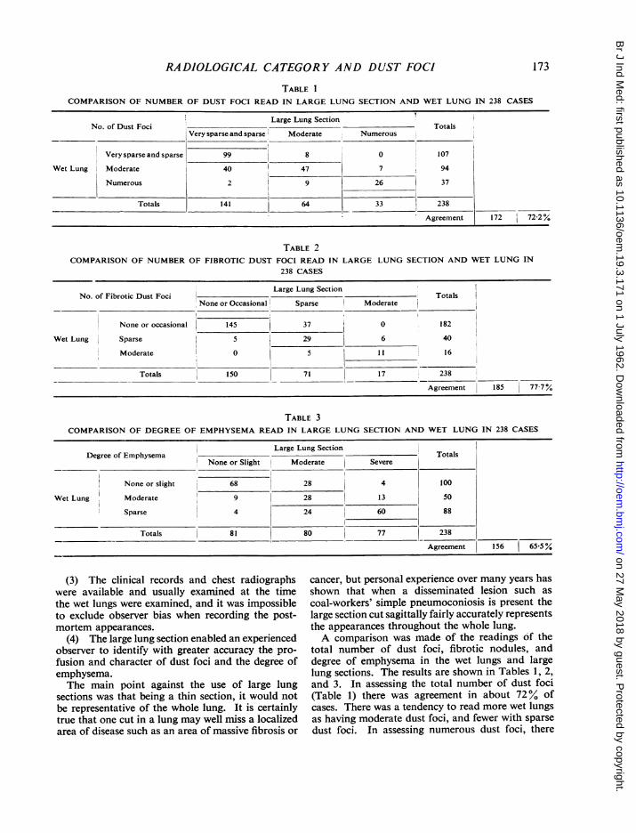
(3) The clinical records and chest radiographswere available and usually examined at the timethe wet lungs were examined, and it was impossibleto exclude observer bias when recording the post-mortem appearances.
(4) The large lung section enabled an experiencedobserver to identify with greater accuracy the pro-fusion and character of dust foci and the degree ofemphysema.The main point against the use of large lung
sections was that being a thin section, it would notbe representative of the whole lung. It is certainlytrue that one cut in a lung may well miss a localizedarea of disease such as an area of massive fibrosis or
cancer, but personal experience over many years hasshown that when a disseminated lesion such ascoal-workers' simple pneumoconiosis is present thelarge section cut sagittally fairly accurately representsthe appearances throughout the whole lung.A comparison was made of the readings 6f the
total number of dust foci, fibrotic nodules, anddegree of emphysema in the wet lungs and largelung sections. The results are shown in Tables 1, 2,and 3. In assessing the total number of dust foci(Table 1) there was agreement in about 72% ofcases. There was a tendency to read more wet lungsas having moderate dust foci, and fewer with sparsedust foci. In assessing numerous dust foci, there
on 27 May 2018 by guest. P
rotected by copyright.http://oem
.bmj.com
/B
r J Ind Med: first published as 10.1136/oem
.19.3.171 on 1 July 1962. Dow
nloaded from

BRITISH JOURNAL OF INDUSTRIAL MEDICINE
TABLE 4 TABLE 8RST AND SECOND READINGS AGE AND RADIOLOGICAL CATEGORY
OF RADIOLOGICAL CATEGORIES IN 238 CASES
Radiological First Reading !_Totals__Category Totals
0 if2 3
0 14 5 0 0 19
1 4 71 19 0 94SecondReading 2 0 5 60 8 73
3 0 0 7 45 52
Totals 18 81 86 53 238
Agreement 190 80%
Radiological CategoryAge (years) _ _-
0 1 2 3 Totals
20- 0 0 1 0 130- 0 0 2 0 240- 3 6 3 4 1650- 7 30 21 11 6960- 6 35 38 32 II170+ 2 10 21 6 39
Totals 18 81 86 53 238
TABLE 5COMPARISON OF FIRST AND SECOND READINGS OF NUMBER OF DUST FOCI IN 238 CASES
First ReadingNumber of Dust Foci TotalsVery Sparse Moderate Numerous
and Sparse
Very sparse and sparse 136 20 0 156
Second Reading Moderate 5 41 7 53
Numerous 0 3 26 29
Totals 141 64 33 238
Agreement 203 85 3
TABLE 6COMPARISON OF FIRST AND SECOND READINGS OF NUMBER OF FIBROTIC NODULES IN 238 CASES
First ReadingNo. of Fibrotic Nodules Totals
None or Occasional Sparse Moderate
None or occasional 123 9 0 132
Second Reading Sparse 27 57 5 89
Moderate 0 5 12 17
Totals 150 71 17 238
Agreement 192 j 80-7%
TABLE 7COMPARISON OF FIRST AND SECOND READINGS OF DEGREE OF EMPHYSEMA IN 238 CASES
First ReadingDegree of Emphysema Totals
None or Slight Moderate Severe
None or slight 75 20 0 95
Second Reading Moderate 6 53 13 72
Severe 0 7 64 71
Totals 81 80 77 238
Agreement 192 80-7%
174
COMPARISON OF FIT
on 27 May 2018 by guest. P
rotected by copyright.http://oem
.bmj.com
/B
r J Ind Med: first published as 10.1136/oem
.19.3.171 on 1 July 1962. Dow
nloaded from

RADIOLOGICAL CATEGORY AND DUST FOCI
was fair agreement apart from two cases read asnumerous in the wet lung and sparse in the largelung section. There was agreement in about 78%of cases in reading fibrotic nodules (Table 2). Atendency to read a higher proportion with fibroticnodules was observed in the large lung section. Inassessing the degree of emphysema there was lessagreement (about 65%) (Table 3). Moderate andsevere emphysema was recorded more frequently inthe large lung section than in the wet lung. Of the80 cases assessed as having moderate emphysema inthe large lung sections, approximately equal numberswere put in all three categories when viewing the wetlungs. This suggests that emphysema is easier to seein the lung section than in the wet lung.As the comparison of the readings of wet lungs
and large lung sections for dust foci was fairly goodit was decided that the readings of the large lungsections were preferable for radiological correlation,if only because it eliminated the observer bias whenreading wet lungs with a prior knowledge of theradiological appearances.Most of the chest radiographs were taken at the
Cardiff Pneumoconiosis Panel where the techniqueof exposure and development of films is fairly con-sistent. The radiological classification of pneumo-coniosis was based on the International LabourOrganization (I.L.O.) Classification (1953), currentlyfollowed by the Pneumoconiosis Medical Panels.The films were read by the author without know-ledge of the pathology.The large lung sections and the chest radiographs
were re-read by the same observer after an intervalof three months, and the first readings of the largelung sections were correlated with the first readingsof the radiological category of the comparable lungfield.
ResultsRepeatability of Assessments of Radiographs and
Lung Sections.-The comparison of first and secondreadings is shown in Tables 4, 5, 6, and 7. Therewas agreement in about 80% of cases in the readingof radiological category, number of fibrotic nodulesand emphysema, and in about 85% in the readingsof the total number of dust foci. There was a slighttendency at the second reading to read more ascategory 1 and fewer as category 2; fewer as moderateand more as sparse dust foci; more with sparsefibrotic nodules; and more cases without emphysema.
Age Incidence.-The age incidence of the 238 casesis shown in Table 8. Only 19 (8%) were under 50,and 39 (16-4 %) 70 or over. The majority werebetween the ages of 50 and 69; 69 (29 %) were 50 to59, and Il l (46-6 %) 60 to 69.
Relation of Radiological Category and Total DustFoci (Table 9 and Fig. 4).-All cases with category 0had very sparse or sparse dust foci. With increasingradiological category there was a fall in the pro-portion of cases with very sparse and sparse dustfoci and an increase in the prevalence of moderateand numerous dust foci.The relation between radiological category and
dust foci was somewhat improved if cases with Ashadows were excluded. There was a higher preva-lence of sparse dust foci in category 3 cases withA shadows (38 %) as compared with category 3without A shadows (13 %). The numbers were smalland the difference not statistically significant.
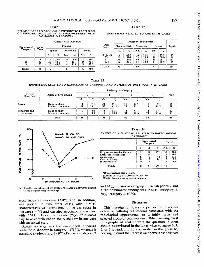
Relation of Radiological Category to Profusion ofFibrotic Nodules.-None of the cases with category 0and only 11 0% of those with category 1 had fibroticnodules in the lungs (Table 10 and Fig. 5). Incontrast to this, fibrotic nodules were found in about440% of cases with category 2 and about 770% ofcases with category 3. There were no cases withnumerous fibrotic nodules. The relation betweenradiological category and profusion of fibroticnodules is, therefore, clearer than with total dustfoci.When the cases with A shadows are excluded the
percentage of category 2 cases with fibrotic noduleswas 28-3 instead of 44-2. The proportion of caseswith fibrotic nodules was about three times higherin those with A shadows than in those without Ashadows (Table 11).
Relation of Radiological Category and Em-physema.-Table 12 related emphysema to age.There was a rise in the prevalence of emphysemawith increasing age up to 69; this was most strikingbetween the under 50 and 50 to 59 age groups.
Fig. 6. relates emphysema to radiological categoryin two age groups (a) below 60 and (b) 60 and over.The prevalence of emphysema was highest in bothage groups in those with category 0 and not veryclosely related to the other radiological categories,although there was a tendency for those withcategory 1 to have the smallest amount ofemphysema.
Table 13 relates emphysema to radiologicalcategory and number of dust foci. There were nocases of category 0 with moderate or numerous dustfoci, and of the 12 cases of category 1 with moderateor numerous dust foci, five had none or slightemphysema and seven moderate or severe. Thisfinding does not support a widely held belief thatemphysema may obscure radiological dust opacities
175
on 27 May 2018 by guest. P
rotected by copyright.http://oem
.bmj.com
/B
r J Ind Med: first published as 10.1136/oem
.19.3.171 on 1 July 1962. Dow
nloaded from

BRITISH JOURNAL OF INDUSTRIAL MEDICINE
100
90
80
70
0
zla4
I.
60-
50-
40-
30-
20-
lo.
(is Cases) (81 Cases)(86 Cases) (53 Cases)
2I 3RADIOLOGICAL CATEGORY
VERY SPARSE SPARSE
MODERATE NUMEROUS
FIG. 4.-The prevalence of sparse, moderate, and numerous dust fociin each radiological category.
and "convert" a category 2 or 3 to 0 or 1. If thiswere so there should have been a disproportionatenumber of cases with moderate and numerous dustfoci in categories 0 and 1 with post-mortem evidenceof moderate or severe emphysema.
Pathology Associated with Category A Shadows.-Table 14 shows that A shadows were accountable
8o
70
60
0
5.1-
z
-i
4c
Iii
50z
40
30
20
10.
a SPARSE
0 MODERATE
-H_0 I 2
RADIOLOGICAL CATEGORY3
FIG. 5.-The prevalence of fibrotic nodules in each radiologicalcategory.
TABLE 10RELATION OF RADIOLOGICAL CATEGORY TO PROFUSION
OF FIBROTIC NODULES IN 238 CASES
Fibrotic NodulesRadiological No. of F TtlCategory Cases Sparse Moderate* Totals
No. % No.| % No. %0 18 0 0.0 0 0.0 0 0.01 81 9 11-1 0 0.0 9 11*12 86 33 38-4 5 5 8 38 44-23 53 29 54-7 12 22-6 41 77-3
Totals 238 71 17 88 37 0
*There were no cases with numerous fibrotic nodules.
pathologically mainly by progressive massive fibrosis(P.M.F.) (51-4 %), large fibrotic nodules (2517 %), andapical scars (186 %). Cancer of the lung was the sole
TABLE 9CORRELATION OF RADIOLOGICAL CATEGORY AND NUMBER OF DUST FOCI IN 238 CASES
Number of Dust Foci
Category No. of Cases Very Sparse Sparse Moderate Numerous
No. % No. % No. % No. %0 18 12 66-7 6 33-3 0 00 0 00-I|81 33 40 7 36 44-5 12 14-8 0 00
2 86 1 1-2 39 45-3 30 34 9 16 18-63 53 0 00 14 26-4 22 41-5 17 32-1
Totals 238 46 95 64 33 I
176
I
on 27 May 2018 by guest. P
rotected by copyright.http://oem
.bmj.com
/B
r J Ind Med: first published as 10.1136/oem
.19.3.171 on 1 July 1962. Dow
nloaded from
RADIOLOGICAL CATEGORY AND DUST FOCI
TABLE I1RELATION OF RADIOLOGICAL CATEGORY TO PROFUSIONOF FIBROTIC NODULES IN 70 COAL-WORKERS WITH
CATEGORY A SHADOWS
ICharacter of Dust Fo
Radiological No. of FibroticCategory Cases Sparse Moderate
-No. % No. %
I; 1-I 8 2 25-0 0 0 02 33 20 60-6 4 12-13 29 20 69-0 4 13-8
Totals 70 42 1 8
ici
Totals
TABLE 12
EMPHYSEMA RELATED TO AGE IN 238 CASES
Degree of EmphysemaAge None or Slight Moderate Severe Totals(years)
No. % INo. I%. No. I%YNo. % Up to 49 12 63-2 4 21-6 3 15-2 19
50- 26 37-7 23 33-3 20 29-0 692 25-0 60- 31 27-8 40 36-1 40 36-1 111
24 72-7 70+ 12 30-8 13 33-3 14 35 9 3924 82-8
Totals 81 80 77 23850 71-4 -ill
TABLE 13EMPHYSEMA RELATED TO RADIOLOGICAL CATEGORY AND NUMBER OF DUST FOCI IN 238 CASES
Radiological CategoryNo. of
Dust Foci Degree of Emphysema I 0 2 3 Totals
\|No. % No. % No. % No. %5_Sparse None or slight 4 7-4 32 59-3 14 25-9 4 7-4 54
Moderate or severe 14 16-1 37 42-5 26 29-9 10 11 5 87
Moderate and None or slight 0 0-0 5 18-5 12 44-4 10 37-1 27numerous Moderate or severe 0 0.0 7 10-0 34 48-6 29 41-4 70
Totals 1 18 81 86 53 238
*0--* BELOW 60O-0 60 AND OVER
TABLE 14CAUSES OF A SHADOWS RELATED TO RADIOLOGICAL
CATEGORY
Radiological Totals
Category
I1 2 3 No. I%
Progressive massive fibrosis 1* 18t 17t 36 51-4Large fibrotic nodules I 10 7 18 25-7Apical scars 6 3 41 3 18 6Cancer of lung 0 2 0 2 2-9Bronchiectasis 0 0 1 1 1-4
Totals 8 33 I 29 70
0 0 I 2 3RADIOLOGICAL CATEGORY
FIG. 6.-The prevalence of moderate and severe emphysema relatedto radiological category and age.
gross lesion in two cases (290%) and, in addition,was present in two other cases with P.M.F.Bronchiectasis was considered to be the cause inone case ([-40%) and was also associated in one case
with P.M.F. Interstitial fibrosis ("cystic" disease)may have contributed to the A shadow in one case
with an apical scar.
Apical scarring was the commonest apparentcause for A shadows in category 1 (75 %), whereas itcaused A shadows in only 9% of cases in category 2
*Bronchiectasis also present.tCancer of lung also present in one case.
+Cystic disease also present in one case.
and 14% of cases in category 3. In categories 2 and3 the commonest finding was P.M.F. (category 2,54%; category 3, 60%).
DiscussionThis investigation gives the proportion of certain
definable pathological features associated with theradiological appearances on a fairly large andselected group of coal-workers. When viewing chestradiographs of coal-workers the question is whatshould be envisaged in the lungs when category 0, 1,2, or 3 is read, and how accurate can this guess be,bearing in mind that there is an appreciable observer
177
90.
zhi-i
a.1
in
80
70
60
so
on 27 May 2018 by guest. P
rotected by copyright.http://oem
.bmj.com
/B
r J Ind Med: first published as 10.1136/oem
.19.3.171 on 1 July 1962. Dow
nloaded from
BRITISH JOURNAL OF INDUSTRIAL MEDICINE
error both in reading radiographs and assessing thenumber and character of dust foci found in the lungsafter death? A very close correlation of radiographreadings and post-mortem findings cannot beexpected. Nevertheless, the findings of this investi-gation suggest that there is fairly good correlationbetween the radiological category of simple pneumo-coniosis and the total number of dust foci and pro-portion of fibrotic nodules found in the lungs atpost-mortem examination. The higher the radio-logical category, the greater is the likelihood that thelungs will show a larger number of dust foci andparticularly a greater proportion of fibrotic nodules,and vice versa.
All the 18 cases read as category 0 had very sparseor sparse soft dust foci, and in a high proportion ofcases (66 %) the foci were very sparse. In category 1the majority (85 %) had very sparse (41 %) or sparsedust foci; a small proportion (15%) had moderatefoci. In 89% of cases the foci were soft, and in 11 %sparse fibrotic nodules were present. As soft dustfoci do not show an appreciable degree of colla-genous fibrosis histologically, these findings suggestthat when an experienced observer reads category 0or 1, there will probably be very little simplepneumoconiosis found in the lungs of all cases ofcategory 0 and in most cases with category 1.
In categories 2 and 3 the proportion of cases withsparse dust foci fell (46% and 26% respectively) andthe proportion of moderate (35% and 41 % res-
pectively) and numerous (19% and 32% respectively)rose. In addition fibrotic nodules were present in44% of cases with category 2 and 77% of cases withcategory 3; in about 6% of cases with category 2and 23% in category 3 the fibrotic nodules weremoderate in number.A comparison of cases with and without A
shadows showed no significant difference in the dis-tribution of the total number of dust foci in category2 but a surprisingly higher prevalence of sparse
dust foci in cases of category 3 with A shadows(38%) as compared with those without A shadows(13 %). This theoretically could be attributed to thepossibility that dust had migrated to the P.M.F.lesion. A more likely explanation is that there was
a relatively high proportion of category 3 cases withsparse dust foci most of which were fibrotic.A significant finding in comparing the prevalence
of fibrotic nodules in cases with and without Ashadows was the very much higher proportion withfibrotic nodules in category 2 with A shadows(72-7y% as compared with 28 3%). The probableexplanation is that both fibrotic nodules and thecauses of A shadows-commonly P.M.F. or con-
glomeration of large fibrotic nodules-have a com-mon aetiological factor. In category 3 cases there
was a high proportion of fibrotic nodules in thosewith and without A shadows (82-8 % and 75%respectively). It is also of interest that more than50% of cases with category 3 had A shadows.
Rivers et al. (1960) found that fibrous nodulesoccur mainly in the lungs which contain most dustalthough they may also occur in lungs with a lowdust content. This suggested to the authors that thenodules are probably not produced by dust aloneand they considered infection to be a likely additionalfactor, particularly tuberculosis (cf., Gough, 1947).Fibrous nodules were found by Rivers in 33% ofhis 45 cases. This prevalence of fibrotic nodules issimilar to that found in this investigation (37 %).Rivers et al. also found a clear increase in theaverage weight of total dust with increasing radio-logical category. This was paralleled in this investi-gation with the finding that the proportion of caseswith moderate and numerous dust foci rose withincreasing radiological category.
Rivers et al. concluded that "there is little or norelation between radiological category and thepresence of fibrotic nodules". This appears to be inconflict with the findings of the present investigation.It is to be noted, however, that Rivers did find ahigher prevalence of fibrotic nodules in categories2 and 3 (40% and 67% respectively) as comparedwith categories 0 and 1 (23 %). The difference ofopinion is, therefore, one of interpretation of find-ings rather than actual difference in results. In thisinvestigation it was considered that there was aclearer relation between radiological category andprofusion of fibrotic nodules than with the totalnumber of dust foci. These observations suggestthat the number of fibrotic nodules probably in-fluences the radiological category more than thetotal number of soft dust foci. Indeed personalexperience has shown that in cases with an indisput-able radiological category 3, the lungs at necropsynot infrequently show only sparse but mainly fibroticnodules. The findings of this investigation tend toconfirm this observation in that in 12 of the 14 casesread as category 3 with sparse dust foci, the lesionswere mainly fibrotic. A typical example is shown inFig. 7. Conversely category 0 may be read in lungshaving moderate soft dust foci.One of the more important reasons for relating
emphysema to dust foci and radiological categorywas to test a current belief that emphysema mayobscure radiological dust opacities and thereby"convert" a category 2 or 3 to category 0 or 1.These findings did not support this contention in thatthere was not a disproportionate number of caseswith moderate and numerous dust foci in categories0 or 1 when moderate or severe emphysema waspresent.
178
on 27 May 2018 by guest. P
rotected by copyright.http://oem
.bmj.com
/B
r J Ind Med: first published as 10.1136/oem
.19.3.171 on 1 July 1962. Dow
nloaded from

RADIOLOGICAL CATEGORY AND DUST FOCI... _
.4 .
;, * # * ;
.. s R -;! .; b~~~~
FIG. 7a and b.-Example of large lung section and radiograph of corresponding lung field showing category 3 nodular.
The material used in this investigation may wellhave had an odd distribution of the prevalence ofemphysema. No firm conclusions regarding therelation of emphysema to age, radiological category,and number of dust foci can therefore be drawn.The commonest causes ofA shadows in categories
2 and 3 were P.M.F. and large fibrotic nodules,whereas in category 1 most of the A shadows weredue to apical scars. Other rarer causes found werecancer of the lung, bronchiectasis, and "cystic"disease.
I wish to acknowledge my thanks to the Chief MedicalOfficer, Ministry of Pensions and National Insurance,for permission to publish this paper, and to ProfessorJ. Gough of the Institute of Pathology at the WelshNational School of Medicine who provided the large
lung sections. Much valuable advice and criticism wasgiven by Professor A. L. Cochrane and Dr. J. C. Gilsonof the Pneumoconiosis Research Unit and Professor J.Gough, and I am further indebted to Dr. J. C. Gilsonfor the figures drawn and photographed by members ofhis staff. Dr. Enid Rogers took part in the originalinvestigation and her help and advice are much appreci-ated.
REFERENCESCiba Guest Symposium (1959). Thorax, 14, 286.Gough, J. (1947). In Silicosis, Pneumokoniosis and Dust Suppression
in Mines (Proc. Conf. London, 1947), p. 7. Institution ofMining Engineers and Institution of Mining and Metallurgy,London.James, W. R. L., and Wentworth, J. E. (1949). J. Fac. Radiol.(Lond.), 1, 28.
International Labour Organization (1953). Third International Con-ference of Experts on Pneumoconiosis, Sydney, 1950. Recordof Proceedings, 1, 126. Internal Labour Office, Geneva.
Rivers, D., Wise, M. E., King, E. J., and Nagelschmidt, G. (1960).Brit. J. industr. Med., 17, 87.
Roach, S. A. (1953). ibid., 10, 220.
179
.m.W.A..
6QP"lnN, -.0
on 27 May 2018 by guest. P
rotected by copyright.http://oem
.bmj.com
/B
r J Ind Med: first published as 10.1136/oem
.19.3.171 on 1 July 1962. Dow
nloaded from