Embed Size (px)
Citation preview
20
International Tinnitus Journal, Vol. 18, No 1 (2013)www.tinnitusjournal.com
Correlation between brain cortex metabolic and perfusion functions in subjective idiopathic tinnitus
Abstract
Saeid Mahmoudian1,2,3
Mohammad Farhadi1Saeid Gholami4,7
Fariba Saddadi5Maryam Jalesi1
Ali Reza Karimian6
Maryam Darbeheshti1Sara Momtaz1
Sara Fardin7
1 Laboratory for Auditory Neuroscience, ENT and Head & Neck Research Center, Iran University of Medical Sciences. Tehran, Iran.2 Iran National Science Foundation (INSF), Tehran, Iran.3 Department of Otorhinolaryngology, Medical University of Hannover (MHH) and Center for Systems Neuroscience, Hannover, Germany.4 Department of Radiology and Molecular Imaging, Hospital of the University of Pennsylvania, Philadelphia, USA.5 Agriculture, Medical & Industrial Research School. Tehran, Iran.6 Department of Biomedical Engineering, Faculty of Engineering, University of Isfahan, Isfahan, Iran.7 Firoozgar Clinical Research Developing Center, Firoozgar General Hospital, Iran University of Medical Sciences, Tehran, Iran.Institution: This investigation was supported by Iran National Science Foundation (INSF) and ENT & Head and Neck Research Center of Iran University of Medical Sciences.Send correspondence to:Saeid Mahmoudian.Assistant Professor of Neuroscience. Laboratory for Auditory Neuroscience, ENT and Head & Neck Research Center, Iran University of Medical Sciences, Tehran, Iran.Tel.: +98 (0) 21 66504294Fax: +98 (0) 21 66525329E-mail: [email protected]@mh-hammover.dePaper submitted to the ITJ-SGP (Publishing Management System) on September 3, 2013;and accepted on January 22, 2014. cod. 141
Objectives: Subjective tinnitus has associated with abnormal brain metabolism and perfusion found in functional imaging studies by fluorodeoxyglucose (FDG) and technetium99m (TC99m). But there is no study evaluating the association of brain metabolism and perfusion abnormalities in a group of these subjects. The aim of this study was to investigate if there is any significant correlation between the brain perfusion and metabolism abnormalities in subjects with tinnitus. Materials and Methods: In this cross-sectional study, 52 patients were undergone TC99m-ECD single photon emission computerized tomography (SPECT) scan and F18-FDG positron emission tomography (PET). The results of PET and SPECT scanning were fused with MRI to accurate anatomical localization of abnormalities. The analysis was performed using Kendal’s correlation, t-test and chi square. Results: Assessing these 52 tinnitus subjects (containing 42 males [76.4%]) showed that a significant correlation was found between the brain metabolic function and perfusion (p value 0.001).
ORIGINAL ARTICLEInternational Tinnitus Journal. 2013;18(1):20-28.
Keywords: emission-computed, metabolism, perfusion, positron-emission tomography, single-photon, tinnitus, tomography.
DOI: 10.5935/0946-5448.20130004

21
International Tinnitus Journal, Vol. 18, No 1 (2013)www.tinnitusjournal.com
INTRODUCTION
Tinnitus is the consciousness of sound that arises in the head without an obvious voluntary origin1. The prevalence of chronic tinnitus in general population is between 5%-15% and causes severe impairment of the quality of life in 1% to 3%1,2. Most of tinnitus subjects adapt to this phantom sound. Tinnitus may become a source of significant disability for those who fail to adapt. Therefore, the symptom of tinnitus has forced clinicians to establish protocols for the accuracy of tinnitus diagnosis and treatment3.
A number of mechanisms and suspected origins of tinnitus in the auditory pathway have been hypothesized. Most hypotheses postulate that the generation of tinnitus is in association with cochlear or acoustic nerve or central auditory dysfunction and their interactions4-9. Despite this profusion of assumed locations in the generation of tinnitus, most current hypotheses agree that abnormal neural activity is interpreted and perceived as tinnitus in higher cortical centers (e.g., auditory cortex). Abnormal activity at higher levels of the auditory pathways (auditory nuclei, auditory cortex and associative cortices) may contribute significantly to the generation of tinnitus6,10,11. Central mechanisms must play a determinant role in generating this auditory phantom sensation as it resistant in most cases follow secession of auditory nerve. One hypothesis emphasis that tinnitus is caused by a reorganization of tonotopic maps in the auditory cortex, which leads to an over representation of tinnitus frequencies. Moreover, the participation of the limbic system in generating tinnitus has been postulated12. In recent years, it has been widely acknowledged that maladaptation of central information processing are critically responsible in tinnitus perception and generation13. Many investigations have revealed that tinnitus is linked with increased activity in the central auditory system as confirmed by electrophysiology and neuroimaging studies5,14,15. It could be a critical step in the task of defining the factors that create these phantom sensations and developing rational treatments for this chronic and disabling disorder16.
Subjective idiopathic tinnitus is a symptom that could not be detected by routine clinical and paraclinical examinations. But functional imaging, electrophysiological and psycho-acoustical evaluation are among the tools that could be useful to objectify tinnitus. Advances in brain function imaging techniques have made it possible to identify the brain regions associated with the production of transient, subjective sensations, such as phantom limb pain or hallucinations, as well as perception and processing of sound and tinnitus and associating characteristics such as loudness of tinnitus16-22.
Finding the involved foci in the brain of these patients is important for defining pathophysiology and selecting effective treatment strategy. The results of various functional imaging modalities such as positron emission tomography (PET), single photon emission computed tomography (SPECT) and functional magnetic resonance imaging (FMRI) have shown similar brain abnormalities but there are some studies that have reported different findings in a group of patients5,7,16-30.
Considering the importance of finding the responsible site of tinnitus in the brain and different functional imaging methods for finding the involved foci, this investigation was designed to evaluate correlation of two nuclear imaging methods evaluating brain metabolic perfusion and function using SPECT and PET in subjects with chronic subjective idiopathic tinnitus.
MATERIALS AND METHODS
SubjectsThis cross-sectional study was performed on 52
patients referred to ENT and Head & Neck Research Centre of Iran University of Medical Sciences (IUMS) for evaluation and treatment of their tinnitus, and 6 control subjects from February 2008 to February 2009. All subjects gave written informed consent in accord with the declaration of Helsinki, National Committee of Ethics in Medical Research (Technology and Research Deputy of Hygiene Ministration) and the Committee on Ethics at the ENT and Head & Neck Research Centre of Tehran University of Medical Sciences, radiation safety, and radioactive drug research committees to participate in the study.
Inclusion criteria were: good general health, do not have any invasive therapeutic interventions of the brain, should not be pregnant or have made any decision to become pregnant, no psychiatric disorders or its history (according to a psychiatric evaluation), any treatment for tinnitus during last three months, dementia, seizure or alcohol/drug abuse in last six months, head and neck diseases or space occupying lesions, or any organic disease that could cause tinnitus should not be present. Also MRI with and without Gadolinium injection were performed to rule out any organic lesions in suspected patients. All patients were over 18 years old.
Tinnitus assessmentPitch and Loudness Matching of t innitus
(PMT and LMT) were evaluated in tinnitus subjects in the affected ear to an external tone presented to the contralateral ear. This task was accomplished using a Tinnitus Evaluation Device (TinED®) which includes 6 channels to reconstruct the Most Troublesome Tinnitus (MTT) with a similar frequency and intensity. The accuracy of the calibrating equipment shall

22
International Tinnitus Journal, Vol. 18, No 1 (2013)www.tinnitusjournal.com
be sufficient to determine that the TinED® is within the tolerances permitted by American Standard Specification for Audiometers, S3.6-2004. The subjects had to have LMT over 6 decibel sensation level (dB SL) to be included in this study.
Using Persian version of Tinnitus Handicap Inventory (THI), severity of tinnitus was evaluated and subjects with THI score of 44 or more were considered to have moderate to severe tinnitus31.
ImagingEach patient underwent scanning first with TC99m-
ethyl cysteinate dimer (EDC) and two weeks later with F18-FDG. Then the MRI of patients was fused with the PET and SPECT images using Brain Anatomical Functional Images Coregistration Software (BrainAFICS®).
TC99m Perfusion imagingThe patients were placed in a quiet, dimly lit room
and instructed to keep the eyes open (or use a mask) and the ears unplugged. They were also instructed not to speak, read, or move during 5 min prior to and 5 min post injection. They were asked to think about their tinnitus during the test. A commercial ethyl cysteinate dimer (ECD) preparation was used. After approximately 30 min, each subject received a 15mCi intravenous injection of tracer while they were still lying down. No sedation was used. One hour after intra venous injection of 15 mCi 99mTc-ECD, SPECT scanning was done (120 projections; 40 projections per head; 25 sec/projection). Scanning was performed using a dual head SMV gamma camera, equipped with a pair of low energy, high resolution collimators.
Planar and processed SPECT images were visually assessed by a nuclear medicine physician twice who was blinded to all other clinical and imaging information. The images were visually graded as normal for no appreciable abnormal activity and abnormal for hyper activity. Semiquantitative evaluation of planar images was also performed by drawing regions of interest (ROI) over the lesion and background areas on the anterior, posterior or contralateral hemisphere images without any lesion. Geometric means of the contralateral, posterior and anterior ROI values were used for calculation of lesion-to-background ratio. Transverse views with the best visualization of the lesions were selected for ROI drawing on the SPECT images. Lesion to background ratio was calculated accordingly for all sets of images. All activity ratios were classified to determine the intensity of activity as: 1 > = normal, 1-1.5 mild, 1.5-2 moderate and > 2 as sever hyperactivity. If the ratio was less than 1, the visually suspected sites were considered normal and if more than one, those were classified as abnormal sites or lesions.
F18-FDG Metabolic Function ImagingSerum level of glucose had to be under 6 mmol/dl
after 4 hours fasting. All subjects had an intravenous line. They were lying down with closed eyes (or covered) and unplugged ears in a dark and silent room. They were asked to concentrate on their tinnitus and not to speak, read, or move for at least 5 min prior to injection. No sedation was applied. Approximately 30 min after 5 miliCurie intravenous injection of F18-FDG (Fluoro Deoxy Glucose), scans were obtained. Imaging procedure was performed with a dual head SMV camera, with a pair of low energy, high-resolution collimators. The projections data were processed with a FBP butherworth 5-0.5 to show a 3D view of the brain. Anatomical tissue images were generated and standard circular regions of interest were created for each subject in the study. Standard uptake values were calculated for each region of interest. After subtracting areas with normal uptake from PET images of subjects, an average normal scanning of control group were used. Regions with abnormal uptake were identified. All these abnormal regions constituted eight regions of interests, which were found to be related to tinnitus. These regions consisted of middle temporal, inferotemporal, medial temporal, superior temporal, temporoparietal, frontal, frontoparietal, and parietal areas. These areas were separated by anatomical sulci in three-dimensional images and were localized by one expert.
The criteria to define the hyperactivity of brain were with both visual and semi quantitatively analysis. First an abnormal area of hyperactivity in cerebral gray cortex was found by the expert and then an ROI (1pixel size) was drowning around that area and background. An ROI on the opposite side (as mirror image) for unilateral lesion and on the cerebellum (as reference) for bilateral lesion was analyzed simultaneously. Counts per voxel of all bilateral lesions (hyperactivity) was calculated in cerebral cortex and compared with cerebellum to calculate the activity ratio. Also the ROI value of any unilateral lesion was compared with the value of the ROI on the other side to give an activity ratio as well. All activity ratios were classified to determine the intensity of activity as: 1 > = normal, 1-1.5 mild, 1.5-2 moderate and > 2 as sever hyperactivity. If the ratio was less than 1, the visually suspected sites were considered normal and if more than one, those were classified as abnormal sites or lesions.
MRIMagnetic resonance imagings were obtained
using tool Q11 marked Siemens, 1.5 Tesla, Avanto 18 channels. The participants were kept approximately 8 min without any movement. The images were stored in Dicom format to be applied in Brain Anatomical Functional Images Coregistration Software (BrainAFICS®).

23
International Tinnitus Journal, Vol. 18, No 1 (2013)www.tinnitusjournal.com
Image FusionPET and SPECT imaging are inherently metabolic
and perfusion modalities, so it is sometimes difficult to exactly interpret the anatomical area of disturbed function. Therefore, it is helpful to correlate the relatively coarse functional images to high-resolution anatomical MRI. BrainAFICS®, a software system designed in the ENT and Head and Neck research Center of IUMS, is capable of registering and fusing unsynchronized PET and SPECT with MRIs with different dimensions, in the same subjects (Figures 1 and 2).
If the assessment was not similar, images were assessed by an expert for a third time and repeated reports (in the previous evaluations) were considered to be the result.
Statistical AnalysisFor the association between localization by FDG
and TC99m scans the analysis was performed using Kendall’s tau-b correlation coefficient test. The amount of increase was assessed using Pearson correlation test. The association and difference of qualitative data was tested by chi square and the quantitative data by independent t-test. A probability value less than 0.05 was considered significant. The data are presented as mean ± SD. All analyses were performed using the statistical package for social science version 16 (SPSS v.16).
RESULTS
DescriptionThere were 52 tinnitus subjects and six healthy
subjects in this study with a mean age 48 ± 13.5 and 35.8 ± 12.4 years old and male to female ratio of 40:12 and 3:3, respectively. Of the tinnitus subjects, 16 (31%) had left ear tinnitus, 12 (23%) had right ear tinnitus and 24 (46%) had bilateral tinnitus. The mean duration of tinnitus was 70 ± 57.2 months for left and 82.6 ± 69.3 months for right ears. Within tinnitus subjects 45 (86.5%) had abnormality by FDG and 40 (76.9%) by TC99m in the left hemisphere, and 40 (76.9%) had abnormality by FDG and 39 (75%) by TC99m in the right hemisphere (p < 0.001, Figure 3). Number of abnormal area in PET scan was also significantly associated with SPECT scan (Table 1). Middle temporal, temporoparietal and inferotemporal were the most common abnormal areas detected by F18-FDG PET coincidence imaging and TC99m-EDC SPECT imaging in these patients (Tables 2 and 3).
Figure 1. Involvement of left Infrotemporal area in a subject with left ear tinnitus. Note: Fusion of SPECT and magnetic resonance imaging of a patient shows hyperactivity of left Infrotemporal area.
Figure 2. TC99m-ECD single photon emission computerized tomography scan (A) and F18-FDG positron emission tomography (B) in tinnitus subjects.
Figure 3. Comparison between metabolic (PET) and perfusion (SPECT) function abnormalities.
ValidationAfter final fusion of images, each image was
evaluated and reported by one expert in nuclear medicine twice at separate times. Almost all images were reported similar in both times (r = 0.8, p > 0.001).
24
International Tinnitus Journal, Vol. 18, No 1 (2013)www.tinnitusjournal.com
AbnormalitiesLeft hemisphere
N (%)Right hemisphere
N (%)
PET SPECT PET SPECT
Unifocal 41(78.8%) 28(53.8%) 34(65.4%) 28(53.8%)
Multifocal 4(7.7%) 12(23%) 6(11.5%) 11(21.1%)
No abnormality 7(13.5%) 12(23%) 12(23%) 13(25%)
p value < 0.001* < 0.001*
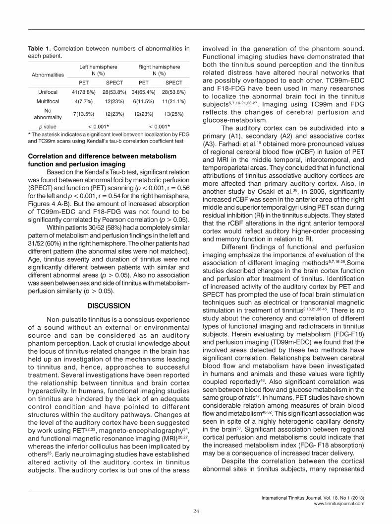
Table 1. Correlation between numbers of abnormalities in each patient.
* The asterisk indicates a significant level between localization by FDG and TC99m scans using Kendall’s tau-b correlation coefficient test
Correlation and difference between metabolism function and perfusion imaging
Based on the Kendal’s Tau-b test, significant relation was found between abnormal foci by metabolic perfusion (SPECT) and function (PET) scanning (p < 0.001, r = 0.56 for the left and p < 0.001, r = 0.54 for the right hemisphere, Figures 4 A-B). But the amount of increased absorption of TC99m-EDC and F18-FDG was not found to be significantly correlated by Pearson correlation (p > 0.05).
Within patients 30/52 (58%) had a completely similar pattern of metabolism and perfusion findings in the left and 31/52 (60%) in the right hemisphere. The other patients had different pattern (the abnormal sites were not matched). Age, tinnitus severity and duration of tinnitus were not significantly different between patients with similar and different abnormal areas (p > 0.05). Also no association was seen between sex and side of tinnitus with metabolism-perfusion similarity (p > 0.05).
DISCUSSION
Non-pulsatile tinnitus is a conscious experience of a sound without an external or environmental source and can be considered as an auditory phantom perception. Lack of crucial knowledge about the locus of tinnitus-related changes in the brain has held up an investigation of the mechanisms leading to tinnitus and, hence, approaches to successful treatment. Several investigations have been reported the relationship between tinnitus and brain cortex hyperactivity. In humans, functional imaging studies on tinnitus are hindered by the lack of an adequate control condition and have pointed to different structures within the auditory pathways. Changes at the level of the auditory cortex have been suggested by work using PET32,33, magneto-encephalography34, and functional magnetic resonance imaging (MRI)20,27, whereas the inferior colliculus has been implicated by others35. Early neuroimaging studies have established altered activity of the auditory cortex in tinnitus subjects. The auditory cortex is but one of the areas
involved in the generation of the phantom sound. Functional imaging studies have demonstrated that both the tinnitus sound perception and the tinnitus related distress have altered neural networks that are possibly overlapped to each other. TC99m-EDC and F18-FDG have been used in many researches to localize the abnormal brain foci in the tinnitus subjects5,7,16-21,23-27. Imaging using TC99m and FDG reflects the changes of cerebral perfusion and glucose-metabolism.
The auditory cortex can be subdivided into a primary (A1), secondary (A2) and associative cortex (A3). Farhadi et al.19 obtained more pronounced values of regional cerebral blood flow (rCBF) in fusion of PET and MRI in the middle temporal, inferotemporal, and temporoparietal areas. They concluded that in functional attributions of tinnitus associative auditory cortices are more affected than primary auditory cortex. Also, in another study by Osaki et al.36, in 2005, significantly increased rCBF was seen in the anterior area of the right middle and superior temporal gyri using PET scan during residual inhibition (RI) in the tinnitus subjects. They stated that the rCBF alterations in the right anterior temporal cortex would reflect auditory higher-order processing and memory function in relation to RI.
Different findings of functional and perfusion imaging emphasize the importance of evaluation of the association of different imaging methods5,7,16-26.Some studies described changes in the brain cortex function and perfusion after treatment of tinnitus. Identification of increased activity of the auditory cortex by PET and SPECT has prompted the use of focal brain stimulation techniques such as electrical or transcranial magnetic stimulation in treatment of tinnitus2,13,21,36-45. There is no study about the coherency and correlation of different types of functional imaging and radiotracers in tinnitus subjects. Herein evaluating by metabolism (FDG-F18) and perfusion imaging (TD99m-EDC) we found that the involved areas detected by these two methods have significant correlation. Relationships between cerebral blood flow and metabolism have been investigated in humans and animals and these values were tightly coupled reportedly46. Also significant correlation was seen between blood flow and glucose metabolism in the same group of rats47. In humans, PET studies have shown considerable relation among measures of brain blood flow and metabolism48-52. This significant association was seen in spite of a highly heterogenic capillary density in the brain53. Significant association between regional cortical perfusion and metabolisms could indicate that the increased metabolism index (FDG- F18 absorption) may be a consequence of increased tracer delivery.
Despite the correlation between the cortical abnormal sites in tinnitus subjects, many represented

25
International Tinnitus Journal, Vol. 18, No 1 (2013)www.tinnitusjournal.com
Region of Interest (ROI) Middle Temporal
Infero-temporal
Medial Temporal
Superior Temporal
Temporo-parietal Frontal Fronto-
parietal Parietal No abnormality
Tinnitus subjects N = 52
SPECT 22 (42.3%) 7 (13.5%) 2 (3.8%) 3 (5%) 10 (19.2%) 1
(1.9%) 5 (9.6%) 4 (7.7%) 12 (23%)
PET 32 (61.5%) 6 (11.5%) 0 1 (1.9%) 7 (13.4%) 1
(1.9%) 0 2 (3.8%) 6 (11.5%)
p value* 0.07* 0.76 0.49 0.61 0.59 1 0.056 0.67 0.19
Controls N = 6
SPECT 1 (16.6%) 0 0 1(16.6%) 1 (16.6%) 0 0 0 4 (66.7%)
PET 2 (33.3%) 0 0 0 0 0 0 0 4 (66.7%)
Table 2. Brain metabolic function and perfusion abnormalities in left hemisphere. N (%)
* Chi square and Fisher’s exact tests were performed to evaluate differences between metabolic function and perfusion in each site.
Evaluated Site Middle Temporal
Infero- temporal
Medial Temporal
Superior Temporal
Temporo- parietal Frontal Fronto-
parietal Parietal No abnormality
Tinnitus subjects
SPECT 19 (36.5%) 7 (13.5%) 3 (5.7%) 3 (5.7%) 11 (21.2%) 1 (1.9%) 5 (9.6%) 3 (5.7%) 13 (25%)
PET 28 (53.8%) 5 (9.6%) 2 (3.8%) 1 (1.9%) 7 (13.4%) 2 (3.8%) 0 1 (1.9%) 12 (23%)
p value* 0.11 0.75 0.95 0.61 0.43 0.95 0.057 0.61 0.81
controlsSPECT 1 (16.6%) 0 0 1 (16.6%) 1 (16.6%) 0 0 0 3 (50%)
PET 3 (50%) 0 0 0 0 0 0 0 3 (50%)
Table 3. Brain metabolic function and perfusion abnormalities in right hemisphere.
* Chi square and Fisher’s exact tests were performed to evaluate differences between metabolic function and perfusion in each site.
Figure 4. Correlation between brain cortex metabolic and perfusion function in the right (A) and left (B) hemispheres. Note: the circles size indicate the number of subjects with similar involvement in each area. MiT: Middle Temporal; IT: Inferotemporal; MT: Medial Temporal; ST: Superior Temporal; TP: Temporoparietal; F: Frontal; FP: Frontoparietal; P: Parietal; NA: No abnormal area.
foci were not similar in these two methods of imaging. This difference seems to be a major concern in different diseases with confirmed brain function and perfusion abnormalities for diagnosis or direct stimulation therapy (epilepsy, depression, glioma)28-30,54-57. Possible factors for incongruent results are: a) differences in molecular structure of ligands caused by adherence of different elements; b) differences in activity level; onset of scanning after injection of the ligand was
later in TC99m SPECT compared with F18-FDG PET coincidence imaging; c) Neurovascular coupling should be considered for unmatched findings. Each of these mechanisms may cause unmatched hyperemia28,58-64. In some investigations uncoupled neuronal activity and haemodynamic responses has been reported in brain cortex of normal rats and human65-67. Additionally regulator factors may be disturbed by tinnitus and tinnitus may be related to abnormalities associated with cerebral
26
International Tinnitus Journal, Vol. 18, No 1 (2013)www.tinnitusjournal.com
microcirculation. Dissociation between function and perfusion has been reported with a number of disorders, including Parkinson’s disease, stroke, migraine, Alzheimer’s disease and early onset dementia59,68.
This was the first study in which the findings of TC99m SPECT and FDG SPECT coincidence imaging in tinnitus subjects has been compared in the same group of tinnitus subjects. In this study we haven’t evaluated the size of the abnormality that may vary by different methods or tracers29.
Evaluating the efficacy of neuronavigated stimulation using different methods of imaging could be useful for finding the accuracy of each type of imaging. Also continuous brain monitoring after tracer injection might give more information about the neurovascular coupling and the time effect of imaging with different tracers.
AcknowledgmentThis study was funded and supported by Iran
National Science Foundation (INSF) and ENT & Head and Neck Research Center of Iran University of Medical Sciences. We are grateful to thank Dr. Martin Reivich from university of Pennsylvania who kindly gave nice comments for improving our work.
Conflict of interestThe authors declare that they have no conflict of
interest.
REFERENCES
1. Davis A, Refaie AE. Epidemiology of tinnitus. In: Tyler R, editor. Tinnitus Handbook. San Diego: Singular Publishing Group; 2000. p.1-23.
2. Plewnia C, Reimold M, Najib A, Reischl G, Plontke SK, Gerloff C. Moderate therapeutic efficacy of positron emission tomography-navigated repetitive transcranial magnetic stimulation for chronic tinnitus: a randomised, controlled pilot study. J Neurol Neurosurg Psychiatry. 2007;78(2):152-6. PMID: 16891384 DOI: http://dx.doi.org/10.1136/jnnp.2006.095612
3. Shulman A, Goldstein B. Subjective idiopathic tinnitus and palliative care: a plan for diagnosis and treatment. Otolaryngol Clin North Am. 2009;42(1):15-37. DOI: http://dx.doi.org/10.1016/j.otc.2008.09.012
4. Matthies C, Samii M. Management of 1000 vestibular schwannomas (acoustic neuromas): clinical presentation. Neurosurgery. 1997;40(1):1-9. PMID: 8971818
5. Cacace AT. Expanding the biological basis of tinnitus: crossmodal origins and the role of neuroplasticity. Hear Res. 2003;175(1-2):112-32. PMID: 12527130 DOI: http://dx.doi.org/10.1016/S0378-5955(02)00717-7
6. Eggermont JJ, Roberts LE. The neuroscience of tinnitus. Trends Neurosci. 2004;27(11):676-82. DOI: http://dx.doi.org/10.1016/j.tins.2004.08.010
7. Shulman A, Goldstein B, Strashun AM. Final common pathway for tinnitus: theoretical and clinical implications of neuroanatomical substrates. Int Tinnitus J. 2009;15(1):5-50.
8. Zenner HP, Ernst A. Cochlear-motor, transduction and signal-transfer tinnitus: models for three types of cochlear tinnitus. Eur Arch Otorhinolaryngol. 1993;249(8):447-54. PMID: 7680210
9. Coles RR. Tinnitus. In: Stephens D, editor. Adult audiology. 1st ed. Oxford: Butterworth-Heinemann; 1997. p.1-34.
10. Hazell JW, Jastreboff PJ. Tinnitus. I: Auditory mechanisms: a model for tinnitus and hearing impairment. J Otolaryngol. 1990;19(1):1-5. PMID: 2179573
11. Bauer CA, Brozoski TJ, Holder TM, Caspary DM. Effects of chronic salicylate on GABAergic activity in rat inferior colliculus. Hear Res. 2000;147(1-2):175-82. PMID: 10962183 DOI: http://dx.doi.org/10.1016/S0378-5955(00)00130-1
12. Mühlau M, Rauschecker JP, Oestreicher E, Gaser C, Röttinger M, Wohlschläger AM, et al. Structural brain changes in tinnitus. Cereb Cortex. 2006;16(9):1283-8. DOI: http://dx.doi.org/10.1093/cercor/bhj070
13. Plewnia C, Reimold M, Najib A, Brehm B, Reischl G, Plontke SK, et al. Dose-dependent attenuation of auditory phantom perception (tinnitus) by PET-guided repetitive transcranial magnetic stimulation. Hum Brain Mapp. 2007;28(3):238-46. DOI: http://dx.doi.org/10.1002/hbm.20270
14. Kaltenbach JA. Neurophysiologic mechanisms of tinnitus. J Am Acad Audiol. 2000;11(3):125-37.
15. Cacace AT, Cousins JP, Parnes SM, Semenoff D, Holmes T, McFarland DJ, et al. Cutaneous-evoked tinnitus. I. Phenomenology, psychophysics and functional imaging. Audiol Neurootol. 1999;4(5):247-57. DOI: http://dx.doi.org/10.1159/000013848
16. Lockwood AH, Salvi RJ, Coad ML, Towsley ML, Wack DS, Murphy BW. The functional neuroanatomy of tinnitus: evidence for limbic system links and neural plasticity. Neurology. 1998;50(1):114-20. PMID: 9443467 DOI: http://dx.doi.org/10.1212/WNL.50.1.114
17. Flor H, Elbert T, Knecht S, Wienbruch C, Pantev C, Birbaumer N, et al. Phantom-limb pain as a perceptual correlate of cortical reorganization following arm amputation. Nature. 1995;375(6531):482-4. PMID: 7777055 DOI: http://dx.doi.org/10.1038/375482a0
18. Silbersweig DA, Stern E, Frith C, Cahill C, Holmes A, Grootoonk S, et al. A functional neuroanatomy of hallucinations in schizophrenia. Nature. 1995;378(6553):176-9. PMID: 7477318 DOI: http://dx.doi.org/10.1038/378176a0
19. Farhadi M, Mahmoudian S, Saddadi F, Karimian AR, Mirzaee M, Ahmadizadeh M, et al. Functional brain abnormalities localized in 55 chronic tinnitus patients: fusion of SPECT coincidence imaging and MRI. J Cereb Blood Flow Metab. 2010;30(4):864-70. DOI: http://dx.doi.org/10.1038/jcbfm.2009.254
20. Giraud AL, Chéry-Croze S, Fischer G, Fischer C, Vighetto A, Grégoire MC, et al. A selective imaging of tinnitus. Neuroreport. 1999;10(1):1-5. DOI: http://dx.doi.org/10.1097/00001756-199901180-00001
21. Shulman A, Strashun AM, Avitable MJ, Lenhardt ML, Goldstein BA. Ultra-high-frequency acoustic stimulation and tinnitus control: a po-sitron emission tomography study. Int Tinnitus J. 2004;10(2):113-25.
22. Lockwood AH, Wack DS, Burkard RF, Coad ML, Reyes SA, Arnold SA. The functional anatomy of gaze-evoked tinnitus and sustained lateral gaze. Neurology. 2001;56(4):472-80. PMID: 11222790 DOI: http://dx.doi.org/10.1212/WNL.56.4.472
23. Crick F, Koch C. The problem of consciousness. Sci Am. 1992;267(3):152-9. PMID: 1502517 DOI: http://dx.doi.org/10.1038/scientificamerican0992-152
24. Shulman A, Strashun A. SPECT imaging of brain and tinnitus. Case reports. In: Heertum RV, Tikofsky A, editors. Cerebral spect imaging. 2nd ed. New York: Raven Press; 1995. p.210-2.
25. Shulman A, Strashun AM, Afriyie M, Aronson F, Abel W, Goldstein B. SPECT Imaging of Brain and Tinnitus-Neurotologic/Neurologic Implications. Int Tinnitus J. 1995;1(1):13-29.
26. Shulman A. A Final Common Pathway for Tinnitus - The Medial Temporal Lobe System. Int Tinnitus J. 1995;1(2):115-26.
27. Mirz F, Pedersen B, Ishizu K, Johannsen P, Ovesen T, Stødkilde-Jørgensen H, et al. Positron emission tomography of cortical centers of tinnitus. Hear Res. 1999;134(1-2):133-44. PMID: 10452383 DOI: http://dx.doi.org/10.1016/S0378-5955(99)00075-1

27
International Tinnitus Journal, Vol. 18, No 1 (2013)www.tinnitusjournal.com
28. Pirotte B, Goldman S, Massager N, David P, Wikler D, Vandesteene A, et al. Comparison of 18F-FDG and 11C-methionine for PET-guided stereotactic brain biopsy of gliomas. J Nucl Med. 2004;45(8):1293-8. PMID: 15299051
29. Debets RM, Sadzot B, van Isselt JW, Brekelmans GJ, Meiners LC, van Huffelen AO, et al. Is 11C-flumazenil PET superior to 18FDG PET and 123I-iomazenil SPECT in presurgical evaluation of temporal lobe epilepsy? J Neurol Neurosurg Psychiatry. 1997;62(2):141-50. PMID: 9048714 DOI: http://dx.doi.org/10.1136/jnnp.62.2.141
30. la Fougère C, Rominger A, Förster S, Geisler J, Bartenstein P. PET and SPECT in epilepsy: a critical review. Epilepsy Behav. 2009;15(1):50-5. DOI: http://dx.doi.org/10.1016/j.yebeh.2009.02.025
31. Mahmoudian S, Shahmiri E, Rouzbahani M, Jafari Z, Keyhani M, Rahimi F, et al. Persian language version of the “Tinnitus Handicap Inventory”: translation, standardization, validity and reliability. Int Tinnitus J. 2011;16(2):93-103.
32. Arnold W, Bartenstein P, Oestreicher E, Römer W, Schwaiger M. Focal metabolic activation in the predominant left auditory cortex in patients suffering from tinnitus: a PET study with [18F]deoxyglucose. ORL J Otorhinolaryngol Relat Spec. 1996;58(4):195-9. DOI: http://dx.doi.org/10.1159/000276835
33. Lockwood AH, Salvi RJ, Coad ML, Towsley ML, Wack DS, Murphy BW. The functional neuroanatomy of tinnitus: evidence for limbic system links and neural plasticity. Neurology. 1998;50(1):114-20. PMID: 9443467 DOI: http://dx.doi.org/10.1212/WNL.50.1.114
34. Mühlnickel W, Elbert T, Taub E, Flor H. Reorganization of auditory cortex in tinnitus. Proc Natl Acad Sci U S A. 1998;95(17):10340-3. DOI: http://dx.doi.org/10.1073/pnas.95.17.10340
35. Melcher JR, Sigalovsky IS, Guinan JJ Jr, Levine RA. Lateralized tinnitus studied with functional magnetic resonance imaging: abnormal inferior colliculus activation. J Neurophysiol. 2000;83(2):1058-72. PMID: 10669517
36. Osaki Y, Nishimura H, Takasawa M, Imaizumi M, Kawashima T, Iwaki T, et al. Neural mechanism of residual inhibition of tinnitus in cochlear implant users. Neuroreport. 2005;16(15):1625-8. DOI: http://dx.doi.org/10.1097/01.wnr.0000183899.85277.08
37. Eichhammer P, Hajak G, Kleinjung T, Landgrebe M, Langguth B. Functional imaging of chronic tinnitus: the use of positron emission tomography. Prog Brain Res. 2007;166:83-8. PMID: 17956774
38. Smith JA, Mennemeier M, Bartel T, Chelette KC, Kimbrell T, Triggs W. Repetitive transcranial magnetic stimulation for tinnitus: a pilot study. Laryngoscope. 2007;117(3):529-34. PMID: 17334317 DOI: http://dx.doi.org/10.1097/MLG.0b013e31802f4154
39. Langguth B, Eichhammer P, Kreutzer A, Maenner P, Marienhagen J, Kleinjung T, et al. The impact of auditory cortex activity on characterizing and treating patients with chronic tinnitus--first results from a PET study. Acta Otolaryngol Suppl. 2006;(556):84-8. DOI: http://dx.doi.org/10.1080/03655230600895317
40. Richter GT, Mennemeier M, Bartel T, Chelette KC, Kimbrell T, Triggs W, et al. Repetitive transcranial magnetic stimulation for tinnitus: a case study. Laryngoscope. 2006;116(10):1867-72. PMID: 17016213 DOI: http://dx.doi.org/10.1097/01.mlg.0000234936.82619.69
41. Kleinjung T, Steffens T, Langguth B, Eichhammer P, Marienhagen J, Hajak G, et al. Treatment of chronic tinnitus with neuronavigated repetitive Transcranial Magnetic Stimulation (rTMS). HNO. 2006;54(6):439-44. PMID: 16170508 DOI: http://dx.doi.org/10.1007/s00106-005-1329-8
42. Kleinjung T, Eichhammer P, Langguth B, Jacob P, Marienhagen J, Hajak G, et al. Long-term effects of repetitive transcranial magnetic stimulation (rTMS) in patients with chronic tinnitus. Otolaryngol Head Neck Surg. 2005;132(4):566-9. PMID: 15806046 DOI: http://dx.doi.org/10.1016/j.otohns.2004.09.134
43. Langguth B, Eichhammer P, Zowe M, Marienhagen J, Kleinjung T, Jacob P, et al. Low frequency repetitive transcranial magnetic stimulation (rTMS) for the treatment of chronic tinnitus--are there long-term effects? Psychiatr Prax. 2004;31 Suppl 1:S52-4.
44. Eichhammer P, Langguth B, Marienhagen J, Kleinjung T, Hajak G. Neuronavigated repetitive transcranial magnetic stimulation in patients with tinnitus: a short case series. Biol Psychiatry. 2003;54(8):862-5. DOI: http://dx.doi.org/10.1016/S0006-3223(02)01896-6
45. Reyes SA, Salvi RJ, Burkard RF, Coad ML, Wack DS, Galantowicz PJ, et al. Brain imaging of the effects of lidocaine on tinnitus. Hear Res. 2002;171(1-2):43-50. PMID: 12204348 DOI: http://dx.doi.org/10.1016/S0378-5955(02)00346-5
46. Reivich M. Blood flow metabolism couple in brain. Res Publ Assoc Res Nerv Ment Dis. 1974;53:125-40. PMID: 4216058
47. Lear JL, Jones SC, Greenberg JH, Fedora TJ, Reivich M. Use of 123I and 14C in a double radionuclide autoradiographic technique for simulta-neous measurement of LCBF and LCMRgl. Theory and method. Stroke. 1981;12(5):589-97. DOI: http://dx.doi.org/10.1161/01.STR.12.5.589
48. Baron JC, Lebrun-Grandie P, Collard P, Crouzel C, Mestelan G, Bousser MG. Noninvasive measurement of blood flow, oxygen consumption, and glucose utilization in the same brain regions in man by positron emission tomography: concise communication. J Nucl Med. 1982;23(5):391-9.
49. Fox PT, Raichle ME, Mintun MA, Dence C. Nonoxidative glucose consumption during focal physiologic neural activity. Scien-ce. 1988;241(4864):462-4. PMID: 3260686 DOI: http://dx.doi.org/10.1126/science.3260686
50. Bentourkia M, Bol A, Ivanoiu A, Labar D, Sibomana M, Coppens A, et al. Comparison of regional cerebral blood flow and glu-cose metabolism in the normal brain: effect of aging. J Neurol Sci. 2000;181(1-2):19-28. PMID: 11099707 DOI: http://dx.doi.org/10.1016/S0022-510X(00)00396-8
51. Mintun MA, Lundstrom BN, Snyder AZ, Vlassenko AG, Shulman GL, Raichle ME. Blood flow and oxygen delivery to human brain during functional activity: theoretical modeling and experimental data. Proc Natl Acad Sci U S A. 2001;98(12):6859-64. PMID: 11381119 DOI: http://dx.doi.org/10.1073/pnas.111164398
52. Gur RC, Ragland JD, Reivich M, Greenberg JH, Alavi A, Gur RE. Regional differences in the coupling between resting cerebral blood flow and metabolism may indicate action preparedness as a default state. Cereb Cortex. 2009;19(2):375-82. DOI: http://dx.doi.org/10.1093/cercor/bhn087
53. Klein B, Kuschinsky W, Schröck H, Vetterlein F. Interdependency of local capillary density, blood flow, and metabolism in rat brains. Am J Physiol. 1986;251(6 Pt 2):H1333-40.
54. Brockmann H, Zobel A, Joe A, Biermann K, Scheef L, Schuhmacher A, et al. The value of HMPAO SPECT in predicting treatment response to citalopram in patients with major depression. Psychiatry Res. 2009;173(2):107-12. PMID: 19540732 DOI: http://dx.doi.org/10.1016/j.pscychresns.2008.10.006
55. Amen D. Brain SPECT imaging in clinical practice. Am J Psychiatry. 2010;167(9):1125. PMID: 20826857 DOI: http://dx.doi.org/10.1176/appi.ajp.2010.10060814
56. Borbély K. Functional imaging (PET and SPECT) in epilepsy. Orv Hetil. 2001;142(44):2405-14. PMID: 11766233
57. Lamusuo S, Ruottinen HM, Knuuti J, Härkönen R, Ruotsalainen U, Bergman J, et al. Comparison of [18F]FDG-PET, [99mTc]-HMPAO--SPECT, and [123I]-iomazenil-SPECT in localising the epileptogenic cortex. J Neurol Neurosurg Psychiatry. 1997;63(6):743-8. DOI: http://dx.doi.org/10.1136/jnnp.63.6.743

28
International Tinnitus Journal, Vol. 18, No 1 (2013)www.tinnitusjournal.com
58. Anderson CM, Nedergaard M. Astrocyte-mediated control of cerebral microcirculation. Trends Neurosci. 2003;26(7):340-4. DOI: http://dx.doi.org/10.1016/S0166-2236(03)00141-3
59. Iadecola C. Neurovascular regulation in the normal brain and in Alzheimer’s disease. Nat Rev Neurosci. 2004;5(5):347-60. DOI: http://dx.doi.org/10.1038/nrn1387
60. Mulligan SJ, MacVicar BA. Calcium transients in astrocyte endfeet cause cerebrovascular constr ict ions. Nature. 2004;431(7005):195-9. PMID: 15356633 DOI: http://dx.doi.org/10.1038/nature02827
61. Filosa JA, Bonev AD, Nelson MT. Calcium dynamics in cortical astrocytes and arterioles during neurovascular coupling. Circ Res. 2004;95(10):e73-81. PMID: 15499024 DOI: http://dx.doi.org/10.1161/01.RES.0000148636.60732.2e
62. Simard M, Arcuino G, Takano T, Liu QS, Nedergaard M. Signaling at the gliovascular interface. J Neurosci. 2003;23(27):9254-62. PMID: 14534260
63. Edvinsson L, Hamel E. Perivascular nerves in brain vessels. In: Edvinsson L, Krause DN, editors. Cerebral blood flow and meta-bolism. New York; Lippincott Williams and Wilkins; 2002. p.43-67.
64. Zonta M, Angulo MC, Gobbo S, Rosengarten B, Hossmann KA, Pozzan T. Neuron-to-astrocyte signaling is central to the dynamic control of brain microcirculation. Nat Neurosci. 2003;6(1):43-50. DOI: http://dx.doi.org/10.1038/nn980
65. Sloan HL, Austin VC, Blamire AM, Schnupp JW, Lowe AS, Allers KA, et al. Regional differences in neurovascular coupling in rat brain as determined by fMRI and electrophysiology. Neuroima-ge. 2010;53(2):399-411. PMID: 20633665 DOI: http://dx.doi.org/10.1016/j.neuroimage.2010.07.014
66. Ito H, Kanno I, Kato C, Sasaki T, Ishii K, Ouchi Y, et al. Database of normal human cerebral blood flow, cerebral blood volume, cerebral oxygen extraction fraction and cerebral metabolic rate of oxygen measured by positron emission tomography with 15O-labelled carbon dioxide or water, carbon monoxide and oxygen: a multicentre study in Japan. Eur J Nucl Med Mol Imaging. 2004;31(5):635-43. PMID: 14730405
67. Ito H, Kanno I, Fukuda H. Human cerebral circulation: positron emission tomography studies. Ann Nucl Med. 2005;19(2):65-74. DOI: http://dx.doi.org/10.1007/BF03027383
68. de la Torre JC. Alzheimer disease as a vascular disorder: noso-logical evidence. Stroke. 2002;33(4):1152-62. PMID: 11935076 DOI: http://dx.doi.org/10.1161/01.STR.0000014421.15948.67