Embed Size (px)
Citation preview

/ J of IMAB. 2012, vol. 18, book 4/ 263
CORRECTING LABIAL THICK AND HIGHATTACHED FRENUM (clinical observation)
Silvia Krusteva1, Mariana Dimitrova2, Hristo Daskalov3, Stiliana Krusteva4
1) Department of Orthodontics,2) Department of Pediatric Dentistry,3) Department of Oral Surgery,4) Student,Faculty of Dental Medicine, Medical University - Plovdiv, Bulgaria.
Journal of IMAB - Annual Proceeding (Scientific Papers) 2012, vol. 18, book 4
ABSTRACTLabial thick and high attached maxillary frenum is
commonly regarded as contributing etiology for maintainingmidline diastema and upper jaw delayed development. Thesurgical modalities used to solve this problem are known tobe quite stressful for children. Dental lasers have recentlybeen increasingly used to treat wide variety of problems inmedicine.
AIM: Using a high energy diode laser to remove ashort, high attached frenum of the upper lip and present theresults of the procedure.
MATERIAL AND METHODS: We performedfrenectomy in 10 randomly selected patients of both sexesaged 7-9 years with short, thick frena of the upper lip. APicasso soft tissue diode laser, class IV, power output 7 W,ë-810 nm was used for the procedure.
RESULTS AND DISCUSSION: The healing processwas uneventful, painless and without edemas developing inthe soft tissues. No inflammation was found in the treatedtissues. The children undergoing the procedure showed nofear. This was the reason why we preferred to use lasers asa modern therapeutic modality for soft tissue correction inthe mouth.
CONCLUSION: Using lasers to remove short, highattached maxillary labial frenum has the benefit of inducingless stress in children than that they experience if anaesthesiaand surgery are administered. Anaesthesia with topicalanaesthetics is used in the procedure. The postoperativeperiod is free of pain and far from severe. This makes thistechnique particularly useful for children.
Key words: maxillary frenum, frenectomy, diastema,laser.
INTRODUCTIONShort, thick maxillary labial frena that have wide
attachment base are commonly considered an etiologicfactor for a diastema and delayed upper jaw development.
They can cause gingival recession and hamper orthodontictreatment. (2)
With low attached frena getting back to normalcondition is expected after the eruption of the permanentincisors and canines. But if frena are short and thick withwide attachment bases there might be a risk of periodontaltissue damage. (4) In such cases the correction procedureshould involve the correction of the attachment site and thelength of the frenum which usually is achieved throughsurgery. Conventional surgical methods usually involveinjection anesthesia. And surgical intervention are apparentlya source of high stress for children.
Dental lasers, because of their wide range of biologicaction, have recently been steadily introduced in dentalmedicine. Low-energy laser radiation is used to stimulate thehealing process in soft tissues and bone lesions. (7) Highenergy laser radiation is used to remove some formationsand lesions on mouth mucosa that require a surgicalintervention. (5, 6, 11) However it is still rarely used inorthodontics. (8, 9)
But lasers can be quite useful in the adjustment ofshort, thick maxillary frenum. The technique requires the useof very small amount of topical anaesthetics and has shownto induce less stress in children during the procedure andafter that. (1)
Different types of high energy lasers using differentwave length can be used for the purpose. (3, 10)
Aim: Using a high energy diode laser to remove ashort, thick upper lip frenum with wide attachment base andpresent the results of the procedure.
MATERIAL AND METHODS.We performed frenectomy in 10 randomly selected
patients of both sexes (age range 7-9 years) with short labialfrena with wide attachment base (Fig. 1).
DOI: 10.5272/jimab.2012184.263
264 / J of IMAB. 2012, vol. 18, book 4/
Fig. 1. Short frenum with wide attachment base.
Local anaesthesia was used in performing theprocedure. A Picasso diode laser, class IV, power output 7W, ë-810 nm designed for work in soft tissues was used (Fig.2). We used one of the inbuilt programmes of the equipmentworking in impulse mode of work and with power outputof 2 W.
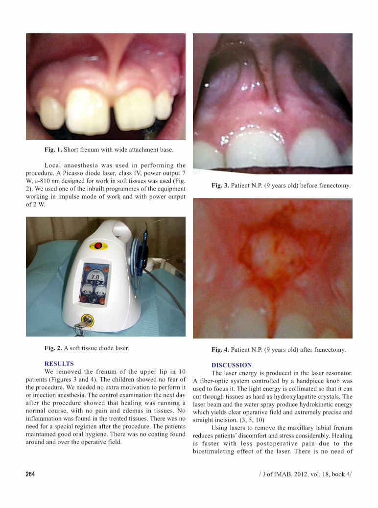
Fig. 3. Patient N.P. (9 years old) before frenectomy.
Fig. 2. A soft tissue diode laser.
RESULTSWe removed the frenum of the upper lip in 10
patients (Figures 3 and 4). The children showed no fear ofthe procedure. We needed no extra motivation to perform itor injection anesthesia. The control examination the next dayafter the procedure showed that healing was running anormal course, with no pain and edemas in tissues. Noinflammation was found in the treated tissues. There was noneed for a special regimen after the procedure. The patientsmaintained good oral hygiene. There was no coating foundaround and over the operative field.
Fig. 4. Patient N.P. (9 years old) after frenectomy.
DISCUSSIONThe laser energy is produced in the laser resonator.
A fiber-optic system controlled by a handpiece knob wasused to focus it. The light energy is collimated so that it cancut through tissues as hard as hydroxylapatite crystals. Thelaser beam and the water spray produce hydrokinetic energywhich yields clear operative field and extremely precise andstraight incision. (3, 5, 10)
Using lasers to remove the maxillary labial frenumreduces patients’ discomfort and stress considerably. Healingis faster with less postoperative pain due to thebiostimulating effect of the laser. There is no need of

/ J of IMAB. 2012, vol. 18, book 4/ 265
Corresponding author:Dr. Silvia Krusteva,Department of Orthodontics, Faculty of Dental Medicine,Medical University Plovdiv,3, Hristo Botev boul., 4000 Plovdiv, Bulgaria.E-mail: [email protected];
1. Arnautska Hr, Krumova V,Georgieva T. Correction of short frenausing ER, CR: YSGG laser (WaterlaseMD). Clinical trial. OrthodonticReview. 2011; 13,2:10-11. [inBulgarian]
2. Dekova L, Mutafchiev V,Krumova V. Atlas of orthodonticprophylaxis. Medicine and sports,Sofia. 1993;144-149. [in Bulgarian]
3. Lalabonova Hr, Firkova E.Lasers in dentistry. Physical medicine,rehabilitation, health. 2006; 4:4-9. [inBulgarian]
4. Mutafchiev V, Krumova V,Yordanov V. Orthodontics for generaldental practitioner. Nemizida, Sofia.2003; 211-222. [in Bulgarian]
5. Borstein E. Proper use of Er
REFERENCES:YAG lasers and contact sapphire tipswhen cutting teeth and bone: scientificprinciples and clinical application.Dent Today. 2004 Aug;23(8):84, 86-89.[PubMed]
6. Lalabonova Hr, Peycheva St,Petrov P. Application of Nd – YAGlaser treatment for oral leukoplakia. Jof IMAB. 2012; 18(4):240-242.[CrossRef]
7. Lalabonova Hr. Low intensitylasers in the surgical protocol if thedental implantology. J of IMAB. 2011;17(2):104-106. [CrossRef].
8. Sarver DM, Yanosky M.Principles of cosmetic dentistry inorthodontics: Part 2, Soft tissue lasertechnology and cosmetic gingivalcontouring. Am J Orthod Dentofacial
Orthop. 2005 Jan;127(1):85-90.[PubMed] [CrossRef]
9. Sarver DM, Yanosky M.Principles of cosmetic dentistry inorthodontics: Part 3, Laser treatmentsfor tooth eruption and soft tissueproblems. Am J Orthod DentofacialOrthop. 2005 Feb;127(2):262-264.[PubMed] [CrossRef]
10. Wigdor HA, Walsh JT Jr,Featherstone JD, Visuri SR, Fried D,Waldvogel JL. Lasers in dentistry.Lasers Surg Med. 1995; 16(2):103-133. [PubMed]
11. Yordanova S, Yordanova M,Tomov G, Lalabonova Hr. Er:YAGlaser application in orthodonticpractice. A case report. J of IMAB.2011, 17(2):129-131. [CrossRef].
postoperative management of the wound. The anesthesia-associated stress and the blood procedure usually so muchfeared by children are avoided. These factors helps in theorthodontic treatment and makes it faster. Laser energystimulates the DNA-RNA-Protein system and this increasesthe mitotic activity of cells. This accelerates the formationand creation of fibroblasts and collagen fibers. Thetechnique stimulates the epithelialization of the surgicalwound and regeneration of the nerve fibers. Lasers areantibacterial so infection is very unlikely as many bacterialand viral colonies are destroyed in the procedure. (1, 3, 8-10) Lymph vessels are sealed, capillaries are constricted, andthe homeostasis improves. The lack of post-operative painis due to the depolarization of the nerve fibres. In this waythere is less need for painkilling agents after the frenectomy.
All problems in social activity that could have been raisedif conventional surgery were performed are avoided. (8,9)And this was the reason why we preferred to use laser energyas a modern therapeutic modality for correction of short andthick frena of the upper lip with wide attachment base.
CONCLUSIONSUsing laser energy to remove a short and thick
maxillary labial frenum has significant advantages over theconventional surgical methods. It reduces the stress childrenexperience by anesthesia and blood procedure. Anesthesiawith topical anesthetics is used in the procedure. Thepostoperative period is free of pain and far from severe. Thismakes this technique particularly useful for children.