Embed Size (px)
Citation preview
Copyright 2010, John Wiley & Sons, Inc.
Chapter 13
The Endocrine System
Copyright 2010, John Wiley & Sons, Inc.
End of Chapter 13
Copyright 2010 John Wiley & Sons, Inc.All rights reserved. Reproduction or translation of this work beyond that permitted in section 117 of the 1976 United States Copyright Act without express permission of the copyright owner is unlawful. Request for further information should be addressed to the Permission Department, John Wiley & Sons, Inc. The purchaser may make back-up copies for his/her own use only and not for distribution or resale. The Publishers assumes no responsibility for errors, omissions, or damages caused by the use of theses programs or from the use of the information herein.
Copyright 2010, John Wiley & Sons, Inc.
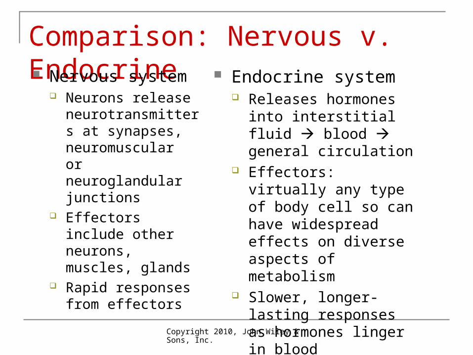
Comparison: Nervous v. Endocrine Nervous system
Neurons release neurotransmitters at synapses, neuromuscular or neuroglandular junctions
Effectors include other neurons, muscles, glands
Rapid responses from effectors
Endocrine system Releases hormones into
interstitial fluid blood general circulation
Effectors: virtually any type of body cell so can have widespread effects on diverse aspects of metabolism
Slower, longer-lasting responses as hormones linger in blood
Copyright 2010, John Wiley & Sons, Inc.
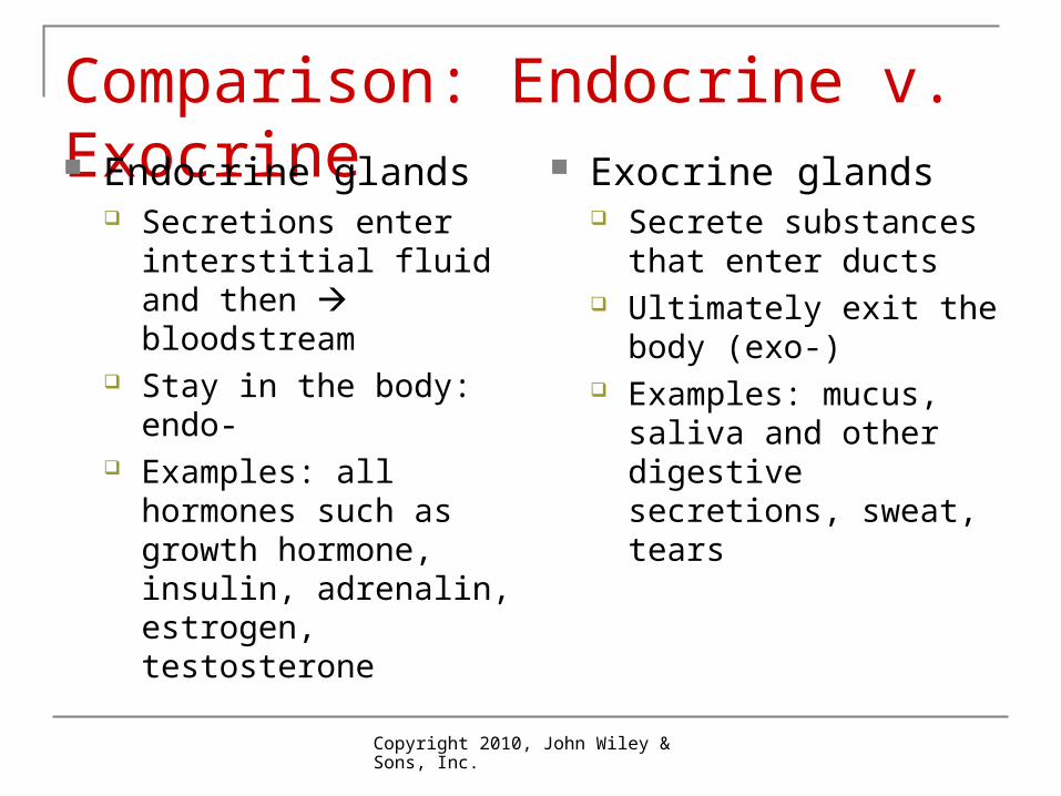
Comparison: Endocrine v. Exocrine Endocrine glands
Secretions enter interstitial fluid and then bloodstream
Stay in the body: endo- Examples: all hormones
such as growth hormone, insulin, adrenalin, estrogen, testosterone
Exocrine glands Secrete substances that
enter ducts Ultimately exit the body
(exo-) Examples: mucus, saliva
and other digestive secretions, sweat, tears
Copyright 2010, John Wiley & Sons, Inc.
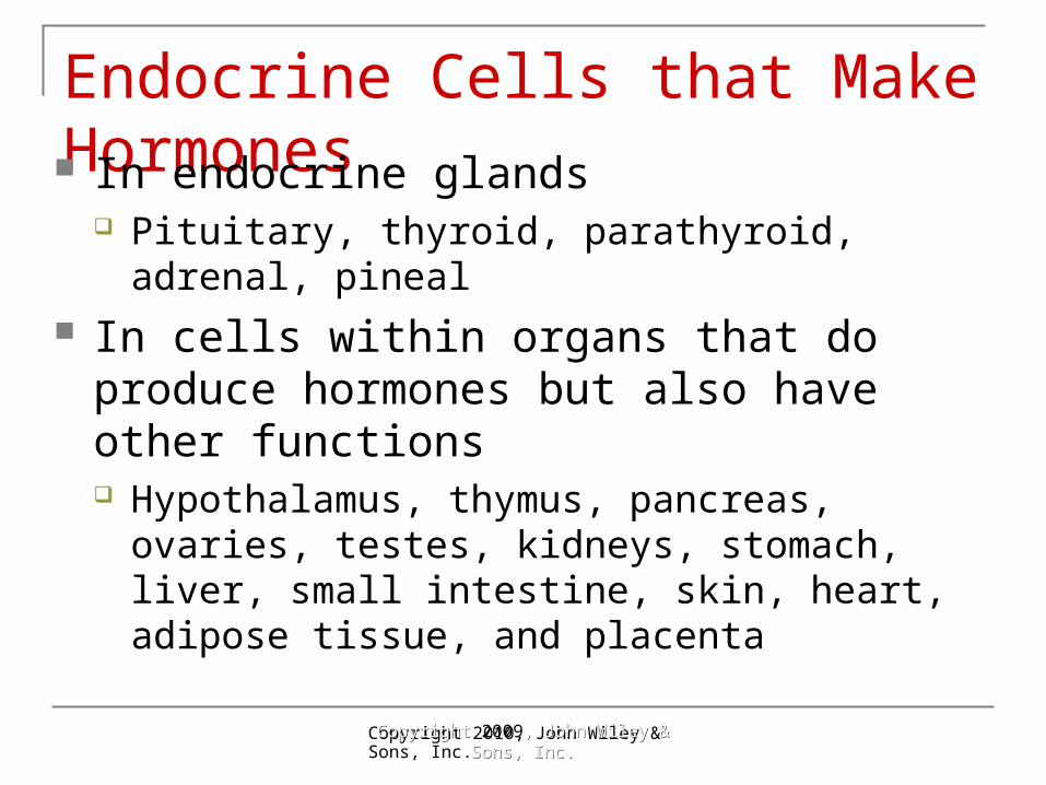
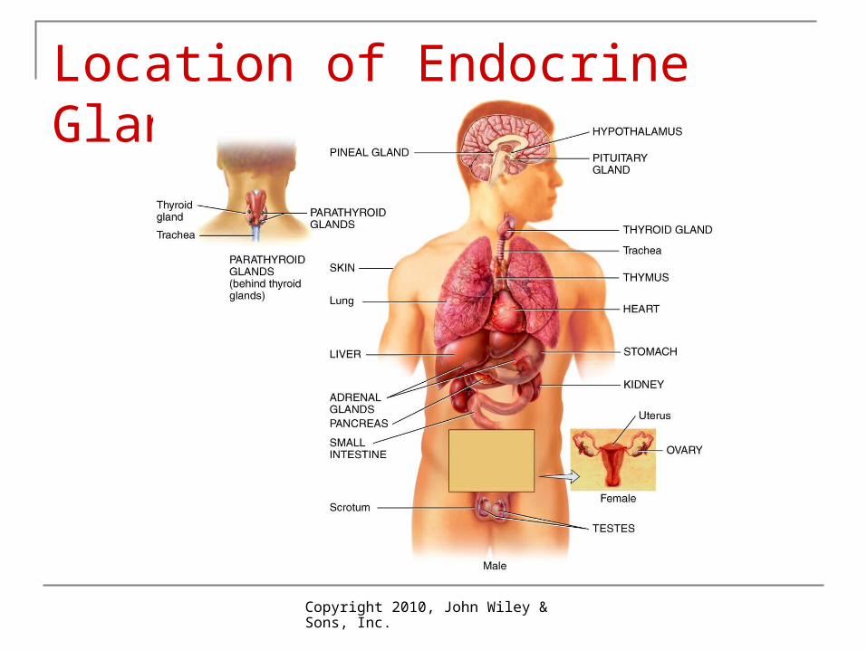
Endocrine Cells that Make Hormones In endocrine glands
Pituitary, thyroid, parathyroid, adrenal, pineal In cells within organs that do produce
hormones but also have other functions Hypothalamus, thymus, pancreas, ovaries,
testes, kidneys, stomach, liver, small intestine, skin, heart, adipose tissue, and placenta
Copyright Copyright 2009, John Wiley & , John Wiley & Sons, Inc.Sons, Inc.
Copyright 2010, John Wiley & Sons, Inc.
Location of Endocrine Glands
Copyright 2010, John Wiley & Sons, Inc.
Hormone Action Hormones are carried in blood stream But only certain cells can be affected by
hormones These target cells have 1000’s of receptors
specific for a particular hormone. Response determined by responding cell:
different cells may respond differently to the same hormone.
Cell may have > 1 type of receptor, so can respond to more than one hormone
Copyright 2010, John Wiley & Sons, Inc.
Hormone Chemistry Lipid-soluble
Steroids, such as testosterone, estrogens Thyroid hormones: T3 and T4 Nitric oxide (NO)
Water-soluble Amino acid derivatives, such as epinephrine,
norepinephrine Peptides: antidiuretic hormone (ADH), oxytocin Proteins: insulin and growth hormone
General action depends on chemistry
Copyright 2010, John Wiley & Sons, Inc.
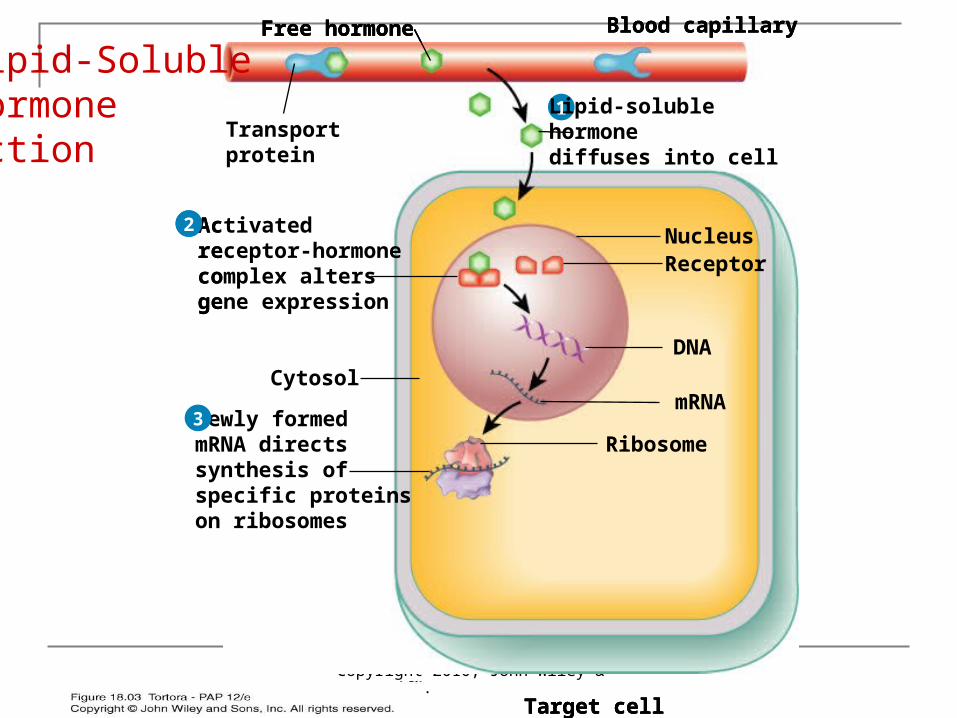
Lipid-Soluble Action Hormone detaches from carrier in blood
stream Diffuses through interstitial fluid and cell
membrane into cell Binds to receptor and activates it Receptor-hormone complex alters gene
expression If new mRNA protein synthesis New proteins alter cell activity
Copyright 2010, John Wiley & Sons, Inc.
1 Lipid-solublehormonediffuses into cell
Blood capillary
Target cell
Transportprotein
Free hormone
1 Lipid-solublehormonediffuses into cell
Blood capillary
Activatedreceptor-hormonecomplex altersgene expression
NucleusReceptor
mRNA
DNA
Cytosol
Target cell
Transportprotein
Free hormone
2
1 Lipid-solublehormonediffuses into cell
Blood capillary
Activatedreceptor-hormonecomplex altersgene expression
NucleusReceptor
mRNANewly formedmRNA directssynthesis ofspecific proteinson ribosomes
DNA
Cytosol
Target cell
Transportprotein
Free hormone
Ribosome
2
3
Lipid-SolubleHormone Action
Copyright 2010, John Wiley & Sons, Inc.
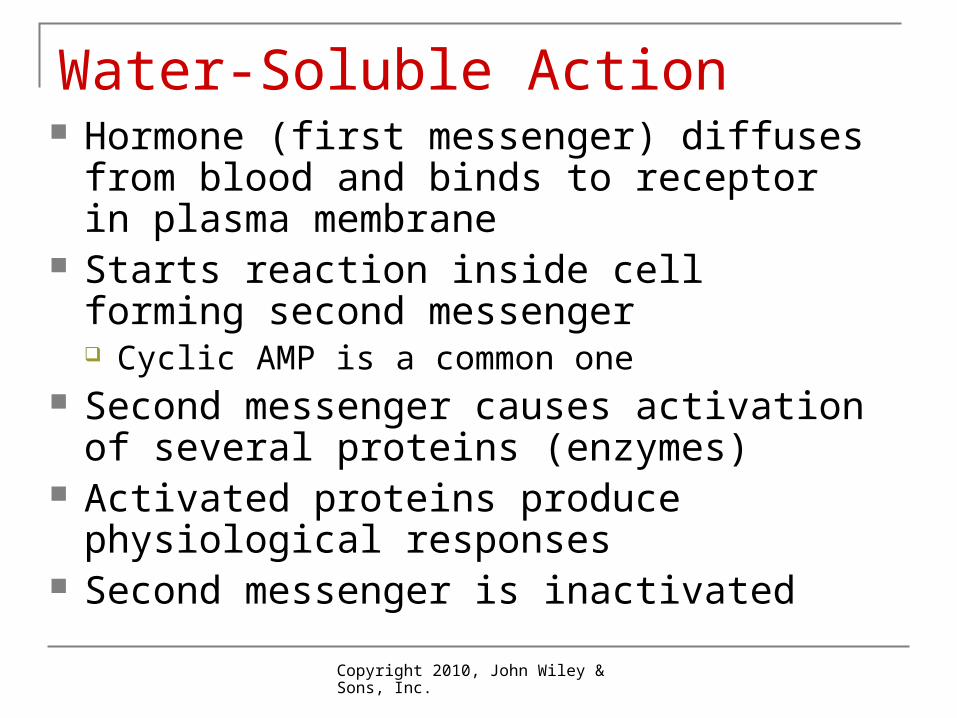
Water-Soluble Action Hormone (first messenger) diffuses from
blood and binds to receptor in plasma membrane
Starts reaction inside cell forming second messenger Cyclic AMP is a common one
Second messenger causes activation of several proteins (enzymes)
Activated proteins produce physiological responses
Second messenger is inactivated
Copyright 2010, John Wiley & Sons, Inc.
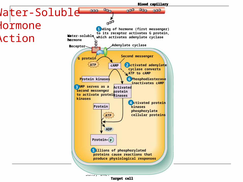
Water-solublehormone
Receptor
G protein
Blood capillary
Binding of hormone (first messenger)to its receptor activates G protein,which activates adenylate cyclase
Adenylate cyclase
Target cell
1
Water-solublehormone
Receptor
G protein
cAMP
Second messenger
Activated adenylatecyclase convertsATP to cAMP
Blood capillary
Binding of hormone (first messenger)to its receptor activates G protein,which activates adenylate cyclase
Adenylate cyclase
Target cell
ATP
1
2
Water-solublehormone
Receptor
cAMP serves as asecond messengerto activate proteinkinases
G protein
Protein kinases
cAMP
Second messenger
Activated adenylatecyclase convertsATP to cAMP
Blood capillary
Binding of hormone (first messenger)to its receptor activates G protein,which activates adenylate cyclase
Adenylate cyclase
Target cell
ATP
1
2
3 Activatedproteinkinases
Water-solublehormone
Receptor
cAMP serves as asecond messengerto activate proteinkinases
G protein
Protein kinases
cAMP
Activatedproteinkinases
Second messenger
Activated adenylatecyclase convertsATP to cAMP
Activated proteinkinasesphosphorylatecellular proteins
Blood capillary
Binding of hormone (first messenger)to its receptor activates G protein,which activates adenylate cyclase
Adenylate cyclase
Target cell
ATP
1
2
4
3
Protein— P
ADP
Protein
ATP
Water-solublehormone
Receptor
cAMP serves as asecond messengerto activate proteinkinases
G protein
Protein kinases
cAMP
Activatedproteinkinases
Protein—
Second messenger
Activated adenylatecyclase convertsATP to cAMP
Activated proteinkinasesphosphorylatecellular proteins
Millions of phosphorylatedproteins cause reactions thatproduce physiological responses
Blood capillary
Binding of hormone (first messenger)to its receptor activates G protein,which activates adenylate cyclase
Adenylate cyclase
Target cell
P
ADP
Protein
ATP
ATP
1
2
4
3
5
Water-solublehormone
Receptor
cAMP serves as asecond messengerto activate proteinkinases
G protein
Protein kinases
cAMP
Activatedproteinkinases
Protein—
Second messenger
Phosphodiesteraseinactivates cAMP
Activated adenylatecyclase convertsATP to cAMP
Activated proteinkinasesphosphorylatecellular proteins
Millions of phosphorylatedproteins cause reactions thatproduce physiological responses
Blood capillary
Binding of hormone (first messenger)to its receptor activates G protein,which activates adenylate cyclase
Adenylate cyclase
Target cell
P
ADP
Protein
ATP
ATP
1
2
6
4
3
5
Water-Soluble Hormone Action
Copyright 2010, John Wiley & Sons, Inc.
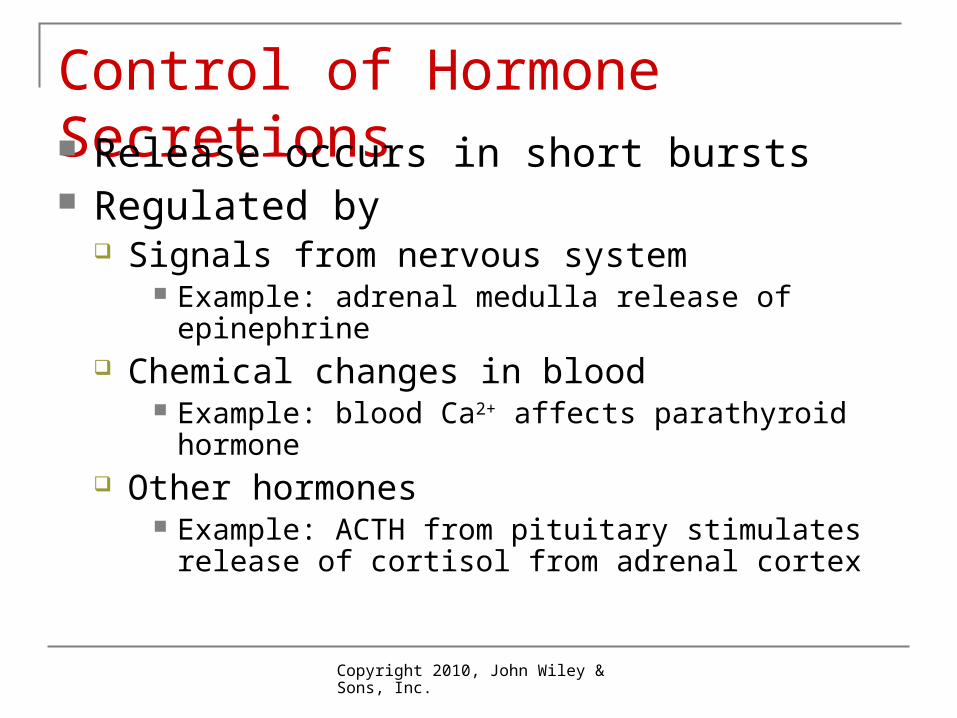
Control of Hormone Secretions Release occurs in short bursts Regulated by
Signals from nervous system Example: adrenal medulla release of epinephrine
Chemical changes in blood Example: blood Ca2+ affects parathyroid hormone
Other hormones Example: ACTH from pituitary stimulates release of
cortisol from adrenal cortex
Copyright 2010, John Wiley & Sons, Inc.
Hormone RegulationInteractions Animation
Introduction to Hormonal Regulation, Secretion, and Concentration
You must be connected to the internet to run this animation.
Copyright 2010, John Wiley & Sons, Inc.
Hypothalamus and Pituitary Serve as major link between nervous and
endocrine systems Hypothalamic cells synthesize
Many releasing and inhibiting hormones Two hormones (oxytocin and ADH) that are then
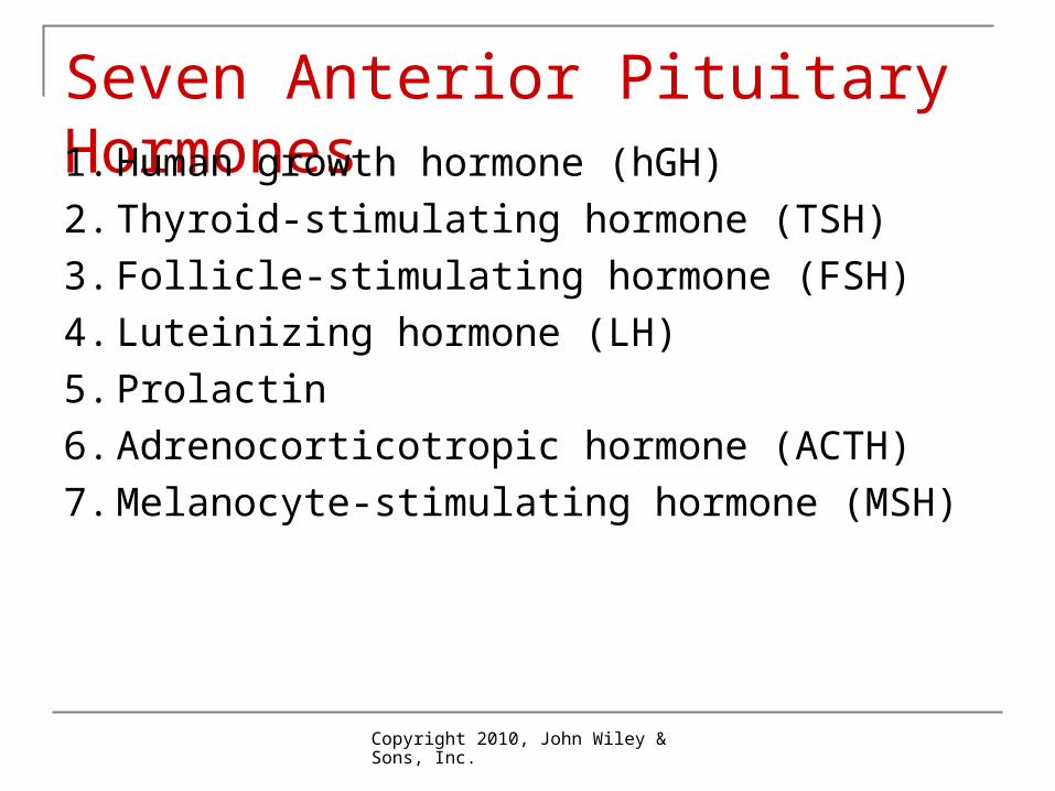
stored and released from the posterior pituitary Anterior pituitary synthesizes 7 hormones Regulate growth, development, metabolism
and homeostasis
Copyright 2010, John Wiley & Sons, Inc.
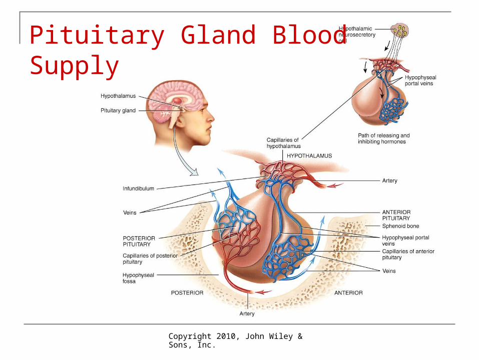
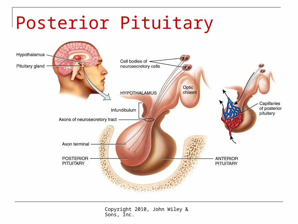
Pituitary Located in depression in sphenoid bone just
inferior to the brain Pituitary is attached to hypothalamus by stalk
(infundibulum) Pituitary has 2 lobes: anterior and posterior
Copyright 2010, John Wiley & Sons, Inc.
Effects of Hypothalamus on Pituitary Axons of hypothalamic neurons
(neurosecretory cells) end near capillaries of hypothalamus
Secrete releasing hormones or inhibiting hormones portal veins
These hormones regulate release of anterior pituitary hormones
Copyright 2010, John Wiley & Sons, Inc.
Pituitary Gland Blood Supply
Copyright 2010, John Wiley & Sons, Inc.
Seven Anterior Pituitary Hormones1. Human growth hormone (hGH)
2. Thyroid-stimulating hormone (TSH)
3. Follicle-stimulating hormone (FSH)
4. Luteinizing hormone (LH)
5. Prolactin
6. Adrenocorticotropic hormone (ACTH)
7. Melanocyte-stimulating hormone (MSH)
Copyright 2010, John Wiley & Sons, Inc.
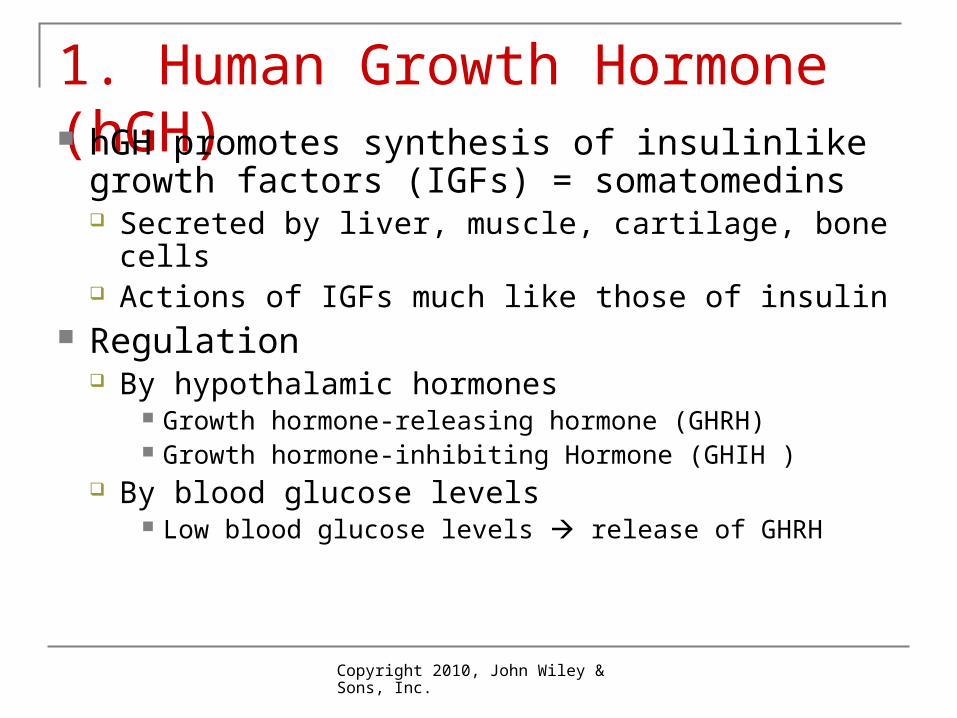
1. Human Growth Hormone (hGH) hGH promotes synthesis of insulinlike growth
factors (IGFs) = somatomedins Secreted by liver, muscle, cartilage, bone cells Actions of IGFs much like those of insulin
Regulation By hypothalamic hormones
Growth hormone-releasing hormone (GHRH) Growth hormone-inhibiting Hormone (GHIH )
By blood glucose levels Low blood glucose levels release of GHRH
Copyright 2010, John Wiley & Sons, Inc.
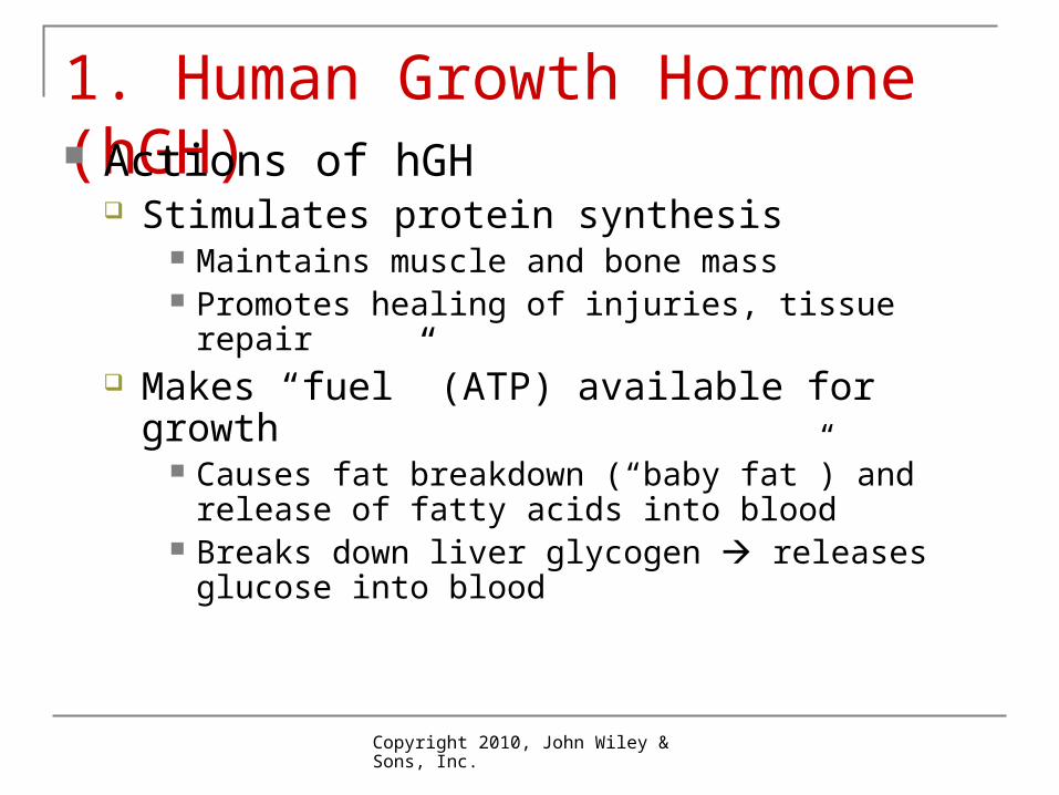
1. Human Growth Hormone (hGH) Actions of hGH
Stimulates protein synthesis Maintains muscle and bone mass Promotes healing of injuries, tissue repair
Makes “fuel” (ATP) available for growth Causes fat breakdown (“baby fat”) and release of fatty
acids into blood Breaks down liver glycogen releases glucose into
blood
Copyright 2010, John Wiley & Sons, Inc.
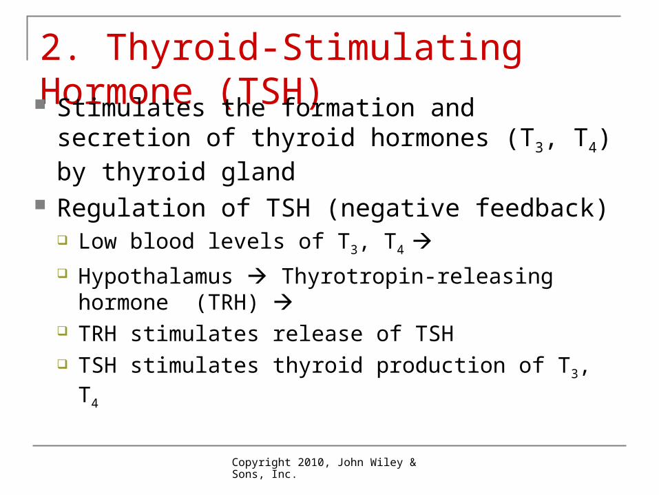
2. Thyroid-Stimulating Hormone (TSH) Stimulates the formation and secretion of
thyroid hormones (T3, T4) by thyroid gland Regulation of TSH (negative feedback)
Low blood levels of T3, T4 Hypothalamus Thyrotropin-releasing hormone
(TRH) TRH stimulates release of TSH TSH stimulates thyroid production of T3, T4
Copyright 2010, John Wiley & Sons, Inc.
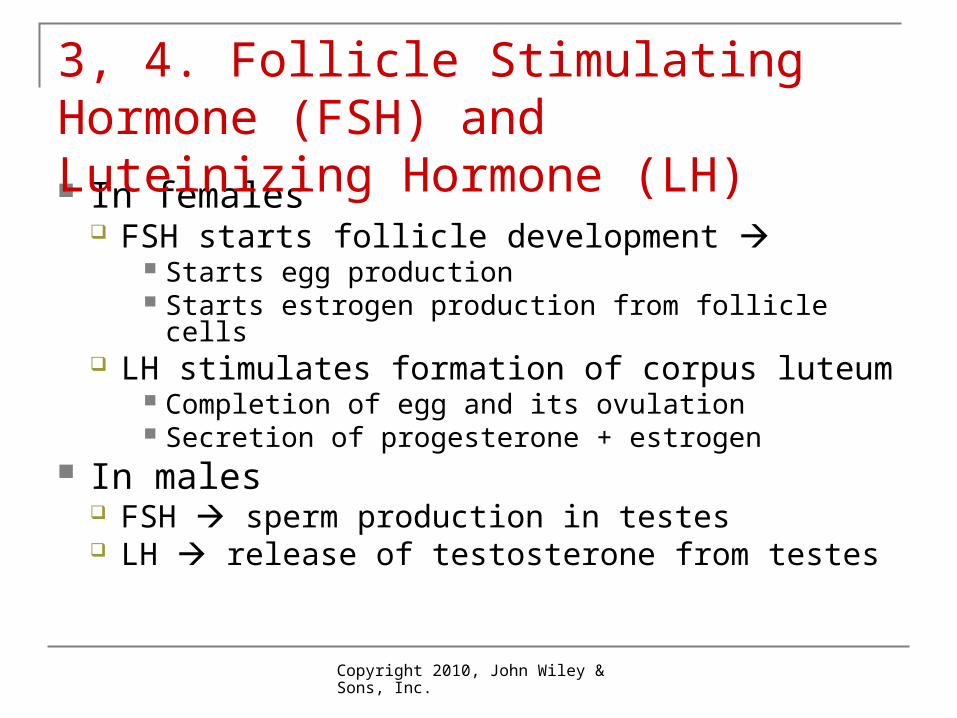
In females FSH starts follicle development
Starts egg production Starts estrogen production from follicle cells
LH stimulates formation of corpus luteum Completion of egg and its ovulation Secretion of progesterone + estrogen
In males FSH sperm production in testes LH release of testosterone from testes
3, 4. Follicle Stimulating Hormone (FSH) and Luteinizing Hormone (LH)
Copyright 2010, John Wiley & Sons, Inc.
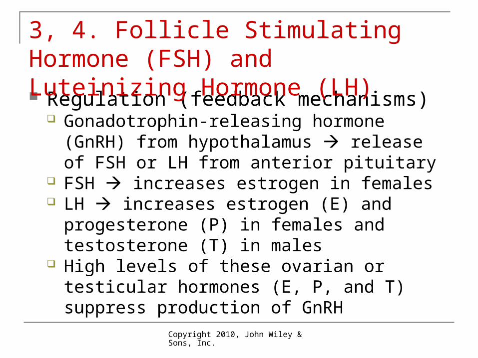
Regulation (feedback mechanisms) Gonadotrophin-releasing hormone (GnRH) from
hypothalamus release of FSH or LH from anterior pituitary
FSH increases estrogen in females LH increases estrogen (E) and progesterone
(P) in females and testosterone (T) in males High levels of these ovarian or testicular
hormones (E, P, and T) suppress production of GnRH
3, 4. Follicle Stimulating Hormone (FSH) and Luteinizing Hormone (LH)
Copyright 2010, John Wiley & Sons, Inc.
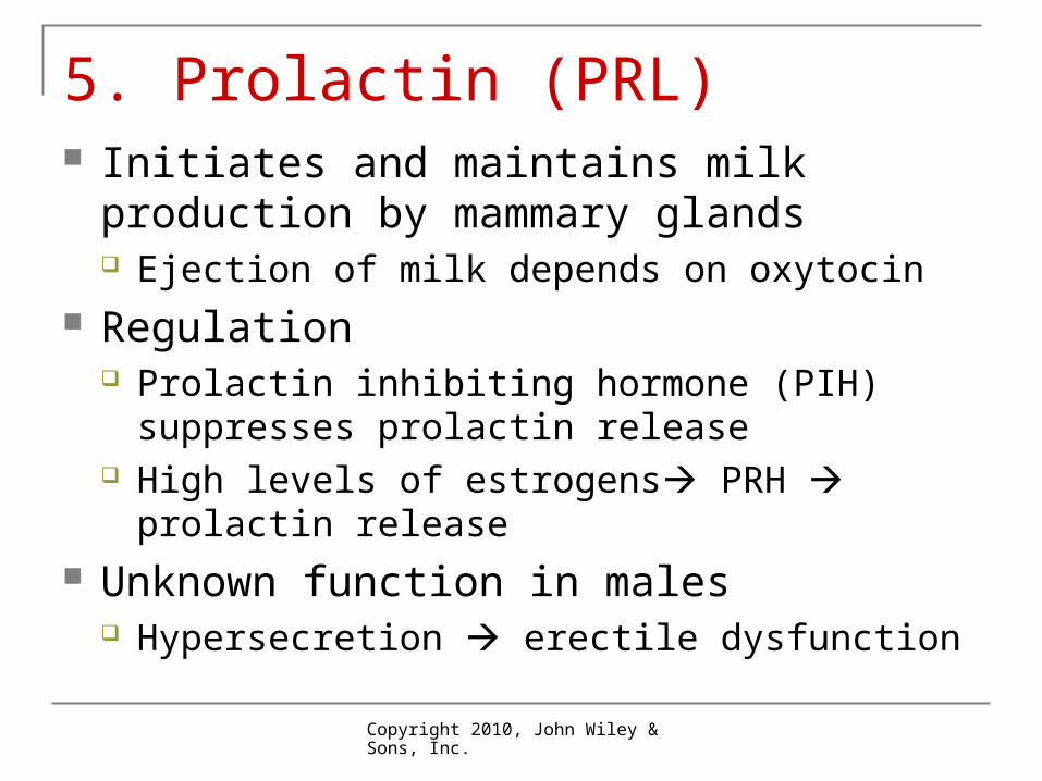
5. Prolactin (PRL) Initiates and maintains milk production by
mammary glands Ejection of milk depends on oxytocin
Regulation Prolactin inhibiting hormone (PIH) suppresses
prolactin release High levels of estrogens PRH prolactin
release Unknown function in males
Hypersecretion erectile dysfunction
Copyright 2010, John Wiley & Sons, Inc.
6. Adrenocorticotropic Hormone(ACTH) Controls production and secretion of
glucocorticoids from adrenal cortex Regulation of ACTH
Corticotrophin releasing hormone (CRH) from hypothalamus stimulates secretion of ACTH
Stress-related stimuli can also stimulate ACTH release
Glucocorticoids inhibit CRH and ACTH release
Copyright 2010, John Wiley & Sons, Inc.
7. Melanocyte Stimulating Hormone (MSH) Small amounts in bloodstream Excess amounts causes skin darkening
Copyright 2010, John Wiley & Sons, Inc.
Posterior Pituitary Hormones made in hypothalamus pass
down axons to posterior pituitary Nerve impulses there cause release of hormones
Two hormones released Oxytocin causes
Smooth muscle contraction of uterus during childbirth Causes “letdown” of milk from glands to ducts Some sexual pleasure during sexual activity
Copyright 2010, John Wiley & Sons, Inc.
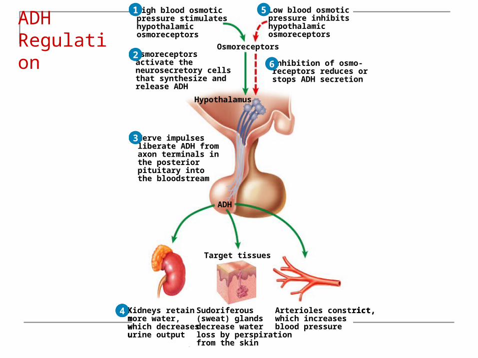
Posterior Pituitary Antidiuretic Hormone (ADH) = vasopressin
Causes kidneys to retain more water Causes vasoconstriction increases blood pressure Dehydration, pain, stress increase ADH secretion
Copyright 2010, John Wiley & Sons, Inc.
Posterior Pituitary
Copyright 2010, John Wiley & Sons, Inc.
Osmoreceptors
High blood osmoticpressure stimulateshypothalamicosmoreceptors
1
Osmoreceptors
High blood osmoticpressure stimulateshypothalamicosmoreceptors
Osmoreceptorsactivate theneurosecretory cellsthat synthesize andrelease ADH
Hypothalamus
1
2Osmoreceptors
High blood osmoticpressure stimulateshypothalamicosmoreceptors
Nerve impulsesliberate ADH fromaxon terminals inthe posteriorpituitary intothe bloodstream
Osmoreceptorsactivate theneurosecretory cellsthat synthesize andrelease ADH
Hypothalamus
ADH
1
2
3
Osmoreceptors
High blood osmoticpressure stimulateshypothalamicosmoreceptors
Nerve impulsesliberate ADH fromaxon terminals inthe posteriorpituitary intothe bloodstream
Osmoreceptorsactivate theneurosecretory cellsthat synthesize andrelease ADH
Hypothalamus
Sudoriferous(sweat) glandsdecrease waterloss by perspirationfrom the skin
Arterioles constrict,which increasesblood pressure
Kidneys retainmore water,which decreasesurine output
ADH
Target tissues
1
2
3
4
Osmoreceptors
High blood osmoticpressure stimulateshypothalamicosmoreceptors
Low blood osmoticpressure inhibitshypothalamicosmoreceptors
Nerve impulsesliberate ADH fromaxon terminals inthe posteriorpituitary intothe bloodstream
Osmoreceptorsactivate theneurosecretory cellsthat synthesize andrelease ADH
Hypothalamus
Sudoriferous(sweat) glandsdecrease waterloss by perspirationfrom the skin
Arterioles constrict,which increasesblood pressure
Kidneys retainmore water,which decreasesurine output
ADH
Target tissues
1
2
3
4
5
Osmoreceptors
High blood osmoticpressure stimulateshypothalamicosmoreceptors
Low blood osmoticpressure inhibitshypothalamicosmoreceptors
Nerve impulsesliberate ADH fromaxon terminals inthe posteriorpituitary intothe bloodstream
Osmoreceptorsactivate theneurosecretory cellsthat synthesize andrelease ADH
Hypothalamus
Inhibition of osmo-receptors reduces orstops ADH secretion
Sudoriferous(sweat) glandsdecrease waterloss by perspirationfrom the skin
Arterioles constrict,which increasesblood pressure
Kidneys retainmore water,which decreasesurine output
ADH
Target tissues
1
2
3
4
5
6
ADH Regulation
Copyright 2010, John Wiley & Sons, Inc.
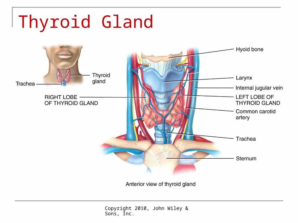
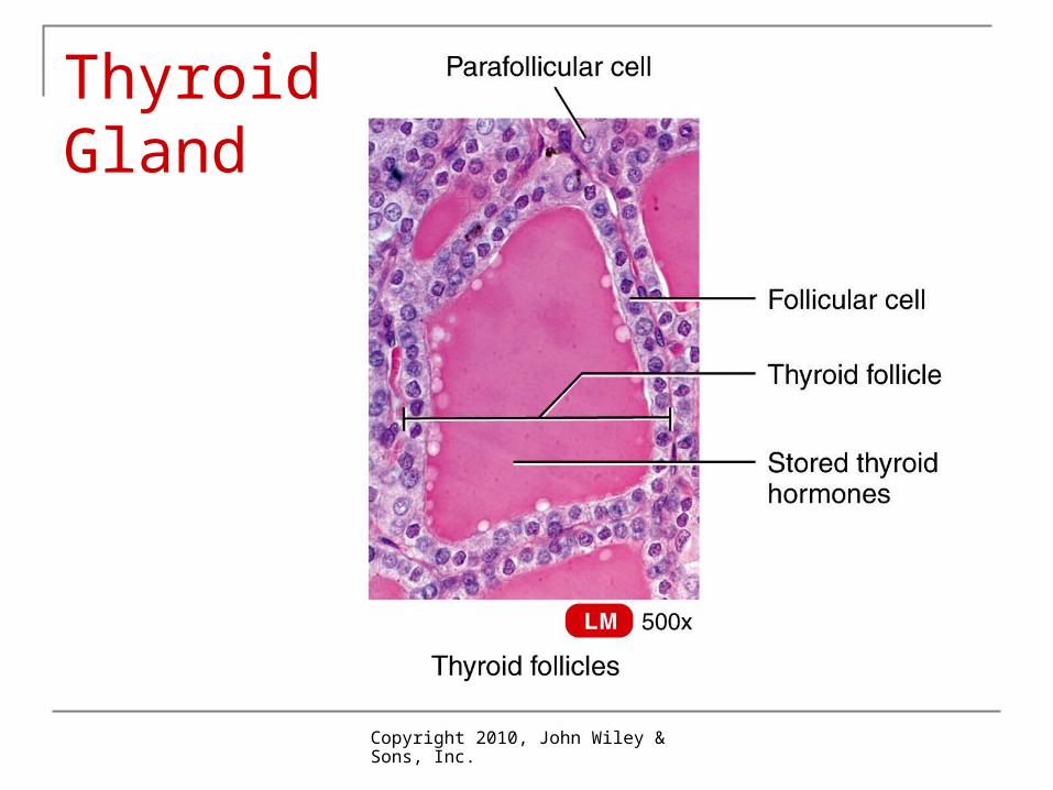
Thyroid Gland Location: inferior to larynx: two lobes Structure and function
Follicular cells produce hormones and store them in follicles
Thyroxin (T4) Triiodothyronine (T3)
Parafollicular cells (C-cells) produce Calcitonin (CT)
Copyright 2010, John Wiley & Sons, Inc.
Thyroid Gland
Copyright 2010, John Wiley & Sons, Inc.
Thyroid Gland
Copyright 2010, John Wiley & Sons, Inc.
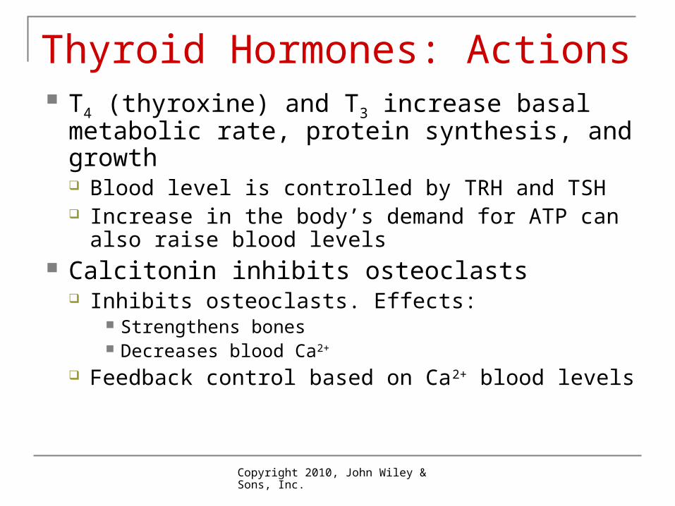
Thyroid Hormones: Actions T4 (thyroxine) and T3 increase basal
metabolic rate, protein synthesis, and growth Blood level is controlled by TRH and TSH Increase in the body’s demand for ATP can also
raise blood levels Calcitonin inhibits osteoclasts
Inhibits osteoclasts. Effects: Strengthens bones Decreases blood Ca2+
Feedback control based on Ca2+ blood levels
Copyright 2010, John Wiley & Sons, Inc.
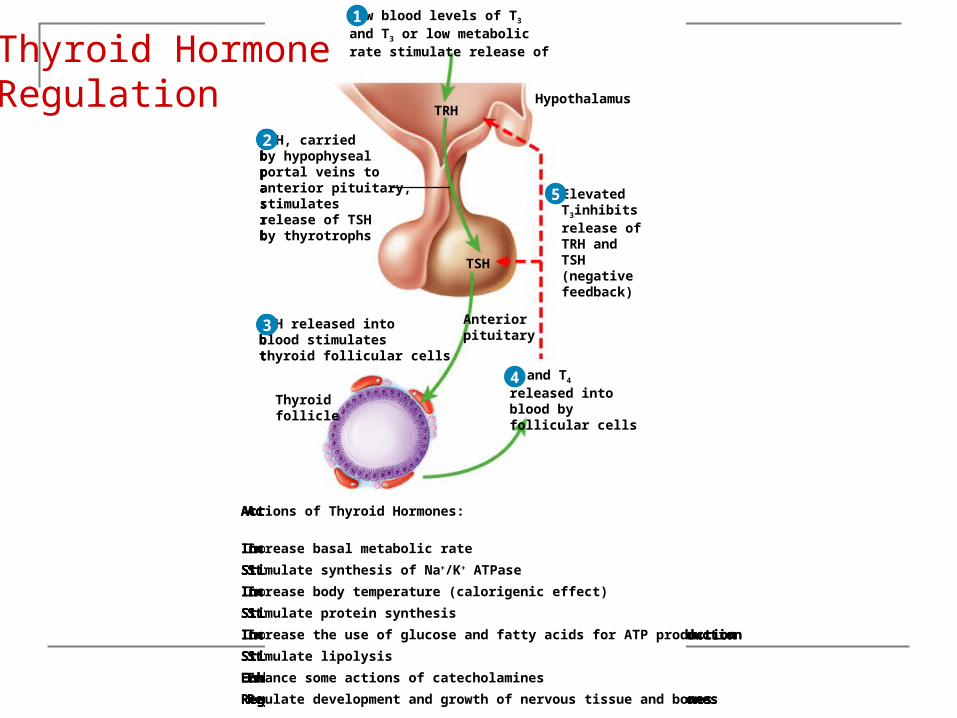
Low blood levels of T3
and T3 or low metabolicrate stimulate release of
HypothalamusTRH
Actions of Thyroid Hormones:
Increase basal metabolic rate
Stimulate synthesis of Na+/K+ ATPase
Increase body temperature (calorigenic effect)
Stimulate protein synthesis
Increase the use of glucose and fatty acids for ATP production
Stimulate lipolysis
Enhance some actions of catecholamines
Regulate development and growth of nervous tissue and bones
1
Anteriorpituitary
TRH, carriedby hypophysealportal veins toanterior pituitary,stimulatesrelease of TSHby thyrotrophs
Low blood levels of T3
and T3 or low metabolicrate stimulate release of
Hypothalamus
TSH
TRH
Actions of Thyroid Hormones:
Increase basal metabolic rate
Stimulate synthesis of Na+/K+ ATPase
Increase body temperature (calorigenic effect)
Stimulate protein synthesis
Increase the use of glucose and fatty acids for ATP production
Stimulate lipolysis
Enhance some actions of catecholamines
Regulate development and growth of nervous tissue and bones
1
2
Anteriorpituitary
TRH, carriedby hypophysealportal veins toanterior pituitary,stimulatesrelease of TSHby thyrotrophs
TSH released intoblood stimulatesthyroid follicular cells
Thyroidfollicle
Low blood levels of T3
and T3 or low metabolicrate stimulate release of
Hypothalamus
Anteriorpituitary
TSH
TRH
Actions of Thyroid Hormones:
Increase basal metabolic rate
Stimulate synthesis of Na+/K+ ATPase
Increase body temperature (calorigenic effect)
Stimulate protein synthesis
Increase the use of glucose and fatty acids for ATP production
Stimulate lipolysis
Enhance some actions of catecholamines
Regulate development and growth of nervous tissue and bones
1
2
3
T3 and T4
released intoblood byfollicular cells
TRH, carriedby hypophysealportal veins toanterior pituitary,stimulatesrelease of TSHby thyrotrophs
TSH released intoblood stimulatesthyroid follicular cells
Thyroidfollicle
Low blood levels of T3
and T3 or low metabolicrate stimulate release of
Hypothalamus
Anteriorpituitary
TSH
TRH
Actions of Thyroid Hormones:
Increase basal metabolic rate
Stimulate synthesis of Na+/K+ ATPase
Increase body temperature (calorigenic effect)
Stimulate protein synthesis
Increase the use of glucose and fatty acids for ATP production
Stimulate lipolysis
Enhance some actions of catecholamines
Regulate development and growth of nervous tissue and bones
1
2
3
4 T3 and T4
released intoblood byfollicular cells
ElevatedT3inhibitsrelease ofTRH andTSH(negativefeedback)
TRH, carriedby hypophysealportal veins toanterior pituitary,stimulatesrelease of TSHby thyrotrophs
TSH released intoblood stimulatesthyroid follicular cells
Thyroidfollicle
Low blood levels of T3
and T3 or low metabolicrate stimulate release of
Hypothalamus
Anteriorpituitary
TSH
TRH
Actions of Thyroid Hormones:
Increase basal metabolic rate
Stimulate synthesis of Na+/K+ ATPase
Increase body temperature (calorigenic effect)
Stimulate protein synthesis
Increase the use of glucose and fatty acids for ATP production
Stimulate lipolysis
Enhance some actions of catecholamines
Regulate development and growth of nervous tissue and bones
1
2
3
5
4
Thyroid HormoneRegulation
Copyright 2010, John Wiley & Sons, Inc.
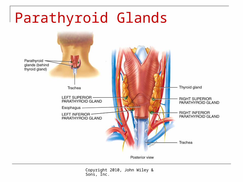
Parathyroid Glands Small round masses in posterior of thyroid
gland Release parathyroid hormone (PTH)
Increases blood Ca2+ in 3 ways Increases number and activity of osteoclasts that break
down bone Slows loss of Ca2+ and Mg2+ in urine Promotes production of calcitriol (vitamin D)
increases rate of Ca2+, Mg2+ and HPO42- absorption in
GI tract increase blood Ca2+
Decreases blood HPO42- by decreasing loss of HPO4
2- in urine
Copyright 2010, John Wiley & Sons, Inc.
Parathyroid Glands
Copyright 2010, John Wiley & Sons, Inc.
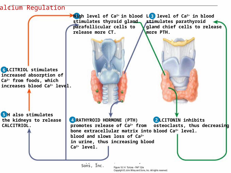
1 High level of Ca2+ in bloodstimulates thyroid glandparafollicular cells to release more CT.
1 High level of Ca2+ in bloodstimulates thyroid glandparafollicular cells to release more CT.
CALCITONIN inhibitsosteoclasts, thus decreasingblood Ca2+ level.
2
1 High level of Ca2+ in bloodstimulates thyroid glandparafollicular cells to release more CT.
Low level of Ca2+ in bloodstimulates parathyroid gland chief cells to release more PTH.
CALCITONIN inhibitsosteoclasts, thus decreasingblood Ca2+ level.
3
2
1 High level of Ca2+ in bloodstimulates thyroid glandparafollicular cells to release more CT.
Low level of Ca2+ in bloodstimulates parathyroid gland chief cells to release more PTH.
CALCITONIN inhibitsosteoclasts, thus decreasingblood Ca2+ level.
PARATHYROID HORMONE (PTH)promotes release of Ca2+ frombone extracellular matrix intoblood and slows loss of Ca2+ in urine, thus increasing bloodCa2+ level.
3
4 2
1
PTH also stimulatesthe kidneys to releaseCALCITRIOL.
High level of Ca2+ in bloodstimulates thyroid glandparafollicular cells to release more CT.
Low level of Ca2+ in bloodstimulates parathyroid gland chief cells to release more PTH.
CALCITONIN inhibitsosteoclasts, thus decreasingblood Ca2+ level.
PARATHYROID HORMONE (PTH)promotes release of Ca2+ frombone extracellular matrix intoblood and slows loss of Ca2+ in urine, thus increasing bloodCa2+ level.
3
4 25
1
CALCITRIOL stimulatesincreased absorption ofCa2+ from foods, whichincreases blood Ca2+ level.
PTH also stimulatesthe kidneys to releaseCALCITRIOL.
High level of Ca2+ in bloodstimulates thyroid glandparafollicular cells to release more CT.
Low level of Ca2+ in bloodstimulates parathyroid gland chief cells to release more PTH.
CALCITONIN inhibitsosteoclasts, thus decreasingblood Ca2+ level.
PARATHYROID HORMONE (PTH)promotes release of Ca2+ frombone extracellular matrix intoblood and slows loss of Ca2+ in urine, thus increasing bloodCa2+ level.
3
4 25
6
Calcium Regulation
Copyright 2010, John Wiley & Sons, Inc.
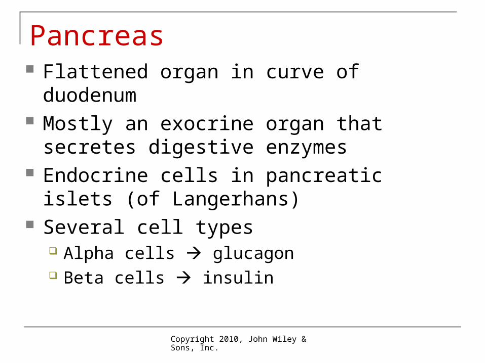
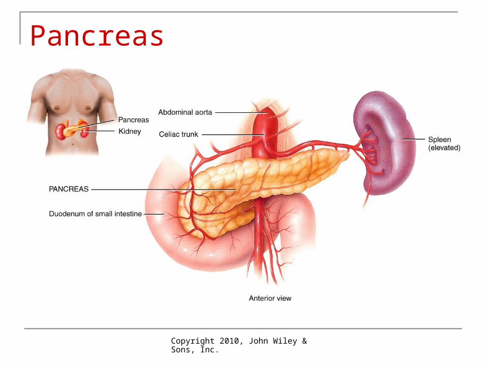
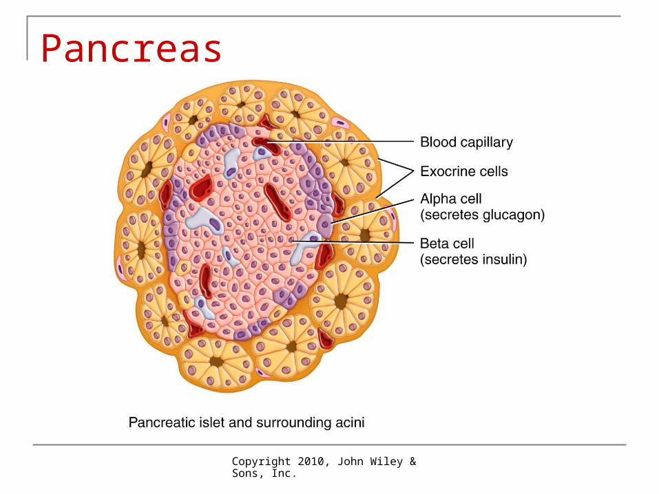
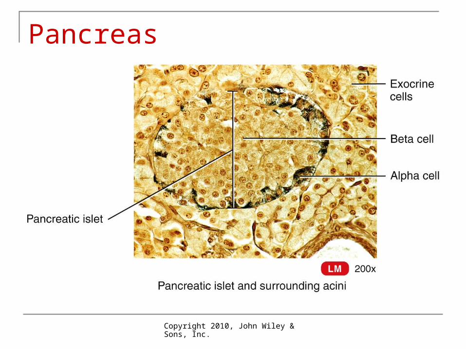
Pancreas Flattened organ in curve of duodenum Mostly an exocrine organ that secretes
digestive enzymes Endocrine cells in pancreatic islets (of
Langerhans) Several cell types
Alpha cells glucagon Beta cells insulin
Copyright 2010, John Wiley & Sons, Inc.
Pancreas
Copyright 2010, John Wiley & Sons, Inc.
Pancreas
Copyright 2010, John Wiley & Sons, Inc.
Pancreas
Copyright 2010, John Wiley & Sons, Inc.
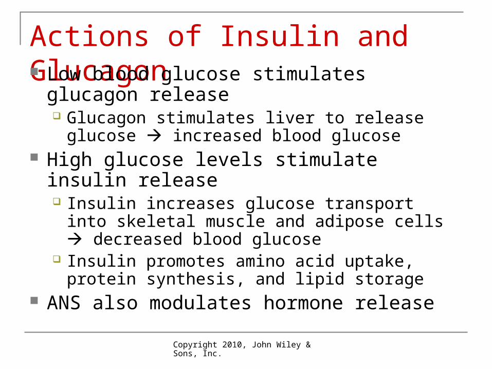
Actions of Insulin and Glucagon Low blood glucose stimulates glucagon
release Glucagon stimulates liver to release glucose
increased blood glucose High glucose levels stimulate insulin release
Insulin increases glucose transport into skeletal muscle and adipose cells decreased blood glucose
Insulin promotes amino acid uptake, protein synthesis, and lipid storage
ANS also modulates hormone release
Copyright 2010, John Wiley & Sons, Inc.
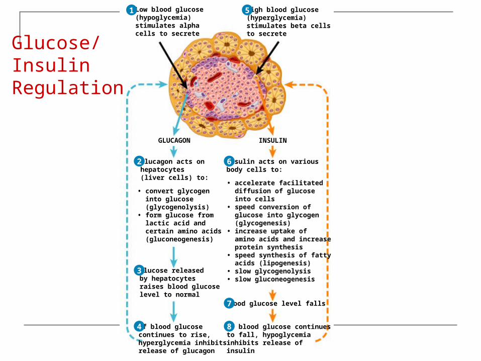
Low blood glucose(hypoglycemia)stimulates alphacells to secrete
1
GLUCAGON
Glucagon acts onhepatocytes(liver cells) to:
• convert glycogen into glucose (glycogenolysis)• form glucose from lactic acid and certain amino acids (gluconeogenesis)
Low blood glucose(hypoglycemia)stimulates alphacells to secrete
GLUCAGON
1
2 Glucagon acts onhepatocytes(liver cells) to:
• convert glycogen into glucose (glycogenolysis)• form glucose from lactic acid and certain amino acids (gluconeogenesis)
Glucose releasedby hepatocytesraises blood glucoselevel to normal
Low blood glucose(hypoglycemia)stimulates alphacells to secrete
GLUCAGON
1
2
3
Glucagon acts onhepatocytes(liver cells) to:
• convert glycogen into glucose (glycogenolysis)• form glucose from lactic acid and certain amino acids (gluconeogenesis)
Glucose releasedby hepatocytesraises blood glucoselevel to normal
If blood glucosecontinues to rise,hyperglycemia inhibitsrelease of glucagon
Low blood glucose(hypoglycemia)stimulates alphacells to secrete
GLUCAGON
1
2
3
4
Glucagon acts onhepatocytes(liver cells) to:
• convert glycogen into glucose (glycogenolysis)• form glucose from lactic acid and certain amino acids (gluconeogenesis)
Glucose releasedby hepatocytesraises blood glucoselevel to normal
If blood glucosecontinues to rise,hyperglycemia inhibitsrelease of glucagon
Low blood glucose(hypoglycemia)stimulates alphacells to secrete
High blood glucose(hyperglycemia)stimulates beta cellsto secrete
GLUCAGON
1 5
2
3
4
INSULIN
Insulin acts on variousbody cells to:
• accelerate facilitated diffusion of glucose into cells• speed conversion of glucose into glycogen (glycogenesis)• increase uptake of amino acids and increase protein synthesis• speed synthesis of fatty acids (lipogenesis)• slow glycogenolysis• slow gluconeogenesis
Glucagon acts onhepatocytes(liver cells) to:
• convert glycogen into glucose (glycogenolysis)• form glucose from lactic acid and certain amino acids (gluconeogenesis)
Glucose releasedby hepatocytesraises blood glucoselevel to normal
If blood glucosecontinues to rise,hyperglycemia inhibitsrelease of glucagon
Low blood glucose(hypoglycemia)stimulates alphacells to secrete
High blood glucose(hyperglycemia)stimulates beta cellsto secrete
INSULINGLUCAGON
1 5
2
3
4
6 Insulin acts on variousbody cells to:
• accelerate facilitated diffusion of glucose into cells• speed conversion of glucose into glycogen (glycogenesis)• increase uptake of amino acids and increase protein synthesis• speed synthesis of fatty acids (lipogenesis)• slow glycogenolysis• slow gluconeogenesis
Blood glucose level falls
Glucagon acts onhepatocytes(liver cells) to:
• convert glycogen into glucose (glycogenolysis)• form glucose from lactic acid and certain amino acids (gluconeogenesis)
Glucose releasedby hepatocytesraises blood glucoselevel to normal
If blood glucosecontinues to rise,hyperglycemia inhibitsrelease of glucagon
Low blood glucose(hypoglycemia)stimulates alphacells to secrete
High blood glucose(hyperglycemia)stimulates beta cellsto secrete
INSULINGLUCAGON
1 5
2
3
4
6
7
Insulin acts on variousbody cells to:
• accelerate facilitated diffusion of glucose into cells• speed conversion of glucose into glycogen (glycogenesis)• increase uptake of amino acids and increase protein synthesis• speed synthesis of fatty acids (lipogenesis)• slow glycogenolysis• slow gluconeogenesis
If blood glucose continuesto fall, hypoglycemiainhibits release ofinsulin
Blood glucose level falls
Glucagon acts onhepatocytes(liver cells) to:
• convert glycogen into glucose (glycogenolysis)• form glucose from lactic acid and certain amino acids (gluconeogenesis)
Glucose releasedby hepatocytesraises blood glucoselevel to normal
If blood glucosecontinues to rise,hyperglycemia inhibitsrelease of glucagon
Low blood glucose(hypoglycemia)stimulates alphacells to secrete
High blood glucose(hyperglycemia)stimulates beta cellsto secrete
INSULINGLUCAGON
1 5
2
3
4
6
7
8
Glucose/InsulinRegulation
Copyright 2010, John Wiley & Sons, Inc.
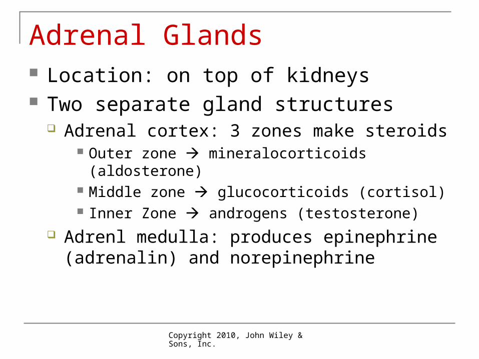
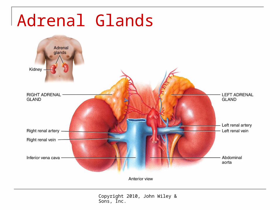
Adrenal Glands Location: on top of kidneys Two separate gland structures
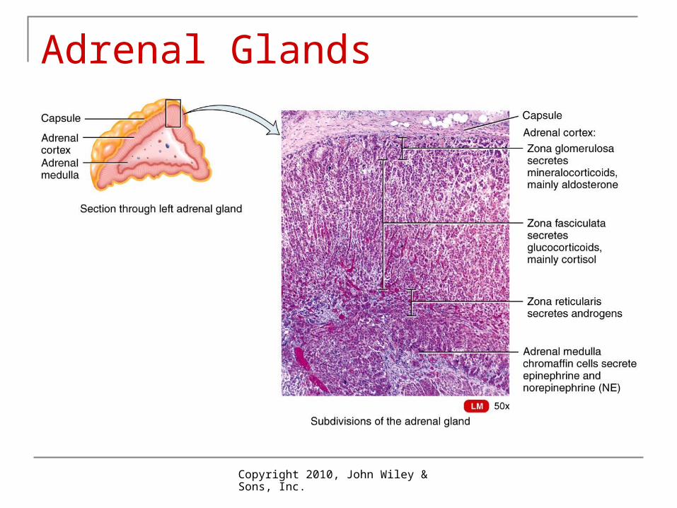
Adrenal cortex: 3 zones make steroids Outer zone mineralocorticoids (aldosterone) Middle zone glucocorticoids (cortisol) Inner Zone androgens (testosterone)
Adrenl medulla: produces epinephrine (adrenalin) and norepinephrine
Copyright 2010, John Wiley & Sons, Inc.
Adrenal Glands
Copyright 2010, John Wiley & Sons, Inc.
Adrenal Glands
Copyright 2010, John Wiley & Sons, Inc.
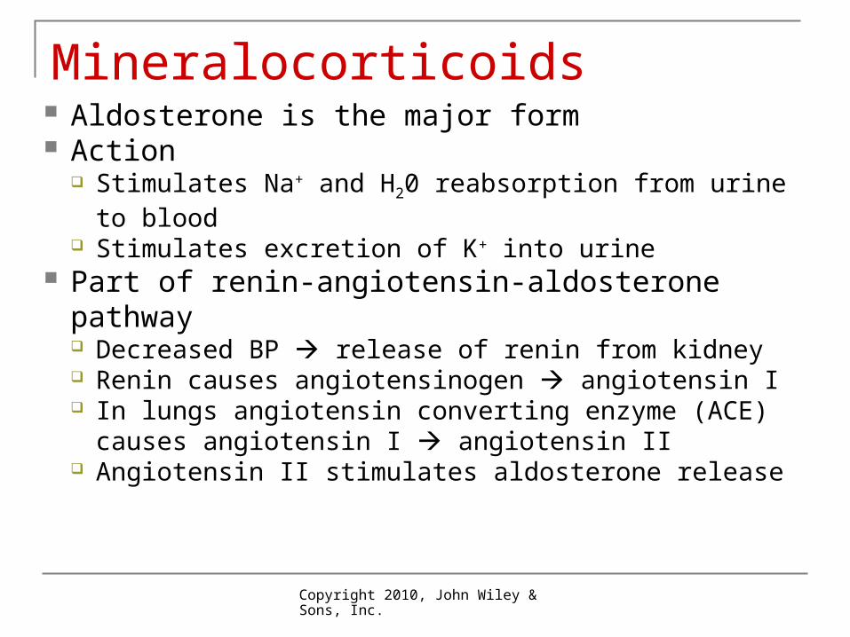
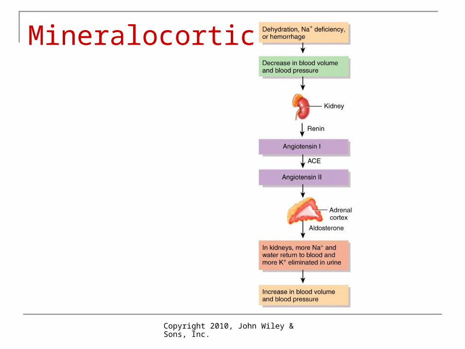
Mineralocorticoids Aldosterone is the major form Action
Stimulates Na+ and H20 reabsorption from urine to blood
Stimulates excretion of K+ into urine Part of renin-angiotensin-aldosterone
pathway Decreased BP release of renin from kidney Renin causes angiotensinogen angiotensin I In lungs angiotensin converting enzyme (ACE)
causes angiotensin I angiotensin II Angiotensin II stimulates aldosterone release
Copyright 2010, John Wiley & Sons, Inc.
Mineralocorticoids
Copyright 2010, John Wiley & Sons, Inc.
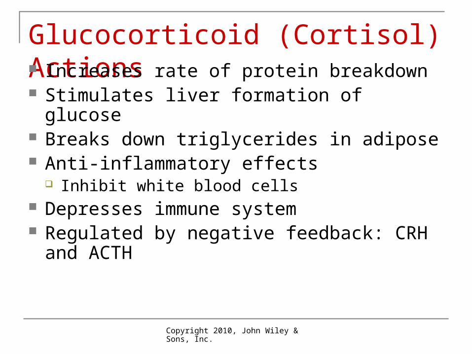
Glucocorticoid (Cortisol) Actions Increases rate of protein breakdown Stimulates liver formation of glucose Breaks down triglycerides in adipose Anti-inflammatory effects
Inhibit white blood cells Depresses immune system Regulated by negative feedback: CRH and
ACTH
Copyright 2010, John Wiley & Sons, Inc.
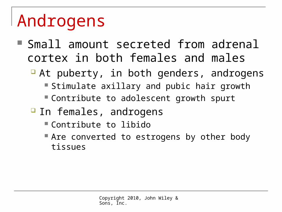
Androgens Small amount secreted from adrenal cortex in
both females and males At puberty, in both genders, androgens
Stimulate axillary and pubic hair growth Contribute to adolescent growth spurt
In females, androgens Contribute to libido Are converted to estrogens by other body tissues
Copyright 2010, John Wiley & Sons, Inc.
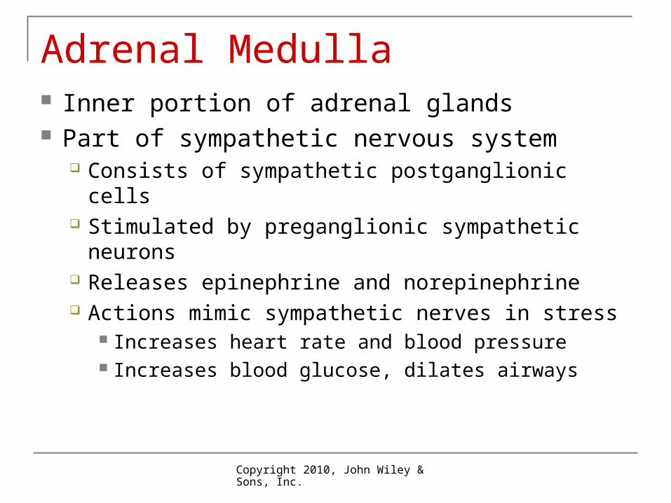
Adrenal Medulla Inner portion of adrenal glands Part of sympathetic nervous system
Consists of sympathetic postganglionic cells Stimulated by preganglionic sympathetic
neurons Releases epinephrine and norepinephrine Actions mimic sympathetic nerves in stress
Increases heart rate and blood pressure Increases blood glucose, dilates airways
Copyright 2010, John Wiley & Sons, Inc.
Gonads: Ovaries and Testes Produce gametes: sperm and oocytes Produce hormones
Testosterone in males Estrogen and progesterone in females Inhibin that inhibits FSH release Relaxin during pregnancy: facilitates birth
Regulated by GnRH from hypothalamus FSH + LH from anterior pituitary
Copyright 2010, John Wiley & Sons, Inc.
Pineal Gland Small gland attached to roof of third ventricle
of brain Produces melatonin Sets body’s biological clock
More released in darkness, less in sunlight
Copyright 2010, John Wiley & Sons, Inc.
Other Hormones Thymus: thymosin GI tract
Gastrin Glucose-dependent insulinotropic peptide (GIP) Secretin Cholecystokinin (CCK)
Kidney: erythropoietin (EPO) Heart: atrial natriuretic peptide (ANP)
Copyright 2010, John Wiley & Sons, Inc.
Other Hormones Adipose tissue: leptin Placenta: human chorionic gonadotropin
(hCG) Prostaglandins (PG) and leukotrienes (LT) Derived from fatty acids Act locally in most tissues and released from
most body cells
Copyright 2010, John Wiley & Sons, Inc.
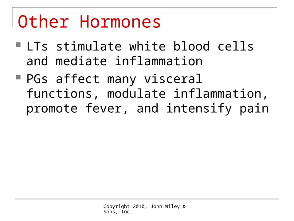
Other Hormones LTs stimulate white blood cells and mediate
inflammation PGs affect many visceral functions, modulate
inflammation, promote fever, and intensify pain
Copyright 2010, John Wiley & Sons, Inc.
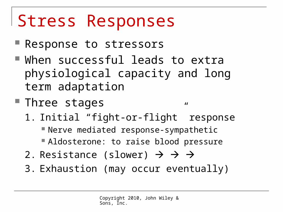
Stress Responses Response to stressors When successful leads to extra physiological
capacity and long term adaptation Three stages
1. Initial “fight-or-flight” response Nerve mediated response-sympathetic Aldosterone: to raise blood pressure
2. Resistance (slower) 3. Exhaustion (may occur eventually)
Copyright 2010, John Wiley & Sons, Inc.
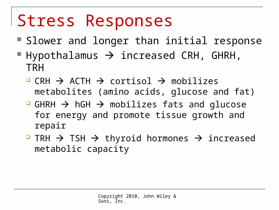
Slower and longer than initial response Hypothalamus increased CRH, GHRH,
TRH CRH ACTH cortisol mobilizes metabolites
(amino acids, glucose and fat) GHRH hGH mobilizes fats and glucose for
energy and promote tissue growth and repair TRH TSH thyroid hormones increased
metabolic capacity
Stress Responses
Copyright 2010, John Wiley & Sons, Inc.
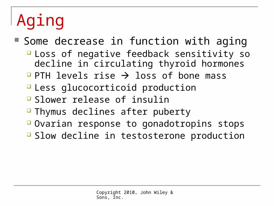
Aging Some decrease in function with aging
Loss of negative feedback sensitivity so decline in circulating thyroid hormones
PTH levels rise loss of bone mass Less glucocorticoid production Slower release of insulin Thymus declines after puberty Ovarian response to gonadotropins stops Slow decline in testosterone production