Embed Size (px)
Citation preview

Copyright © 2006 Thomson Delmar Learning
Chapter 6
Gut Instincts
The Gastrointestinal System
Copyright © 2006 Thomson Delmar Learning
The Digestive System
• The digestive system is referred to as the– digestive system (or tract)– alimentary system– GI system (or tract)
• The digestive system is basically a long, muscular tube that begins at the mouth and ends at the anus
Copyright © 2006 Thomson Delmar Learning
The Digestive System
• Functions of the digestive system– intake and digestion of food and water– absorption of nutrients– elimination of solid wastes
• The combining form for nourishment is aliment/o
Copyright © 2006 Thomson Delmar Learning
Structures of the Digestive System
• Mouth or oral cavity
• Pharynx
• Esophagus
• Stomach
• Small intestines
• Large intestines
• Accessory organs of digestion
Copyright © 2006 Thomson Delmar Learning
Structures of the Mouth
• Mouth or oral cavity– Contains the lips,
cheeks, palates (hard and soft), salivary glands, tongue, teeth, and periodontium
– Combining forms are or/o and stomat/o
• Boundaries of the mouth are the maxilla and mandible (jaw)– Combining form for jaw
is gnath/o– Prognathia means
having an elongated mandible (overshot)
– Brachygnathia means having a shortened mandible (undershot)

Copyright © 2006 Thomson Delmar Learning
Structures of the Digestive System
• Mouth or oral cavity– Lips form the opening
to the oral cavity• Combining forms are
cheil/o and labi/o– Cheeks form the walls
of the oral cavity• Combining form is
bucc/o– The palate forms the
roof of the mouth• Combining form is
palat/o• Rug/o = wrinkle or
fold
• Tongue is a movable muscular organ – Combining forms are
gloss/o and lingu/o– Papillae are the
elevations on the tongue
• Filiform = threadlike• Fungiform =
mushroom-like• Vallate = cup-shaped
Copyright © 2006 Thomson Delmar Learning
Structures of the Mouth
• Teeth are arranged in the maxillary and mandibular arcade– Combining forms are
dent/o, dent/I, and odont/o
• Dentition refers to the teeth as a whole– The primary dentition
is temporary and known as the deciduous dentition
• decidu/o = shedding– The secondary
dentition is permanent
• Dental formula represents the type and number of each tooth type found in that species– Adult dog is 2(I 3/3,
C 1/1, P 4/4, M 2/3)
Copyright © 2006 Thomson Delmar Learning
Tooth Names
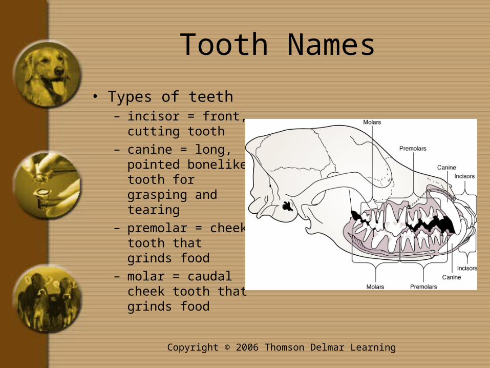
• Types of teeth– incisor = front,
cutting tooth– canine = long,
pointed bonelike tooth for grasping and tearing
– premolar = cheek tooth that grinds food
– molar = caudal cheek tooth that grinds food
Copyright © 2006 Thomson Delmar Learning
Tooth Anatomy
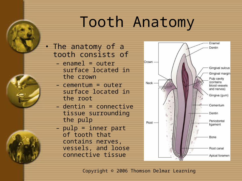
• The anatomy of a tooth consists of– enamel = outer surface
located in the crown – cementum = outer
surface located in the root
– dentin = connective tissue surrounding the pulp
– pulp = inner part of tooth that contains nerves, vessels, and loose connective tissue

Copyright © 2006 Thomson Delmar Learning
Other Mouth Structures
• Gingiva is the mucous membrane that surrounds the teeth– The combining form
for gingiva is gingiv/o
• Salivary glands are groups of cells that secrete saliva– Named for their
location– Combining forms are
sialaden/o and sial/o

Copyright © 2006 Thomson Delmar Learning
The Throat
• The pharynx is the cavity in the caudal oral cavity that joins the respiratory and gastrointestinal systems– Also known as the throat– Combining form is pharyng/o
Copyright © 2006 Thomson Delmar Learning
The Gullet
• The esophagus is a collapsible, muscular tube that leads from the oral cavity to the stomach– Also known as the gullet– Combining form is esophag/o– Enters the stomach through an opening
that is surrounded by a sphincter• Sphincter is a ringlike muscle that constricts
an opening
Copyright © 2006 Thomson Delmar Learning
The Abdomen
• The remaining digestive organs are found in the abdomen– Also known as the peritoneal or
abdominal cavity– Located between the diaphragm and
pelvis– Combining forms are abdomin/o and
celi/o– Combining form for abdomen or flank
is lapar/o

Copyright © 2006 Thomson Delmar Learning
Abdominal Structures
• The peritoneum is the membrane lining that covers the abdominal and pelvic cavity and some of the organs in this area– The layer that lines the abdominal and pelvic
cavities is called the paxetal peritoneum– The layer that covers the abdominal organs is
called the visceral peritoneum– The omentum is a fold of the peritoneum that
connects the stomach to the other visceral organs
Copyright © 2006 Thomson Delmar Learning
The Stomach
• The stomach is a saclike organ that aides in digestion of food– Combining form is gastr/o– Animals can be classified as
monogastric or ruminant• Monogastric animals have one true,
glandular stomach (one that produces secretions)
• Ruminants have one true, glandular stomach plus three forestomachs

Copyright © 2006 Thomson Delmar Learning
Stomach Parts• Parts of the stomach
include– cardia (entrance near
esophagus)– fundus (cranial,
rounded part)– body (main part)– antrum (caudal part)– pylorus (narrow
passage between the stomach and duodenum)
– pyloric sphincter (muscle ring that controls flow of material from the stomach to the small intestine)
– rugae (folds in the mucosa)
Copyright © 2006 Thomson Delmar Learning
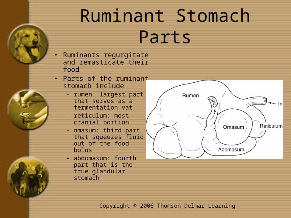
Ruminant Stomach Parts
• Ruminants regurgitate and remasticate their food
• Parts of the ruminant stomach include– rumen: largest part
that serves as a fermentation vat
– reticulum: most cranial portion
– omasum: third part that squeezes fluid out of the food bolus
– abdomasum: fourth part that is the true glandular stomach
Copyright © 2006 Thomson Delmar Learning
Small Intestines
• Small intestine extends from the pylorus to the large intestine
• It is held in place by the mesentery• Enter/o means small intestine• Three segments of the small intestine are
– duodenum: proximal part• duoden/i or duoden/o
– jejunum: middle part• jenun/o
– ileum: distal part• ile/o

Copyright © 2006 Thomson Delmar Learning
Large Intestines• Large intestine extends
from the ileum to the anus• Four segments of the
large intestine are– cecum: pouch that takes
food from the ileum • cec/o
– colon: varies among species
• col/o
– rectum: caudal portion• rect/o
– anus: caudal opening• an/o• proct/o means anus and
rectum together
Copyright © 2006 Thomson Delmar Learning
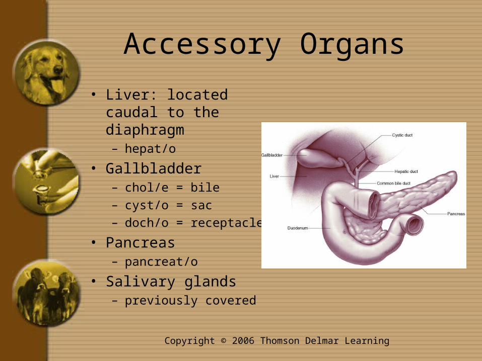
Accessory Organs
• Liver: located caudal to the diaphragm – hepat/o
• Gallbladder– chol/e = bile– cyst/o = sac– doch/o = receptacle
• Pancreas– pancreat/o
• Salivary glands– previously covered
Copyright © 2006 Thomson Delmar Learning
Digestion
• Digestion is the process of breaking down foods into nutrients that the body can use
• Metabolism is the processes involved in the body’s use of nutrients– Meta- means change or beyond– Anabolism is building up of body cells– Catabolism is breakdown of body cells

Copyright © 2006 Thomson Delmar Learning
Absorption
• Absorption is the process of taking digested nutrients into the circulatory system– also called assimilation
• Absorption occurs in the small intestine– Villi are tiny hairlike projections that help
increase the surface area of the small intestine allowing more nutrients to be absorbed
• Vill/i means tuft of hair
– The valleys that result from the projections of the small intestine are called crypts

Copyright © 2006 Thomson Delmar Learning
Path of Digestion
• Food is grasped and collected into the oral cavity– This is called prehension
• Mastication (chewing) breaks food into smaller pieces
• Deglutition moves chewed food into the pharynx and on into the esophagus– The epiglottis closes off the entrance to the trachea
• Food moves down the esophagus by gravity and peristalsis– Peristalsis is a series of wavelike contractions of
smooth muscle• -stalsis means contraction

Copyright © 2006 Thomson Delmar Learning
Peristalsis versus Segmentation
• Food moves through the small intestines by peristalsis and segmentation
• Peristalsis is a series of wavelike contractions that move ingesta caudally toward the anus
• Segmentation involves the side-to- side mixing of ingesta
Copyright © 2006 Thomson Delmar Learning
Medical Terms for the Digestive System
• Additional terms for digestive system tests, pathology, and procedures can be found in the text
• Review the Flash! CD program to make sure you understand these terms