Embed Size (px)
Citation preview
Carnegie Mellon UniversityResearch Showcase @ CMU
Department of Biological Sciences Mellon College of Science
7-3-2006
COPII-Golgi protein interactions regulate COPIIcoat assembly and Golgi size.Yusong GuoCarnegie Mellon University
Adam D. LinstedtCarnegie Mellon University, [email protected]
Follow this and additional works at: http://repository.cmu.edu/biology
Part of the Biology Commons
This Article is brought to you for free and open access by the Mellon College of Science at Research Showcase @ CMU. It has been accepted forinclusion in Department of Biological Sciences by an authorized administrator of Research Showcase @ CMU. For more information, please [email protected].
Published InThe Journal of cell biology, 174, 1, 53-63.
TH
EJ
OU
RN
AL
OF
CE
LL
BIO
LO
GY
JCB: ARTICLE
© The Rockefeller University Press $8.00The Journal of Cell Biology, Vol. 174, No. 1, July 3, 2006 53–63http://www.jcb.org/cgi/doi/10.1083/jcb.200604058
JCB 53
IntroductionEndomembrane compartments create specialized environments
that are optimized for diverse reactions, including protein fold-
ing, quality control, processing, sorting, and turnover. How
these compartments are established and maintained is a ques-
tion of fundamental importance. Compartment size is somehow
coupled to cell growth such that it increases as cells grow and
remains roughly constant in postmitotic cells. On the other
hand, during mitosis or differentiation, or in response to stress,
secretory compartments can undergo extensive up- or down-
regulation depending on cell type, developmental timing, or
stress condition (Knaapen et al., 1997; Lee and Linstedt, 1999;
Lu et al., 2001; Colanzi et al., 2003; Yu et al., 2003). Thus, ho-
meostatic mechanisms that maintain a proper balance of mem-
brane input and output at each compartment must be suffi ciently
fl exible to allow extensive, rapid, and reversible changes.
Although signaling pathways, transcriptional events, and
other higher order processes undoubtedly participate (Buccione
et al., 1996; Ikonomov et al., 2001), compartment homeostasis
might fundamentally rely on intrinsic features of the vesicle coat
machinery. Transport vesicles typically fuse shortly after their
formation, indicating that vesicle formation is rate limiting.
Thus, the net fl ux of membrane through the Golgi apparatus,
for example, would be determined by the rates at which vesi-
cle coats acting at the ER and endosomes contribute input
of vesicles and the rates at which vesicle coats acting at the
Golgi drive vesicle export. What, then, determines the rate of
vesicle production?
Assembly of the coat protein complex (COP) II coat on
the ER membrane is initiated by guanine nucleotide exchange
of the GTPase Sar1p (Futai et al., 2004). The presence of
Sar1p-GTP leads to the successive recruitment of the coat com-
ponents Sec23p–Sec24p and Sec13p–Sec31p (Matsuoka et al.,
1998). COPII coat assembly is opposed by GTP hydrolysis by
Sar1p triggered by Sec23p GAP activity and amplifi ed by the
presence of Sec13p–Sec31p (Yoshihisa et al., 1993; Antonny
et al., 2001). Although each step may play a regulatory role that
infl uences the rate of vesicle production, the self-terminating
property of COPII assembly suggests that additional factors
stabilize the coat on the membrane and regulate overall rate.
Evidence suggests that cargo molecules in the ER membrane
contribute (Aridor and Traub, 2002; Forster et al., 2006; Sato
and Nakano, 2005). For example, synchronized export of ve-
sicular stomatitus virus G protein (VSVG) stimulates COPII
vesicle budding, and inhibition of protein synthesis depresses it
(Aridor et al., 1999).
If compartment homeostasis is determined by vesicle
production rates and vesicle production rates are infl uenced
by cargo concentration, then it is important to ask whether
COPII–Golgi protein interactions regulate COPII coat assembly and Golgi size
Yusong Guo and Adam D. Linstedt
Department of Biological Sciences, Carnegie Mellon University, Pittsburgh, PA 15213
Under experimental conditions, the Golgi apparatus
can undergo de novo biogenesis from the en do-
plasmic reticulum (ER), involving a rapid phase of
growth followed by a return to steady state, but the mech-
anisms that control growth are unknown. Quantifi cation
of coat protein complex (COP) II assembly revealed a dra-
matic up-regulation at exit sites driven by increased levels
of Golgi proteins in the ER. Analysis in a permeabilized
cell assay indicated that up-regulation of COPII assembly
occurred in the absence GTP hydrolysis and any cytosolic
factors other than the COPII prebudding complex Sar1p–
Sec23p–Sec24p. Remarkably, acting via a direct interac-
tion with Sar1p, increased expression of the Golgi enzyme
N-acetylgalactosaminyl transferase-2 induced increased
COPII assembly on the ER and an overall increase in the
size of the Golgi apparatus. These results suggest that
direct interactions between Golgi proteins exiting the ER
and COPII components regulate ER exit, providing a vari-
able exit rate mechanism that ensures homeostasis of the
Golgi apparatus.
Correspondence to Adam D. Linstedt: [email protected]
Abbreviations used in this paper: BFA, brefeldin A; COP, coat protein complex; GalNAcT2, N-acetylgalactosaminyl transferase-2; NRK, normal rat kidney; VSVG, vesicular stomatitus virus G protein; VTC, vesicular-tubular cluster.
The online version of this article contains supplemental material.
on October 3, 2014
jcb.rupress.orgD
ownloaded from
Published July 3, 2006
http://jcb.rupress.org/content/suppl/2006/06/29/jcb.200604058.DC1.html Supplemental Material can be found at:

JCB • VOLUME 174 • NUMBER 1 • 2006 54
compartment size is a function of cargo abundance. Here, we
test the hypothesis that Golgi residents, in the guise of cargo at
the ER, infl uence the size of the Golgi apparatus by regulating
COPII assembly and thereby determining the extent of mem-
brane input to the Golgi.
ResultsOsmotic stress causes Golgi collapse and dispersal of Golgi
components in the ER; yet, after cell volume recovery, the
Golgi apparatus completely reassembles (Lee and Linstedt,
1999). Biogenesis of the Golgi apparatus from the ER is reca-
pitulated using drug washout after sequential treatment with
brefeldin A (BFA), to inhibit Arf1, and H89, to block COPII
assembly. That is, the Golgi apparatus effi ciently and synchro-
nously reassembles from the ER (Puri and Linstedt, 2003).
Inhibitor reversal is rapid, as indicated by restoration of mem-
brane recruitment of Sec13 and β-COP at the earliest time
points tested (Puri and Linstedt, 2003). During biogenesis,
Golgi growth is rapid until steady state is reestablished, imply-
ing a temporary shift in the input/output balance at the Golgi
that favors input.
To test the involvement of increased COPII assembly, we
used morphological assays to determine the levels of COPII and
Golgi assembly in cells before and during BFA/H89 washout.
In normal rat kidney (NRK) cells, the Golgi marker giantin
yielded the expected juxtanuclear ribbon structure before treat-
ment, a dispersed ER pattern upon BFA/H89 treatment, and,
during washout, a punctate vesicular-tubular cluster (VTC) pat-
tern followed by reestablishment of the juxtanuclear Golgi
ribbon (Fig. 1, A–D). In the same cells, staining of the COPII
component Sec13 was restricted to ER exit sites before treat-
ment, absent during treatment, and reestablished at exit sites
during washout (Fig. 1, E–H). Signifi cantly, quantifi cation (see
Materials and methods) revealed a reproducible peak of COPII
assembly (Fig. 1 I). The transient up-regulation of COPII as-
sembly was threefold (P = 0.006) greater than steady-state lev-
els and coincided with the rapid phase of giantin emergence
from the ER. HeLa cells, which exhibit signifi cantly slower
Golgi assembly (Puri and Linstedt, 2003), also yielded a peak in
COPII assembly (twofold; P = 0.01), and this coincided in time
with the delayed emergence of giantin from the ER, thereby
confi rming COPII up-regulation and its correlation with Golgi
exit from the ER. Further, single HeLa cells expressing Sec13-
GFP (Hammond and Glick, 2000) and imaged live at consecu-
tive 2-min intervals while undergoing Golgi assembly during
BFA/H89 washout also exhibited transient up-regulation in
COPII assembly (Fig. 1 J). In contrast, untreated control cells
exhibited relatively stable levels of COPII assembly (Fig. 1 J).
In sum, a transient up-regulation in COPII assembly occurs dur-
ing de novo biogenesis of the Golgi from the ER and coincides
with Golgi egress marked by giantin.
The values shown represent, on a per-cell basis, the total
above-threshold Sec13 fl uorescence, which is a composite of
Figure 1. Golgi biogenesis coincides with in-creased COPII assembly. (A–H) NRK cells were untreated (A and E) or treated with BFA/H89 (30 min BFA and 10 min H89) followed by the indicated times of washout to allow Golgi assembly. The methanol-fi xed cells were costained with anti-giantin (A–D) and anti-Sec13 antibodies (E–H). Single optical sections are shown. Bar, 10 μm. (I) To quantify COPII assembly and ER exit of giantin over time, total above-threshold fl uorescence levels of Sec13 and giantin per NRK cell were determined (mean ± SEM; >15 cells each). Coincident with giantin exit, COPII assembly increased before returning to steady-state levels for untreated cells (dashed lines). (J) Quantifi ed COPII assembly based on GFP-Sec13 expres-sion in HeLa cells is compared for a represen-tative cell after BFA/H89 washout and two untreated control cells. Imaging was at 2-min intervals, and values are corrected and nor-malized to adjust for slight photobleaching and to allow direct comparison. (K) Means per cell for the number, area, and intensity of above-threshold Sec13 objects are compared over time during BFA + H89 washout in NRK cells. Note that increases in exit sites, i.e., object number, and mean size account for the total COPII assembly increase shown in I.
on October 3, 2014
jcb.rupress.orgD
ownloaded from
Published July 3, 2006
COPII ASSEMBLY AND GOLGI SIZE • GUO AND LINSTEDT 55
the number of exit sites per cell, their size, and their intensity.
Analysis of these parameters indicated that the change in total
fl uorescence was due to both increased size of exit sites and
change in number (Fig. 1 K). Thus, up-regulation during Golgi
biogenesis involves up-regulated assembly at preexisting sites
as well as the formation of new ER exit sites.
ER-localized Golgi proteins may activate COPII recruit-
ment at new and preexisting sites. As a test, we used BFA
treatment to induce redistribution of Golgi enzymes to the ER.
Unlike the combined BFA/H89 treatment, BFA alone, which
inhibits Arf1 guanine nucleotide exchange factor (Donaldson
et al., 1992; Helms and Rothman, 1992), does not prevent COPII
assembly (Orci et al., 1993; Bednarek et al., 1995; Ward et al.,
2001). Indeed, in BFA-treated NRK cells, Sec13 remained lo-
calized to exit sites, whereas the Golgi enzyme mannosidase II
was dispersed in the ER (unpublished data). Importantly, Sec13
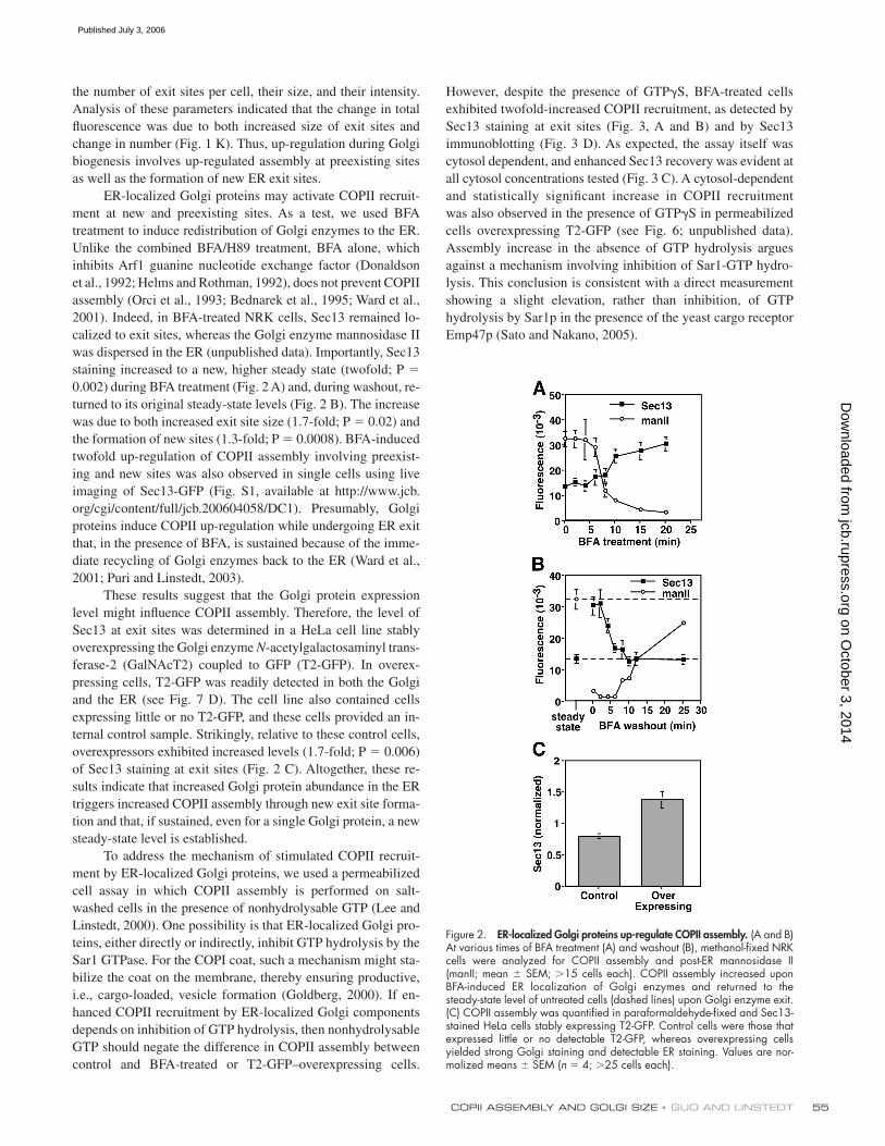
staining increased to a new, higher steady state (twofold; P =
0.002) during BFA treatment (Fig. 2 A) and, during washout, re-
turned to its original steady-state levels (Fig. 2 B). The increase
was due to both increased exit site size (1.7-fold; P = 0.02) and
the formation of new sites (1.3-fold; P = 0.0008). BFA-induced
twofold up-regulation of COPII assembly involving preexist-
ing and new sites was also observed in single cells using live
imaging of Sec13-GFP (Fig. S1, available at http://www.jcb.
org/cgi/content/full/jcb.200604058/DC1). Presumably, Golgi
proteins induce COPII up-regulation while undergoing ER exit
that, in the presence of BFA, is sustained because of the imme-
diate recycling of Golgi enzymes back to the ER (Ward et al.,
2001; Puri and Linstedt, 2003).
These results suggest that the Golgi protein expression
level might infl uence COPII assembly. Therefore, the level of
Sec13 at exit sites was determined in a HeLa cell line stably
overexpressing the Golgi enzyme N-acetylgalactosaminyl trans-
ferase-2 (GalNAcT2) coupled to GFP (T2-GFP). In overex-
pressing cells, T2-GFP was readily detected in both the Golgi
and the ER (see Fig. 7 D). The cell line also contained cells
expressing little or no T2-GFP, and these cells provided an in-
ternal control sample. Strikingly, relative to these control cells,
overexpressors exhibited increased levels (1.7-fold; P = 0.006)
of Sec13 staining at exit sites (Fig. 2 C). Altogether, these re-
sults indicate that increased Golgi protein abundance in the ER
triggers increased COPII assembly through new exit site forma-
tion and that, if sustained, even for a single Golgi protein, a new
steady-state level is established.
To address the mechanism of stimulated COPII recruit-
ment by ER-localized Golgi proteins, we used a permeabilized
cell assay in which COPII assembly is performed on salt-
washed cells in the presence of nonhydrolysable GTP (Lee and
Linstedt, 2000). One possibility is that ER-localized Golgi pro-
teins, either directly or indirectly, inhibit GTP hydrolysis by the
Sar1 GTPase. For the COPI coat, such a mechanism might sta-
bilize the coat on the membrane, thereby ensuring productive,
i.e., cargo-loaded, vesicle formation (Goldberg, 2000). If en-
hanced COPII recruitment by ER-localized Golgi components
depends on inhibition of GTP hydrolysis, then nonhydrolysable
GTP should negate the difference in COPII assembly between
control and BFA-treated or T2-GFP–overexpressing cells.
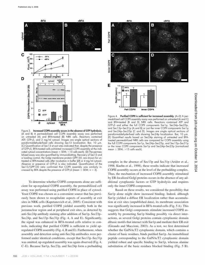
However, despite the presence of GTPγS, BFA-treated cells
exhibited twofold-increased COPII recruitment, as detected by
Sec13 staining at exit sites (Fig. 3, A and B) and by Sec13
immuno blotting (Fig. 3 D). As expected, the assay itself was
cytosol dependent, and enhanced Sec13 recovery was evident at
all cytosol concentrations tested (Fig. 3 C). A cytosol-dependent
and statistically signifi cant increase in COPII recruitment
was also observed in the presence of GTPγS in permeabilized
cells overexpressing T2-GFP (see Fig. 6; unpublished data).
Assembly increase in the absence of GTP hydrolysis argues
against a mechanism involving inhibition of Sar1-GTP hydro-
lysis. This conclusion is consistent with a direct measurement
showing a slight elevation, rather than inhibition, of GTP
hydrolysis by Sar1p in the presence of the yeast cargo receptor
Emp47p (Sato and Nakano, 2005).
Figure 2. ER-localized Golgi proteins up-regulate COPII assembly. (A and B) At various times of BFA treatment (A) and washout (B), methanol-fi xed NRK cells were analyzed for COPII assembly and post-ER mannosidase II (manII; mean ± SEM; >15 cells each). COPII assembly increased upon BFA-induced ER localization of Golgi enzymes and returned to the steady-state level of untreated cells (dashed lines) upon Golgi enzyme exit. (C) COPII assembly was quantifi ed in paraformaldehyde-fi xed and Sec13-stained HeLa cells stably expressing T2-GFP. Control cells were those that expressed little or no detectable T2-GFP, whereas overexpressing cells yielded strong Golgi staining and detectable ER staining. Values are nor-malized means ± SEM (n = 4; >25 cells each).
on October 3, 2014
jcb.rupress.orgD
ownloaded from
Published July 3, 2006
JCB • VOLUME 174 • NUMBER 1 • 2006 56
To determine whether COPII components alone are suffi -
cient for up-regulated COPII assembly, the permeabilized cell
assay was performed using purifi ed COPII in place of cytosol.
Yeast COPII was chosen as a convenient source that has previ-
ously been shown to recapitulate aspects of assembly at exit
sites in NRK cells (Kapetanovich et al., 2005). Consistent with
previous work, purifi ed COPII yielded assembly both in the
juxtanuclear region and at peripheral exit sites, as detected by
anti-Sec24p antibody staining after addition of Sar1p, Sec23p–
Sec24p, and Sec13p–Sec31p (Fig. 4, A and E). Signifi cantly,
the signal was enhanced in BFA-treated cells relative to con-
trols, indicating that purifi ed COPII is suffi cient to yield up-
regulated COPII assembly (Fig. 4, B and E). Furthermore, when
assembly and detection using anti-Sec24p antibodies were per-
formed under identical conditions, except that Sec13p–Sec31p
was omitted, up-regulated assembly was again observed (Fig. 4,
C–E). Because Sar1p, Sec23p, and Sec24p form a prebudding
complex in the absence of Sec13p and Sec31p (Aridor et al.,
1998; Kuehn et al., 1998), these results indicate that increased
COPII assembly occurs at the level of the prebudding complex.
Thus, the mechanism of increased COPII assembly stimulated
by ER-localized Golgi proteins occurs in the absence of any ad-
ditional cytoplasmic factors or GTP hydrolysis and requires
only the inner COPII components.
Based on these results, we considered the possibility that
Sar1p alone might show increased binding. Indeed, although
Sar1p yielded a diffuse ER localization rather than accumula-
tion at exit sites (unpublished data), its membrane association
was signifi cantly increased in BFA-treated cells (Fig. 5 A). This
suggests that Golgi components stimulate increased COPII as-
sembly by promoting Sar1p binding possibly via direct inter-
actions, as several Golgi proteins contain cytoplasmic domain
dibasic motifs that interact with Sar1p and mediate their ER exit
(Giraudo and Maccioni, 2003). As a test, we fi rst determined
whether the GalNAcT2 cytoplasmic domain, which contains a
cluster of basic residues, binds purifi ed Sar1p. An immobilized
peptide corresponding to the GalNAcT2 cytoplasmic domain
yielded robust and specifi c binding to Sar1p, whereas alanine
substitution of the basic residues blocked binding (Fig. 5 B).
Figure 3. Increased COPII assembly occurs in the absence of GTP hydrolysis. (A and B) A permeabilized cell COPII assembly assay was performed on untreated (A) and BFA-treated (B) NRK cells. Reactions contained ATP, GTPγS, and 2 mg/ml cytosol. Images are single optical sections of paraformaldehyde-fi xed cells showing Sec13 localization. Bar, 10 μm. (C) Quantifi cation of Sec13 at exit sites indicated that, despite the presence of GTPγS, BFA-treated cells exhibited increased COPII assembly at the indi-cated cytosol concentrations (mean ± SEM; >15 cells each). (D) The permea-bilized assay was also quantifi ed by immunoblotting. Recovery of Sec13 and a loading control, the Golgi membrane protein GPP130, are shown for un-treated or BFA-treated cells after incubation in buffer (∅) or 4 mg/ml cytosol. Absence or presence of GTPγS is also indicated. Quantifi cation of the Sec13/GPP130 ratio confi rmed that COPII assembly was similarly in-creased by BFA despite the presence of GTPγS (mean ± SEM; n = 3).
Figure 4. Purifi ed COPII is suffi cient for increased assembly. (A–D) A per-meabilized cell COPII assembly assay was performed on untreated (A and C) and BFA-treated (B and D) NRK cells. Reactions contained ATP and GTPγS and either the full COPII components Sar1p, Sec24p–Sec23p, and Sec13p–Sec31p (A and B) or just the inner COPII components Sar1p and Sec24p–Sec23p (C and D). Images are single optical sections of paraformaldehyde-fi xed cells showing Sec24p localization. Bar, 10 μm. (E) Quantifi ed results based on Sec24p staining of untreated and BFA-treated permeabilized NRK cells are compared for COPII assembly using the full COPII components Sar1p, Sec24p–Sec23p, and Sec13p–Sec31p or the inner COPII components Sar1p and Sec24p–Sec23p (normalized mean ± SEM; >15 cells each).
on October 3, 2014
jcb.rupress.orgD
ownloaded from
Published July 3, 2006
COPII ASSEMBLY AND GOLGI SIZE • GUO AND LINSTEDT 57
Binding occurred in the absence of added GTP but was en-
hanced by the presence of GTPγS. Thus, GalNAcT2 is capable
of directly binding Sar1p, and it may stabilize Sar1p on the ER
membrane, leading to enhanced COPII assembly.
Next, we asked whether presence of the GalNAcT2 cyto-
plasmic domain peptide would inhibit up-regulation of COPII
assembly in cells overexpressing T2-GFP. Increasing concentra-
tions of the alanine-substituted control peptide and GalNAcT2
cytoplasmic domain peptide were added together with cytosol
and GTPγS to salt-washed permeabilized cells, and T2-GFP and
Sec13 levels were determined (images presented in Fig. S2, avail-
able at http://www.jcb.org/cgi/content/full/jcb.200604058/DC1).
As expected, cells overexpressing T2-GFP yielded increased
COPII assembly compared with adjacent low expressors and
the alanine-substituted control peptide (R→A) exerted little
or no effect (Fig. 6 A). In contrast, the GalNAcT2 peptide
potently inhibited the COPII assembly up-regulation triggered
by T2-GFP overexpression (Fig. 6 B). A moderate inhibition
of basal COPII assembly was also observed at higher peptide
concentrations, indicating the involvement of the Sar1p dibasic
binding site in COPII recruitment generally. Analysis of the area
and number of Sec13-labeled exit sites per cell indicated that
inhibition occurred at both up-regulated, preexisting sites and
at newly formed exit sites (Fig. S3). In conclusion, increased
T2-GFP in the ER stimulates new COPII assembly via direct
binding to Sar1p. This offers an important variation on recent
work showing stabilization of Sec23p–Sec24p membrane con-
tact by cargo in the presence of multiple rounds of GTP hydro-
lysis by Sar1p (Sato and Nakano, 2005; Forster et al., 2006).
Further, these results reveal an elegant solution to the
problem of transiently accelerated Golgi growth in the biogenesis
assay. Increased availability of Golgi proteins in the ER
stimulates COPII assembly via direct interaction with the coat,
Figure 5. Purifi ed Sar1p shows enhanced recruitment and binds GalNAcT2. (A) Untreated or BFA-treated permeabilized NRK cells were incubated with-out or with Sar1p in absence or presence of GTPγS. Recovery of mem-brane bound Sar1p and the loading control GPP130 was determined by immunoblotting. The quantifi ed result, showing enhanced Sar1p binding to cells with ER-localized Golgi enzymes, is the ratio of Sar1p/GPP130 at each condition (mean ± SD; n = 2). (B) Purifi ed Sar1p was tested for bind-ing to the cytoplasmic domain peptide of GalNAcT2 (wild type [wt] = MRRRSRC) or a version with alanine substituted for arginine (R→A = MAAASAC). After incubation in the presence or absence of GTPγS, recov-ery of Sar1p bound to unconjugated (∅) or peptide-conjugated thiopropyl Sepharose 6B beads was determined by immunoblotting. Each reaction contained 0.5 μg Sar1p, and 10% of this was also analyzed (T). The quan-tifi ed results showing the percentage bound indicate a specifi c interaction (mean ± SD; n = 2).
Figure 6. Binding of Sar1p to GalNAcT2 underlies assembly increase. (A and B) Permeabilized HeLa cells stably expressing GFP-T2 were incubated with GTPγS and 4 mg/ml cytosol containing the indicated concentration of R→A, the alanine-substituted peptide (A), or wt, the wild-type GalNAcT2 cytoplasmic domain peptide (B). COPII assembly, based on Sec13, was then quantifi ed in T2-GFP–overexpressing cells and adjacent non- or low-expressing cells (mean ± SEM; >20 cells each). Representative images are presented in Fig. S2 (available at http://www.jcb.org/cgi/content/full/jcb.200604058/DC1). In contrast to the control, the GalNAcT2 peptide blocked the enhanced COPII assembly induced by GFP-T2 overexpression.
on October 3, 2014
jcb.rupress.orgD
ownloaded from
Published July 3, 2006
JCB • VOLUME 174 • NUMBER 1 • 2006 58
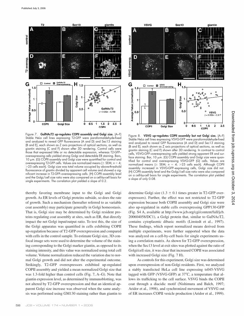
thereby favoring membrane input to the Golgi and Golgi
growth. As ER levels of Golgi proteins subside, so does the rate
of growth. Such a mechanism (hereafter referred to as variable
coat assembly) may participate generally in Golgi homeostasis.
That is, Golgi size may be determined by Golgi resident pro-
teins regulating coat assembly at sites, such as ER, that directly
impact the net Golgi input/output ratio. To test this, the size of
the Golgi apparatus was quantifi ed in cells exhibiting COPII
up-regulation because of T2-GFP overexpression and compared
with cells in the control sample. To estimate Golgi size, 3D con-
focal image sets were used to determine the volume of the stain-
ing corresponding to the Golgi marker giantin, as opposed to its
staining intensity, and this value was normalized using total cell
volume. Volume normalization reduced the variation due to nor-
mal Golgi growth and did not alter the experimental outcome.
Strikingly, T2-GFP overexpressors exhibited up-regulated
COPII assembly and yielded a mean normalized Golgi size that
was 1.3-fold higher than control cells (Fig. 7, A–G). Note that
giantin expression level, as determined by immunoblotting, was
not altered by T2-GFP overexpression and that an identical ap-
parent Golgi size increase was observed when the same analy-
sis was performed using GM130 staining rather than giantin to
determine Golgi size (1.3 ± 0.1 times greater in T2-GFP over-
expressors). Further, the effect was not restricted to T2-GFP
expression because both COPII assembly and Golgi size were
also up-regulated in stable cells overexpressing GPP130-GFP
(Fig. S4 A, available at http://www.jcb.org/cgi/content/full/jcb.
200604058/DC1), a Golgi protein that, similar to GalNAcT2,
contains cytoplasmic dibasic motifs (Linstedt et al., 1997).
These fi ndings, which report normalized means derived from
multiple experiments, were further supported when the data
was analyzed on a cell-by-cell basis for single experiments us-
ing a correlation matrix. As shown for T2-GFP overexpression,
when the Sec13 level at exit sites was plotted against the ratio of
Golgi/cell size, it was clear that increased COPII was associated
with increased Golgi size (Fig. 7 H).
As controls for this experiment, Golgi size was determined
upon overexpression of non-Golgi residents. First, we analyzed
a stably transfected HeLa cell line expressing ts045-VSVG
tagged with GFP (VSVG-GFP) at 37°C, a temperature that al-
lows its traffi cking to the cell surface. VSVG binds the COPII
coat through a diacidic motif (Nishimura and Balch, 1997;
Aridor et al., 1998), and synchronized movement of VSVG out
of ER increases COPII vesicle production (Aridor et al., 1999).
Figure 7. GalNAcT2 up-regulates COPII assembly and Golgi size. (A–F) Stable HeLa cell lines expressing T2-GFP were paraformaldehyde-fi xed and analyzed to reveal GFP fl uorescence (A and D) and Sec13 staining (B and E), each shown as Z axis projections of optical sections, as well as giantin staining (C and F) shown after 3D rendering. Control cells were those that expressed little or no detectable expression, whereas T2-GFP–overexpressing cells yielded strong Golgi and detectable ER staining. Bars, 10 μm. (G) COPII assembly and Golgi size were quantifi ed for control and overexpressing T2-GFP cells. Values are normalized means (± SEM; n = 4; >25 cells each). Golgi size was total volume occupied by above-threshold fl uorescence of giantin divided by apparent cell volume and showed a sig-nifi cant increase in T2-GFP–overexpressing cells. (H) COPII assembly level and the Golgi/cell size ratio were also compared on a cell-by-cell basis for single experiments. The correlation plot yielded a slope of 0.2.
Figure 8. VSVG up-regulates COPII assembly but not Golgi size. (A–F) Stable HeLa cell lines expressing VSVG-GFP were paraformaldehyde-fi xed and analyzed to reveal GFP fl uorescence (A and D) and Sec13 staining (B and E), each shown as Z axis projections of optical sections, as well as giantin staining (C and F) shown after 3D rendering. In contrast to control cells, VSVG-GFP–overexpressing cells yielded strong apparent ER and sur-face staining. Bar, 10 μm. (G) COPII assembly and Golgi size were quan-tifi ed for control and overexpressing VSVG-GFP (G) cells. Values are normalized means (± SEM; n = 4; >25 cells each). Although COPII assembly increased in VSVG-GFP–expressing cells, Golgi size did not. (H) COPII assembly level and the Golgi/cell size ratio were also compared on a cell-by-cell basis for single experiments. The correlation plot yielded a slope of only 0.08.
on October 3, 2014
jcb.rupress.orgD
ownloaded from
Published July 3, 2006

COPII ASSEMBLY AND GOLGI SIZE • GUO AND LINSTEDT 59
Consistent with this, compared with cells in a control sample,
we observed a twofold increase in Sec13 levels at exit sites in
cells expressing VSVG-GFP, yet there was no signifi cant differ-
ence in Golgi size induced by VSVG-GFP expression (Fig. 8,
A–G). Using GM130 to measure Golgi size yielded identical
results. Analysis of the data from single experiments on a cell-
by-cell basis also supported these fi ndings. In contrast to the case
for T2-GFP, the COPII up-regulation induced by VSVG expres-
sion correlated poorly, if at all, with Golgi size (Fig. 8 H). Even
under conditions of high-level VSVG-GFP expression induced
by transient transfection (1.5-fold higher than T2-GFP based
on total fl uorescence per cell), there was no signifi cant change
in apparent Golgi size (1.0 ± 0.1–fold). If the COPII assembly
increase induced by VSVG-GFP corresponded to increased in-
put to the Golgi, as would be expected, then VSVG-GFP must
have also induced a compensatory increase in output to account
for the lack of change in Golgi size. Indeed, a transient Golgi
size increase was previously observed coincident with a wave
of VSVG passing through the Golgi (Aridor et al., 1999; Trucco
et al., 2004), and we also observed increased Golgi size if we
analyzed cells 20 min after releasing ER-accumulated VSVG-
GFP using temperature shift (unpublished data). As further
controls, we analyzed cells expressing the ER-localized protein
Sec61-GFP (Voeltz et al., 2002) and the secreted protein albumin.
Comparison of expressing to nonexpressing cells indicated that
Sec61-GFP expression (at a level equal to T2-GFP based on
total fl uorescence per cell) altered neither COPII assembly nor
Golgi size (Fig. S4 B) and that albumin expression modestly
elevated COPII assembly but did not alter Golgi size (Fig. S4 C).
In sum, these results suggest that COPII assembly can be induced
by proteins rapidly exiting the ER and that, in the case of Golgi
residents, this can lead to sustained changes in Golgi size.
If T2-GFP infl uences Golgi size via its interaction
with the COPII coat, then interfering with this interaction should
alter both COPII assembly and Golgi size. Consistent with this,
it was shown in Fig. 6 in permeabilized cells that the GalNAcT2
cytoplasmic domain peptide, which binds Sar1p, blocks the up-
regulation of COPII assembly induced by T2-GFP expression.
Therefore, we microinjected this peptide or the alanine-
substituted control peptide into nonexpressing and T2-GFP–
expressing cells and, after 45 min, determined COPII assembly
levels and Golgi size in cells marked by coinjected fl uorescent
dextran (Fig. 9, A–H). Microinjection itself led to an unexplained
minimal increase in dissociated Golgi elements, but it did not
alter steady-state changes in COPII assembly or Golgi size. That
is, similar to noninjected cells, T2-GFP–overexpressing cells
injected with the control peptide exhibited signifi cant increases
in both COPII assembly and Golgi size (Fig. 9, I and J, R→A).
Figure 9. Sar1p–GalNAcT2 interaction un-derlies Golgi size increase. (A–H) HeLa cells stably transfected with T2-GFP were micro-injected with the alanine-substituted control pep-tide (A–D; R→A) or the GalNAcT2 cytoplasmic domain peptide (E–H; wild type [wt]) at 1 mM in the presence of fl uorescent dextran as a microinjection marker. After 45 min, the cells were paraformaldehyde-fi xed and analyzed to reveal dextran and GFP fl uorescence (red and green, respectively; A, C, E, and G), as well as giantin staining shown after 3D rendering (B, D, F, and H). Control cells were those that exhibited little or no detectable expression, whereas T2-GFP–overexpressing cells yielded strong Golgi and detectable ER staining. Bars, 10 μm. (I and J) COPII assembly (I) and Golgi size (J) were then quantifi ed for all control or T2-GFP–overexpressing microinjected cells. Values are normalized means (± SD; n = 2; ≥12 cells each). Note that the GalNAcT2 pep-tide, but not the control peptide, blocked both the up-regulation of COPII assembly and the increase in Golgi size induced by T2-GFP.
on October 3, 2014
jcb.rupress.orgD
ownloaded from
Published July 3, 2006

JCB • VOLUME 174 • NUMBER 1 • 2006 60
In contrast, the GalNAcT2 peptide blocked both the COPII as-
sembly increase and the Golgi size increase (Fig. 9, I and J, wt)
strongly suggesting that the Golgi size increase evident in
T2-GFP–expressing cells is a direct consequence of the COPII–
Golgi protein interaction.
DiscussionCOPII assembly was regulated by the availability of Golgi pro-
teins in the ER that was due to, in the case of T2-GFP, a direct
interaction between the Golgi protein cytoplasmic domain and
Sar1p. Transient availability of Golgi proteins in the ER trig-
gered transient COPII up-regulation, explaining the burst of
Golgi growth that accompanies Golgi biogenesis from the ER.
Further, when sustained, increased availability of Golgi proteins
in the ER established a new, higher steady-state level of COPII
assembly, and this accompanied a stable increase in Golgi size.
Thus, our work shows for the fi rst time that variable coat as-
sembly regulated by compartment residents signifi cantly im-
pacts organelle homeostasis.
The binding of cargo to assembling coats is widely recog-
nized to underlie the local enrichment of cargo at bud sites and,
thus, sorting (Springer et al., 1999; Bonifacino and Glick, 2004).
Less well recognized is that the capacity of cargo to bind coats
infl uences coat assembly by increasing avidity of the coat for
the membrane (Miller et al., 1991; Callus et al., 1996; Le Borgne
and Hofl ack, 1997; Aridor et al., 1999). GDP bound Sar1p inter-
acted with GalNAcT2, suggesting that, as shown in the model
(Fig. 10 A), increased GalNAcT2 in the ER membrane recruited
more Sar1p-GDP and, after Sec12p-mediated GTP exchange,
this led to increased formation of Sec23–Sec24 prebudding
complexes. Whereas Sar1 is diffusely localized on the ER mem-
brane, assembled COPII is not. A simple explanation is that lat-
eral interactions between coat components accounts for the
concentration of COPII into new and larger sites. That is, up-
regulated Sar1 recruitment increases COPII components on the
ER membrane, which in turn form new and larger clusters. It
was very recently shown that secretory cargo infl uences forma-
tion of ER-to-Golgi tubular carriers (Simpson et al., 2006). It is
unclear whether this is mechanistically related to ER exit site
formation, but it may be that cargo-induced new and/or larger
ER exit sites give rise to tubular carriers.
Cargo binding to the GDP form of Sar1p may be generally
involved because, in addition to the dibasic-containing Golgi en-
zymes (Giraudo and Maccioni, 2003) and VSVG (Aridor et al.,
2001), at least two SNAREs, Bet1p and Bos1p, also bind Sar1p-
GDP (Springer and Schekman, 1998). Because all these proteins
also bind Sar1p-GTP, an additional effect may be stabilization of
Sar1p-GTP on the membrane. However, based on the failure of
inhibition of GTP hydrolysis by Sar1p to induce COPII assembly
up-regulation, Sar1p-GTP stabilization is not suffi cient. Further,
the best described cargo–COPII interactions involve binding sites
on the Sec24p subunit (Miller et al., 2003; Mossessova et al.,
2003), and SNARE binding at these sites stabilizes membrane
binding of Sec23–Sec24p complex during multiple rounds of
Sar1p-GTP hydrolysis (Sato and Nakano, 2005). Thus, cargo–
coat interactions exert multilevel control of coat assembly.
Variable coat assembly regulated by compartment resi-
dents could profoundly impact organelle homeostasis (Fig. 10 B).
Steady-state Golgi growth refl ects the balance of all input and
output reactions. Input reactions could be most sensitive to
Golgi protein expression level, whereas output reactions might
be most sensitive to the level of non-Golgi residents, such as
proteins that rapidly recycle to the ER and newly synthesized
proteins headed for distal compartments, including outside the
cell, the cell surface, and lysosomes. If so, increased Golgi pro-
tein synthesis would cause increased Golgi growth because of
increased input from the ER with a less dramatic change in exit
from the Golgi. Because of the necessary presence of targeting
and fusion factors accompanying the increased input from the
ER, there is likely an increase in Golgi-to-ER recycling. An ex-
planation for why this does not offset the size increase is that
recycling involves less material and therefore less membrane.
In contrast to Golgi protein synthesis, increased synthesis of
plasma membrane proteins would cause a transient increase in
Golgi size, as has been observed (Aridor et al., 1999; Trucco
et al., 2004), but little lasting change in Golgi growth. That is,
any increase in input to the Golgi from the ER would shortly
thereafter be offset by a corresponding increase in exit from the
Figure 10. Model depicting variable exit rate mechanism. (A) COPII as-sembly is shown at steady state and after up-regulation by increased avail-ability of Golgi residents in the ER membrane. The dibasic signal in the cytoplasmic domain of Golgi residents recruits more Sar1p-GDP to the ER membrane. After nucleotide exchange catalyzed by Sec12p and assembly of the prebudding complex, lateral interactions of the coat components leads to new and larger exit site formation. (B) Golgi input/output path-ways are schematized before (steady state) or after either increased Golgi resident expression (Golgi growth) or increased plasma membrane protein expression (PM growth). Golgi residents up-regulate COPII assembly, lead-ing to increased ER exit (denoted by larger arrow) and Golgi growth. Plasma membrane proteins up-regulate both ER exit and Golgi exit, lead-ing to, at most, a transient increase in Golgi size.
on October 3, 2014
jcb.rupress.orgD
ownloaded from
Published July 3, 2006

COPII ASSEMBLY AND GOLGI SIZE • GUO AND LINSTEDT 61
Golgi, possibly because of cargo–coat interactions at the TGN
(Nishimura et al., 2002). Thus, homeostasis of endomembrane
compartments might be partly determined by the level of com-
partment residents because of their infl uence on coat assembly
and vesicle traffi cking kinetics. Paralleling the sensitivity of
COPII assembly to VSVG levels, assembly of the clathrin–AP2
complex is sensitive to transferrin receptor and mannose-6
phosphate receptor levels (Miller et al., 1991; Callus et al.,
1996; Le Borgne and Hofl ack, 1997). Future work may reveal
that the cargo sensitivity of all coats extends to the level of com-
partment residents and that the sum of the consequent inter-
actions establishes compartment size. Interestingly, other
complex biological structures, such as the yeast spindle pole
body, show size regulation via expression level (Bullitt et al.,
1997; Elliott et al., 1999).
Variable coat assembly provides a straightforward way in
which cells can respond to dramatic, reversible changes in the
integrity of an organelle, such as the Golgi apparatus, and
restore its original state. Conceivably, variable coat assembly
also drives sustained changes in organelle size, such as those
that occur during differentiation, by responding to coordinated
changes in expression of organelle residents.
Materials and methodsCell culture and immunofl uorescenceNRK cells, HepG2 cells, HeLa cells, or HeLa cell lines stably expressing the GFP-tagged proteins GalNacT2 (T2-GFP), ts045-VSVG (VSVG-GFP), or GPP130 (GPP130-GFP) were maintained as described previously (Puri and Linstedt, 2003). BFA (Sigma-Aldrich) and H89 (Toronto Research) were used in media at 2.5 μg/ml and 100 μM, respectively. For transient transfections, HeLa cells were transfected with plasmids encoding GFP-tagged Sec13 (Hammond and Glick, 2000) using calcium phosphate or VSVG- or Sec61-GFP using Transfectol (GeneChoice). Immunofl uorescence was performed as described previously (Puri and Linstedt, 2003). The anti-bodies and their dilutions were mouse anti-giantin at 1:100 (Linstedt and Hauri, 1993); rabbit anti-Sec13 at 1:500 (Kapetanovich et al., 2005); rabbit anti-Sec24p at 1:500 (provided by T.H. Lee, Carnegie Mellon Uni-versity, Pittsburgh, PA); mouse anti–mannosidase II at 1:10,000 (Covance); rabbit anti-GM130 at 1:500 (Puthenveedu and Linstedt, 2001); FITC- conjugated anti–albumin at 1:100 (Biotrend); Cy5-labeled goat anti–mouse or anti–rabbit at 1:500 (Zymed Laboratories); and rhodamine-labeled goat anti–rabbit at 1:500 (Zymed Laboratories).
Image analysisMicroscopy was performed using a spinning disk confocal scan head equipped with three-line laser and independent excitation and emission fi lter wheels (PerkinElmer) and a 12-bit digital camera (Orca ER; Hama-matsu) mounted on a microscope (Axiovert 200; Carl Zeiss MicroImag-ing, Inc.) with a 100×, 1.4 NA apochromat oil-immersion objective (Carl Zeiss MicroImaging, Inc.). Single optical sections or sections at 0.3-μm spacing were acquired using ImagingSuite software (PerkinElmer). Individual experiments were performed with identical laser output levels, exposure times, and scaling. At least six representative fi elds, each con-taining 3–5 cells, were taken. Total fl uorescence of Sec13 at peripheral sites was quantifi ed using ImageJ on single optical sections as follows. For each experiment, a single fi xed threshold was manually chosen (based on comparison to the original gray-scale images) and applied to all images. Individual cells were then selected with the free-hand tool, and the total above-threshold fl uorescence was determined using either the analyze particles or measure functions. For quantifi cation of Golgi size, the ana-lyze particles function was used after thresholding to yield, on a per-cell basis, the sum of each section’s Golgi area as determined by giantin or GM130 staining. Cell volume was estimated by summing the area in each section outlined manually based on the diffuse background staining of Sec13. To allow direct comparison of distinct experiments given small changes in staining intensity, COPII assembly and Golgi size values were
normalized by dividing by the mean values of the entire dataset for a given experiment. To compare expression level of GFP constructs, single optical sections were acquired using identical settings and the 12-bit im-ages were background subtracted using a fi xed value (202). Means of the total fl uorescence per cell were compared (>10 cells each). Live imaging was performed 2 d after transfection in Optimem (Invitrogen) containing 10% fetal bovine serum on a 37°C stage using 300-ms exposures every 2 min. For each time point, fl uorescence intensity in objects was deter-mined as just described. These values were then adjusted to correct for the slight rate of photobleaching based on the photobleaching rate of an identically captured movie for a control cell outside of the dataset. The val-ues for a given cell were then normalized by dividing each by the mean value of the last fi ve time points for that cell. This allowed direct compari-son of distinct cells. Neither the correction nor the normalization altered the pattern of fl uorescence changes.
Permeabilized cell assaysThe morphological permeabilized cell COPII assembly assay was per-formed as described previously (Kapetanovich et al., 2005). NRK cells at 70% confl uence on 12-mm glass coverslips were treated with BFA for 30 min, washed 3 × 0.5 ml with cold DME, and washed 2 × 0.5 ml with cold KOAc buffer (115 mM KOAc, 2.5 mM MgOAc, 25 mM Hepes, pH 7.2, and 1 mM dithiothreitol). The washed cells were permeabilized 6 min at RT in 0.5 ml 0.03 mg/ml digitonin in KOAc buffer followed by 3 × 0.5 ml washes in cold KOAc buffer. After transfer to parafi lm, the coverslips were incubated at 37°C for 10 min in 50 μl KOAc buffer containing either rab-bit liver cytosol, at the indicated concentrations, or purifi ed yeast Sar1p (1 μg/ml), Sec23p–Sec24p (3 μg/ml), and Sec13p–Sec31p (7 μg/ml). Where indicated, each reaction also contained 500 μM GTPγS and an ATP regeneration system (0.5 mM ATP, 0.5 mM UTP, 50 μM GTP, 5 mM creatine phosphate, 25 μg/ml creatine phosphokinase, 0.05 mM EGTA, and 0.5 mM MgCl2). The coverslips were then washed in cold KOAc buf-fer and fi xed and stained as described. The morphological assay was slightly modifi ed for the peptide inhibition experiments. T2-GFP cells were used instead of NRK and, before use, the cytosol was preincubated at 4°C 1 h in 20-μl reactions containing 0.2 mg cytosol, 500 μM GTPγS, the ATP regeneration system, 1 mM PMSF, and synthetic peptides (see Peptide binding) at the indicated concentrations.
The immunoblot-based assay was essentially the same except that cells were grown in 35-mm dishes and volumes were scaled accordingly. Instead of fi xation, the washed cells were scraped into lysis buffer (1% TX-100, 2 mM EDTA, 50 mM Tris, pH 8.0, 1 mM PMSF, and 150 mM NaCl) and incubated on ice for 10 min with vortexing followed by centrifugation at 14,000 rpm for 5 min. The cleared lysate was precipitated using trichlo-roacetic acid and analyzed by immunoblotting (Linstedt and Hauri, 1993) using enhanced chemiluminescence (Pierce Chemical Co.) with acquisition by the LAS-3000 imaging system (Fujifi lm). Antibodies used were rabbit anti-Sec13 at 1:1,000 (Kapetanovich et al., 2005), rabbit anti-GPP130 at 1:1,000 (Puri et al., 2002), and peroxidase-conjugated anti–rabbit at 1:5,000 (Bio-Rad Laboratories).
Peptide bindingSynthetic peptides (MRRRSRC and MAAASAC) were purchased from Gene-Script and coupled via the added C-terminal cysteine residue to Sepharose 6B beads according to previous work (Dominguez et al., 1998; Giraudo and Maccioni, 2003) and manufacturer instructions (GE Healthcare) in re-actions containing 100 μl 14 mg/ml peptide, 500 μl beads, and 600 μl coupling buffer (50 mM Tris, pH 7.3, and 0.5 M NaCl). Coupling effi -ciency was determined by the measuring 2-thiopyridone release at 343 nm. The beads (14-μl aliquots) were then blocked by a 40-min 4°C in-cubation in 0.5 ml 5 mM β-mercaptoethanol, 50 mM NaOAc, 0.5 M NaCl, pH 4.5, followed by washing and a 2-h 4°C incubation in binding buffer (20 mM Hepes, pH 7.2, 250 mM sorbitol, 70 mM KOAc, 1 mM Mg[OAc]2, and 1 mg/ml bovine serum albumin). For binding experi-ments, 0.5 μg Sar1p was preincubated for 30 min at 4°C in the presence or absence of 500 μM GTPγS in a total volume of 14 μl binding buffer. To this was added 16 μl buffer containing 7 μl beads containing 5 nmoles of peptide for 1 h at 4°C. The beads were washed 4 × 500 μl for 1 min each with binding buffer lacking albumin and analyzed for Sar1p recov-ery by immunoblot as described in the previous paragraph using rabbit anti-Sar1p antibody at 1:2,000.
MicroinjectionThe MRRRSRC or MAAASAC peptides were microinjected into T2-GFP cells at 1 mM in water containing 0.25 mg/ml Alexa 568–conjugated
on October 3, 2014
jcb.rupress.orgD
ownloaded from
Published July 3, 2006
JCB • VOLUME 174 • NUMBER 1 • 2006 62
dextran (Invitrogen) using a FemtoJet system with InjectMan-NI2 micro-manipulator (Brinkman). The injected cells were maintained at 37°C in Optimem containing 10% serum for 45 min followed by analysis of COPII assembly using anti-Sec13 and Golgi size using anti-giantin as des-cribed in Image analysis.
Online supplemental materialFig. S1 shows up-regulation of COPII assembly at exit sites using represen-tative frames and quantifi ed data from live imaging experiments in which Sec13-GFP was visualized in control cells or upon BFA treatment. Fig. S2 and Fig. S3 show the specifi c inhibition of T2-GFP–induced COPII up- regulation by addition of the GalNAcT2 cytoplasmic domain peptide to the permeabilized cell assay. Representative images are presented in Fig. S2; total quantifi ed Sec13 fl uorescence is presented in Fig. 6; and quantifi -cation of number, size, and intensity of fl uorescent objects (ER exit sites) is presented in Fig. S3. Fig. S4 shows that Golgi size is increased by over-expression of the Golgi protein GPP130 but not by overexpression of the ER protein Sec61 or the secreted protein albumin using representative images and quantifi ed data. Online supplemental material is available at http://www.jcb.org/cgi/content/full/jcb.200604058/DC1.
We thank Dr. T.H. Lee for COPII reagents and expert advice, the Linstedt laboratory (especially Dr. M.A. Puthenveedu for invaluable suggestions and T. Feinstein for help in generating stable cell lines), and Russell Schwartz for modeling work that sparked interest in this project.
This work was supported by National Institutes of Health grant GM-56779 to A.D. Linstedt.
Submitted: 12 April 2006Accepted: 30 May 2006
ReferencesAntonny, B., D. Madden, S. Hamamoto, L. Orci, and R. Schekman. 2001.
Dynamics of the COPII coat with GTP and stable analogues. Nat. Cell Biol. 3:531–537.
Aridor, M., and L.M. Traub. 2002. Cargo selection in vesicular transport: the making and breaking of a coat. Traffi c. 3:537–546.
Aridor, M., J. Weissman, S. Bannykh, C. Nuoffer, and W.E. Balch. 1998. Cargo selection by the COPII budding machinery during export from the ER. J. Cell Biol. 141:61–70.
Aridor, M., S.I. Bannykh, T. Rowe, and W.E. Balch. 1999. Cargo can modulate COPII vesicle formation from the endoplasmic reticulum. J. Biol. Chem. 274:4389–4399.
Aridor, M., K.N. Fish, S. Bannykh, J. Weissman, T.H. Roberts, J. Lippincott-Schwartz, and W.E. Balch. 2001. The Sar1 GTPase coordinates biosyn-thetic cargo selection with endoplasmic reticulum export site assembly. J. Cell Biol. 152:213–229.
Bednarek, S.Y., M. Ravazzola, M. Hosobuchi, M. Amherdt, A. Perrelet, R. Schekman, and L. Orci. 1995. COPI- and COPII-coated vesicles bud directly from the endoplasmic reticulum in yeast. Cell. 83:1183–1196.
Bonifacino, J.S., and B.S. Glick. 2004. The mechanisms of vesicle budding and fusion. Cell. 116:153–166.
Buccione, R., S. Bannykh, I. Santone, M. Baldassarre, F. Facchiano, Y. Bozzi, G. Di Tullio, A. Mironov, A. Luini, and M.A. De Matteis. 1996. Regulation of constitutive exocytic transport by membrane receptors. A biochemical and morphometric study. J. Biol. Chem. 271:3523–3533.
Bullitt, E., M.P. Rout, J.V. Kilmartin, and C.W. Akey. 1997. The yeast spin-dle pole body is assembled around a central crystal of Spc42p. Cell. 89:1077–1086.
Callus, B.A., B.J. Iacopetta, L.C. Kuhn, and E.H. Morgan. 1996. Effects of over-e xpression of the transferrin receptor on the rates of transferrin recycling and uptake of non-transferrin-bound iron. Eur. J. Biochem. 238:463–469.
Colanzi, A., C. Suetterlin, and V. Malhotra. 2003. Cell-cycle-specifi c Golgi fragmentation: how and why? Curr. Opin. Cell Biol. 15:462–467.
Dominguez, M., K. Dejgaard, J. Fullekrug, S. Dahan, A. Fazel, J.P. Paccaud, D.Y. Thomas, J.J. Bergeron, and T. Nilsson. 1998. gp25L/emp24/p24 protein family members of the cis-Golgi network bind both COP I and II coatomer. J. Cell Biol. 140:751–765.
Donaldson, J.G., D. Finazzi, and R.D. Klausner. 1992. Brefeldin A inhibits Golgi membrane-catalysed exchange of guanine nucleotide onto ARF protein. Nature. 360:350–352.
Elliott, S., M. Knop, G. Schlenstedt, and E. Schiebel. 1999. Spc29p is a com-ponent of the Spc110p subcomplex and is essential for spindle pole body duplication. Proc. Natl. Acad. Sci. USA. 96:6205–6210.
Forster, R., M. Weiss, T. Zimmermann, E.G. Reynaud, F. Verissimo, D.J. Stephens, and R. Pepperkok. 2006. Secretory cargo regulates the turnover of COPII subunits at single ER exit sites. Curr. Biol. 16:173–179.
Futai, E., S. Hamamoto, L. Orci, and R. Schekman. 2004. GTP/GDP exchange by Sec12p enables COPII vesicle bud formation on synthetic liposomes. EMBO J. 23:4146–4155.
Giraudo, C.G., and H.J. Maccioni. 2003. Endoplasmic reticulum export of glyco-syltransferases depends on interaction of a cytoplasmic dibasic motif with Sar1. Mol. Biol. Cell. 14:3753–3766.
Goldberg, J. 2000. Decoding of sorting signals by coatomer through a GTPase switch in the COPI coat complex. Cell. 100:671–679.
Hammond, A.T., and B.S. Glick. 2000. Dynamics of transitional endoplasmic reticulum sites in vertebrate cells. Mol. Biol. Cell. 11:3013–3030.
Helms, J.B., and J.E. Rothman. 1992. Inhibition by brefeldin A of a Golgi mem-brane enzyme that catalyses exchange of guanine nucleotide bound to ARF. Nature. 360:352–354.
Ikonomov, O.C., D. Sbrissa, and A. Shisheva. 2001. Mammalian cell morphol-ogy and endocytic membrane homeostasis require enzymatically active phosphoinositide 5-kinase PIKfyve. J. Biol. Chem. 276:26141–26147.
Kapetanovich, L., C. Baughman, and T.H. Lee. 2005. Nm23H2 facilitates coat protein complex II assembly and endoplasmic reticulum export in mam-malian cells. Mol. Biol. Cell. 16:835–848.
Knaapen, M.W., B.C. Vrolijk, and A.C. Wenink. 1997. Ultrastructural changes of the myocardium in the embryonic rat heart. Anat. Rec. 248:233–241.
Kuehn, M.J., J.M. Herrmann, and R. Schekman. 1998. COPII-cargo inter-actions direct protein sorting into ER-derived transport vesicles. Nature. 391:187–190.
Le Borgne, R., and B. Hofl ack. 1997. Mannose 6-phosphate receptors regu-late the formation of clathrin-coated vesicles in the TGN. J. Cell Biol. 137:335–345.
Lee, T.H., and A.D. Linstedt. 1999. Osmotically induced cell volume changes alter anterograde and retrograde transport, Golgi structure, and COPI dissociation. Mol. Biol. Cell. 10:1445–1462.
Lee, T.H., and A.D. Linstedt. 2000. Potential role for protein kinases in regula-tion of bidirectional endoplasmic reticulum-to-Golgi transport revealed by protein kinase inhibitor H89. Mol. Biol. Cell. 11:2577–2590.
Linstedt, A.D., and H.P. Hauri. 1993. Giantin, a novel conserved Golgi mem-brane protein containing a cytoplasmic domain of at least 350 kDa. Mol. Biol. Cell. 4:679–693.
Linstedt, A.D., A. Mehta, J. Suhan, H. Reggio, and H.P. Hauri. 1997. Sequence and overexpression of GPP130/GIMPc: evidence for saturable pH- sensitive targeting of a type II early Golgi membrane protein. Mol. Biol. Cell. 8:1073–1087.
Lu, Z., D. Joseph, E. Bugnard, K.J. Zaal, and E. Ralston. 2001. Golgi complex reorganization during muscle differentiation: visualization in living cells and mechanism. Mol. Biol. Cell. 12:795–808.
Matsuoka, K., L. Orci, M. Amherdt, S.Y. Bednarek, S. Hamamoto, R. Schekman, and T. Yeung. 1998. COPII-coated vesicle formation reconstituted with purifi ed coat proteins and chemically defi ned liposomes. Cell. 93:263–275.
Miller, E.A., T.H. Beilharz, P.N. Malkus, M.C. Lee, S. Hamamoto, L. Orci, and R. Schekman. 2003. Multiple cargo binding sites on the COPII subunit Sec24p ensure capture of diverse membrane proteins into transport vesicles. Cell. 114:497–509.
Miller, K., M. Shipman, I.S. Trowbridge, and C.R. Hopkins. 1991. Transferrin receptors promote the formation of clathrin lattices. Cell. 65:621–632.
Mossessova, E., L.C. Bickford, and J. Goldberg. 2003. SNARE selectivity of the COPII coat. Cell. 114:483–495.
Nishimura, N., and W.E. Balch. 1997. A di-acidic signal required for selective export from the endoplasmic reticulum. Science. 277:556–558.
Nishimura, N., H. Plutner, K. Hahn, and W.E. Balch. 2002. The delta subunit of AP-3 is required for effi cient transport of VSV-G from the trans-Golgi network to the cell surface. Proc. Natl. Acad. Sci. USA. 99:6755–6760.
Orci, L., A. Perrelet, M. Ravazzola, F.T. Wieland, R. Schekman, and J.E. Rothman. 1993. “BFA bodies”: a subcompartment of the endoplasmic reticulum. Proc. Natl. Acad. Sci. USA. 90:11089–11093.
Puri, S., and A.D. Linstedt. 2003. Capacity of the Golgi apparatus for biogenesis from the endoplasmic reticulum. Mol. Biol. Cell. 14:5011–5018.
Puri, S., C. Bachert, C.J. Fimmel, and A.D. Linstedt. 2002. Cycling of early Golgi proteins via the cell surface and endosomes upon lumenal pH disruption. Traffi c. 3:641–653.
Puthenveedu, M.A., and A.D. Linstedt. 2001. Evidence that Golgi structure depends on a p115 activity that is independent of the vesicle tether com-ponents giantin and GM130. J. Cell Biol. 155:227–238.
Sato, K., and A. Nakano. 2005. Dissection of COPII subunit-cargo assembly and disassembly kinetics during Sar1p-GTP hydrolysis. Nat. Struct. Mol. Biol. 12:167–174.
on October 3, 2014
jcb.rupress.orgD
ownloaded from
Published July 3, 2006

COPII ASSEMBLY AND GOLGI SIZE • GUO AND LINSTEDT 63
Simpson, J.C., T. Nilsson, and R. Pepperkok. 2006. Biogenesis of tubular ER-to-Golgi transport intermediates. Mol. Biol. Cell. 17:723–737.
Springer, S., and R. Schekman. 1998. Nucleation of COPII vesicular coat complex by endoplasmic reticulum to Golgi vesicle SNAREs. Science. 281:698–700.
Springer, S., A. Spang, and R. Schekman. 1999. A primer on vesicle budding. Cell. 97:145–148.
Trucco, A., R.S. Polishchuk, O. Martella, A. Di Pentima, A. Fusella, D. Di Giandomenico, E. San Pietro, G.V. Beznoussenko, E.V. Polishchuk, M. Baldassarre, et al. 2004. Secretory traffi c triggers the formation of tubular continuities across Golgi sub-compartments. Nat. Cell Biol. 6:1071–1081.
Voeltz, G.K., M.M. Rolls, and T.A. Rapoport. 2002. Structural organization of the endoplasmic reticulum. EMBO Rep. 3:944–950.
Ward, T.H., R.S. Polishchuk, S. Caplan, K. Hirschberg, and J. Lippincott-Schwartz. 2001. Maintenance of Golgi structure and function depends on the integrity of ER export. J. Cell Biol. 155:557–570.
Yoshihisa, T., C. Barlowe, and R. Schekman. 1993. Requirement for a GTPase-activating protein in vesicle budding from the endoplasmic reticulum. Science. 259:1466–1468.
Yu, W., L.E. O’Brien, F. Wang, H. Bourne, K.E. Mostov, and M.M. Zegers. 2003. Hepatocyte growth factor switches orientation of polarity and mode of movement during morphogenesis of multicellular epithelial structures. Mol. Biol. Cell. 14:748–763.
on October 3, 2014
jcb.rupress.orgD
ownloaded from
Published July 3, 2006


![A Golgi-Released Subpopulation of the Trans-Golgi · A Golgi-Released Subpopulation of the Trans-Golgi Network Mediates Protein Secretion in Arabidopsis1[OPEN] Tomohiro Uemura,a,b,2,3,4](https://img.dokumen.tips/doc/110x75/5eda9f5a09f66a09130ba5a1/a-golgi-released-subpopulation-of-the-trans-golgi-a-golgi-released-subpopulation.jpg)